EP0252060A1 - DNA-Sequenz, rekombinante DNA-Moleküle und Verfahren zur Herstellung des von Newcastle-Krankheitsvirus-Fusionproteins oder der gleichwertigen Polypeptide, erhaltene Produkte und Präparate, die sie enthalten - Google Patents
DNA-Sequenz, rekombinante DNA-Moleküle und Verfahren zur Herstellung des von Newcastle-Krankheitsvirus-Fusionproteins oder der gleichwertigen Polypeptide, erhaltene Produkte und Präparate, die sie enthalten Download PDFInfo
- Publication number
- EP0252060A1 EP0252060A1 EP87870091A EP87870091A EP0252060A1 EP 0252060 A1 EP0252060 A1 EP 0252060A1 EP 87870091 A EP87870091 A EP 87870091A EP 87870091 A EP87870091 A EP 87870091A EP 0252060 A1 EP0252060 A1 EP 0252060A1
- Authority
- EP
- European Patent Office
- Prior art keywords
- newcastle disease
- disease virus
- fusion protein
- polypeptide
- recombinant dna
- Prior art date
- Legal status (The legal status is an assumption and is not a legal conclusion. Google has not performed a legal analysis and makes no representation as to the accuracy of the status listed.)
- Withdrawn
Links
- 108090000765 processed proteins & peptides Proteins 0.000 title claims abstract description 20
- 229920001184 polypeptide Polymers 0.000 title claims abstract description 18
- 102000004196 processed proteins & peptides Human genes 0.000 title claims abstract description 18
- 241000711404 Avian avulavirus 1 Species 0.000 title claims description 45
- 238000000034 method Methods 0.000 title claims description 11
- 108091028043 Nucleic acid sequence Proteins 0.000 title claims description 9
- 108020004511 Recombinant DNA Proteins 0.000 title claims description 9
- 108010059722 Viral Fusion Proteins Proteins 0.000 title claims description 8
- 238000004519 manufacturing process Methods 0.000 title claims description 4
- 238000002360 preparation method Methods 0.000 title description 3
- 102000037865 fusion proteins Human genes 0.000 claims abstract description 19
- 108020001507 fusion proteins Proteins 0.000 claims abstract description 19
- 240000004808 Saccharomyces cerevisiae Species 0.000 claims abstract description 18
- 239000002299 complementary DNA Substances 0.000 claims description 11
- 108091034057 RNA (poly(A)) Proteins 0.000 claims description 10
- 230000014509 gene expression Effects 0.000 claims description 10
- 239000000203 mixture Substances 0.000 claims description 9
- 208000015181 infectious disease Diseases 0.000 claims description 8
- 230000028993 immune response Effects 0.000 claims description 7
- 102000053602 DNA Human genes 0.000 claims description 6
- 239000012634 fragment Substances 0.000 claims description 6
- 239000013604 expression vector Substances 0.000 claims description 5
- 239000013598 vector Substances 0.000 claims description 5
- 101150034814 F gene Proteins 0.000 claims description 4
- 239000013605 shuttle vector Substances 0.000 claims description 4
- 230000001580 bacterial effect Effects 0.000 claims description 2
- 244000005700 microbiome Species 0.000 claims description 2
- 230000008569 process Effects 0.000 claims description 2
- 230000035515 penetration Effects 0.000 claims 2
- 238000003306 harvesting Methods 0.000 claims 1
- 230000001939 inductive effect Effects 0.000 claims 1
- 230000001105 regulatory effect Effects 0.000 claims 1
- 230000004936 stimulating effect Effects 0.000 claims 1
- 230000001131 transforming effect Effects 0.000 claims 1
- 241000700605 Viruses Species 0.000 abstract description 14
- 239000002773 nucleotide Substances 0.000 abstract description 9
- 125000003729 nucleotide group Chemical group 0.000 abstract description 9
- 230000004927 fusion Effects 0.000 abstract description 8
- 241000588724 Escherichia coli Species 0.000 abstract description 7
- 208000010359 Newcastle Disease Diseases 0.000 abstract description 2
- 238000002255 vaccination Methods 0.000 abstract description 2
- 238000003745 diagnosis Methods 0.000 abstract 1
- 108090000623 proteins and genes Proteins 0.000 description 25
- 210000004027 cell Anatomy 0.000 description 20
- 239000013612 plasmid Substances 0.000 description 14
- 235000018102 proteins Nutrition 0.000 description 14
- 102000004169 proteins and genes Human genes 0.000 description 14
- 150000001413 amino acids Chemical group 0.000 description 13
- 229960005486 vaccine Drugs 0.000 description 13
- FAPWRFPIFSIZLT-UHFFFAOYSA-M Sodium chloride Chemical compound [Na+].[Cl-] FAPWRFPIFSIZLT-UHFFFAOYSA-M 0.000 description 10
- 230000003612 virological effect Effects 0.000 description 10
- 230000002209 hydrophobic effect Effects 0.000 description 9
- 108020004999 messenger RNA Proteins 0.000 description 9
- 108020004635 Complementary DNA Proteins 0.000 description 8
- 108020004414 DNA Proteins 0.000 description 8
- 108010068327 4-hydroxyphenylpyruvate dioxygenase Proteins 0.000 description 7
- 229940024606 amino acid Drugs 0.000 description 7
- 235000001014 amino acid Nutrition 0.000 description 7
- 230000013595 glycosylation Effects 0.000 description 7
- 238000006206 glycosylation reaction Methods 0.000 description 7
- 239000012528 membrane Substances 0.000 description 7
- KCXVZYZYPLLWCC-UHFFFAOYSA-N EDTA Chemical compound OC(=O)CN(CC(O)=O)CCN(CC(O)=O)CC(O)=O KCXVZYZYPLLWCC-UHFFFAOYSA-N 0.000 description 6
- 239000000020 Nitrocellulose Substances 0.000 description 6
- 239000000872 buffer Substances 0.000 description 6
- 238000003776 cleavage reaction Methods 0.000 description 6
- 229920001220 nitrocellulos Polymers 0.000 description 6
- 230000007017 scission Effects 0.000 description 6
- 108091026890 Coding region Proteins 0.000 description 5
- 108010076504 Protein Sorting Signals Proteins 0.000 description 5
- 239000007983 Tris buffer Substances 0.000 description 5
- 239000000284 extract Substances 0.000 description 5
- 238000009396 hybridization Methods 0.000 description 5
- 239000011780 sodium chloride Substances 0.000 description 5
- LENZDBCJOHFCAS-UHFFFAOYSA-N tris Chemical compound OCC(N)(CO)CO LENZDBCJOHFCAS-UHFFFAOYSA-N 0.000 description 5
- HEDRZPFGACZZDS-UHFFFAOYSA-N Chloroform Chemical compound ClC(Cl)Cl HEDRZPFGACZZDS-UHFFFAOYSA-N 0.000 description 4
- 101001014029 Helix pomatia Copper-metallothionein Proteins 0.000 description 4
- ISWSIDIOOBJBQZ-UHFFFAOYSA-N Phenol Chemical compound OC1=CC=CC=C1 ISWSIDIOOBJBQZ-UHFFFAOYSA-N 0.000 description 4
- 239000012083 RIPA buffer Substances 0.000 description 4
- 101001014025 Saccharomyces cerevisiae (strain ATCC 204508 / S288c) Copper metallothionein 1-1 Proteins 0.000 description 4
- 101001014027 Saccharomyces cerevisiae (strain ATCC 204508 / S288c) Copper metallothionein 1-2 Proteins 0.000 description 4
- 150000001720 carbohydrates Chemical group 0.000 description 4
- 238000010353 genetic engineering Methods 0.000 description 4
- 230000006698 induction Effects 0.000 description 4
- PHTQWCKDNZKARW-UHFFFAOYSA-N isoamylol Chemical compound CC(C)CCO PHTQWCKDNZKARW-UHFFFAOYSA-N 0.000 description 4
- 238000012163 sequencing technique Methods 0.000 description 4
- 238000012360 testing method Methods 0.000 description 4
- 238000012546 transfer Methods 0.000 description 4
- 102000006410 Apoproteins Human genes 0.000 description 3
- 108010083590 Apoproteins Proteins 0.000 description 3
- LFQSCWFLJHTTHZ-UHFFFAOYSA-N Ethanol Chemical compound CCO LFQSCWFLJHTTHZ-UHFFFAOYSA-N 0.000 description 3
- 101710160621 Fusion glycoprotein F0 Proteins 0.000 description 3
- PEDCQBHIVMGVHV-UHFFFAOYSA-N Glycerine Chemical compound OCC(O)CO PEDCQBHIVMGVHV-UHFFFAOYSA-N 0.000 description 3
- 241000711920 Human orthopneumovirus Species 0.000 description 3
- OKKJLVBELUTLKV-UHFFFAOYSA-N Methanol Chemical compound OC OKKJLVBELUTLKV-UHFFFAOYSA-N 0.000 description 3
- 108010067390 Viral Proteins Proteins 0.000 description 3
- 238000010367 cloning Methods 0.000 description 3
- 238000010276 construction Methods 0.000 description 3
- 239000013613 expression plasmid Substances 0.000 description 3
- 239000000499 gel Substances 0.000 description 3
- 239000002609 medium Substances 0.000 description 3
- 230000034217 membrane fusion Effects 0.000 description 3
- 230000014616 translation Effects 0.000 description 3
- 108091032973 (ribonucleotides)n+m Proteins 0.000 description 2
- QKNYBSVHEMOAJP-UHFFFAOYSA-N 2-amino-2-(hydroxymethyl)propane-1,3-diol;hydron;chloride Chemical compound Cl.OCC(N)(CO)CO QKNYBSVHEMOAJP-UHFFFAOYSA-N 0.000 description 2
- 108020004705 Codon Proteins 0.000 description 2
- DHMQDGOQFOQNFH-UHFFFAOYSA-N Glycine Chemical compound NCC(O)=O DHMQDGOQFOQNFH-UHFFFAOYSA-N 0.000 description 2
- 108090000288 Glycoproteins Proteins 0.000 description 2
- 102000003886 Glycoproteins Human genes 0.000 description 2
- 101000926057 Human herpesvirus 2 (strain G) Envelope glycoprotein C Proteins 0.000 description 2
- 241000711408 Murine respirovirus Species 0.000 description 2
- 239000004098 Tetracycline Substances 0.000 description 2
- XSQUKJJJFZCRTK-UHFFFAOYSA-N Urea Chemical compound NC(N)=O XSQUKJJJFZCRTK-UHFFFAOYSA-N 0.000 description 2
- 206010046865 Vaccinia virus infection Diseases 0.000 description 2
- 241000269370 Xenopus <genus> Species 0.000 description 2
- RJURFGZVJUQBHK-UHFFFAOYSA-N actinomycin D Natural products CC1OC(=O)C(C(C)C)N(C)C(=O)CN(C)C(=O)C2CCCN2C(=O)C(C(C)C)NC(=O)C1NC(=O)C1=C(N)C(=O)C(C)=C2OC(C(C)=CC=C3C(=O)NC4C(=O)NC(C(N5CCCC5C(=O)N(C)CC(=O)N(C)C(C(C)C)C(=O)OC4C)=O)C(C)C)=C3N=C21 RJURFGZVJUQBHK-UHFFFAOYSA-N 0.000 description 2
- 230000004913 activation Effects 0.000 description 2
- 239000002671 adjuvant Substances 0.000 description 2
- 229960000723 ampicillin Drugs 0.000 description 2
- AVKUERGKIZMTKX-NJBDSQKTSA-N ampicillin Chemical compound C1([C@@H](N)C(=O)N[C@H]2[C@H]3SC([C@@H](N3C2=O)C(O)=O)(C)C)=CC=CC=C1 AVKUERGKIZMTKX-NJBDSQKTSA-N 0.000 description 2
- 238000004873 anchoring Methods 0.000 description 2
- 238000013459 approach Methods 0.000 description 2
- 230000015572 biosynthetic process Effects 0.000 description 2
- 210000000170 cell membrane Anatomy 0.000 description 2
- 238000005119 centrifugation Methods 0.000 description 2
- ARUVKPQLZAKDPS-UHFFFAOYSA-L copper(II) sulfate Chemical compound [Cu+2].[O-][S+2]([O-])([O-])[O-] ARUVKPQLZAKDPS-UHFFFAOYSA-L 0.000 description 2
- 229910000366 copper(II) sulfate Inorganic materials 0.000 description 2
- 238000001514 detection method Methods 0.000 description 2
- 201000010099 disease Diseases 0.000 description 2
- 208000037265 diseases, disorders, signs and symptoms Diseases 0.000 description 2
- 238000001962 electrophoresis Methods 0.000 description 2
- 239000012149 elution buffer Substances 0.000 description 2
- 239000011521 glass Substances 0.000 description 2
- 238000011065 in-situ storage Methods 0.000 description 2
- KWGKDLIKAYFUFQ-UHFFFAOYSA-M lithium chloride Chemical compound [Li+].[Cl-] KWGKDLIKAYFUFQ-UHFFFAOYSA-M 0.000 description 2
- 239000003550 marker Substances 0.000 description 2
- 239000000463 material Substances 0.000 description 2
- 230000003472 neutralizing effect Effects 0.000 description 2
- 210000000287 oocyte Anatomy 0.000 description 2
- 239000008188 pellet Substances 0.000 description 2
- 244000144977 poultry Species 0.000 description 2
- 239000002243 precursor Substances 0.000 description 2
- 230000010076 replication Effects 0.000 description 2
- 230000000241 respiratory effect Effects 0.000 description 2
- 238000001179 sorption measurement Methods 0.000 description 2
- 238000003786 synthesis reaction Methods 0.000 description 2
- 229960002180 tetracycline Drugs 0.000 description 2
- 229930101283 tetracycline Natural products 0.000 description 2
- 235000019364 tetracycline Nutrition 0.000 description 2
- 150000003522 tetracyclines Chemical class 0.000 description 2
- 238000013519 translation Methods 0.000 description 2
- 208000007089 vaccinia Diseases 0.000 description 2
- 238000001262 western blot Methods 0.000 description 2
- 102000040650 (ribonucleotides)n+m Human genes 0.000 description 1
- 101710163881 5,6-dihydroxyindole-2-carboxylic acid oxidase Proteins 0.000 description 1
- 229930024421 Adenine Natural products 0.000 description 1
- GFFGJBXGBJISGV-UHFFFAOYSA-N Adenine Chemical compound NC1=NC=NC2=C1N=CN2 GFFGJBXGBJISGV-UHFFFAOYSA-N 0.000 description 1
- 102100024265 Beta-ureidopropionase Human genes 0.000 description 1
- 108091003079 Bovine Serum Albumin Proteins 0.000 description 1
- 101100327917 Caenorhabditis elegans chup-1 gene Proteins 0.000 description 1
- 208000003322 Coinfection Diseases 0.000 description 1
- 102000004594 DNA Polymerase I Human genes 0.000 description 1
- 108010017826 DNA Polymerase I Proteins 0.000 description 1
- 108020003215 DNA Probes Proteins 0.000 description 1
- 239000003298 DNA probe Substances 0.000 description 1
- 108010092160 Dactinomycin Proteins 0.000 description 1
- 239000006145 Eagle's minimal essential medium Substances 0.000 description 1
- 108010067770 Endopeptidase K Proteins 0.000 description 1
- 241000287828 Gallus gallus Species 0.000 description 1
- WQZGKKKJIJFFOK-GASJEMHNSA-N Glucose Natural products OC[C@H]1OC(O)[C@H](O)[C@@H](O)[C@@H]1O WQZGKKKJIJFFOK-GASJEMHNSA-N 0.000 description 1
- 241000238631 Hexapoda Species 0.000 description 1
- 101000761934 Homo sapiens Beta-ureidopropionase Proteins 0.000 description 1
- 102100034343 Integrase Human genes 0.000 description 1
- 239000007836 KH2PO4 Substances 0.000 description 1
- 101150062031 L gene Proteins 0.000 description 1
- HNDVDQJCIGZPNO-YFKPBYRVSA-N L-histidine Chemical compound OC(=O)[C@@H](N)CC1=CN=CN1 HNDVDQJCIGZPNO-YFKPBYRVSA-N 0.000 description 1
- QIVBCDIJIAJPQS-VIFPVBQESA-N L-tryptophane Chemical compound C1=CC=C2C(C[C@H](N)C(O)=O)=CNC2=C1 QIVBCDIJIAJPQS-VIFPVBQESA-N 0.000 description 1
- 241001559185 Mammalian rubulavirus 5 Species 0.000 description 1
- 101100285000 Neurospora crassa (strain ATCC 24698 / 74-OR23-1A / CBS 708.71 / DSM 1257 / FGSC 987) his-3 gene Proteins 0.000 description 1
- 238000000636 Northern blotting Methods 0.000 description 1
- 101710149086 Nuclease S1 Proteins 0.000 description 1
- 108010038807 Oligopeptides Proteins 0.000 description 1
- 102000015636 Oligopeptides Human genes 0.000 description 1
- 241000283973 Oryctolagus cuniculus Species 0.000 description 1
- 206010033799 Paralysis Diseases 0.000 description 1
- 108091005804 Peptidases Proteins 0.000 description 1
- 102000035195 Peptidases Human genes 0.000 description 1
- JWBLQDDHSDGEGR-DRZSPHRISA-N Phe-Ile Chemical compound CC[C@H](C)[C@@H](C(O)=O)NC(=O)[C@@H](N)CC1=CC=CC=C1 JWBLQDDHSDGEGR-DRZSPHRISA-N 0.000 description 1
- 108010092799 RNA-directed DNA polymerase Proteins 0.000 description 1
- 238000012300 Sequence Analysis Methods 0.000 description 1
- 101710173693 Short transient receptor potential channel 1 Proteins 0.000 description 1
- 108020004682 Single-Stranded DNA Proteins 0.000 description 1
- 108091027568 Single-stranded nucleotide Proteins 0.000 description 1
- 229930006000 Sucrose Natural products 0.000 description 1
- CZMRCDWAGMRECN-UGDNZRGBSA-N Sucrose Chemical compound O[C@H]1[C@H](O)[C@@H](CO)O[C@@]1(CO)O[C@@H]1[C@H](O)[C@@H](O)[C@H](O)[C@@H](CO)O1 CZMRCDWAGMRECN-UGDNZRGBSA-N 0.000 description 1
- 229920004890 Triton X-100 Polymers 0.000 description 1
- 239000013504 Triton X-100 Substances 0.000 description 1
- QIVBCDIJIAJPQS-UHFFFAOYSA-N Tryptophan Natural products C1=CC=C2C(CC(N)C(O)=O)=CNC2=C1 QIVBCDIJIAJPQS-UHFFFAOYSA-N 0.000 description 1
- YJQCOFNZVFGCAF-UHFFFAOYSA-N Tunicamycin II Natural products O1C(CC(O)C2C(C(O)C(O2)N2C(NC(=O)C=C2)=O)O)C(O)C(O)C(NC(=O)C=CCCCCCCCCC(C)C)C1OC1OC(CO)C(O)C(O)C1NC(C)=O YJQCOFNZVFGCAF-UHFFFAOYSA-N 0.000 description 1
- ISAKRJDGNUQOIC-UHFFFAOYSA-N Uracil Chemical group O=C1C=CNC(=O)N1 ISAKRJDGNUQOIC-UHFFFAOYSA-N 0.000 description 1
- 108020000999 Viral RNA Proteins 0.000 description 1
- 208000036142 Viral infection Diseases 0.000 description 1
- 239000002253 acid Substances 0.000 description 1
- RJURFGZVJUQBHK-IIXSONLDSA-N actinomycin D Chemical compound C[C@H]1OC(=O)[C@H](C(C)C)N(C)C(=O)CN(C)C(=O)[C@@H]2CCCN2C(=O)[C@@H](C(C)C)NC(=O)[C@H]1NC(=O)C1=C(N)C(=O)C(C)=C2OC(C(C)=CC=C3C(=O)N[C@@H]4C(=O)N[C@@H](C(N5CCC[C@H]5C(=O)N(C)CC(=O)N(C)[C@@H](C(C)C)C(=O)O[C@@H]4C)=O)C(C)C)=C3N=C21 RJURFGZVJUQBHK-IIXSONLDSA-N 0.000 description 1
- 229960000643 adenine Drugs 0.000 description 1
- 239000011543 agarose gel Substances 0.000 description 1
- WNROFYMDJYEPJX-UHFFFAOYSA-K aluminium hydroxide Chemical compound [OH-].[OH-].[OH-].[Al+3] WNROFYMDJYEPJX-UHFFFAOYSA-K 0.000 description 1
- 125000003277 amino group Chemical group 0.000 description 1
- 238000004458 analytical method Methods 0.000 description 1
- 239000003242 anti bacterial agent Substances 0.000 description 1
- 239000007864 aqueous solution Substances 0.000 description 1
- 125000000613 asparagine group Chemical group N[C@@H](CC(N)=O)C(=O)* 0.000 description 1
- 230000002238 attenuated effect Effects 0.000 description 1
- 206010064097 avian influenza Diseases 0.000 description 1
- 239000011324 bead Substances 0.000 description 1
- 230000008901 benefit Effects 0.000 description 1
- 230000003115 biocidal effect Effects 0.000 description 1
- 230000004071 biological effect Effects 0.000 description 1
- 229940098773 bovine serum albumin Drugs 0.000 description 1
- 210000004899 c-terminal region Anatomy 0.000 description 1
- 239000004202 carbamide Substances 0.000 description 1
- 230000015556 catabolic process Effects 0.000 description 1
- 230000001413 cellular effect Effects 0.000 description 1
- 229920002678 cellulose Polymers 0.000 description 1
- 239000001913 cellulose Substances 0.000 description 1
- 238000006243 chemical reaction Methods 0.000 description 1
- 239000003795 chemical substances by application Substances 0.000 description 1
- 230000000295 complement effect Effects 0.000 description 1
- 210000005220 cytoplasmic tail Anatomy 0.000 description 1
- 229960000640 dactinomycin Drugs 0.000 description 1
- 238000006731 degradation reaction Methods 0.000 description 1
- 238000004925 denaturation Methods 0.000 description 1
- 230000036425 denaturation Effects 0.000 description 1
- 230000029087 digestion Effects 0.000 description 1
- 238000010790 dilution Methods 0.000 description 1
- 239000012895 dilution Substances 0.000 description 1
- BNIILDVGGAEEIG-UHFFFAOYSA-L disodium hydrogen phosphate Chemical compound [Na+].[Na+].OP([O-])([O-])=O BNIILDVGGAEEIG-UHFFFAOYSA-L 0.000 description 1
- 229910000397 disodium phosphate Inorganic materials 0.000 description 1
- 235000019800 disodium phosphate Nutrition 0.000 description 1
- 239000012153 distilled water Substances 0.000 description 1
- VHJLVAABSRFDPM-QWWZWVQMSA-N dithiothreitol Chemical compound SC[C@@H](O)[C@H](O)CS VHJLVAABSRFDPM-QWWZWVQMSA-N 0.000 description 1
- 210000002257 embryonic structure Anatomy 0.000 description 1
- 210000002472 endoplasmic reticulum Anatomy 0.000 description 1
- 238000000605 extraction Methods 0.000 description 1
- 238000009472 formulation Methods 0.000 description 1
- 230000008014 freezing Effects 0.000 description 1
- 238000007710 freezing Methods 0.000 description 1
- -1 gall Proteins 0.000 description 1
- 239000008103 glucose Substances 0.000 description 1
- 238000010438 heat treatment Methods 0.000 description 1
- HNDVDQJCIGZPNO-UHFFFAOYSA-N histidine Natural products OC(=O)C(N)CC1=CN=CN1 HNDVDQJCIGZPNO-UHFFFAOYSA-N 0.000 description 1
- 125000001165 hydrophobic group Chemical group 0.000 description 1
- 230000001900 immune effect Effects 0.000 description 1
- 230000002163 immunogen Effects 0.000 description 1
- 238000001114 immunoprecipitation Methods 0.000 description 1
- 239000000411 inducer Substances 0.000 description 1
- 230000002458 infectious effect Effects 0.000 description 1
- 230000002401 inhibitory effect Effects 0.000 description 1
- 238000003780 insertion Methods 0.000 description 1
- 230000037431 insertion Effects 0.000 description 1
- 230000003993 interaction Effects 0.000 description 1
- 230000003834 intracellular effect Effects 0.000 description 1
- 238000007918 intramuscular administration Methods 0.000 description 1
- 125000005647 linker group Chemical group 0.000 description 1
- 239000006166 lysate Substances 0.000 description 1
- 210000004962 mammalian cell Anatomy 0.000 description 1
- 238000005259 measurement Methods 0.000 description 1
- 229910000402 monopotassium phosphate Inorganic materials 0.000 description 1
- 235000019796 monopotassium phosphate Nutrition 0.000 description 1
- 210000004897 n-terminal region Anatomy 0.000 description 1
- 238000006386 neutralization reaction Methods 0.000 description 1
- IJGRMHOSHXDMSA-UHFFFAOYSA-N nitrogen Substances N#N IJGRMHOSHXDMSA-UHFFFAOYSA-N 0.000 description 1
- 229910052757 nitrogen Inorganic materials 0.000 description 1
- QJGQUHMNIGDVPM-UHFFFAOYSA-N nitrogen group Chemical group [N] QJGQUHMNIGDVPM-UHFFFAOYSA-N 0.000 description 1
- 108020004707 nucleic acids Proteins 0.000 description 1
- 102000039446 nucleic acids Human genes 0.000 description 1
- 150000007523 nucleic acids Chemical class 0.000 description 1
- 230000036961 partial effect Effects 0.000 description 1
- 239000013600 plasmid vector Substances 0.000 description 1
- 229920002401 polyacrylamide Polymers 0.000 description 1
- GNSKLFRGEWLPPA-UHFFFAOYSA-M potassium dihydrogen phosphate Chemical compound [K+].OP(O)([O-])=O GNSKLFRGEWLPPA-UHFFFAOYSA-M 0.000 description 1
- 238000001556 precipitation Methods 0.000 description 1
- 230000002265 prevention Effects 0.000 description 1
- 239000000047 product Substances 0.000 description 1
- 230000000644 propagated effect Effects 0.000 description 1
- 238000001243 protein synthesis Methods 0.000 description 1
- 108091008146 restriction endonucleases Proteins 0.000 description 1
- 210000001995 reticulocyte Anatomy 0.000 description 1
- 239000000523 sample Substances 0.000 description 1
- 238000010187 selection method Methods 0.000 description 1
- 125000003607 serino group Chemical group [H]N([H])[C@]([H])(C(=O)[*])C(O[H])([H])[H] 0.000 description 1
- 238000002791 soaking Methods 0.000 description 1
- 239000001509 sodium citrate Substances 0.000 description 1
- NLJMYIDDQXHKNR-UHFFFAOYSA-K sodium citrate Chemical compound O.O.[Na+].[Na+].[Na+].[O-]C(=O)CC(O)(CC([O-])=O)C([O-])=O NLJMYIDDQXHKNR-UHFFFAOYSA-K 0.000 description 1
- 238000002415 sodium dodecyl sulfate polyacrylamide gel electrophoresis Methods 0.000 description 1
- 230000007480 spreading Effects 0.000 description 1
- 238000003892 spreading Methods 0.000 description 1
- 230000000638 stimulation Effects 0.000 description 1
- 238000007920 subcutaneous administration Methods 0.000 description 1
- 239000000126 substance Substances 0.000 description 1
- 239000005720 sucrose Substances 0.000 description 1
- 239000006228 supernatant Substances 0.000 description 1
- 239000000725 suspension Substances 0.000 description 1
- 208000024891 symptom Diseases 0.000 description 1
- 230000005945 translocation Effects 0.000 description 1
- ZHSGGJXRNHWHRS-VIDYELAYSA-N tunicamycin Chemical compound O([C@H]1[C@@H]([C@H]([C@@H](O)[C@@H](CC(O)[C@@H]2[C@H]([C@@H](O)[C@@H](O2)N2C(NC(=O)C=C2)=O)O)O1)O)NC(=O)/C=C/CC(C)C)[C@H]1O[C@H](CO)[C@@H](O)[C@H](O)[C@H]1NC(C)=O ZHSGGJXRNHWHRS-VIDYELAYSA-N 0.000 description 1
- MEYZYGMYMLNUHJ-UHFFFAOYSA-N tunicamycin Natural products CC(C)CCCCCCCCCC=CC(=O)NC1C(O)C(O)C(CC(O)C2OC(C(O)C2O)N3C=CC(=O)NC3=O)OC1OC4OC(CO)C(O)C(O)C4NC(=O)C MEYZYGMYMLNUHJ-UHFFFAOYSA-N 0.000 description 1
- 230000009385 viral infection Effects 0.000 description 1
- 230000001018 virulence Effects 0.000 description 1
- XLYOFNOQVPJJNP-UHFFFAOYSA-N water Chemical compound O XLYOFNOQVPJJNP-UHFFFAOYSA-N 0.000 description 1
Images




Classifications
-
- C—CHEMISTRY; METALLURGY
- C07—ORGANIC CHEMISTRY
- C07K—PEPTIDES
- C07K14/00—Peptides having more than 20 amino acids; Gastrins; Somatostatins; Melanotropins; Derivatives thereof
- C07K14/005—Peptides having more than 20 amino acids; Gastrins; Somatostatins; Melanotropins; Derivatives thereof from viruses
-
- A—HUMAN NECESSITIES
- A61—MEDICAL OR VETERINARY SCIENCE; HYGIENE
- A61K—PREPARATIONS FOR MEDICAL, DENTAL OR TOILETRY PURPOSES
- A61K39/00—Medicinal preparations containing antigens or antibodies
-
- C—CHEMISTRY; METALLURGY
- C12—BIOCHEMISTRY; BEER; SPIRITS; WINE; VINEGAR; MICROBIOLOGY; ENZYMOLOGY; MUTATION OR GENETIC ENGINEERING
- C12N—MICROORGANISMS OR ENZYMES; COMPOSITIONS THEREOF; PROPAGATING, PRESERVING, OR MAINTAINING MICROORGANISMS; MUTATION OR GENETIC ENGINEERING; CULTURE MEDIA
- C12N2760/00—MICROORGANISMS OR ENZYMES; COMPOSITIONS THEREOF; PROPAGATING, PRESERVING, OR MAINTAINING MICROORGANISMS; MUTATION OR GENETIC ENGINEERING; CULTURE MEDIA ssRNA viruses negative-sense
- C12N2760/00011—Details
- C12N2760/18011—Paramyxoviridae
- C12N2760/18111—Avulavirus, e.g. Newcastle disease virus
- C12N2760/18122—New viral proteins or individual genes, new structural or functional aspects of known viral proteins or genes
Definitions
- the present invention relates to a DNA sequence, recombinant DNA molecules and a process for the production of the fusion protein of Newcastle disease virus or equivalent polypeptides, the products obtained and the preparations containing them, more particularly for diagnostic or vaccination purposes.
- Newcastle disease sometimes called 'bird flu', is one of the most important diseases among those that affect poultry.
- the agent responsible is a paramyxovirus: Newcastle disease virus (NDV).
- NDV Newcastle disease virus
- Viral infection causes the appearance of respiratory symptoms sometimes accompanied by severe nervous complications leading to paralysis and death, causing serious economic hardship in the poultry industry.
- Candidates for such a vaccine were the two glycoproteins present in the membrane of the virus, ie NH and F (Ray R. et al.: J. Inf. Dis. 152 ; 1219-1230; 1985).
- the two proteins form spicule-like projections bristling on the outer face of the membrane of the virus and, together, they are involved in viral interactions at the level of the cell membrane: NH is responsible for the adsorption of the virus while F is involved in the fusion of viral and cellular membranes.
- the fusion protein, F is essential for the infectivity of the virus (Nagai Y.
- the F protein is first synthesized as an inactive precursor (F0) and is then specifically cleaved by a proteolytic enzyme from the host so as to provide two polypeptides linked by disulfide bridges (F1 ) and F2) (Nagai Y. et al. loc. cit .; Nagai Y. et al. Virology 77 ; 125-134; 1977).
- This intracellular cleavage (Morrisson T. et al. J. Virol. 53 ; 851-857; 1985) generates a new amino group on the F1 polypeptide and has proved essential for the biological activity of F (Nagai Y. et al. loc. cit.).
- the amino acid sequence of the N-terminal region of F1 is very hydrophobic and highly conserved among several paramyxoviruses (Choppin PA et al. J. Inf. Dis. 143 ; 352-363; 1981). These findings suggested that this new amino-terminal region could interfere with the target cell membrane in the membrane fusion reaction. This possibility has been confirmed by studies in which synthetic oligopeptides, whose sequence mimics the amino-terminal group of F1, specifically inhibited membrane fusion by several paramyxoviruses (Richardson CD et al. Virology 131 ; 518-532; 1983 and Choppin PW et al. Loc. Cit.).
- NH and F both induce neutralizing antibodies (Lê Long et al. J. Virol. 57 ; 1198-1202; 1986).
- F. appears to be an essential element for a vaccine (Merz DC et al. Loc. Cit. And Choppin PW et al. Loc. Cit.).
- anti-F antibodies completely prevent the spread of infection, by inhibiting two types of fusion: external fusion caused by infectious viruses released from infected cells and fusion from the inside which leads to cell-to-cell infection spreading through membrane fusion (Merz DC et al. loc. cit. and Choppin PW et al. loc. cit.). Consequently, it has been considered that, to be effective, a vaccine must induce antibodies against glycoprotein F and that pure glycoprotein F would be the ideal immunogen for such a vaccine (Choppin MW et al. Loc. Cit.).
- Fusion proteins have been detected not only in Newcastle disease virus (NDV) but also in other paramyxoviruses (e.g. SV5 and Sendai virus) and in human respiratory syncytial viruses (HRSV) and, before This invention, the DNA sequences encoding the fusion proteins of the SV5, Sendai and HRSV viruses were determined (Paterson RG et al. Proc. Natl. Acad. Sci. USA 81 ; 6706-6710; 1984; Blumberg BM et al. J. Gen. Virol. 66 ; 317-331; 1985; Elango N. et al. Nucl. Ac. Res. 13 ; 1559-1574; 1985 and Collins PL et al. Proc. Natl. Acad. Sci. USA 81 ; 7683-7687; 1984).
- NDV Newcastle disease virus
- HRSV human respiratory syncytial viruses
- paramyxovirus fusion proteins whose sequences are known have in general a rather hydrophobic character and exhibit 3 characteristic hydrophobic regions, namely the signal sequence, the N-terminal end of F1 and the domain of anchoring.
- NDV F protein Prior to the present invention, the NDV F protein had been detected by SDS-polyacrylamide gel electrophoresis (Nagai Y. et al. Loc. Cit.); however the structure of the F protein was unknown and it could not be obtained in substantial quantity.
- a library comprising NDV gene sequences had been constructed containing clones having at least part of the F gene but this gene had not been expressed (Chambers Ph. Et al. J. Gen. Virol. 67 ; 475- 486; 1986).
- the invention consists of a recombinant DNA molecule which comprises a DNA sequence coding for the fusion protein of Newcastle disease virus or a fragment or derivative thereof coding for a polypeptide which may induce an immune response to the fusion protein of Newcastle disease virus.
- the invention is the recombinant DNA molecule constituting an expression vector which has the coding sequence for the fusion protein operatively linked to an expression control sequence and a microorganism or a cell thus transformed. .
- the present invention is the NDV fusion protein prepared by recombinant DNA techniques and a vaccine which comprises said protein and which induces an immune response against NDV in a volatile.
- a bank of clones was constructed in the well-known plasmid pBR322, by insertion of cDNAs synthesized from poly (A) +-mRNA of cells infected with NDV. After identification of the viral clones by hybridization tests, the sequence of a cDNA which contains the entire coding region for the fusion glycoprotein was determined. In this way, it was found that the encoded polypeptide is 526 amino acids in length and contains 4 potential glycosylation sites (one on F2 and three on F1).
- the single-strand cDNA nucleotide sequence of the Newcastle disease virus fusion protein is given in Figure 1 as is also the amino acid sequence of the native F protein (F0) which is deduced therefrom.
- F0 is cleaved proteolytically to give two apoproteins, F1 and F2, linked together by disulfide bridges.
- the N-terminal end of F1 was determined directly by Edman degradation (Choppin PW et al. loc. cit.): this is a hydrophobic region which can be recognized on the complete sequence of F in Figure 1 (a and b)) (Phe-Ile -Gly-Ala ..., i.e. residues 104 and following). in essence, this sequence is identical to the published sequence, except for one position (residue 111: Ser instead of Gly).
- an uncharged region (residue 480-512) which has the strongest hydrophobicity of the whole protein and is flanked on each side by strongly domains loaded.
- cytoplasmic tail a relatively polar region of 14 amino acids (residues 513-516) which interacts with the protein of the internal membrane, M, before or during assembly of the virus (Peeples, ME et al. J. Virol. 51 ; 81-90; 1984).
- the glycosylation of the NDV fusion protein has been shown to be completely inhibited in the presence of tunicamycin (Morrisson TG et al. J. Virol. 36 ; 171-180; 1980; Lê Long et al. Loc. Cit), an antibiotic which prevents nitrogen bonding of the side carbohydrate chains to asparagine residues. Examination of the sequences shows four potential glycosylation sites (Asn-X-Ser or Asn-X-Thr): one on F2 (residues 72-74) and three on F1 (residues 189-191, 354-356 and 435- 437). It is known that the apoprotein F2 is glycosylated (Scheid A. et al.
- fusion protein of the Newcastle disease virus different sources of virus are accessible (for example, the Italian, Beaudette, Ulster and Queensland strains).
- host / vector systems are also accessible, eg. ex. E. coli bacteria and expression plasmids in which a heterologous gene can be expressed under the control of a viral or bacterial promoter (Harris TJR Genetic Engineering 4 ; R. Williamson ed. Academic Press, London; 127-185; 1983); the yeasts S. cerevisiae and S. pombe and the shuttle vectors which are also expression plasmids in which a heterologous gene can be expressed under the control of a yeast promoter (Beggs JD Genetic Engineering 2 , R. Williamson ed.
- E. coli express the F protein of NDV.
- S. cerevisiae which is used.
- a suspension of the polypeptide preparation obtained is prepared in an isotonic aqueous solution which is preferably supplemented with an adjuvant suitable for intramuscular or subcutaneous administration, such as for example the complete adjuvant of Freund or aluminum hydroxide, as is well known in the art of vaccine formulation, and the composition is distributed in glass vials containing for example 100 to 1,000 effective doses of vaccine.
- the vials are freeze-dried and then sealed tightly.
- the vaccine Before administration, the vaccine is reconstituted extemporaneously by the addition of pyrogen-free distilled water.
- VIRUS Newcastle disease virus, Italian strain, was obtained from the International Reference Laboratory, Weybridge, United Kingdom. It has been propagated in 9-11 day old chicken embryos and used as a typical virus source. The virus was purified as described by Lê Long et al. (loc. cit.)
- actinomycin D was added to a final concentration of 2 ⁇ g / ml and, four hours later, the cells were washed twice with TNE buffer (0.01M Tris pH 8.3, 0 , 15M NaCl, 0.001M EDTA) and lysed in TNE-SDS buffer (0.01M Tris pH 8.3, 0.15M NaCl, 0.001M EDTA, 0.5% SDS) containing 400 ⁇ g / ml proteinase K. After adding a volume of phenol: chloroform: isoamyl alcohol (25: 24: 1), the ADL was passed through the mixer and the mixture was extracted twice with phenol: chloroform: isoamyl alcohol (1 volume).
- the nucleic acids were sedimented by centrifugation gation and dissolved in an elution buffer (0.1M Tris pH 7.5, 0.001M EDTA, 0.5 M SDS) and the mixture was brought to 2M LiCl concentration and incubated at 4 ° C for 12 hours . After centrifugation, the pellet was dissolved in 2 ml of hybridization buffer (0.1M Tris pH 7.5, 0.35M NaCl, 0.001 M EDTA, 0.5% SDS) and passed over 1.5 ml of an oligo (dT) -cellulose column. The DNAs containing the poly (A) were eluted with the elution buffer.
- an elution buffer 0.1M Tris pH 7.5, 0.001M EDTA, 0.5 M SDS
- the poly (A) +-mRNAs were tested for their stimulation of protein synthesis in rabbit reticulocyte lysates (Palham HRB et al. Eur. J. Biochem. 67 ; 247-256; 1976) and in Xenopus oocytes (Gurdon JB et al. Nature 233 ; 177-182; 1971); the viral proteins synthesized were detected by immunoprecipitation using specific anti-NDV antibodies, as described by Lê Long et al. (loc. cit.).
- the cDNAs were then inserted into the PstI site of the DNA of the plasmid pBR322 with oligo tail (dG) by hybridization (dG) 4- (dC) 4 and the ligation mixture was used to transform the strain.
- MM294 from E. coli hsd R17, i.e. r m ) giving it resistance to tetracycline.
- Clones containing viral cDNAs as inserts were detected by hybridization of the colonies in situ (Grunstein M. et al. Loc. Cit.) Using as probe of 32P-labeled 50S viral RNA (isolated on sucrose gradient (15-30% w / w)). The viral clones were then classified into different groups by carrying out other colony hybridization tests in situ , using as DNA probe plasmids DNA chosen at random and containing a cDNA insert and labeled in32P by nick translation.
- IDENTIFICATION OF cDNA Taking advantage of the knowledge of the coding attributes of NDV mRNAs (Collins PL et al. J. Virol. 43 , 1024-1031; 1982), the mRNAs were separated by poly (A) +-mRNA electrophoresis. infected cells using 1.5% agarose gels in 0.025M sodium citrate, pH 3.5, containing 6M urea and RNA transfer was performed on nitrocellulose. The nitrocellulose sheet was then cut into several bands and each of them was hybridized with a cDNA labeled with 32P by nick translation.
- the complementary identification of the cDNAs was carried out by the hybridization-selection method, as described by Riezman H. et al. (Embo J. 12 ; 2161-2168; 1983). Each mRNA selected by this method was translated into Xenopus oocytes (Gurdon JB et al. Loc. Cit.) And the proteins obtained were immunoprecipitated by anti-NDV antibodies (Lê Long et al. Loc. Cit.).
- DNA SEQUENCE ANALYSIS The DNA sequence was determined by the chemical method of Maxam and Gilbert and the Side dideoxy sequencing method.
- Figure 2 shows that, except for one clone (i.e. clone I.5 which hybridizes to a 35S mRNA, which is a transcript of the L gene), each DNA hybridizes to a large band , an 18S mRNA and with a weaker band, localized in the 22S region. As this latter region was found to contain polycistronic transcripts of NDV genes (Collins PL et al. Loc. Cit.), The results obtained allow a clear identification of four groups and, in particular, they show that clone II. 14 contains at least part of the F sequence (Collins PL et al. Loc. Cit.).
- the recombinant cDNA clone (II.14) was chosen for the sequential DNA analysis for the reason that it has the longest insert among the clones containing specific F sequences.
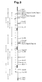
- the sequencing was carried out according to the strategy described in FIG. 3. More than 94% of the sequence of each strand of the coding region for the F protein was determined.
- the entire nucleotide sequence of the insert of clone II.14, in the mRNA direction, is indicated in Figure 1. It contains 1764 base pairs excluding the poly (A) region and the GC tails. There is only one long reading phase, starting from ATG (nucleotides 68-70) and ending with an ocher codon termination (nucleotides 1646-1648). This terminal codon is followed by four additional terminators in phase.
- the coding region codes for a protein of 526 amino acids (M r 56,464, without taking into account the carbohydrate composition), which has three characteristic hydrophobic regions.
- F2 probably extends from residue 19 to residue 98 and consists of 80 amino acids (M r 8.304 without taking account of the carbohydrate composition);
- F1 is a polypeptide of 423 amino acids (residues 104-526; M r 45,541 in non-glycosylated form).
- F presents 4 potential glycosylation sites: one on F2 and three on F1.
- Example 2 Expression of the NDV fusion protein in yeast.
- the E. coli strain used is the strain MM294 ( hsd R17, i.e. R m ). 2 / YEAST.
- the S. cerevisiae strain used is the strain BR10 (MAT alpha, trpl -1, His 4-519, his-3, gall, BUP1 r) (TR Butt et al Proc Natl Acad Sci 81; across 3332 -3336; 1984).
- the yeast shuttle vector pCD192 described in FIG. 4 is a multicopy plasmid containing (i) a part of the well-known 2- ⁇ m plasmid (form B) as the origin of replication yeast, (ii) the yeast gene TRP-1 (Struhl K. et al. Proc. Natl. Acad. Sci. USA 76 , 1035-1039; 1979) as a selection marker in yeast, (iii) a large fraction of the well-known plasmid PBR322, as the origin of replication in E. coli and a selection marker (Ap) into which is inserted (iv) the expression cassette derived from pYSK12 (Butt TR et al. loc.
- the expression cassette contains the beginning of the CUP-1 locus, that is to say the Cu-MT promoter (Butt TR et al. Loc. Cit.) An EcoRI-PvuII-EcoRI linkage group. and the termination of the CYCl gene, that is to say the CYCl terminator (Smith M. et al. Cell 16 ; 753-761; 1979).
- This plasmid therefore contains a single PvuII restriction site which is located between the promoter and the terminator.
- the strategy for the construction of the expression plasmid containing the NDV fusion gene is described in FIG. 4.
- the ligation mixture was used to transform the E. coli MM294 strain giving it resistance to ampicillin.
- the clones were then screened for the presence of the insert and for its orientation towards the promoter, by digestion with EcoRI and AvaI respectively, allowing the identification of plasmids having the correct orientation of F.
- Such a plasmid is pYDE1.
- the yeast strain BR10 was transformed with pYDE1 using intact cells, as described by Ito H. et al. J. Bacteriol. 153 ; 163-168; 1983; the transformed clones being selected on minimal medium without tryptophan (0.67% of nitrogenous base medium for yeast, 2% of glucose with addition of adenine, uracyl and histidine (in each case 20 ⁇ / ml).
- Cu-Mt promoter was then induced as described by Butt TR et al (loc. cit.) with the exception that the induction time was 2 hours and that the inducer was CuSO4 at a final concentration of 0.1 mM.
- the cell extracts were prepared by treatment with glass beads in buffer A (50mM Tris HCl pH 8.0, 2mM EDTA, 0.1mM dithio threitol, 5% glycerol) (Butt TR et al. loc. cit.). An aliquot of the total extract (pellet and supernatant) was then subjected to electrophoresis on a 10% polyacrylamide-SDS gel to separate the proteins which were then transferred to a nitrocellulose filter. The transfer was carried out for 2 hours under 250mA in 0.025M Tris; 0.2M glycine; 20% methanol (pH 8.3).
- buffer A 50mM Tris HCl pH 8.0, 2mM EDTA, 0.1mM dithio threitol, 5% glycerol
- the nitrocellulose filter was washed 3 times at ordinary temperature for 5 minutes by soaking in PBS buffer (140mM NaCl, 2.5mM KCl, 1.5mM KH2PO4, 7.8mM Na2HPO4) and then incubated at 37 ° C for one hour in PBS buffer with 3% bovine serum albumin.
- PBS buffer 140mM NaCl, 2.5mM KCl, 1.5mM KH2PO4, 7.8mM Na2HPO4
- the nitrocellulose filter After 3 washes for five minutes at room temperature in RIPA buffer (150mM NaCl, 20mM tris-HCl pH 7.5, 10mM EDTA, 0.5% Triton X-100, 0.1% SDS), the nitrocellulose filter has then incubated at 37 ° C for 1 hour in a polyclonal antibody directed against NDV and diluted 1,000 times in RIPA buffer and then incubated at the same temperature and for the same duration in 40 ⁇ Ci of protein A labeled with 125I in 40 ml of RIPA buffer (protein A had been iodized to a specific radioactivity of 35 ⁇ Ci / ⁇ g). The nitrocellulose filter was finally washed six times with RIPA buffer, dried and autoradiographed with Kodak X-AR5 film.
- RIPA buffer 150mM NaCl, 20mM tris-HCl pH 7.5, 10mM EDTA, 0.5% Triton X-100, 0.1% SDS
- the Cu-MT promoter was induced with CuSO4 0.1M for 2 hours and cell extracts were prepared and then analyzed in Western blot assays with a polyclonal antibody against NDv (Italian strain).
- FIG. 5 shows the appearance of two bands of approximately 50 kd, immediately below the native F1 polypeptide while no band is present in the induction of the BR10 / pCD192 control. It therefore seems that protein F is synthesized in yeast in two forms of slightly different molecular weights.
Landscapes
- Chemical & Material Sciences (AREA)
- Organic Chemistry (AREA)
- Health & Medical Sciences (AREA)
- Life Sciences & Earth Sciences (AREA)
- Proteomics, Peptides & Aminoacids (AREA)
- Biochemistry (AREA)
- Biophysics (AREA)
- Virology (AREA)
- Medicinal Chemistry (AREA)
- General Health & Medical Sciences (AREA)
- Molecular Biology (AREA)
- Genetics & Genomics (AREA)
- Gastroenterology & Hepatology (AREA)
- Medicines Containing Antibodies Or Antigens For Use As Internal Diagnostic Agents (AREA)
- Preparation Of Compounds By Using Micro-Organisms (AREA)
- Peptides Or Proteins (AREA)
Applications Claiming Priority (2)
| Application Number | Priority Date | Filing Date | Title |
|---|---|---|---|
| US88037186A | 1986-06-30 | 1986-06-30 | |
| US880371 | 1986-06-30 |
Publications (1)
| Publication Number | Publication Date |
|---|---|
| EP0252060A1 true EP0252060A1 (de) | 1988-01-07 |
Family
ID=25376110
Family Applications (1)
| Application Number | Title | Priority Date | Filing Date |
|---|---|---|---|
| EP87870091A Withdrawn EP0252060A1 (de) | 1986-06-30 | 1987-06-29 | DNA-Sequenz, rekombinante DNA-Moleküle und Verfahren zur Herstellung des von Newcastle-Krankheitsvirus-Fusionproteins oder der gleichwertigen Polypeptide, erhaltene Produkte und Präparate, die sie enthalten |
Country Status (6)
| Country | Link |
|---|---|
| EP (1) | EP0252060A1 (de) |
| JP (1) | JPS6324888A (de) |
| AU (1) | AU7492087A (de) |
| DK (1) | DK335987A (de) |
| PT (1) | PT85220B (de) |
| ZA (1) | ZA874677B (de) |
Cited By (5)
| Publication number | Priority date | Publication date | Assignee | Title |
|---|---|---|---|---|
| US5310678A (en) * | 1985-12-18 | 1994-05-10 | National Research Development Corporation | Newcastle disease virus gene clones |
| WO2000077218A1 (en) * | 1999-06-10 | 2000-12-21 | Agricultural Research Council | Vaccine for newcastle disease virus |
| KR100454870B1 (ko) * | 2001-07-30 | 2004-11-03 | 주식회사 인트론바이오테크놀로지 | 뉴캐슬병 바이러스의 f 단백질과 hn 단백질 및 그의유전자들 |
| EP1578374A4 (de) * | 2002-12-06 | 2006-12-13 | Wyeth Corp | Escape-mutanten des newcastle-disease-virus als marker-vakzine |
| CN1293195C (zh) * | 2005-09-02 | 2007-01-03 | 中国农业科学院哈尔滨兽医研究所 | 新城疫LaSota疫苗株反向遗传操作系统及其应用 |
Families Citing this family (1)
| Publication number | Priority date | Publication date | Assignee | Title |
|---|---|---|---|---|
| CA1340203C (en) * | 1987-09-16 | 1998-12-15 | Noboru Yanagida | Recombinant apivoxvirus |
Citations (1)
| Publication number | Priority date | Publication date | Assignee | Title |
|---|---|---|---|---|
| EP0227414A2 (de) * | 1985-12-18 | 1987-07-01 | Btg International Limited | Gen-Klone des Newcastle-Krankheit-Virus |
-
1987
- 1987-06-29 JP JP62165127A patent/JPS6324888A/ja active Pending
- 1987-06-29 ZA ZA874677A patent/ZA874677B/xx unknown
- 1987-06-29 EP EP87870091A patent/EP0252060A1/de not_active Withdrawn
- 1987-06-29 AU AU74920/87A patent/AU7492087A/en not_active Abandoned
- 1987-06-30 DK DK335987A patent/DK335987A/da not_active Application Discontinuation
- 1987-06-30 PT PT85220A patent/PT85220B/pt not_active IP Right Cessation
Patent Citations (1)
| Publication number | Priority date | Publication date | Assignee | Title |
|---|---|---|---|---|
| EP0227414A2 (de) * | 1985-12-18 | 1987-07-01 | Btg International Limited | Gen-Klone des Newcastle-Krankheit-Virus |
Non-Patent Citations (4)
| Title |
|---|
| BIOLOGICAL ABSTRACTS, vol. 84, type 8/4/1, résumé no. 45858, Philadelphia, US; H. SATO et al.: "Molecular cloning and nucleotide sequence of P M and F genes of Newcastle disease virus avirulent strain D26", & VIRUS RES 1987, vol. 7, no. 3, p241-256 * |
| CHEMICAL ABSTRACTS, vol. 106, no. 1, 5 janvier 1987, page 113, résumé no. 1125p, Columbus, Ohio, US; L. McGINNES et al.: "Nucleotide sequence of the gene encoding the Newcastle disease virus fusion protein and comparisons of paramyxovirus fusion protein sequences", & VIRUS RES. 1986, 5(4), 343-56 * |
| CHEMICAL ABSTRACTS, vol. 106, no. 1, 5 janvier 1987, page 171, résumé no. 97046n, Columbus, Ohio, US; P. CHAMBERS et al.: "Nucleotide sequence of the gene encoding the fusion glycoprotein of Newcastle disease virus", & J. GEN. VIROL. 1986, 67(12), 2685-94 * |
| PROC. NATL. ACAD. SCI. USA, vol. 81, December 1984, page 7683, US; P.L. COLLINS et al.: "Nucleotide sequence of the gene encoding the fusion (F) glycoprotein of human respiratory syncytial virus" * |
Cited By (5)
| Publication number | Priority date | Publication date | Assignee | Title |
|---|---|---|---|---|
| US5310678A (en) * | 1985-12-18 | 1994-05-10 | National Research Development Corporation | Newcastle disease virus gene clones |
| WO2000077218A1 (en) * | 1999-06-10 | 2000-12-21 | Agricultural Research Council | Vaccine for newcastle disease virus |
| KR100454870B1 (ko) * | 2001-07-30 | 2004-11-03 | 주식회사 인트론바이오테크놀로지 | 뉴캐슬병 바이러스의 f 단백질과 hn 단백질 및 그의유전자들 |
| EP1578374A4 (de) * | 2002-12-06 | 2006-12-13 | Wyeth Corp | Escape-mutanten des newcastle-disease-virus als marker-vakzine |
| CN1293195C (zh) * | 2005-09-02 | 2007-01-03 | 中国农业科学院哈尔滨兽医研究所 | 新城疫LaSota疫苗株反向遗传操作系统及其应用 |
Also Published As
| Publication number | Publication date |
|---|---|
| ZA874677B (en) | 1988-11-30 |
| PT85220A (en) | 1987-07-01 |
| AU7492087A (en) | 1988-01-07 |
| JPS6324888A (ja) | 1988-02-02 |
| DK335987A (da) | 1987-12-31 |
| DK335987D0 (da) | 1987-06-30 |
| PT85220B (pt) | 1990-07-31 |
Similar Documents
| Publication | Publication Date | Title |
|---|---|---|
| Gao et al. | Sequence of mRNA coding for bindin, a species-specific sea urchin sperm protein required for fertilization. | |
| Müller et al. | Induction of c-fos during myelomonocytic differentiation and macrophage proliferation | |
| Kato et al. | The paramyxovirus, Sendai virus, V protein encodes a luxury function required for viral pathogenesis | |
| JP2534246B2 (ja) | ニユ−カツスル病ウイルス遺伝子クロ−ン | |
| JP3399943B2 (ja) | 癌胎児性抗原を発現する組換えウイルスとその使用方法 | |
| US5141742A (en) | Vaccines against melanoma | |
| KR101745029B1 (ko) | 재조합 조류 파라믹소바이러스 백신 및 이의 제조 및 사용 방법 | |
| LU86765A1 (fr) | Vaccins contre le melanome | |
| JPH03502687A (ja) | レスピラトリイ・シンシチアル・ウイルス:ワクチンおよび診断法 | |
| HK137794A (en) | Expression of human proapolipoprotein a-1 | |
| JPH11512609A (ja) | クローン化されたヌクレオチド配列からの感染性RSウイルス(respiratory syncytial virus)の生産 | |
| US20250295755A1 (en) | Mrna vaccine composition | |
| WO2002042326A1 (en) | A method of expression and agents identified thereby | |
| US6706523B2 (en) | Attenuated rabies virus with nucleoprotein mutation at the phosphorylation site for vaccination against rabies and gene therapy in the CNS | |
| CA1290267C (en) | Human anti-inflammatory phospholipase inhibitor protein | |
| WO2016034110A1 (zh) | 通过miRNA抑制埃博拉病毒的方法 | |
| JP2003512817A (ja) | 免疫調節分子を発現する組み換え呼吸系発疹ウィルスの生成 | |
| TW202222821A (zh) | 用於預防及/或治療covid-19之組合物及方法 | |
| CN118440937B (zh) | 新型的人合胞病毒RSV B mRNA疫苗 | |
| JP2003506039A (ja) | cDNAからの流行性耳下腺炎ウイルスのレスキュー | |
| JP2026509040A (ja) | 呼吸器合胞体ウイルスワクチン、並びにその製造方法および使用 | |
| EP0268501B1 (de) | Proteine mit Glutathion-S-Transferase-Aktivität, DNA-Sequenzen, Antikörper, Poxviren und Arzneimittel zur Prävention von Schistosomiasis | |
| EP0252060A1 (de) | DNA-Sequenz, rekombinante DNA-Moleküle und Verfahren zur Herstellung des von Newcastle-Krankheitsvirus-Fusionproteins oder der gleichwertigen Polypeptide, erhaltene Produkte und Präparate, die sie enthalten | |
| CN112029792B (zh) | 一种具有抗炎作用的鸭病毒性肝炎卵黄抗体及其制备方法和应用 | |
| JPH11514864A (ja) | ヘルペスウイルスのrfhv/kshv亜科の糖タンパク質b |
Legal Events
| Date | Code | Title | Description |
|---|---|---|---|
| PUAI | Public reference made under article 153(3) epc to a published international application that has entered the european phase |
Free format text: ORIGINAL CODE: 0009012 |
|
| 17P | Request for examination filed |
Effective date: 19870713 |
|
| AK | Designated contracting states |
Kind code of ref document: A1 Designated state(s): AT BE CH DE ES FR GB GR IT LI LU NL SE |
|
| RAP1 | Party data changed (applicant data changed or rights of an application transferred) |
Owner name: SMITHKLINE BECKMAN - ANIMAL HEALTH PRODUCTS S.A. |
|
| RAP3 | Party data changed (applicant data changed or rights of an application transferred) |
Owner name: SMITHKLINE BECKMAN - ANIMAL HEALTH PRODUCTS |
|
| 17Q | First examination report despatched |
Effective date: 19900704 |
|
| 18D | Application deemed to be withdrawn |
Effective date: 19901115 |
|
| 18RA | Request filed for re-establishment of rights before grant |
Effective date: 19910531 |
|
| 18RR | Decision to grant the request for re-establishment of rights before grant |
Free format text: 910716 ANGENOMMEN |
|
| REG | Reference to a national code |
Ref country code: DE Ref legal event code: 8570 |
|
| D18D | Application deemed to be withdrawn (deleted) | ||
| STAA | Information on the status of an ep patent application or granted ep patent |
Free format text: STATUS: THE APPLICATION IS DEEMED TO BE WITHDRAWN |
|
| R18D | Application deemed to be withdrawn (corrected) |
Effective date: 19950503 |
|
| RIN1 | Information on inventor provided before grant (corrected) |
Inventor name: ESPION, DANIELE Inventor name: BURNY, ARSENE Inventor name: DEBOUCK, CHRISTINE |