EP0253646A2 - Monoklonale anti-menschliche Magenkrebs-Antikörper - Google Patents
Monoklonale anti-menschliche Magenkrebs-Antikörper Download PDFInfo
- Publication number
- EP0253646A2 EP0253646A2 EP87306257A EP87306257A EP0253646A2 EP 0253646 A2 EP0253646 A2 EP 0253646A2 EP 87306257 A EP87306257 A EP 87306257A EP 87306257 A EP87306257 A EP 87306257A EP 0253646 A2 EP0253646 A2 EP 0253646A2
- Authority
- EP
- European Patent Office
- Prior art keywords
- cells
- monoclonal antibody
- antibody
- cancer
- human
- Prior art date
- Legal status (The legal status is an assumption and is not a legal conclusion. Google has not performed a legal analysis and makes no representation as to the accuracy of the status listed.)
- Granted
Links
Images


Classifications
-
- C—CHEMISTRY; METALLURGY
- C07—ORGANIC CHEMISTRY
- C07K—PEPTIDES
- C07K16/00—Immunoglobulins [IG], e.g. monoclonal or polyclonal antibodies
- C07K16/18—Immunoglobulins [IG], e.g. monoclonal or polyclonal antibodies against material from animals or humans
- C07K16/28—Immunoglobulins [IG], e.g. monoclonal or polyclonal antibodies against material from animals or humans against receptors, cell surface antigens or cell surface determinants
- C07K16/30—Immunoglobulins [IG], e.g. monoclonal or polyclonal antibodies against material from animals or humans against receptors, cell surface antigens or cell surface determinants from tumour cells
- C07K16/303—Liver or Pancreas
-
- C—CHEMISTRY; METALLURGY
- C07—ORGANIC CHEMISTRY
- C07K—PEPTIDES
- C07K16/00—Immunoglobulins [IG], e.g. monoclonal or polyclonal antibodies
- C07K16/18—Immunoglobulins [IG], e.g. monoclonal or polyclonal antibodies against material from animals or humans
- C07K16/28—Immunoglobulins [IG], e.g. monoclonal or polyclonal antibodies against material from animals or humans against receptors, cell surface antigens or cell surface determinants
- C07K16/30—Immunoglobulins [IG], e.g. monoclonal or polyclonal antibodies against material from animals or humans against receptors, cell surface antigens or cell surface determinants from tumour cells
- C07K16/3046—Stomach, Intestines
-
- Y—GENERAL TAGGING OF NEW TECHNOLOGICAL DEVELOPMENTS; GENERAL TAGGING OF CROSS-SECTIONAL TECHNOLOGIES SPANNING OVER SEVERAL SECTIONS OF THE IPC; TECHNICAL SUBJECTS COVERED BY FORMER USPC CROSS-REFERENCE ART COLLECTIONS [XRACs] AND DIGESTS
- Y10—TECHNICAL SUBJECTS COVERED BY FORMER USPC
- Y10S—TECHNICAL SUBJECTS COVERED BY FORMER USPC CROSS-REFERENCE ART COLLECTIONS [XRACs] AND DIGESTS
- Y10S436/00—Chemistry: analytical and immunological testing
- Y10S436/811—Test for named disease, body condition or organ function
- Y10S436/813—Cancer
-
- Y—GENERAL TAGGING OF NEW TECHNOLOGICAL DEVELOPMENTS; GENERAL TAGGING OF CROSS-SECTIONAL TECHNOLOGIES SPANNING OVER SEVERAL SECTIONS OF THE IPC; TECHNICAL SUBJECTS COVERED BY FORMER USPC CROSS-REFERENCE ART COLLECTIONS [XRACs] AND DIGESTS
- Y10—TECHNICAL SUBJECTS COVERED BY FORMER USPC
- Y10S—TECHNICAL SUBJECTS COVERED BY FORMER USPC CROSS-REFERENCE ART COLLECTIONS [XRACs] AND DIGESTS
- Y10S530/00—Chemistry: natural resins or derivatives; peptides or proteins; lignins or reaction products thereof
- Y10S530/808—Materials and products related to genetic engineering or hybrid or fused cell technology, e.g. hybridoma, monoclonal products
- Y10S530/809—Fused cells, e.g. hybridoma
Definitions
- the present invention relates to monoclonal antibodies which react with digestive system cancer, and which are useful in the diagnosis of digestive system cancer.
- Carcinoembryonic antigen is a known marker for digestive system cancer, and methods for the detection of digestive system cancer using both anti-CEA serum (polyclonal antibody) and anti-CEA monoclonal antibodies have been proposed.
- the positive rate using CEA measurement is 30 to 60%, which is too low for the successful screening of digestive system cancer patients.
- a monoclonal antibody has been produced showing strong reactivity with digestive system cancer, especially with pancreatic cancer, and which is capable of detecting the presence of digestive system cancer in respect of the samples which give negative results in the serodiagnosis using NS19-9 or DuPan-2 monoclonal antibody.
- the monoclonal antibody is of the IgG 1 class and is capable of recognizing sialylated glycoproteins and glycolipids as antigens.
- the monoclonal antibody is obtained by immunizing a mammalian animal, preferably mice, with human gastric cancer tissue membrane preparations, and fusing spleen cells from the immunized animal with myeloma cells, preferably a murine myeloma cell line, to generate hybridomas, selecting from among the hybridomas obtained a hybridoma clone producing the monoclonal antibody having specificity to human gastric cancer, and cultivating the selected hybridoma either in a suitable culture medium or administering the hybridoma to a host animal, e.g. a mouse, to thereby cause hybridoma cell propagation in the ascitic fluid, followed by separation, from the culture or the ascitic fluid, of the desired antibody.
- a host animal e.g. a mouse
- mice 3 to 10 weeks old, preferably 8 weeks old, are immunized with human gastric cancer cells, tissues or membrane preparations derived from such tissues to cause mice to generate antibody-producing cells in the spleen, lymph node and peripheral blood. Mice that have immunological tolerance as a result of pretreatment with normal human stomach cells should preferably be used.
- the immunization is generally performed by administering human gastric cancer cells (10 6 to 10 7 cells per animal), human gastric cancer tissues, or membrane preparations (membrane fragments) derived from such tissues (10 to 500 ⁇ g per animal) together with an appropriate adjuvant (e.g.
- the normal human and tumour tissues used may be obtained from autopsies or surgical operations.
- the tissues are immediately frozen and stored at -80°C.
- For membrane components the tissues are thawed at 4°C in PBS containing 1 mM phenylmethyl sulfonyl fluoride. After mincing they are disrupted with an ultra disperser (LK-21; Yamato, Tokyo, Japan) and homogenized with a Teflon-glass homogenizer (Teflon is a Registered Trade Mark).
- the homogenate is centrifuged at 100,000 x g, and then the pellet is resuspended at 1 mg protein in 1 ml of PBS and stored at -80°C.
- the membrane preparations of normal or tumour cells or tissues are distributed into wells of a 96-well EIA plate (product of Flow Laboratories) (100 to 200 ⁇ l per well). After allowing the membrane preparations to stand overnight for two nights at 4°C, the supernatant is removed from the plate and then the plate is washed well with deionized water or phosphate-buffered saline (PBS; 1.83 g of disodium phosphate, 0.21 g of monopotassium phosphate and 7.65 g of sodium chloride in each litre of distilled water, pH 7.2).
- PBS phosphate-buffered saline
- BSA bovine serum albumin
- an ABTS substrate solution prepared by dissolving 550 mg of 2,2'-azinobis (3-ethylbenzothiazoline-6-sulfonic acid) diammonium salt in 1 liter of 0.1 M citrate buffer (pH 4.2) and adding, just prior to use, hydrogen peroxide to a concentration of 1 ⁇ l/ml] is applied and the color developed is measured in terms of the absorbance OD 415nm .
- Those mice that strongly react with the gastric cancer cells, tissues or membrane preparations thereof are used as human gastric cancer-immunized mice, namely as sources of supply of antibody-producing cells for the hybridoma production.
- the target cells are cultivated on a Falcon 3072 plate, 0.25% glutaraldehyde-PBS is added and, after allowing it to stand at room temperature for 1-2 hours, the plate is washed well with PBS. Then, 100-200 ⁇ l of 1% BSA-PBS is added and, after 2 hours of standing, the plate is washed well with deionized water or PBS and submitted to antibody titer determination, which is conducted in the same manner as the case where an ordinary antigen-coated plate is used.
- human gastric cancer cells, tissues or membrane preparations are intraperitoneally administered to the immunized mice in a dose of 2 to 5 x 10 6 cells per animal or 20 to 400 ug per animal 3-4 days prior to the fusion treatment.
- the spleen is extirpated, cut into fragments in MEM (product of Nissui Pharmaceutical), loosened up with a pair of forceps, and centrifuged at 1,200 rpm for 5 minutes. The supernatant is discarded, and the sediment is deprived of erythrocytes by treatment with Tris-ammonium chloride buffer (pH 7.65) for 1-2 minutes, washed three times with MEM, and used as the spleen cells' for fusion.
- MEM product of Nissui Pharmaceutical
- a mouse-derived established myeloma cell line is preferably used.
- Usable examples of such cell line include the 8- azaguanine resistant murine (BALB/c-derived) myeloma cell lines P3-X63Ag8-Ul (P3-Ul) [Current Topics in Microbiology and Immunology-1] P3-NSI/1-Ag4.1 (NS-1) [European J. Immunology, 6, 511-519 (1976)], SP2/0-Agl4 (SP-2) [Nature, 276, 269-270 (1978)], P3-X63-Ag8 653 (653) [J.
- BALB/c-derived myeloma cell lines P3-X63Ag8-Ul (P3-Ul) [Current Topics in Microbiology and Immunology-1]
- P3-NSI/1-Ag4.1 (NS-1) [European J. Immunology, 6, 511-519 (1976)]
- the suspension obtained is distributed, in 1 mt- portions, into the wells of a 24-well incubation plate. Incubation is carried out in a 5% C0 2 incubator at 37°C for 24 hours. HAT medium [normal medium supplemented with hypoxanthine (10- 4 M), thymidine (1.5 x 10- 5 M) and aminopterine (4 x 10- 7 M)] is added to the incubation plate (1 ml per well) and incubation is conducted for a further 24 hours. Thereafter, 1 mt of the culture supernatant is discarded and the same volume of fresh HAT medium is added at 24-hour intervals for 2 days. The incubation in the C0 2 incubator at 37°C is continued for 10-14 days.
- HAT medium normal medium supplemented with hypoxanthine (10- 4 M), thymidine (1.5 x 10- 5 M) and aminopterine (4 x 10- 7 M)
- HT medium HAT medium minus aminopterine
- a portion of the culture supernatant is collected and assayed for antibody titer relative to human gastric cancer by the above-mentioned enzyme immunoassay technique.
- the reactivities with normal human cells or tissues and membrane preparations thereof, among others are also determined by a similar method, and those wells for which selective reactivity with human gastric cancer cells or tissues or membrane preparations thereof is shown are selected.
- cloning is repeated twice by the limiting dilution technique. In this way, those clones for which high antibody titer values are stably obtainable relative to human gastric cancer cells or tissues or membrane preparations thereof are selected as anti-human gastric cancer monoclonal antibody-producing hybridoma cell lines.
- mice treated with pristane are intraperitoneally injected with the anti-human gastric cancer monoclonal antibody-producing hybridoma cells obtained in procedure (3) above at a dose of 2-4 x 10 6 cells per animal.
- the hybridoma cells produce ascites carcinoma in the mice.
- the ascitic fluid is collected from such mice, centrifuged (3,000 rpm, 5.
- the isotype of the antibody is determined by Ouchterlony's method (double immunodiffusion) [Seibutsukagaku Jikkenho (Methods in Experimental Biochemistry), vol. 15, Introduction to Experimental Immunology, p. 74, Gakkai Shuppan Center, 1981].
- the quantity of protein is estimated by the Folin's method, followed by calculation based on the absorbance at 280 nm [1.4 (OD 280 ) approximately corresponds to 1 mg of immunoglobulin per mt].
- the monoclonal antibodies thus obtained are evaluated for specificity characteristics based on (1) the reactivities with normal and tumor tissues and membrane preparations thereof derived from a variety of human organs obtained from a plurality of subjects, (2) the reactivities with a variety of normal human or tumor cell lines or human fetal cell line, or membrane preparations derived therefrom, (3) the reactivity with the hitherto known carcinoembryonic antigen (CEA) and (4) the reactivities with healthy human-derived and patient-derived sera, and the like as determined by an appropriate assay technique, such as the enzyme immunoassay method, fluorescent antibody method, immunohistological staining method (ABC method), etc. Those monoclonal antibodies that react with human gastric cancer and do not exhibit reactivity with the other antigens in any evaluation test are selected.
- CEA carcinoembryonic antigen
- the serodiagnosis is performed as follows:
- the antigens gastric cancer membrane preparations, cultured gastric cancer cell lines, gastric cancer tissues
- reagents such as enzymes (e.g. neuraminidase, protease) or periodic acid and then reacted with the monoclonal antibodies.
- enzymes e.g. neuraminidase, protease
- periodic acid e.g. neuraminidase, protease
- the subsequent comparison for differences in reactivity with the monoclonal antibodies between the original antigens without such pretreatment and the antigens pretreated in the above manner can elucidate the chemical characteristics of the antigenic sites which the monoclonal antibodies recognize.
- mice Normal human stomach tissue membrane preparations were administered intravenously to new-born C57BL/6 mice (purchased from Shizuoka Agricultural Cooperative Association for Laboratory Animals) within 24 postnatal hours at a dose of 1 mg of proteins per animal. After the lapse of 8 weeks, the mice were intraperitoneally administered with human gastric cancer membrane preparations (100 ⁇ g of proteins per animal) together with aluminium hydroxide gel (2 mg per animal) and killed B.pertussis vaccine (1 x 10 9 per animal), followed by 3 to 5 immunizations with the same antigen without adjuvant at a dose of 100 gg per animal on the protein basis at 1 to 2 week intervals. From among these immunized mice, those mice whose antisera intensely reacted with human gastric cancer cells or tissues or membrane preparations derived therefrom were selected, and spleen cells were prepared from such mice and submitted to cell fusion.
- the 8-azaguanine-resistant murine myeloma cell line P3-U1 was cultivated in normal medium to thereby secure not less than 2 x 10 7 cells at the time of cell fusion, and submitted to cell fusion as a parent strain.
- the spleen cells and myeloma cells obtained in (1) and (2) respectively were used in a ratio of 5:1 and subjected to fusion according to the procedure mentioned hereinabove. After cultivation in HAT medium at 37°C under 5% C0 2 for 14 days, fused cells were selected and, after change of the medium to HT medium, cultivation was continued. Based on the results of anti-human gastric cancer antibody titer determination, active wells were selected and, after change of the medium to normal medium, cloning was repeated twice.
- the hybridoma cell line AMC-462 having no reactivity with normal human cells or tissues or other cancers and having specific reactivity with human gastric cancer, as determined by various assay methods, was thus selected.
- Hybridoma cell line AMC-462 has been deposited under the terms of the Budapest Treaty with the European Collection of Animal Cell Cultures, Great Britain, on 8th May, 1986 as ECACC Deposit No. 86050801.
- Pristane-treated 8-week old female C57BL/6 mice were intraperitoneally injected with the hybridoma cell line AMC-462 obtained in (3) at a dose of 4 x 10 6 cells per animal. In 10-21 days, the hybridoma produced ascites carcinoma.
- the ascitic fluid was collected from ascitic fluid-bearing mice (5-10 ml per animal), deprived of solids by centrifugation (3,000 rpm, 5 minutes), subjected to salting out with 40% ammonium sulfate, dialyzed against 0.04 M phosphate buffer (pH 8.0) supplemented with NaCl (0.03 M), and passed through a DE52 (product of Whatman) column (bed volume 50 mQ) at a flow rate of 20-30 ml/hr. An IgG l fraction was collected and used as purified antibody.
- the measurement was performed by enzyme-linked immunosorbent assay (ELISA) as follows.
- a solution of 0.1 mg/mi membrane preparations from tissues was distributed in 50 ⁇ l portions into the wells of a 96-well plate for EIA (purchased from Limbro). After allowing it to stand at 37°C for 2 hours or at 4°C overnight, the plate in which the membrane preparations from tissues had been fixed was washed with PBS. Then PBS supplemented with 10% fetal calf serum was distributed into the wells (100 ⁇ l per well). The plate in which the active residues of the fixed membrane preparations from tissues had been protected was washed with PBS. The first antibody (AMC-462) was distributed into the wells (50 ⁇ l per well), followed by allowing the plate to stand at 37°C for 1-2 hours or at 4°C overnight to carry out the reaction between the target and the antibody.
- EIA purchased from Limbro
- the cell lines were cultivated in the wells of a 96-well plate for cultivation (purchased from Limbro). After the cells were confluent in the plate, the immune reaction was allowed to proceed in a similar manner to the case of the membrane preparations from tissues described above except that the reactions of the first antibody and of second antibody concerned were respectively carried out at room temperature for 30 minutes. After the color development, the reaction was terminated by transferring the reaction mixture into a 96-well plate for analysis.
- the immune reaction was carried out in a similar manner to the case of the membrane preparations from tissues except that CEA instead of the membrane preparations from tissues was used.
- the absorbance at 415 nm was measured, putting the absorbance at 490 nm as the control.
- AMC-462 is reactive with not only gastric cancer but also digestive system cancer such as pancreatic cancer, colorectal cancer, etc. But since AMC-462 is non-reactive with CEA, AMC-462 is different from anti-CEA antibody. The results indicate that it is possible to make a pathologic diagnosis of digestive system cancer by immunohistochemical staining using AMC-462.
- a suspension of AMC-462 (10 ⁇ g/ml) was distributed in 50- ⁇ l portions into the wells of a 96-well plate for ELISA (purchased from Flow Laboratories). After allowing it to stand overnight at 4°C, the plate was washed with PBS. Then, 1% BSA-PBS was added (200 ⁇ l per well). After overnight standing, the plate was washed well with PBS.
- biotin-labeled anti-gastric cancer monoclonal antibody AMC-462 (10 ⁇ g/ml) was added as the second antibody (100 ⁇ l per well).
- the plate was allowed to stand overnight at 4°C and, then, washed well with PBS.
- Avidin-biotin-peroxidase product of Vector
- 10 ug/mk was distributed "in 100 ⁇ l-portions into the wells, and the plate was allowed to stand at room temperature for 1 hour and then washed with PBS. Thereafter, the ABTS substrate solution was added in an amount of 100 ⁇ l per well and the reaction was allowed to proceed at room temperature for 30 minutes and then terminated by adding 5% SDS solution (100 ⁇ l per well).
- CA19-9 antigen defined by NS19-9
- D uPan-2 antigen defined by DuPan-2
- Fig. 2 shows the comparison of the amounts of CA19-9 with the amounts of the antigen defined by the monoclonal antibody AMC-462 according to the present invention in the same serum samples.
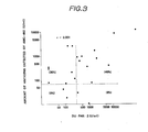
- Fig. 3 shows the comparison between the amounts of the antigen defined by DuPan-2 and the amounts of the antigen defined by AMC-462 in the same serum samples.
- the amounts of antigen in the serum samples from pancreatic cancer patients measured by the serodiagnosis system using AMC-462 of the present invention highly correlate with the amounts of CA19-9. But, 5% of the samples which gives negative results for CA19-9 gives positive results in the serodiagnosis system using AMC-462.
- Fig. 3 indicates that the amounts of the antigens of cancer defined by the present invention little correlate with the amounts of DuPan-2. From the point of view of the positive rate, AMC-462 can be more effective for serodiagnosis of pancreatic cancer than DuPan-2.
- Example 1 and 3 indicate that the antigen detectable in the serodiagnosis-system using AMC-462 was different from CEA and DuPan-2 which were known tumor markers of digestive system cancer, especially pancreatic cancer.
- the binding test was carried out by the method of sandwich ELISA.
- suspensions of AMC-462 or NS19-9 (10 ⁇ g/ml) as the first antibody were distributed into the wells of a 96- well plate for EIA (50 ⁇ l/well) and fixed on the bottom of the well.
- the plate was blocked by 1% BSA-PBS and then the serum samples from pancreatic cancer patients containing both the antigen defined by CA19-9 and the antigen defined by AMC-462 were added in an amount of 50 ⁇ l per well. After well washing, the monoclonal antibody NS19-9 . or AMC-462 (0.1 - 50 ug/mk) was added as an inhibiting antibody and the plate was washed well.
- biotin-labeled monoclonal antibody NS19-9 or biotin-labeled monoclonal antibody AMC-462 was added as the second antibody and the plate was washed well.
- Avidin-biotin-peroxidase was distributed into the wells and then the plate was washed well. Thereafter, the ABTS substrate solution was added and an enzyme reaction was allowed to proceed and then terminated by adding SDS solution. For each well, the absorbance at 415 nm was measured.
- A) the first antibody AMC-462 (10 ⁇ g/ml) the second antibody : biotin-labeled AMC-462 (0.1-50 ⁇ g/ml)
Landscapes
- Health & Medical Sciences (AREA)
- Chemical & Material Sciences (AREA)
- Organic Chemistry (AREA)
- Life Sciences & Earth Sciences (AREA)
- Immunology (AREA)
- General Health & Medical Sciences (AREA)
- Biochemistry (AREA)
- Biophysics (AREA)
- Cell Biology (AREA)
- Genetics & Genomics (AREA)
- Medicinal Chemistry (AREA)
- Molecular Biology (AREA)
- Proteomics, Peptides & Aminoacids (AREA)
- Gastroenterology & Hepatology (AREA)
- Preparation Of Compounds By Using Micro-Organisms (AREA)
- Peptides Or Proteins (AREA)
- Medicines Containing Antibodies Or Antigens For Use As Internal Diagnostic Agents (AREA)
Applications Claiming Priority (2)
| Application Number | Priority Date | Filing Date | Title |
|---|---|---|---|
| JP61166138A JPH0673470B2 (ja) | 1986-07-15 | 1986-07-15 | 抗ヒト胃癌単クロ−ン性抗体amc−462 |
| JP166138/86 | 1986-07-15 |
Publications (3)
| Publication Number | Publication Date |
|---|---|
| EP0253646A2 true EP0253646A2 (de) | 1988-01-20 |
| EP0253646A3 EP0253646A3 (en) | 1990-01-31 |
| EP0253646B1 EP0253646B1 (de) | 1993-05-12 |
Family
ID=15825742
Family Applications (1)
| Application Number | Title | Priority Date | Filing Date |
|---|---|---|---|
| EP87306257A Expired - Lifetime EP0253646B1 (de) | 1986-07-15 | 1987-07-15 | Monoklonale anti-menschliche Magenkrebs-Antikörper |
Country Status (5)
| Country | Link |
|---|---|
| US (1) | US5051355A (de) |
| EP (1) | EP0253646B1 (de) |
| JP (1) | JPH0673470B2 (de) |
| CA (1) | CA1320460C (de) |
| DE (1) | DE3785795T2 (de) |
Cited By (3)
| Publication number | Priority date | Publication date | Assignee | Title |
|---|---|---|---|---|
| EP0339632A3 (de) * | 1988-04-28 | 1990-09-19 | Kyowa Hakko Kogyo Kabushiki Kaisha | Testmethode für menschliche Krebsantigene |
| EP0362332A4 (en) * | 1988-03-04 | 1991-04-10 | New England Deaconess Hospital | Carcinoma orosomucoid-related antigen, a monoclonal antibody thereto, and their uses |
| CN112292398A (zh) * | 2018-03-30 | 2021-01-29 | 积水医疗株式会社 | 与dupan-2抗原特异性反应的单克隆抗体及其制造方法 |
Families Citing this family (4)
| Publication number | Priority date | Publication date | Assignee | Title |
|---|---|---|---|---|
| US5160723A (en) * | 1985-04-19 | 1992-11-03 | Sloan-Kettering Institute For Cancer Research | Method of imaging colorectal carcinoma lesion and composition for use therein |
| WO2003060121A2 (en) * | 2001-12-21 | 2003-07-24 | Diadexus, Inc. | Compositions and methods relating to gastric specific genes and proteins |
| EP2972375A2 (de) * | 2013-03-13 | 2016-01-20 | Creatics LLC | Verfahren und zusammensetzungen zur erkennung von bauchspeicheldrüsenkrebs |
| FR3078887B1 (fr) | 2018-03-14 | 2020-12-18 | Urodelia | Vaccins autologues contre le cancer |
Family Cites Families (15)
| Publication number | Priority date | Publication date | Assignee | Title |
|---|---|---|---|---|
| US4471057A (en) * | 1981-06-30 | 1984-09-11 | The Wistar Institute | Detection of colorectal carcinoma |
| WO1984000758A1 (en) * | 1982-08-09 | 1984-03-01 | Centocor Inc | Immunoassay for carbohydrate antigenic determinant |
| JPS59128397A (ja) * | 1983-01-13 | 1984-07-24 | Nippon Koutai Kenkyusho:Kk | 抗ヒト胃癌抗体 |
| JPS59205327A (ja) * | 1983-03-11 | 1984-11-20 | スロ−ン−ケツタリング・インステイテユ−ト・フオ−・キヤンサ−・リサ−チ | ヒト膀胱及び尿管がんに対するモノクロ−ナル抗体及び方法 |
| US4579827A (en) * | 1983-03-11 | 1986-04-01 | Sloan-Kettering Institute For Cancer Research | Monoclonal antibodies to human gastrointestinal cancers and hybridoma method of production of the monoclonal antibodies |
| JPS60190721A (ja) * | 1984-03-12 | 1985-09-28 | Kyowa Hakko Kogyo Co Ltd | 抗腫瘍特異的単クロ−ン性抗体の製造法 |
| US4683200A (en) * | 1984-05-17 | 1987-07-28 | Setsuo Hirohashi | Monoclonal antibody to human cancer antigen and method for producing same |
| JPS60243026A (ja) * | 1984-05-17 | 1985-12-03 | Akio Hirohashi | モノクロ−ナル抗体 |
| NZ212419A (en) * | 1984-06-25 | 1988-08-30 | Mucan Diagnostics Pty Ltd | In vitro diagnostic test for detecting cancer cells producing mucin antigens |
| JPS6144900A (ja) * | 1984-08-08 | 1986-03-04 | Green Cross Corp:The | モノクロ−ナル抗体及び癌抗原検出用試薬 |
| DE3584280D1 (de) * | 1984-10-26 | 1991-11-07 | Wakunaga Seiyaku Kk | Monoklonaler antikoerper gegen menschlichen krebs. |
| EP0199141A3 (de) * | 1985-04-19 | 1988-07-20 | Sloan-Kettering Institute For Cancer Research | Monoklonale Antikörper gegen humanen Magen-Darmkrebs |
| JPS61250000A (ja) * | 1985-04-27 | 1986-11-07 | Green Cross Corp:The | モノクロ−ナル抗体 |
| JPS6236398A (ja) * | 1985-08-12 | 1987-02-17 | Green Cross Corp:The | モノクロ−ナル抗体 |
| JPS62123200A (ja) * | 1985-11-22 | 1987-06-04 | Green Cross Corp:The | モノクロ−ナル抗体 |
-
1986
- 1986-07-15 JP JP61166138A patent/JPH0673470B2/ja not_active Expired - Fee Related
-
1987
- 1987-07-14 CA CA000542064A patent/CA1320460C/en not_active Expired - Fee Related
- 1987-07-15 DE DE8787306257T patent/DE3785795T2/de not_active Expired - Fee Related
- 1987-07-15 EP EP87306257A patent/EP0253646B1/de not_active Expired - Lifetime
-
1989
- 1989-12-06 US US07/445,160 patent/US5051355A/en not_active Expired - Fee Related
Cited By (3)
| Publication number | Priority date | Publication date | Assignee | Title |
|---|---|---|---|---|
| EP0362332A4 (en) * | 1988-03-04 | 1991-04-10 | New England Deaconess Hospital | Carcinoma orosomucoid-related antigen, a monoclonal antibody thereto, and their uses |
| EP0339632A3 (de) * | 1988-04-28 | 1990-09-19 | Kyowa Hakko Kogyo Kabushiki Kaisha | Testmethode für menschliche Krebsantigene |
| CN112292398A (zh) * | 2018-03-30 | 2021-01-29 | 积水医疗株式会社 | 与dupan-2抗原特异性反应的单克隆抗体及其制造方法 |
Also Published As
| Publication number | Publication date |
|---|---|
| US5051355A (en) | 1991-09-24 |
| JPS6321562A (ja) | 1988-01-29 |
| CA1320460C (en) | 1993-07-20 |
| EP0253646B1 (de) | 1993-05-12 |
| EP0253646A3 (en) | 1990-01-31 |
| DE3785795T2 (de) | 1993-09-02 |
| DE3785795D1 (de) | 1993-06-17 |
| JPH0673470B2 (ja) | 1994-09-21 |
Similar Documents
| Publication | Publication Date | Title |
|---|---|---|
| US4892935A (en) | Anti-human pulmonary carcinoma monoclonal antibody | |
| US5185432A (en) | Monoclonal antibodies and antigen for human non-small cell lung carcinoma and other certain human carcinomas | |
| US4800155A (en) | Human monoclonal antibody to lung carcinoma and hybridoma producing the same | |
| CA1320460C (en) | Anti-human gastric cancer monoclonal antibody | |
| EP0156578B1 (de) | Verfahren zur Herstellung von tumorspezifische monoklonale Antikörper produzierenden Hybridomazellen | |
| EP0171083B1 (de) | Monoklonaler Antikörper, Verfahren zu seiner Herstellung, den monoklonalen Antikörper enthaltendes Reagenz zum Nachweiss eines Krebsantigens und Verfahren zu seiner Herstellung | |
| EP0218257B1 (de) | Spezifischer monoklonaler Antikörper gegen menschliches Lungenadenokarzinom | |
| Anderson et al. | Monoclonal antibodies to human malignant mesothelioma | |
| EP0272113A2 (de) | Monoklonale Antikörper gegen menschlichen Krebs | |
| EP0155172A2 (de) | Monoklonale Antikörper gegen menschlichen Lungenkrebs | |
| EP0339633A2 (de) | Monoklonaler Antikörper gegen menschlichen Magenkrebs | |
| Kudo et al. | A novel human monoclonal antibody directed to a tumor‐associated antigen | |
| US5081032A (en) | Anti-human pulmonary adenocarcinoma monoclonal antibody | |
| EP0266188B1 (de) | Monoklonale Antikörper gegen menschliche mesotheliale Zellen | |
| US5552291A (en) | Anti-human pulmonary adenocarcinoma specific monoclonal antibody | |
| EP0235817A2 (de) | Monoklonaler Antikörper gegen menschlichen Magenkrebs | |
| CA1294905C (en) | Anti-lafora body monoclonal antibody | |
| EP0508282A2 (de) | Anti-idiotypische monoklonale Antikörper | |
| US5580740A (en) | Antihuman pulmonary adenocarcinoma monoclonal antibody | |
| EP0285143B1 (de) | Monoclonaler Antikörper gegen menschliches Lungenadenokarzinom | |
| JP2626979B2 (ja) | ヒト癌細胞に対するモノクロナール抗体4c1および該抗体を産生するハイブリドーマ | |
| EP0285052A2 (de) | Monoklonaler Antikörper | |
| JPH0213393A (ja) | 抗ヒト肺腺癌単クローン性抗体 | |
| JPH06102038B2 (ja) | モノクロ−ナル抗体 |
Legal Events
| Date | Code | Title | Description |
|---|---|---|---|
| PUAI | Public reference made under article 153(3) epc to a published international application that has entered the european phase |
Free format text: ORIGINAL CODE: 0009012 |
|
| AK | Designated contracting states |
Kind code of ref document: A2 Designated state(s): DE FR GB |
|
| PUAL | Search report despatched |
Free format text: ORIGINAL CODE: 0009013 |
|
| AK | Designated contracting states |
Kind code of ref document: A3 Designated state(s): DE FR GB |
|
| 17P | Request for examination filed |
Effective date: 19900616 |
|
| 17Q | First examination report despatched |
Effective date: 19910710 |
|
| GRAA | (expected) grant |
Free format text: ORIGINAL CODE: 0009210 |
|
| AK | Designated contracting states |
Kind code of ref document: B1 Designated state(s): DE FR GB |
|
| REF | Corresponds to: |
Ref document number: 3785795 Country of ref document: DE Date of ref document: 19930617 |
|
| ET | Fr: translation filed | ||
| PGFP | Annual fee paid to national office [announced via postgrant information from national office to epo] |
Ref country code: DE Payment date: 20000710 Year of fee payment: 14 |
|
| PGFP | Annual fee paid to national office [announced via postgrant information from national office to epo] |
Ref country code: FR Payment date: 20000711 Year of fee payment: 14 |
|
| PGFP | Annual fee paid to national office [announced via postgrant information from national office to epo] |
Ref country code: GB Payment date: 20000713 Year of fee payment: 14 |
|
| PG25 | Lapsed in a contracting state [announced via postgrant information from national office to epo] |
Ref country code: GB Free format text: LAPSE BECAUSE OF NON-PAYMENT OF DUE FEES Effective date: 20010715 |
|
| GBPC | Gb: european patent ceased through non-payment of renewal fee |
Effective date: 20010715 |
|
| PG25 | Lapsed in a contracting state [announced via postgrant information from national office to epo] |
Ref country code: FR Free format text: LAPSE BECAUSE OF NON-PAYMENT OF DUE FEES Effective date: 20020329 |
|
| PG25 | Lapsed in a contracting state [announced via postgrant information from national office to epo] |
Ref country code: DE Free format text: LAPSE BECAUSE OF NON-PAYMENT OF DUE FEES Effective date: 20020501 |
|
| REG | Reference to a national code |
Ref country code: FR Ref legal event code: ST |
|
| PLBE | No opposition filed within time limit |
Free format text: ORIGINAL CODE: 0009261 |
|
| STAA | Information on the status of an ep patent application or granted ep patent |
Free format text: STATUS: NO OPPOSITION FILED WITHIN TIME LIMIT |