EP0254081A2 - Verfahren zur Feststellung spezifisches Protein - Google Patents
Verfahren zur Feststellung spezifisches Protein Download PDFInfo
- Publication number
- EP0254081A2 EP0254081A2 EP87109352A EP87109352A EP0254081A2 EP 0254081 A2 EP0254081 A2 EP 0254081A2 EP 87109352 A EP87109352 A EP 87109352A EP 87109352 A EP87109352 A EP 87109352A EP 0254081 A2 EP0254081 A2 EP 0254081A2
- Authority
- EP
- European Patent Office
- Prior art keywords
- detecting
- protein
- peptide
- test sample
- monoclonal antibodies
- Prior art date
- Legal status (The legal status is an assumption and is not a legal conclusion. Google has not performed a legal analysis and makes no representation as to the accuracy of the status listed.)
- Withdrawn
Links
Images


Classifications
-
- G—PHYSICS
- G01—MEASURING; TESTING
- G01N—INVESTIGATING OR ANALYSING MATERIALS BY DETERMINING THEIR CHEMICAL OR PHYSICAL PROPERTIES
- G01N33/00—Investigating or analysing materials by specific methods not covered by groups G01N1/00 - G01N31/00
- G01N33/48—Biological material, e.g. blood, urine; Haemocytometers
- G01N33/50—Chemical analysis of biological material, e.g. blood, urine; Testing involving biospecific ligand binding methods; Immunological testing
- G01N33/53—Immunoassay; Biospecific binding assay; Materials therefor
- G01N33/576—Immunoassay; Biospecific binding assay; Materials therefor for hepatitis
-
- G—PHYSICS
- G01—MEASURING; TESTING
- G01N—INVESTIGATING OR ANALYSING MATERIALS BY DETERMINING THEIR CHEMICAL OR PHYSICAL PROPERTIES
- G01N33/00—Investigating or analysing materials by specific methods not covered by groups G01N1/00 - G01N31/00
- G01N33/48—Biological material, e.g. blood, urine; Haemocytometers
- G01N33/50—Chemical analysis of biological material, e.g. blood, urine; Testing involving biospecific ligand binding methods; Immunological testing
- G01N33/53—Immunoassay; Biospecific binding assay; Materials therefor
- G01N33/543—Immunoassay; Biospecific binding assay; Materials therefor with an insoluble carrier for immobilising immunochemicals
- G01N33/54366—Apparatus specially adapted for solid-phase testing
-
- G—PHYSICS
- G01—MEASURING; TESTING
- G01N—INVESTIGATING OR ANALYSING MATERIALS BY DETERMINING THEIR CHEMICAL OR PHYSICAL PROPERTIES
- G01N33/00—Investigating or analysing materials by specific methods not covered by groups G01N1/00 - G01N31/00
- G01N33/48—Biological material, e.g. blood, urine; Haemocytometers
- G01N33/50—Chemical analysis of biological material, e.g. blood, urine; Testing involving biospecific ligand binding methods; Immunological testing
- G01N33/53—Immunoassay; Biospecific binding assay; Materials therefor
- G01N33/543—Immunoassay; Biospecific binding assay; Materials therefor with an insoluble carrier for immobilising immunochemicals
- G01N33/54393—Improving reaction conditions or stability, e.g. by coating or irradiation of surface, by reduction of non-specific binding, by promotion of specific binding
-
- G—PHYSICS
- G01—MEASURING; TESTING
- G01N—INVESTIGATING OR ANALYSING MATERIALS BY DETERMINING THEIR CHEMICAL OR PHYSICAL PROPERTIES
- G01N33/00—Investigating or analysing materials by specific methods not covered by groups G01N1/00 - G01N31/00
- G01N33/48—Biological material, e.g. blood, urine; Haemocytometers
- G01N33/50—Chemical analysis of biological material, e.g. blood, urine; Testing involving biospecific ligand binding methods; Immunological testing
- G01N33/53—Immunoassay; Biospecific binding assay; Materials therefor
- G01N33/543—Immunoassay; Biospecific binding assay; Materials therefor with an insoluble carrier for immobilising immunochemicals
- G01N33/551—Immunoassay; Biospecific binding assay; Materials therefor with an insoluble carrier for immobilising immunochemicals the carrier being inorganic
- G01N33/553—Metal or metal coated
-
- G—PHYSICS
- G01—MEASURING; TESTING
- G01N—INVESTIGATING OR ANALYSING MATERIALS BY DETERMINING THEIR CHEMICAL OR PHYSICAL PROPERTIES
- G01N33/00—Investigating or analysing materials by specific methods not covered by groups G01N1/00 - G01N31/00
- G01N33/48—Biological material, e.g. blood, urine; Haemocytometers
- G01N33/50—Chemical analysis of biological material, e.g. blood, urine; Testing involving biospecific ligand binding methods; Immunological testing
- G01N33/53—Immunoassay; Biospecific binding assay; Materials therefor
- G01N33/577—Immunoassay; Biospecific binding assay; Materials therefor involving monoclonal antibodies binding reaction mechanisms characterised by the use of monoclonal antibodies
-
- G—PHYSICS
- G01—MEASURING; TESTING
- G01N—INVESTIGATING OR ANALYSING MATERIALS BY DETERMINING THEIR CHEMICAL OR PHYSICAL PROPERTIES
- G01N33/00—Investigating or analysing materials by specific methods not covered by groups G01N1/00 - G01N31/00
- G01N33/48—Biological material, e.g. blood, urine; Haemocytometers
- G01N33/50—Chemical analysis of biological material, e.g. blood, urine; Testing involving biospecific ligand binding methods; Immunological testing
- G01N33/58—Chemical analysis of biological material, e.g. blood, urine; Testing involving biospecific ligand binding methods; Immunological testing involving labelled substances
- G01N33/585—Chemical analysis of biological material, e.g. blood, urine; Testing involving biospecific ligand binding methods; Immunological testing involving labelled substances with a particulate label, e.g. coloured latex
Definitions
- the present invention relates to a simple and rapid method of detecting very small amounts of a specific protein in fluids using with the immunolo - gical reaction, and to the reagents and a set of equipments to make the detection.
- the domain applicable to this detecting method includes the kits for the detection of various specific proteins in the fundamental research, various clinical diagnostic agents, and the kits for the detection of various diseases in agriculture, stock raising and marine products industry, and the like.
- the detection of specific proteins is an indispensable means.
- the detection of specific proteins has become an essential factor. Namely, in certain diseases, the proteins specific to the diseases (for example, hormone, enzyme, etc.) are always released into the body fluids. In these cases, not only the diseases can be diagnos- able through the detection of specific proteins thereof, but also the lapse of diseases can be known through the detection followed up with the lapse of time.
- the detection of specific protein should have high reliability.
- high accuracy capable of detecting specific protein specifically is required.
- the method of detecting specific protein which has been used most widely hitherto is that used the immunological antigen-antibody reaction.
- This is the method wherein the antibody which recognizes the specific protein to become antigen is prepared based on the property that the antibody recognizes the antigenic substance specifically, this is allowed to react with specific protein in the test sample through antigen-antibody reaction, and the result is detected using secondary phenomena such as, for example, coagulation reaction etc. and labels.
- the qualitative detecting method is one to detect the existence of more than a fixed amount of protein by simple means such as tinting and is applied generally to clinical diagnostic agents etc.
- the quantitative detecting method is a system to determine the specific protein quantitatively and has higher sensitivity and accuracy compared with the qualitative detecting method, so that it is used widely for fundamental research and clinical diagnosis.
- the radioimmunoassay method which was developed by S.A. Berson and R. Yallow in 1959, has become established today as an indispensable one.
- the principle of the detecting method is to quantify the antigen-antibody reaction in whatever form and to directly determine the antigen or antibody.
- the method of labeling the antigen or antibody with something as a means therefor is called labeling immunoassay method and the quantification is made on the basis of this label. Since the radioimmunoassay method uses a radioactive isotope with high sensitivity as a label, it can detect quantitatively even extremely small amounts of protein antigen. Therefore, it has contributed significantly not only to the fundamental research but also .to the clinical diagnosis.
- Preparing two kinds of antibodies specific to an antigen one is allowed to bond to the solid phase and the other is bonded with enzyme to become a label to obtain a complex.
- the test sample solution is added to the solid phase bonded with antibody and unreacted substances are removed by washing sufficiently.
- the enzyme-antibody complex is allowed to react, a bonded matter in "sandwich" form composed of the antibody- antigenic protein complex is produced on the surface of the solid phase.
- the enzymic activity at the complex bonded on the surface at the solid phase is determined to quantify the specific antigenic. protein in the test sample.
- a problem in this detecting method is the need of completely removing the unreacted enzyme-antibody complex. This is because an enzyme with high specific activity is used in order to raise the sensibility, therefore if there is even a slight amounts of this complex remaining, the background of the measurement is raised. The shortcoming of using the enzyme as a label cannot be avoided and the difficulty in the procedure removing the unreacted agents cannot be eliminated. Accordingly, several simple qualitative methods which applied the sandwich method such as enzyme immunoassay methods are devised, but all of them have the same problem described above and there remains a problematic point that perfect accuracy cannot be obtained.
- the detecting method required most urgently at present is one in which special equipment and apparatus are unnecessary and highly accurate and reliable data can be obtained rapidly by a simple procedure. For instance, when clinical diagnosis is urgently needed after an operation, etc., it is essential to make judgment accurately and still rapidly for the sake of preparing subsequent treatment. It is natural that high accuracy and rapidity are sought on the results obtained by the diagnostic agent, which becomes the material for the judgment.
- the invention relates to a method of detecting specific protein characterized in that, by the use of monoclonal antibodies or a combination of a polyclonal antibody and a kind of a monoclonal antibody having high specificity.
- the specific protein which is included in the test sample is prefiltered for removing the obstractive residue, and is inserted in a sandwich form between the antibodies, and detected visually as a tinted spot produced directly on the test piece of paper with the detecting agent (metallic colloidal solution bonded with another monoclonal antibody).
- Fig. 1 is diagram illustrated showing one example of the fundamental principle of the invention.
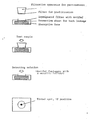
- Fig. 2 is a diagram illustrating an example (2 - 2) of the invention.
- Fig. 3 is a diagram showing clearly the performance of the invention.
- the monoclonal antibody is defined as "a pure antibody against only one antigen determinant" (Dictionary of Biology, 3rd edition, Iwanami Bookstore), the specificity thereof against antigen is exceptionally high compared with the usual antibody (called polyclonal antibody to monoclonal antibody). For this reason, utilizing this property, an improvement is made in raising the accuracy by using this monoclonal antibody in the method of detecting specific protein applied to the conventional antigen-antibody reaction.
- the detection accuracy has been enhanced and the specificity has been raised by double antigen-antibody reactions.
- the direct detecting method of labeling the immunoassay method was applied and, as a detecting substance, colloidal metal particles (metallic colloidal particles) were used. These metallic colloidal particles are tinted with a unique color due to their respective physical properties and therefore, the result of the antigen-antibody reaction can be directly and visually displayed.
- the colloidal metal particles are known in the field of immunochemistry research.
- colloidal gold particles are widely used. These are ones having made from aurichloric acid colloidal under particular conditions and are tinted from pink to reddish purple depending on the diameter of the colloidal gold particle. when this colloidal gold and the protein are placed under certain conditions, they are bonded through noncovalent and electrostatic adsorption to form a stable conjugate. Since this is a bonding not necessitated by chemical modification, there is no denaturation of the protein nor in the physiological activity reduced. In the world of immunohistology, colloidal gold is already known as an immunostainer for microscopic research, and has been used for many years. The metallic colloidal particles represented by this colloidal gold have been adopted as a detecting substance for specific protein.
- the method of detecting specific protein utilizing this metallic colloid was already reported as the sol particle immunoassay method (SPIA method) (Japanese Unexamined Patent Publication No. Sho 55- 15100).
- the clinical diagnostic agent uses colloidal gold among other metallic colloids (trade name "Predictor Color D") is already on the market.
- the principle thereof lies in the fact that the test sample is added to the solution of gold colloid-antibody conjugate and the coagulation of the conjugate taking place through the immunosedimentation reaction between antigenic substance in the sample and antibody is judged by the accompanying fading of the solution of colloidal gold (from red to colorless).
- the sensitivity is low (more than 400 international units of hCG in 1 liter of urine) and the detection time of required is long (about 30 minutes).
- the amount at detecting substance is very small, there remains a problem also in the point of accuracy because the fading is so indistince, it is difficult to make an accurate judgment.
- the fading occurs even when it is negative.
- the result of the judgment cannot be preserved.
- the fading of the conjugate of the metallic colloid in the state of solution was not untilized, but a method capable of clear and accurate judgment by anyone and still capable of the preservation of the result thereof has been devised.
- a method of removing the obstructive substances which become the origin of the high background at the time of detection has also been devised together.
- Fig. 1 the fundamental principle of the invention is shown.
- two monoclonal antibodies against a plurality of mutually different epitopes are used.
- P indicates the specific protein or peptide to be detected, and the monoclonal antibody against "epitope 1" on this P molecule is expressed by AntiPml and that against "epitope 2" is expressed by AntiPm2.
- AntiPml forms the conjugate with metallic colloid (M) and is made up of the detecting solution after the H buffer solution is added.
- AntiPm2 is impregnated into an absorptive carrier (FP) and, after air-drying, the carrier is dipped into a buffer solution containing the blocking agent (B, eg. bovine serum albumin etc.) to mask the surface of FP except for the areas where AntiPm2 has been bonded thereto for the purpose of the preventing bonding with nonspecific protein.
- the blocking agent eg. bovine serum albumin etc.
- the specific protein P in a fixed quantity of the solution of test sample is caught with AntiPm2 on FP.
- the specific protein P is recognized by both monoclonal antibodies 'of AntiPml and AntiPm2 and, as shown in the diagram, the complex composed of AntiPm2-specific protein-AntiPml-conjugate with colloidal gold is produced on the absorptive carrier FP through the antigen-antibody reaction.
- the result is that only the complex bonded on the surface of FP remains as a tinted spot to be identified.
- the former When using the polyclonal antibody in combination with the monoclonal antibody having high specificity, the former may be regarded as AntiPm2 and the latter as AntiPml.
- the specific protein can be also detected semiquantitatively similarly to the so-called dot blotting method used most frequently in the domain of fundamental biochemistry without the necessity of analytical instruments.
- test samples are original urine, etc.
- unknown substances stay on the detecting test paper to catch. the conjugate of metallic colloid nonspecifically on the test paper, so that the phenomenon to render the background high upon detection tends to occur, but, in the invention, a filtration apparatus for the pretreatment of the test sample has been devised as a means to overcome this.
- the filtration apparatus is compared of a nylon membrane filter coated with a solution of skim milk, pH buffer solution, etc. (refer to Fig. 2).
- this coating treatment it is possible to prevent the protein in the test sample from being caught nonspecifically on the filter during the prefiltration stage.
- p H can be kept as optimum as possible.
- both the deteacting sensitivity and the detecting accuracy are improved drastically (refer to Table 1).
- pH buffer solution In order to make the conjugate of metallic colloid stable in the reaction solution, pH buffer solution, stabilizers, etc. were added to the conjugate solution of metallic colloid.
- the specific protein detectable by this detecting method should have two or more kinds of epitopes or the epitope specific to the antigen thereof.
- many proteins have subunits structure. Accordingly, in the case of protein consisting of a plurality of subunits, only a plurality of monoclonal antibodies specific to the epitopes on the surface of the protein of respective subunits has only to be obtained and, even in the case of protein or peptide not having a subunit structure, it can be detected specifically by the present detecting method, even with the existence of a plurality of different epitopes.
- the invention provides a simple and rapid method of detecting the specific protein, wherein the specificity thereof has been enhanced and the accuracy has been raised by the recognition of specific protein using a plurality of monoclonal antibodies or a combination of a monoclonal antibody having high specificity and a polyclonal antibody, and, at the same time, the sensitivity has been increased by using stable metallic colloid as a detecting agent so as to be detected visually.
- hCG human chorionic gonadotropin
- hCG is a hormone secreted specifically at the early days of pregnancy or in the cases of certain kinds of chorionic and nonchorionic tumors.
- various methods to detect hCG released into urine with anti-hCG antibodies have been devised and are used widely as the indicator not only for the diagnosis of pregnancy but also for the diagnosis and therapy of tumors aforementioned.
- hCG is glycoprotein hormone consisting of both ⁇ -and p-subunits. Since the d-subunit therebetween has a structure that very closely resembles the respective subunits of hLH (human luteinizing hormone), hFSH (human follicle-stimulating hormone) and hTSH (human thyroid-stimulating hormone), the crossing property with conventional polyclonal antibodies is high and, in particular, the possibility of having a false positive reaction cannot be eliminated. Particularly, when detecting extremely low concentrations of hCG as in the case of prognosis at the time of abortion, and the diagnosis of extrauterine pregnancy, etc., the conventional polyclonal antibodies could not be used at all.
- mice immunized with commercial hCG was allowed to fuse with myeloma cells by the common method to obtain hybridoma. This was submitted to cloning and, using ⁇ -and ⁇ -subunits as standard substances, the specificity of the monoclonal antibody was assayed for each clone by the ELISA method.
- anti- ⁇ -hCG monoclonal antibody was impregnated into the filter. After air-drying, this was dipped into the solutions of BSA etc. to block it and then air dried to fix it to the supporter. On the other hand, anti- ⁇ -hCG antibody was bonded to the commercial solution of gold colloid as usual.
- the procedure is as follows: The above mentioned filter aforementioned is dipped for a fixed period of time (for example, 1 minute) into the urine which has been pretreated by the filtration apparatus, to catch hCG in the urine by ant - ⁇ -hCG monoclonal antibodies on the filter. Then, this filter is placed in the solution of conjugate of gold colloid and allowed to stand for some time (about 10 minutes). Thereafter, the filter is taken out and, after rinsing slightly, it is dried in air.
- a fixed period of time for example, 1 minute
- Two components are obtained similarly to (2 - 1). Namely, the filter impregnated with the anti- ⁇ -hCG monoclonal antibody and the gold colloidal solution conjugated with the anti-p-hCG monoclonal antibody are provided.
- the former is placed on the top surface of the cylindrical vessel and the lower section is filled up with a highly hygroscopic material (refer to Fig. 2).
- a sample of the urine to be tested is taken into an injection tube, this is submitted to prefiltration with the filtration apparatus, and a fixed amount of this is added drop by drop onto the center of the test paper of the kit to catch the hCG on the filter. Following this, the solution of conjugate of colloidal gold is added drop by drop. if it is possitive, a reddish purple spot is observed immediately at the center.
- hCG of a concentration of as low as 5 IU/liter could be detected.
- the sensitivity is over 100 times greater than that of the conventional simplified detecting method.
- the specific protein is immediately identified as a spot after the addition of conjugate of colloidal gold.
- the background upon detection happens to become high
- the background at the time of detection, disappears completely to make it possible to drastically enhance the detecting sensitivity and the detecting accuracy for a specific protein.
- the necessary time for this method is less than two minutes, which shows the unparalleled rapidity.
- the result obtained remains - 18 - on the test paper which makes it possible to transport it by the way of the post office etc. It is also possible to preserve it as a permanent record.
- the detection is not hindered to any degree in very high concectration of one hundred million IU/liter and, because of the general absence of false negative called "prozone phenomenon", so the test sample can be used without dilution.
- the invention comprises the following:
Landscapes
- Health & Medical Sciences (AREA)
- Life Sciences & Earth Sciences (AREA)
- Immunology (AREA)
- Engineering & Computer Science (AREA)
- Chemical & Material Sciences (AREA)
- Molecular Biology (AREA)
- Biomedical Technology (AREA)
- Urology & Nephrology (AREA)
- Hematology (AREA)
- Biotechnology (AREA)
- General Health & Medical Sciences (AREA)
- Cell Biology (AREA)
- Pathology (AREA)
- Food Science & Technology (AREA)
- Medicinal Chemistry (AREA)
- Physics & Mathematics (AREA)
- Analytical Chemistry (AREA)
- Biochemistry (AREA)
- Microbiology (AREA)
- General Physics & Mathematics (AREA)
- Chemical Kinetics & Catalysis (AREA)
- Communicable Diseases (AREA)
- Inorganic Chemistry (AREA)
- Peptides Or Proteins (AREA)
- Investigating Or Analysing Biological Materials (AREA)
- Preparation Of Compounds By Using Micro-Organisms (AREA)
Applications Claiming Priority (2)
| Application Number | Priority Date | Filing Date | Title |
|---|---|---|---|
| JP153707/86 | 1986-06-30 | ||
| JP15370786 | 1986-06-30 |
Publications (2)
| Publication Number | Publication Date |
|---|---|
| EP0254081A2 true EP0254081A2 (de) | 1988-01-27 |
| EP0254081A3 EP0254081A3 (de) | 1988-04-20 |
Family
ID=15568341
Family Applications (1)
| Application Number | Title | Priority Date | Filing Date |
|---|---|---|---|
| EP87109352A Withdrawn EP0254081A3 (de) | 1986-06-30 | 1987-06-29 | Verfahren zur Feststellung spezifisches Protein |
Country Status (2)
| Country | Link |
|---|---|
| EP (1) | EP0254081A3 (de) |
| JP (1) | JPS63127160A (de) |
Cited By (9)
| Publication number | Priority date | Publication date | Assignee | Title |
|---|---|---|---|---|
| EP0258963A3 (de) * | 1986-06-09 | 1988-09-07 | Ortho Diagnostic Systems Inc. | Immunoassay mit Verwendung von kolloidalem Gold |
| WO1990009588A1 (en) * | 1989-02-09 | 1990-08-23 | Eastman Kodak Company | Immunoassay kit containing multiple level filter |
| EP0401913A1 (de) * | 1989-06-05 | 1990-12-12 | Janssen Pharmaceutica N.V. | Festphasen-Test zur Verwendung mit einem physikalischen Entwickler |
| EP0327395A3 (de) * | 1988-02-05 | 1990-12-27 | Idexx Corp. | Assaysatz und Verfahren |
| US5202267A (en) * | 1988-04-04 | 1993-04-13 | Hygeia Sciences, Inc. | Sol capture immunoassay kit and procedure |
| WO1994014071A1 (fr) * | 1992-12-10 | 1994-06-23 | Novik, Anatoly Matveevich | Methode de diagnostic des tumeurs malignes chez l'etre humain |
| US5851777A (en) * | 1996-02-05 | 1998-12-22 | Dade Behring Inc. | Homogeneous sol-sol assay |
| EP1933153A1 (de) * | 2006-12-15 | 2008-06-18 | Mikrogen Molekularbiologische Entwicklungs-GmbH | Testsystem zur parallelen Durchführung von Multiparameter-Nachweisreaktionen |
| US8460877B2 (en) | 2004-01-23 | 2013-06-11 | Arkray, Inc. | Method of protein measurement |
Families Citing this family (3)
| Publication number | Priority date | Publication date | Assignee | Title |
|---|---|---|---|---|
| JPH02141665A (ja) * | 1988-11-24 | 1990-05-31 | Godo Shiyusei Kk | 糞便中のヘモグロビンの検出方法 |
| JP3126383B2 (ja) * | 1991-03-28 | 2001-01-22 | 株式会社アズウェル | 簡易な分析装置 |
| JP3978153B2 (ja) | 2003-06-12 | 2007-09-19 | 富士フイルム株式会社 | 光干渉基材、標的検出用基材、並びに、標的検出装置及び標的検出方法 |
Family Cites Families (7)
| Publication number | Priority date | Publication date | Assignee | Title |
|---|---|---|---|---|
| JPS549078B1 (de) * | 1971-02-23 | 1979-04-20 | ||
| NL7807532A (nl) * | 1978-07-13 | 1980-01-15 | Akzo Nv | Metaal-immunotest. |
| JPS57136165A (en) * | 1981-02-18 | 1982-08-23 | Mochida Pharmaceut Co Ltd | Immunological measuring reagent |
| JPS60501271A (ja) * | 1983-05-06 | 1985-08-08 | ザ・トラステイ−ズ・オブ・コロンビア・ユニヴア−シテイ・イン・ザ・シテイ・オブ・ニユ−・ヨ−ク | ヒト絨毛性ゴナドトロピンのイムノアツセイ |
| US4595661A (en) * | 1983-11-18 | 1986-06-17 | Beckman Instruments, Inc. | Immunoassays and kits for use therein which include low affinity antibodies for reducing the hook effect |
| GB8331514D0 (en) * | 1983-11-25 | 1984-01-04 | Janssen Pharmaceutica Nv | Visualization method |
| GR850899B (de) * | 1984-04-12 | 1985-11-25 | Gen Hospital Corp |
-
1987
- 1987-06-29 EP EP87109352A patent/EP0254081A3/de not_active Withdrawn
- 1987-06-29 JP JP16197987A patent/JPS63127160A/ja active Pending
Cited By (10)
| Publication number | Priority date | Publication date | Assignee | Title |
|---|---|---|---|---|
| EP0258963A3 (de) * | 1986-06-09 | 1988-09-07 | Ortho Diagnostic Systems Inc. | Immunoassay mit Verwendung von kolloidalem Gold |
| EP0327395A3 (de) * | 1988-02-05 | 1990-12-27 | Idexx Corp. | Assaysatz und Verfahren |
| US5202267A (en) * | 1988-04-04 | 1993-04-13 | Hygeia Sciences, Inc. | Sol capture immunoassay kit and procedure |
| WO1990009588A1 (en) * | 1989-02-09 | 1990-08-23 | Eastman Kodak Company | Immunoassay kit containing multiple level filter |
| EP0401913A1 (de) * | 1989-06-05 | 1990-12-12 | Janssen Pharmaceutica N.V. | Festphasen-Test zur Verwendung mit einem physikalischen Entwickler |
| US5710049A (en) * | 1989-06-05 | 1998-01-20 | Janssen Pharmaceutica, N.V. | Solid phase assay for use with a physical developer |
| WO1994014071A1 (fr) * | 1992-12-10 | 1994-06-23 | Novik, Anatoly Matveevich | Methode de diagnostic des tumeurs malignes chez l'etre humain |
| US5851777A (en) * | 1996-02-05 | 1998-12-22 | Dade Behring Inc. | Homogeneous sol-sol assay |
| US8460877B2 (en) | 2004-01-23 | 2013-06-11 | Arkray, Inc. | Method of protein measurement |
| EP1933153A1 (de) * | 2006-12-15 | 2008-06-18 | Mikrogen Molekularbiologische Entwicklungs-GmbH | Testsystem zur parallelen Durchführung von Multiparameter-Nachweisreaktionen |
Also Published As
| Publication number | Publication date |
|---|---|
| JPS63127160A (ja) | 1988-05-31 |
| EP0254081A3 (de) | 1988-04-20 |
Similar Documents
| Publication | Publication Date | Title |
|---|---|---|
| DK160108C (da) | Fremgangsmåde og udstyr til direkte eller indirekte påvisning af reaktion mellem et specifikt bindingsmiddel og den tilsvarende acceptorsubstans | |
| US4397960A (en) | Immunoassays using F(AB')2 fragments | |
| EP0398913B1 (de) | Testverfahren und reagenzsatz dazu | |
| US5958790A (en) | Solid phase transverse diffusion assay | |
| EP3904878B1 (de) | Immunchromatografischer streifen zur schwangerschaftsdiagnose mit mehrfachen testlinien und schwangerschaftsdiagnosekit damit | |
| EP0207152B1 (de) | Festphasendiffusionstextverfahren | |
| US5876935A (en) | Luminescent specific binding assay | |
| EP0254081A2 (de) | Verfahren zur Feststellung spezifisches Protein | |
| EP3244189A1 (de) | Verfahren und vorrichtung für quantitativen analyt-assay | |
| KR930703611A (ko) | 양막파열을 감지하는 진단방법과 이방법을 이용한 테스트키트 | |
| WO1985000663A1 (en) | Immunometric assay using polyclonal and monoclonal antibodies and a kit for use therein | |
| HK1000970B (en) | Solid phase diffusion assay | |
| US6753189B1 (en) | Detection apparatus and method for the same | |
| US4522922A (en) | Soluble assay | |
| US4725556A (en) | Process of immunoassay of a substance in a liquid sample containing a similar substance | |
| JPS58211662A (ja) | 試験試料中の被検体を測定するための均一系結合分析方法および該方法に用いる試薬系 | |
| IE58254B1 (en) | Process and reagent for the determination of a polyvalent antigen | |
| EP0245509B1 (de) | Immuntestverfahren | |
| EP0303980A2 (de) | Träger beschichtet mit Antigen oder Antikörper | |
| EP0756173A1 (de) | Reagenz zum nachweis von substanzen und verfahren zur diagnose der rheumatoiden arthritis | |
| Madhu et al. | Interferences in Diagnostic Immunoassay Procedures | |
| CN120504740A (zh) | 一种促甲状腺激素抗体及其在荧光免疫层析检测中的应用 | |
| JP4013270B2 (ja) | 尿中生理活性ペプチドの測定法及び測定用試薬 | |
| JPS60364A (ja) | 新規な抗原定量法 | |
| Mack | Development of an enzyme-linked immunosorbent assay for porcine relaxin |
Legal Events
| Date | Code | Title | Description |
|---|---|---|---|
| PUAI | Public reference made under article 153(3) epc to a published international application that has entered the european phase |
Free format text: ORIGINAL CODE: 0009012 |
|
| AK | Designated contracting states |
Kind code of ref document: A2 Designated state(s): DE FR GB NL |
|
| PUAL | Search report despatched |
Free format text: ORIGINAL CODE: 0009013 |
|
| AK | Designated contracting states |
Kind code of ref document: A3 Designated state(s): DE FR GB NL |
|
| STAA | Information on the status of an ep patent application or granted ep patent |
Free format text: STATUS: THE APPLICATION IS DEEMED TO BE WITHDRAWN |
|
| 18D | Application deemed to be withdrawn |
Effective date: 19881021 |