EP0257759A2 - Teilchen zum Kalibrieren von Durchflusszytometriegeräten und anderen Analysevorrichtungen - Google Patents
Teilchen zum Kalibrieren von Durchflusszytometriegeräten und anderen Analysevorrichtungen Download PDFInfo
- Publication number
- EP0257759A2 EP0257759A2 EP87306144A EP87306144A EP0257759A2 EP 0257759 A2 EP0257759 A2 EP 0257759A2 EP 87306144 A EP87306144 A EP 87306144A EP 87306144 A EP87306144 A EP 87306144A EP 0257759 A2 EP0257759 A2 EP 0257759A2
- Authority
- EP
- European Patent Office
- Prior art keywords
- instrument
- particles
- calibration
- signal
- fluorescence
- Prior art date
- Legal status (The legal status is an assumption and is not a legal conclusion. Google has not performed a legal analysis and makes no representation as to the accuracy of the status listed.)
- Withdrawn
Links
Images

Classifications
-
- G—PHYSICS
- G01—MEASURING; TESTING
- G01N—INVESTIGATING OR ANALYSING MATERIALS BY DETERMINING THEIR CHEMICAL OR PHYSICAL PROPERTIES
- G01N15/00—Investigating characteristics of particles; Investigating permeability, pore-volume or surface-area of porous materials
- G01N15/10—Investigating individual particles
- G01N15/1012—Calibrating particle analysers; References therefor
-
- G—PHYSICS
- G01—MEASURING; TESTING
- G01N—INVESTIGATING OR ANALYSING MATERIALS BY DETERMINING THEIR CHEMICAL OR PHYSICAL PROPERTIES
- G01N15/00—Investigating characteristics of particles; Investigating permeability, pore-volume or surface-area of porous materials
- G01N15/10—Investigating individual particles
- G01N15/14—Optical investigation techniques, e.g. flow cytometry
- G01N15/1456—Optical investigation techniques, e.g. flow cytometry without spatial resolution of the texture or inner structure of the particle, e.g. processing of pulse signals
-
- G—PHYSICS
- G01—MEASURING; TESTING
- G01N—INVESTIGATING OR ANALYSING MATERIALS BY DETERMINING THEIR CHEMICAL OR PHYSICAL PROPERTIES
- G01N15/00—Investigating characteristics of particles; Investigating permeability, pore-volume or surface-area of porous materials
- G01N15/10—Investigating individual particles
- G01N2015/1006—Investigating individual particles for cytology
-
- G—PHYSICS
- G01—MEASURING; TESTING
- G01N—INVESTIGATING OR ANALYSING MATERIALS BY DETERMINING THEIR CHEMICAL OR PHYSICAL PROPERTIES
- G01N15/00—Investigating characteristics of particles; Investigating permeability, pore-volume or surface-area of porous materials
- G01N15/10—Investigating individual particles
- G01N15/1012—Calibrating particle analysers; References therefor
- G01N2015/1014—Constitution of reference particles
-
- G—PHYSICS
- G01—MEASURING; TESTING
- G01N—INVESTIGATING OR ANALYSING MATERIALS BY DETERMINING THEIR CHEMICAL OR PHYSICAL PROPERTIES
- G01N15/00—Investigating characteristics of particles; Investigating permeability, pore-volume or surface-area of porous materials
- G01N15/10—Investigating individual particles
- G01N15/14—Optical investigation techniques, e.g. flow cytometry
- G01N2015/1477—Multiparameters
Definitions
- Characteristics of particles may be determined by techniques in which the particles remain relatively fixed while being analyzed or while the particles are moving in a stream or carried in a suspension.
- Flow cytometers are known and available for analyzing or detecting certain characteristics of particles which are in motion.
- cells or other biological particles are caused to flow in a liquid stream so that each particle, preferably one at a time, passes through a sensing region which measures physical or chemical characteristics of the particles.
- signals may be detected for association with different characteristics of the particles, including electrical, acoustical and radioactive, flow cytometers commonly rely on optical signals for the analysis of particles passing through the instrument.
- Flow cytometers and other biological particle analysis instruments are normally calibrated with particles which simulate or approximate the types of particles or cells which are expected to undergo actual analysis.
- calibration particles should be selected so that they have characteristics similar to the particles to be tested for, such as size, volume, surface characteristics, granularity properties, color features (cellular stains, dyes, immunofluorescent tags and the like).
- Past and current calibration procedures for flow cytometry instruments include the utilization of chicken red blood cells for the calibration steps. While chicken red blood cells are reliable for some aspects of the calibration procedures, they, nevertheless, are not entirely satisfactory, particularly because of spectral deficiencies in some light-related characteristics.
- microspheric beads have become available for calibrating cellular analysis instruments.
- the method of the present invention for calibrating an instrument for using that instrument to obtain at least one light-related signal from particles under analysis comprises directing an incident beam of light at calibration particles having one or more known characteristics related to the particles expected to be analyzed. Both a light signal and a noise signal from the calibration particles are detected.
- the light signal is detected from one type of particle in the calibration mix, such as a fluorescent particle
- the noise signal is detected from another type of particle in the calibration mix, such as a non-fluorescent particle.
- a measurement is made of the ratio of the detected light signal to the detected noise signal, and this ratio is reported. The measured ratio is compared to a predetermined ratio which represents a threshold for minimum instrument performance.
- This method includes adjusting, if the predetermined ratio has not been attained, the operation of the instrument, while the calibration particles are within the incident beam of light, until the measured ratio reaches, or exceeds, the predetermined ratio whereby the instrument is calibrated for subsequently obtaining the light signal from particles to be analyzed.
- the method is employed for calibrating a flow cytometry instrument and includes passing calibration particles, having known characteristics related to the particles expected to be tested, in a liquid flow stream so that each calibration particle passes, substantially one at at time, through an incident beam of light. Both a light signal and a noise signal relating to the calibration particles are detected and the signal to noise ratio is determined. This ratio is reported as a measured separation value between the light signal and the noise signal. The measured separation value is compared to a predetermined separation value representative of a threshold for minimum instrument performance. Adjustment of the operation of the instrument is performed, if the predetermined separation value has not been attained, until the measured separation value reaches the predetermined separation value whereby the instrument is calibrated for subsequently obtaining light signals from particles to be analyzed.
- a further aspect of the present invention relates to particle standards, for calibrating fluorescence analysis instruments, comprising osmium tetroxide-fixed chicken red blood cells, which are incapable of emitting fluorescence, including autofluorescence, until reacted with a specific fluorophore.
- calibration methods and materials provide substantial improvements over currently known and used calibration techniques.
- the present invention allows the operator to calibrate a particle analysis instrument by relying on calibration standards and/or thresholds which have been predetermined and are made available to the operator.
- the threshold values for establishing minimum performance of the instrument are displayed to the operator during the calibration procedures so that a calibration target is known.
- Guesswork and eyeballing techniques of calibration are eliminated by the calibration procedures of the present invention so that calibration errors are reduced or eliminated, and reproducibility of tests is enhanced.
- the calibration procedures of the present invention are particularly suitable for flow cytometry instruments since proper calibration assures proper alignment of the optical features of the instrument, accounts for spectral cross-talk by employment of compensation techniques, and provides information to the operator about the sensitivity of the instrument particularly for optimizing the performance of the instrument for immunofluorescent analysis of particles. It should be pointed out that the advantages and features of the present invention extend beyond flow cytometry instruments, and may be utilized in other analysis instruments such as automated microscopes, fluorescence microscopes, quantitative microscopes, image analyzers, and the like.
- the calibration technique to be described below, and in conjunction with the drawings is associated with a flow cytometry instrument.
- the exemplary calibration procedures to be described are associated with a FACS® analyzer marketed by Becton Dickinson Immunocytometry Systems, Mountain View, California.
- the histogram display of Fig. l and the dot population displays of Figs. 2-4 are typical displays seen on the screen of the FACS analyzer during the calibration procedures.
- Appropriate software to carry out the calibration procedures, according to the principles of the present invention, may be provided for the flow cytometry instrument to facilitate the various calibration procedures and functions.
- the flow cytometry instrument to be calibrated includes a nozzle or like device for providing a liquid flow stream of particles to be analyzed.
- the liquid flow stream of particles is ensheathed in a sheath fluid so that the particle stream may be hydrodynamically focused as it flows through an incident beam of light normally directed at right angles to the stream of particle flow.
- light characteristics associated with each particle may be detected. Therefore, one or more light detectors for measuring fluorescence, light scatter (in one or more directions) absorbance and the like may be included. These light detectors include photomultiplier tubes (PMT's) which convert light signals to electrical signals.
- PMT's photomultiplier tubes
- an electrical impedance measurement may also be taken as the particles pass through an orifice.
- This impedance measurement is related to particle volume and is based on the well-known Coulter principle.
- PMT's are provided for detecting two different colors of fluorescence and light scatter, in this instance side scatter, for measuring scatter from the particles at substantially 90° with respect to the incident beam of light.
- the FACS analyzer further provides for the measurement of electrical impedance associated with particle volume.
- the calibration techniques of the present invention are intended to inform the operator of the instrument that the instrument is calibrated based on predetermined threshold values representative of minimum instrument performance. These minimum threshold values provide an indication of sensitivity of the instrument for carrying out particle analysis, particularly immunofluorescent performance. For example, in addition to side scatter and volume, the specific flow cytometry instrument being described is capable of detecting two different colors of fluorescence, for a total of four different parameters.
- the first fluorescence color of particles to be analyzed will be in the green spectral region associated with particles labeled with fluorescein representing FITC stained cells; the second fluorescence color of particles to be analyzed will be in the red spectral region associated with phycoerythrin labeled particles representing PE stained cells.
- fluorescein representing FITC stained cells
- the second fluorescence color of particles to be analyzed will be in the red spectral region associated with phycoerythrin labeled particles representing PE stained cells.
- fluorochromes or labeling agents may be employed, if desired, depending upon the tests to be conducted and the particles or cells to be analyzed.
- the optical elements of the instrument could be out of alignment.
- the tests to be performed relate to data acquisition for monitoring particles of the immune system, such as leucocyte samples
- the operator may desire to determine whether the instrument is calibrated to the proper sensitivity for conducting these tests.
- Instrument sensitivity performance and the results provided thereby are illustrated in Fig. l.
- the sensitivity test is merely a confirmatory test to determine whether the instrument is properly calibrated for analyzing certain types of particles, such as leucocytes or other immune system cells or particles.
- a reagent kit which preferably includes three containers, each having different plastic microspheres or beads therein.
- the different beads are maintained in phosphate buffered saline (PBS) containing gelatin, 0.l% Tween 20 and 0.l% sodium azide as a preservative.
- PBS phosphate buffered saline
- FITC Fluorescein labeled
- PE Phycoerythrin labeled
- Unlabeled beads at approximately 4.5 microns diameter, are included in the third container.
- the concentration of the beads in each of the three containers is approximately l06 particles per milliliter.
- a sample tube is prepared by the operator by mixing the FITC, PE and unlabeled beads in a l:l:l ratio by adding one drop of each of the three reagents to three milliliters of sterile PBS. This sample tube with the mixture is then inserted into the FACS analyzer, which is operated in known fashion so that the beads pass substantially one at a time through an incident beam of light.
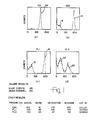
- Fig. l represents the display on the instrument screen of the four parameters provided in the form of a histogram.
- the Y axis of the histograms are particle counts and the X axis is the number of electrical channels over which data acquisition is made. In the embodiments being described, there are 255 channels represented along the X axis of the various histograms. It is preferred that the histograms be reported or displayed in a log scale relationship so that the separation of histogram peaks between noise and signal is seen as a difference, but is actually expressed as a linearized ratio.
- the light-related signals are displayed in Figs. l(b),(c) and (d) relating to side scatter, FITC and PE, respectively.
- the side scatter signal is illustrated by curve l2
- noise is illustrated in region l4.
- FITC signal is illustrated by curve l6
- noise is illustrated in region l8.
- PE signal is illustrated by curve 20, while noise is indicated within region 22.
- volume displayed in Fig. l(a) the volume signal is displayed under curve 24; noise detection is displayed below the histograms as the number of noise events.
- noise is actually introduced so that a separation value between noise and signal may be measured. This is necessary because the noise represented by region l4 of Fig. l(b) does not result from unlabeled cells as occurs in the fluorescence signal.
- side scatter (SSC) noise is triggered on an unrelated signal to generate a reference signal for noise.
- the FL 2 signal is arbitrarily chosen to generate a reference signal.
- Noise is introduced into the side scatter signal by adjusting the FL 2 controls on the flow cytometry instrument so that the event rate (or flow rate of particles) through the instrument is about double the event rate for obtaining data with respect to volume, FL l and FL 2.
- noise occurs in the side scatter signal so that the linearized amount of separation between the noise and scatter signals may be determined.
- the screen of the instrument being calibrated may be programmed to display the results in the form of a digital readout.
- Software for the instrument, including calibration procedures, may be readily programmed to provide information relating to the accumulated data. Therefore, it can be seen on Fig. l, below the histograms, that volume results are reported in digital readout form so that noise events and mean channel signal may be read by the operator.
- the volume mean channel should be approximately mid-scale in the volume histogram. A mid-scale reading indicates that the orifice through which the particles flow is correctly chosen and that the volume controls on the instrument are properly set. A noise event level preferably below 500 is indicative of proper set up of the instrument.
- Fig. l also displays on the screen the digital readings associated with the respective histograms of Figs. l(b),(c) and (d).
- the various parameters of SSC, FL l and FL 2 are listed along with readings for signal, noise, separation, minimum and lot ID. Lot ID and minimum values are related to each other.
- the operator Prior to inserting the mixture of calibration beads into the instrument, the operator is instructed to feed the lot ID number into the program for regulating the calibration procedures. Since the calibration beads may have variable properties including different fluorescence brightness, intensity and the like, calibration of the instrument should take into account these variable properties.
- different predetermined separation values are established as minimum or threshold values for minimum instrument performance. Since the software may be preprogrammed, the predetermined minimum separation value is established for display on Fig. l as long as the lot ID number has been fed into the program by the operator at the outset of the calibration procedures.
- Fig. l Displayed at the bottom of Fig. l are the average mean channel values for signal and noise for side scatter, FL l and FL 2, associated with the histograms of Figs. l(b),(c) and (d), respectively.
- the side scatter signal is displayed as having a mean average channel at l92, while the mean average channel for noise is at l6.
- the separation between these signal and noise results is reported at l76.
- the predetermined minimum separation value for side scatter signal is reported at l60. This minimum side scatter separation value is the desired level for the instrument to be sufficiently calibrated for reliably making side scatter measurements. Therefore, since the actual measured separation is at l76, the minimum is exceeded. Accordingly, the instrument is deemed to be calibrated for subsequent tests of particles in which side scatter is a signal to be detected.
- FITC labeled beads from the first container of the reagent kit described above.
- a tube of FITC labeled bead suspension may be prepared by adding one drop of FITC bead reagent to l milliliter of sterile PBS. This tube is then inserted into the FACS analyzer, and in conjunction with the calibration software, the screen displays dot population plots as illustrated in Fig. 2.
- Fig. 2(a) illustrates FL 2 vs FL l
- Fig. 2(b) illustrates side scatter (SSC) vs FL l.
- Fig. 2(a) calibration is attained when the dot population for the green (FITC) beads is located near the center of the display.
- the fluorescence PMT voltages (on the instrument) may be adjusted to move the dot population to this location.
- the operator With respect to the FL l and FL 2 signal averages, printed below the dot plots, the operator assures calibration by maximizing these signals.
- Optical elements of the instrument such as the condenser lens controls, are turned or adjusted to maximize these signal averages, which should be maximized at approximately the same time.
- a target value is the goal for calibration.
- the target for the SSC signal channel average is about l90.
- the operator may adjust the side scatter (SSC) PMT voltage on the instrument until the SSC signal average reaches approximately l90.
- Other adjustments of the optical elements or PMT's may be needed to achieve the target level for the SSC signal average.
- Fig. 3 illustrates typical dot plot populations associated with PMT adjustment.
- Fig. 3(a) is a dot plot of FL 2 vs FL l
- Figs. 3(b) is a dot plot of side scatter (SSC) vs volume.
- SSC side scatter
- the operator employs a different calibration particle procedure from those described above.
- the operator preferably prepares a tube of unlabeled bead suspension by adding one drop of unlabeled beads, from the unlabeled bead container of the above-described reagent kit, to one milliliter of sterile PBS.
- Fig. 3 In addition to the dot plots, Fig. 3 also displays three numbers representing the channel mean averages for the populations of the three parameters of FL l, FL 2 and SSC. While the calibration particles are flowing through the instrument at a controlled, known rate, the operator aims at a predetermined target for values of FL l average, FL 2 average and SSC average.
- Another adjustment may be made in establishing the calibration of the instrument.
- This additional adjustment relates to compensation for spectral cross-talk or overlap in the event that two-color fluorescence analysis is to be performed. This procedure facilitates the adjustment for fluorescence compensation so that undesired green signal will be removed from the red (PE stained) population, and undesired red signal will be removed from the green (FITC stained) population.
- the operator preferably starts with a tube of calibration particles similar to that prepared with respect to the sensitivity test described above.
- This tube is prepared with a l:l:l mixture of FITC, PE and unlabeled beads by adding one drop of each of the three reagents, from the above-described containers, to three milliliters of sterile PBS.
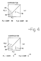
- the screen displays a real-time dot plot, representing uncompensated fluorescence, such as seen in Fig. 4(a).
- the three separate dot populations displayed in Fig. 4(a) represent the unstained, green and red stained calibration beads.
- Unstained beads are typically located inside the square in the lower left corner of the display, illustrated by region 28.
- Red (PE stained) beads are typically located above the unstained population, indicated by region 30.
- the green population is to the right of the unstained population, indicated by region 32.
- regions 28,30 and 32 are only approximate.
- Below the dot plot of Fig. 4(a) is a digital display of FL l compensation and FL 2 compensation. If these digital values are not fluctuating at or close to zero, adjustments should be made for fluorescence compensation.
- Compensation is provided for two-color fluorescence analysis by adjusting FL l compensation and FL 2 compensation controls on the instrument until the compensation values listed on the screen are as close to zero as possible. After adjustment is made, the compensation value should fluctuate and vary equally above and below zero. With respect to FL l compensation, the compensated value represents the difference between the mean horizontal channel for the unstained population and the mean horizontal channel for the green stained population. As the FL l compensation control is adjusted on the instrument and the value on the screen approaches zero, the green population 32 on the dot plot should move downwardly until it is even with the unstained population 28 on a horizontal line parallel to the horizontal axis, as seen in Fig. 4(b).
- the mean channel difference listed next to FL 2 compensation on the screen represents the difference between the mean vertical channel for the unstained and red stained populations.
- the FL 2 compensation control is adjusted on the instrument and the value on the screen approaches zero, the red population 30 on the dot plot should move horizontally until it is even with the unstained population 28 on a vertical line parallel to the vertical axis, as illustrated in Fig. 4(b).
- These adjustments of the FL l and FL 2 controls achieve fluorescence compensation by electrically subtracting a proportion of the unwanted green signal from the red population, and by electrically subtracting a proportion of the unwanted red signal from the green population. Compensation is achieved when the predetermined values of fluorescence compensation on the screen, set by the computer program at zero, are met. If acceptable or target settings cannot be achieved, the operator may be required to return to the alignment and PMT adjustment procedures as explained above.
- the operator may return to the sensitivity test described above in conjunction with Fig. l. If the minimum separation values are achieved for each of the parameters being calibrated, the operator can proceed to use the instrument with full reliance that it is properly calibrated for the tests to be conducted.
- a variety of particles may be used as standards.
- biological samples such as particles or cells may be employed as long as these samples either have, or may be treated to have, characteristics similar to the actual particles to undergo analysis.
- chicken red blood cells may be employed with proper selection techniques.
- a modified form of chicken red blood cells may be used as particle standards.
- fluorescently labeled osmiumtetroxide-fixed chicken red blood cells may serve as particle standards since they have the size and scatter properties of biological cells.
- osmiumtetroxide fixation quenches the autofluorescence of cells.
- These cells may be biotinilated, and subsequently reacted with a fluorophore conjugated to avidin in known fashion. Fluorescence of the avidin complex, bound to the cells, is not quenched by the osmiumtetroxide on the cells.
- the specific fluorophore conjugated to the avidin has spectral properties characteristic for cell-surface staining. Many different fluorophores may be used in the conjugates, including those to be described below.
- the most preferred type of calibration particles for the present invention are plastic microbeads.
- Such beads may be fabricated with substantially uniform diameter, and have surface characteristics which are suitable for surface binding of fluorophores or other colorimetric marking agents.
- Substantially spherical beads may be formed having a diameter between 0.5 and 20 microns, and most preferably between 2 and 8 microns, so that these calibration beads are of the same order of magnitude in size as leucocytes.
- the beads are usually made in solid form, they may be hollow inside and could be vesicles, such as liposomes or other microcarriers.
- the beads do not have to be perfect spheres in order to function in accordance with the present invention.
- Plastic materials such as polystyrene, polyacrylamide and other latex materials may be employed for fabricating the beads, and other plastic materials such as polyvinylchloride, polypropylene and the like may also be utilized.
- fluorescein and the phycoerythrin labels for the beads described above other fluorescence labels, such as allophycocyanin, Texas red, rhodamine, rhodamine-type compounds and other stains may be employed as fluorescent labels for purposes of the present invention.
- the beads are preferably provided as an aqueous solution so that the beads are present in concentrations ranging from l05 to l08 particles per milliliter, and preferably having a concentration of at least l06 particles per milliliter.
- the present invention provides a method and materials for calibrating an instrument prior to using that instrument for obtaining at least one light-related signal from particles under analysis.
- the present invention may be used with appropriate software to display minimum values of signal to noise ratios which should be met in order to determine that the instrument is calibrated for a specific light-related parameter.
- By reporting the signal to noise ratio as a differential on the linear scale operator convenience is not only facilitated, but guesswork, eyeballing and errors are substantially reduced or eliminated.
- the techniques and materials of this invention also provide quality assurance for clinical diagnostic applications.
Landscapes
- Chemical & Material Sciences (AREA)
- Biochemistry (AREA)
- General Physics & Mathematics (AREA)
- Health & Medical Sciences (AREA)
- Life Sciences & Earth Sciences (AREA)
- Analytical Chemistry (AREA)
- Dispersion Chemistry (AREA)
- General Health & Medical Sciences (AREA)
- Physics & Mathematics (AREA)
- Immunology (AREA)
- Pathology (AREA)
- Investigating Or Analysing Biological Materials (AREA)
- Investigating, Analyzing Materials By Fluorescence Or Luminescence (AREA)
- Investigating Or Analysing Materials By The Use Of Chemical Reactions (AREA)
- Investigating Or Analysing Materials By Optical Means (AREA)
Applications Claiming Priority (2)
| Application Number | Priority Date | Filing Date | Title |
|---|---|---|---|
| US901860 | 1986-08-29 | ||
| US06/901,860 US4704891A (en) | 1986-08-29 | 1986-08-29 | Method and materials for calibrating flow cytometers and other analysis instruments |
Publications (2)
| Publication Number | Publication Date |
|---|---|
| EP0257759A2 true EP0257759A2 (de) | 1988-03-02 |
| EP0257759A3 EP0257759A3 (de) | 1989-10-18 |
Family
ID=25414938
Family Applications (3)
| Application Number | Title | Priority Date | Filing Date |
|---|---|---|---|
| EP87306145A Expired - Lifetime EP0258983B1 (de) | 1986-08-29 | 1987-07-10 | Verfahren zum Kalibrieren von Durchflusszytometriegeräten |
| EP87306144A Withdrawn EP0257759A3 (de) | 1986-08-29 | 1987-07-10 | Teilchen zum Kalibrieren von Durchflusszytometriegeräten und anderen Analysevorrichtungen |
| EP87306143A Expired - Lifetime EP0258982B1 (de) | 1986-08-29 | 1987-07-10 | Mittel zum Kalibrieren von Durchflusszytometriegeräten und anderen Analysevorrichtungen |
Family Applications Before (1)
| Application Number | Title | Priority Date | Filing Date |
|---|---|---|---|
| EP87306145A Expired - Lifetime EP0258983B1 (de) | 1986-08-29 | 1987-07-10 | Verfahren zum Kalibrieren von Durchflusszytometriegeräten |
Family Applications After (1)
| Application Number | Title | Priority Date | Filing Date |
|---|---|---|---|
| EP87306143A Expired - Lifetime EP0258982B1 (de) | 1986-08-29 | 1987-07-10 | Mittel zum Kalibrieren von Durchflusszytometriegeräten und anderen Analysevorrichtungen |
Country Status (7)
| Country | Link |
|---|---|
| US (1) | US4704891A (de) |
| EP (3) | EP0258983B1 (de) |
| JP (3) | JPS6363942A (de) |
| AT (2) | ATE75042T1 (de) |
| DE (2) | DE3778253D1 (de) |
| ES (2) | ES2031507T3 (de) |
| GR (2) | GR3004885T3 (de) |
Cited By (1)
| Publication number | Priority date | Publication date | Assignee | Title |
|---|---|---|---|---|
| GB2499796A (en) * | 2012-02-28 | 2013-09-04 | Ojk Consulting Ltd | Method and system for calibrating a flow cytometer |
Families Citing this family (95)
| Publication number | Priority date | Publication date | Assignee | Title |
|---|---|---|---|---|
| DK164144C (da) * | 1988-11-17 | 1992-10-12 | Slagteriernes Forskningsinst | Fremgangsmaade ved kontrol eller justering af maaleapparater med en optisk sonde og medium til anvendelse ved fremgangsmaaden |
| CA2021658C (en) * | 1989-08-25 | 2001-10-09 | Myron J. Block | Multiplex immunoassay system |
| US5853984A (en) * | 1990-06-11 | 1998-12-29 | Nexstar Pharmaceuticals, Inc. | Use of nucleic acid ligands in flow cytometry |
| IE76732B1 (en) * | 1990-08-07 | 1997-11-05 | Becton Dickinson Co | One step test for absolute counts |
| JP2913219B2 (ja) * | 1991-03-06 | 1999-06-28 | 株式会社シマ研究所 | 尿沈渣検査成績管理方法 |
| US5627040A (en) * | 1991-08-28 | 1997-05-06 | Becton Dickinson And Company | Flow cytometric method for autoclustering cells |
| US5451525A (en) * | 1992-02-14 | 1995-09-19 | Coulter Corporation | Method and materials for determining particle count in a flow cytometer |
| DE69326731T2 (de) | 1992-09-04 | 2000-05-18 | Becton Dickinson And Co., Franklin Lakes | Kontroll-Teilchen für die Zellzählung und Instrumentenlinearität |
| US5620842A (en) * | 1995-03-29 | 1997-04-15 | Becton Dickinson And Company | Determination of the number of fluorescent molecules on calibration beads for flow cytometry |
| JP3875754B2 (ja) * | 1995-11-17 | 2007-01-31 | シスメックス株式会社 | フローサイトメータ用標準液 |
| US6014904A (en) * | 1996-05-09 | 2000-01-18 | Becton, Dickinson And Company | Method for classifying multi-parameter data |
| EP2264427B1 (de) | 1997-01-31 | 2017-05-03 | Xy, Llc | Optisches Gerät mit fokussierendem Reflektor zur Konvergenz der Strahlung auf einen Partikelfluss, und zugehöriges Analyseverfahren |
| US6074879A (en) * | 1997-06-23 | 2000-06-13 | Bayer Corporation | Synthetic polymer particles for use as standards and calibrators in flow cytometry |
| US6071689A (en) | 1997-12-31 | 2000-06-06 | Xy, Inc. | System for improving yield of sexed embryos in mammals |
| US6149867A (en) | 1997-12-31 | 2000-11-21 | Xy, Inc. | Sheath fluids and collection systems for sex-specific cytometer sorting of sperm |
| US6197520B1 (en) * | 1999-08-13 | 2001-03-06 | University Of Utah Research Foundation | Solution-based color compensation adjusted for temperature and electronic gains |
| US7208265B1 (en) | 1999-11-24 | 2007-04-24 | Xy, Inc. | Method of cryopreserving selected sperm cells |
| US6809804B1 (en) | 2000-05-11 | 2004-10-26 | Becton, Dickinson And Company | System and method for providing improved event reading and data processing capabilities in a flow cytometer |
| JP2004501358A (ja) | 2000-05-11 | 2004-01-15 | ベクトン・ディキンソン・アンド・カンパニー | 最適な境界を有する平滑化された多角形を使用して散布図中のクラスタを識別するシステム |
| US7713687B2 (en) | 2000-11-29 | 2010-05-11 | Xy, Inc. | System to separate frozen-thawed spermatozoa into x-chromosome bearing and y-chromosome bearing populations |
| WO2002043486A1 (en) | 2000-11-29 | 2002-06-06 | Xy, Inc. | System for in-vitro fertilization with spermatozoa separated into x-chromosome and y-chromosome bearing populations |
| JP4595067B2 (ja) | 2002-08-01 | 2010-12-08 | エックスワイ,エルエルシー | 低圧精子細胞分離システム |
| US8486618B2 (en) | 2002-08-01 | 2013-07-16 | Xy, Llc | Heterogeneous inseminate system |
| CA2534394C (en) | 2002-08-15 | 2013-01-08 | Xy, Inc. | High resolution flow cytometer |
| US7169548B2 (en) | 2002-09-13 | 2007-01-30 | Xy, Inc. | Sperm cell processing and preservation systems |
| BRPI0408857B1 (pt) | 2003-03-28 | 2018-09-11 | Inguran Llc | aparelho, métodos e processos para separar partículas e para prover esperma de animal separado por sexo |
| ES2541121T3 (es) | 2003-05-15 | 2015-07-16 | Xy, Llc | Clasificación eficiente de células haploides por sistemas de citometría de flujo |
| KR101135138B1 (ko) * | 2003-08-13 | 2012-04-16 | 루미넥스 코포레이션 | 유세포 분석기식 측정 시스템의 하나 이상의 파라미터의제어 방법 |
| JP4850711B2 (ja) * | 2003-10-02 | 2012-01-11 | ベックマン コールター, インコーポレイテッド | 血液サンプルの有核赤血球の光学測定のリファレンスコントロール |
| DK2801363T3 (en) | 2004-03-29 | 2018-05-28 | Inguran Llc | PROCEDURE FOR STORING SORTED SPERMATOZOES |
| CA2574499C (en) | 2004-07-22 | 2016-11-29 | Monsanto Technology Llc | Process for enriching a population of sperm cells |
| DE102004044717B8 (de) * | 2004-09-10 | 2010-12-16 | BAM Bundesanstalt für Materialforschung und -prüfung | Verfahren und Kit zur Kalibrierung eines Photolumineszenzmesssystems |
| EP1744145B1 (de) | 2005-07-12 | 2015-09-09 | Sysmex Corporation | Standardmaterial für ein Teilchenanalysegerät |
| JP4953710B2 (ja) * | 2005-07-12 | 2012-06-13 | シスメックス株式会社 | 尿中有形成分分析装置用標準物質 |
| JP4748719B2 (ja) * | 2006-03-02 | 2011-08-17 | 国立大学法人大阪大学 | 生細胞内の特定タンパク質の定量方法および標準蛍光マイクロビーズの作製方法 |
| CN100386054C (zh) * | 2006-04-24 | 2008-05-07 | 西安交通大学 | 用于脉搏血氧仿真器的光散射介质及制备方法 |
| US20090105963A1 (en) * | 2006-05-13 | 2009-04-23 | Jesper Laursen | Methods for Flow Cytometry Analyses of Un-Lysed Cells from Biological Fluids |
| JP5010443B2 (ja) * | 2006-12-20 | 2012-08-29 | シスメックス株式会社 | 血球分析装置および血球分析方法 |
| CN101226190B (zh) * | 2007-01-17 | 2013-07-03 | 深圳迈瑞生物医疗电子股份有限公司 | 流式细胞术的自动分类方法和装置 |
| DE102007014413B4 (de) * | 2007-03-17 | 2016-02-04 | DüRR DENTAL AG | Verfahren zum Auswerten von Fluoreszenzbildsätzen und Vorrichtung zu seiner Durchführung |
| US8102528B2 (en) * | 2007-09-13 | 2012-01-24 | Brightwell Technologies Inc. | Particle standard and method of calibrating or validating an optical particle analyzer |
| WO2009042423A1 (en) * | 2007-09-27 | 2009-04-02 | Cellatope Corporation | Methods and compositions for improved diagnostic assays |
| US8187885B2 (en) * | 2009-05-07 | 2012-05-29 | Nodality, Inc. | Microbead kit and method for quantitative calibration and performance monitoring of a fluorescence instrument |
| US20120065092A1 (en) | 2010-09-14 | 2012-03-15 | Wai Hobert | Fusion analyte cytometric bead assay, and systems and kits for performing the same |
| EP2630492B1 (de) * | 2010-10-21 | 2018-03-07 | Nexcelom Bioscience LLC | Kügelchen mit interner fokusreferenz zur abbildung einer zytometrie |
| WO2012065004A2 (en) | 2010-11-12 | 2012-05-18 | Incelldx, Inc. | Methods and systems for predicting whether a subject has a cervical intraepithelial neoplasia (cin) lesion from a suspension sample of cervical cells |
| JP2013146263A (ja) * | 2011-12-21 | 2013-08-01 | Azbil Corp | 微生物検出装置の校正方法、及び微生物検出装置の校正キット |
| ES2928973T3 (es) * | 2012-01-11 | 2022-11-23 | Takeda Pharmaceuticals Co | Caracterización de partículas subvisibles usando un analizador de partículas |
| US20150177115A1 (en) | 2012-04-06 | 2015-06-25 | Slingshot Biosciences | Hydrogel particles with tunable optical properties |
| ITTV20130026A1 (it) * | 2013-02-27 | 2014-08-28 | Texa Spa | Metodo e sistema per calibrare uno strumento analizzatore di particolato di un gas |
| US10364277B2 (en) | 2013-07-01 | 2019-07-30 | Newsouth Innovations Pty Limited | Diagnosis and treatment of autoimmune diseases |
| WO2016089521A1 (en) | 2014-12-04 | 2016-06-09 | Becton, Dickinson And Company | Flow cytometry cell sorting systems and methods of using the same |
| WO2016093970A1 (en) | 2014-12-10 | 2016-06-16 | Becton, Dickinson And Company | Methods for optically aligning light collection components and optically aligned light collection systems thereof |
| JP6692834B2 (ja) | 2015-02-09 | 2020-05-13 | スリングショット バイオサイエンシーズ, インコーポレイテッド | 調整可能な光学特性を有するヒドロゲル粒子およびその使用の方法 |
| EP3259574B1 (de) | 2015-02-18 | 2024-03-27 | Becton, Dickinson and Company | Optische erfassungssysteme und verfahren zur verwendung davon |
| WO2016144652A1 (en) | 2015-03-12 | 2016-09-15 | Becton, Dickinson And Company | Polymeric bodipy dyes and methods for using the same |
| CN107428918B (zh) | 2015-03-12 | 2021-07-30 | 贝克顿·迪金森公司 | 紫外线吸收聚合物染料及其使用方法 |
| CN107923836B (zh) | 2015-06-17 | 2020-12-01 | 贝克顿·迪金森公司 | 具有可移除散射杆的光学检测器散射帽组件及其使用方法 |
| US10876953B2 (en) | 2015-07-15 | 2020-12-29 | Becton, Dickinson And Company | System and method for label selection |
| WO2017011626A1 (en) * | 2015-07-15 | 2017-01-19 | Becton, Dickinson And Company | System and method for adjusting cytometer measurements |
| KR102600559B1 (ko) | 2015-10-13 | 2023-11-10 | 벡톤 디킨슨 앤드 컴퍼니 | 다중모드 형광 이미징 유동 세포 계측 시스템 |
| WO2017105928A1 (en) | 2015-12-16 | 2017-06-22 | Becton, Dickinson And Company | Photostable fluorescent polymeric tandem dyes including luminescent metal complexes |
| EP3426296A4 (de) | 2016-03-10 | 2019-11-20 | Becton, Dickinson and Company | Verfahren zur bewertung einer zellulären probe für her-2/neu-expression und zusammensetzungen zur durchführung davon |
| JP6237806B2 (ja) * | 2016-03-16 | 2017-11-29 | ソニー株式会社 | 微小粒子分取装置 |
| KR102527830B1 (ko) | 2016-03-17 | 2023-05-02 | 벡톤 디킨슨 앤드 컴퍼니 | 고효율 형광 유세포 분석기를 사용하는 세포 선별 |
| GB2548807A (en) * | 2016-03-23 | 2017-10-04 | Sony Corp | Information processing apparatus,second information processing apparatus, system,method and computer program product |
| CN105928753B (zh) * | 2016-04-18 | 2019-01-15 | 中国科学院苏州生物医学工程技术研究所 | 流式细胞仪校准用鸡红细胞的制备方法 |
| US11408812B2 (en) | 2016-04-22 | 2022-08-09 | Becton, Dickinson And Company | High density deposition for array production |
| KR102381176B1 (ko) | 2016-05-12 | 2022-04-01 | 비디 바이오사이언시스 | 개선된 이미지 해상도를 갖는 형광 이미징 유세포 분석법 |
| CN109661439A (zh) | 2016-07-07 | 2019-04-19 | 贝克顿·迪金森公司 | 发荧光的水溶剂化共轭聚合物 |
| CN109477785B (zh) | 2016-09-13 | 2022-09-27 | 贝克顿·迪金森公司 | 具有光学均衡的流式细胞仪 |
| CN109073530B (zh) | 2016-10-03 | 2022-04-26 | 贝克顿·迪金森公司 | 用于确定流式细胞仪中的流体流的液滴延迟的方法和系统 |
| JP7097885B2 (ja) | 2016-12-12 | 2022-07-08 | ベクトン・ディキンソン・アンド・カンパニー | 水溶性ポリマー色素 |
| US10684275B2 (en) | 2016-12-14 | 2020-06-16 | Becton, Dickinson And Company | Methods and compositions for obtaining a tuberculosis assessment in a subject |
| CN110337331B (zh) | 2017-02-08 | 2022-04-26 | 贝克顿·迪金森公司 | 干染料试剂装置以及制造和使用所述干染料试剂装置的方法 |
| EP3586104A4 (de) | 2017-02-27 | 2020-12-16 | Becton, Dickinson and Company | Lichtdetektionssysteme und verfahren zur verwendung davon |
| JP6955385B2 (ja) * | 2017-07-14 | 2021-10-27 | 株式会社堀場製作所 | 粒子分析装置における光照射を調整するためのモニター装置 |
| KR102838795B1 (ko) | 2018-01-23 | 2025-07-25 | 벡톤 디킨슨 앤드 컴퍼니 | 광 검출을 동적 차폐하기 위한 시스템 및 이를 사용하는 방법 |
| EP3775052B1 (de) | 2018-03-30 | 2024-06-05 | Becton, Dickinson and Company | Wasserlösliche polymere farbstoffe mit seitenständigen chromophoren |
| EP3785017B1 (de) | 2018-04-27 | 2025-09-10 | Becton, Dickinson And Company | Sammelsysteme für stromzytometrisch sortierte proben und verfahren zur verwendung davon |
| JP7416729B2 (ja) | 2018-06-19 | 2024-01-17 | ベクトン・ディキンソン・アンド・カンパニー | 検出器アレイのための可変多重化スイッチ、システム、およびその使用方法 |
| CN113785187B (zh) | 2019-03-22 | 2025-01-28 | 贝克顿·迪金森公司 | 使用射频多路复用激发数据对荧光成像进行光谱解混 |
| KR20220129585A (ko) | 2020-01-24 | 2022-09-23 | 슬링샷 바이오사이언시즈 인코포레이티드 | 세포-유사 보정 입자를 위한 조성물 및 방법 |
| US12436080B2 (en) * | 2020-02-24 | 2025-10-07 | Corning Incorporated | Calibration set including nanoparticles of nylon-6 and dyes |
| WO2021173719A1 (en) | 2020-02-25 | 2021-09-02 | Becton, Dickinson And Company | Bi-specific probes to enable the use of single-cell samples as single color compensation control |
| JP7807395B2 (ja) | 2020-05-04 | 2026-01-27 | スリングショット バイオサイエンシーズ, インコーポレイテッド | 多重化アッセイのためのパッシブ光学バーコード付与のための組成物および方法 |
| WO2022132985A1 (en) * | 2020-12-16 | 2022-06-23 | Juno Therapeutics, Inc. | Threshold gating for flow cytometry methods |
| JP2024518755A (ja) | 2021-04-23 | 2024-05-02 | ベクトン・ディキンソン・アンド・カンパニー | 分析器及び/又は選別器式のフロー式粒子分析器用の流体管理システム |
| EP4155349A1 (de) | 2021-09-24 | 2023-03-29 | Becton, Dickinson and Company | Wasserlösliche gelbe grünabsorbierende farbstoffe |
| AU2022376541A1 (en) | 2021-10-29 | 2024-04-18 | Slingshot Biosciences, Inc. | Hydrogel particles as feeder cells and as synthetic antigen presenting cells |
| CN119907921A (zh) | 2022-05-05 | 2025-04-29 | 弹弓生物科学公司 | 用于血液学的作为红细胞模拟物的工程化颗粒和含有工程化颗粒的组合物 |
| CA3257621A1 (en) | 2022-06-02 | 2023-12-07 | Slingshot Biosciences, Inc. | APOPTOTIC CELL MIMETIC |
| CN120712108A (zh) | 2022-10-26 | 2025-09-26 | 弹弓生物科学公司 | 具有可调整光学性质的大小可调整合成颗粒和用于使用其进行免疫细胞激活的方法 |
| WO2025049609A1 (en) | 2023-08-29 | 2025-03-06 | Slingshot Biosciences, Inc. | Cd34 stem cell mimics |
| US20250137905A1 (en) | 2023-10-27 | 2025-05-01 | Becton, Dickinson And Company | Fluid Supply Systems Having a Flow Control Circuit and Single Fluidic Connection, and Methods of Use Thereof |
Family Cites Families (12)
| Publication number | Priority date | Publication date | Assignee | Title |
|---|---|---|---|---|
| US3406121A (en) * | 1965-10-01 | 1968-10-15 | Dade Reagents Inc | Blood cell counting standard and method of preparing the same |
| US3412037A (en) * | 1966-01-26 | 1968-11-19 | Technicon Corp | Method and means for the calibration of particle counting apparatus |
| US3791517A (en) * | 1973-03-05 | 1974-02-12 | Bio Physics Systems Inc | Digital fluidic amplifier particle sorter |
| US4135821A (en) * | 1977-03-22 | 1979-01-23 | The United States Of America As Represented By The United States Department Of Energy | Calibration of optical particle-size analyzer |
| US4331862A (en) * | 1979-02-23 | 1982-05-25 | Ryan Wayne L | Method for calibrating a particle counting machine and a calibration standard therefor |
| US4380392A (en) * | 1981-03-18 | 1983-04-19 | Karabegov Mikhail A | Method and apparatus for calibration of instruments serving to count and to determine the size of particles suspended in dispersion medium |
| US4438390A (en) * | 1981-03-23 | 1984-03-20 | Coulter Electronics, Inc. | Tandem sensing zones for improved signal-to-noise ratio in particle analyzer |
| US4434647A (en) * | 1981-07-27 | 1984-03-06 | Lockheed Corporation | Dynamic spot calibration for automatic particle counters |
| US4475236A (en) * | 1981-11-12 | 1984-10-02 | Ortho Diagnostic Systems Inc. | Method for counting overlapping cell populations in a distribution histogram |
| US4499052A (en) * | 1982-08-30 | 1985-02-12 | Becton, Dickinson And Company | Apparatus for distinguishing multiple subpopulations of cells |
| JPS59184862A (ja) * | 1983-04-05 | 1984-10-20 | ベクトン・ディッキンソン・アンド・カンパニ− | 試料内細胞の複数副次集団を識別する方法と装置 |
| US4596464A (en) * | 1983-10-14 | 1986-06-24 | Ortho Diagnostic Systems, Inc. | Screening method for red cell abnormality |
-
1986
- 1986-08-29 US US06/901,860 patent/US4704891A/en not_active Expired - Lifetime
-
1987
- 1987-07-10 AT AT87306145T patent/ATE75042T1/de not_active IP Right Cessation
- 1987-07-10 EP EP87306145A patent/EP0258983B1/de not_active Expired - Lifetime
- 1987-07-10 DE DE8787306145T patent/DE3778253D1/de not_active Expired - Lifetime
- 1987-07-10 AT AT87306143T patent/ATE80947T1/de not_active IP Right Cessation
- 1987-07-10 EP EP87306144A patent/EP0257759A3/de not_active Withdrawn
- 1987-07-10 EP EP87306143A patent/EP0258982B1/de not_active Expired - Lifetime
- 1987-07-10 DE DE8787306143T patent/DE3781855T2/de not_active Expired - Lifetime
- 1987-07-10 ES ES198787306145T patent/ES2031507T3/es not_active Expired - Lifetime
- 1987-07-10 ES ES198787306143T patent/ES2035068T3/es not_active Expired - Lifetime
- 1987-08-28 JP JP62214975A patent/JPS6363942A/ja active Granted
- 1987-11-06 JP JP62280841A patent/JPS63177038A/ja active Granted
- 1987-11-06 JP JP62280840A patent/JPS63171343A/ja active Granted
-
1992
- 1992-06-11 GR GR920401228T patent/GR3004885T3/el unknown
- 1992-12-02 GR GR920402771T patent/GR3006407T3/el unknown
Cited By (3)
| Publication number | Priority date | Publication date | Assignee | Title |
|---|---|---|---|---|
| GB2499796A (en) * | 2012-02-28 | 2013-09-04 | Ojk Consulting Ltd | Method and system for calibrating a flow cytometer |
| US9068915B2 (en) | 2012-02-28 | 2015-06-30 | Ojk Consulting Ltd. | Method and system for calibrating a flow cytometer |
| GB2499796B (en) * | 2012-02-28 | 2016-06-15 | Ojk Consulting Ltd | Method and system for calibrating a flow cytometer |
Also Published As
| Publication number | Publication date |
|---|---|
| GR3004885T3 (de) | 1993-04-28 |
| EP0258983B1 (de) | 1992-04-15 |
| EP0258982A2 (de) | 1988-03-09 |
| JPH0350211B2 (de) | 1991-08-01 |
| EP0257759A3 (de) | 1989-10-18 |
| JPS63171343A (ja) | 1988-07-15 |
| ATE75042T1 (de) | 1992-05-15 |
| DE3781855T2 (de) | 1993-04-29 |
| EP0258983A2 (de) | 1988-03-09 |
| US4704891A (en) | 1987-11-10 |
| GR3006407T3 (de) | 1993-06-21 |
| JPH0435709B2 (de) | 1992-06-11 |
| ATE80947T1 (de) | 1992-10-15 |
| JPH0350212B2 (de) | 1991-08-01 |
| DE3778253D1 (de) | 1992-05-21 |
| ES2035068T3 (es) | 1993-04-16 |
| EP0258982A3 (en) | 1989-10-25 |
| ES2031507T3 (es) | 1992-12-16 |
| JPS6363942A (ja) | 1988-03-22 |
| EP0258983A3 (en) | 1989-09-13 |
| DE3781855D1 (de) | 1992-10-29 |
| EP0258982B1 (de) | 1992-09-23 |
| JPS63177038A (ja) | 1988-07-21 |
Similar Documents
| Publication | Publication Date | Title |
|---|---|---|
| EP0258982B1 (de) | Mittel zum Kalibrieren von Durchflusszytometriegeräten und anderen Analysevorrichtungen | |
| US4867908A (en) | Method and materials for calibrating flow cytometers and other analysis instruments | |
| US11726031B2 (en) | Fluorescent spectrum correcting method and fluorescent spectrum measuring device | |
| US5325168A (en) | Apparatus and method for analyzing cells in urine | |
| US5747349A (en) | Fluorescent reporter beads for fluid analysis | |
| CN103823051B (zh) | 利用红细胞内含有的血红蛋白的本征色素沉着来确定血样的红细胞指数的方法及设备 | |
| US5325169A (en) | Apparatus and method for analyzing cells in urine | |
| US5616501A (en) | Reticulocyte analyzing method and apparatus utilizing light scatter techniques | |
| DE60034370T2 (de) | Verfahren und vorrichtung zur analyse von zellen in einer vollblutprobe | |
| US5798827A (en) | Apparatus and method for determination of individual red blood cell shape | |
| EP1063974B1 (de) | Analyse von ruhenden mit antikoagulans behandelten vollblutproben | |
| US4499052A (en) | Apparatus for distinguishing multiple subpopulations of cells | |
| DE60109616T2 (de) | Immunoassay und immunoassayvorrichtung | |
| US7344890B2 (en) | Method for discriminating platelets from red blood cells | |
| EP0029662A1 (de) | Automatisiertes Verfahren zur Zellvolumen-Bestimmung | |
| JP5178530B2 (ja) | 有核赤血球の測定方法 | |
| JPS61225656A (ja) | 検体検査装置 | |
| CN103941026A (zh) | 标本分析仪 | |
| US6448085B1 (en) | Quality control material and calibrator for nucleated red blood cell tested on hematology analyzer | |
| Traganos | Flow cytometry: principles and applications. I | |
| EP0678742B1 (de) | Überwachungsverfahren einer Färbelösung für Partikelanalysen und Kalibrierungsverfahren von Partikelanalysen | |
| Bakke | The principles of flow cytometry | |
| US4157499A (en) | Blood cell counter having dual testing heads | |
| Erotocritou et al. | Flow Cytometry | |
| Lewis | AIJTJ1OMTION IN HAEMPITOLOGY-PBEST MD FUTURE TflEIWS |
Legal Events
| Date | Code | Title | Description |
|---|---|---|---|
| PUAI | Public reference made under article 153(3) epc to a published international application that has entered the european phase |
Free format text: ORIGINAL CODE: 0009012 |
|
| AK | Designated contracting states |
Kind code of ref document: A2 Designated state(s): AT BE CH DE ES FR GB GR IT LI NL SE |
|
| PUAL | Search report despatched |
Free format text: ORIGINAL CODE: 0009013 |
|
| AK | Designated contracting states |
Kind code of ref document: A3 Designated state(s): AT BE CH DE ES FR GB GR IT LI NL SE |
|
| RHK1 | Main classification (correction) |
Ipc: G01N 15/14 |
|
| 17P | Request for examination filed |
Effective date: 19900326 |
|
| 17Q | First examination report despatched |
Effective date: 19910613 |
|
| STAA | Information on the status of an ep patent application or granted ep patent |
Free format text: STATUS: THE APPLICATION IS DEEMED TO BE WITHDRAWN |
|
| 18D | Application deemed to be withdrawn |
Effective date: 19911224 |
|
| RIN1 | Information on inventor provided before grant (corrected) |
Inventor name: CHEN, CHIA HUEI Inventor name: LOKEN, MICHAEL R. Inventor name: RECKTENWALD, DIETHER J. Inventor name: KERNDT, RICKIE S. |