EP0267615A2 - Zusammensetzungen und Methoden zur Behandlung von Krebs unter Benutzung von monoklonalen Antikörpern gegen GD3 Ganglioside mit IL-2 - Google Patents
Zusammensetzungen und Methoden zur Behandlung von Krebs unter Benutzung von monoklonalen Antikörpern gegen GD3 Ganglioside mit IL-2 Download PDFInfo
- Publication number
- EP0267615A2 EP0267615A2 EP87116730A EP87116730A EP0267615A2 EP 0267615 A2 EP0267615 A2 EP 0267615A2 EP 87116730 A EP87116730 A EP 87116730A EP 87116730 A EP87116730 A EP 87116730A EP 0267615 A2 EP0267615 A2 EP 0267615A2
- Authority
- EP
- European Patent Office
- Prior art keywords
- cells
- mab
- proliferation
- monoclonal antibody
- cell
- Prior art date
- Legal status (The legal status is an assumption and is not a legal conclusion. Google has not performed a legal analysis and makes no representation as to the accuracy of the status listed.)
- Withdrawn
Links
Images


Classifications
-
- A—HUMAN NECESSITIES
- A61—MEDICAL OR VETERINARY SCIENCE; HYGIENE
- A61K—PREPARATIONS FOR MEDICAL, DENTAL OR TOILETRY PURPOSES
- A61K39/00—Medicinal preparations containing antigens or antibodies
- A61K39/395—Antibodies; Immunoglobulins; Immune serum, e.g. antilymphocytic serum
-
- A—HUMAN NECESSITIES
- A61—MEDICAL OR VETERINARY SCIENCE; HYGIENE
- A61P—SPECIFIC THERAPEUTIC ACTIVITY OF CHEMICAL COMPOUNDS OR MEDICINAL PREPARATIONS
- A61P35/00—Antineoplastic agents
Definitions
- Monoclonal antibody, or its F(ab ⁇ )2 portion, specific for G D3 ganglioside stimulates lymphocyte proliferation in human T cells.
- the effect also shows induction of IL-2 receptors and IL-2 secretion by lymphocytes as well as an increase in the percent of G D3 + cells.
- This T-cell stimulation effect is augmented by exogeneous IL-2, PHA and PMA.
- Figure 1 shows R24 staining of thymocytes, fetal thymus, adult lymph node and lymphocytes.
- Figure 2 shows thin layer chromatography and densitometric scanning of ganglioside preparations.
- Figure 3 shows inhibition by purified gangliosides of mAb R24 reactivity.
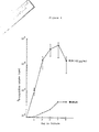
- Figure 4 shows proliferation of E+ lymphocytes with R24.
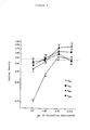
- FIG. 5 shows the mitogenic effect of different concentrations of R24.
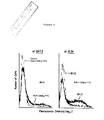
- Figure 6 shows flow-cytometer analysis of E+ cells stimulated with anti-CD3 (OKT-3) antibody or R24.
- Figure 7 shows effect of PHA, anti-T cell mAb and PMA on mAb R24 induced proliferation of E+ cells.
- MAb R24 Immunofluorescence staining of fetal thymus (a-c) and adult lymph node (d) with mAb R24.
- MAb R24 reacts with thymocytes in subcortical areas (a), around blood vessels (b), and near Hassal's corpuscles (c).
- MAb R24 stains patches of lymphocytes in paracortical areas (d).
- E+ cells stimulated with anti-CD3 (OKT-3) antibody (2.5 ng/mL) or mAb R24 (100 ug/mL).
- E+ cells at initiation of cultures (day 0) or day 5 of cultures were incubated with mAb R24 (10 ug/106 cells) followed by incubation with FITC-labeled goat-anti-mouse immunoglobulin (GAMIg FITC).
- GMIg FITC FITC-labeled goat-anti-mouse immunoglobulin
- Cells were analyzed by a FACS IV.
- Gangliosides are a class of glycosphingolipids that contain sialylated oligosccharide groups attached to a lipid core structure. Although the exact function of gangliosides is not known, it has been suggested that they play a role in cell signaling and recognition (Hakomori, S., (1981) Ann. Rev. Biochem. 50 :733; Yu, R.K., et al. (1984) Adv. Exp. Med. Biol. 174 :87). Specific gangliosides have been implicated as receptors for viruses (Markwell, M.A.K., et al. (1981) Proc. Natl. Acad. Sci.
- gangliosides can induce neural cell proliferation, increase neurite formation (Ferrari, G., et al. (1983) Brain Res. 284 :215; Tsuji, S., et al. (1983) J. Biochem. (Tokyo) 94 :303; Nakajuma, J., et al. (1986) Biochim. Biophys. Acta. 876 :65) and facilitate adhesion of neural cells (Blackburn, C.C., et al. (1986) J. Biol. Chem. 261 :2873).
- Mouse monoclonal antibodies (mAB) R24 (IgG3), C5 (IgG3) and K9 (IgM) have been shown to react with G D3 (Dippold, W., et al. (1980) Proc. Natl. Acad. SCI. USA 77 :6114; Pukel, C., et al. (1982) J. Exp. Med. 155:1133).
- Mouse mAb 3F8 (IgG3) (a gift from Dr. Nai-Kong Cheung, Case-Western Reserve University, Cleveland, Ohio) recognizes G D2 ganglioside (Saito, M., et al. (1985) Biochem. Biophys. Res. Comm.
- mouse mAb 10-11 (IgM) recognizes G M2 (Natoli, E., et al. (1986) Cancer Res. 46 :4116), and mouse mAb F36/22 (IgG3) reacts with a high molecular weight cell surface glycoprotein expressed by breast carcinoma cells but not by lymphoid cells (Papsidero, L.D., et al. (1984) Cancer Res. 44 :4653).
- MAb R24 was used as hybridoma supernatant or prepared from ascites of mice bearing hybridoma tumors.
- Preparations of mAbs R24, 3F8, C5 and F36/22 from ascites were purified by precipitation with a saturated ammonium sulfate solution added to a final concentration of 35% (w/v).
- IgG3 antibody preparations were applied to a protein A Sepharose (Pharmacia, Inc., Piscataway, NJ) column and eluted with 0.5M citrate/0.15M NaCl, pH 4.0, buffer.
- Eluted protein fractions were run through a Sephadex G25M (Pharmacia, Inc.) column and eluted with phosphate buffered saline (PBS), pH 6.0.
- Monoclonal antibodies OKT-3, OKT-4, OKT-6, OKT-8, and OKT-11, either unconjugated or conjugated with FITC were purchased from Ortho Diagnostics Systems (Raritan, NJ). OKT-3, OKT-8 and OKT-11 antibodies were extensively dialyzed before use in the proliferation assays.
- Anti-IL-2 receptor monoclonal antibody labeled with FITC and Leu-2a, Leu-3a and Leu-4 conjugated to phycoerythrin (PE) were purchased from Becton-Dickinson, Inc., Mountainview, CA. Recombinant IL-2 was kindly provided by Cetus (Emeryville, CA)
- Histologically normal adult human lymph nodes were obtained from surgical pathology specimens of patients without cancer within 1-2 hrs after resection. Specimens of fetal thymus were obtained from elective abortions. Fetal gestational age was determined by crown-rump, crown-hell and weight measurements. Tissues were immersed in isopenthane pre-cooled in liquid nitrogen, embedded in OCT compound in cryomolds (Miles Labs, Inc., Naperville, IL) and stored at -70°C.
- tissue sections (4-8 microns thick) were used unfixed or fixed for 10 min with acetone at 4-8°C. Tissue sections were stained by indirect immunofluorescence or peroxidase methods as previously described (Cordon-Cardo, C., et al. (1986) Int. J. Cancer 37 :667). Briefly, tissue sections were incubated in goat (10% v/v) suppressor serum for 20 min, then primary antibody for 1 hr. MAb R24 was used at concentrations between 20-40 ug/mL in the form of hybridoma supernatant or purified mouse ascites.
- PBMC Peripheral blood mononuclear cells
- adherent cells In order to collect adherent cells, up to 3 x 10⁷ PBMC were incubated 90 min at 37°C in 5% CO2 in a total volume of 10 mL in Falcon 3003 tissue culture dishes (Falcon Labware Div., Becton-Dickinson, Oxnard, CA). Nonadherent cells were separated by gently swirling the dishes and slowly pipetting off the medium. Dishes were washed three times with medium, and adherent cells were harvested using a rubber policeman. The adherent cell fraction contained more than 90% monocytes/macrophages as judged by alpha-naphthyl-acetate-esterase stain.
- PBMC non-adherent PBMC were rosetted with neuraminidase-treated sheep red blood cells according to a method modified from Weiner et al. (Weiner, M.S., et al. (1973) Blood 42 :939). This fraction contained less than 2% monocytes/macrophages and more than 90% of cells reacted with monoclonal antibody OKT-3. THis cell population is designated :"E+ cells".
- 106 PBMC in 0.1 mL were incubated with appropriate dilutions of unconjugated monoclonal antibody for 30 min on ice, washed three times in cold PBS/2% bovine serum albumin (BSA), and then incubated on ice with 0.1 mL of a 1:40 dilution of affinity purified goat anti-mouse IgG F(ab ⁇ )2-FITC (Cappel Lab., Cochranville, PA).
- BSA bovine serum albumin
- H-50 Cytofluorograph Ortho Diagnostic Systems, Inc., Raritan, NJ
- FACS IV Becton-Dickinson
- a forward angle scatter versus right-angle green fluorescence cytogram was generated in order to gate around the region of interest (i.e. to differentiate lymphocytic, monocytic and granulocytic cells) and to exclude cell debris and dying cells.
- H-50 cytofluorograph an argon-ion laser, tuned to 488 nm around gated regions, was set around lymphocytic cells. From this gated region, green fluorescence (FITC) histograms were generated.
- FITC green fluorescence
- FITC green
- PE red
- PBMC or E+ cells were incubated in triplicate microwell cultures (Costar 3596 plates, Costar, Inc., Cambridge, MA) in the presence or absence of mAb R24.
- Other factors evaluated in proliferation assays included recombinant IL-2 (Cetus Corporation), phytohemmaglutinin (PHA) (Gibco Labs, Grand Island, NY), phorbol-myristate-acetate (PMA) (Sigma Chemicals, St. Louis, MO), and protein A (Pharmacia, Inc.). At indicated time points, 100 ul of supernatant were removed from each well to be assayed for IL-2 activity.
- IL-2 assays a mouse IL-2-dependent cytotoxic T cell line was used as previously described (Welte, K., et al. (1983) supra ).
- One unit of activity in this assay is equivalent to one unit of activity of the IL-2 standard from the Biological Response Modifier program, National Institutes of Health.
- E+ cells were adjusted to a concentration of 106 cells/mL. E+ cells were incubated at room temperature with different concentrations of mAb R24 (100, 10, 1ug/mL) for one hour, washed and incubated with a complement source (prescreened human serum diluted 1:8 in Veronal buffered medium) at 37°C for 2 hrs. Cells were washed three times and resuspended for use in proliferation assays. Control cells were treated with PBS followed by complement source alone or with mAb R24 (100 ug/mL) followed by Veronal buffered medium without complement source.
- mAb R24 100, 10, 1ug/mL
- G D3 was purified from human melanoma tissue and G D2 was purified from human brain, as described below.
- G M1 was purchased from Sulpeco, Inc. (Bellafonte, PA).
- G M2 was prepared by treating G M1 with beta-galactosidase (from Dr. George W. Jourdian, University of Michigan, Ann Arbor, MI) as described by Cahan et al. (Cahan, D.L., et al. (1982) Proc. Natl. Acad. Sci. USA 79 :7629). Purity of ganglioside preparations was verified by thin layer chromatography.
- Glycolipids were isolated by a modification of the method described by Saito and Kahomori (Saito, T., et al. (1971) J. Lipid Res. 12 :257). Briefly, cells were homogenized in chloroform:methanol (C:M) 2:1 and extracted in a 100-fold volume of C:M. The homogenate was filtered and re-extracted twice in C:M, first 1:1, then 1:2. Filtrates were evaporated in a rotary evaporator, acetylated, run through a florisil column, de-acetylated and diaglyzed against H2O for 24 hrs.
- C:M chloroform:methanol
- the sample was then re-evaporated and suspended in C:M:H2O (30:60:8) and applied to a DEAE-Sephadex (Pharmacia, Inc.) column equilibrated with C:M:0.8M sodium acetate (30:60:8).
- silica gel plates (Analtech, Newark, Delaware) were activated by heating to 100°C for 1 hr. N-propanol:ammonium hydroxide:H2O (60:95:11.4) was used to develop chromatographs. After the solvent had migrated 12 cm from the origin, plates were air-dried, baked 10 min at 120°C, cooled to room temperature and sprayed with resorcinol-HCl. Densitometry was evaluated using a Shimadzu CS-930 thin layer chromatography scanner (Kyoto, Japan).
- Ganglioside preparations from E+ cells were applied to microtest plates (Bellco Glass, Inc., Vineland, NJ). The plates were air-dried for two hrs and blocked with 1% BSA for two hrs. MAb R24 20 ug/mL was incubated with various concentrations of ganglioside preparations for two hrs and then added to Microtest wells and incubated for one hr at room temperature. Plates were washed three times and incubated for 45 min with anti-mouse IgG conjugated to alkaline phosphatase (Sigma Chemical Corp., St. Louis, MO) diluted 1:200. Plates were washed and incubated with diethanolamine substrate for 20 min at 37°C. Reactivity was evaluated on an Artec Systems Corp. model 210 reader (Farmingdale, NJ).
- MAb R24 was tested for its ability to react with protein or glycoprotein antigens of unstimulated E+ cells or E+ cells stimulated with mAb R24 (100 ug/mL). Extracts were prepared from detergent solubilized E+ cells (in 0.5% NP-40, 0.01M Tris, pH 7.5, 0.15M NaCl and 2mM MgCl2) or from sonicated extracts of membrane preparations solubilized in 2% (w/v) SDS. Extracts were labeled with 125I by the chloramine T method (Greenwood, F.C., et al. (1963) Biochem. J. 89 :114).
- MAb R24, anti-HLA A,B,C antibody W6/32 (Barnstable, C.J., et al. (1978) Cell 14 :9).
- OKT-3 or control antisera (normal mouse serum) was incubated with labeled extracts for 1 hr and rabbit anti-mouse Ig overnight at 4°C followed by immunoprecipitation with protein A Sepharose (Pharmacia, Inc.), as previously described (Lloyd, K.O., et al. (1981) J. Immunol. 126 :2408). Immunoprecipitates were eluted with 2% SDS and analyzed on 9% gels by SDS/PAGE.
- MAb R24 Reacts with Thymocytes and Lymphocytes :
- Mab R24 was found to react with subpopulations of cells in thymus tissues from three 12-14 week old-fetuses.
- R24+ thymocytes were localized in subcortical areas and in patches around blood vessels and Hassal's corpuscles ( Figure 1a-c). Identical results were observed in tissues stained by immunofluorescence and immunoperoxidase techniques.
- mAb R24 was reacting with thymocytes, consecutive 4 micron sections were stained with mAbs R24 or OKT-6; R24+ cells were shown to react with the thymocyte marker OKT-6.
- lymph nodes derived from two healthy adults mAb R24 stained small clusters of lymphocytes around blood vessels in interfollicular and pericortical areas ( Figure 1d).
- MAb R24 reacted with 13.9 ⁇ 5.5% (standard deviation, S.D.) of PBMC from 13 healthy individuals (range 7.4-24.3% R24 positive cells). Analysis by two-color flow cytometry to PBMC showed that mAB R24 reacted with a subpopulation of T lymphocytes. In resting T cells derived from peripheral blood of 13 healthy persons, mAb R24 reacted with 14.8 ⁇ 5.7% (S.D.) of CD3+ cells (range 5.0-25.5%), with 20.1 ⁇ 11.7% (S.D.) of CD8+ cells (range 7.0-44.0%) and with 14.0 ⁇ 6.7% (S.D.) of CD4+ T cells (range 6.0-29.7%).
- MAb R24 Reacts with G D3 Ganglioside on T Lymphocytes :
- G D3 comprises 3-5% of total gangliosides ( Figure 2b), while unstimulated E+ cells contained lower levels of G D3 .
- MAb R24 reacted with a purified ganglioside fraction derived from peripheral blood T cells, and this reactivity was specifically inhibited by purified G D3 but not by G M2 , G M3 or G D2 ( Figure 3).
- mAb R24 detected G D3 ganglioside on T cells and that mAb R24 was not reacting with other gangliosides present in the T cell preparation.
- mAb R24 did not react with protein components from peripheral blood E+ cells by Western blot analysis or by radioimmunoprecipitation of extracts labeled with 125I by the chloramine T method.
- MAb R24 was found to stimulate the growth of both PBMC and E+ cells.
- the kinetics of 3H-thymidine uptake by cells cultured in the presence of 100 ug/mL of mAB R24 are shown in Figure 4. Maximum proliferation was observed after 5 to 7 days of stimulation.
- Figure 5a shows the effect of various concentrations of mAb R24 on proliferation of E+ cells between 2 and 5 days in culture. A dose-dependent response of proliferation was observed at all time points tested. A concentration of 20 ug/mL gave a significant increase of 3H-thymidine uptake over background (cultured in medium alone) at all days tested. At concentrations as low as 2 ug/mL, mAb R24 still produced a detectable increase in proliferation.
- MAb R24 prepared by various methods induced similar levels of proliferation of E+ cells at equivalent protein concentrations. Stimulation was produced by supernatants from the R24 hybridoma, unpurified ascites from R24-hybridoma bearing mice, and ascites purified by ammonium sulfate precipitation with or without protein A affinity chromatography (Table 1). Protein A itself induced only minimal proliferation of E+ cells (2379 ⁇ 145 cpm 3H-thymmidine uptake at day 5 with 5ug/mL protein A).
- FIG. 5b demonstrates the uptake of 3H-thymidine of PBMC during stimulation by different concentrations (2, 20 and 200 ug/mL) of mAb R24 F(ab ⁇ )2 fragments.
- Incubation of cells with F(ab ⁇ )2 fragments of a control IgG3 monoclonal antibody mAb 3F8 (anti-G D2 ) produced no increase in proliferation.
- the level of proliferation during stimulation with F(ab ⁇ )2 fragments was one-half the level of proliferation induced by the same concentration of intact R24 antibody molecules (e.g., 30,000 cpm for 200 ug/mL of whole mAb R24 versus 18,000 cpm with 200 ug/mL of F(ab ⁇ )2.
- MAb R24 induced equivalent stimulation of PBMC cultures containing 10-20% macrophages and cultures of E+ cells depleted of macrophages (confirmed by flow cytometry analysis) as measured by expression of IL-2 receptors and level of cell proliferation (Table 4).
- PHA and anti-CD3 mAb two mitogens that require accessory cells, did not stimulate these E+ cultures depleted of macrophages.
- macrophages were added back to E+ cells (to 5% and 10% of the final cell number), there was no change in induction of proliferation by mAb R24 (Table 4).
- Figure 7 presents results of representative experiments (at day 5 in culture).
- PHA at concentrations of 1% (v/v) increased R24-induced proliferation of E+ cells approximately 1.5 fold, while 1% PHA alone was minimally mitogenic (less than 10% of the level of proliferation achieved with 1% PHA in the presence of macrophages).
- Anti-CD3 (OKT-3) stimulation is also dependent on the presence of macrophages.
- Anti-CD3 at concentrations of 2.5 ng/mL, 25 ng/ML and 250 ng/mL did not augment or inhibit R24-induced stimulation of E+ cells depleted of macrophages (Figure 7).
- Anti-CD2 did not include stimulation of E+ cells when used alone, but anti-CD2 did inhibit mAB R24 induced proliferation (60% inhibition at 1ug/mL and 33% inhibition at 100 ng/mL) (Figure 7).
- Anti-CD8 which is the same subclass as anti-CD2 and anti-CD3 and is known to inhibit IL-2 production and proliferation induced by anti-CD3 (Welte, K., et al. (1983) supra ), did not inhibit or augment stimulation by mAb R24.
- the proliferation induced by mAb R24 as measured by 3H-thymidine uptake was the result of IL-2 production and IL-2 receptor expression (Table 1).
- exogenous IL-2 could increase mAb R24-induced proliferation
- recombinant IL-2 100 U/mL was added to mAb R24-stimulated cultures.
- exogenous IL-2 substantially increased the proliferation of E+ cells when compared to stimulation by mAb R24 alone.
- Protein A (recombinant material from Pharmacia Corp., Piscataway, N.J.) also increases proliferation of T cells and can serve as a stimulatory agent as shown.
- G D3 is expressed on cell types derived from the neuroectoderm, including melanocytes, adrenal medullary cells, glia, neurons, and islet cells of the pancreas (Real, F.X., et al. (1985) Cancer Res. 45 :4401). With the finding that G D3 is expressed by a subpopulation of thymocytes and peripheral blood lymphocytes, G D3 joins a class of antigens which are shared by neuroectoderm-derived cells and cells of the immune system.
- G D3 may play a crucial role in cell signaling in these differentiation pathways.
- binding to G D3 induces T cell activation, including the expression of IL-2 receptors, the production of IL-2 and cell proliferation.
- T cells can be stimulated by IgM, IgG and F(ab ⁇ )2 fragments that react with G D3 .
- G D3 As serum contains G D3 , it is possible that G D3 on the surface of lymphocytes is the result of passive adsorption. However, several lines of evidence suggest that G D3 is actively synthesized by T cells. First, G D3 is selectively expressed at high levels by only a small subpopulation of T cells rather than by a broad population of cells. Second, the number of T cells expressing G D3 increased substantially in tissue culture in the presence of anti-G D3 antibodies. Third, G D3 can be detected on T cell leukemia cell lines as well as fresh lymphocytic leukemia cells.
- the level of G D3 expression by the T cell leukemia cell line CEM has been shown to increase during stimulation with the phorbol ester PMA (Kiguchi, K., et al. (1986) Cancer Res. 46 :3027).
- Gangliosides have been shown to interact with signals and receptors that influence growth and differentiation of cells. Gangliosides added exogenously to growing cells have been found to alter binding affinity of growth factors to their receptors and to affect the state of tyrosine phosphorylation of growth factor receptors (Bremer, E.G., et al. (1984) J. Biol. Chem. 259 :6818; Bremer, E.G., et al. (1986) J. Biol. Chem. 261 :2434).
- T cells A variety of glycoprotein molecules on the surface of T cells can mediate activation of T cells, including the IL-2 receptor, the Ti/CD3 molecular complex, and the CD2 antigen (Weiss, M.J., et al. (1984) Proc. Natl. Acad. Sci. USA 81 :6836). Activation by these receptors generally requires accessory Fc receptor-positive cells such as marcrophages and soluble components (e.g. IL-1) or PMA (Hara, T., et al. (1985) J. Exp. Med. 161 :641). An exception is the CD2 receptor which is expressed early in T cell differentiation in the thymus (Meuer, S.C., et al.
- G D3 Activation by binding to G D3 , like CD2-mediated activation, does not appear to depend on accessory cells. Despite the diversity of cell surface receptors available for activation, only limited pathways appear to be available for transduction of signals inside the cell (Nishizuka, Y. (1986) Science 233 :305; Imboden, J.B., et al. (1985) J. Exp. Med. 161 :446). It is not clear whether G D3 binding by mAb initiates activation at the level of known ligand/surface receptors or through a separate, yet unidentified receptor complex.
- gangliosides may bind IL-2, thereby inhibiting mitogen-induced proliferation of lymphocytes (Robb, R.J. (1986) J. Immunol. 136 :971; Parker, J., et al. (1984) FEBS letters 170 :391).
- gangliosides may compete for IL-2 reactivity by sequestering or inactivating IL-2.
- Robb et al. Robb, R.J. (1986) J. Immunol.
- gangliosides are able to decrease the expression of IL-2 receptor (Tac antigen) on PHA-induced blasts.
- IL-2 receptor Tac antigen
- cellular gangliosides could be involved in the suppression of IL-2 receptor expression on resting T cells.
- binding of IL-2 or antibodies to critical gangliosides on T cells could permit full expression of IL-2 receptors. This would also be compatible with the observations that IL-2 is able to upregulate its own receptor (Reem, G.H., et al. (1984) Science 225 :429; Welte, K., et al. (1984) J. Exp. Med.
- the stimulation of T cells by anti-G D3 antibodies can be augmented by exogenous IL-2, PHA, and PMA, but not by anti-CD3 mAb.
- the inhibition of G D3 -mediated activation by monoclonal antibodies to CD2 suggests that G D3 might be associated with the CD3 molecule, but there are other possible explanations for this finding e.g. downregulation of CD3-mediated activation by binding to CD2 (Meuer, S.C., et al. (1983) Science 222 :1239).
- G D3 + T cells appear to be essential for initial activation by mAb R24 since depletion of this population abrogates the proliferative response.
- MAb R24 can kill target cells by complement-mediated cytotoxicity and antibody-dependent cellular cytotoxicity.
- mAb R24 can kill target cells by complement-mediated cytotoxicity and antibody-dependent cellular cytotoxicity.
- a preliminary clinical trial was initiated to evaluate mAb R24 in the treatment of patients with metastatic melanoma (Houghton, A.N., et al. (1985) Proc. Natl. Acad. Sci. USA 82 :1242).
- Intravenous infusion of mAb R24 induced clinical signs and symptoms of inflammation around tumors and regression or disappearance of lesions in some patients.
- treatment with mAb R24 produced complement deposition and infiltration of T cells (CD3+/CD8+/Ia+) in tumors.
- T cell activation Although several biological properties of mAb R24 may contribute to its anti-tumor activity, the potential role of T cell activation is particularly interesting. This view is supported by the finding of T cell infiltrates in regressing tumors after treatment with R24 but not in tumor biopsies prior to treatment. In addition, cytotoxic T cell clones have been established from a patent treated with R24. This T cell clones specifically kill autologous melanoma cells and not normal tissues or allogeneic melanomas. The possible activation of T cells in treated patients is a complex issue since mAb R24 can also lyse target T cells in the presence of human complement and thereby abolish T cell stimulation (Table 3).
- mAb R24 The ability of mAb R24 to lyse host T cells may partially explain the observation that tumor regressions have generally been seen only after treatment with low doses but not high doses of mAb R24 (Houghton, A.N., et al. (1985) Proc. Natl. Acad. Sci. USA 82 :1242). It will be possible to investigate this issue since F(ab ⁇ )2 fragments of mAb R24 can stimulate T cells but do not mediate complement lysis (P. Chapman, unpublished observation). F(ab ⁇ )2 fragments will be investigated in a clinical trial in order to evaluate their potential role in cancer treatment.
- Recombinant protein A is another stimulatory agent.
- Protein A can be either injected intravenously or blood can be removed from the body and passed over protein A and then reinfused ( ex vivo ). In each case in animal models, tumor regressions have been seen.
- PHA or PMA use would most likely be in ex vivo treatment together with IL-2 and/or R24.
- the IL-2 range for treatment is approximately from a low dose of less than 1 million units to a high dose of greater than 3 million units of IL-2.
- a mid range is considered to be approximately 1-3 million units of IL-2.
Landscapes
- Health & Medical Sciences (AREA)
- Chemical & Material Sciences (AREA)
- Medicinal Chemistry (AREA)
- Life Sciences & Earth Sciences (AREA)
- Animal Behavior & Ethology (AREA)
- Veterinary Medicine (AREA)
- Public Health (AREA)
- General Health & Medical Sciences (AREA)
- Pharmacology & Pharmacy (AREA)
- Nuclear Medicine, Radiotherapy & Molecular Imaging (AREA)
- Microbiology (AREA)
- General Chemical & Material Sciences (AREA)
- Chemical Kinetics & Catalysis (AREA)
- Engineering & Computer Science (AREA)
- Bioinformatics & Cheminformatics (AREA)
- Immunology (AREA)
- Organic Chemistry (AREA)
- Mycology (AREA)
- Epidemiology (AREA)
- Preparation Of Compounds By Using Micro-Organisms (AREA)
- Medicines Containing Antibodies Or Antigens For Use As Internal Diagnostic Agents (AREA)
- Micro-Organisms Or Cultivation Processes Thereof (AREA)
- Medicines That Contain Protein Lipid Enzymes And Other Medicines (AREA)
Applications Claiming Priority (2)
| Application Number | Priority Date | Filing Date | Title |
|---|---|---|---|
| US93029286A | 1986-11-13 | 1986-11-13 | |
| US930292 | 1986-11-13 |
Publications (2)
| Publication Number | Publication Date |
|---|---|
| EP0267615A2 true EP0267615A2 (de) | 1988-05-18 |
| EP0267615A3 EP0267615A3 (de) | 1989-05-03 |
Family
ID=25459157
Family Applications (1)
| Application Number | Title | Priority Date | Filing Date |
|---|---|---|---|
| EP87116730A Withdrawn EP0267615A3 (de) | 1986-11-13 | 1987-11-12 | Zusammensetzungen und Methoden zur Behandlung von Krebs unter Benutzung von monoklonalen Antikörpern gegen GD3 Ganglioside mit IL-2 |
Country Status (6)
| Country | Link |
|---|---|
| EP (1) | EP0267615A3 (de) |
| JP (1) | JPS6485930A (de) |
| AU (1) | AU617295B2 (de) |
| CA (1) | CA1341374C (de) |
| IE (1) | IE873057L (de) |
| PT (1) | PT86128B (de) |
Cited By (7)
| Publication number | Priority date | Publication date | Assignee | Title |
|---|---|---|---|---|
| EP0332879A3 (de) * | 1988-02-19 | 1991-05-02 | Mect Corporation | Monoklonaleantikörper zur Erkennung von unnatürlichen Gangliosiden GD3 |
| EP0533199A3 (en) * | 1991-09-18 | 1993-10-06 | Kyowa Hakko Kogyo Co., Ltd. | Application to a chimeric antibody directed against ganglioside gd3 |
| WO1999024045A1 (en) * | 1997-11-10 | 1999-05-20 | Arch Development Corporation | Methods for treatment of tumors and tumor cells using ex vivo activated t cells |
| WO2002078739A1 (en) * | 2001-03-29 | 2002-10-10 | Kyowa Hakko Kogyo Co., Ltd. | Drugs containing genetically modified antibody against ganglioside gd3 |
| EP1200476A4 (de) * | 1999-07-23 | 2004-11-17 | Univ Massachusetts | Antitumorantikörper, proteine und deren verwendung |
| US8444972B2 (en) | 1999-07-23 | 2013-05-21 | University Of Massachusetts | Antitumor antibodies, proteins, and uses thereof |
| US12006354B2 (en) | 2017-05-24 | 2024-06-11 | Novartis Ag | Antibody-IL2 engrafted proteins and methods of use in the treatment of cancer |
Families Citing this family (1)
| Publication number | Priority date | Publication date | Assignee | Title |
|---|---|---|---|---|
| CH679277A5 (de) * | 1989-02-09 | 1992-01-31 | Sandoz Ag |
Family Cites Families (2)
| Publication number | Priority date | Publication date | Assignee | Title |
|---|---|---|---|---|
| DE3319751A1 (de) * | 1983-05-31 | 1984-12-06 | Max-Planck-Gesellschaft zur Förderung der Wissenschaften e.V., 3400 Göttingen | Verfahren zur gewinnung von gegen tumorantigene gerichteten monoklonalen antikoerpern und diese enthaltende arzneimittel |
| US5179018A (en) * | 1983-10-14 | 1993-01-12 | Centocor, Inc. | Mamalian monoclonal antibodies against endotoxin of gram-negative bacteria |
-
1987
- 1987-11-12 AU AU81176/87A patent/AU617295B2/en not_active Ceased
- 1987-11-12 EP EP87116730A patent/EP0267615A3/de not_active Withdrawn
- 1987-11-12 CA CA000551669A patent/CA1341374C/en not_active Expired - Fee Related
- 1987-11-12 IE IE873057A patent/IE873057L/xx unknown
- 1987-11-13 JP JP62287179A patent/JPS6485930A/ja active Pending
- 1987-11-13 PT PT86128A patent/PT86128B/pt not_active IP Right Cessation
Non-Patent Citations (6)
| Title |
|---|
| AUSTRALIAN AND NEW ZELAND JOURNAL OF MEDICINE, vol. 16, 1986, page 152; P. HERSEY et al.: "Potentation of Lymphocyte Responses by M.AB to Gangliside GD3" * |
| CHEMICAL ABSTRACTS, vol. 100, no. 10, 5th March 1984, abstract no. 83938u, Columbus, Ohio, US; W. DIPPOLD et al.: "Inhibition of human melanoma cell growth in vitro by monoclonal anti-GD3-ganglioside antibody"; & Cancer Res., vol. 44, no. 2, 1984, pages 806-810 * |
| CHEMICAL ABSTRACTS, vol. 103, no. 13, 30th September 1985, abstract no. 121350t, Columbus, Ohio, US; D.A. CHERESH et al.: "Disialoganglioside GD3 omn human melanoma serves al a relevant target antigen for monoclonal antibody-mediated tumor cytolysis"; & Proc. Natl. Acad. Sci. U.S.A., vol. 82, no. 15, 1985, pages 5155-5159 * |
| CHEMICAL ABSTRACTS, vol. 105, no. 19, 10th November 1986, abstract no. 189138b, Columbus, Ohio, US; M. KAMBER et al.: "Phorbol myristate acetate-induced proliferation of an IL-2-dependent T-cell line: action of PMA is independent of IL-2 and cannot be mimicked by diacylglycerols"; & Cell. Immunol., vol. 102, no. 1, 1986, pages 177-186 * |
| CHEMICAL ABSTRACTS, vol. 105, no. 25, 22nd December 1986, abstract no. 224175k, Columbus, Ohio, US; C. HONSIK: "Lymphokine-activated killer cells targeted by monoclonal antibodies to the disialogangliosides GD2 and GD3 specifically lyse human tumor cells of neuroectodermal origin"; & Proc. Natl. ACad. Sci. U.S.A., 1986, vol. 83, no. 20, pages 7893-7897 * |
| CHEMICAL ABSTRACTS, vol. 99, no. 22, 28th November 1983, abstract no. 193025n, Columbus, Ohio, US; S. MOTOI et al.: "Cytotoxic activity of killer T cells induced by PHA"; & Dig. Organ. Immunol., vol. 10, 1983, pages 201-205 * |
Cited By (15)
| Publication number | Priority date | Publication date | Assignee | Title |
|---|---|---|---|---|
| EP0332879A3 (de) * | 1988-02-19 | 1991-05-02 | Mect Corporation | Monoklonaleantikörper zur Erkennung von unnatürlichen Gangliosiden GD3 |
| US5173420A (en) * | 1988-02-19 | 1992-12-22 | Mect Corporation | Monoclonal antibody recognizing un-natural ganglioside gd3 |
| US6495666B2 (en) | 1991-09-18 | 2002-12-17 | Kyowa Hakko Kogyo Co., Ltd. | Polypeptide composing human chimeric antibody |
| US5750078A (en) * | 1991-09-18 | 1998-05-12 | Kyowa Hakko Kogyo Co., Ltd. | Process for producing humanized chimera antibody |
| US5807548A (en) * | 1991-09-18 | 1998-09-15 | Kyowa Hakko Kogyo Co., Ltd. | Method of treating cancer using a chimera antibody |
| US5866692A (en) * | 1991-09-18 | 1999-02-02 | Kyowa Hakko Kogyo Co., Ltd. | Process for producing humanized chimera antibody |
| US6437098B1 (en) | 1991-09-18 | 2002-08-20 | Kyowa Hakko Kogyo Co., Ltd. | Human chimeric antibody specific for the ganglioside GD3 |
| EP0533199A3 (en) * | 1991-09-18 | 1993-10-06 | Kyowa Hakko Kogyo Co., Ltd. | Application to a chimeric antibody directed against ganglioside gd3 |
| US6965024B2 (en) | 1991-09-18 | 2005-11-15 | Kyowa Hakko Kogyo Co., Ltd. | Process for producing humanized chimera antibody |
| US7045129B2 (en) | 1991-09-18 | 2006-05-16 | Kyowa Hakko Kogyo Co., Ltd. | Method of treating cancer including administering a human chimeric antibody specific for the ganglioside GD3 |
| WO1999024045A1 (en) * | 1997-11-10 | 1999-05-20 | Arch Development Corporation | Methods for treatment of tumors and tumor cells using ex vivo activated t cells |
| EP1200476A4 (de) * | 1999-07-23 | 2004-11-17 | Univ Massachusetts | Antitumorantikörper, proteine und deren verwendung |
| US8444972B2 (en) | 1999-07-23 | 2013-05-21 | University Of Massachusetts | Antitumor antibodies, proteins, and uses thereof |
| WO2002078739A1 (en) * | 2001-03-29 | 2002-10-10 | Kyowa Hakko Kogyo Co., Ltd. | Drugs containing genetically modified antibody against ganglioside gd3 |
| US12006354B2 (en) | 2017-05-24 | 2024-06-11 | Novartis Ag | Antibody-IL2 engrafted proteins and methods of use in the treatment of cancer |
Also Published As
| Publication number | Publication date |
|---|---|
| CA1341374C (en) | 2002-07-09 |
| EP0267615A3 (de) | 1989-05-03 |
| PT86128B (pt) | 1990-11-20 |
| AU8117687A (en) | 1988-05-19 |
| PT86128A (en) | 1987-12-01 |
| AU617295B2 (en) | 1991-11-28 |
| JPS6485930A (en) | 1989-03-30 |
| IE873057L (en) | 1988-05-13 |
Similar Documents
| Publication | Publication Date | Title |
|---|---|---|
| Welte et al. | Stimulation of T lymphocyte proliferation by monoclonal antibodies against GD3 ganglioside. | |
| Takahashi et al. | Immunoglobulin G3 monoclonal antibody directed to Tn antigen (tumor-associated α-N-acetylgalactosaminyl epitope) that does not cross-react with blood group A antigen | |
| Seon et al. | Long-lasting complete inhibition of human solid tumors in SCID mice by targeting endothelial cells of tumor vasculature with antihuman endoglin immunotoxin. | |
| Chang et al. | Expression of disialogangliosides GD2 and GD3 on human soft tissue sarcomas | |
| Vadhan-Raj et al. | Phase I trial of a mouse monoclonal antibody against GD3 ganglioside in patients with melanoma: induction of inflammatory responses at tumor sites. | |
| Wang et al. | Stimulation of tumor‐cell growth by alpha‐fetoprotein | |
| Shen et al. | Heteroantibody-mediated cytotoxicity: antibody to the high affinity Fc receptor for IgG mediates cytotoxicity by human monocytes that is enhanced by interferon-gamma and is not blocked by human IgG. | |
| JP2003508355A (ja) | 卵巣癌細胞及び骨髄腫細胞の表面糖タンパク質、それへの抗体及びその使用 | |
| Bernard et al. | AT cell surface molecule different from CD2 is involved in spontaneous rosette formation with erythrocytes. | |
| Gilhus et al. | Rabbit antiserum to a citric acid extract of human skeletal muscle staining thymomas from myasthenia gravis patients | |
| Loop et al. | Two human tumor‐associated antigens, p155 and p210, detected by monoclonal antibodies | |
| CA1339583C (en) | Monoclonal antibody nuh2 capable of inactivating motility of human sperm, antigen defined by said monoclonal antibody and methods of using said monoclonal antibody and antigen | |
| CA2319688A1 (en) | Specific antibodies against mammary tumor-associated mucin, method for production and use | |
| CA1341374C (en) | Compositions and method for treatment of cancer using monoclonal antibody against gd3 ganglioside together with il-2 | |
| Fox et al. | Salivary gland lymphocytes in primary Sjogren's syndrome lack lymphocyte subsets defined by Leu-7 and Leu-11 antigens. | |
| Ito et al. | Carbohydrates as antigenic determinants of tumor‐associated antigens recognized by monoclonal anti‐tumor antibodies produced in a syngeneic system | |
| US5104652A (en) | Compositions and method for treatment of cancer using monoclonal antibody against GD3 ganglioside together with IL-2 | |
| Lebacq‐Verheyden et al. | Rat AL2, AL3, AL4 and AL5 monoclonal antibodies bind to the common acute lymphoblastic leukaemia antigen (CALLA gp 100) | |
| Santoni et al. | Rat natural killer cells synthesize fibronectin. Possible involvement in the cytotoxic function. | |
| Axelsson et al. | The large sialoglycoprotein of human lymphocytes. I. Distribution on T and B lineage cells as revealed by a monospecific chicken antibody | |
| Dirienzo et al. | α1-Acid glycoprotein (α1-AGP) on the membrane of human lymphocytes: possible involvement in cellular activation | |
| Dubey et al. | Unique proliferation-associated marker expressed on activated and transformed human cells defined by monoclonal antibody | |
| IE921810A1 (en) | Me20: monoclonal antibodies and antigen for human melanoma | |
| Erb et al. | Characterization of a Human—Human Hybridoma Antibody, C-OU1, Directed Against a Colon Tumor-Associated Antigen | |
| JPH03502280A (ja) | T細胞系hsb‐2およびt細胞性慢性リンパ性白血病(t‐cll)細胞を加えて成長させた、正常ヒトtおよびbリンパ球ならびに単球と反応するマウス単クローン性抗体 |
Legal Events
| Date | Code | Title | Description |
|---|---|---|---|
| PUAI | Public reference made under article 153(3) epc to a published international application that has entered the european phase |
Free format text: ORIGINAL CODE: 0009012 |
|
| AK | Designated contracting states |
Kind code of ref document: A2 Designated state(s): AT BE CH DE ES FR GB GR IT LI LU NL SE |
|
| PUAL | Search report despatched |
Free format text: ORIGINAL CODE: 0009013 |
|
| AK | Designated contracting states |
Kind code of ref document: A3 Designated state(s): AT BE CH DE ES FR GB GR IT LI LU NL SE |
|
| 17P | Request for examination filed |
Effective date: 19891024 |
|
| 17Q | First examination report despatched |
Effective date: 19911030 |
|
| STAA | Information on the status of an ep patent application or granted ep patent |
Free format text: STATUS: THE APPLICATION IS DEEMED TO BE WITHDRAWN |
|
| 18D | Application deemed to be withdrawn |
Effective date: 19940705 |
|
| RIN1 | Information on inventor provided before grant (corrected) |
Inventor name: MILLER, GLENN Inventor name: WELTE, KARL Inventor name: CHAPMAN, PAUL Inventor name: HOUGHTON, ALAN N. Inventor name: OLD, LLOYD J. |