EP0310056A2 - T-Zellen-Wachstumsfaktor aus Thymusdrüsen-Stroma und Verfahren zu seiner Herstellung - Google Patents
T-Zellen-Wachstumsfaktor aus Thymusdrüsen-Stroma und Verfahren zu seiner Herstellung Download PDFInfo
- Publication number
- EP0310056A2 EP0310056A2 EP88116054A EP88116054A EP0310056A2 EP 0310056 A2 EP0310056 A2 EP 0310056A2 EP 88116054 A EP88116054 A EP 88116054A EP 88116054 A EP88116054 A EP 88116054A EP 0310056 A2 EP0310056 A2 EP 0310056A2
- Authority
- EP
- European Patent Office
- Prior art keywords
- tstgf
- cell
- cells
- activity
- tscf
- Prior art date
- Legal status (The legal status is an assumption and is not a legal conclusion. Google has not performed a legal analysis and makes no representation as to the accuracy of the status listed.)
- Withdrawn
Links
- 102000000588 Interleukin-2 Human genes 0.000 title claims abstract description 111
- 108010002350 Interleukin-2 Proteins 0.000 title claims abstract description 110
- 230000002992 thymic effect Effects 0.000 title claims abstract description 85
- 238000000034 method Methods 0.000 title claims abstract description 23
- 238000004519 manufacturing process Methods 0.000 title claims abstract description 12
- 230000008569 process Effects 0.000 title claims abstract description 7
- 210000004027 cell Anatomy 0.000 claims abstract description 153
- 210000001744 T-lymphocyte Anatomy 0.000 claims abstract description 78
- 239000006228 supernatant Substances 0.000 claims abstract description 73
- 230000012010 growth Effects 0.000 claims abstract description 39
- 239000001963 growth medium Substances 0.000 claims abstract description 8
- 238000012258 culturing Methods 0.000 claims abstract description 6
- 230000001737 promoting effect Effects 0.000 claims abstract description 4
- 239000012228 culture supernatant Substances 0.000 claims description 29
- 238000011098 chromatofocusing Methods 0.000 claims description 15
- 210000002443 helper t lymphocyte Anatomy 0.000 claims description 8
- VHJLVAABSRFDPM-QWWZWVQMSA-N dithiothreitol Chemical compound SC[C@@H](O)[C@H](O)CS VHJLVAABSRFDPM-QWWZWVQMSA-N 0.000 claims description 7
- 102000004142 Trypsin Human genes 0.000 claims description 5
- 108090000631 Trypsin Proteins 0.000 claims description 5
- 239000012588 trypsin Substances 0.000 claims description 5
- 238000010438 heat treatment Methods 0.000 claims description 4
- 238000004440 column chromatography Methods 0.000 claims description 3
- BFSVOASYOCHEOV-UHFFFAOYSA-N 2-diethylaminoethanol Chemical compound CCN(CC)CCO BFSVOASYOCHEOV-UHFFFAOYSA-N 0.000 claims 1
- 230000000694 effects Effects 0.000 description 130
- 230000035755 proliferation Effects 0.000 description 97
- 102000004388 Interleukin-4 Human genes 0.000 description 75
- 108090000978 Interleukin-4 Proteins 0.000 description 75
- PHEDXBVPIONUQT-RGYGYFBISA-N phorbol 13-acetate 12-myristate Chemical compound C([C@]1(O)C(=O)C(C)=C[C@H]1[C@@]1(O)[C@H](C)[C@H]2OC(=O)CCCCCCCCCCCCC)C(CO)=C[C@H]1[C@H]1[C@]2(OC(C)=O)C1(C)C PHEDXBVPIONUQT-RGYGYFBISA-N 0.000 description 45
- 101001002634 Homo sapiens Interleukin-1 alpha Proteins 0.000 description 42
- 102100020881 Interleukin-1 alpha Human genes 0.000 description 42
- 102000007644 Colony-Stimulating Factors Human genes 0.000 description 28
- 108010071942 Colony-Stimulating Factors Proteins 0.000 description 28
- 238000002360 preparation method Methods 0.000 description 23
- 239000000427 antigen Substances 0.000 description 22
- 102000036639 antigens Human genes 0.000 description 22
- 108091007433 antigens Proteins 0.000 description 22
- 238000003556 assay Methods 0.000 description 22
- 239000002356 single layer Substances 0.000 description 22
- 102000004127 Cytokines Human genes 0.000 description 20
- 108090000695 Cytokines Proteins 0.000 description 20
- 102000015696 Interleukins Human genes 0.000 description 20
- 108010063738 Interleukins Proteins 0.000 description 20
- 239000000523 sample Substances 0.000 description 20
- FAPWRFPIFSIZLT-UHFFFAOYSA-M Sodium chloride Chemical compound [Na+].[Cl-] FAPWRFPIFSIZLT-UHFFFAOYSA-M 0.000 description 18
- 238000011282 treatment Methods 0.000 description 18
- 101001033279 Homo sapiens Interleukin-3 Proteins 0.000 description 16
- 102100039064 Interleukin-3 Human genes 0.000 description 16
- 108020003175 receptors Proteins 0.000 description 16
- 102000005962 receptors Human genes 0.000 description 16
- 230000000638 stimulation Effects 0.000 description 16
- 239000000872 buffer Substances 0.000 description 15
- 108010017213 Granulocyte-Macrophage Colony-Stimulating Factor Proteins 0.000 description 14
- 108010050904 Interferons Proteins 0.000 description 14
- 102000014150 Interferons Human genes 0.000 description 14
- 102100039620 Granulocyte-macrophage colony-stimulating factor Human genes 0.000 description 13
- 239000002609 medium Substances 0.000 description 13
- 210000001541 thymus gland Anatomy 0.000 description 13
- 238000002474 experimental method Methods 0.000 description 11
- 230000006870 function Effects 0.000 description 11
- 230000001939 inductive effect Effects 0.000 description 11
- 230000003389 potentiating effect Effects 0.000 description 11
- 102000008072 Lymphokines Human genes 0.000 description 10
- 108010074338 Lymphokines Proteins 0.000 description 10
- 231100000673 dose–response relationship Toxicity 0.000 description 10
- 230000003328 fibroblastic effect Effects 0.000 description 10
- 239000000499 gel Substances 0.000 description 10
- 230000007246 mechanism Effects 0.000 description 10
- 239000012981 Hank's balanced salt solution Substances 0.000 description 9
- 238000007792 addition Methods 0.000 description 9
- 230000010261 cell growth Effects 0.000 description 9
- 230000001419 dependent effect Effects 0.000 description 9
- 229940079322 interferon Drugs 0.000 description 9
- 230000001404 mediated effect Effects 0.000 description 9
- 102000004169 proteins and genes Human genes 0.000 description 9
- 108090000623 proteins and genes Proteins 0.000 description 9
- 239000011780 sodium chloride Substances 0.000 description 9
- NHBKXEKEPDILRR-UHFFFAOYSA-N 2,3-bis(butanoylsulfanyl)propyl butanoate Chemical compound CCCC(=O)OCC(SC(=O)CCC)CSC(=O)CCC NHBKXEKEPDILRR-UHFFFAOYSA-N 0.000 description 8
- 108010002616 Interleukin-5 Proteins 0.000 description 8
- 241001529936 Murinae Species 0.000 description 8
- 239000012506 Sephacryl® Substances 0.000 description 8
- IQFYYKKMVGJFEH-XLPZGREQSA-N Thymidine Chemical compound O=C1NC(=O)C(C)=CN1[C@@H]1O[C@H](CO)[C@@H](O)C1 IQFYYKKMVGJFEH-XLPZGREQSA-N 0.000 description 8
- IQFYYKKMVGJFEH-UHFFFAOYSA-N beta-L-thymidine Natural products O=C1NC(=O)C(C)=CN1C1OC(CO)C(O)C1 IQFYYKKMVGJFEH-UHFFFAOYSA-N 0.000 description 8
- 238000012360 testing method Methods 0.000 description 8
- JDIIGWSSTNUWGK-UHFFFAOYSA-N 1h-imidazol-3-ium;chloride Chemical compound [Cl-].[NH2+]1C=CN=C1 JDIIGWSSTNUWGK-UHFFFAOYSA-N 0.000 description 7
- 108091003079 Bovine Serum Albumin Proteins 0.000 description 7
- 238000004458 analytical method Methods 0.000 description 7
- 238000011534 incubation Methods 0.000 description 7
- 229940047122 interleukins Drugs 0.000 description 7
- 230000004044 response Effects 0.000 description 7
- 238000002415 sodium dodecyl sulfate polyacrylamide gel electrophoresis Methods 0.000 description 7
- 108010084313 CD58 Antigens Proteins 0.000 description 6
- 108010062580 Concanavalin A Proteins 0.000 description 6
- 102000000743 Interleukin-5 Human genes 0.000 description 6
- 102000013967 Monokines Human genes 0.000 description 6
- 108010050619 Monokines Proteins 0.000 description 6
- 238000010790 dilution Methods 0.000 description 6
- 239000012895 dilution Substances 0.000 description 6
- 238000010348 incorporation Methods 0.000 description 6
- 210000004698 lymphocyte Anatomy 0.000 description 6
- 238000005259 measurement Methods 0.000 description 6
- 238000004091 panning Methods 0.000 description 6
- 230000037361 pathway Effects 0.000 description 6
- 230000009696 proliferative response Effects 0.000 description 6
- 210000002536 stromal cell Anatomy 0.000 description 6
- DGVVWUTYPXICAM-UHFFFAOYSA-N β‐Mercaptoethanol Chemical compound OCCS DGVVWUTYPXICAM-UHFFFAOYSA-N 0.000 description 6
- 241000283707 Capra Species 0.000 description 5
- 102000010789 Interleukin-2 Receptors Human genes 0.000 description 5
- 108010038453 Interleukin-2 Receptors Proteins 0.000 description 5
- 101100438957 Mus musculus Cd8a gene Proteins 0.000 description 5
- 108010047620 Phytohemagglutinins Proteins 0.000 description 5
- DBMJMQXJHONAFJ-UHFFFAOYSA-M Sodium laurylsulphate Chemical compound [Na+].CCCCCCCCCCCCOS([O-])(=O)=O DBMJMQXJHONAFJ-UHFFFAOYSA-M 0.000 description 5
- 230000001464 adherent effect Effects 0.000 description 5
- 210000000612 antigen-presenting cell Anatomy 0.000 description 5
- 238000004587 chromatography analysis Methods 0.000 description 5
- 230000000295 complement effect Effects 0.000 description 5
- 230000004069 differentiation Effects 0.000 description 5
- 238000010828 elution Methods 0.000 description 5
- MHMNJMPURVTYEJ-UHFFFAOYSA-N fluorescein-5-isothiocyanate Chemical compound O1C(=O)C2=CC(N=C=S)=CC=C2C21C1=CC=C(O)C=C1OC1=CC(O)=CC=C21 MHMNJMPURVTYEJ-UHFFFAOYSA-N 0.000 description 5
- 239000012737 fresh medium Substances 0.000 description 5
- 239000011521 glass Substances 0.000 description 5
- 230000003993 interaction Effects 0.000 description 5
- 229940047124 interferons Drugs 0.000 description 5
- 210000002540 macrophage Anatomy 0.000 description 5
- 239000000203 mixture Substances 0.000 description 5
- 230000001885 phytohemagglutinin Effects 0.000 description 5
- 230000035935 pregnancy Effects 0.000 description 5
- 230000009257 reactivity Effects 0.000 description 5
- 210000004989 spleen cell Anatomy 0.000 description 5
- 239000000126 substance Substances 0.000 description 5
- 230000002195 synergetic effect Effects 0.000 description 5
- QKNYBSVHEMOAJP-UHFFFAOYSA-N 2-amino-2-(hydroxymethyl)propane-1,3-diol;hydron;chloride Chemical compound Cl.OCC(N)(CO)CO QKNYBSVHEMOAJP-UHFFFAOYSA-N 0.000 description 4
- HRPVXLWXLXDGHG-UHFFFAOYSA-N Acrylamide Chemical compound NC(=O)C=C HRPVXLWXLXDGHG-UHFFFAOYSA-N 0.000 description 4
- 241000699666 Mus <mouse, genus> Species 0.000 description 4
- 241000699670 Mus sp. Species 0.000 description 4
- 239000013543 active substance Substances 0.000 description 4
- 239000000654 additive Substances 0.000 description 4
- 230000003305 autocrine Effects 0.000 description 4
- 210000003719 b-lymphocyte Anatomy 0.000 description 4
- 210000002798 bone marrow cell Anatomy 0.000 description 4
- 239000012141 concentrate Substances 0.000 description 4
- 239000012468 concentrated sample Substances 0.000 description 4
- 238000011161 development Methods 0.000 description 4
- 230000018109 developmental process Effects 0.000 description 4
- 239000012091 fetal bovine serum Substances 0.000 description 4
- 210000000943 fetal thymocyte Anatomy 0.000 description 4
- 230000004048 modification Effects 0.000 description 4
- 238000012986 modification Methods 0.000 description 4
- 230000002062 proliferating effect Effects 0.000 description 4
- 210000001519 tissue Anatomy 0.000 description 4
- 229920001817 Agar Polymers 0.000 description 3
- 230000006820 DNA synthesis Effects 0.000 description 3
- 239000012983 Dulbecco’s minimal essential medium Substances 0.000 description 3
- 206010028980 Neoplasm Diseases 0.000 description 3
- 230000006044 T cell activation Effects 0.000 description 3
- 230000000996 additive effect Effects 0.000 description 3
- 239000008272 agar Substances 0.000 description 3
- 210000001185 bone marrow Anatomy 0.000 description 3
- 229940098773 bovine serum albumin Drugs 0.000 description 3
- 201000011510 cancer Diseases 0.000 description 3
- 238000004113 cell culture Methods 0.000 description 3
- 239000006285 cell suspension Substances 0.000 description 3
- 230000005757 colony formation Effects 0.000 description 3
- 239000003636 conditioned culture medium Substances 0.000 description 3
- 239000003102 growth factor Substances 0.000 description 3
- 238000003306 harvesting Methods 0.000 description 3
- 238000000338 in vitro Methods 0.000 description 3
- PGHMRUGBZOYCAA-ADZNBVRBSA-N ionomycin Chemical compound O1[C@H](C[C@H](O)[C@H](C)[C@H](O)[C@H](C)/C=C/C[C@@H](C)C[C@@H](C)C(/O)=C/C(=O)[C@@H](C)C[C@@H](C)C[C@@H](CCC(O)=O)C)CC[C@@]1(C)[C@@H]1O[C@](C)([C@@H](C)O)CC1 PGHMRUGBZOYCAA-ADZNBVRBSA-N 0.000 description 3
- PGHMRUGBZOYCAA-UHFFFAOYSA-N ionomycin Natural products O1C(CC(O)C(C)C(O)C(C)C=CCC(C)CC(C)C(O)=CC(=O)C(C)CC(C)CC(CCC(O)=O)C)CCC1(C)C1OC(C)(C(C)O)CC1 PGHMRUGBZOYCAA-UHFFFAOYSA-N 0.000 description 3
- 230000035800 maturation Effects 0.000 description 3
- 239000012528 membrane Substances 0.000 description 3
- 238000004264 monolayer culture Methods 0.000 description 3
- 238000000746 purification Methods 0.000 description 3
- 230000008093 supporting effect Effects 0.000 description 3
- 230000003442 weekly effect Effects 0.000 description 3
- IVLXQGJVBGMLRR-UHFFFAOYSA-N 2-aminoacetic acid;hydron;chloride Chemical compound Cl.NCC(O)=O IVLXQGJVBGMLRR-UHFFFAOYSA-N 0.000 description 2
- NHZLNPMOSADWGC-UHFFFAOYSA-N 4-amino-N-(2-quinoxalinyl)benzenesulfonamide Chemical compound C1=CC(N)=CC=C1S(=O)(=O)NC1=CN=C(C=CC=C2)C2=N1 NHZLNPMOSADWGC-UHFFFAOYSA-N 0.000 description 2
- 206010003445 Ascites Diseases 0.000 description 2
- 101100393023 Crocus sativus GLT2 gene Proteins 0.000 description 2
- DHMQDGOQFOQNFH-UHFFFAOYSA-N Glycine Chemical compound NCC(O)=O DHMQDGOQFOQNFH-UHFFFAOYSA-N 0.000 description 2
- 206010061598 Immunodeficiency Diseases 0.000 description 2
- 208000029462 Immunodeficiency disease Diseases 0.000 description 2
- 108010047761 Interferon-alpha Proteins 0.000 description 2
- 102000006992 Interferon-alpha Human genes 0.000 description 2
- 241000609499 Palicourea Species 0.000 description 2
- VMHLLURERBWHNL-UHFFFAOYSA-M Sodium acetate Chemical compound [Na+].CC([O-])=O VMHLLURERBWHNL-UHFFFAOYSA-M 0.000 description 2
- PXIPVTKHYLBLMZ-UHFFFAOYSA-N Sodium azide Chemical compound [Na+].[N-]=[N+]=[N-] PXIPVTKHYLBLMZ-UHFFFAOYSA-N 0.000 description 2
- 230000006052 T cell proliferation Effects 0.000 description 2
- 108091008874 T cell receptors Proteins 0.000 description 2
- 102000016266 T-Cell Antigen Receptors Human genes 0.000 description 2
- 239000002269 analeptic agent Substances 0.000 description 2
- 210000003567 ascitic fluid Anatomy 0.000 description 2
- 230000001413 cellular effect Effects 0.000 description 2
- 238000005119 centrifugation Methods 0.000 description 2
- 239000003795 chemical substances by application Substances 0.000 description 2
- 238000010367 cloning Methods 0.000 description 2
- 201000010099 disease Diseases 0.000 description 2
- 208000037265 diseases, disorders, signs and symptoms Diseases 0.000 description 2
- 239000012153 distilled water Substances 0.000 description 2
- 239000012634 fragment Substances 0.000 description 2
- 210000004408 hybridoma Anatomy 0.000 description 2
- 230000001900 immune effect Effects 0.000 description 2
- 230000007813 immunodeficiency Effects 0.000 description 2
- 208000015181 infectious disease Diseases 0.000 description 2
- 230000005764 inhibitory process Effects 0.000 description 2
- 230000000977 initiatory effect Effects 0.000 description 2
- 238000002955 isolation Methods 0.000 description 2
- 108010045069 keyhole-limpet hemocyanin Proteins 0.000 description 2
- 239000007788 liquid Substances 0.000 description 2
- 238000012423 maintenance Methods 0.000 description 2
- 230000014759 maintenance of location Effects 0.000 description 2
- 239000000463 material Substances 0.000 description 2
- ZIUHHBKFKCYYJD-UHFFFAOYSA-N n,n'-methylenebisacrylamide Chemical compound C=CC(=O)NCNC(=O)C=C ZIUHHBKFKCYYJD-UHFFFAOYSA-N 0.000 description 2
- OHDXDNUPVVYWOV-UHFFFAOYSA-N n-methyl-1-(2-naphthalen-1-ylsulfanylphenyl)methanamine Chemical compound CNCC1=CC=CC=C1SC1=CC=CC2=CC=CC=C12 OHDXDNUPVVYWOV-UHFFFAOYSA-N 0.000 description 2
- 238000010899 nucleation Methods 0.000 description 2
- 238000002264 polyacrylamide gel electrophoresis Methods 0.000 description 2
- 239000013641 positive control Substances 0.000 description 2
- 150000003839 salts Chemical class 0.000 description 2
- 235000017281 sodium acetate Nutrition 0.000 description 2
- 239000001632 sodium acetate Substances 0.000 description 2
- 238000010186 staining Methods 0.000 description 2
- UCSJYZPVAKXKNQ-HZYVHMACSA-N streptomycin Chemical compound CN[C@H]1[C@H](O)[C@@H](O)[C@H](CO)O[C@H]1O[C@@H]1[C@](C=O)(O)[C@H](C)O[C@H]1O[C@@H]1[C@@H](NC(N)=N)[C@H](O)[C@@H](NC(N)=N)[C@H](O)[C@H]1O UCSJYZPVAKXKNQ-HZYVHMACSA-N 0.000 description 2
- 239000000725 suspension Substances 0.000 description 2
- 238000000108 ultra-filtration Methods 0.000 description 2
- XLYOFNOQVPJJNP-UHFFFAOYSA-N water Chemical compound O XLYOFNOQVPJJNP-UHFFFAOYSA-N 0.000 description 2
- 208000030507 AIDS Diseases 0.000 description 1
- 230000020955 B cell costimulation Effects 0.000 description 1
- 238000011725 BALB/c mouse Methods 0.000 description 1
- 238000011740 C57BL/6 mouse Methods 0.000 description 1
- 210000001239 CD8-positive, alpha-beta cytotoxic T lymphocyte Anatomy 0.000 description 1
- 229920002271 DEAE-Sepharose Polymers 0.000 description 1
- CEAZRRDELHUEMR-URQXQFDESA-N Gentamicin Chemical compound O1[C@H](C(C)NC)CC[C@@H](N)[C@H]1O[C@H]1[C@H](O)[C@@H](O[C@@H]2[C@@H]([C@@H](NC)[C@@](C)(O)CO2)O)[C@H](N)C[C@@H]1N CEAZRRDELHUEMR-URQXQFDESA-N 0.000 description 1
- 229930182566 Gentamicin Natural products 0.000 description 1
- 239000004471 Glycine Substances 0.000 description 1
- 102000004457 Granulocyte-Macrophage Colony-Stimulating Factor Human genes 0.000 description 1
- 206010020751 Hypersensitivity Diseases 0.000 description 1
- 102100022339 Integrin alpha-L Human genes 0.000 description 1
- 102100026720 Interferon beta Human genes 0.000 description 1
- 102100037850 Interferon gamma Human genes 0.000 description 1
- 108090000467 Interferon-beta Proteins 0.000 description 1
- 108010074328 Interferon-gamma Proteins 0.000 description 1
- 102000000589 Interleukin-1 Human genes 0.000 description 1
- 108010002352 Interleukin-1 Proteins 0.000 description 1
- 108010064548 Lymphocyte Function-Associated Antigen-1 Proteins 0.000 description 1
- 101000934342 Mus musculus T-cell surface glycoprotein CD5 Proteins 0.000 description 1
- 241000204031 Mycoplasma Species 0.000 description 1
- 241000204003 Mycoplasmatales Species 0.000 description 1
- 241000283973 Oryctolagus cuniculus Species 0.000 description 1
- 229930182555 Penicillin Natural products 0.000 description 1
- JGSARLDLIJGVTE-MBNYWOFBSA-N Penicillin G Chemical compound N([C@H]1[C@H]2SC([C@@H](N2C1=O)C(O)=O)(C)C)C(=O)CC1=CC=CC=C1 JGSARLDLIJGVTE-MBNYWOFBSA-N 0.000 description 1
- BELBBZDIHDAJOR-UHFFFAOYSA-N Phenolsulfonephthalein Chemical compound C1=CC(O)=CC=C1C1(C=2C=CC(O)=CC=2)C2=CC=CC=C2S(=O)(=O)O1 BELBBZDIHDAJOR-UHFFFAOYSA-N 0.000 description 1
- 239000012980 RPMI-1640 medium Substances 0.000 description 1
- 101150052863 THY1 gene Proteins 0.000 description 1
- 239000007983 Tris buffer Substances 0.000 description 1
- 238000002835 absorbance Methods 0.000 description 1
- 230000002378 acidificating effect Effects 0.000 description 1
- 230000009471 action Effects 0.000 description 1
- 230000003213 activating effect Effects 0.000 description 1
- 230000004913 activation Effects 0.000 description 1
- 208000026935 allergic disease Diseases 0.000 description 1
- 230000007815 allergy Effects 0.000 description 1
- BFNBIHQBYMNNAN-UHFFFAOYSA-N ammonium sulfate Chemical compound N.N.OS(O)(=O)=O BFNBIHQBYMNNAN-UHFFFAOYSA-N 0.000 description 1
- 229910052921 ammonium sulfate Inorganic materials 0.000 description 1
- 235000011130 ammonium sulphate Nutrition 0.000 description 1
- 230000003321 amplification Effects 0.000 description 1
- 210000003484 anatomy Anatomy 0.000 description 1
- 238000010171 animal model Methods 0.000 description 1
- 239000003242 anti bacterial agent Substances 0.000 description 1
- 229940088710 antibiotic agent Drugs 0.000 description 1
- 230000007503 antigenic stimulation Effects 0.000 description 1
- 239000012911 assay medium Substances 0.000 description 1
- 230000004071 biological effect Effects 0.000 description 1
- 230000008827 biological function Effects 0.000 description 1
- 230000008512 biological response Effects 0.000 description 1
- 230000011748 cell maturation Effects 0.000 description 1
- 239000013553 cell monolayer Substances 0.000 description 1
- 230000008859 change Effects 0.000 description 1
- 239000003153 chemical reaction reagent Substances 0.000 description 1
- 210000001728 clone cell Anatomy 0.000 description 1
- 230000002281 colonystimulating effect Effects 0.000 description 1
- 238000011109 contamination Methods 0.000 description 1
- 238000010924 continuous production Methods 0.000 description 1
- 238000007796 conventional method Methods 0.000 description 1
- 238000001816 cooling Methods 0.000 description 1
- 230000004940 costimulation Effects 0.000 description 1
- 230000000139 costimulatory effect Effects 0.000 description 1
- 210000004748 cultured cell Anatomy 0.000 description 1
- 230000003247 decreasing effect Effects 0.000 description 1
- 238000012217 deletion Methods 0.000 description 1
- 230000037430 deletion Effects 0.000 description 1
- 230000009699 differential effect Effects 0.000 description 1
- 229940079593 drug Drugs 0.000 description 1
- 239000003814 drug Substances 0.000 description 1
- 238000001962 electrophoresis Methods 0.000 description 1
- 230000008030 elimination Effects 0.000 description 1
- 238000003379 elimination reaction Methods 0.000 description 1
- 210000003386 epithelial cell of thymus gland Anatomy 0.000 description 1
- 230000001747 exhibiting effect Effects 0.000 description 1
- 230000001605 fetal effect Effects 0.000 description 1
- 239000012847 fine chemical Substances 0.000 description 1
- 230000005714 functional activity Effects 0.000 description 1
- 108010074605 gamma-Globulins Proteins 0.000 description 1
- 238000001641 gel filtration chromatography Methods 0.000 description 1
- 238000002523 gelfiltration Methods 0.000 description 1
- 210000003714 granulocyte Anatomy 0.000 description 1
- 230000036737 immune function Effects 0.000 description 1
- 230000028993 immune response Effects 0.000 description 1
- 238000003125 immunofluorescent labeling Methods 0.000 description 1
- 238000009169 immunotherapy Methods 0.000 description 1
- 230000006698 induction Effects 0.000 description 1
- 230000008611 intercellular interaction Effects 0.000 description 1
- 238000011835 investigation Methods 0.000 description 1
- 238000004255 ion exchange chromatography Methods 0.000 description 1
- 210000002510 keratinocyte Anatomy 0.000 description 1
- 239000010410 layer Substances 0.000 description 1
- 239000003446 ligand Substances 0.000 description 1
- 210000001165 lymph node Anatomy 0.000 description 1
- 230000002101 lytic effect Effects 0.000 description 1
- 230000003211 malignant effect Effects 0.000 description 1
- 239000003550 marker Substances 0.000 description 1
- 239000003226 mitogen Substances 0.000 description 1
- 230000002297 mitogenic effect Effects 0.000 description 1
- 238000007799 mixed lymphocyte reaction assay Methods 0.000 description 1
- 230000009456 molecular mechanism Effects 0.000 description 1
- 210000001616 monocyte Anatomy 0.000 description 1
- 230000000877 morphologic effect Effects 0.000 description 1
- 238000011206 morphological examination Methods 0.000 description 1
- 238000003199 nucleic acid amplification method Methods 0.000 description 1
- 210000000056 organ Anatomy 0.000 description 1
- 229940049954 penicillin Drugs 0.000 description 1
- 238000003359 percent control normalization Methods 0.000 description 1
- 230000002093 peripheral effect Effects 0.000 description 1
- 229960003531 phenolsulfonphthalein Drugs 0.000 description 1
- 230000035790 physiological processes and functions Effects 0.000 description 1
- 238000001556 precipitation Methods 0.000 description 1
- 239000002243 precursor Substances 0.000 description 1
- 239000000047 product Substances 0.000 description 1
- XJMOSONTPMZWPB-UHFFFAOYSA-M propidium iodide Chemical compound [I-].[I-].C12=CC(N)=CC=C2C2=CC=C(N)C=C2[N+](CCC[N+](C)(CC)CC)=C1C1=CC=CC=C1 XJMOSONTPMZWPB-UHFFFAOYSA-M 0.000 description 1
- 230000017854 proteolysis Effects 0.000 description 1
- 239000012521 purified sample Substances 0.000 description 1
- 239000004172 quinoline yellow Substances 0.000 description 1
- 238000011084 recovery Methods 0.000 description 1
- 230000009467 reduction Effects 0.000 description 1
- 230000002629 repopulating effect Effects 0.000 description 1
- 230000000717 retained effect Effects 0.000 description 1
- 239000012146 running buffer Substances 0.000 description 1
- 238000000926 separation method Methods 0.000 description 1
- 238000013207 serial dilution Methods 0.000 description 1
- 230000019491 signal transduction Effects 0.000 description 1
- 239000000243 solution Substances 0.000 description 1
- 238000004611 spectroscopical analysis Methods 0.000 description 1
- 210000000952 spleen Anatomy 0.000 description 1
- 230000003393 splenic effect Effects 0.000 description 1
- 230000004936 stimulating effect Effects 0.000 description 1
- 238000005728 strengthening Methods 0.000 description 1
- 229960005322 streptomycin Drugs 0.000 description 1
- 210000004367 thymic lymphocyte Anatomy 0.000 description 1
- LENZDBCJOHFCAS-UHFFFAOYSA-N tris Chemical compound OCC(N)(CO)CO LENZDBCJOHFCAS-UHFFFAOYSA-N 0.000 description 1
- 231100000588 tumorigenic Toxicity 0.000 description 1
- 230000000381 tumorigenic effect Effects 0.000 description 1
- VBEQCZHXXJYVRD-GACYYNSASA-N uroanthelone Chemical compound C([C@@H](C(=O)N[C@H](C(=O)N[C@@H](CS)C(=O)N[C@@H](CC(N)=O)C(=O)N[C@@H](CS)C(=O)N[C@H](C(=O)N[C@@H]([C@@H](C)CC)C(=O)NCC(=O)N[C@@H](CC=1C=CC(O)=CC=1)C(=O)N[C@@H](CO)C(=O)NCC(=O)N[C@@H](CC(O)=O)C(=O)N[C@@H](CCCNC(N)=N)C(=O)N[C@@H](CS)C(=O)N[C@@H](CCC(N)=O)C(=O)N[C@@H]([C@@H](C)O)C(=O)N[C@@H](CCCNC(N)=N)C(=O)N[C@@H](CC(O)=O)C(=O)N[C@@H](CC(C)C)C(=O)N[C@@H](CCCNC(N)=N)C(=O)N[C@@H](CC=1C2=CC=CC=C2NC=1)C(=O)N[C@@H](CC=1C2=CC=CC=C2NC=1)C(=O)N[C@@H](CCC(O)=O)C(=O)N[C@@H](CC(C)C)C(=O)N[C@@H](CCCNC(N)=N)C(O)=O)C(C)C)[C@@H](C)O)NC(=O)[C@H](CO)NC(=O)[C@H](CC(O)=O)NC(=O)[C@H](CC(C)C)NC(=O)[C@H](CO)NC(=O)[C@H](CCC(O)=O)NC(=O)[C@@H](NC(=O)[C@H](CC=1NC=NC=1)NC(=O)[C@H](CCSC)NC(=O)[C@H](CS)NC(=O)[C@@H](NC(=O)CNC(=O)CNC(=O)[C@H](CC(N)=O)NC(=O)[C@H](CC(C)C)NC(=O)[C@H](CS)NC(=O)[C@H](CC=1C=CC(O)=CC=1)NC(=O)CNC(=O)[C@H](CC(O)=O)NC(=O)[C@H](CC=1C=CC(O)=CC=1)NC(=O)[C@H](CO)NC(=O)[C@H](CO)NC(=O)[C@H]1N(CCC1)C(=O)[C@H](CS)NC(=O)CNC(=O)[C@H]1N(CCC1)C(=O)[C@H](CC=1C=CC(O)=CC=1)NC(=O)[C@H](CO)NC(=O)[C@@H](N)CC(N)=O)C(C)C)[C@@H](C)CC)C1=CC=C(O)C=C1 VBEQCZHXXJYVRD-GACYYNSASA-N 0.000 description 1
- 238000005406 washing Methods 0.000 description 1
Images


Classifications
-
- C—CHEMISTRY; METALLURGY
- C12—BIOCHEMISTRY; BEER; SPIRITS; WINE; VINEGAR; MICROBIOLOGY; ENZYMOLOGY; MUTATION OR GENETIC ENGINEERING
- C12N—MICROORGANISMS OR ENZYMES; COMPOSITIONS THEREOF; PROPAGATING, PRESERVING, OR MAINTAINING MICROORGANISMS; MUTATION OR GENETIC ENGINEERING; CULTURE MEDIA
- C12N15/00—Mutation or genetic engineering; DNA or RNA concerning genetic engineering, vectors, e.g. plasmids, or their isolation, preparation or purification; Use of hosts therefor
-
- C—CHEMISTRY; METALLURGY
- C07—ORGANIC CHEMISTRY
- C07K—PEPTIDES
- C07K14/00—Peptides having more than 20 amino acids; Gastrins; Somatostatins; Melanotropins; Derivatives thereof
- C07K14/435—Peptides having more than 20 amino acids; Gastrins; Somatostatins; Melanotropins; Derivatives thereof from animals; from humans
- C07K14/52—Cytokines; Lymphokines; Interferons
Definitions
- This invention relates to a thymic stroma-derived T cell growth factor (hereafter abbreviated as TSTGF) and to its production.
- TSTGF thymic stroma-derived T cell growth factor
- the thymic stroma is an essential microenvironment for the proper development of precursors of the T cell lineage into immunocompetent peripheral T lymphocytes (Phisiol. Rev. 47:437, 1967, Cancer Res. 28:21, 1968, Ann. N.Y. Acad. Sci. 249:493, 1975, Immunol. Rev. 42:138, 1978, J. Immunol. 123:984, 1979).
- the thymic stroma is composed of at least three components: fibroblastic, epithelial and macrophage-like cells (J. Reticuloendothel. Soc. 26:385, 1979), all of which may be involved in the differentiation and/or proliferation of the T cell lineage in the thymus (J.
- the thymic stroma may exert its effect on functionally immature thymocytes not only through processes involving cell contact, but also through events that are mediated by humoral factors.
- a variety of soluble mediators derived from thymic stromal components has been reported to affect the maturation of immature thymocytes (Thymus 1:163, 1979, Prog. Allergy 21:342, 1976, In Biological Responses in Cancer, Vol. I, E. Mihick, ed. Pleum. New York, P. 219, 1982), expression of marker antigen on a cell surface, and reactivities in proliferative responses.
- the thymic stroma cell line in a fibroblastic form which was established by us produces a factor capable of inducing the proliferation of various T cell clones.
- the said factor is entirely different from interleukin (IL) 1, 2, 3, 4, 5 and 6, and also from known cytokines including interferon in terms of the function.
- IL interleukin
- this factor is also produced by the primary explant of thymic stroma cell. Therefore, this invention provides a thymic stroma-derived T cell growth factor having new properties and a method for preparing it.
- T cell growth factors Various cytokines and monokines have so far been developed as T cell growth factors and are being applied to clinical use. However, there has not been attained so much effect as originally expected and discovery of other T cell growth factors is urgently needed at present. Under such circumstances, the presentation of a T cell growth factor having novel properties will make a new development in the clinical application of T cell growth factors and will contribute to the the medicines attempting the treatment of diseases of immunodeficiency, the artificial strengthening of the immune response in various infections and cancers as well as the accelerated repopulation of T lymphocytes after bone marrow reconstruction.
- the cloned thymic stroma cell line TSCF recently developed by us supported the proliferation of the IL2-dependent, antigen-specific helper T cell (Th) clone 9-16 without requiring interleukin (IL)2 and antigen.
- Th antigen-specific helper T cell
- Such proliferation was also induced by a factor produced in the culture supernatant of the aforesaid thymic stroma cell line TSCF.
- This substance namely thymic stroma cell-derived T cell growth factor (TSTGF) could induce proliferation of various T cell clones.
- TSTGF thymic stroma cell-derived T cell growth factor
- the action, brought about by TSTGF, of accelerating the proliferation of T cell is dose-dependent, and could induce the proliferation of T cell while retaining the antigen specificity of the T cell clone.
- TSTGF is functionally different from IL2 and IL4 which represent the major factor with T cell growth activity.
- TSTGF activity was also obtained from the supernatant of the primary explant of thymic stroma.
- the TSTGF mentioned here is a novel physiologically active substance.
- the T cell growth factor TSTGF in the culture supernatant (SN) of the cloned fibroblastic thymic stroma cell line was purified with DEAE-Sephacel chromatography and PBE 94 chromatofocusing, and as a result, its properties have been determined. Namely, the original culture supernatant did not show any of IL1, IL2, IL3, IL4, IL5 and interferon activities, but it contained a considerable amount of IL6 and colony-stimulating factor (CSF) activity in addition to T cell growth factor (TSTGF) activity. However, no IL6 and colony-stimulating factor of any type were detected in the purified preparation of TSTGF.
- CSF colony-stimulating factor
- the T cell growth factor has an isoelectric point (pI) of about 6.0.
- pI isoelectric point
- SDS sodium dodecyl sulfate
- m.w. molecular weight of about 25,000.
- DTT dithiothreitol
- the TSTGF namely the new thymic stroma-derived T cell growth factor, is a protein with a molecular weight of about 25,000 and pI of 6.0 and having disulfide bonds in its intact molecule which are indispensable to maintain and exhibit its biological activity.
- TSTGF shown in this invention is produced from the cloned thymic stroma cell line (TSCF) and the primary explant of thymic cell, and accelerates the proliferation of various T cell clones. It is a new active substance clearly different from the cytokines reported heretofore. Therefore, the factor designated as TSTGF in this invention is one of the thymic stroma-derived physiologically active substance relating to the differentiation and proliferation of T cells.
- TSTGF TSTGF-derived neurotrophic factor
- TSCF SN contained IL6 and colony-stimulating activity; of the colonies obtained by TSCF SN, 90% or more was of macrophage type while L cell culture SN containing stronger macrophage CSF (M-CSF) activity did not show any TSTGF activity.
- M-CSF macrophage CSF
- IL6 did not exhibit any TSTGF activity in the system herein described. From these, therefore, TSTGF activity is not mediated by IL6 or M-CSF contaminated in the SN.
- TSTGF growth promotion mediated by TSTGF was not affected by anti-IL2 receptor or anti-IL4 monoclonal antibody.
- the proliferation induced by the relevant interleukin was inhibited almost completely.
- the important differences between TSTGF and IL2 or IL4 were shown by their differential effect on various T cell clones. The TSTGF induced, to various extent, the proliferation of helper T cell clones, but it failed to support the proliferation of various CTL clones such as CTLL-2 and CT6 which are widely distributed.
- TSTGF is functionally different from the cytokines reported heretofore.
- the TSTGF did not support the proliferation of HT-2 T cell line which is supported not only by IL-2 but also by keratinocyte-derived T cell growth factor (KTGF) (Proc. Natl. Acad. Sci. USA 83:4451, 1986).
- KTGF keratinocyte-derived T cell growth factor
- TSTGF functions to promote the growth of T cells.
- This factor induces the proliferation of T cells. From the present invention, however, some of these possibilities can be excluded.
- TSTGF displayed its effect of inducing proliferation even under conditions in which IL2 receptor was almost completely blocked. Therefore, TSTGF is not considered to be the LFA-3 molecule itself and the effect of TSTGF is considered to be exerted through a mechanism different from that the proliferation of T cells is brought about by the cellular adhesion involving LFA-3-CD2 molecules.
- TSTGF is produced by thymic stroma cells which originally interact with thymocytes in the thymus. It is therefore considered that, through this factor, T cells in the thymus receive a signal for proliferation. Moreover, since IL-2 and IL-4 do not participate in this process, it is postulated that TSTGF contributes to the expansion of T cell progenitors emigrating into the thymus from the bone marrow, before the expression of functional IL2 receptor on thymocytes.
- TSTGF induces the proliferation of various subset cells of thymic T cell partially immature thymocytes in addition to T cell clones disclosed in this specification.
- TSTGF TSTGF
- the supernatant of TSCF culture was fractionated by continuous processes with DEAE-Sephacel and PBE 94 columns. And the fractions containing TSTGF activity were pooled. The sample thus pooled was separated from the contaminated proteins which are inactive and constitute the greater part of the TSCF culture supernatant. As a result, the specific activity increases about 300 times on protein basis. This was taken as partially purified preparation of TSTGF.
- the original of TSCF supernatant did not show IL1, IL2, IL3, IL4, IL5, or interferon activity of any detectable level, but it contained a considerable amount of IL6 and macrophage CSF activities. However, in the partially purified preparation no IL6 and CSF activities were not detected.
- the partially purified TSTGF did not contain any other CSF of detectable level such as GM-CSF which has recently been reported as capable of inducing the proliferation of HT-2 cell, a helper T cell clone (J. Immunol. 138:4208, 1987, J. Immunol. 138:4293, 1987). From these facts, it was revealed that the partially purified preparation could be used to determine the physicochemical propertied of TSTGF as shown below.
- TSTGF The properties of TSTGF are determined from a series of analyses using the partially purified preparation as follows: (a) According to analyses by Sephacryl S-200 chromatography and SDS-PAGE, the molecular weight of the TSTGF activity is about 25,000. (Figs. 8 and 9); (b) Isoelectric point (pI) measured by the chromatofocusing is about 6.0 (Fig. 6); (c) Stable against each treatment with heat (65°C, 10 min.) and with pH 2.0 - 9.0 (Table 6); (d) Highly susceptible to each treatment with trypsin and DTT (Table 7), which suggests that the disulfide bond plays an crucial role in the TSTGF activity.
- TSTGF is different in properties from various factors which have hitherto been reported to relate to the proliferation of T cells (J. Immunol. 138:4208, 1987, J. Immunol. 138:4293, 1987, Immunological Rev. 63:173, 1982, Immunological Rev. 78:185, 1984, Ann. Rev. Immunol. 5:429, 1987, Methods in Enzymology 116:588, 1985, J. Immunol. 138:3314, 1987). These differences in properties are summarized in Table 11. From these facts, it has been concluded that TSTGF is a T cell growth factor clearly different from any known cytokines including the factors listed in Table 11.
- TSTGF thymic T lymphocytes
- TSTGF virtually acts on fresh thymic lymphocytes as well as established cell lines as mentioned above. Since TSTGF is produced by thymic stroma-derived cloned cell line (TSCF) and primary explant of thymic stroma, it is presumed that TSTGF contributes to differentiation and proliferation of thymic T cells by especially effecting immature thymocytes.
- TSCF thymic stroma-derived cloned cell line
- IL1 was revealed to be capable of replacing the role of PMA in the above costimulation cultures and inducing enhanced proliferation of double negative (L3T4 ⁇ Lyt2 ⁇ ) immature thymocytes in combination with TSTGF.
- TSTGF plus IL2 or IL4 also exhibited appreciable or moderate synergistic effect on the growth of such double-negative thymocytes, its magnitude was weaker compared to that obtained by TSTGF plus IL1.
- the TSTGF of this invention is a new T cell growth factor having physicochemical and functional characteristics clearly different from those of any known cytokines which exert T cell growth activity.
- the TSTGF derived from thymic stroma cells is considered to play an important role in the differentiation and proliferation of the lymphocytes of T cell lineage which emigrated into the thymus.
- the TSTGF of this invention promotes the proliferation of T cells including immature thymocytes. Therefore, the TSTGF in this invention can be applied to immunotherapy for diseases, such as AIDS, resulting from immunodeficiency and for various infections and malignant tumors.
- IL interleukin
- TSTGF thymic stroma-derived T cell growth factor
- Th helper T cell
- KLH keyhole limpet hemocyanin
- Con A concanavalin A
- SN supernatant
- INF interferon
- TSCE thymic stroma cell line in epithelial form
- TSCF thymic stroma cell line in fibroblastic form.
- mice Female C3H/HeN and BALB/c mice, 6 - 8 weeks of age, were purchased from Charles River Laboratory (Japan), Kanagawa, Japan and Shizuoka Experimental Animal Laboratory, Shizuoka, Japan, respectively.
- Th Three helper T cell (Th) clones, 9-16, 8-E and 8-5 (specific for KLH and restricted to I-E k (9-16) or restricted to I-A b (8-E and 8-5)) were used.
- Other two Th clones are autoreactive anti-I-A b (GLT2) and anti-I-E k (BR2) Th clones.
- IMP clone is an anti-K k allo-CTL clone.
- Con A SN culture supernatant of murine concanavalin A (Con A)-stimulated lymphocytes, with X-ray-irradiated spleen cells as feeder and with addition of KLH, if necessary.
- Other three IL2-dependent cell lines, CTLL-2 (J. Exp. Med. 149:273, 1979), CT6 and HT-2 (J. Exp. Med. 150:1510, 1979) were maintained in cultures containing murine Con A SN.
- TSCE thymic stromal cell line in epithelial form
- TSCF thymic stromal cell line in fibroblastic form
- the primary monolayer culture of thymic stroma cells was conducted by a modification of the conventional method (Proc. Natl. Acad. Sci. USA 80:6005, 1983). Briefly, 1 - 2 x 10⁸ cells of C3H/He thymocytes were cultures in a 25-cm2 culture flask in 10 ml of culture medium described below. Two days after start of culturing, thymocytes were gently washed away, and scattered few adherent cells were fed with 50% fresh medium and 50% conditioned medium obtained after removing, by centrifugation, thymocytes from the primary thymic explant culture. Thereafter, the primary cultures were maintained by weekly feeding with a similar 50:50 mixture of fresh and conditioned media.
- TSTGF thymic stroma-derived T cell growth factor
- the supernatants were harvested from cultures of established thymic stroma cell lines or primary monolayer cultures of thymic stroma cells one week after the monolayer reached confluency.
- DMEM Dulbecco's minimal essential medium
- Anti-IL2 receptor (7D4) and anti-IL4 (11B11) monoclonal antibodies described in Proc. Natl. Acad. Sci. USA 80: 5694, 1983, and Nature 315:333, 1985, respectively, were used.
- Step-wise dilutions (100 ⁇ l) of test samples (culture supernatants of thymic stroma cell lines) were added to a 1 - 5 x 10⁵ cells/ml Th clone suspension (100 ⁇ l) in wells of flat-bottomed 96-well microplates (No. 25860: Corning Glass Works) and incubated at 37°C in CO2 incubator (95% air / 5% CO2) for 2 days, unless otherwise indicated.
- the cells were pulsed with 0.5 ⁇ Ci/well of [3H] thymidine for 7 hr and then harvested onto a glass filter with an automated sample harvester (Bellco Glass Inc., Vineland, NJ), and the radioactivity incorporated into the DNA was measured with a liquid scintillation spectrometer (Aloka Co., ltd., Tokyo).
- the assay to detect IL1 activity with the use of the T cell clone LBRM-33 1A5 that produces IL2 in the presence of IL1 and phytohemagglutinin (PHA) is described in Proc. Natl. Acad. Sci. USA 78:1133, 1981. Briefly, 1 x 10⁵ of LBRM-33 1A5 cells were cultured in 200 ⁇ l of assay medium for 20 hr at 37°C in flat-bottomed 96-well microplates. This medium contains PHA (2 ⁇ g/ml) and step-wise diluted standard IL1 or test sample.
- cell-free SN was collected from each well and tested for IL2 activity in an IL2 assay system (described below) using IL2-dependent T cell line CTLL-2. By thus measuring IL2 activity, IL1 activity in the test sample was determined.
- IL2 activity was assayed by measuring [3H] thymidine uptake of IL-2-dependent murine T cell line CTLL-2 (J. Exp. Med. 152:759, 1980).
- CTLL-2 IL-2-dependent murine T cell line
- 1 x 10⁴ CTLL-2 cells were cultured in a volume of 200 ⁇ l containing test samples, with different concentrations of recombinant murine IL2 as standard. After 24 hr of incubation at 37°C, cells were pulsed with 0.5 ⁇ Ci/well of [3H] thymidine (TdR) and cells were harvested 8 hr later.
- TdR [3H] thymidine
- IL3 activity was assayed by measuring the [3H] thymidine uptake of IL3-dependent cell line FDC-P2 (J. Exp. Med. 152:1036, 1980).
- FDC-P2 J. Exp. Med. 152:1036, 1980.
- 2 x 10⁴ FDC-P2 cells were cultured in 200 ⁇ l of a culture solution containing different dilutions of the recombinant murine IL3 or test samples. After 24 hr of incubation, cells were pulsed with 0.5 ⁇ Ci/well of [3H] TdR and harvested 4 hr later.
- IL4 (BSF-1) activity was measured by a modification of the anti-IgM costimulation assay method (J. Exp. Med. 155:914, 1982). Briefly, BALB/c splenic B cells were prepared by the generally known method (J. Exp. Med. 154:1681, 1981, J. Immunol. 130:2219, 1983), and 105 purified B cells were cultured for 3 days with various dilutions of recombinant murine IL4 or test samples with or without anti-IgM at 5 ⁇ g/ml. DNA synthesis was assessed by a final 16-hr pulse with 1 ⁇ Ci/well of [3H] TdR.
- Th clone by a fibroblastic form of thymic stroma-derived cell line or its culture SN
- TSCF fibroblastic form
- TSCE epidermal form
- the yield in cell number of 9-16 Th clone co-cultured with TSCF monolayer for 9 days showed an approximately 3-5 fold increase as compared with the initial input cell number in the absence of KLH and I-E k antigen-presenting cells or exogenous lymphokines. This was in contrast to a decrease in cell number of 9-16 Th clone cultured alone or co-cultured with TSCE or BALB/3T3.
- the ability of TSCF monolayer to support the growth of 9-16 Th clone was also found in culture SN of TSCF (Expt. 2 of Table 1).
- Thymic stroma-derived cell line which was established recently and abbreviated as TSCF in this specification has the capability to support the growth of Th clone without requiring KLH and I-E k antigen-presenting cells or exogenous lymphokines. Moreover, it produces, in the culture supernatant, a soluble factor of factors capable of supporting such growth. Thus, the said factor was named as thymic stroma-derived T cell growth factor (TSTGF).
- TSTGF thymic stroma-derived T cell growth factor
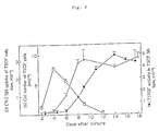
- TSCF cells and their culture SN were harvested from the one well of duplicate TSCF cultures on various days after seeding in 24-well plate. And the number of TSCF cells harvested and the TSTGF activity in the harvested culture SN were measured.
- the DNA synthesis of TSCF themselves was measured by [3H] TdR uptake in the other well of duplicate cultures. The results are summarized in Fig. 2. The growth of TSCF themselves as evaluated by the increase in cell number continued until around day 8 of the culture when TSCF formed a confluent monolayer. Meanwhile, the peak response of [3H] TdR uptake by TSCF preceded the increase in the number of these cells.
- the peak was seen on day 4 of the culture, and began to decrease after the cell monolayer acquired confluency.
- the TSTGF activity in TSCF culture became detectable around day 8 or 9 concurrently with the acquisition of confluence by TSCF monolayer. Thereafter, the activity continued to increase, and the maximum activity was obtained 12 - 14 days after seeding.
- the TSCF culture SN harvested at this timing was used as a source of TSTGF in the following experiments.
- Th clone maintained with addition of TSTGF would retain its antigen-reactivity.
- 9-16 Th clone was cultured either on TSCF monolayer or with TSCF culture SN (TSTGF) for 4 weeks by weekly change of culture medium, and tested for the reactivity to stimulation with KLH + I-E k antigen-presenting cells (Table 2).
- the results demonstrate that the 9-16 Th clone harvested after culturing on TSCF monolayer or with TSTGF did not acquire capability to proliferate spontaneously like malignant transformed cells but clearly retained antigen (KLH)-specific reactivity. Therefore, even in the absence of antigen + antigen-presenting cells, the Th clone proliferation is promoted by TSTGF, while retaining its antigen-reactivity.
- lymphokines and monokines produced by lymphocytes and monocytes have so far been reported. Therefore, a study was made as to whether any known lymphokine or monokine activity can be detected in culture SN of TSCF monolayer. Recombinant lymphokines and standard culture SNs containing lymphokines were used as positive control in the corresponding assay systems. And by comparing them with culture SN of TSCF, IL1, IL2, IL3, IL4 and IL5 activities therein were tested. A representative result summarized in Table 3 shows that positive controls of IL1, IL2, IL3 and IL4 functioned to proliferate or activate the corresponding responding cells, whereas culture SN of TSCF did not show such interleukin activity of detectable level. It was further shown by other experiments that IL5 and interferon (IFN) activities were not detected in TSCF SN.
- IFN interferon
- TSTGF lymphokines which have been known as T cell growth factor
- type or types of lymphokine are capable of promoting the proliferation of 9-16 Th clone.
- Interleukins IL1, IL2, IL3 and IL4
- interferons IFN- ⁇ , IFN- ⁇ and IFN- ⁇
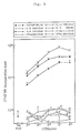
- the results in Fig. 3 demonstrate that the proliferation of 9-16 Th clone was induced by IL2 and IL4 as well as TSTGF but not by IL1, IL3 and interferons.
- Fig. 4 demonstrate that the proliferation of 9-16 Th clone was supported by IL2 and IL4 (right-hand-side panels) and that such proliferation was almost completely inhibited by the addition of anti-IL2 receptor or anti-IL4 antibody. In contrast to this, TSTGF-induced proliferation of 9-16 Th clone was little affected by the existence of any of these antibodies (left-hand-side panels). These results indicate that TSTGF induced the proliferation of Th clone in a mechanism which is entirely different from that in IL2 or IL4 and that TSTGF is functionally different from IL2 and IL4.
- TSTGF was capable of proliferating various but not all types of Th clones (each different in antigen specificity and restriction specificity to major histocompatible antigen (MHC)), though in varying degree of proliferation in each clone.
- MHC major histocompatible antigen
- TSTGF failed to support the proliferation of newly established CTL clones such as anti-K k (IMP) clone used in this experiment and also of various CTL clones used widely heretofore.
- IMP anti-K k
- TSTGF is functionally different from IL2 or IL4.
- a C3H/HeN thymic cell suspension containing both thymocytes and thymic stromal cells was seeded on culture flasks.
- Adherent cells were cultured after discarding non-adherent cells (mainly thymocytes and dead cells) and fed with 50% fresh medium and 50% conditioned medium.
- TSCF thymocytes and dead cells
- the adherent cells obtained from the primary thymic cultures were similar to TSCF cell line not only in morphological characteristics but also in the ability to support the proliferation of Th clone.
- 9-16 Th cells adhered to TSCF and primary thymic culture cells and proliferated though to different extent.
- thymic stroma-derived cell line TSCF was established by us. Morphologically, this cell line has a fibroblastic appearance. In the following experiments, a cloned cell line of this TSCF was used.
- Test samples (crude or partially purified TSTGF) (100 ⁇ l) were added to wells of 96-well flat-bottomed microplates (No. 25860: Corning Glass Works) to which 100 ⁇ l of 9-16 Th clone suspension (1 x 10⁵ cells/ml) had been placed and incubated in a CO2 incubator (95% air/5% CO2) for 2 days at 37°C. Then, these cells were pulsed with 0.5 ⁇ Ci/well of [3H] thymidine (New England Nuclear, Boston, MA) and 7 hr. later harvested onto the glass filter with an automated sample harvester (Bellco Glass Inc., Vineland, NJ), and the incorporated radioactivity was measured with a scintillation spectrometer (Aloka Co., Ltd., Tokyo, Japan).
- CSF colony-stimulating factor
- the 3H-TdR incorporation assay method developed for measuring CSF activity is basically a modification of the existing assay methods for interleukin 1 (J. Immunol. 116:1466, 1976) and interleukin 2 (Mol. Immunol. 17:579, 1980) with the exceptions that the number of responding cells is different and an exogenous primary mitogenic signal is not required.
- the culture supernatant containing TSTGF activity was dialyzed against 25 mM imidazole-HCl buffer (pH 7.5) and applied to a column of DEAE-Sephacel (3.0 x 45 cm) equilibrated with the same buffer.
- the column was washed with 1000 ml of the same buffer and eluted with 25 mM imidazole-HCl buffer (pH 7.5) containing 0.08 M sodium chloride at a flow rate of 20 ml/hr.
- Fractions of 50 ml each were collected and dialyzed against Hanks balanced salt solution (HBSS), and measurements were made on TSTGF activity and protein content by O.D. 280 nm.
- the protein remaining in the column was further eluted with 25 mM imidazole-HCl buffer (pH 7.5) containing 0.2 M sodium chloride. Then, measurement was made on the activity in a similar manner.
- TSTGF activity-containing fractions obtained from PBE 94 column was concentrated 10-fold by ultrafiltration by using an Amicon 8400 cell YM10 membrane (Amicon corp., Lexington, MA). The concentrate was then dialyzed against HBSS buffer. Two ml of concentrated sample was applied to a 1.5 x 9.5 cm Sephacryl S-200 (Pharmacia Fine Chemicals) column equilibrated with the same buffer. Fractions (2.5 ml) were collected, dialyzed with HBSS, and assayed for TSTGF activity.
- the TSTGF-containing concentrated sample after chromatofocusing was analyzed by discontinuous SDS-PAGE (0.75 mm x 16 cm x 20 cm slab gel) with an upper gel (1 cm) consisting of 2.88% acrylamide, 0.12% bis-acrylamide, 0.125 M Tris-HCl (pH 6.8) and 0.1% SDS, and a lower gel (8 cm) consisting of 11.52% acrylamide, 0.48% bis-acrylamide, 0.375 M Tris-HCl (pH 8.8) and 0.1% SDS (Methods in Enzymol. 116:493, 1985).
- the running buffer contained 0.025 M Tris base, 0.192 M glycine and 0.1% SDS.
- the gel was run at 30 V until the sample entered the lower gel (about 1 h) and then at 120 V until completion (about 4 hr). After electrophoresis, the SDS-PAGE gels were sliced into 5 mm fragments. Individual fragments were eluted with 1 ml of RPM11640 medium containing 10 mg/ml bovine serum albumin, which were then used for measuring TSTGF activity. The gels loaded with the sample or standard proteins were stained by the method of Merril et al. (Science 211:1437, 1981).
- TSTGF Fifty ⁇ l sample of partially purified TSTGF obtained by DEAE-Sephacel chromatography and PBE 94 chromatofocusing was treated with trypsin (15 ⁇ g/ml, 37°C, 2 hr) or dithiothreitol (DTT) (50 ml, 25°C, 2 hr).
- trypsin 15 ⁇ g/ml, 37°C, 2 hr
- DTT dithiothreitol
- the TSTGF activity was monitored by the proliferation of helper T cell (Th) clone 9-16.
- TSCF culture supernatant was collected 1 week after the TSCF cell line acquired confluence.
- elution was performed by serially increasing a concentration of sodium chloride. The results showed that the majority of the TSTGF activity was eluted of 0.08 M sodium chloride. Therefore, to achieve partial purification, about 1 l of culture supernatant was applied to DEAE-Sephacel column and eluted with buffer containing 0.08 M sodium chloride. As a result, all TSTGF activity was recovered in this fraction (Fig. 5). It is interesting that the greater part of the TSTGF activity is eluted at a comparatively early stage with 0.08 M sodium chloride and clearly separated from the major peaks of proteins eluted with 0.08 M NaCl and with 0.5 M NaCl.
- TSTGF activity-containing fractions thus obtained from DEAE-Sephacel column were further applied to a PBE 94 chromatofocusing column.
- a typical elution profile is shown in Fig. 6.
- the TSTGF activity was eluted as a single peak having pI value of about 6.0.
- TSTGF activity was similarly eluted as a single peak with pI of 6.0.
- TSTGF activity-containing fractions obtained by separating DEAE-Sephacel-fractions and the chromatofocusing were pooled and used as a partially purified TSTGF preparation for the following experiments.
- the recovery ratio of TSTGF activity after being applied to DEAE-Sepharose and chromatofocusing columns was about 20%, and increase in specific activity based on protein concentration was about 300 times at this stage.
- TSTGF recombinant IL2
- the titers of the TSTGF activity of concentrated and unconcentrated partially purified TSTGF preparation were calculated by comparison with dose response by TSTGF concentrate. The result demonstrates that the titers were about 60 and 0.7 U/ml respectively in terms of IL-2 titer.
- TSTGF activity was assessed by its ability to induce the proliferation of 9-16 cells.
- analysis of TSTGF by Sephacryl S-200 gel filtration showed its apparent molecular weight is 25,000.
- the same concentrated sample was also analyzed by SDS-PAGE (12.5% acrylamide) under nonreducing conditions.
- SDS-PAGE (12.5% acrylamide) under nonreducing conditions.
- the gel was sliced into 0.5 cm pieces and proteins were eluted from each sliced gel. Each fraction was assayed for TSTGF activity with the use of 9-16 Th cells.
- TSTGF activity ws reproducibly detected in fractions having a molecular weight of about 25,000.
- a representative experiment is shown in Fig. 9.
- TSTGF The stability of TSTGF was examined by subjecting partially purified TSTGF preparation to various treatments.
- the activity was found to be resistant to a moderate heat treatment (65°C, 10 min) but stable under acidic (pH 2.0) and alkaline (pH 9.0) conditions, while the activity partially decreased at pH of around 4.0 -5.0 by some unknown reason (Table 6).
- it was susceptible to proteolysis with trypsin and to reduction by DTT (10 mM DDT, 2 hr) (Table 7).
- TSTGF is different from any of the previously described cytokines including those capable of inducing T cell proliferation such as IL2, IL4 and GM-CSF.
- TSTGF functions as a potent T cell growth factor (TCGF) for various antigen-specific helper T cell clones.
- TSTGF functions as a potent T cell growth factor (TCGF) for various antigen-specific helper T cell clones.
- TSTGF contributes to the growth of thymocytes.
- the GK1.5 monoclonal antibody-producing hybridoma cells were inoculated into pristane-primed BALB/c nu/nu mice intraperitoneally and ascitic fluids were collected. Culture supernatants (SNs) from 3.155 and 2C11 monoclonal antibody-producing hybridomas were also collected. Gamma-globulin fractions of cell-free ascites and culture SNs were obtained by precipitation at 40% saturation with ammonium sulfate. Fluorescein isothiocyanate (FITC) was conjugated to GK1.5 or 2C11 monoclonal antibody by regular methods, followed by DE-52 (Whatman Biochemicals Ltd., England) ion-exchange chromatography.
- FITC Fluorescein isothiocyanate
- the F/P ratio of FITC/GK1.5 or FITC/2C11 was 1.6 or 6.8.
- FITC-conjugated anti-Lyt2, anti-Thy1.2 and mouse anti-rat IgG were the products of Becton Dickinson Immunocytometry Systems, Mountain View, CA, of Bio Yeda Ltd., Rehovot, Israel and of Jackson Immunoresearch Laboratories, INC., West Grove, PA., respectively.
- Phorbol myristate acetate (PMA) was purchased from Sigma Chemical Co. (St. Louis. MO.).
- the treated cells were washed and then incubated for 1 hr at 37°C on tissue culture grade petri dishes (Corining Glass Works, NY.) pretreated for 2 hr at 37°C with goat anti-mouse Ig (Cappel, Malvern, PA.) that cross-reacts with rat Ig.
- the nonadherent cells were collected by gently pipetting. This procedure yielded a population of L3T4 ⁇ Lyt2 ⁇ thymocytes representing 0.5 - 1.0 % of the starting thymocyte population.
- the double-negative adult thymocyte population (3 x 10⁴/well) was cultured with TSTGF plus PMA.
- Table 13 the double-negative thymocytes failed to respond to either stimulant alone, whereas the co-stimulation with these two stimulation resulted in appreciable proliferation of the immature thymoctye population.
- Fig. 11 also illustrates that the TSTGF promotes the proliferation of double-negative thymocytes in a dose-dependent manner when combined with PMA.
- the same cell concentrate (3 x 10⁴/well) of unfractionated thymocytes were unable to exhibit potent proliferation in the presence of TSTGF and PMA (Exp.
- Table 13 the results of Table 13 indicate that the combination of TSTGF plus PMA induces the growth-promoting effect mainly on the double negative immature adult thymocytes. It is additionally shown in Table 13 that this stimulant-combination is also capable of supporting the growth of fetal thymocytes on 14 day gestation that have been considered to be double-negative (J. Immunol. 133: 1117, 1984, Immunol. Rev. 82: 141, 1984, Immunol. Rev. 82: 79, 1984).
- TSTGF induces the growth-promotion of double-negative thymocytes without stimulating IL2- or IL4-dependent autocrine mechanism.
- IL1 has a potent costimulatory activity for the growth of double-negative thymocytes in combination with TSTGF
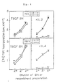
- TSTGF acts on immature thymocytes for their proliferative response but its ability depends on the presence of PMA as a costimulator. Since PMA is a non-physiological chemical, interesting is the question of whether the function of PMA can be replaced by some of physiological factors. To answer this question, various cytokines of T cell and non-T cell origins were tested for their capability of functioning as a costimulator of TSTGF to support the growth of double-negative thymoctyes. The results shown in Table 15 demonstrate that IL1 can promote the growth of double-negative thymocytes by synergy with TSTGF as efficiently as PMA.
- IL1 was the most effective co-stimulant, additional experiments were performed to more accurately investigate the synergistic effect between TSTGF and IL1 (Fig. 13).
- the co-stimulatory effect of IL1 was dose-dependent and low doses of IL1 between 0.02 and 0.2 U/ml still produced appreciable co-stimulatory activity (Fig. 13A). It was also shown that the proliferation of double-negative thymocytes induced by TSTGF plus IL1 exhibited the peak response around 3 days after the initiation of cultures and that the magnitude as well as the timing of the peak response in cultures stimulated with TSTGF plus IL1 coincide with those obtained by TSTGF plus PMA (Fig. 13B).
- IL1 was revealed to be the most effective replacement of PMA as a co-stimulant of TSTGF for the growth of double-negative thymocytes
- Table 15 also demonstrated that their proliferation was inducible by another combination of TSTGF plus the principal TCGF (IL2 or IL4) to an appreciable extent.
- IL2 or IL4 the principal TCGF
- double-negative thymocytes were cultured in the presence of TSTGF, IL1 and IL2 or IL4. The results of Fig.
- TSTGF 3 cytokines
- IL1 and IL2 3 cytokines
- anti-T3 (2C11) antibody was included along with anti-L3T4 and anti-Lyt2 antibodies in both the complement treatment and panning step.
- the efficacy of anti-T3 antibody to deplete T3 bright thymocytes is shown in the results of Fig. 15.
- Double-negative thymocyte population contained both T3 ⁇ or dull and T3 bright subsets, whereas T3 bright cell-depleted subset consisted of a large number of T3 ⁇ and small number of T3 dull thymocytes.
- Table 16 investigates the capacity of thus far used cytokines, either alone or in various combinations, to induce the proliferation of L3T4 ⁇ Lyt2 ⁇ T3 ⁇ or dull thymocyte subpopulation.
- the results demonstrate that the magnitude of the proliferation was more potent in the stimulation with TSTGF, IL1 and IL2 or IL4 than in any combination of the above two cytokines.
- Fig. 14 shows that more than additive growth-promoting effect was observed in the L3T4 ⁇ Lyt2 ⁇ T3 ⁇ or dull thymocyte population when stimulated especially with a combination of TSTGF, IL1 and IL4.
- Table 1 Capacity of a thymic stroma-derived cell line or its culture SN to support the growth of Th clone in vitro Expt. Monolayer a or SN Growth of Th clone b : Cell No. (x 10 ⁇ 4) at Input Harvest 1 none 10.0 1.3 TSCF monolayer 10.0 30.1 TSCE monolayer 10.0 7.2 BALB/3T3 monolayer 10.0 5.2 2 none 5.0 0.2 TSCF monolayer 5.0 27.5 TSCF SN (25%) 5.0 46.2 a. Monolayers were prepared from two types (fibroblastic form; TSCF and epithelial form; TSCE) of thymic stroma-derived cell lines or BALB/3T3 cell line. b.
- Table 2 Retention of antigen-reactivity in Th clone after growth-maintenance by TSCF monolayer or its SN Stimulation a with [3H]TdR uptake by 9-16 (cpm) after pre-culture with b : TSCF monolayer TSCF SN none 199(1.23) 90(1.12) I-E k spleen cells 1970(1.08) 222(1.76) KLH+I-E k spleen cells 11290(1.02) 11570(1.13) a.
- 9-16 cells (1 x 10⁴/well) were stimulated by 2000 R X ray-irradiated C3H/He spleen cells (5 x 10⁴/well) as I-E k antigen-presenting cells with or without KLH antigen (8 ⁇ g/ml).
- 9-16 cells were pre-cultured with TSCF monolayer or its SN for 4 weeks in the absence of I-E k and KLH antigens.
- Culture medium was weekly changed with fresh medium for the pre-culture with TSCF monolayer or with fresh medium containing 25% TSCF SN for the pre-culture with TSCF SN.
- LBRM-33 1A5 cells were exposed to various dilutions of rIL1 or TSTGF (25% TSCF SN) in the presence of PHA (2 ⁇ g/ml).
- IL1 activity was obtained by measuring IL2 activity produced in these culture SN with the use of IL2-dependent CTLL-2.
- IL4 activity was assessed by measuring the proliferation of B cells by co-stimulation with anti-mouse IgM and TSCF SN or rIL4 as control.
- WEHI SN was obtained from culture SN of WEHI-3 cell line. Table 4 Effect of TSTGF on the proliferation of various T cell clones T cell clone a [3H]TdR incorporation in the presence of Type Name Cell No.
- T cell clones (1 or 5 x 10⁴/well) were cultured with rIL2 (20 ⁇ /ml), rIL4 (25 ⁇ /ml) or TSTGF (50% v/v) of culture SN condensed into 0.2 volume by a centricon membrane for 2 days in 96-well microplate.
- TSTGF preparation was heated (indicated) or added to 3-fold volumes of either 0.2 M glycine-HCl (for pH 2.0), 0.2 M sodium acetate (for pH 5.0) or 0.2 M Tris-HCl (for pH 9.0). Samples were then dialyzed against HBSS. b. Dialyzed sample (50 ⁇ l) was added to each well of 105/well 9-16 Th clone. 3H-TdR uptake by the same number of 9-16 cells in the absence of TSTGF sample was 120.
- CTLL-2 (1 ⁇ 104/well) or HT-2 (1 ⁇ 104/well) cells were stimulated with either recombinant IL2 (rIL2), rIL4 or partially purified TSTGF.
- rIL2 recombinant IL2
- Table 10 Elimination of CSF activity in partially purified TSTGF preparation Factor a TSTGF activity b CSF activity c 3H-TdR uptake cpm x 103 Total colony No.
- TSTGF activity was assessed by 3H-TdR uptake of 105 9-16 Th clone.
- Colony formation and 3H-TdR incorporation assays were performed with cells from the same bone marrow preparation. Colony formation was determined after 10 day incubation. Incorporation of 3H-TdR was measured after 72 hr incubation. Table 11 Comparison of functions and properties between various factors capable of contributing to T cell proliferation Factor (mice) Proliferation of: M.W.
- L3T4 ⁇ Lyt2 ⁇ adult thymocytes (3 x 10⁴/well) or fetal thymocytes on 14 day gestation (2 x 10⁴/well) were cultured for 3 days with various stimulating agents at the same doses as in Table 12.
- Table 14 Comparison between TSTGF and various ILs on the ability to promote the growth of double negative thymocytes in the presence of PMA Stimulation with a 3H-TdR uptake (cpm) PMA(-) PMA(+) medium 1082 ⁇ 241 1354 ⁇ 189 TSTGF 3162 ⁇ 184 19060 ⁇ 529 rIL2 2004 ⁇ 685 2250 ⁇ 51 rIL4 1950 ⁇ 136 17390 ⁇ 576 Ionomycin 2645 ⁇ 563 35381 ⁇ 3283 a. Double negative (L3T4 ⁇ Lyt2 ⁇ ) adult thymocytes were stimulated with various agents at the same doses in Table 12.
- TSTGF(-) TSTGF(+) TSTGF(-) TSTGF(+) medium 1293 ⁇ 537 1657 ⁇ 255 2190 ⁇ 1401 3895 ⁇ 389 PMA 603 ⁇ 111 12372 ⁇ 1209 1424 ⁇ 555 19040 ⁇ 2856 rIL1 ⁇ 374 ⁇ 21 9119 ⁇ 918 1224 ⁇ 465 21548 ⁇ 24 rIL2 628 ⁇ 65 5232 ⁇ 854 1475 ⁇ 265 9873 ⁇ 691 rIL4 373 ⁇ 63 7045 ⁇ 1327 1936 ⁇ 341 10007 ⁇ 1001 rIL6 b 253 ⁇ 21 2398 ⁇ 89 2897 ⁇ 637 3873 ⁇ 309 a.
- Table 16 Enhanced proliferation of L3T4 ⁇ Lyt2 ⁇ T3 - or dull thymocytes by a combination of [TSTGF + IL1] and IL2 or IL4 Stimulation with a 3H-TdR uptake (cpm) Exp. 1 Exp.
Landscapes
- Health & Medical Sciences (AREA)
- Life Sciences & Earth Sciences (AREA)
- Chemical & Material Sciences (AREA)
- Genetics & Genomics (AREA)
- Organic Chemistry (AREA)
- Engineering & Computer Science (AREA)
- Zoology (AREA)
- General Health & Medical Sciences (AREA)
- Biomedical Technology (AREA)
- Biophysics (AREA)
- General Engineering & Computer Science (AREA)
- Molecular Biology (AREA)
- Biotechnology (AREA)
- Biochemistry (AREA)
- Bioinformatics & Cheminformatics (AREA)
- Wood Science & Technology (AREA)
- Physics & Mathematics (AREA)
- Proteomics, Peptides & Aminoacids (AREA)
- Medicinal Chemistry (AREA)
- Plant Pathology (AREA)
- Gastroenterology & Hepatology (AREA)
- Microbiology (AREA)
- Toxicology (AREA)
- Preparation Of Compounds By Using Micro-Organisms (AREA)
- Medicines That Contain Protein Lipid Enzymes And Other Medicines (AREA)
- Medicines Containing Material From Animals Or Micro-Organisms (AREA)
- Micro-Organisms Or Cultivation Processes Thereof (AREA)
- Peptides Or Proteins (AREA)
Applications Claiming Priority (2)
| Application Number | Priority Date | Filing Date | Title |
|---|---|---|---|
| JP248616/87 | 1987-09-30 | ||
| JP62248616A JPS6490199A (en) | 1987-09-30 | 1987-09-30 | T cell growth factor derived from thymic interstitial cell and production thereof |
Publications (2)
| Publication Number | Publication Date |
|---|---|
| EP0310056A2 true EP0310056A2 (de) | 1989-04-05 |
| EP0310056A3 EP0310056A3 (de) | 1989-06-14 |
Family
ID=17180768
Family Applications (1)
| Application Number | Title | Priority Date | Filing Date |
|---|---|---|---|
| EP88116054A Withdrawn EP0310056A3 (de) | 1987-09-30 | 1988-09-29 | T-Zellen-Wachstumsfaktor aus Thymusdrüsen-Stroma und Verfahren zu seiner Herstellung |
Country Status (7)
| Country | Link |
|---|---|
| EP (1) | EP0310056A3 (de) |
| JP (1) | JPS6490199A (de) |
| KR (1) | KR940003650B1 (de) |
| AU (1) | AU607000B2 (de) |
| DK (1) | DK543388A (de) |
| HU (1) | HUT48305A (de) |
| NZ (1) | NZ226356A (de) |
Cited By (6)
| Publication number | Priority date | Publication date | Assignee | Title |
|---|---|---|---|---|
| WO1993017700A1 (en) * | 1992-03-13 | 1993-09-16 | Beardsley Terry R | Immune-enhancing agent for therapeutic use in immunocompromised hosts |
| WO1993022437A1 (en) * | 1992-04-30 | 1993-11-11 | N.V. Innogenetics S.A. | New polypeptides and peptides, nucleic acids coding for them, and their use in the field of tumor therapy, inflammation or immunology |
| US5346988A (en) * | 1990-06-20 | 1994-09-13 | Tampa Bay Research Institute | Process for obtaining cellular protein having anti-HIV activity |
| WO1995034317A1 (en) * | 1994-06-10 | 1995-12-21 | Olavi Kajander | Growth factor preparation, its production and use |
| US5643881A (en) * | 1994-08-04 | 1997-07-01 | Tampa Bay Research Institute | Protein having anti-HIV activity and method for obtaining it |
| CN107299138A (zh) * | 2014-04-09 | 2017-10-27 | 上海欣生源药业有限公司 | Cxcl4单抗治疗肿瘤及化疗后肿瘤加速再增殖基因筛选法 |
Families Citing this family (4)
| Publication number | Priority date | Publication date | Assignee | Title |
|---|---|---|---|---|
| IT1196958B (it) * | 1986-07-10 | 1988-11-25 | Ellem Ind Farmaceutica | Derivato di timo attivo per via orale, procedimenti per la sua preparazione e relative composizioni farmaceutiche |
| ZA887773B (en) * | 1987-10-26 | 1989-07-26 | Immunex Corp | Interleukin-7 |
| CA2000695A1 (en) * | 1989-03-16 | 1990-09-16 | Nancy M. Hedberg | Cytokine |
| JPH04186696A (ja) * | 1990-11-16 | 1992-07-03 | Mitsubishi Electric Corp | ボンディング装置 |
Family Cites Families (3)
| Publication number | Priority date | Publication date | Assignee | Title |
|---|---|---|---|---|
| EP0163543A3 (de) * | 1984-06-01 | 1987-08-12 | VXR, Inc. | Markzellenanreger |
| EP0302459A3 (de) * | 1987-08-04 | 1990-02-07 | Sumitomo Pharmaceuticals Company, Limited | Thermostabiler Zellwachstumsfaktor aus menschlicher Plazenta und Verfahren zu seiner Herstellung |
| ATE149176T1 (de) * | 1987-10-02 | 1997-03-15 | Univ Rockefeller | Von makrophagen abstammender entzündungsvermittler(mip-1-alpha et mip-1-beta) |
-
1987
- 1987-09-30 JP JP62248616A patent/JPS6490199A/ja active Pending
-
1988
- 1988-09-27 NZ NZ226356A patent/NZ226356A/xx unknown
- 1988-09-29 HU HU885070A patent/HUT48305A/hu unknown
- 1988-09-29 EP EP88116054A patent/EP0310056A3/de not_active Withdrawn
- 1988-09-29 DK DK543388A patent/DK543388A/da not_active Application Discontinuation
- 1988-09-30 KR KR1019880012844A patent/KR940003650B1/ko not_active Expired - Fee Related
- 1988-09-30 AU AU23348/88A patent/AU607000B2/en not_active Ceased
Cited By (10)
| Publication number | Priority date | Publication date | Assignee | Title |
|---|---|---|---|---|
| US5346988A (en) * | 1990-06-20 | 1994-09-13 | Tampa Bay Research Institute | Process for obtaining cellular protein having anti-HIV activity |
| US5480782A (en) * | 1990-06-20 | 1996-01-02 | Tampa Bay Research Institute | Process for obtaining cellular protein having anti-HIV activity |
| WO1993017700A1 (en) * | 1992-03-13 | 1993-09-16 | Beardsley Terry R | Immune-enhancing agent for therapeutic use in immunocompromised hosts |
| WO1993022437A1 (en) * | 1992-04-30 | 1993-11-11 | N.V. Innogenetics S.A. | New polypeptides and peptides, nucleic acids coding for them, and their use in the field of tumor therapy, inflammation or immunology |
| US5981277A (en) * | 1992-04-30 | 1999-11-09 | N.V. Innogenetics S.A. | Polypeptides and peptides, nucleic acids coding for them, and their use in the field of tumor therapy, inflammation or immunology |
| WO1995034317A1 (en) * | 1994-06-10 | 1995-12-21 | Olavi Kajander | Growth factor preparation, its production and use |
| US5776778A (en) * | 1994-06-10 | 1998-07-07 | Kajander; Olavi | Growth factor preparation of thymocyte cell culture medium its production and use |
| US5643881A (en) * | 1994-08-04 | 1997-07-01 | Tampa Bay Research Institute | Protein having anti-HIV activity and method for obtaining it |
| CN107299138A (zh) * | 2014-04-09 | 2017-10-27 | 上海欣生源药业有限公司 | Cxcl4单抗治疗肿瘤及化疗后肿瘤加速再增殖基因筛选法 |
| CN107299138B (zh) * | 2014-04-09 | 2021-05-07 | 上海欣生源药业有限公司 | Cxcl4单抗治疗肿瘤及化疗后肿瘤加速再增殖基因筛选法 |
Also Published As
| Publication number | Publication date |
|---|---|
| EP0310056A3 (de) | 1989-06-14 |
| AU2334888A (en) | 1989-04-06 |
| AU607000B2 (en) | 1991-02-21 |
| HUT48305A (en) | 1989-05-29 |
| DK543388A (da) | 1989-03-31 |
| KR940003650B1 (ko) | 1994-04-25 |
| DK543388D0 (da) | 1988-09-29 |
| JPS6490199A (en) | 1989-04-06 |
| KR890005272A (ko) | 1989-05-13 |
| NZ226356A (en) | 1991-08-27 |
Similar Documents
| Publication | Publication Date | Title |
|---|---|---|
| Tosato et al. | Identification of interleukin-6 as an autocrine growth factor for Epstein-Barr virus-immortalized B cells | |
| Sutherland et al. | Differential regulation of primitive human hematopoietic cells in long-term cultures maintained on genetically engineered murine stromal cells | |
| Firestein et al. | A new murine CD4+ T cell subset with an unrestricted cytokine profile. | |
| Lotz et al. | B cell stimulating factor 2/interleukin 6 is a costimulant for human thymocytes and T lymphocytes. | |
| Raghavachar et al. | T lymphocyte control of human eosinophilic granulopoiesis. Clonal analysis in an idiopathic hypereosinophilic syndrome. | |
| Prystowsky et al. | Alloreactive cloned T cell lines. VI. Multiple lymphokine activities secreted by helper and cytolytic cloned T lymphocytes. | |
| Erard et al. | Interleukin 2 is both necessary and sufficient for the growth and differentiation of lectin-stimulated cytolytic T lymphocyte precursors. | |
| Nabel et al. | Multiple biologic activities of a cloned inducer T-cell population. | |
| EP0473724B1 (de) | Menschliches cytokin, interleukin-9 | |
| Smith et al. | Functional and molecular characteristics of T-cell growth factor | |
| Herrmann et al. | In vitro regulation of human hematopoiesis by natural killer cells: analysis at a clonal level | |
| Lopez-Botet et al. | Relationship between IL 2 synthesis and the proliferative response to PHA in different primary immunodeficiencies. | |
| Ihle | Biochemical and biological properties of interleukin-3: a lymphokine mediating the differentiation of a lineage of cells that includes prothymocytes and mastlike cells | |
| Platzer et al. | Human pluripotent hemopoietic colony stimulating factor: activities on human and murine cells | |
| Fischer et al. | Granulocyte‐macrophage colony‐stimulating factor‐cultured bone marrow‐derived macrophages reveal accessory cell function and synthesis of MHC class II determinants in the absence of external stimuli | |
| Nakoinz et al. | Differentiation of the IL-3-dependent NFS-60 cell line and adaption to growth in macrophage colony-stimulating factor. | |
| Bressler et al. | Inhibition of the growth of IL-3-dependent mast cells from murine bone marrow by recombinant granulocyte macrophage-colony-stimulating factor. | |
| Vie et al. | Estimation by limiting dilution analysis of human IL 2-secreting T cells: detection of IL 2 produced by single lymphokine-secreting T cells. | |
| Tennant et al. | Abnormalities of adherent layers grown from bone marrow of patients with myelodysplasia | |
| Sakabe et al. | Functional differences between subpopulations of mobilized peripheral blood-derived CD34+ cells expressing different levels of HLA-DR, CD33, CD38 and c-kit antigens | |
| EP0310056A2 (de) | T-Zellen-Wachstumsfaktor aus Thymusdrüsen-Stroma und Verfahren zu seiner Herstellung | |
| Talmadge et al. | Proliferation of human peripheral blood lymphocytes induced by recombinant human interleukin 2: contribution of large granular lymphocytes and T lymphocytes | |
| Kelso et al. | Interleukin 2 enhancement of lymphokine secretion by T lymphocytes: analysis of established clones and primary limiting dilution microcultures. | |
| Kelvin et al. | Interleukin 3 and cell cycle progression | |
| Kobayashi et al. | Interleukin-3 is significantly more effective than other colony-stimulating factors in long-term maintenance of human bone marrow-derived colony-forming cells in vitro |
Legal Events
| Date | Code | Title | Description |
|---|---|---|---|
| PUAI | Public reference made under article 153(3) epc to a published international application that has entered the european phase |
Free format text: ORIGINAL CODE: 0009012 |
|
| AK | Designated contracting states |
Kind code of ref document: A2 Designated state(s): AT BE CH DE ES FR GB GR IT LI LU NL SE |
|
| PUAL | Search report despatched |
Free format text: ORIGINAL CODE: 0009013 |
|
| AK | Designated contracting states |
Kind code of ref document: A3 Designated state(s): AT BE CH DE ES FR GB GR IT LI LU NL SE |
|
| 17P | Request for examination filed |
Effective date: 19891011 |
|
| 17Q | First examination report despatched |
Effective date: 19911212 |
|
| STAA | Information on the status of an ep patent application or granted ep patent |
Free format text: STATUS: THE APPLICATION IS DEEMED TO BE WITHDRAWN |
|
| 18D | Application deemed to be withdrawn |
Effective date: 19920423 |