EP0310132A2 - Immunassay mit Anwendung von IgG Einfangantikörper und ein Markierungssystem bestehend aus mehreren monoklonalen Antikörpern - Google Patents
Immunassay mit Anwendung von IgG Einfangantikörper und ein Markierungssystem bestehend aus mehreren monoklonalen Antikörpern Download PDFInfo
- Publication number
- EP0310132A2 EP0310132A2 EP88116274A EP88116274A EP0310132A2 EP 0310132 A2 EP0310132 A2 EP 0310132A2 EP 88116274 A EP88116274 A EP 88116274A EP 88116274 A EP88116274 A EP 88116274A EP 0310132 A2 EP0310132 A2 EP 0310132A2
- Authority
- EP
- European Patent Office
- Prior art keywords
- monoclonal antibodies
- immunoassay
- labeled
- binding pair
- reporter
- Prior art date
- Legal status (The legal status is an assumption and is not a legal conclusion. Google has not performed a legal analysis and makes no representation as to the accuracy of the status listed.)
- Withdrawn
Links
Images


Classifications
-
- G—PHYSICS
- G01—MEASURING; TESTING
- G01N—INVESTIGATING OR ANALYSING MATERIALS BY DETERMINING THEIR CHEMICAL OR PHYSICAL PROPERTIES
- G01N33/00—Investigating or analysing materials by specific methods not covered by groups G01N1/00 - G01N31/00
- G01N33/48—Biological material, e.g. blood, urine; Haemocytometers
- G01N33/50—Chemical analysis of biological material, e.g. blood, urine; Testing involving biospecific ligand binding methods; Immunological testing
- G01N33/58—Chemical analysis of biological material, e.g. blood, urine; Testing involving biospecific ligand binding methods; Immunological testing involving labelled substances
-
- G—PHYSICS
- G01—MEASURING; TESTING
- G01N—INVESTIGATING OR ANALYSING MATERIALS BY DETERMINING THEIR CHEMICAL OR PHYSICAL PROPERTIES
- G01N33/00—Investigating or analysing materials by specific methods not covered by groups G01N1/00 - G01N31/00
- G01N33/48—Biological material, e.g. blood, urine; Haemocytometers
- G01N33/50—Chemical analysis of biological material, e.g. blood, urine; Testing involving biospecific ligand binding methods; Immunological testing
- G01N33/53—Immunoassay; Biospecific binding assay; Materials therefor
- G01N33/543—Immunoassay; Biospecific binding assay; Materials therefor with an insoluble carrier for immobilising immunochemicals
- G01N33/54306—Solid-phase reaction mechanisms
-
- G—PHYSICS
- G01—MEASURING; TESTING
- G01N—INVESTIGATING OR ANALYSING MATERIALS BY DETERMINING THEIR CHEMICAL OR PHYSICAL PROPERTIES
- G01N33/00—Investigating or analysing materials by specific methods not covered by groups G01N1/00 - G01N31/00
- G01N33/48—Biological material, e.g. blood, urine; Haemocytometers
- G01N33/50—Chemical analysis of biological material, e.g. blood, urine; Testing involving biospecific ligand binding methods; Immunological testing
- G01N33/53—Immunoassay; Biospecific binding assay; Materials therefor
- G01N33/569—Immunoassay; Biospecific binding assay; Materials therefor for microorganisms, e.g. protozoa, bacteria, viruses
- G01N33/56983—Viruses
-
- G—PHYSICS
- G01—MEASURING; TESTING
- G01N—INVESTIGATING OR ANALYSING MATERIALS BY DETERMINING THEIR CHEMICAL OR PHYSICAL PROPERTIES
- G01N33/00—Investigating or analysing materials by specific methods not covered by groups G01N1/00 - G01N31/00
- G01N33/48—Biological material, e.g. blood, urine; Haemocytometers
- G01N33/50—Chemical analysis of biological material, e.g. blood, urine; Testing involving biospecific ligand binding methods; Immunological testing
- G01N33/53—Immunoassay; Biospecific binding assay; Materials therefor
- G01N33/577—Immunoassay; Biospecific binding assay; Materials therefor involving monoclonal antibodies binding reaction mechanisms characterised by the use of monoclonal antibodies
Definitions
- This invention relates generally to immunoassays and more specifically to solid phase immunoassays utilizing a labeled multiple monoclonal antibody detection system.
- monoclonal antibodies can be difficult to attach to a solid phase surface either by nonspecific absorption or covalent attachment techniques and researchers have encountered difficulty labeling monoclonals with various reporter groups using standard labeling techniques.
- U.S. Patent 4,659,678 relates to a method of solid phase immunoassay having a reduced incubation period which is achieved by reacting a sample and antibody reagents in the liquid phase and then capturing the reaction product with an insolubilized anti-antibody generated against the unlabeled antibody reagent.
- U.S. Patent 4,535,057 describes a solid phase immunoassay for detecting HSV antigens or anti-HSV IgM immunoglobulin in biological fluids by using monoclonal IgM HSV virus/antigen specific antibodies and a readably labeled, preferably biotin-avidin based, detection system.
- U.S. Patent 4,430,437 describes test methods using monoclonal antibodies directed to HSV type 1 and type 2 nucleocapsid proteins.
- U.S. Patent 4,271,140 describes a solid phase immunoassay using an enzyme based detection system.
- U.S. Patent 4,514,505 describes radioimmunoassays with enhanced sensitivity for polypeptides (e.g. human chorionic gonadotropin) by utilizing a mixture of monoclonal antibodies.
- polypeptides e.g. human chorionic gonadotropin
- U.S. Patent 4,228,237 describes insoluble phase enzyme immunoassays employing labeled avidin and biotin labeled reagent to detect a ligand.
- U.S. Patent Re. 31,006 describes an insoluble phase immunoassay using an enzyme based detection system.
- Adler-Storthz et al., J. Clinical Microbiology, Vol. 18, No. 6, pp. 1329-1334 (Dec. 1983) describe a biotin-avidin amplified enzyme-linked immunosorbent assay in which a biotinylated mouse monoclonal IgM antibody to HSV was used to detect HSV-1 and HSV-2.
- Syva Co. Product Insert for Herpes Simplex Virus-1/Herpes Simplex Virus-2 Culture Confirmation/Typing Test (1984) describes a test to identify and type herpes simplex virus in tissue culture using multiple monoclonal antibodies labeled with fluorescein isothiocyanate.
- Goldstein, et al., J. Infectious Diseases, Vol. 147, No. 51, pp. 829-837 (May 1983) developed a panel of unlabeled monoclonal antibodies reactive with either herpes simplex virus-1 or herpes simplex virus-2 which can be used reliably in immunofluorescent tests to serotype herpes simplex virus in culture and also for direct antigen detection and simultaneous typing of clinical specimens. These monoclonal antibodies were used separately and they were unlabeled.
- This invention is directed to solid phase immunoassays and kits for detecting or quantitating a substance having multiple epitopes utilizing principally IgG capture antibody and a cocktail of at least two labeled monoclonal antibodies as the detection system wherein each monoclonal antibody recognizes a distinct antigenic determinant.
- the monclonals may be labeled with a component of a reporter system or they may be labeled with a member of a specific binding pair. When the monoclonals are labeled with a component of a reporter system, they may be detected and/or quantitated immediately.
- the monoclonals when labeled with a member of a specific binding pair they must be reacted with the second member of the binding pair which may be unlabeled or labeled. If the second member of the binding pair is labeled, then after the reaction ceases, detection and/or quantification may follow. If the second member of the binding pair is unlabeled then reaction of the bound pair with a component of a reporter system is needed in order to detect or quantify the substance suspected to be present.
- This invention is directed to an immunoassay for detecting or quantitating a substance having multiple epitopes comprising:
- the immunoassay can be run as a forward sandwich assay in which the capture antibody is attached to a solid support followed by the addition of sample suspected to contain a substance having multiple epitopes. Once the reaction ceases, unreacted substance is washed away and a known quantity of the cocktail containing multiple labeled antibodies is added to the mixture.
- Immunoassays according to this invention can also be run as simultaneous sandwich assays in which both the insolubilized capture antibody and cocktail of labeled antibodies and sample are added at the same time.
- the capture antibody may be any immunoglobulin.
- the preferred embodiment of this invention utilizes principally IgG as the insolubilized capture antibody.
- the support may be any one of a wide variety of supports, and as representative examples of suitable supports there may be mentioned: synthetic polymer supports, such as polystyrene, polypropylene, substituted polystyrene, e.g., aminated or carboxylated polystyrene, polyacrylamides, polyamides, polyvinylchloride, etc.; glass beads, agarose; etc.
- the supports may include reactive groups; e.g., carboxyl groups etc. to permit direct linking of the substance to the support.
- the support consists of polystyrene microtiter plate wells.
- the substance which is being detected and/or quantitated is characterized by having multiple epitopes.
- polypeptides and proteins polysaccharides, nucleic acids, and combinations thereof.
- proteins are nucleoproteins, glycoproteins, scleroproteins, proteoglycans, mucoproteins, histones, albumins, insulin, pepsin and the like.
- viruses include adenoviruses; herpes viruses such as herpes simplex, varicella zoster, herpes virus B, cytomegalovirus, Epstein-Barr virus, human herpes virus type 6; pox viruses; picornaviruses; orthomyxoviruses; paramyxoviruses; coronaviruses; rhabdoviruses; togaviruses; bunyaviruses; arenaviruses; rubella virus; arboviruses; reoviruses; hepatitis; retroviruses; oncogenic viruses and the like.
- herpes viruses such as herpes simplex, varicella zoster, herpes virus B, cytomegalovirus, Epstein-Barr virus, human herpes virus type 6
- pox viruses picornaviruses
- orthomyxoviruses paramyxoviruses
- coronaviruses coronaviruses
- the capture antibody may be either polyclonal or monoclonal. Attachment to the solid support, whether direct or indirect, covalent or non-covalent, may be achieved using well-known techniques.
- all of the monoclonal antibodies are labeled using conventional techniques. They be labeled with a component of a reporter system or with a member of a specific binding pair.
- Specific binding pairs may be of the immune or non-immune type. Immune specific binding pairs are exemplified by antigen-antibody systems or hapten/anti-hapten systems. There can be mentioned fluorescein/anti-fluorescein, dinitrophenyl/anti-dinitrophenyl, biotin/anti-biotin, peptide/anti-peptide and the like.
- the antibody member of the specific binding pair may be produced by customary methods familiar to those skilled in the art. Such methods involve immunizing an animal with the antigen member of the specific binding pair. If the antigen member of the specific binding pair is not immunogenic, i.e., a hapten, it may be covalently coupled to a carrier protein to render it immunogenic.
- Non-immune binding pairs include systems wherein the two components share a natural affinity for each other but are not antibodies.
- Exemplary non-immune pairs are biotin-streptavidin, intrinisic factor-vitamin B12, folic acid-folate binding protein and the like.
- Biotin may be covalently coupled to monoclonal antibodies by utilizing commercially available active derivatives. Some of these are biotin-N-hydroxysuccinimide which binds to amine groups on proteins; biotin hydrazide which binds to carbohydrate moieties, aldehydes and carboxyl groups via a carbodiimide coupling; and biotin maleimide and iodoacetyl biotin which bind to sulfhydryl groups.
- Fluorescein can be coupled to protein amine groups using fluorescein isothiocyanate. Dinitrophenyl groups can be coupled to protein amine groups using 2,4-dinitrobenzene sulfonate or 2,4-dinitrofluorobenzene. Other standard methods of conjugation may be employed to couple monoclonal antibodies to a member of a specific binding pair including dialdehyde, carbodiimide coupling, homofunctional crosslinking, and heterobifunctional crosslinking. Carbodiimide coupling is an effective method of coupling carboxyl groups on one substance to amine groups on another. Carbodiimide coupling is facilitated by using the commercially available reagent, 1-ethyl-3-(dimethylaminpropyl)-carbodiimide (EDAC).
- EDAC 1-ethyl-3-(dimethylaminpropyl)-carbodiimide
- Homobifunctional crosslinkers including the bifunctional imidoesters and bifunctional N-hydroxysuccinimide esters, are commercially available and are employed for coupling amine groups on one substance to amine groups on another.
- Heterobifunctional crosslinkers are reagents which possess different functional groups.
- the most common commercially available heterobifunctional crosslinkers have an amine reactive N-hydroxysuccinimide ester as one functional group, and a sulfhydryl reactive group as the second functional group.
- the most common sulfhydryl reactive groups are maleimides, pyridyl disulfides and active halogens.
- One of the functional groups may be a photoactive aryl nitrene, which upon irradiation reacts with a variety of groups.
- the preferred mode according to this inventions is conjugating monoclonal antibodies with biotin via an N-hydroxysuccinimide ester.
- reporting system refers to the reporter selected and any means of linking the reporter to the monoclonal antibody or to a component of a specific binding pair.
- a reporter may be linked directly or indirectly, covalently or non-covalently to the monoclonal antibodies or to a member of a specific binding pair.
- Reporters may be radioactive isotopes, enzymes, fluorogenic, chemiluminescent or electrochemical materials. Two commonly used radioisotopes are 125I and 3H. Standard radioactive isotopic labeling procedures include the chloramine T, lactoperoxidase and Bolton-Hunter methods for 125I and reductive methylation for 3H.
- Enzymes are also used as reporters for immunoassays. These include, but are not limited to, horseradish peroxidase, alkaline phosphatase, ⁇ -galactosidase, glucose oxidase, luciferase, ⁇ -lactamase, urease and lysozyme. Labeling with enzymes is facilitated by using dialdehyde, carbodiimide coupling, homobifunctional crosslinkers and heterobifunctional crosslinkers as described above for labeling monoclonal antibodies with members of specific binding pairs. The labeling method chosen depends on the functional groups available on the enzyme and the material to be labeled, and the tolerance of both to the conjugation conditions.
- the labeling method used in the present invention may be one of, but not limited to, any conventional methods currently employed including those described by Engvall and Pearlmann, Immunochemistry 8, 871 (1971), Avrameas and Ternynck, Immunochemistry 8, 1175 (1971), Ishikawa et al., J. Immunoassay 4(3):209-327 (1983) and Jablonski, Anal. Biochem. 148:199 (1985).
- Labeling may be accomplished by indirect methods such as using spacers or other members of specific binding pairs. An example of this is the detection of biotinylated antibodies with unlabeled streptavidin and biotinylated enzyme, with the unlabeled streptavidin and biotinylated enzyme being added either sequentially or simultaneously. Detection of enzyme activity may be facilitated by measuring chromogenic, fluorogenic, chemiluminescent or electrochemical changes by commonly known methods.
- Reporters may be fluorogenic or chemiluminescent in nature. In addition, reporters may be detectable by electrochemical means. Some methods of labeling with these reporters are described above.
- the enzyme horseradish peroxidase is used as the reporter and o-phenylenediamine/H2O2 is used as the substrate for chromogenic detection.
- the monoclonal antibodies useful in the present invention may be of the IgG type as well as other immunoglobulins.
- these monoclonals may be directed to a multitude of antigenic determinants depending upon the target.
- antigenic determinants associated with nucleocapsid proteins, glycoproteins or viral envelopes.
- this invention also employs a collection and transport system specifically for the transport of samples for testing by immunoassay according to the invention.
- the collection system utilizes any appropriate means of collecting a virus and the transport system utilizes a tube containing buffered detergent solution. After sample collection, the specimen is immersed in the buffered detergent. Direct immersion lyses the virus in the sample thus making more antigen accessible for testing and stablilizes the antigen during transport.
- the solution also contains a carrier protein which minimizes nonimmunologic binding of the proteins in the sample.
- nonionic detergents such as polyoxyethylene ethers and alkylphenoxypolyethoxy compounds
- ionic detergents such as alkyl/aryl sulfonates/sulfates
- zwitterionic detergents such as bile salts and derivatives thereof such as deoxycholic acid and alkyl saccharides such as octyl glucopyranoside and dodecyl maltoside.
- Exemplary carrier proteins include but are not limited to serum, albumin, gelatin and casein.
- the buffer is phosphate buffered saline
- the detergent is NONIDET P 40 (NP 40)
- the carrier protein is bovine serum albumin.
- kits of reagents for carrying out the assays of the invention.
- a kit according to the present invention may comprise (a) a support containing principally IgG antibodies, (b) a quantity of at least two monoclonal antibodies, wherein each monoclonal antibody recognizes a distinct antigenic epitope, and wherein each monoclonal antibody is selected from the group consisting of (1) monoclonal antibodies labeled with a reporter and (2) monoclonal antibodies labeled with a first member of a specific binding pair; (c) a quantity of the second member of the specific binding pair which may be labeled or unlabeled when (b) (2) is used: and (d) a quantity of a component of a reporter system when the second member of the binding pair is unlabeled.
- kits of reagents comprises all of the components (a)-(d) recited above and, in addition, a fifth component (e) which is a collection and transport system consisting of one or more plastic shafted cotton swabs and a tube containing a buffered detergent solution comprising buffer, detergent and a carrier protein.
- a fifth component (e) which is a collection and transport system consisting of one or more plastic shafted cotton swabs and a tube containing a buffered detergent solution comprising buffer, detergent and a carrier protein.
- the method of the present invention has a number of significant advantages such as increased sensitivity and specificity as evidenced by the data presented in Tables 1, 2 and 3.
- Tables 1 and 2 present clinical data obtained by using the kit as referred to in Example 3. These clinical data demonstrate the high sensitivity and specificity achieved by using an immunoassay performed in accordance with the invention.
- Table 2 presents comparative data demonstrating the superiority of the instant invention over comparable methods.
- Table 3 compares results obtained using the instant invention against data set forth in the product inserts of the OrthoTM HSV ELISA and the Fairleigh Dickinson HSV ELISA.
- the Ortho test referred to in Tables 2 and 3 is a forward sandwich assay using polyclonal detection.
- the Fairleigh Dickinson test discussed in Table 3 is a simultaneous sandwich assay using polyclonal detection.
- the data for the invention were obtained by using the kit as referred to in Example 3.
- Sensitivity % of culture positive samples that tested positive by ELISA.
- Specificity % of culture negative samples that tested negative by ELISA.
- TABLE 3 COMPARISON OF TESTS FOR DETECTION OF HERPES SIMPLEX VIRUS INVENTION ORTHO (a) FAIRLEIGH (b) DICKINSON CULTURE SENSITIVITY 95% 55% 61% 100% SPECIFICITY 100% 99% 92% 100% SPECIMEN COLLECTION MEDIUM HERPTRAN(TM) (h) VTM (c) VTM (c) TEST TIME 4 hrs 4.5 hrs 1 hr 1-7 days TIME TO POSITIVE 4 hrs 4.5 hrs 1 hr 1-7 days METHOD ELISA ELISA ELISA CULTURE LABEL SA-HRP (d) -OPD HRP-OPD (e) HRP-ABTS (f) CPE (g) Notes: (a) Product Insert, OrthoTM HSV Antigen ELISA
- VTM represents viral transport medium.
- SA-HRP-OPD is streptavidin-horseradish peroxidase-orthophenylene-diamine.
- HRP-OPD is horseradish peroxidase-orthophenylenediamine.
- HRP-ABTS is horseradish peroxidase-2,2′-Azino-bis-(3-ethylbenzthiazolinesulfonic Acid).
- CPE is cytopathic effect.
- HERPTRANTM specimen collection system is discussed in Example 3.
- the monoclonal antibodies were prepared according to the methods of L. Pereira, et al., as described in Infection and Immunity, Vol. 29. No. 2, pp. 724-732 (Aug. 1980) and Oi, et al. as described in Immunoglobulin Producing Hybrid cell lines, p. 351-371, in B. Mishell and S. Schiigi (ed.), Selected Methods in Cellular Immunology, W. H. Freeman Co., San Francisco.
- Epitope specificities were determined by competing unlabeled antibody with biotin labeled antibody for solid phase antigen. When an unlabeled antibody did not inhibit the binding of a labeled antibody, it was determined that the antibodies were directed against different epitopes.
- Table 4 as discussed in Example 2 sets forth the correlation between the monoclonal antibodies and their particular specificities.
- monoclonals identified as 55306-55308 in this example have the same specificities set forth in Table 4 but a different concentration was used.
- each detector monoclonal antibody was used at a 1/1000 dilution.
- Monoclonal antibodies were purified from ascites fluid by salt fractionation with 40% saturated ammonium sulfate and gel filtration on Sephacryl S-300 (Pharmacia, Inc., Piscataway, NJ). The monoclonal antibodies were biotinylated as described below. To reiterate, each detector monclonal antibody was used at a 1/1000 dilution.
- Antibody solutions were dialyzed against 0.1M NaHCO3 buffer, pH 8.4, and adjusted to a concentration of 1 mg in 1 ml.
- a 50 fold molar excess of a biotin-N-hydroxysuccinimide ester (commercially available from a variety of sources, including Sigma Chemical Co., St. Louis, MO and Calbiochem, San Diego, CA) in 0.1 ml dimethylsulfoxide was added and allowed to react for 2 hours at room temperature, at which time 0.1 ml of 2M tris-HCl pH 8.0 was added. After 15 minutes, 1 ml of 1% bovine serum albumin (Sigma) in phosphate buffered saline (PBS) pH 7.4 was added and the solution dialyzed against PBS. The biotinylated antibodies were stored at 4°C.
- Polystyrene microtiter plate wells were coated with 0.1 ml polyclonal antibody reactive against both HSV-1 and HSV-2 (Dako Corp., Santa Barbara, CA) diluted in 0.1 M carbonate buffer, pH 9.6, and incubated overnight at 4°C.
- the wells were washed with PBS containing 0.05% Tween 20 (PBS-T), and blocked with 2% bovine serum albumin (BSA) in carbonate buffer for 1 hour at room temperature.
- the wells were washed with PBS-T and 0.1 ml of HSV-1 antigen diluted in 1% BSA-PBS-T was added and incubated for 1 hour at 37°C.
- biotinylated detector antibody diluted in 1% BSA-PBS-T was added and incubated for 30 minutes at room temperature.
- Combinations of two biotinylated monoclonal antibodies were prepared by mixing one part of each of the detectors (i.e., each antibody contributed one half of the total mass of detector antibody).
- a combination of the three antibodies was prepared by mixing one part of each detector (i.e., each contributed one third of the total mass).
- the wells were washed with PBS-T and 0.1 ml of a streptavidin-horseradish peroxidase conjugate (Bethesda Research Laboratories, Gaithersburg, MD), diluted in 1% BSA-PBS-T, was added and incubated for 15 minutes at room temperature.
- the wells were washed with PBS-T and 0.1 ml of peroxidase substrate (0.05% o-phenylenediamine, 0.01% H2O2 in 0.1 M citrate buffer, pH 5.0) was added. Following color development, the reaction was stopped with H2SO4, and the absorbance at 490 nm determined.
- optical densities were determined by subtracting the signal of a blank well (with no antigen present) from the signal generated by the sample well.
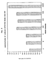
- the intensity of the signal produced by each of the three detectors was noticeably different, with 55306 giving the greatest signal and 55307, the poorest.
- the combination of any two or three detector antibodies generated a greater signal than that of any individual antibody having a mass corresponding to the total mass of any combination. This clearly demonstrates the synergistic effect of combining detector antibodies.
- Table 4 sets forth the specificity and concentration of each monoclonal antibody used in the three Examples 2 and 3 and the specificity of monoclonal antibodies used in Example 1.
- Table 4 HSV Antibodies Ab Specificity Concentration 55306 HSV 1 and 2 gD 1/1600 55307 HSV 1 and 2 gD 1/800 55308 HSV 1 and 2 gD 1/1600 55309 HSV 1 gC 1/800 55310 HSV 1 gB 1/1600 55311 HSV 2 gG 1/800 55312 HSV 2 gG 1/1600 55313 HSV 2 gB 1/1600 55314 HSV 2 gG 1/1600
- Polystyrene microtiter plate wells were coated with 0.1 ml polyclonal antibody reactive against both HSV-1 and HSV-2 (Dako Corp., Santa Barbara, CA) diluted in 0.1 M carbonate buffer, pH 9.6, and incubated overnight at 4°C.
- the wells were washed with PBS containing 0.05% Tween 20 (PBS-T), and blocked with 2% bovine serum albumin (BSA) in carbonate buffer for 1 hour at room temperature.
- the wells were washed with PBS-T and 0.1 ml of HSV-1 antigen diluted in 1% BSA-PBS-T was added and incubated for 2 hours at 37°C.
- the wells were washed with PBS-T and 0.1 ml of biotinylated detector antibody, or cocktail of antibodies, diluted in 50% normal rabbit serum-0.1% sodium caseinate-PBS was added, and incubated for 30 minutes at room temperature.
- the wells were washed with PBS-T and 0.1 ml of a streptavidin-horseradish peroxidase conjugate, diluted in 1% BSA-PBS-T, was added and incubated for 15 minutes at room temperature.
- the wells were washed with PBS-T and 0.1 ml of peroxidase substrate (0.05% o-phenylenediamine, 0.01% H2O2 in 0.1M citrate buffer, pH 5.0) was added. Following color development, the reaction was stopped with H2SO4, and the absorbance at 490 nm determined.
- Figure 2 shows the comparison of a single monoclonal detector compared to a cocktail of nine monoclonal antibodies. The cutoffs were determined by adding 0.05 to the blank readings. Use of a monoclonal antibody detector cocktail results in improved assay sensitivity, as well as an approximately five fold lower detection limit of viral antigen.
- a solid support polystyrene microtiter plate coated with antibody reactive against herpes simplex virus-1 and herpes simplex virus-2.
- b a quantity of multiple biotinylated monoclonal antibodies specific for herpes simplex virus-1 and herpes simplex virus-2.
- enzyme labeled streptavidin streptavidin-horseradish peroxidase.
- a substrate for determining enzyme activity o-phenylenediamine/H2O2.
- Duplicate specimens were collected with cotton swabs, one placed into medium for viral culture, and the second placed into Du Pont HerptranTM medium for ELISA. These specimens were collected from obstetric patients or patients suspected to have herpes whether or not these patients manifested visible signs of infection.
- the HerptranTM medium consists of phosphate buffered saline, NONIDET P 40 (NP 40) and bovine serum albumin. Specimens for culture were either cultured immediately or stored at -70°C. Specimens for ELISA testing were stored at 4°C until tested.
- CMV Cytomegalovirus
- Three anti-CMV monoclonal antibodies (designated as A, B, and C) were prepared, purified and biotinylated as described in Example 1. Optimal concentrations were determined using a dilution series of detector antibody, selecting the concentration giving maximum signal to background.
- Table 5 sets forth the specificity and concentration of each monoclonal antibody discussed in Example 4.
- Table 5 Ab Specificity Concentration A glycoprotein A 1/733 B glycoprotein A 1/1467 C glycoprotein A 1/2933
- Polystyrene microtiter plate wells were coated with a polyclonal rabbit anti-CMV antibody diluted in 0.1 M carbonate buffer pH 9.6 overnight at 4°C.
- the wells were washed with PBS-T and blocked with 2% BSA in carbonate buffer for 2 hours at 37°C.
- the wells were washed with PBS-T.
- CMV antigen diluted in Du Pont HerptranTM
- the wells were washed with PBS-T and detector antibody and combinations of detector antibodies diluted to their optimal concentrations in 1% BSA-PBS-T were added and incubated for 1 hour at room temperature.
- the wells were washed with PBS-T and a streptavidin-horseradish peroxidase conjugate, diluted in 1% BSA-PBS-T was added and incubated for 15 minutes at room temperature.
- the wells were washed with PBS-T an a peroxidase substrate (0.05% o-phenylenediamine, 0.01% H2O2, in citrate buffer pH 5.0) was added. After color development, the reaction was stopped with H2SO4, and the absorbance was determined at 490 nm.
- Figure 3 illustrates the improved performance resulting from the utilization of combinations of monoclonal antibodies as compared to single monoclonal antibody detectors for CMV.
Landscapes
- Health & Medical Sciences (AREA)
- Life Sciences & Earth Sciences (AREA)
- Immunology (AREA)
- Engineering & Computer Science (AREA)
- Chemical & Material Sciences (AREA)
- Urology & Nephrology (AREA)
- Hematology (AREA)
- Biomedical Technology (AREA)
- Molecular Biology (AREA)
- Medicinal Chemistry (AREA)
- Analytical Chemistry (AREA)
- Cell Biology (AREA)
- Pathology (AREA)
- Food Science & Technology (AREA)
- Biotechnology (AREA)
- Physics & Mathematics (AREA)
- Microbiology (AREA)
- Biochemistry (AREA)
- General Health & Medical Sciences (AREA)
- General Physics & Mathematics (AREA)
- Chemical Kinetics & Catalysis (AREA)
- Virology (AREA)
- Tropical Medicine & Parasitology (AREA)
- Preparation Of Compounds By Using Micro-Organisms (AREA)
- Peptides Or Proteins (AREA)
Applications Claiming Priority (2)
| Application Number | Priority Date | Filing Date | Title |
|---|---|---|---|
| US10364087A | 1987-10-02 | 1987-10-02 | |
| US103640 | 2002-03-21 |
Publications (2)
| Publication Number | Publication Date |
|---|---|
| EP0310132A2 true EP0310132A2 (de) | 1989-04-05 |
| EP0310132A3 EP0310132A3 (de) | 1990-10-10 |
Family
ID=22296237
Family Applications (1)
| Application Number | Title | Priority Date | Filing Date |
|---|---|---|---|
| EP19880116274 Withdrawn EP0310132A3 (de) | 1987-10-02 | 1988-10-01 | Immunassay mit Anwendung von IgG Einfangantikörper und ein Markierungssystem bestehend aus mehreren monoklonalen Antikörpern |
Country Status (5)
| Country | Link |
|---|---|
| EP (1) | EP0310132A3 (de) |
| JP (1) | JPH01163661A (de) |
| DK (1) | DK546988A (de) |
| FI (1) | FI884505L (de) |
| NO (1) | NO884368L (de) |
Cited By (17)
| Publication number | Priority date | Publication date | Assignee | Title |
|---|---|---|---|---|
| GB2270158A (en) * | 1992-08-03 | 1994-03-02 | Marconi Gec Ltd | Immunoassay using two detectable species |
| US5443956A (en) * | 1985-01-29 | 1995-08-22 | Oncogene Science, Inc. | Detection, quantitation and classification of RAS proteins in body fluids and tissues |
| WO1996003652A1 (de) * | 1994-07-25 | 1996-02-08 | Boehringer Mannheim Gmbh | Bestimmung von spezifischem immunglobulin unter verwendung multipler antigene |
| US5723304A (en) * | 1992-08-03 | 1998-03-03 | Gec-Marconi Limited | Immunological detection using two detectable labels |
| US6200764B1 (en) | 1985-01-29 | 2001-03-13 | Bayer Corporation | Detection, quantitation and classification of ras proteins in body fluids and tissues |
| WO2004011947A1 (en) * | 2002-07-29 | 2004-02-05 | I-Stat Corporation | Multiple hybrid immunoassay |
| US7501287B2 (en) * | 2001-04-23 | 2009-03-10 | Axis-Shield Asa | Transcobalamin II assay method |
| US8163499B2 (en) | 2008-02-25 | 2012-04-24 | Prometheus Laboratories Inc. | Drug selection for breast cancer therapy using antibody-based arrays |
| US8304195B2 (en) | 2001-10-12 | 2012-11-06 | Life Technologies Corporation | Antibody complexes and methods for immunolabeling |
| US8658388B2 (en) | 2006-09-21 | 2014-02-25 | Nestec S.A. | Antibody-based arrays for detecting multiple signal transducers in rate circulating cells |
| CN104730251A (zh) * | 2014-11-28 | 2015-06-24 | 威海纽普生物技术有限公司 | 肌钙蛋白i检测试剂盒及检测方法 |
| US9250243B2 (en) | 2006-09-21 | 2016-02-02 | Nestec S.A. | Drug selection for lung cancer therapy using antibody-based arrays |
| US9664683B2 (en) | 2011-09-02 | 2017-05-30 | Pierian Holdings, Inc. | Profiling of signal pathway proteins to determine therapeutic efficacy |
| US9719995B2 (en) | 2011-02-03 | 2017-08-01 | Pierian Holdings, Inc. | Drug selection for colorectal cancer therapy using receptor tyrosine kinase profiling |
| US9834596B2 (en) | 2012-07-03 | 2017-12-05 | Washington University | Antibodies to tau |
| US9957317B2 (en) | 2014-06-27 | 2018-05-01 | C2N Diagnostics, Llc | Humanized anti-tau antibodies |
| US10473640B2 (en) | 2006-09-21 | 2019-11-12 | Société des Produits Nestlé S.A. | Drug selection for gastric cancer therapy using antibody-based arrays |
Families Citing this family (2)
| Publication number | Priority date | Publication date | Assignee | Title |
|---|---|---|---|---|
| US8323903B2 (en) | 2001-10-12 | 2012-12-04 | Life Technologies Corporation | Antibody complexes and methods for immunolabeling |
| WO2008036802A2 (en) * | 2006-09-21 | 2008-03-27 | Prometheus Laboratories Inc. | Antibody-based arrays for detecting multiple signal transducers in rare circulating cells |
Family Cites Families (4)
| Publication number | Priority date | Publication date | Assignee | Title |
|---|---|---|---|---|
| NL185309C (nl) * | 1980-07-28 | 1990-03-01 | Akzo Nv | Methode voor het bepalen van antigenen met behulp van twee of meer monoklonale antilichamen alsmede immuunreagens. |
| SE8006424L (sv) * | 1980-09-12 | 1982-03-13 | Jolla Cancer Res Found | Sett att bestemma ett antigen i losning |
| US4514505A (en) * | 1982-05-21 | 1985-04-30 | The Trustees Of Columbia University In The City Of New York | Monoclonal antibody mixtures and use thereof for enhanced sensitivity immunoassays |
| US4595661A (en) * | 1983-11-18 | 1986-06-17 | Beckman Instruments, Inc. | Immunoassays and kits for use therein which include low affinity antibodies for reducing the hook effect |
-
1988
- 1988-09-30 NO NO88884368A patent/NO884368L/no unknown
- 1988-09-30 JP JP63244725A patent/JPH01163661A/ja active Pending
- 1988-09-30 DK DK546988A patent/DK546988A/da not_active Application Discontinuation
- 1988-09-30 FI FI884505A patent/FI884505L/fi not_active Application Discontinuation
- 1988-10-01 EP EP19880116274 patent/EP0310132A3/de not_active Withdrawn
Cited By (31)
| Publication number | Priority date | Publication date | Assignee | Title |
|---|---|---|---|---|
| US5443956A (en) * | 1985-01-29 | 1995-08-22 | Oncogene Science, Inc. | Detection, quantitation and classification of RAS proteins in body fluids and tissues |
| US6200764B1 (en) | 1985-01-29 | 2001-03-13 | Bayer Corporation | Detection, quantitation and classification of ras proteins in body fluids and tissues |
| GB2270158A (en) * | 1992-08-03 | 1994-03-02 | Marconi Gec Ltd | Immunoassay using two detectable species |
| GB2270158B (en) * | 1992-08-03 | 1997-03-19 | Marconi Gec Ltd | Detection |
| US5723304A (en) * | 1992-08-03 | 1998-03-03 | Gec-Marconi Limited | Immunological detection using two detectable labels |
| WO1996003652A1 (de) * | 1994-07-25 | 1996-02-08 | Boehringer Mannheim Gmbh | Bestimmung von spezifischem immunglobulin unter verwendung multipler antigene |
| US6613530B1 (en) | 1994-07-25 | 2003-09-02 | Roche Diagnostics Gmbh | Determination of a specific immunoglobulin using multiple antigens |
| US9140710B2 (en) | 2001-04-23 | 2015-09-22 | Axis-Shield Asa | Transcobalamin II assay method |
| US7501287B2 (en) * | 2001-04-23 | 2009-03-10 | Axis-Shield Asa | Transcobalamin II assay method |
| US8535894B2 (en) | 2001-10-12 | 2013-09-17 | Life Technologies Corporation | Antibody complexes and methods for immunolabeling |
| US8304195B2 (en) | 2001-10-12 | 2012-11-06 | Life Technologies Corporation | Antibody complexes and methods for immunolabeling |
| US10641767B2 (en) | 2002-07-29 | 2020-05-05 | Abbott Point Of Care Inc. | Multiple hybrid immunoassay |
| US9995744B2 (en) | 2002-07-29 | 2018-06-12 | Abbott Point Of Care Inc. | Multiple hybrid immunoassay |
| WO2004011947A1 (en) * | 2002-07-29 | 2004-02-05 | I-Stat Corporation | Multiple hybrid immunoassay |
| US9267939B2 (en) | 2002-07-29 | 2016-02-23 | Abbott Point Of Care Inc. | Multiple hybrid immunoassay |
| US10473640B2 (en) | 2006-09-21 | 2019-11-12 | Société des Produits Nestlé S.A. | Drug selection for gastric cancer therapy using antibody-based arrays |
| US8658388B2 (en) | 2006-09-21 | 2014-02-25 | Nestec S.A. | Antibody-based arrays for detecting multiple signal transducers in rate circulating cells |
| US9250243B2 (en) | 2006-09-21 | 2016-02-02 | Nestec S.A. | Drug selection for lung cancer therapy using antibody-based arrays |
| US10527622B2 (en) | 2006-09-21 | 2020-01-07 | Société des Produits Nestlé S.A. | Antibody-based arrays for detecting multiple signal transducers in rare circulating cells |
| US9285369B2 (en) | 2006-09-21 | 2016-03-15 | Nestec S.A. | Antibody-based arrays for detecting multiple signal transducers in rare circulating cells |
| US9575066B2 (en) | 2006-09-21 | 2017-02-21 | Nestec S.A. | Antibody-based arrays for detecting multiple signal transducers in rare circulating cells |
| US8609349B2 (en) | 2008-02-25 | 2013-12-17 | Nestec S.A. | Drug selection for breast cancer therapy using antibody-based arrays |
| US9274116B2 (en) | 2008-02-25 | 2016-03-01 | Nestec S.A. | Drug selection for breast cancer therapy using antibody-based arrays |
| US10436786B2 (en) | 2008-02-25 | 2019-10-08 | Société des Produits Nestlé S.A. | Methods for detecting truncated receptors using antibody-based arrays |
| US8163499B2 (en) | 2008-02-25 | 2012-04-24 | Prometheus Laboratories Inc. | Drug selection for breast cancer therapy using antibody-based arrays |
| US9719995B2 (en) | 2011-02-03 | 2017-08-01 | Pierian Holdings, Inc. | Drug selection for colorectal cancer therapy using receptor tyrosine kinase profiling |
| US10401364B2 (en) | 2011-02-03 | 2019-09-03 | Soiété Des Produits Nestlé S.A. | Drug selection for colorectal cancer therapy using receptor tyrosine kinase profiling |
| US9664683B2 (en) | 2011-09-02 | 2017-05-30 | Pierian Holdings, Inc. | Profiling of signal pathway proteins to determine therapeutic efficacy |
| US9834596B2 (en) | 2012-07-03 | 2017-12-05 | Washington University | Antibodies to tau |
| US9957317B2 (en) | 2014-06-27 | 2018-05-01 | C2N Diagnostics, Llc | Humanized anti-tau antibodies |
| CN104730251A (zh) * | 2014-11-28 | 2015-06-24 | 威海纽普生物技术有限公司 | 肌钙蛋白i检测试剂盒及检测方法 |
Also Published As
| Publication number | Publication date |
|---|---|
| DK546988D0 (da) | 1988-09-30 |
| NO884368D0 (no) | 1988-09-30 |
| JPH01163661A (ja) | 1989-06-27 |
| FI884505A0 (fi) | 1988-09-30 |
| FI884505A7 (fi) | 1989-04-03 |
| NO884368L (no) | 1989-04-03 |
| EP0310132A3 (de) | 1990-10-10 |
| DK546988A (da) | 1989-04-03 |
| FI884505L (fi) | 1989-04-03 |
Similar Documents
| Publication | Publication Date | Title |
|---|---|---|
| Yolken | Enzyme-linked immunosorbent assay (ELISA): a practical tool for rapid diagnosis of viruses and other infectious agents | |
| US4016043A (en) | Enzymatic immunological method for the determination of antigens and antibodies | |
| US6489129B1 (en) | Antigen-specific IgM detection | |
| US4228237A (en) | Methods for the detection and determination of ligands | |
| US4347311A (en) | Enzyme immunoassay for determining antigen specific antibodies and test kit for carrying out this assay | |
| EP0345462B1 (de) | Immuntest für HIV-1-Antigene unter Benutzung von F(AB')2-Fragmenten als Sonde | |
| JPH01163661A (ja) | IgG捕捉抗体および多重単クローン性抗体検出系を用いたイムノアツセイ | |
| JP2636331B2 (ja) | 抗原特異的な抗体の一段階測定法およびそれに適する試薬 | |
| JP2599058B2 (ja) | クラス特異的な免疫グロブリン抗体の免疫分析法 | |
| EP0315364A2 (de) | Gleichzeitiger Immunassay zur Bestimmung von Antigenen und Antikörpern | |
| IE62974B1 (en) | Method for determination of a polyvalent substance using an immunoaggregate | |
| EP0355099B2 (de) | Immunoglobulin-bestimmungsverfahren | |
| EP0345277B1 (de) | Nachweis von analyt in partikel enthaltenden proben | |
| US5124245A (en) | Wash composition, test kit and their use to determine a herpes simplex viral antigen | |
| CA2008100A1 (en) | Method for the determination of immunologically detectable substance | |
| USRE32696E (en) | Enzymatic immunological method for determination of antigens and antibodies | |
| EP0366673B1 (de) | Immunassay-verfahren | |
| EP0303980A2 (de) | Träger beschichtet mit Antigen oder Antikörper | |
| EP0389301A2 (de) | Reagenzkomplex für Immunoessay | |
| US5277589A (en) | Process for the determination of antibodies | |
| US5210039A (en) | Wash composition, test kit and their use to determine a herpes simplex viral antigen | |
| EP0529057B1 (de) | Denaturierte transportproteine zur verbesserung von enzymgebundenen immunoassays (elisa) | |
| EP0262217A4 (de) | Festphase-analyse-verfahren. | |
| WO1989007271A1 (en) | Immunoassays using monoclonal antibodies directed against natural binding proteins | |
| JPH0792455B2 (ja) | 免疫学的試験用水性溶媒 |
Legal Events
| Date | Code | Title | Description |
|---|---|---|---|
| PUAI | Public reference made under article 153(3) epc to a published international application that has entered the european phase |
Free format text: ORIGINAL CODE: 0009012 |
|
| AK | Designated contracting states |
Kind code of ref document: A2 Designated state(s): BE CH DE FR GB IT LI LU NL SE |
|
| PUAL | Search report despatched |
Free format text: ORIGINAL CODE: 0009013 |
|
| AK | Designated contracting states |
Kind code of ref document: A3 Designated state(s): BE CH DE FR GB IT LI LU NL SE |
|
| 17P | Request for examination filed |
Effective date: 19901206 |
|
| 17Q | First examination report despatched |
Effective date: 19920709 |
|
| STAA | Information on the status of an ep patent application or granted ep patent |
Free format text: STATUS: THE APPLICATION HAS BEEN WITHDRAWN |
|
| 18W | Application withdrawn |
Withdrawal date: 19930526 |