EP0312942A2 - Proteine mit Prourokinase-Aktivität, ihre Herstellung und Verwendung - Google Patents
Proteine mit Prourokinase-Aktivität, ihre Herstellung und Verwendung Download PDFInfo
- Publication number
- EP0312942A2 EP0312942A2 EP88117188A EP88117188A EP0312942A2 EP 0312942 A2 EP0312942 A2 EP 0312942A2 EP 88117188 A EP88117188 A EP 88117188A EP 88117188 A EP88117188 A EP 88117188A EP 0312942 A2 EP0312942 A2 EP 0312942A2
- Authority
- EP
- European Patent Office
- Prior art keywords
- prourokinase
- polypeptides
- puk
- gene
- expression
- Prior art date
- Legal status (The legal status is an assumption and is not a legal conclusion. Google has not performed a legal analysis and makes no representation as to the accuracy of the status listed.)
- Withdrawn
Links
Images



Classifications
-
- C—CHEMISTRY; METALLURGY
- C12—BIOCHEMISTRY; BEER; SPIRITS; WINE; VINEGAR; MICROBIOLOGY; ENZYMOLOGY; MUTATION OR GENETIC ENGINEERING
- C12N—MICROORGANISMS OR ENZYMES; COMPOSITIONS THEREOF; PROPAGATING, PRESERVING, OR MAINTAINING MICROORGANISMS; MUTATION OR GENETIC ENGINEERING; CULTURE MEDIA
- C12N9/00—Enzymes; Proenzymes; Compositions thereof; Processes for preparing, activating, inhibiting, separating or purifying enzymes
- C12N9/14—Hydrolases (3)
- C12N9/48—Hydrolases (3) acting on peptide bonds (3.4)
- C12N9/50—Proteinases, e.g. Endopeptidases (3.4.21-3.4.25)
- C12N9/64—Proteinases, e.g. Endopeptidases (3.4.21-3.4.25) derived from animal tissue
- C12N9/6421—Proteinases, e.g. Endopeptidases (3.4.21-3.4.25) derived from animal tissue from mammals
- C12N9/6424—Serine endopeptidases (3.4.21)
- C12N9/6456—Plasminogen activators
- C12N9/6462—Plasminogen activators u-Plasminogen activator (3.4.21.73), i.e. urokinase
-
- A—HUMAN NECESSITIES
- A61—MEDICAL OR VETERINARY SCIENCE; HYGIENE
- A61P—SPECIFIC THERAPEUTIC ACTIVITY OF CHEMICAL COMPOUNDS OR MEDICINAL PREPARATIONS
- A61P7/00—Drugs for disorders of the blood or the extracellular fluid
- A61P7/02—Antithrombotic agents; Anticoagulants; Platelet aggregation inhibitors
-
- C—CHEMISTRY; METALLURGY
- C12—BIOCHEMISTRY; BEER; SPIRITS; WINE; VINEGAR; MICROBIOLOGY; ENZYMOLOGY; MUTATION OR GENETIC ENGINEERING
- C12Y—ENZYMES
- C12Y304/00—Hydrolases acting on peptide bonds, i.e. peptidases (3.4)
- C12Y304/21—Serine endopeptidases (3.4.21)
- C12Y304/21073—Serine endopeptidases (3.4.21) u-Plasminogen activator (3.4.21.73), i.e. urokinase
Definitions
- the present invention relates to new polypeptides with prourokinase activity, processes for their preparation and their use in combating diseases.
- the plasminogen activator prourokinase also called “single chain urokinase plasminogenactivator (SCUPA)” or “kidney plasminogenactivator” (KPA)
- PKA plasminogen activator prourokinase
- SCUPA single chain urokinase plasminogenactivator
- KPA kidney plasminogenactivator
- LUK low molecular weight urokinase
- Both the one and the two chain forms have fibrinolytic properties.
- the two-chain molecules have a systemic effect. They degrade both fibrin and fibrinogen throughout the body, and life-threatening bleeding often occurs during application.
- the single-chain forms alone are clot-specific and show little systemic lysis.
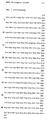
- polypeptides of the formula I can carry a glycosyl residue on the amino acid 302 (Asn, see Fig. 1).
- the invention further relates to DNA sequences which code for the polypeptides of the formula I and to vectors which contain these DNA sequences.
- the new polypeptides can be produced using known genetic engineering techniques.
- the starting point for the constructions described here is a cDNA clone, hereinafter called pU20, which contains the coding sequence of prourokinase and 5′- and 3′- untranslated regions.
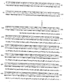
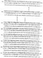
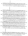
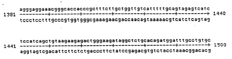
- the partial sequence of the cDNA clone, which encompasses the coding region and from which the protein sequence (FIG. 1) is derived, is shown in FIG. 2.
- the mature protein begins after a so-called "leader" sequence at Ser1 and terminates after Leu411.
- pU20 was obtained from the human, prourokinase-producing tumor cell line Detroit 562 (ATCC No. CCL 138): m-RNA was isolated from this cell line and transcribed into double-stranded cDNA.
- Parts of the DNA sequence of pU20 are easily accessible with the help of restriction endonucleases. These fragments, optionally in conjunction with chemically synthesized oligonucleotides, adapters or gene fragments, can be used to clone the DNA sequences which code for the new polypeptides.
- the installation of the gene fragments or synthetic DNA sequences in cloning vectors for example the commercially available plasmids pBR 322, puc 18 and puc 19, is carried out in a known manner.
- the genes or gene fragments can also be provided with suitable chemically synthesized control regions or control regions isolated from bacteria or phages, which enable expression of the proteins.
- hybrid plasmids obtained in this way into suitable host organisms are also known and has been described in detail.
- the hybrid plasmids can also be provided with corresponding signal sequences which allow the polypeptides to be secreted into the periplasm of E. coli.
- eukaryotic control regions must be used here.
- vectors which e.g. puts the foreign gene to be expressed under the control of the viral SV 40 promoter.
- Such plasmids have no eukaryotic origin of replication. After the transformation into permissive mammalian cells, clones are isolated which carry copies of these plasmids integrated in the genome.
- the genes to be expressed can be cloned under the control of a eukaryotic promoter onto vectors based on the bovine papilloma virus.
- Such expression plasmids remain episomally in a copy number of 20 to 100 in the permissive mouse fibroblast cell line C127.
- the prourokinase When expressed in eukaryotic cells, the prourokinase also receives a glycoside side chain at the amino acid 302 (Asn). Bacteria are unable to synthesize glycoside side chains. Most eukaryotic proteins expressed in bacteria, as well as prourokinase and the polypeptides derived from it according to the invention, occur in the cell as denatured inclusion bodies and have to be refolded by protein chemistry. Furthermore, bacteria are often unable to split off the initiator amino acid methionine from the finished protein. The cleavage of the terminal methionine when expressed in bacteria and in some cases the prevention of the formation of inclusion bodies can be achieved by using secretion systems.
- the N-terminus often still has an amino acid that is important for the cleavage site, e.g. Alanine.
- an amino acid that is important for the cleavage site e.g. Alanine.
- expression in bacteria poses many problems, like the prourokinase, these are the expression system of choice if the resulting crude protein can be renatured into the active enzyme. Therefore, expression in bacteria is preferred here.
- the new active ingredients can be used for thrombolysis in arterial and venous occlusive diseases and show improved properties compared to human prourokinase.
- the invention therefore also relates to medicaments which contain at least one of the new polypeptides, optionally in a pharmaceutically acceptable carrier or binder.
- the new proteins can also be used together with other fibrinolytics, such as urokinase, TPA, streptokinase, derivatives derived from these three parties, or sodium pentosan polysulfate.
- TPA and derived proteins with similar activity are particularly suitable as combination components.
- the prourokinase-producing human tumor cell line Detroit 562 (ATCC No. CCL 138) was in "Eagle's minimal essential" medium with 3% NaHCO3, 10% fetal calf serum, 1% amino acids, 2.4% Hepes pH 7.5 at 37 ° C and 5% CO2 atmosphere pulled to confluence.
- the medium was poured off, the cells washed with physiological saline and disrupted in lysis buffer (6M guanidinium isothiocyanate, 5 mM sodium citrate (pH 7), 0.1 M 2-mercaptoethanol, 0.5% sarkosyl).
- the RNA was sedimented through a 5.7 M CsCl cushion overnight at 100,000 g.
- the fraction containing polyA + RNA was separated by two affinity chromatography on oligo (dT) cellulose.
- the polyA + RNA was rewritten into single-stranded cDNA.
- the second strand was synthesized using E. coli DNA polymerase I. About 10-20 dG residues were synthesized on the double-stranded cDNA with the aid of the enzyme terminal transferase at each 5′-end.
- the same procedure was followed with the commercially available plasmid puC 18, which was linearized with the restriction endonuclease SphI and about 10 to 20 dC residues were synthesized at its 5 'ends as described above. Both DNAs were fused together and the hybrid was transformed into competent cells of the E. coli strain HB 101. The clones were plated on LB plates with 100 ⁇ g / ml ampicillin as a selection marker and the colonies were transferred to nitrocellulose.
- the bacteria on the filters were replicated, lysed and the denatured DNA firmly bound to the filter.
- oligonucleotide probes with homology to the published DNA sequence of the human prourokinase were produced. These probes had the following sequences: 5 ′ TCAGCAGCACATAGCAT 3 ′ 5 ′ AACCAGGGCTGGTTCTC 3 ′ 5 ′ ACCATGCACTCTTGGAC 3 ′ 5 ′ TGCTGGTCACAGGTCAT 3 ′
- Fig. 2 shows the sequence of the coding region and neighboring areas.
- the starting point was the plasmid pU20 (Example 1).
- the fragment of the prourokinase gene from nucleotide 162 to 1329 was cut out of this by a limited TaqI / BamHI digestion and isolated by gel electrophoresis (fragment 1).
- the plasmid puc 18 was linearized with EcoRI and SalI, which resulted in fragment 2.
- Fragments 1 and 2 were ligated with the oligonucleotide adapters 3/4 and 5/6, which restore the respective 5'-end (3/4) and 3'-end (5/6) of the prourokinase gene.
- oligonucleotide 3/4 the EcoRI site at the 5'-end was deleted.
- the hybrid plasmid pPUK was obtained according to FIG. 3, which made the prourokinase gene available as an easily clonable ClaI / SalI "cassette" for conversion into different expression vectors.
- the hybrid plasmid pPUK was opened with ClaI and the protruding 5 'ends were filled in with Klenow fragment of DNA polymerase I and dCTP and dGTP.
- This strain contains the episome F′i Q with the lac repressor, which represses the prourokinase gene under normal growth conditions by adjusting the isopropyl ⁇ -D-thiogalactoside (IPTG) content to 0.5 mM in the In the logarithmic growth phase, the expression of the prourokinase gene was induced, and the prourokinase was denatured intracellularly as so-called "inclusion bodies” e Prourokinase can be renatured and purified using protein chemistry.
- IPTG isopropyl ⁇ -D-thiogalactoside
- the plasmid pPUK (Fig. 3, Example 2) was completely cleaved with BalI and then partially with EcoRI. The fragment containing the vector was purified by gel electrophoresis and ligated with the synthetic oligonucleotides 84/85, 86/87 or 88/89:
- polypeptides claimed according to the invention were produced by genetic engineering in E. coli as described in Example 2.
- polypeptides described above can be purified and renatured after their expression in E. coli using a standard method:
- the cell pellet (100 g wet mixture) was taken up in 500 ml (50 mM phosphate buffer pH 8.0, 10 mM EDTA, 1 mM DTT and 0.005% Tween 80) and resuspended.
- the cells disrupted by ultrasound were centrifuged off and the pellet with the inclusion bodies was washed with 50 mM phosphate buffer, pH 8.0, 1 M NaCl, 1 mM DTT, 0.005% Tween 80.
- the washed inclusion bodies were centrifuged off (10 min, 10,000 g) and dissolved in 6 M guanidinium hydrochloride. This was done by stirring for 30 min at room temperature in 200 ml of 50 mM phosphate buffer pH 8.0, 5 mM DTT, 1 mM EDTA, 6 M guanidinium hydrochloride and 0.005% Tween 80. Undissolved protein was centrifuged off.
- the PUK mutein was renatured by diluting this solution to a protein concentration between 0.2 and 0.3 mg / ml in 50 mM phosphate buffer pH 9.0, 1.5 M guanidinium hydrochloride, 0.5 M arginine, 1 mM GSH, 0.1mM GSSG, 1mM EDTA, 0.005% Tween 80.
- Protein purification was done by ammonium sulfate precipitation, affinity chromatography and ion exchange chromatography. For this purpose, ammonium sulfate (between 40 to 70%) was added to the renaturation solution, the pellet was taken up in 50 mM phosphate, 1 M guanidinium hydrochloride, 0.1 M NaCl, 0.005% Tween 80 and applied to a Zn chelate (Pharmacia) chromatography column and eluted with 50 mM EDTA in the above buffer at 4 ° C.
- the eluted protein was dialyzed at 4 ° C against 10 mM phosphate, 10 mM NaCl, 0.005% Tween 80 and applied to an S-Sepharose (Pharmacia).
- the PUK mutein was eluted from the column with 50 mM phosphate, 400 mM NaCl, 0.005% Tween 80.
- the specific activities of the purified proteins in the direct amidolytic test with the substrate S-2444 are in the range from 0.5 to 1.5x10⁵ U / mg.
- Thrombin cleavage of the prourokinase behind Arg 156 results in a 2 chain molecule that can no longer be activated and is no longer available for clot lysis.
- Polypeptides with amino acid exchange in pos. 156 can no longer be inactivated by thrombin or only to a significantly lesser extent.
- the thrombin inactivation test was carried out as follows: Prourokinase (or its muteins) is diluted to 6000 U / ml (50 mM phosphate, pH 6.7, 300 mM NaCl, 0.005% Tween 80) and with thrombin (5 U / ml) Incubated at 37 ° C. Aliquots are removed at intervals of 15 min and measured in the amidolytic test.
- the PUK mutein (Gln 156 or Leu 156) was used in the thrombin inhibition test.
- Fig. 7 shows that this mutein still showed 100% of the initial activity after 30 min incubation with 5 U thrombin (o-o).
- the amino acid exchange at position 156 of the prourokinase (Arg ⁇ Gln) or Leu therefore means that thrombin on the mutein is no longer proteolytically active and no inactive two-chain material is formed.
Landscapes
- Health & Medical Sciences (AREA)
- Chemical & Material Sciences (AREA)
- Engineering & Computer Science (AREA)
- Life Sciences & Earth Sciences (AREA)
- Organic Chemistry (AREA)
- Bioinformatics & Cheminformatics (AREA)
- Wood Science & Technology (AREA)
- Zoology (AREA)
- General Health & Medical Sciences (AREA)
- Genetics & Genomics (AREA)
- Medicinal Chemistry (AREA)
- Biochemistry (AREA)
- General Engineering & Computer Science (AREA)
- Biomedical Technology (AREA)
- General Chemical & Material Sciences (AREA)
- Microbiology (AREA)
- Veterinary Medicine (AREA)
- Animal Behavior & Ethology (AREA)
- Molecular Biology (AREA)
- Biotechnology (AREA)
- Pharmacology & Pharmacy (AREA)
- Public Health (AREA)
- Nuclear Medicine, Radiotherapy & Molecular Imaging (AREA)
- Chemical Kinetics & Catalysis (AREA)
- Hematology (AREA)
- Diabetes (AREA)
- Medicines That Contain Protein Lipid Enzymes And Other Medicines (AREA)
- Enzymes And Modification Thereof (AREA)
- Peptides Or Proteins (AREA)
- Preparation Of Compounds By Using Micro-Organisms (AREA)
Abstract
Description
- Die vorliegende Erfindung betrifft neue Polypeptide mit Prourokinase-Aktivität, Verfahren zu deren Herstellung und deren Verwendung bei der Bekämpfung von Krankheiten.
- Der Plasminogenaktivator Prourokinase (PUK), auch "single chain urokinase plasminogenactivator (SCUPA)" oder "kidney plasminogenactivator" (KPA) genannt, ist ein Einketten-Polypeptid aus 411 Aminosäuren und einem Molekulargewicht von etwa 54 kD. Abb. 1 zeigt die Aminosäuresequenz der reifen Prourokinase (Z. Physiol. Chem. 363, 133, 1043, 1155 (1982)). Aus der Prourokinse entsteht durch limitierte Proteasespaltung der Lys¹⁵⁷-Pheó¹⁵⁸-Bindung ein Zweikettenmolekül, die hochmolekulare Urokinase (HUK). Diese besitzt dasselbe Molekulargewicht wie die Prourokinase. Durch eine weitere proteolytische Spaltung zwischen Lys¹³⁵ und Lys¹³⁶ entsteht die niedermolekulare Urokinase (LUK) mit einem Molekulargewicht von etwa 33 kD. Die LUK ist wie die HUK ein Zweikettenmolekül. Alle drei Formen sind im humanen Urin oder im humanen Serum als natürliche Formen nachgewiesen worden (J. Biol. Chem. 257, 3276 (1982) und 261, 1267 (1986)).
- Sowohl die Ein-, als auch die Zweikettenformen besitzen fibrinolytische Eigenschaften. Die Zweikettenmoleküle wirken allerdings systemisch. Sie degradieren sowohl Fibrin als auch Fibrinogen überall im Körper, und es kommt bei der Applikation häufig zu lebensbedrohenden Blutungen. Allein die Einkettenformen sind gerinnselspezifisch und zeigen nur eine geringe systemische Lyse.
- Es wurde nun gefunden, daß Polypeptide der Formel I
R-PUK1‾155-X-PUK157‾411
worin R ein Wasserstoffatom, Ala oder Met,
PUK1‾155 und PUK157‾411 für die Aminosäuren 1 bis 155 und 157 bis 411 der Prourokinase und
X Ala, Asn, Asp, Cys, Gln, Glu, Gly, His, Ile, Leu, Lys, Met, Phe, Pro, Ser, Thr, Trp, Try oder Val darstellen, verbesserte Eigenschaften besitzen und enzymatisch stabiler sind. - Die Polypeptide der Formel I können an der Aminosäure 302 (Asn, vgl. Abb. 1) einen Glykosylrest tragen.
- Die Aminosäurereste 1 bis 155 und 157 bis 411 der Prourokinase sind aus Abb. 1 ersichtlich.
- Die Erfindung betrifft weiter DNA-Sequenzen, die für die Polypeptide der Formel I kodieren, sowie Vektoren, die diese DNA-Sequenzen enthalten.
- Die neuen Polypeptide lassen sich gentechnologisch nach bekannten Verfahren herstellen. Ausgangspunkt für die hier beschriebenen Konstruktionen ist ein cDNA-Klon, im folgenden pU20 genannt, der die kodierende Sequenz von Prourokinase und noch 5′- und 3′- nicht translatierte Bereiche beinhaltet. Die Teilsequenz des cDNA-Klons, der die kodierende Region umfaßt und aus der sich die Proteinsequenz (Abb. 1) ableitet, ist in Abb. 2 dargestellt. Das reife Protein beginnt nach einer sogenannten "leader"-Sequenz bei Ser¹ und terminiert nach Leu⁴¹¹.
- pU20 wurde aus der humanen, Prourokinase produzierenden Tumorzellinie Detroit 562 (ATCC No. CCL 138) erhalten: Aus dieser Zellinie wurde m-RNA isoliert und in doppelsträngige cDNA umgeschrieben.
- Nach Einsetzen dieser cDNA in den kommerziell erhältlichen Klonierungsvektor puc 18 wurde eine cDNA-Bibliothek angelegt. Die dabei verwendeten Methoden sind beispielsweise in Maniatis et al, "Molecular Cloning", CSH-Press nachzulesen. Auch das "Screenen" solcher Genbänke mit radioaktiv markierten Oligonukleotidsonden ist inzwischen eine vielfach verwendete und beschriebene Methode.
- Teile der DNA-Sequenz von pU20 sind mit Hilfe von Restriktionsendonukleasen leicht zugänglich. Diese Fragmente, gegebenenfalls in Verbindung mit chemisch synthetisierten Oligonukleotiden, Adaptoren oder Genfragmenten können benutzt werden, um die DNA-Sequenzen, die für die neuen Polypeptide kodieren, zu klonieren. Der Einbau der Genfragmente bzw. synthetischen DNA-Sequenzen in Klonierungsvektoren, beispielsweise die handelsüblichen Plasmide pBR 322, puc 18 und puc 19, erfolgt in bekannter Weise. Auch können die Gene oder Genfragmente mit geeigneten chemisch synthetisierten oder aus Bakterien bzw. Phagen isolierten Kontrollregionen versehen werden, die die Expression der Proteine ermöglichen. Die Transformation der so erhaltenen Hybridplasmide in geeignete Wirtsorganismen ist ebenfalls bekannt und eingehend beschrieben. Auch können die Hybridplasmide mit entsprechenden Signalsequenzen versehen werden, die die Sekretion der Polypeptide in das Periplasma von E.coli erlauben.
- Eine Expression in Mammalia-Zellkultur bzw. Hefe oder Insektenzellen ist in analoger Weise möglich: Hierbei müssen allerdings eukaryontische Kontrollregionen verwendet werden. Bei der Expression in Mammaliazellen kann man Vektoren verwenden, die z.B. das zu exprimierende Fremdgen unter die Kontrolle des viralen SV 40 Promotors setzt. Solche Plasmide besitzen keinen eukaryontischen Replikationsursprung. Man isoliert nach der Transformation in permissive Mammaliazellen Klone, die Kopien dieser Plasmide integriert im Genom tragen. In gleicher Weise kann man die zu exprimierenden Gene unter der Kontrolle eines eukaryontischen Promotors auf Vektoren auf der Basis des Rinderpapillom-Virus klonieren. Solche Expressionsplasmide bleiben episomal in einer Kopienzahl von 20 bis 100 in der permissiven Mausfibroblastenzellinie C127.
- Diese eukaryontischen Expressionssysteme besitzen den Vorteil, daß sie in der Lage sind, ihre Produkte effektiv und meist in nativer Form zu sekretieren. Ferner besitzen sie die Fähigkeit, ihre Produkte posttranslationell zu modifizieren.
- So erhält die Prourokinase bei einer Expression in Eukaryontenzellen noch eine Glykosidseitenkette an der Aminosäure 302 (Asn). Bakterien sind nicht in der Lage, Glycosidseitenketten zu synthetisieren. Die meisten in Bakterien exprimierten eukaryontischen Proteine, wie auch die Prourokinase und die von ihr erfindungsgemäß abgeleiteten Polypeptide, fallen in der Zelle als denaturierte Einschlußkörper an und müssen proteinchemisch zurückgefaltet werden. Ferner sind Bakterien oft nicht in der Lage, die Initiatoraminosäure Methionin vom fertigen Protein abzuspalten. Die Abspaltung des endständigen Methionins bei der Expression in Bakterien und in manchen Fällen die Verhinderung der Bildung von Einschlußkörpern kann durch die Verwendung von Sekretionssystemen erreicht werden. Bei der Verwendung solcher Systeme besitzt dann allerdings häufig der N-Terminus noch eine für die Spaltstelle wichtige Aminosäure, wie z.B. Alanin. Obwohl die Expression in Bakterien viele Probleme aufwirft, sind diese, wie bei der Prourokinase das Expressionssystem der Wahl, wenn sich das anfallende Rohprotein zum aktiven Enzym renaturieren läßt. Daher wird hier die Expression in Bakterien bevorzugt.
- Aufgrund der Degeneration des genetischen Codes ist es auch möglich, andere DNA-Sequenzen, z.B. chemisch synthetisierte Prourokinasegene oder Teile davon mit anderen Tripletts als die hier beschriebenen für die Expression der neuen Polypeptide zu benutzen.
- Die neuen Wirkstoffe können zur Thrombolyse bei arteriellen und venösen Verschlußkrankheiten eingesetzt werden und zeigen verbesserte Eigenschaften gegenüber humaner Prourokinase.
- Gegenstand der Erfindung sind daher auch Arzneimittel, die mindestens eines der neuen Polypeptide enthalten, gegebenenfalls in einem pharmazeutisch verträglichen Träger oder Bindemittel.
- Die neuen Proteine können auch zusammen mit anderen Fibrinolytika, wie Urokinase, TPA, Streptokinase, von diesen drei Parteien abgeleiteten Derivaten oder Natriumpentosanpolysulfat, angewendet werden. Besonders eignen sich TPA und davon abgeleitete Proteine mit ähnlicher Aktivität als Kombinationsbestandteile.
- Weitere Ausgestaltungen der Erfindung sind in den folgenden Beispielen näher beschrieben.
- Die Prourokinase produzierende humane Tumorzellinie Detroit 562 (ATCC Nr. CCL 138) wurde in "Eagle's minimal essential"-Medium mit 3 % NaHCO₃, 10 % fötalem Kälberserum, 1 % Aminosäuren, 2,4 % Hepes pH 7,5 bei 37°C und 5 % CO₂-Atmosphäre bis zur Konfluenz gezogen. Das Medium wurde abgegossen, die Zellen mit physiologischer Kochsalzlösung gewaschen und in Lysepuffer (6M Guanidiniumisothiocyanat, 5 mM Natriumcitrat (pH 7), 0,1 M 2-Mercaptoethanol, 0,5 % Sarkosyl) aufgeschlossen. Die RNA wurde durch ein 5,7 M CsCl-Kissen über Nacht bei 100.000 g sedimentiert. Die polyA+-RNA enthaltende Fraktion wurde durch zweimalige Affinitätschromatographie an oligo(dT)-Cellulose abgetrennt.
- Mit Hilfe des Enzyms AMV-Reverse Transkriptase und oligo(dT)12-18 als Starter wurde die polyA+-RNA in einsträngige cDNA umgeschrieben. Die Synthese des zweiten Strangs erfolgte mit E.coli-DNA-Polymerase I. An die doppelsträngige cDNA wurden mit Hilfe des Enzyms Terminale Transferase an jedem 5′-Ende ca. 10-20 dG-Reste ansynthetisiert. Ebenso wurde mit dem kommerziell erhältlichen Plasmid puC 18 verfahren, das mit der Restriktionsendonuklease SphI linearisiert wurde und an dessen 5′-Enden wie oben beschrieben ca. 10 bis 20 dC-Reste ansynthetisiert wurden. Beide DNAs wurden miteinander verschmolzen und das Hybrid in kompetente Zellen des E.coli-Stammes HB 101 transformiert. Die Klone wurden auf LB-Platten mit 100 µg/ml Ampicillin als Selektionsmarker ausplattiert und die Kolonien auf Nitrocellulose übertragen.
- Die Bakterien auf den Filtern wurden repliziert, lysiert und die denaturierte DNA fest an das Filter gebunden.
- Mit Hilfe eines DNA-Synthesizers (Applied Biosystems, Typ 380A) wurden vier 17er Oligonukleotidsonden mit Homologie zur publizierten DNA-Sequenz der humanen Prourokinase hergestellt. Diese Sonden hatten folgende Sequenzen:
5′ TCAGCAGCACATAGCAT 3′
5′ AACCAGGGCTGGTTCTC 3′
5′ ACCATGCACTCTTGGAC 3′
5′ TGCTGGTCACAGGTCAT 3′
Die Oligonukleotidsonden wurden am 5′-Ende mit γ-32p-ATP markiert und die Nitrocellulosefilter in 6xSET Puffer (1xSET = 0,15 M NaCl, 0,015 M Tris-HCl pH 7,4, 0,001 M EDTA) 0,1 % SDS und 10 % Dextransulfat über Nacht bei 42°C unter Schütteln mit den Sonden inkubiert. Nach intensivem Waschen der Filter in 6 x SET/0,1 % SDS bei 42°C wurden die Filter mit einem Röntgenfilm exponiert und Klone mit Sequenzhomologie ermittelt. Die homologen Klone wurden isoliert und vereinzelt. - Einer der so isolierten Klone war pU20. Abb. 2 zeigt die Sequenz der kodierenden Region und benachbarter Bereiche.
- Ausgangspunkt war das Plasmid pU20 (Beispiel 1). Aus diesem wurde durch eine limitierte TaqI/BamHI-Verdauung das Fragment des Prourokinasegens von Nukleotid 162 bis 1329 herausgeschnitten und gelelektrophoretisch isoliert (Fragment 1). Das Plasmid puc 18 wurde mit EcoRI und SalI linarisiert, dadurch entstand das Fragment 2.
- Fragmente 1 und 2 wurden mit den Oligonukleotidadaptoren 3/4 und 5/6 ligiert, die das jeweilige 5′-Ende (3/4) bzw. 3′-Ende (5/6) des Prourokinasegens wieder herstellen. Durch die Verwendung von Oligonukleotid 3/4 wurde die EcoRI-Schnittstelle am 5′-Ende deletiert. So erhielt man gemäß Abb. 3 das Hybridplasmid pPUK, das das Prourokinase-Gen als leicht klonierbare ClaI/SalI-"Kassette" zum Umsetzen in unterschiedliche Expressionsvektoren verfügbar machte.


- Das Hybridplasmid pPUK wurde mit ClaI geöffnet und die überstehenden 5′-Enden mit Klenow Fragment der DNA Polymerase I und dCTP und dGTP aufgefüllt. Das Fragment mit dem Urokinasegen erhielt man durch Nachschneiden mit der Endonuklease SalI und gelelektrophoretischer Aufreinigung. Dieses Fragment wurde in den kommerziell erhältlichen Expressionsvektor pKK 223-3 (Pharmacia Bestell-Nr. 27-4935-01) eingesetzt, den man mit EcoRI geöffnet, die überhängenden Enden mit "mung bean-nuclease (mung bean = Mungbohne) nach Angaben des Herstellers entfernt und anschließend mit SalI nachgeschnitten hatte. Nach der Ligation der Fragmente erhielt man das Expressionsplasmid pKK 223/PUK gemäß Abb. 4. Das Plasmid wurde in kompetent gemachte Zellen des E.coli Stammes K12 JM105 (Pharmacia Bestell-Nr. 27-1550-01) transformiert. Dieser Stamm enthält das Episom F′iQ mit dem lac-Repressor, der unter den üblichen Wachstumsbedingungen das Prourokinasegen reprimiert. Durch Einstellen des Gehalts an Isopropyl β-D-Thiogalactosid (IPTG) auf 0,5 mM in der logarithmischen Wachstumsphase wurden die Expression des Prourokinasegens induziert. Dabei fiel die Prourokinase intrazellulär denaturiert als sogenannte "Einschlußkörper" an. Diese konnten nach Aufschluß der Bakterien gereinigt und daraus die Prourokinase proteinchemisch renatuiert und aufgereinigt werden.
- Das Plasmid pPUK (Abb. 3, Beispiel 2) wurde mit BalI vollständig und dann mit EcoRI partiell gespalten. Das den Vektor enthaltende Fragment wurde gelelektrophoretisch aufgereinigt und mit den synthetischen Oligonukleotiden 84/85, 86/87 oder 88/89 ligiert:

- So entstanden die Hybridplasmide pPUK Lys, pPUK Gln und pPUK Leu (Abb. 5). Mit Hilfe dieser DNA-Sequenzen wurden wie in Beispiel 2 beschrieben die erfindungsgemäß beanspruchten Polypeptide gentechnologisch in E.coli hergestellt.
- Die oben beschriebenen Polypeptide lassen sich nach ihrer Expression in E.coli nach einem Standardverfahren aufreinigen und renaturieren:
- Das Zellpellet (100 g Naßgemisch) wurde in 500 ml (50 mM Phosphatpuffer pH 8,0, 10 mM EDTA, 1 mM DTT und 0,005 % Tween 80) aufgenommen und resuspendiert. Die mittels Ultraschall aufgeschlossenen Zellen wurden abzentrifugiert und das Pellet mit den Einschlußkörpern mit 50 mM Phosphatpuffer, pH 8,0, 1 M NaCl, 1 mM DTT, 0,005 % Tween 80 gewaschen.
- Die gewaschenen Einschlußkörper wurden abzentrifugiert (10 min, 10.000 g) und in 6 M Guanidiniumhydrochlorid gelöst. Dies geschah durch Rühren für 30 min bei Raumtemperatur in 200 ml 50 mM Phosphatpuffer pH 8,0, 5 mM DTT, 1 mM EDTA, 6 M Guanidiniumhydrochlorid und 0,005 % Tween 80. Nicht gelöstes Protein wurde abzentrifugiert. Die Renaturierung des PUK-Muteins geschah durch Ausverdünnung dieser Lösung auf eine Proteinkonzentration zwischen 0,2 und 0,3 mg/ml in 50 mM Phosphatpuffer pH 9,0, 1,5 M Guanidiniumhydrochlorid, 0,5 M Arginin, 1 mM GSH, 0,1 mM GSSG, 1 mM EDTA, 0,005 % Tween 80.
- Die Proteinreinigung erfolgte durch eine Ammoniumsulfatpräzipitation, Affinitätschromatographie und Ionenaustauschchromatographie. Dazu wurde die Renaturierungslösung mit Ammoniumsulfat (zwischen 40 bis 70 %) versetzt, das Pellet in 50 mM Phosphat, 1 M Guanidiniumhydrochlorid, 0,1 M NaCl, 0,005 % Tween 80 aufgenommen und auf eine Zn-Chelat-(Pharmacia)-Chromatographiesäule aufgetragen und mit 50 mM EDTA in obigem Puffer bei 4°C eluiert. Das eluierte Protein wurde bei 4°C gegen 10 mM Phosphat, 10 mM NaCl, 0,005 % Tween 80 dialysiert und auf eine S-Sepharose (Pharmacia) aufgetragen. Das PUK-Mutein wurde mit 50 mM Phosphat, 400 mM NaCl, 0,005 % Tween 80 von der Säule eluiert.
- Die spezifischen Aktivitäten der gereinigten Proteine im direkten amidolytischen Test mit dem Substrat S-2444 (Kali-Vitrum) liegen im Bereich von 0,5 bis 1,5x10⁵ U/mg.
- Durch eine Thrombinspaltung der Prourokinase hinter Arg 156 entsteht ein 2 Ketten-Molekül, das nicht mehr aktivierbar ist und für eine Gerinnsel-Lyse nicht mehr zur Verfügung steht. Die oben beschriebenen Polypeptide mit Aminosäureaustausch in Pos. 156 sind nicht mehr oder nur in signifikant geringerem Ausmaß durch Thrombin inaktivierbar.
- Stellvertretend für die oben beschriebenen Polypeptide soll diese verbesserte Eigenschaft für jene Moleküle demonstriert werden, die in Pos. 156 ein Glutamin oder ein Leucin tragen:
- Der Thrombininaktivierungstest wurde wie folgt durchgeführt: Prourokinase (bzw. deren Muteine) wird auf 6000 U/ml verdünnt (50 mM Phosphat, pH 6,7, 300 mM NaCl, 0,005 % Tween 80) und mit Thrombin (5 U/ml) bei 37°C inkubiert. In Zeitabständen von 15 min werden Aliquote entnommen und im amidolytischen Test gemessen.
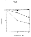
- Es zeigte sich (vgl. Abb. 6), daß die Aktivität der Prourokinase im amidolytischen Test mit steigenden Thrombineinheiten abnahm. So hatte die aktivierte Prourokinase mit 2 U (x--x) Thrombin nach Inkubation von 15 min noch 86 %, nach 30 min noch 59 % seiner Ausgangsaktivität, während mit 10 U Thrombin bei gleicher Inkubationsdauer (x...x) noch 50 % bzw. 36 % der Anfangsaktivität gemessen werden konnte. Eine Inkubation der Prourokinase unter identischen Bedingungen ohne Thrombin zeigte über einen ZUeitraum von 1 h immer noch 100 % der Ausgangsaktivität. An Stelle der Prourokinase wurde nun das PUK-Mutein (Gln 156 oder Leu 156) im Thrombininhibierungstest eingesetzt. Abb. 7 macht deutlich, daß dieses Mutein nach 30 min Inkubation mit 5 U Thrombin noch 100 % der Ausgangsaktivität zeigte (o-o). Der Aminosäureaustausch an der Stelle 156 der Prourokinase (Arg→Gln) oder Leu bewirkt demnach, daß Thrombin am Mutein nicht mehr proteolytisch wirksam ist und kein inaktives Zweikettenmaterial entsteht.
Claims (6)
R-PUK1‾155-X-PUK157‾411
worin R ein Wasserstoffatom, Ala oder Met
PUK1⁻155 und PUK157⁻411 die Aminosäuren 1 bis 155 und 157 bis 411 der Prourokinase und
X Ala, Asn, Asp, Cys, Gln, Glu, Gly, His, Ile, Leu, Lys, Met, Phe, Pro, Ser, Thr, Trp, Try oder Val darstellen.
Applications Claiming Priority (2)
| Application Number | Priority Date | Filing Date | Title |
|---|---|---|---|
| DE19873735916 DE3735916A1 (de) | 1987-10-23 | 1987-10-23 | Neue polypeptide, ihre herstellung und verwendung |
| DE3735916 | 1987-10-23 |
Publications (2)
| Publication Number | Publication Date |
|---|---|
| EP0312942A2 true EP0312942A2 (de) | 1989-04-26 |
| EP0312942A3 EP0312942A3 (de) | 1990-05-02 |
Family
ID=6338942
Family Applications (1)
| Application Number | Title | Priority Date | Filing Date |
|---|---|---|---|
| EP88117188A Withdrawn EP0312942A3 (de) | 1987-10-23 | 1988-10-15 | Proteine mit Prourokinase-Aktivität, ihre Herstellung und Verwendung |
Country Status (4)
| Country | Link |
|---|---|
| EP (1) | EP0312942A3 (de) |
| JP (1) | JPH01160482A (de) |
| DE (1) | DE3735916A1 (de) |
| DK (1) | DK587588A (de) |
Cited By (3)
| Publication number | Priority date | Publication date | Assignee | Title |
|---|---|---|---|---|
| EP0405285A1 (de) * | 1989-06-19 | 1991-01-02 | Kyowa Hakko Kogyo Co., Ltd. | Plasminogenaktivator |
| US5316934A (en) * | 1989-06-13 | 1994-05-31 | Nippon Soda Co., Ltd. | Effective thrombosis mediated by human prourokinase-like polypeptides with increased binding affinity for thrombosis |
| CN100441683C (zh) * | 2006-03-24 | 2008-12-10 | 中国人民解放军军事医学科学院生物工程研究所 | 低分子量尿激酶突变体及其表达载体 |
Family Cites Families (2)
| Publication number | Priority date | Publication date | Assignee | Title |
|---|---|---|---|---|
| EP0210279B1 (de) * | 1985-01-25 | 1991-08-14 | Sagami Chemical Research Center | Stabilisierte menschliche prourokinase |
| US5219569A (en) * | 1985-04-22 | 1993-06-15 | Genentech, Inc. | Protease resistant urokinase |
-
1987
- 1987-10-23 DE DE19873735916 patent/DE3735916A1/de not_active Withdrawn
-
1988
- 1988-10-15 EP EP88117188A patent/EP0312942A3/de not_active Withdrawn
- 1988-10-21 DK DK587588A patent/DK587588A/da not_active Application Discontinuation
- 1988-10-24 JP JP63266255A patent/JPH01160482A/ja active Pending
Cited By (5)
| Publication number | Priority date | Publication date | Assignee | Title |
|---|---|---|---|---|
| US5316934A (en) * | 1989-06-13 | 1994-05-31 | Nippon Soda Co., Ltd. | Effective thrombosis mediated by human prourokinase-like polypeptides with increased binding affinity for thrombosis |
| EP0405285A1 (de) * | 1989-06-19 | 1991-01-02 | Kyowa Hakko Kogyo Co., Ltd. | Plasminogenaktivator |
| US5160735A (en) * | 1989-06-19 | 1992-11-03 | Kyowa Hakko Kogyo Co. Ltd. | Plasminogen activator |
| US5342775A (en) * | 1989-06-19 | 1994-08-30 | Kyowa Hakko Kogyo Co. Ltd. | Plasminogen activator |
| CN100441683C (zh) * | 2006-03-24 | 2008-12-10 | 中国人民解放军军事医学科学院生物工程研究所 | 低分子量尿激酶突变体及其表达载体 |
Also Published As
| Publication number | Publication date |
|---|---|
| DE3735916A1 (de) | 1989-05-03 |
| DK587588A (da) | 1989-06-14 |
| DK587588D0 (da) | 1988-10-21 |
| JPH01160482A (ja) | 1989-06-23 |
| EP0312942A3 (de) | 1990-05-02 |
Similar Documents
| Publication | Publication Date | Title |
|---|---|---|
| DE3751128T2 (de) | Rekombinante gereinigte Protease, Nexin. | |
| DE3588149T2 (de) | Rekombinante DNS-Verfahren und dadurch hergestellte Produkte | |
| DE3786767T2 (de) | Neues Polypeptid. | |
| DE3687651T2 (de) | Analoge des gewebespezifischen plasminogenaktivators. | |
| DE69012888T2 (de) | Plasminogenaktivator. | |
| DE3316297C2 (de) | ||
| DE68923741T2 (de) | Fledermausspeichel-Plasminogenaktivatoren. | |
| DE3587875T2 (de) | Rekombinantverfahren zur herstellung von serinproteaseinhibitoren, sowie dns-sequenzen dazu. | |
| DE3903581A1 (de) | Gewebs-plasminogenaktivator-derivat | |
| DE69432608T2 (de) | Expression von inhibitoren vom plasminogenaktivator vom urokinasetyp | |
| DE3643158A1 (de) | Gewebs-plasminogen-aktivator(tpa) - derivat und seine herstellung | |
| DE69027948T2 (de) | Reinigung von rekombinantem, menschlichem Interleukin-3 | |
| EP0408945B1 (de) | Plasmide, deren Herstellung und deren Verwendung bei der Gewinnung eines Plasminogenaktivators | |
| DE3689977T2 (de) | Dns-sequenzen, rekombinante dns-moleküle und verfahren zur herstellung menschlicher lipocortinähnlicher polypeptide. | |
| EP0496327B1 (de) | Verfahren zur Herstellung von Polypeptiden mit Prourokinase-Aktivität | |
| EP0244627A2 (de) | Expressionsvektoren zur Gewinnung von Polypeptiden | |
| DE3780199T2 (de) | Modifiziertes enzym. | |
| DE3735916A1 (de) | Neue polypeptide, ihre herstellung und verwendung | |
| EP0312941A2 (de) | Polypeptide mit Prourokinase-Aktivität, ihre Herstellung und Verwendung | |
| EP0447468B1 (de) | Ancrod-proteine, ihre herstellung und verwendung | |
| DE3877197T2 (de) | Verfahren zur herstellung von hirudin. | |
| EP0403598A1 (de) | tPA-ÄHNLICHE POLYPEPTIDE, IHRE HERSTELLUNG UND VERWENDUNG | |
| EP0540560B1 (de) | Ancrod ähnliche proteine, ihre herstellung und verwendung | |
| EP0242835B1 (de) | Verfahren zur Herstellung von Plasminogen-Aktivatoren in Prokaryonten | |
| DE3854195T2 (de) | Polypeptid-verbindungen. |
Legal Events
| Date | Code | Title | Description |
|---|---|---|---|
| PUAI | Public reference made under article 153(3) epc to a published international application that has entered the european phase |
Free format text: ORIGINAL CODE: 0009012 |
|
| AK | Designated contracting states |
Kind code of ref document: A2 Designated state(s): AT BE CH DE ES FR GB IT LI NL |
|
| PUAL | Search report despatched |
Free format text: ORIGINAL CODE: 0009013 |
|
| AK | Designated contracting states |
Kind code of ref document: A3 Designated state(s): AT BE CH DE ES FR GB IT LI NL |
|
| 17P | Request for examination filed |
Effective date: 19900329 |
|
| 18W | Application withdrawn |
Withdrawal date: 19910107 |
|
| STAA | Information on the status of an ep patent application or granted ep patent |
Free format text: STATUS: THE APPLICATION HAS BEEN WITHDRAWN |
|
| R18W | Application withdrawn (corrected) |
Effective date: 19910107 |