EP0314109A2 - Composition biorésorbable de collagène pour le traitement de blessures sur la peau - Google Patents
Composition biorésorbable de collagène pour le traitement de blessures sur la peau Download PDFInfo
- Publication number
- EP0314109A2 EP0314109A2 EP88117850A EP88117850A EP0314109A2 EP 0314109 A2 EP0314109 A2 EP 0314109A2 EP 88117850 A EP88117850 A EP 88117850A EP 88117850 A EP88117850 A EP 88117850A EP 0314109 A2 EP0314109 A2 EP 0314109A2
- Authority
- EP
- European Patent Office
- Prior art keywords
- collagen
- sponge
- flake product
- biodegradable
- product
- Prior art date
- Legal status (The legal status is an assumption and is not a legal conclusion. Google has not performed a legal analysis and makes no representation as to the accuracy of the status listed.)
- Withdrawn
Links
Images
Classifications
-
- A—HUMAN NECESSITIES
- A61—MEDICAL OR VETERINARY SCIENCE; HYGIENE
- A61K—PREPARATIONS FOR MEDICAL, DENTAL OR TOILETRY PURPOSES
- A61K9/00—Medicinal preparations characterised by special physical form
- A61K9/70—Web, sheet or filament bases ; Films; Fibres of the matrix type containing drug
-
- A—HUMAN NECESSITIES
- A61—MEDICAL OR VETERINARY SCIENCE; HYGIENE
- A61L—METHODS OR APPARATUS FOR STERILISING MATERIALS OR OBJECTS IN GENERAL; DISINFECTION, STERILISATION OR DEODORISATION OF AIR; CHEMICAL ASPECTS OF BANDAGES, DRESSINGS, ABSORBENT PADS OR SURGICAL ARTICLES; MATERIALS FOR BANDAGES, DRESSINGS, ABSORBENT PADS OR SURGICAL ARTICLES
- A61L15/00—Chemical aspects of, or use of materials for, bandages, dressings or absorbent pads
- A61L15/16—Bandages, dressings or absorbent pads for physiological fluids such as urine or blood, e.g. sanitary towels, tampons
- A61L15/22—Bandages, dressings or absorbent pads for physiological fluids such as urine or blood, e.g. sanitary towels, tampons containing macromolecular materials
- A61L15/32—Proteins, polypeptides; Degradation products or derivatives thereof, e.g. albumin, collagen, fibrin, gelatin
- A61L15/325—Collagen
-
- A—HUMAN NECESSITIES
- A61—MEDICAL OR VETERINARY SCIENCE; HYGIENE
- A61L—METHODS OR APPARATUS FOR STERILISING MATERIALS OR OBJECTS IN GENERAL; DISINFECTION, STERILISATION OR DEODORISATION OF AIR; CHEMICAL ASPECTS OF BANDAGES, DRESSINGS, ABSORBENT PADS OR SURGICAL ARTICLES; MATERIALS FOR BANDAGES, DRESSINGS, ABSORBENT PADS OR SURGICAL ARTICLES
- A61L15/00—Chemical aspects of, or use of materials for, bandages, dressings or absorbent pads
- A61L15/16—Bandages, dressings or absorbent pads for physiological fluids such as urine or blood, e.g. sanitary towels, tampons
- A61L15/42—Use of materials characterised by their function or physical properties
- A61L15/425—Porous materials, e.g. foams or sponges
-
- A—HUMAN NECESSITIES
- A61—MEDICAL OR VETERINARY SCIENCE; HYGIENE
- A61L—METHODS OR APPARATUS FOR STERILISING MATERIALS OR OBJECTS IN GENERAL; DISINFECTION, STERILISATION OR DEODORISATION OF AIR; CHEMICAL ASPECTS OF BANDAGES, DRESSINGS, ABSORBENT PADS OR SURGICAL ARTICLES; MATERIALS FOR BANDAGES, DRESSINGS, ABSORBENT PADS OR SURGICAL ARTICLES
- A61L15/00—Chemical aspects of, or use of materials for, bandages, dressings or absorbent pads
- A61L15/16—Bandages, dressings or absorbent pads for physiological fluids such as urine or blood, e.g. sanitary towels, tampons
- A61L15/42—Use of materials characterised by their function or physical properties
- A61L15/44—Medicaments
-
- A—HUMAN NECESSITIES
- A61—MEDICAL OR VETERINARY SCIENCE; HYGIENE
- A61L—METHODS OR APPARATUS FOR STERILISING MATERIALS OR OBJECTS IN GENERAL; DISINFECTION, STERILISATION OR DEODORISATION OF AIR; CHEMICAL ASPECTS OF BANDAGES, DRESSINGS, ABSORBENT PADS OR SURGICAL ARTICLES; MATERIALS FOR BANDAGES, DRESSINGS, ABSORBENT PADS OR SURGICAL ARTICLES
- A61L15/00—Chemical aspects of, or use of materials for, bandages, dressings or absorbent pads
- A61L15/16—Bandages, dressings or absorbent pads for physiological fluids such as urine or blood, e.g. sanitary towels, tampons
- A61L15/42—Use of materials characterised by their function or physical properties
- A61L15/64—Use of materials characterised by their function or physical properties specially adapted to be resorbable inside the body
-
- A—HUMAN NECESSITIES
- A61—MEDICAL OR VETERINARY SCIENCE; HYGIENE
- A61L—METHODS OR APPARATUS FOR STERILISING MATERIALS OR OBJECTS IN GENERAL; DISINFECTION, STERILISATION OR DEODORISATION OF AIR; CHEMICAL ASPECTS OF BANDAGES, DRESSINGS, ABSORBENT PADS OR SURGICAL ARTICLES; MATERIALS FOR BANDAGES, DRESSINGS, ABSORBENT PADS OR SURGICAL ARTICLES
- A61L26/00—Chemical aspects of, or use of materials for, wound dressings or bandages in liquid, gel or powder form
- A61L26/0009—Chemical aspects of, or use of materials for, wound dressings or bandages in liquid, gel or powder form containing macromolecular materials
- A61L26/0028—Polypeptides; Proteins; Degradation products thereof
- A61L26/0033—Collagen
-
- A—HUMAN NECESSITIES
- A61—MEDICAL OR VETERINARY SCIENCE; HYGIENE
- A61L—METHODS OR APPARATUS FOR STERILISING MATERIALS OR OBJECTS IN GENERAL; DISINFECTION, STERILISATION OR DEODORISATION OF AIR; CHEMICAL ASPECTS OF BANDAGES, DRESSINGS, ABSORBENT PADS OR SURGICAL ARTICLES; MATERIALS FOR BANDAGES, DRESSINGS, ABSORBENT PADS OR SURGICAL ARTICLES
- A61L27/00—Materials for grafts or prostheses or for coating grafts or prostheses
- A61L27/14—Macromolecular materials
- A61L27/22—Polypeptides or derivatives thereof, e.g. degradation products
- A61L27/24—Collagen
-
- A—HUMAN NECESSITIES
- A61—MEDICAL OR VETERINARY SCIENCE; HYGIENE
- A61L—METHODS OR APPARATUS FOR STERILISING MATERIALS OR OBJECTS IN GENERAL; DISINFECTION, STERILISATION OR DEODORISATION OF AIR; CHEMICAL ASPECTS OF BANDAGES, DRESSINGS, ABSORBENT PADS OR SURGICAL ARTICLES; MATERIALS FOR BANDAGES, DRESSINGS, ABSORBENT PADS OR SURGICAL ARTICLES
- A61L27/00—Materials for grafts or prostheses or for coating grafts or prostheses
- A61L27/50—Materials characterised by their function or physical properties, e.g. injectable or lubricating compositions, shape-memory materials, surface modified materials
- A61L27/56—Porous materials, e.g. foams or sponges
-
- A—HUMAN NECESSITIES
- A61—MEDICAL OR VETERINARY SCIENCE; HYGIENE
- A61L—METHODS OR APPARATUS FOR STERILISING MATERIALS OR OBJECTS IN GENERAL; DISINFECTION, STERILISATION OR DEODORISATION OF AIR; CHEMICAL ASPECTS OF BANDAGES, DRESSINGS, ABSORBENT PADS OR SURGICAL ARTICLES; MATERIALS FOR BANDAGES, DRESSINGS, ABSORBENT PADS OR SURGICAL ARTICLES
- A61L27/00—Materials for grafts or prostheses or for coating grafts or prostheses
- A61L27/50—Materials characterised by their function or physical properties, e.g. injectable or lubricating compositions, shape-memory materials, surface modified materials
- A61L27/58—Materials at least partially resorbable by the body
-
- C—CHEMISTRY; METALLURGY
- C08—ORGANIC MACROMOLECULAR COMPOUNDS; THEIR PREPARATION OR CHEMICAL WORKING-UP; COMPOSITIONS BASED THEREON
- C08L—COMPOSITIONS OF MACROMOLECULAR COMPOUNDS
- C08L89/00—Compositions of proteins; Compositions of derivatives thereof
- C08L89/04—Products derived from waste materials, e.g. horn, hoof or hair
- C08L89/06—Products derived from waste materials, e.g. horn, hoof or hair derived from leather or skin, e.g. gelatin
-
- A—HUMAN NECESSITIES
- A61—MEDICAL OR VETERINARY SCIENCE; HYGIENE
- A61L—METHODS OR APPARATUS FOR STERILISING MATERIALS OR OBJECTS IN GENERAL; DISINFECTION, STERILISATION OR DEODORISATION OF AIR; CHEMICAL ASPECTS OF BANDAGES, DRESSINGS, ABSORBENT PADS OR SURGICAL ARTICLES; MATERIALS FOR BANDAGES, DRESSINGS, ABSORBENT PADS OR SURGICAL ARTICLES
- A61L2300/00—Biologically active materials used in bandages, wound dressings, absorbent pads or medical devices
- A61L2300/20—Biologically active materials used in bandages, wound dressings, absorbent pads or medical devices containing or releasing organic materials
- A61L2300/252—Polypeptides, proteins, e.g. glycoproteins, lipoproteins, cytokines
-
- A—HUMAN NECESSITIES
- A61—MEDICAL OR VETERINARY SCIENCE; HYGIENE
- A61L—METHODS OR APPARATUS FOR STERILISING MATERIALS OR OBJECTS IN GENERAL; DISINFECTION, STERILISATION OR DEODORISATION OF AIR; CHEMICAL ASPECTS OF BANDAGES, DRESSINGS, ABSORBENT PADS OR SURGICAL ARTICLES; MATERIALS FOR BANDAGES, DRESSINGS, ABSORBENT PADS OR SURGICAL ARTICLES
- A61L2300/00—Biologically active materials used in bandages, wound dressings, absorbent pads or medical devices
- A61L2300/40—Biologically active materials used in bandages, wound dressings, absorbent pads or medical devices characterised by a specific therapeutic activity or mode of action
- A61L2300/412—Tissue-regenerating or healing or proliferative agents
Definitions
- the invention relates to the field of wound treatment, particularly epidermal and skin wounds, more especially pressure ulcers, like decubitus ulcers with sponge-like collagen compositions.
- the invention is of particular importance for the treatment of patients suffering from such ulcers.
- the invention also relates to a collagen-type product, particularly a new collagen flake product, also especially useful for treatment of human pressure skin ulcers, like decubitus ulcers.
- the invention allows to apply a therapeutic method for treating epidermal and skin ulcers especially of humans, which comprises periodically treating the wound with a collagen flake product of the invention.
- the flake product of the invention need not be of a cross-linked collagen.
- the invention allows to apply a therapeutic method for treating pressure ulcers with improved sponge-like, cross-linked collagen products which preferably contain fibronectin (FN) and/or hyaluronic acid (HA).
- FN fibronectin
- HA hyaluronic acid
- the invention allows to apply a method for treating deep animal dermal wounds or pressure ulcers, especially human decubitus ulcers by treatment with a cross-linked collagen matrix or type I collagen sponge, or with a new collagen flake product.
- the invention provides further biocompatible synthetic polymeric materials for wound treatment such as pressure ulcers, with sponges and flake products.
- the invention allows to apply therapeutic methods for the treatment of dermal wounds with such biocompatible synthetic materials to promote healing and form new tissue.
- the invention provides various useful products for such purposes.
- the invention allows to apply a method for promoting optimum tissue ingrowth implants.
- the invention provides these various materials, products and other aspects further disclosed herein for treatment of warm-blooded animals, mammals, especially humans but also for animals of the equine, canine, bovine, and other species. Of course the more important and serious needs are for human patients.
- the invention also provides products useful in fields of application other than wound repair, tissue reconstructions, implants and the like but also in cosmetic surgery.
- the invention also provides tissue, skin, etc., materials which have all the characteristics of human or animal living tissue for applications inside wounds, or outside of the body, and applications where all the normal tissue-like characteristics are desired to be simulated.
- the invention provides several new products and compositions, and makes available methods of treatment which are a significant contribution to the medical and healing arts.
- the invention deals principally with an important problem of pressure ulcers. This problem is assuming increasingly greater importance because of the growth of our aging population and because there is no satisfactory method or product available for dealing with the problem of pressure ulcers.
- collagen-based materials such as sponges, sheets, sutures and coatings have been used in animal and human studies as dermal dressings and implants because of its bicompatibility and because the collagen-based material has been demonstrated to provide what has been called a "scaffold" for tissue ingrowth and to play an active role in tissue organization during repair.
- the problem occurs frequently with patients subject to prolonged bedrest like quadriplegics and paraplegics who suffer skin loss due to the effects of localized pressure.
- the resulting pressure sores also known as decubitus ulcers, exhibit dermal erosion and loss of the epidermis and skin appendages.
- stage 1 and/or 2 ulcers are traditionaly referred to as stage 1 and/or 2 ulcers when the superficial skin layer has been traumatized, abraded and removed (approximately to a depth of 0.1 mm) and where the lesion has progressed to form a wound that can be 5 to 6 or more centimeters (cm) deep and up to approximately 13 cm wide and several cm long. Such wounds also typically show an edge of the skin which has separated from the skin in a flap-like shape.
- ulcers have been identified and classified as follows:
- repair of large open wounds involves several events including cellular migration, biosynthesis of connective tissue components and other factors, deposition and remodeling of granulation tissue.
- the remodeling phase has been shown to be accelerated in the presence of type I collagen porous matrix.
- Type I collagen attracts fibroblasts in cell cultures and appears to cause directed migration of cells.
- Fibronectin is known to increase chemoattraction and spreading of fibroblasts in vitro, and is found in large amounts in dermis during embryonic skin development and in healing wounds.
- Hyaluronic acid is found in high concentrations during embryonic development, is associated with cell movement and differentiation and is the first connective tissue glycosaminoglycan to appear in the extracellular matrix during wound repair.
- a variety of low molecular weight cellular products have been shown to stimulate proliferation and enhance cellular biosynthesis of connective tissue components. These factors include epidermal growth factor, platelet-derived growth factor, fibroblast growth factor, eye-derived growth factor and cartilage-derived growth factor. Heparin has been shown to induce the formation of capillaries.
- type I collagen sponges enhance the healing of excised wounds in an animal model.
- the interest in the studies was to enhance the healing of chronic wounds using connective tissue macromolecules, growth factors and cells to reconstitute synthetic dermal and epidermal replacements.
- the work focused on the interaction of fibroblasts and epidermal cells with type I cross-linked collagen sponge in vitro cell cultures.
- Natural insoluble collagen as used herein means and refers to collagen which cannot be dissolved in an aqueous acidic or alkaline or in any inorganic salt solution without chemical modification and includes hides, splits, and other mammalian or reptilian coverings. More particularly, “natural insoluble collagen” means and refers to the corium which is the intermediate layer of a bovine hide between the grain and the flesh sides.
- Collagen constitutes the connective tissue and is the major type of fibrous protein in higher vertebrae. Collagen in its natural state exists in a triple chain helix along with a constant periodicity between aligned triple chains.
- the triple helical configuration of collagen is sometimes referred to as a rod and a group of molecules align themselves with an axial periodicity of about 640 A.
- Type I collagen Although there are several types of collagen, the major type is referred to as "type I" which is the major collagen of skin, bones and tendons. Type I collagen has a chain composition of [alpha 1(I)2 alpha 2 (I)]. The alpha 1 (I) and alpha 2 (I) chains are homologous.
- the flake and sponge products be included in the invention when made from types of collagens other than collagen type I, for instance types II, which corresponds to [alpha (II)]3; type III which corresponds to [alpha I (III)]3; type V which corresponds to [alpha I (IV)]3 and [alpha 2 (IV)]3 and type V which corresponds to [alpha I (V) ]2 alpha 2(V).
- types II which corresponds to [alpha (II)]3
- type III which corresponds to [alpha I (III)]3
- type V which corresponds to [alpha I (IV)]3 and [alpha 2 (IV)]3
- type V which corresponds to [alpha I (V) ]2 alpha 2(V).
- Type I is principally distributed (as noted above) in tissue like skin, tendon, bone, dentin and fascia; the other types are distributed in cartilage, notochord, vitreous body; skin uterus, blood vessels, "reticulin” fibers, kidney glomeruli, lens capsule; basement laminae of smooth and striated muscle cells; exoskeleton of fibroblasts and other mesenchymal cells.
- the different types of collagen are built of different but related collagen polypeptides. The chains differ in the extent to which their proline, lysine and cysteine residues are modified. See, Characteristics of Different Types of Collagen, in Darnell, above cited (page 179, Table 5-6).
- An objective of this invention is to provide such products and a reliable method for making such products. Many variables have been considered and tried by workers in the prior art; none, as far as could be determined, found the set of conditions which yielded reliably a satisfactory product.
- a collagen product e.g. a sponge
- the process comprises dispersing a finely divided inorganic acid like hydrochloric acid at a pH preferably between about 1.0 and 4.0, even more preferably from about 2.0 to about 3.75.
- the collagen is preferably in concentrations of about 0.5 to about 1% weight/volume.
- the temperature at which the operations are performed is in the range of about 15 to 35°C, preferably 20-30°C. A temperature of 22 ⁇ 2°C has been observed.
- the dispersing or blending is performed with any suitable mechanical blending means such as a stirrer (Osterizer Blender), for a time sufficient to accomplish a thorough dispersion.
- the dispersion is then deaerated under appropriate vacuum like less than 0.4 millitorr.
- the collagen dispersion is then frozen under conditions conducive to obtain a fibrous structure which contains pores, ideally of substantially uniform average pore size, preferably of about 100 ⁇ 50 ⁇ m and containing channels connecting the exterior of the sponge to the inside, generally connecting one side of the product to the other, and pores that open into the channels.
- the collagen dispersion is freeze-dried at a temperature range of about -20 to -35°C, preferably at about -30°C in a bath of a lower alkanol, like ethanol.
- Preferably air gaps between the sponge body and the container are minimized.
- cross-linking can be performed by any method known in the art.
- cross-linking is performed in accordance with a two-step process resulting in formation of peptide bonds on oppositely charged side chains. See K. Weadock, R.M. Olson, F.H. Silver, Evaluation of Collagen cross-linking techniques. Biomater. Med. Denies Artif. Organs, 11, 293-318 (1984), (reference 10).
- the cross-linking methods disclosed herein result in a stable cross-linked sponge with stable pores and channels (which show no skrinkage of the pores or the channels).
- the products so obtained are sponges which are characterized by a dual porosity, as described further below.
- a flake product is made which also has dual porosity as will be described further below.
- the products of the invention ideally show a highly fibrous structure associated with an average pore size of about 100 ⁇ 50 ⁇ m containing channels which offer an ideal structure for a collagen-based material for tissue ingrowth as needed for an "artificial" dermis.
- Fibroblast behavior does, it has been noted, have a relationship to pore size; tissue ingrowth into the porous sponge occurs when the pore size is between about 50 to 300 ⁇ m, ideally when pore size is about 80 to 120, often about 100 ⁇ m.
- the collagen flake product of the invention may be described as follows.
- the product comprises collagen flakes generally non-uniform in size, length and thickness.
- the fibers are organized or positioned randomly in a plane generally horizontal.
- the flakes define between them non-uniform channels some of which extend throughout the product from the surface deep into the interior, or cross the entire product and form a labyrinthal-type structure.
- the collagen flakes also have interconnecting pores, the pores having openings into the channels.
- the flake product of the invention can have a large proportion of channels communicating with the pores, a proportion of the pores though interconnecting with each other do not all open into the channels.
- the dual type of porosity (channels and pores) and flake-like structure has an enhanced beneficial and healing effect on the wound.
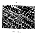
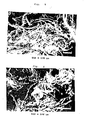
- the morphology of the flake product shows a network of individual collagen fibers, generally at least about 1 ⁇ m in diameter forming a web-like mesh, as well as an aggregate of collagen fibers which appear to form a sheet-like structure with channels between the individual collagen fibers and between the sheets of collagen fibers.
- the channels connect surface pores and pores in the interior of the sponge.
- the collagen fibers contain pores the greatest majority of which (at least 50%, generally over 80% or 90% by volume), are interconnecting with each other; and a significant proportion of the pores, e.g., 50 to 80%, connect to the channels.
- the flakes are constituted of the collagen fibers, it is evident that the flakes can be as long as the fibers; the thickness of the flake can also vary but will generally be less than the length or width of the flake.
- FIGs. 1 and 2 A typical morphology of a flake product of the invention is seen in Figs. 1 and 2.
- the channels of the flake product of the invention appear cylindrical or spherical.
- the pores are generally uniform in average diameter, preferably between 5 to 300 ⁇ m, ideally from about 20 to 110 ⁇ m, even better about 30 to 75 i.e., about 50 ⁇ m. From diffusion studies it has been observed that no channel is so small as to exclude a macromolecule up to 100,000 in molecular weight.
- the collagen flake can vary in length often between about 0.1 to 3 cm, commonly about 1 cm.
- the type of application or use influences the type of collagen product best suited for the application. For large wounds, it is acceptable for the flake of the flake product to be longer than for small wounds.
- the flakes of the product of the invention can be longer than the individual fibers themselves since the fibers often, if not generally, will align themselves length-wise partially or totally overlapping other fibers, to form the flake (or mat-like structures) of the invention.
- the flake products of the invention have a surface area which is markedly greater than that of collagen sponges, often from about 20% to at least 50% or even to about 80% greater than that of sponges.
- the flake products of the invention may or may not be of cross-linked collagen; both types are acceptable.
- the sponge product used in the invention is preferably cross-linked, and most preferably contain FN and/or HA.
- the flake product of the invention is distinguishable over collagen powders. Contrary to expectations, collagen powders were very unsatisfactory in the principal application of the products of the invention, i.e., as wound dressing.
- the porosity of powder is virtually of one type of pores; no channels could be observed and the pores were of very small size, in the order of not over about 10 ⁇ m, generally 1 to 5 ⁇ m. Absorbancy of the fluid of the wound was unsatisfactory; handling of the powder and packing of the wound were very difficult.
- the process of the invention for making the flake product of the invention comprises treating a collagen sponge product with shearing means under conditions of minimum pressure.
- the shearing process cuts slices or lamellae of the sponge with the minimum application of pressure.
- the dry sponge product is exposed to cutting means that slices-off pieces or slices or flakes of the collagen sponge and minimizes destroying the porous structure of the collagen.
- the resulting flake can vary in size from about 0.1 mm to about 1mm in average thickness. However, the thickness of the flake can be adjusted by adjusting the cutting or shearing means. Multiple shearing blades may be used, in which case the distance separating two such adjacent means determine, at least in part, the thickness of the flake.
- the process of the invention therefore comprises causing a shearing strain to be applied to a sponge, the strain resulting from forces that cause or tend to cause contiguous means to slide or move relative to each other in a direction substantially parallel to each other.
- the shearing stress causes lamellae or slices of the sponge to be cut off the porous body and the body sheared into numerous flakes.
- the compression stress during (before and after) the shearing is preferably minimal or essentailly nil.
- the sponge may be advanced toward a shearing means, which will cut off flakes and remove the flakes from contact with the shearing means.
- the means for shearing the porous sponge are not critical. Any suitable means which accomplish this purpose is suitable.
- a device which is quite satisfactory comprises a container for the sponge which includes sharp propeller-like knives in an elongated tube-like cylinder which allows the sliced-off flakes to be removed upwardly from the vicinity of the shearing means.
- a Willey mill has been found quite satisfactory.
- the device can be made to operate in such a manner that gravity will remove the sliced-off flakes from the vicinity of the blades.
- the invention contemplates mechanical means for accomplishing the shearing. Likewise other means which accomplish the equivalent can be used, like ultra sonic devices and the like.
- the flake product of the invention need not be made from a porous collagen sponge but may be made directly from the collagen fibers without going through the intermediary of the sponge.
- the flake products made of synthetic biocompatible polymers sponge can be made directly from the collagen fibers without going through the intermediary of the sponge.
- the flakes can be formed into mat-like or flake-like structures.
- a satisfactory flake product for the purpose of the invention can vary over a wide range.
- Preferred is a flake product that includes virtually no powder particles (particles which pass a 40 mesh screen) and where the majority i.e., over 50%, preferably over 80% to about 100% of the flakes will pass through a 10 to 30 mesh screen.
- the fibers are generally 0.1 mm to 3 mm (or more) of average length, often about 1 mm long, or longer as described above. It is understood that considerable latitude is allowed as long as the product is a flake product of the collagen (or of the biocompatible synthetic polymer).
- Suitable separating means like a screen, separate the flakes from smaller particles, like powder of sponge.
- the conditions under which the shearing process is carried out can vary considerably.
- the shearing process can be carried out concurrently with, prior to, or following other steps or treatment of the collagen of the invention.
- the temperature at which the shearing process is carried out is generally any temperature which will not disintegrate or damage the starting and final product, such as in the range from above freezing to about 60°C, preferably about room temperature.
- the length of time for which the shearing process is carried out is until the desired flake product is obtained, a flake product having the morphology described above.
- One skilled in the art can without any undue experimentation adjust the time and speed of the shearing means.
- the atmosphere under which the shearing process is carried out can be air, or any other desirable atmosphere.
- the process may be carried out under vacuum.
- the atmosphere is dry air.
- the atmosphere may be sterile or at least filtered air.
- the product is preferably sterilized after the shearing process.
- the collagen may be cross-linked during the shearing. That is, the collagen may be first freeze-dried and then cross-linked, either prior to, concurrently or subsequently to the shearing process.
- the atmosphere in the shearing apparatus there may be provided an appropriate atmosphere like vapor of the desired cross-linking agent.
- any of the chemicals which have been used in the prior art may be vaporized, such as an aldehyde, carbodiimides and the like and the polymerization may be concurrently carried out (or prior, or after) the shearing process.
- an appropriate sterilization gas such as ethylene oxide, may be used.
- the process of the invention may be carried out in a batch-like or continuous manner. In either, larger chunks of the product may be separated from the flake product and recycled to be sheared to the appropriate size.
- Biocompatible synthetic polymers that are contemplated for the uses disclosed in conjunction with the invention include those described in Transdermal Controlled Systemic Medications, Marcel Dekker, Inc., (1987), in the section on Polymeric Materials of Skin Biocompatibility, Kevin S. Weadock et al..
- Typical polymers are the polyurethanes, polyethylenes, silicone polymers, hydrogels and others. Polymers which are free of additives used during manufacture are considered least likely to cause allergic or tumorigenic responses.
- the synthetic polymers may or may not be biodegradable.
- the synthetic biocompatible polymers are manufactured as described above, for instance from porous polyurethanes and the like.
- pores in the collagen sponge (or synthetics) which were interconnected (or not interconnected) but positioned within the interior of the sponge structure now connect to surfaces of the flakes and/or to the channels between the flakes.
- the morphology of the products of the invention are typified by this dual porosity and also by a remarkable increase in surface area. This morphology is much more ideal and conducive to attracting fibroblasts and epithethial cells and thereby promoting synthesis of the new collagen in a wound.
- HA or FN In the treatment of ulcers in accordance with the invention, the role of HA or FN should also be taken into account.
- HA the synthesis of collagen was increased at day 3 post-seeding in comparison to that observed in the presence of FN or collagen sponge alone.
- FN deposition of collagen into matrix was increased particularly at days 7 and 9 post-seeding in comparison to that observed when cells were grown on collagen sponge alone.
- FN and HA both enhance wound healing in vivo, they both do not appear to influence the healing process in the same manner and at the same time.
- ECM extra cellular matrix
- the growth rate and collagen synthesis are increased. This may be explained by increased cell division or by an increase in the number of cells that migrated into the interior of the sponge containing HA or FN when compared to cells grown on the collagen sponge alone.
- the biochemical composition also influences the infiltration of cells (observed by light microscopy).
- the presence of FN or HA affected cell infiltration into a collagen sponge.
- cells observed in the porous spaces may be bound directly to HA and not in direct contact with collagen, since HA may be situated within the porous spaces of the sponge.
- direct contact of cells with HA bound to collagen fibers may allow the cells to proliferate and synthesize ECM in contrast to that observed when cells are in direct contact with the collagen fibers alone.
- Previous studies suggest that direct exposure of fibroblasts to gel of HA inhibits cellular mobility and multiplication. The results of the work in connection with the invention suggests that the combination of HA and collagen fibers is important in enhancing cellular mobility and replication.
- fibroblasts grown on collagen sponges containing FN or HA proliferate and deposit more newly synthesized collagen into the matrix compared with fibroblasts grown on the unmodified matrix or on plastic.
- HA in the matrix encourages cellular infiltration into the pores and channels of the sponge while FN induces cell attachment to the collagen fibers of the sponge.
- Incorporation of HA and FN into the collagen sponge enhances cell mobility and replication in the collagen sponge and improves its properties as an artificial connective tissue.
- the presence of FN and/or HA sponges in the treatment of pressure ulcers likewise contributes to speedier and improved healing.
- the sponge product used in the invention and the flake product of and used in the invention may contain any number of additives that are conducive to healing the wounds, like hormones, bactericides and growth promoters, immunogens, antibiotics and the like.
- Collagen sponges were prepared for cell culture studies and human studies as described previously (Doillon et al., 1984). Type I collagen from cow hide was dispersed at 0.5% (W/V) in HCl solution (pH 3.0), freeze-dried and crosslinked according to Weadock et al., (1984). Collagen sponges were sterilized by exposure to 2.5 M rads of gamma irradiation.
- Fibronectin was extracted from fresh bovine blood as described by Ruoslahti et al., (1982) and was found by polyacrylamide gel electrophoresis to be composed of two polypeptide chains with a molecular weight of about 220,000 after reduction (Brokaw et al., 1985). FN was dissolved in 0.1 M ammonium acetate.
- Hyaluronic Acid from Sigma Chemical Co. (grade III; potassium salt) was used and dissolved in HCl solution (pH 3.0).
- FN and HA solutions were mixed with the collagen dispersion in a Waring blender. Mixtures containing weight ratios of 1:99 FN to collagen and 1:19 of HA of collagen were prepared. Tissue ingrowth was maximized with these concentrations of HA and FN (Doillon and Silver, 1986a).
- Sponges can also be prepared in accordance with the methods disclosed in Doillon et al. (References 3, 4, 5 and 6).
- Fibroblasts were grown on collagen sponges after the sponge pH was stabilized by immersion in serum-free Dulbecco's Modified Eagle Medium (DMEM) (Gibco Laboratories) for three days. Fibroblasts were derived from embryonic chick tendons as described by Kao et al (1975) and cultured in DMEM supplemented with 100 units/ml penicillin, 100 ⁇ g/ml streptomycin, ascorbic acid (10 ⁇ g/ml added daily) and 5% fetal bovine serum (FBS) (Gibco Laboratories). Fibroblasts were plated as primary cultures at concentrations of 1.5 x 10⁵ to 3.2 x 10⁵ cells/cm2.
- DMEM Dulbecco's Modified Eagle Medium
- Epidermal cells were obtained from pieces of guinea pig skin that were excised, defatted and cut into 1 mm2 pieces (Doillon et al., 1986c). The pieces of skin were washed in culture medium composed of a 1 to 1 mixture of DMEM and Ham's F 12 Medium (F12) (DMEM/F12; supplied by Sigma Chemical Co.) containing 100 ⁇ g/ml of penicillin, 10 ⁇ g/ml streptomycin and 3 ⁇ g/ml of amphotericin B. Pieces of skin were then placed in a DMEM/F12 solution containing 5 mg/ml of collagenase (Copper Biomedical) for 90 minutes. The digested skin pieces were then sedimented for 10 minutes under gravity. The sediment rich in epidermal cells was suspended, counted and used for seeding.
- DMEM/F12 Ham's F 12 Medium
- Epidermal cells were cultured in DMEM/F12 supplemented with antibiotics, insulin (1 ⁇ g/ml, hydrocortisone (20 ⁇ g/ml and 15% FBS (Doillon et al., 1986c). Cells were plated as primary cultures at a concentration of 2 x 10⁶ cells/cm2. Cells were seeded on each collagen sponge as described for fibroblasts. After 2 hours, the remaining culture medium was added. Cells were grown at 37°C in a tissue culture incubator in a 5% CO2 atmosphere. The medium was replaced every 2 days. The FBS was decreased from 15% to 10% over a one week period.
- Fibroblast cultures were labeled at days 1, 3, 5, 7 and 9 post-seeding using DMEM containing either 2 uCi/ml [3H] thymidine or 2 ⁇ Ci/ml [14C] proline (New England Nuclear).
- the labeling medium contained 10 ⁇ g/ml ascorbate. Fibroblasts were incubated with [3H] thymidine for four hours. The medium was removed, a 0.5 N perchloric acid solution added and cells were collected as previously described (Doillon et al., 1987). After sonication, the precipitated DNA was washed two times with 0.5 N perchloric acid and heated at 90°C for 90 minutes. After centrifugation, the supernatant was counted in a liquid scintillation counter (Aquasol-2, New England Nuclear).
- fibroblast cultures were incubated with 2 uCl/ml [14C] proline for four hours. The medium was then separated from the cells and fresh DMEM was added to the cells. Cell and medium fractions containing radiolabeled [14C] proline were treated and protease inhibitors (Kao et al., 1977) and sonicated. The samples were brought to a final concentration of 2% sodium dodecyl sulfate (SDV), dialyzed against SDS sample buffer containing 2% (W/V) SDS, 10% (V/V) glycerol, 0.005% bromophenol blue in 0.124 Tris-HCL ph 6.8, and an aliquot was counted in a liquid scintillation counter.
- SDV sodium dodecyl sulfate
- Epidermal cell cultures were labeled between days 15 and 22 using the methods described for the fibroblast cell cultures (See above).
- Sterilized collagen flakes were applied to non-infected dermal ulcers ranging from about 1 to 10 cm2 in surface area. Only ulcers classified as stages 2 or 3 were used in this study. The degree of subcutaneous erosion varied from patient to patient but in no case was the material placed over exposed tendon, muscle or bone (stage 4). The patients ranged in age from about 35 to 70 years of age. At least 6 patients were studied in the control and collagen treated groups.
- Both control and collagen treated wounds were cared for using the protocol that follows. All wounds were washed with a 1% (W/V) solution of hydrogen peroxide followed by normal saline. The collagen flakes were then packed into the wound (except for controls). All wounds were then covered with saline-wetted cotton gauze. A layer of dry cotton gauze was then applied and taped to surrounding healthy skin. All wounds were washed with hydrogen peroxide and saline and rebandaged daily. Wounds treated with the collagen flakes were repacked daily after wounds were washed with saline.
- type I collagen sponges enhance repair of animal dermal wounds by organizing the spatial deposition of newly synthesized collagen and accelerating remodeling.
- incorporation of hyaluronic acid and fibronectin into a type I collagen sponge results in increased numbers of fibroblasts that migrate into the collagen-based sponge and consequently an increased deposition of newly synthesized collagen is observed.
- the deep pores or channels are generally interconnecting forming a labryrinthal-like structure; not all channels are interconnecting and not all channels have openings to the exterior.
- plastic embedded sponge sections showed a porous structure of pores varying from about 20 to 200 ⁇ m.
- Channel structure was regularly observed when HA and FN were both associated at 1% with collagen. In the latter case, "open" channels were frequently seen with large interchannel connections.
- Fibroblasts grown on plastic adopt a flattened shape and synthesize some extracellular matrix (ECM) (see Table 1).
- ECM extracellular matrix
- cells grown on a collagen sponge form several confluent layers of elongated cells and deposit a large amount of ECM.
- by day 9 only about 25% of the collagen sponges are infiltrated with fibroblasts.
- Fibroblasts grown on collagen sponges containing 5% (W/W) hyaluronic acid are found throughout the collagen sponge primarily in pores that are formed by the collagen fibers of the sponge.
- Fibroblasts grown on collagen sponges containing 1% (W/W) fibronectin were observed to be attached and elongated along the collagen fibers and infiltrated throughout the sponge. In the presence of hyaluronic acid or fibronectin several superficial layers of confluent cells are seen on the surface of the collagen sponge.
- Fibroblasts grown on plastic for 3 days in cell culture incorporated about five times more of [14C] proline counts (collagen synthesis) than were incorporated into cells grown on a type I collagen sponge. This result suggests that the presence of a collagen matrix inhibits collagen synthesis by fibroblasts.
- [3H] thymidine incorporation (cell replication) into fibroblasts after three days in cell culture was similar for cells grown on plastic and on a collagen sponge; however, [3H] thymidine incorporation was increased by the addition of 5% HA or 1% FN. By day 9, these trends were still present (see Table 2) although the differences were less noticeable.
- Fibroblasts grown on all substrates synthesized type I procollagen, type I collagen and several processing intermediates based on [14 C] proline incorporation and fluorography The results suggest that the interaction between a porous type I collagen substrate and fibroblasts leads to inhibition of collagen production.
- Cell replication and collagen synthesis can be increased by addition of small amounts of HA or FN such as 0.1% to 5.0%. Larger amounts e.g., 10% do not seem to have the desired effect.
- Collagen synthesis and cell replication were evaluated for epidermal cells grown on a type I collagen sponge (see Table 4).
- Epidermal cells formed more layers of stratified cells when grown on collagen than when grown on plastic. Tight packing of cells on the sponge surface was observed which was not observed when these cells were grown on plastic.
- some cultures showed evidence of a basement membrane-like structure between the collagen sponge and a layer of basal cells.
- epidermal cells formed clusters that resembled primitive glands.
- the collagen sponge did not inhibit incorporation of [14C] labeled proline into collagen.
- Treatment of an ulcer in stage 4 is carried out in like manner. Healing of the wound to stage 2 and 3 and then stage 1 was observed. Treatment takes longer in view of the severity of the wound.
- Distilled water was added until the total volume of the processing mixture reached 14 liters. Air at a pressure of 6 psi was introduced into the tank for 5 minutes to create a homogenous mixture. This mixture then was left to sediment for 20 minutes. After complete sedimentation occurred the liquid phase was drained and fresh distilled water was added until the total volume reached 14 liters. This procedure was repeated three times.
- the material was washed with two liters of distilled water, poured into plastic trays and then placed into a freezer until frozen solid. The frozen material was then placed in the cold trap of a freeze dryer with the shelf maintained at 0°C. A vacuum of 10 microns was then applied for 48-96 hours. The vacuum was then released and material removed.
- Collagen was identified by standard procedure, such as by sodium dodecyl sulfate polacrylamide gel electrophoresis and amino acid analysis as typical of type I collagen free of non-collageneous protein contamination.
- a collagen flake product of the invention is prepared as follows.
- a collagen dispersion is prepared in the following manner. A 1N solution of HCL is slowly added added to 120 ml of distilled water until the pH is 3.0 at room temperature. The contents of the beaker are then emptied into a 200 ml graduated cylinder.
- the dispersion is emptied into a sidearm flask (600 ml). A vacuum of 100 microns is applied to the dispersion at room temperatures until the air bubbles are removed. This procedure may require up to 15 minutes.
- One liter of the 1% V/W collagen dispersion is poured into a 10" x 20" x 1" tray and frozen at -30°C and freeze-dried.
- a 3" x 3" by 1 ⁇ 4" piece of freeze dried collagen sponge is crosslinked by heating to -110°C in a vacuum for 72 hours followed by exposure to aqueous vapors of a 10% solution of cyanamide for 24 hours.
- Crosslinked freeze-dried collagen sponge is placed in a Waring blender and blended for 5 minutes at low speed (3,000 rpm). Flakes of collagen are formed.
- the collagen flakes are about 1 mm in thickness and 1 mm to 1 cm in length and width. Pore sizes range from 100 to 10,000 ⁇ m.
- 1 g of collagen flakes is sealed in a plastic bag and sterilized by exposure to 2.5 M rads gamma radiation.
- the collagen flake product is ready for treating patients suffering from decubitus ulcers which exhibit dermal erosion and loss of epidermis.
- decubitus ulcers Patients suffering of pressure sores known as decubitus ulcers were treated by the conventional protocol which included daily irrigation of the wound and gauze bandaging. No decrease in wound area over a six week period is observed.
- the protocol for treatment of patients was modified as follows.
- the nurses pack the wound and cover the edges of surrounding skin with the collagen flakes (removed from the plastic bag) until the flake level was approximately level with wound.
- the collagen flake packed wound is then bandaged with gauze. After 1 day the packing of collagen flake is removed, the wound is washed and irrigated with saline. It is observed that not all of the collagen flake product is removed but there is evidence of cellular ingrowth which remains in the wound, especially on the bottom.
- the wound is repacked with a fresh supply of collagen flake.
- the procedure is repeated on a daily basis for six to twelve weeks. Gradually it is observed that the capillaries at the base of the wound turn gradually from a very pale cream color to a pale pink and reddish color indicating the development of vasculature and initiation of some blood circulation.
- the flap near the edge of the wound typical of decubitus ulcers gradually starts adhering and forming new tissue joining it to the surrounding edges of the wound, thus gradually closing the wound.
- the wound is gradually closed by new ingrowth of tissue gradually starting from the bottom and the side and invading the cavity of the wound.
- the new tissue has essentially filled the wound.
- the wound is then ready for grafting-on tissue such as by autograft.
- grafting-on tissue such as by autograft.
- an epidermal growth factor is added.
- Each package can contain different amounts of the flake product.
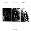
- Figures 1A, 1B and 1C show the gradual closing of a severe decubitus ulcer as a result of treatment with the collagen flake product of the invention.
- photograph C taken after 11 ⁇ 2 months, it is noted that the color of the newly grown tissue has assumed a pinkish light color symptomatic of increased blood circulation and the formation of capillaries.
- the flake product not only absorbs the fluid due to the edema but also absorbs the bacteria in the wound.
- the flake product can be used in conjunction with a collagen sponge.
- the wound is first packed with the flakes (especially in the difficult to reach parts), then the sponge placed on top, followed by an occlusive dressing.
- the flake product of the invention is useful also for treatment of internal wounds, as in liver surgery.
- the collagen product is believed to stimulate blood clotting, platelet aggregation (through ADP release), and to attract white cells into the collagen fibrous structure.
- the cells migrate through the pores and as they are filled, the cells attach themselves to the collagen fibers. Then an extracellular (connective tissue) is formed.
- the matrix is gradually disintegrated, and endothelial cells extend forming new capillaries.
- the new tissue is reconstituted.
- the open wounds can be gradually closed in 7-14 days and thus a new implant will be developed.
- the preparation of collagen sponge with a moisture vapor barrier is carried out as follows.
- Insoluble collagen (1.2 g) is added to 120 ml of dilute HCL solution pH 3.0 and the mixture is dispersed in a Waring Blender at 2,000 RPM for 1 minute. The dispersion is then poured into a vacuum flask and deaerated at a vacuum of less than 10 mtorr for 10 minutes. Deaerated collagen dispersion is then cooled to 0°C, frozen at -30°C, and freeze-dried at a vacuum of less than 10 mtorr. A 1 mm layer of Silastic Medical Grade adhesive is applied to the surface of the collagen sponge using a spatula. The Silastic diffusion control layer is cured by application of a vacuum of 100 mtorr for a period of 2 hours at 22°C. The resultant complex of the diffusion control and sponge layers are placed sponge side down on the wound.
- Treatment of the wound is carried out as described above.
- Collagen modulates, cell shape and cytoskeleton of embryonic corneal and fibroma fibroblasts: distribution of actin, -actinin and myosin. Dev . Biol . 92 : 107-122.
Landscapes
- Health & Medical Sciences (AREA)
- Chemical & Material Sciences (AREA)
- Life Sciences & Earth Sciences (AREA)
- General Health & Medical Sciences (AREA)
- Veterinary Medicine (AREA)
- Public Health (AREA)
- Epidemiology (AREA)
- Animal Behavior & Ethology (AREA)
- Engineering & Computer Science (AREA)
- Materials Engineering (AREA)
- Medicinal Chemistry (AREA)
- Hematology (AREA)
- Dermatology (AREA)
- Oral & Maxillofacial Surgery (AREA)
- Transplantation (AREA)
- Dispersion Chemistry (AREA)
- Pharmacology & Pharmacy (AREA)
- Bioinformatics & Cheminformatics (AREA)
- Chemical Kinetics & Catalysis (AREA)
- Polymers & Plastics (AREA)
- Organic Chemistry (AREA)
- Biophysics (AREA)
- Materials For Medical Uses (AREA)
Applications Claiming Priority (2)
| Application Number | Priority Date | Filing Date | Title |
|---|---|---|---|
| US113547 | 1987-10-26 | ||
| US07/113,547 US4925924A (en) | 1984-03-27 | 1987-10-26 | Biocompatible synthetic and collagen compositions having a dual-type porosity for treatment of wounds and pressure ulcers and therapeutic methods thereof |
Publications (2)
| Publication Number | Publication Date |
|---|---|
| EP0314109A2 true EP0314109A2 (fr) | 1989-05-03 |
| EP0314109A3 EP0314109A3 (fr) | 1989-06-07 |
Family
ID=22350072
Family Applications (1)
| Application Number | Title | Priority Date | Filing Date |
|---|---|---|---|
| EP88117850A Withdrawn EP0314109A3 (fr) | 1987-10-26 | 1988-10-26 | Composition biorésorbable de collagène pour le traitement de blessures sur la peau |
Country Status (4)
| Country | Link |
|---|---|
| US (1) | US4925924A (fr) |
| EP (1) | EP0314109A3 (fr) |
| JP (1) | JP2834155B2 (fr) |
| CA (1) | CA1336405C (fr) |
Cited By (17)
| Publication number | Priority date | Publication date | Assignee | Title |
|---|---|---|---|---|
| EP0360180A3 (fr) * | 1988-09-22 | 1991-01-23 | LTS Lohmann Therapie-Systeme GmbH & Co. KG | Procédé de fabrication de mousses de collagène sous forme de bandes sans fin et leur application en médecine, cosmétiques et hygiène |
| WO1992013003A1 (fr) * | 1991-01-18 | 1992-08-06 | University College London | Proteine d'adhesion cellulaire a orientation macroscopique pour le traitement des blessures |
| EP0562864A1 (fr) * | 1992-03-25 | 1993-09-29 | JOHNSON & JOHNSON MEDICAL, INC. | Eponges hétéromorphes contenant des substances actives |
| US5565210A (en) * | 1993-03-22 | 1996-10-15 | Johnson & Johnson Medical, Inc. | Bioabsorbable wound implant materials |
| USRE35399E (en) * | 1990-04-24 | 1996-12-10 | Eisenberg; Mark | Composite living skin equivalents |
| EP0747068A1 (fr) * | 1995-05-30 | 1996-12-11 | JOHNSON & JOHNSON MEDICAL, INC. | Matériaux absorbables pour implants à porosité contrÔlée |
| US5610148A (en) * | 1991-01-18 | 1997-03-11 | University College London | Macroscopically oriented cell adhesion protein for wound treatment |
| US6649162B1 (en) * | 1996-04-04 | 2003-11-18 | Baxter Aktiengesellschaft | Hemostatic sponge based on collagen |
| WO2006018147A3 (fr) * | 2004-08-13 | 2006-06-15 | Phenion Gmbh & Co Kg | Matrice de collagene reticule servant a produire un equivalent de peau |
| EP1593399A3 (fr) * | 2004-05-07 | 2007-10-03 | Resorba Wundversorgung GmbH & Co. KG | Pansement pour plaie biorésorbable à base de collagène |
| EP3207943A4 (fr) * | 2014-10-16 | 2018-06-27 | Takata, Hideyasu | Matériau thérapeutique pour ulcère de la peau |
| WO2023007444A1 (fr) * | 2021-07-29 | 2023-02-02 | Acera Surgical, Inc. | Matrice de fibres combinées macro et microporeuses à l'échelle hybride |
| US12109334B2 (en) | 2012-09-21 | 2024-10-08 | Washington University | Three dimensional electrospun biomedical patch for facilitating tissue repair |
| US12144716B2 (en) | 2010-06-17 | 2024-11-19 | Washington University | Biomedical patches with aligned fibers |
| US12167853B2 (en) | 2021-09-07 | 2024-12-17 | Acera Surgical, Inc. | Non-woven graft materials for nerve repair and regeneration |
| US12263269B2 (en) | 2021-07-29 | 2025-04-01 | Acera Surgical, Inc. | Particle-form hybrid-scale fiber matrix |
| US12564658B2 (en) | 2016-05-12 | 2026-03-03 | Acera Surgical, Inc. | Tissue substitute materials and methods for tissue repair |
Families Citing this family (68)
| Publication number | Priority date | Publication date | Assignee | Title |
|---|---|---|---|---|
| DE3936568C2 (de) * | 1989-11-03 | 1997-06-19 | Karlheinz Prof Dr Dr Schmidt | Wirkstoffkomplex für die Herstellung von biologischen Teilen in Form von Organen für Lebewesen; Verfahren zum Herstellen desselben und seine Verwendung |
| US5270168A (en) * | 1990-02-21 | 1993-12-14 | Board Of Regents, The University Of Texas System | Method for diagnosing non-healing ulcers |
| JPH03295561A (ja) * | 1990-04-16 | 1991-12-26 | Koken Co Ltd | 癒青防止膜 |
| JPH0783759B2 (ja) * | 1990-06-29 | 1995-09-13 | 株式会社高研 | コラーゲン繊維止血材及びその製造方法 |
| US5705178A (en) * | 1991-05-31 | 1998-01-06 | Gliatech, Inc. | Methods and compositions based on inhibition of cell invasion and fibrosis by anionic polymers |
| US5605938A (en) * | 1991-05-31 | 1997-02-25 | Gliatech, Inc. | Methods and compositions for inhibition of cell invasion and fibrosis using dextran sulfate |
| US6033437A (en) | 1991-09-30 | 2000-03-07 | Orbital Implant Technology | Pegs for orbital implants |
| DE4227681C2 (de) * | 1992-08-21 | 1995-05-18 | Becker & Co Naturinwerk | Wundabdeckungsmaterial auf der Basis von Kollagenfasern und Verfahren zu seiner Herstellung |
| US5928635A (en) * | 1994-12-07 | 1999-07-27 | Schmidt; Karlheinz | Process for producing active agent complexes |
| CA2119064A1 (fr) * | 1993-03-17 | 1994-09-18 | Richard A. Berg | Systeme d'analyse dermo-epidermique in vitro |
| US5383896A (en) * | 1993-05-25 | 1995-01-24 | Gershony; Gary | Vascular sealing device |
| US5868778A (en) * | 1995-10-27 | 1999-02-09 | Vascular Solutions, Inc. | Vascular sealing apparatus and method |
| US6017359A (en) * | 1993-05-25 | 2000-01-25 | Vascular Solutions, Inc. | Vascular sealing apparatus |
| CA2140053C (fr) * | 1994-02-09 | 2000-04-04 | Joel S. Rosenblatt | Systeme a base de collagene pour l'administration de drogue injectable, et utilisation |
| US5554106A (en) * | 1994-10-13 | 1996-09-10 | Quinton Instrument Company | Hydrocolloid exit site dressing |
| US5569207A (en) * | 1994-10-13 | 1996-10-29 | Quinton Instrument Company | Hydrocolloid dressing |
| US5709934A (en) * | 1994-11-22 | 1998-01-20 | Tissue Engineering, Inc. | Bipolymer foams having extracellular matrix particulates |
| US5891558A (en) * | 1994-11-22 | 1999-04-06 | Tissue Engineering, Inc. | Biopolymer foams for use in tissue repair and reconstruction |
| US7112320B1 (en) | 1995-06-07 | 2006-09-26 | Andre Beaulieu | Solid wound healing formulations containing fibronectin |
| US5877149A (en) | 1995-06-07 | 1999-03-02 | Beaulieu; Andre | Deepithelialized skin diffusion cell system |
| US5641483A (en) * | 1995-06-07 | 1997-06-24 | Beaulieu; Andre | Wound healing formulations containing human plasma fibronectin |
| US6528483B2 (en) | 1995-06-07 | 2003-03-04 | André Beaulieu | Method of producing concentrated non-buffered solutions of fibronectin |
| US6666892B2 (en) * | 1996-08-23 | 2003-12-23 | Cook Biotech Incorporated | Multi-formed collagenous biomaterial medical device |
| US8716227B2 (en) * | 1996-08-23 | 2014-05-06 | Cook Biotech Incorporated | Graft prosthesis, materials and methods |
| SK22499A3 (en) * | 1996-08-23 | 1999-10-08 | Cook Biotech Inc | Graft prosthesis, materials and methods |
| US5849336A (en) * | 1997-07-02 | 1998-12-15 | Abbott Laboratories | Method using sturgeon notochord for alleviating the symptoms of arthritis |
| US6063117A (en) * | 1998-01-22 | 2000-05-16 | Perry; Arthur C. | Porous orbital implant structure |
| US8882850B2 (en) * | 1998-12-01 | 2014-11-11 | Cook Biotech Incorporated | Multi-formed collagenous biomaterial medical device |
| US7039453B2 (en) * | 2000-02-08 | 2006-05-02 | Tarun Mullick | Miniature ingestible capsule |
| AU2000231640A1 (en) * | 2000-03-03 | 2001-09-12 | Rolf Siegel | Agent for the treatment of wounds |
| US20030211793A1 (en) * | 2001-03-05 | 2003-11-13 | Eugene Bell | Injectable bio-compatible material and methods of use |
| US6613348B1 (en) * | 2001-07-31 | 2003-09-02 | Manoj K. Jain | Process of controlling absorbency in collagen flakes |
| US20030114061A1 (en) * | 2001-12-13 | 2003-06-19 | Kazuhisa Matsuda | Adhesion preventive membrane, method of producing a collagen single strand, collagen nonwoven fabric and method and apparatus for producing the same |
| AU2003211461C1 (en) | 2002-03-04 | 2008-10-23 | New X-National Technology K.K. | Closed cell culture system |
| US7166133B2 (en) | 2002-06-13 | 2007-01-23 | Kensey Nash Corporation | Devices and methods for treating defects in the tissue of a living being |
| US20040176855A1 (en) * | 2003-03-07 | 2004-09-09 | Acell, Inc. | Decellularized liver for repair of tissue and treatment of organ deficiency |
| US20040175366A1 (en) * | 2003-03-07 | 2004-09-09 | Acell, Inc. | Scaffold for cell growth and differentiation |
| WO2005004928A2 (fr) * | 2003-04-04 | 2005-01-20 | W.R. Grace & Co.-Conn. | Eponges de collagene poreuses en particules |
| EP1722834B1 (fr) | 2003-12-22 | 2012-06-27 | Regentis Biomaterials Ltd. | Matrice comprenant un squelette proteique reticule naturel |
| US7842667B2 (en) | 2003-12-22 | 2010-11-30 | Regentis Biomaterials Ltd. | Matrix composed of a naturally-occurring protein backbone cross linked by a synthetic polymer and methods of generating and using same |
| EP1731180B1 (fr) * | 2004-03-31 | 2017-10-11 | Nipro Corporation | Kit antiadhésif, procédé pour produire celui-ci et procédé consistant à empêcher l'adhérence |
| US20050271712A1 (en) * | 2004-06-04 | 2005-12-08 | Shannon Donald T | Multilayer wound covering and therapeutic methods thereof |
| DK1793872T3 (en) | 2004-09-30 | 2016-07-25 | Covalon Tech Inc | NON-ADHESIVE ELASTIC GELATIN Matrices |
| US7993738B2 (en) * | 2004-11-30 | 2011-08-09 | The Regents Of The University Of Michigan | Modified porous materials and method of forming the same |
| WO2007049848A1 (fr) * | 2005-10-25 | 2007-05-03 | Young Chul Choi | Masque contenant un tampon de sterilisation detachable et procede pour sa fabrication |
| US20070224237A1 (en) * | 2006-03-24 | 2007-09-27 | Julia Hwang | Barbed sutures having a therapeutic agent thereon |
| US7671014B2 (en) * | 2006-08-14 | 2010-03-02 | Warsaw Orthopedic, Inc. | Flowable carrier matrix and methods for delivering to a patient |
| WO2008130068A1 (fr) * | 2007-04-23 | 2008-10-30 | Modern Cell & Tissue Technologies Inc. | Procede de preparation d'un echafaudage polymere poreux a partir de glace seche |
| US20090004271A1 (en) * | 2007-06-29 | 2009-01-01 | Brown Laura J | Morselized foam for wound treatment |
| US8088163B1 (en) | 2008-02-06 | 2012-01-03 | Kleiner Jeffrey B | Tools and methods for spinal fusion |
| US20210378834A1 (en) | 2008-05-22 | 2021-12-09 | Spinal Surgical Strategies, Inc., A Nevada Corporation D/B/A Kleiner Device Labs | Spinal fusion cage system with inserter |
| US8366748B2 (en) | 2008-12-05 | 2013-02-05 | Kleiner Jeffrey | Apparatus and method of spinal implant and fusion |
| US9247943B1 (en) | 2009-02-06 | 2016-02-02 | Kleiner Intellectual Property, Llc | Devices and methods for preparing an intervertebral workspace |
| US9186193B2 (en) | 2009-09-18 | 2015-11-17 | Spinal Surgical Strategies, Llc | Fusion cage with combined biological delivery system |
| USD723682S1 (en) | 2013-05-03 | 2015-03-03 | Spinal Surgical Strategies, Llc | Bone graft delivery tool |
| US8685031B2 (en) | 2009-09-18 | 2014-04-01 | Spinal Surgical Strategies, Llc | Bone graft delivery system |
| US10245159B1 (en) | 2009-09-18 | 2019-04-02 | Spinal Surgical Strategies, Llc | Bone graft delivery system and method for using same |
| US9173694B2 (en) | 2009-09-18 | 2015-11-03 | Spinal Surgical Strategies, Llc | Fusion cage with combined biological delivery system |
| US20170238984A1 (en) | 2009-09-18 | 2017-08-24 | Spinal Surgical Strategies, Llc | Bone graft delivery device with positioning handle |
| US10973656B2 (en) | 2009-09-18 | 2021-04-13 | Spinal Surgical Strategies, Inc. | Bone graft delivery system and method for using same |
| US9629729B2 (en) | 2009-09-18 | 2017-04-25 | Spinal Surgical Strategies, Llc | Biological delivery system with adaptable fusion cage interface |
| USD750249S1 (en) | 2014-10-20 | 2016-02-23 | Spinal Surgical Strategies, Llc | Expandable fusion cage |
| US9060877B2 (en) | 2009-09-18 | 2015-06-23 | Spinal Surgical Strategies, Llc | Fusion cage with combined biological delivery system |
| US8906028B2 (en) | 2009-09-18 | 2014-12-09 | Spinal Surgical Strategies, Llc | Bone graft delivery device and method of using the same |
| US9238090B1 (en) | 2014-12-24 | 2016-01-19 | Fettech, Llc | Tissue-based compositions |
| KR102536494B1 (ko) * | 2015-05-11 | 2023-05-25 | 도레이 카부시키가이샤 | 고분자막 및 그것을 사용한 분산액과 집적체 |
| USD797290S1 (en) | 2015-10-19 | 2017-09-12 | Spinal Surgical Strategies, Llc | Bone graft delivery tool |
| WO2023004359A2 (fr) * | 2021-07-21 | 2023-01-26 | Organogenesis Inc. | Compositions de collagène de porc et leurs méthodes d'utilisation |
Family Cites Families (11)
| Publication number | Priority date | Publication date | Assignee | Title |
|---|---|---|---|---|
| GB1144552A (en) * | 1965-03-01 | 1969-03-05 | Fmc Corp | Method of producing fibrous bodies and fibrous bodies produced thereby |
| US3823212A (en) * | 1968-11-27 | 1974-07-09 | Freudenberg C Fa | Process for the production of collagen fiber fabrics in the form of felt-like membranes or sponge-like layers |
| US4233360A (en) * | 1975-10-22 | 1980-11-11 | Collagen Corporation | Non-antigenic collagen and articles of manufacture |
| DE2734503C2 (de) * | 1977-07-30 | 1984-04-05 | Fa. Carl Freudenberg, 6940 Weinheim | Verfahren zur Herstellung von Kollagenschwamm |
| US4412947A (en) * | 1979-09-12 | 1983-11-01 | Seton Company | Collagen sponge |
| CA1156529A (fr) * | 1980-06-12 | 1983-11-08 | Fred A. Serravallo | Tissu de fibres de collagene adhesif et hemostatique |
| US4424208A (en) * | 1982-01-11 | 1984-01-03 | Collagen Corporation | Collagen implant material and method for augmenting soft tissue |
| MX163953B (es) * | 1984-03-27 | 1992-07-03 | Univ New Jersey Med | Procedimiento para preparar una matriz biodegradable a base de colageno |
| US4837285A (en) * | 1984-03-27 | 1989-06-06 | Medimatrix | Collagen matrix beads for soft tissue repair |
| JPS6144825A (ja) * | 1984-08-09 | 1986-03-04 | Unitika Ltd | 止血剤 |
| JPS62246371A (ja) * | 1986-04-19 | 1987-10-27 | 株式会社 高研 | 人工皮膚及びその製造方法 |
-
1987
- 1987-10-26 US US07/113,547 patent/US4925924A/en not_active Expired - Lifetime
-
1988
- 1988-10-25 CA CA000581248A patent/CA1336405C/fr not_active Expired - Fee Related
- 1988-10-26 EP EP88117850A patent/EP0314109A3/fr not_active Withdrawn
- 1988-10-26 JP JP63268337A patent/JP2834155B2/ja not_active Expired - Fee Related
Cited By (27)
| Publication number | Priority date | Publication date | Assignee | Title |
|---|---|---|---|---|
| EP0360180A3 (fr) * | 1988-09-22 | 1991-01-23 | LTS Lohmann Therapie-Systeme GmbH & Co. KG | Procédé de fabrication de mousses de collagène sous forme de bandes sans fin et leur application en médecine, cosmétiques et hygiène |
| USRE35399E (en) * | 1990-04-24 | 1996-12-10 | Eisenberg; Mark | Composite living skin equivalents |
| WO1992013003A1 (fr) * | 1991-01-18 | 1992-08-06 | University College London | Proteine d'adhesion cellulaire a orientation macroscopique pour le traitement des blessures |
| AU662314B2 (en) * | 1991-01-18 | 1995-08-31 | Intercytex Limited | Macroscopically oriented cell adhesion protein for wound treatment |
| ES2085198A1 (es) * | 1991-01-18 | 1996-05-16 | Univ London | Un procedimiento para la preparacion de una proteina adherente a celulas macroscopicamente orientada y porosa. |
| US5610148A (en) * | 1991-01-18 | 1997-03-11 | University College London | Macroscopically oriented cell adhesion protein for wound treatment |
| EP0562864A1 (fr) * | 1992-03-25 | 1993-09-29 | JOHNSON & JOHNSON MEDICAL, INC. | Eponges hétéromorphes contenant des substances actives |
| US5466462A (en) * | 1992-03-25 | 1995-11-14 | Johnson & Johnson Medical, Inc. | Heteromorphic sponges containing active agents |
| US5700476A (en) * | 1992-03-25 | 1997-12-23 | Johnson & Johnson Medical, Inc. | Heteromorphic sponges containing active agents |
| US5565210A (en) * | 1993-03-22 | 1996-10-15 | Johnson & Johnson Medical, Inc. | Bioabsorbable wound implant materials |
| EP0747068A1 (fr) * | 1995-05-30 | 1996-12-11 | JOHNSON & JOHNSON MEDICAL, INC. | Matériaux absorbables pour implants à porosité contrÔlée |
| US5869080A (en) * | 1995-05-30 | 1999-02-09 | Johnson & Johnson Medical, Inc. | Absorbable implant materials having controlled porosity |
| US6649162B1 (en) * | 1996-04-04 | 2003-11-18 | Baxter Aktiengesellschaft | Hemostatic sponge based on collagen |
| EP1593399A3 (fr) * | 2004-05-07 | 2007-10-03 | Resorba Wundversorgung GmbH & Co. KG | Pansement pour plaie biorésorbable à base de collagène |
| US7604816B2 (en) | 2004-05-07 | 2009-10-20 | Resorba Wundversorgung Gmbh & Co. Kg | Bio-absorbable collagen-based wound dressing |
| WO2006018147A3 (fr) * | 2004-08-13 | 2006-06-15 | Phenion Gmbh & Co Kg | Matrice de collagene reticule servant a produire un equivalent de peau |
| US12144716B2 (en) | 2010-06-17 | 2024-11-19 | Washington University | Biomedical patches with aligned fibers |
| US12491061B2 (en) | 2010-06-17 | 2025-12-09 | Washington University | Biomedical patches with aligned fibers |
| US12246114B2 (en) | 2012-09-21 | 2025-03-11 | Washington University | Biomedical patches with spatially arranged fibers |
| US12109334B2 (en) | 2012-09-21 | 2024-10-08 | Washington University | Three dimensional electrospun biomedical patch for facilitating tissue repair |
| EP3207943A4 (fr) * | 2014-10-16 | 2018-06-27 | Takata, Hideyasu | Matériau thérapeutique pour ulcère de la peau |
| US11654155B2 (en) | 2014-10-16 | 2023-05-23 | Hideyasu Takata | Therapeutic material for skin ulcer |
| US12564658B2 (en) | 2016-05-12 | 2026-03-03 | Acera Surgical, Inc. | Tissue substitute materials and methods for tissue repair |
| US12201749B2 (en) | 2021-07-29 | 2025-01-21 | Acera Surgical, Inc. | Combined macro and micro-porous hybrid-scale fiber matrix |
| US12263269B2 (en) | 2021-07-29 | 2025-04-01 | Acera Surgical, Inc. | Particle-form hybrid-scale fiber matrix |
| WO2023007444A1 (fr) * | 2021-07-29 | 2023-02-02 | Acera Surgical, Inc. | Matrice de fibres combinées macro et microporeuses à l'échelle hybride |
| US12167853B2 (en) | 2021-09-07 | 2024-12-17 | Acera Surgical, Inc. | Non-woven graft materials for nerve repair and regeneration |
Also Published As
| Publication number | Publication date |
|---|---|
| EP0314109A3 (fr) | 1989-06-07 |
| CA1336405C (fr) | 1995-07-25 |
| JP2834155B2 (ja) | 1998-12-09 |
| US4925924A (en) | 1990-05-15 |
| JPH0257263A (ja) | 1990-02-27 |
Similar Documents
| Publication | Publication Date | Title |
|---|---|---|
| US4925924A (en) | Biocompatible synthetic and collagen compositions having a dual-type porosity for treatment of wounds and pressure ulcers and therapeutic methods thereof | |
| Doillon et al. | Collagen-based wound dressing: Effects of hyaluronic acid and firponectin on wound healing | |
| JP5060653B2 (ja) | エラスチン及びコラーゲンを用いた架橋物及びその用途 | |
| US6274163B1 (en) | Keratinous protein material for wound healing applications and method | |
| US20110305745A1 (en) | Pollulan regenerative matrix | |
| US10149924B1 (en) | Ready to use biodegradable and biocompatible artificial skin substitute and a method of preparation thereof | |
| Hyder et al. | Freeze‐dried, cross‐linked bovine type I collagen: Analysis of properties | |
| CN113924132B (zh) | 用于伤口护理的新型多糖基水凝胶支架 | |
| Arkhipova et al. | Effects of fibroin microcarriers on inflammation and regeneration of deep skin wounds in mice | |
| JPH1147258A (ja) | ゼラチンとコラーゲンとを含有する医用基材 | |
| KR20210105252A (ko) | 수축 제어가 가능한 진피층 개발, 및 이를 이용한 균일한 성능의 인공피부 의 제조 | |
| Doillon et al. | Fibroblast and epidermal cell-type I collagen interactions: cell culture and human studies | |
| CN105816915A (zh) | 间充质干细胞组织工程支架及其制备方法和应用 | |
| Rank et al. | Flexor tendon specimens in organ cultures | |
| Denkbas et al. | EGF loaded chitosan sponges as wound dressing material | |
| RU2430743C2 (ru) | Биологически активное раневое покрытие | |
| Shree et al. | Methodologies of autologous skin cell spray graft | |
| CA3137987C (fr) | Nouveaux echafaudages d'hydrogel a base de polysaccharide pour soins de plaie | |
| RU2616866C1 (ru) | Биорезорбируемый микроноситель для доставки клеток в область заживления и регенерации ран | |
| Anand et al. | Methodologies of Autologous Skin Cell Spray Graft | |
| Martin et al. | Epidermal stem cells and their use in regenerative applications for severe cutaneous injuries | |
| GB2593203A (en) | Combinations, kits and methods | |
| EP4237023A1 (fr) | Échafaudages tissulaires | |
| Marchal | Therapeutic strategies for skin regeneration based on biomedical substitutes | |
| de la Cierva | A. GALLARDO, GA ABRAHAM, C. ELVIRA, B. VÁZQUEZ |
Legal Events
| Date | Code | Title | Description |
|---|---|---|---|
| PUAI | Public reference made under article 153(3) epc to a published international application that has entered the european phase |
Free format text: ORIGINAL CODE: 0009012 |
|
| PUAL | Search report despatched |
Free format text: ORIGINAL CODE: 0009013 |
|
| AK | Designated contracting states |
Kind code of ref document: A2 Designated state(s): AT BE CH DE ES FR GB GR IT LI LU NL SE |
|
| AK | Designated contracting states |
Kind code of ref document: A3 Designated state(s): AT BE CH DE ES FR GB GR IT LI LU NL SE |
|
| 17P | Request for examination filed |
Effective date: 19891206 |
|
| 17Q | First examination report despatched |
Effective date: 19910528 |
|
| STAA | Information on the status of an ep patent application or granted ep patent |
Free format text: STATUS: THE APPLICATION IS DEEMED TO BE WITHDRAWN |
|
| 18D | Application deemed to be withdrawn |
Effective date: 19920408 |