EP0326423B1 - Vectors, compounds and methods for expression of a hum adenocarcinoma antigen - Google Patents
Vectors, compounds and methods for expression of a hum adenocarcinoma antigen Download PDFInfo
- Publication number
- EP0326423B1 EP0326423B1 EP89300836A EP89300836A EP0326423B1 EP 0326423 B1 EP0326423 B1 EP 0326423B1 EP 89300836 A EP89300836 A EP 89300836A EP 89300836 A EP89300836 A EP 89300836A EP 0326423 B1 EP0326423 B1 EP 0326423B1
- Authority
- EP
- European Patent Office
- Prior art keywords
- plasmid
- residue
- dna
- fragment
- restriction
- Prior art date
- Legal status (The legal status is an assumption and is not a legal conclusion. Google has not performed a legal analysis and makes no representation as to the accuracy of the status listed.)
- Expired - Lifetime
Links
- 238000000034 method Methods 0.000 title claims description 138
- 239000013598 vector Substances 0.000 title claims description 49
- 150000001875 compounds Chemical class 0.000 title claims description 26
- 230000014509 gene expression Effects 0.000 title claims description 23
- 108091007433 antigens Proteins 0.000 title description 27
- 239000000427 antigen Substances 0.000 title description 26
- 102000036639 antigens Human genes 0.000 title description 26
- 208000009956 adenocarcinoma Diseases 0.000 title description 9
- 239000013612 plasmid Substances 0.000 claims description 679
- 108020004414 DNA Proteins 0.000 claims description 462
- 239000012634 fragment Substances 0.000 claims description 327
- 101000920667 Homo sapiens Epithelial cell adhesion molecule Proteins 0.000 claims description 93
- 102100031940 Epithelial cell adhesion molecule Human genes 0.000 claims description 92
- 230000006870 function Effects 0.000 claims description 76
- 241000588724 Escherichia coli Species 0.000 claims description 28
- 125000000539 amino acid group Chemical group 0.000 claims description 28
- 239000013604 expression vector Substances 0.000 claims description 21
- 108020004511 Recombinant DNA Proteins 0.000 claims description 20
- 108091028043 Nucleic acid sequence Proteins 0.000 claims description 17
- 125000003295 alanine group Chemical group N[C@@H](C)C(=O)* 0.000 claims description 8
- 241000701161 unidentified adenovirus Species 0.000 claims description 8
- 125000000637 arginyl group Chemical group N[C@@H](CCCNC(N)=N)C(=O)* 0.000 claims description 7
- 125000001360 methionine group Chemical group N[C@@H](CCSC)C(=O)* 0.000 claims description 7
- 125000002480 thymidyl group Chemical group 0.000 claims description 7
- DCXYFEDJOCDNAF-UHFFFAOYSA-N Asparagine Natural products OC(=O)C(N)CC(N)=O DCXYFEDJOCDNAF-UHFFFAOYSA-N 0.000 claims description 6
- 108091033380 Coding strand Proteins 0.000 claims description 6
- KDXKERNSBIXSRK-UHFFFAOYSA-N Lysine Natural products NCCCCC(N)C(O)=O KDXKERNSBIXSRK-UHFFFAOYSA-N 0.000 claims description 6
- 125000000613 asparagine group Chemical group N[C@@H](CC(N)=O)C(=O)* 0.000 claims description 6
- CKLJMWTZIZZHCS-REOHCLBHSA-N aspartic acid group Chemical group N[C@@H](CC(=O)O)C(=O)O CKLJMWTZIZZHCS-REOHCLBHSA-N 0.000 claims description 6
- 125000000151 cysteine group Chemical group N[C@@H](CS)C(=O)* 0.000 claims description 6
- 125000000291 glutamic acid group Chemical group N[C@@H](CCC(O)=O)C(=O)* 0.000 claims description 6
- 125000000404 glutamine group Chemical group N[C@@H](CCC(N)=O)C(=O)* 0.000 claims description 6
- 125000003630 glycyl group Chemical group [H]N([H])C([H])([H])C(*)=O 0.000 claims description 6
- 125000000487 histidyl group Chemical group [H]N([H])C(C(=O)O*)C([H])([H])C1=C([H])N([H])C([H])=N1 0.000 claims description 6
- 125000000741 isoleucyl group Chemical group [H]N([H])C(C(C([H])([H])[H])C([H])([H])C([H])([H])[H])C(=O)O* 0.000 claims description 6
- 125000001909 leucine group Chemical group [H]N(*)C(C(*)=O)C([H])([H])C(C([H])([H])[H])C([H])([H])[H] 0.000 claims description 6
- 125000003588 lysine group Chemical group [H]N([H])C([H])([H])C([H])([H])C([H])([H])C([H])([H])C([H])(N([H])[H])C(*)=O 0.000 claims description 6
- COLNVLDHVKWLRT-QMMMGPOBSA-N phenylalanine group Chemical group N[C@@H](CC1=CC=CC=C1)C(=O)O COLNVLDHVKWLRT-QMMMGPOBSA-N 0.000 claims description 6
- 125000001500 prolyl group Chemical group [H]N1C([H])(C(=O)[*])C([H])([H])C([H])([H])C1([H])[H] 0.000 claims description 6
- 125000003607 serino group Chemical group [H]N([H])[C@]([H])(C(=O)[*])C(O[H])([H])[H] 0.000 claims description 6
- 125000000341 threoninyl group Chemical group [H]OC([H])(C([H])([H])[H])C([H])(N([H])[H])C(*)=O 0.000 claims description 6
- 125000000430 tryptophan group Chemical group [H]N([H])C(C(=O)O*)C([H])([H])C1=C([H])N([H])C2=C([H])C([H])=C([H])C([H])=C12 0.000 claims description 6
- 125000001493 tyrosinyl group Chemical group [H]OC1=C([H])C([H])=C(C([H])=C1[H])C([H])([H])C([H])(N([H])[H])C(*)=O 0.000 claims description 6
- 125000002987 valine group Chemical group [H]N([H])C([H])(C(*)=O)C([H])(C([H])([H])[H])C([H])([H])[H] 0.000 claims description 6
- 125000003275 alpha amino acid group Chemical group 0.000 claims description 4
- 238000012258 culturing Methods 0.000 claims description 3
- 230000001131 transforming effect Effects 0.000 claims description 3
- 108091008146 restriction endonucleases Proteins 0.000 description 191
- 210000004027 cell Anatomy 0.000 description 171
- 238000006243 chemical reaction Methods 0.000 description 159
- LFQSCWFLJHTTHZ-UHFFFAOYSA-N Ethanol Chemical compound CCO LFQSCWFLJHTTHZ-UHFFFAOYSA-N 0.000 description 156
- 229910001868 water Inorganic materials 0.000 description 130
- XLYOFNOQVPJJNP-UHFFFAOYSA-N water Chemical compound O XLYOFNOQVPJJNP-UHFFFAOYSA-N 0.000 description 128
- 239000000872 buffer Substances 0.000 description 116
- 241001646716 Escherichia coli K-12 Species 0.000 description 110
- 239000000243 solution Substances 0.000 description 102
- TWRXJAOTZQYOKJ-UHFFFAOYSA-L Magnesium chloride Chemical compound [Mg+2].[Cl-].[Cl-] TWRXJAOTZQYOKJ-UHFFFAOYSA-L 0.000 description 92
- FAPWRFPIFSIZLT-UHFFFAOYSA-M Sodium chloride Chemical compound [Na+].[Cl-] FAPWRFPIFSIZLT-UHFFFAOYSA-M 0.000 description 84
- 108090000623 proteins and genes Proteins 0.000 description 73
- 238000010276 construction Methods 0.000 description 63
- 239000007984 Tris EDTA buffer Substances 0.000 description 58
- 239000000499 gel Substances 0.000 description 53
- HEDRZPFGACZZDS-UHFFFAOYSA-N Chloroform Chemical compound ClC(Cl)Cl HEDRZPFGACZZDS-UHFFFAOYSA-N 0.000 description 46
- 229910001629 magnesium chloride Inorganic materials 0.000 description 46
- QKNYBSVHEMOAJP-UHFFFAOYSA-N 2-amino-2-(hydroxymethyl)propane-1,3-diol;hydron;chloride Chemical compound Cl.OCC(N)(CO)CO QKNYBSVHEMOAJP-UHFFFAOYSA-N 0.000 description 44
- 239000000203 mixture Substances 0.000 description 43
- ISWSIDIOOBJBQZ-UHFFFAOYSA-N Phenol Chemical compound OC1=CC=CC=C1 ISWSIDIOOBJBQZ-UHFFFAOYSA-N 0.000 description 42
- 239000011780 sodium chloride Substances 0.000 description 42
- 230000029087 digestion Effects 0.000 description 41
- 239000011541 reaction mixture Substances 0.000 description 37
- 229960000723 ampicillin Drugs 0.000 description 34
- AVKUERGKIZMTKX-NJBDSQKTSA-N ampicillin Chemical compound C1([C@@H](N)C(=O)N[C@H]2[C@H]3SC([C@@H](N3C2=O)C(O)=O)(C)C)=CC=CC=C1 AVKUERGKIZMTKX-NJBDSQKTSA-N 0.000 description 34
- 101150074155 DHFR gene Proteins 0.000 description 32
- 239000011543 agarose gel Substances 0.000 description 32
- 238000005119 centrifugation Methods 0.000 description 31
- 239000008188 pellet Substances 0.000 description 29
- 239000000047 product Substances 0.000 description 29
- 102000012410 DNA Ligases Human genes 0.000 description 27
- 108010061982 DNA Ligases Proteins 0.000 description 27
- 102000003960 Ligases Human genes 0.000 description 27
- 108090000364 Ligases Proteins 0.000 description 27
- 241000829111 Human polyomavirus 1 Species 0.000 description 23
- 238000011534 incubation Methods 0.000 description 23
- 239000004098 Tetracycline Substances 0.000 description 22
- 229960002180 tetracycline Drugs 0.000 description 22
- 229930101283 tetracycline Natural products 0.000 description 22
- 235000019364 tetracycline Nutrition 0.000 description 22
- 150000003522 tetracyclines Chemical class 0.000 description 22
- DGVVWUTYPXICAM-UHFFFAOYSA-N β‐Mercaptoethanol Chemical compound OCCS DGVVWUTYPXICAM-UHFFFAOYSA-N 0.000 description 21
- 230000009466 transformation Effects 0.000 description 20
- 238000007792 addition Methods 0.000 description 19
- 108091026890 Coding region Proteins 0.000 description 18
- 239000006228 supernatant Substances 0.000 description 18
- 229920001817 Agar Polymers 0.000 description 17
- 239000008272 agar Substances 0.000 description 17
- 238000009585 enzyme analysis Methods 0.000 description 17
- 239000012145 high-salt buffer Substances 0.000 description 17
- 238000011160 research Methods 0.000 description 17
- 102000004190 Enzymes Human genes 0.000 description 16
- 108090000790 Enzymes Proteins 0.000 description 16
- 229940088598 enzyme Drugs 0.000 description 16
- 239000011535 reaction buffer Substances 0.000 description 16
- 238000000605 extraction Methods 0.000 description 15
- 235000018102 proteins Nutrition 0.000 description 15
- 102000004169 proteins and genes Human genes 0.000 description 15
- 230000010076 replication Effects 0.000 description 15
- 102000003978 Tissue Plasminogen Activator Human genes 0.000 description 14
- 108090000373 Tissue Plasminogen Activator Proteins 0.000 description 14
- 229960000187 tissue plasminogen activator Drugs 0.000 description 14
- YQYJSBFKSSDGFO-UHFFFAOYSA-N Epihygromycin Natural products OC1C(O)C(C(=O)C)OC1OC(C(=C1)O)=CC=C1C=C(C)C(=O)NC1C(O)C(O)C2OCOC2C1O YQYJSBFKSSDGFO-UHFFFAOYSA-N 0.000 description 13
- 239000003623 enhancer Substances 0.000 description 13
- KCXVZYZYPLLWCC-UHFFFAOYSA-N EDTA Chemical compound OC(=O)CN(CC(O)=O)CCN(CC(O)=O)CC(O)=O KCXVZYZYPLLWCC-UHFFFAOYSA-N 0.000 description 12
- 239000003550 marker Substances 0.000 description 12
- ZMMJGEGLRURXTF-UHFFFAOYSA-N ethidium bromide Chemical compound [Br-].C12=CC(N)=CC=C2C2=CC=C(N)C=C2[N+](CC)=C1C1=CC=CC=C1 ZMMJGEGLRURXTF-UHFFFAOYSA-N 0.000 description 11
- 229960005542 ethidium bromide Drugs 0.000 description 11
- 229920002401 polyacrylamide Polymers 0.000 description 11
- 102000053602 DNA Human genes 0.000 description 10
- 206010028980 Neoplasm Diseases 0.000 description 10
- 108091000080 Phosphotransferase Proteins 0.000 description 10
- 241000700605 Viruses Species 0.000 description 10
- 239000001110 calcium chloride Substances 0.000 description 10
- 229910001628 calcium chloride Inorganic materials 0.000 description 10
- 102000020233 phosphotransferase Human genes 0.000 description 10
- ATHGHQPFGPMSJY-UHFFFAOYSA-N spermidine Chemical compound NCCCCNCCCN ATHGHQPFGPMSJY-UHFFFAOYSA-N 0.000 description 10
- 238000011282 treatment Methods 0.000 description 10
- 102000002260 Alkaline Phosphatase Human genes 0.000 description 9
- 108020004774 Alkaline Phosphatase Proteins 0.000 description 9
- 239000002609 medium Substances 0.000 description 9
- OPIFSICVWOWJMJ-AEOCFKNESA-N 5-bromo-4-chloro-3-indolyl beta-D-galactoside Chemical compound O[C@@H]1[C@@H](O)[C@@H](O)[C@@H](CO)O[C@H]1OC1=CNC2=CC=C(Br)C(Cl)=C12 OPIFSICVWOWJMJ-AEOCFKNESA-N 0.000 description 8
- UXVMQQNJUSDDNG-UHFFFAOYSA-L Calcium chloride Chemical compound [Cl-].[Cl-].[Ca+2] UXVMQQNJUSDDNG-UHFFFAOYSA-L 0.000 description 8
- CSNNHWWHGAXBCP-UHFFFAOYSA-L Magnesium sulfate Chemical compound [Mg+2].[O-][S+2]([O-])([O-])[O-] CSNNHWWHGAXBCP-UHFFFAOYSA-L 0.000 description 8
- VMHLLURERBWHNL-UHFFFAOYSA-M Sodium acetate Chemical compound [Na+].CC([O-])=O VMHLLURERBWHNL-UHFFFAOYSA-M 0.000 description 8
- 235000001014 amino acid Nutrition 0.000 description 8
- 229940024606 amino acid Drugs 0.000 description 8
- 150000001413 amino acids Chemical class 0.000 description 8
- 235000011148 calcium chloride Nutrition 0.000 description 8
- 238000010367 cloning Methods 0.000 description 8
- 238000003745 diagnosis Methods 0.000 description 8
- 238000013518 transcription Methods 0.000 description 8
- 230000035897 transcription Effects 0.000 description 8
- 210000003527 eukaryotic cell Anatomy 0.000 description 7
- 101150066555 lacZ gene Proteins 0.000 description 7
- 108090000765 processed proteins & peptides Proteins 0.000 description 7
- XDMCWZFLLGVIID-SXPRBRBTSA-N O-(3-O-D-galactosyl-N-acetyl-beta-D-galactosaminyl)-L-serine Chemical compound CC(=O)N[C@H]1[C@H](OC[C@H]([NH3+])C([O-])=O)O[C@H](CO)[C@H](O)[C@@H]1OC1[C@H](O)[C@@H](O)[C@@H](O)[C@@H](CO)O1 XDMCWZFLLGVIID-SXPRBRBTSA-N 0.000 description 6
- 108010021757 Polynucleotide 5'-Hydroxyl-Kinase Proteins 0.000 description 6
- 102000008422 Polynucleotide 5'-hydroxyl-kinase Human genes 0.000 description 6
- 108010076504 Protein Sorting Signals Proteins 0.000 description 6
- 108020004682 Single-Stranded DNA Proteins 0.000 description 6
- DBMJMQXJHONAFJ-UHFFFAOYSA-M Sodium laurylsulphate Chemical compound [Na+].CCCCCCCCCCCCOS([O-])(=O)=O DBMJMQXJHONAFJ-UHFFFAOYSA-M 0.000 description 6
- OSQPUMRCKZAIOZ-UHFFFAOYSA-N carbon dioxide;ethanol Chemical compound CCO.O=C=O OSQPUMRCKZAIOZ-UHFFFAOYSA-N 0.000 description 6
- 238000001962 electrophoresis Methods 0.000 description 6
- 238000012869 ethanol precipitation Methods 0.000 description 6
- 238000002955 isolation Methods 0.000 description 6
- 239000012528 membrane Substances 0.000 description 6
- 238000002703 mutagenesis Methods 0.000 description 6
- 231100000350 mutagenesis Toxicity 0.000 description 6
- 239000002773 nucleotide Substances 0.000 description 6
- 125000003729 nucleotide group Chemical group 0.000 description 6
- 150000002989 phenols Chemical class 0.000 description 6
- SCVFZCLFOSHCOH-UHFFFAOYSA-M potassium acetate Chemical compound [K+].CC([O-])=O SCVFZCLFOSHCOH-UHFFFAOYSA-M 0.000 description 6
- 125000002924 primary amino group Chemical group [H]N([H])* 0.000 description 6
- 230000003362 replicative effect Effects 0.000 description 6
- 150000003839 salts Chemical class 0.000 description 6
- 238000002560 therapeutic procedure Methods 0.000 description 6
- 241000894006 Bacteria Species 0.000 description 5
- 241000701109 Human adenovirus 2 Species 0.000 description 5
- 239000006137 Luria-Bertani broth Substances 0.000 description 5
- 241001135910 Phage M13mp18 Species 0.000 description 5
- 101710086015 RNA ligase Proteins 0.000 description 5
- 108010022394 Threonine synthase Proteins 0.000 description 5
- AIYUHDOJVYHVIT-UHFFFAOYSA-M caesium chloride Chemical compound [Cl-].[Cs+] AIYUHDOJVYHVIT-UHFFFAOYSA-M 0.000 description 5
- 230000001413 cellular effect Effects 0.000 description 5
- 230000001086 cytosolic effect Effects 0.000 description 5
- 230000000694 effects Effects 0.000 description 5
- BPHPUYQFMNQIOC-NXRLNHOXSA-N isopropyl beta-D-thiogalactopyranoside Chemical compound CC(C)S[C@@H]1O[C@H](CO)[C@H](O)[C@H](O)[C@H]1O BPHPUYQFMNQIOC-NXRLNHOXSA-N 0.000 description 5
- 229920001184 polypeptide Polymers 0.000 description 5
- 230000004481 post-translational protein modification Effects 0.000 description 5
- 238000001556 precipitation Methods 0.000 description 5
- 238000002360 preparation method Methods 0.000 description 5
- 102000004196 processed proteins & peptides Human genes 0.000 description 5
- 229940063673 spermidine Drugs 0.000 description 5
- BFSVOASYOCHEOV-UHFFFAOYSA-N 2-diethylaminoethanol Chemical compound CCN(CC)CCO BFSVOASYOCHEOV-UHFFFAOYSA-N 0.000 description 4
- 229920000936 Agarose Polymers 0.000 description 4
- 241000701959 Escherichia virus Lambda Species 0.000 description 4
- 206010058467 Lung neoplasm malignant Diseases 0.000 description 4
- 229930193140 Neomycin Natural products 0.000 description 4
- 239000007983 Tris buffer Substances 0.000 description 4
- 230000003213 activating effect Effects 0.000 description 4
- 238000005273 aeration Methods 0.000 description 4
- 210000004899 c-terminal region Anatomy 0.000 description 4
- 201000011510 cancer Diseases 0.000 description 4
- 238000003776 cleavage reaction Methods 0.000 description 4
- 230000000295 complement effect Effects 0.000 description 4
- 102000004419 dihydrofolate reductase Human genes 0.000 description 4
- 239000001963 growth medium Substances 0.000 description 4
- PHTQWCKDNZKARW-UHFFFAOYSA-N isoamylol Chemical compound CC(C)CCO PHTQWCKDNZKARW-UHFFFAOYSA-N 0.000 description 4
- 201000005202 lung cancer Diseases 0.000 description 4
- 208000020816 lung neoplasm Diseases 0.000 description 4
- 229910052943 magnesium sulfate Inorganic materials 0.000 description 4
- 235000019341 magnesium sulphate Nutrition 0.000 description 4
- 108020004999 messenger RNA Proteins 0.000 description 4
- 229960004927 neomycin Drugs 0.000 description 4
- 230000008569 process Effects 0.000 description 4
- 239000000523 sample Substances 0.000 description 4
- 230000007017 scission Effects 0.000 description 4
- 238000012163 sequencing technique Methods 0.000 description 4
- 238000002741 site-directed mutagenesis Methods 0.000 description 4
- 230000002194 synthesizing effect Effects 0.000 description 4
- 230000014616 translation Effects 0.000 description 4
- 238000013519 translation Methods 0.000 description 4
- LENZDBCJOHFCAS-UHFFFAOYSA-N tris Chemical compound OCC(N)(CO)CO LENZDBCJOHFCAS-UHFFFAOYSA-N 0.000 description 4
- HRPVXLWXLXDGHG-UHFFFAOYSA-N Acrylamide Chemical compound NC(=O)C=C HRPVXLWXLXDGHG-UHFFFAOYSA-N 0.000 description 3
- USFZMSVCRYTOJT-UHFFFAOYSA-N Ammonium acetate Chemical compound N.CC(O)=O USFZMSVCRYTOJT-UHFFFAOYSA-N 0.000 description 3
- 241000701822 Bovine papillomavirus Species 0.000 description 3
- 102000004594 DNA Polymerase I Human genes 0.000 description 3
- 108010017826 DNA Polymerase I Proteins 0.000 description 3
- 108090000204 Dipeptidase 1 Proteins 0.000 description 3
- 108700028146 Genetic Enhancer Elements Proteins 0.000 description 3
- OUYCCCASQSFEME-QMMMGPOBSA-N L-tyrosine Chemical compound OC(=O)[C@@H](N)CC1=CC=C(O)C=C1 OUYCCCASQSFEME-QMMMGPOBSA-N 0.000 description 3
- 108010090054 Membrane Glycoproteins Proteins 0.000 description 3
- 102000012750 Membrane Glycoproteins Human genes 0.000 description 3
- 241001465754 Metazoa Species 0.000 description 3
- 102000016943 Muramidase Human genes 0.000 description 3
- 108010014251 Muramidase Proteins 0.000 description 3
- 241001529936 Murinae Species 0.000 description 3
- 108010062010 N-Acetylmuramoyl-L-alanine Amidase Proteins 0.000 description 3
- 241000364057 Peoria Species 0.000 description 3
- HEMHJVSKTPXQMS-UHFFFAOYSA-M Sodium hydroxide Chemical compound [OH-].[Na+] HEMHJVSKTPXQMS-UHFFFAOYSA-M 0.000 description 3
- 238000002835 absorbance Methods 0.000 description 3
- 238000000246 agarose gel electrophoresis Methods 0.000 description 3
- 238000004458 analytical method Methods 0.000 description 3
- 102000005936 beta-Galactosidase Human genes 0.000 description 3
- 108010005774 beta-Galactosidase Proteins 0.000 description 3
- 239000013599 cloning vector Substances 0.000 description 3
- 239000002299 complementary DNA Substances 0.000 description 3
- 238000000502 dialysis Methods 0.000 description 3
- 201000010099 disease Diseases 0.000 description 3
- 208000037265 diseases, disorders, signs and symptoms Diseases 0.000 description 3
- 239000011536 extraction buffer Substances 0.000 description 3
- 230000001900 immune effect Effects 0.000 description 3
- 229930027917 kanamycin Natural products 0.000 description 3
- 229960000318 kanamycin Drugs 0.000 description 3
- SBUJHOSQTJFQJX-NOAMYHISSA-N kanamycin Chemical compound O[C@@H]1[C@@H](O)[C@H](O)[C@@H](CN)O[C@@H]1O[C@H]1[C@H](O)[C@@H](O[C@@H]2[C@@H]([C@@H](N)[C@H](O)[C@@H](CO)O2)O)[C@H](N)C[C@@H]1N SBUJHOSQTJFQJX-NOAMYHISSA-N 0.000 description 3
- 229930182823 kanamycin A Natural products 0.000 description 3
- 239000004325 lysozyme Substances 0.000 description 3
- 229960000274 lysozyme Drugs 0.000 description 3
- 235000010335 lysozyme Nutrition 0.000 description 3
- 230000004048 modification Effects 0.000 description 3
- 238000012986 modification Methods 0.000 description 3
- 238000010369 molecular cloning Methods 0.000 description 3
- 230000035772 mutation Effects 0.000 description 3
- 235000011056 potassium acetate Nutrition 0.000 description 3
- 239000002244 precipitate Substances 0.000 description 3
- 238000004393 prognosis Methods 0.000 description 3
- 230000009257 reactivity Effects 0.000 description 3
- 210000002845 virion Anatomy 0.000 description 3
- QTBSBXVTEAMEQO-UHFFFAOYSA-N Acetic acid Chemical compound CC(O)=O QTBSBXVTEAMEQO-UHFFFAOYSA-N 0.000 description 2
- 108091003079 Bovine Serum Albumin Proteins 0.000 description 2
- 108010035563 Chloramphenicol O-acetyltransferase Proteins 0.000 description 2
- 238000001712 DNA sequencing Methods 0.000 description 2
- 101100364969 Dictyostelium discoideum scai gene Proteins 0.000 description 2
- 239000006144 Dulbecco’s modified Eagle's medium Substances 0.000 description 2
- 101500025568 Homo sapiens Saposin-D Proteins 0.000 description 2
- 108700002232 Immediate-Early Genes Proteins 0.000 description 2
- KFZMGEQAYNKOFK-UHFFFAOYSA-N Isopropanol Chemical compound CC(C)O KFZMGEQAYNKOFK-UHFFFAOYSA-N 0.000 description 2
- FBOZXECLQNJBKD-ZDUSSCGKSA-N L-methotrexate Chemical compound C=1N=C2N=C(N)N=C(N)C2=NC=1CN(C)C1=CC=C(C(=O)N[C@@H](CCC(O)=O)C(O)=O)C=C1 FBOZXECLQNJBKD-ZDUSSCGKSA-N 0.000 description 2
- 239000006142 Luria-Bertani Agar Substances 0.000 description 2
- 108010052285 Membrane Proteins Proteins 0.000 description 2
- 108090000157 Metallothionein Proteins 0.000 description 2
- 101100364971 Mus musculus Scai gene Proteins 0.000 description 2
- 229920002584 Polyethylene Glycol 6000 Polymers 0.000 description 2
- 230000009471 action Effects 0.000 description 2
- 230000003321 amplification Effects 0.000 description 2
- 230000003698 anagen phase Effects 0.000 description 2
- 238000000137 annealing Methods 0.000 description 2
- 244000309466 calf Species 0.000 description 2
- 229940041514 candida albicans extract Drugs 0.000 description 2
- 108091092356 cellular DNA Proteins 0.000 description 2
- 230000008859 change Effects 0.000 description 2
- 239000007795 chemical reaction product Substances 0.000 description 2
- 239000003795 chemical substances by application Substances 0.000 description 2
- 229960005091 chloramphenicol Drugs 0.000 description 2
- WIIZWVCIJKGZOK-RKDXNWHRSA-N chloramphenicol Chemical compound ClC(Cl)C(=O)N[C@H](CO)[C@H](O)C1=CC=C([N+]([O-])=O)C=C1 WIIZWVCIJKGZOK-RKDXNWHRSA-N 0.000 description 2
- 239000013611 chromosomal DNA Substances 0.000 description 2
- 210000000349 chromosome Anatomy 0.000 description 2
- 210000001072 colon Anatomy 0.000 description 2
- 230000009089 cytolysis Effects 0.000 description 2
- 230000000120 cytopathologic effect Effects 0.000 description 2
- SUYVUBYJARFZHO-RRKCRQDMSA-N dATP Chemical compound C1=NC=2C(N)=NC=NC=2N1[C@H]1C[C@H](O)[C@@H](COP(O)(=O)OP(O)(=O)OP(O)(O)=O)O1 SUYVUBYJARFZHO-RRKCRQDMSA-N 0.000 description 2
- SUYVUBYJARFZHO-UHFFFAOYSA-N dATP Natural products C1=NC=2C(N)=NC=NC=2N1C1CC(O)C(COP(O)(=O)OP(O)(=O)OP(O)(O)=O)O1 SUYVUBYJARFZHO-UHFFFAOYSA-N 0.000 description 2
- RGWHQCVHVJXOKC-SHYZEUOFSA-J dCTP(4-) Chemical compound O=C1N=C(N)C=CN1[C@@H]1O[C@H](COP([O-])(=O)OP([O-])(=O)OP([O-])([O-])=O)[C@@H](O)C1 RGWHQCVHVJXOKC-SHYZEUOFSA-J 0.000 description 2
- HAAZLUGHYHWQIW-KVQBGUIXSA-N dGTP Chemical compound C1=NC=2C(=O)NC(N)=NC=2N1[C@H]1C[C@H](O)[C@@H](COP(O)(=O)OP(O)(=O)OP(O)(O)=O)O1 HAAZLUGHYHWQIW-KVQBGUIXSA-N 0.000 description 2
- 230000007423 decrease Effects 0.000 description 2
- 238000011161 development Methods 0.000 description 2
- 230000018109 developmental process Effects 0.000 description 2
- VHJLVAABSRFDPM-QWWZWVQMSA-N dithiothreitol Chemical compound SC[C@@H](O)[C@H](O)CS VHJLVAABSRFDPM-QWWZWVQMSA-N 0.000 description 2
- 239000012091 fetal bovine serum Substances 0.000 description 2
- 238000001502 gel electrophoresis Methods 0.000 description 2
- 239000011491 glass wool Substances 0.000 description 2
- 230000013595 glycosylation Effects 0.000 description 2
- 238000006206 glycosylation reaction Methods 0.000 description 2
- 210000005260 human cell Anatomy 0.000 description 2
- 229940100689 human protein c Drugs 0.000 description 2
- FDGQSTZJBFJUBT-UHFFFAOYSA-N hypoxanthine Chemical compound O=C1NC=NC2=C1NC=N2 FDGQSTZJBFJUBT-UHFFFAOYSA-N 0.000 description 2
- 238000000338 in vitro Methods 0.000 description 2
- 208000015181 infectious disease Diseases 0.000 description 2
- 238000003780 insertion Methods 0.000 description 2
- 230000037431 insertion Effects 0.000 description 2
- 210000000936 intestine Anatomy 0.000 description 2
- 210000003292 kidney cell Anatomy 0.000 description 2
- 231100000518 lethal Toxicity 0.000 description 2
- 230000001665 lethal effect Effects 0.000 description 2
- 239000007788 liquid Substances 0.000 description 2
- KWGKDLIKAYFUFQ-UHFFFAOYSA-M lithium chloride Chemical compound [Li+].[Cl-] KWGKDLIKAYFUFQ-UHFFFAOYSA-M 0.000 description 2
- 238000004519 manufacturing process Methods 0.000 description 2
- 229960000485 methotrexate Drugs 0.000 description 2
- 238000002156 mixing Methods 0.000 description 2
- 231100000219 mutagenic Toxicity 0.000 description 2
- 230000003505 mutagenic effect Effects 0.000 description 2
- 238000003199 nucleic acid amplification method Methods 0.000 description 2
- 229920003023 plastic Polymers 0.000 description 2
- 239000004033 plastic Substances 0.000 description 2
- 238000012545 processing Methods 0.000 description 2
- 230000009465 prokaryotic expression Effects 0.000 description 2
- 238000000746 purification Methods 0.000 description 2
- 230000028327 secretion Effects 0.000 description 2
- 239000001632 sodium acetate Substances 0.000 description 2
- 235000017281 sodium acetate Nutrition 0.000 description 2
- 238000010186 staining Methods 0.000 description 2
- 238000013456 study Methods 0.000 description 2
- 238000006467 substitution reaction Methods 0.000 description 2
- 238000012360 testing method Methods 0.000 description 2
- RWQNBRDOKXIBIV-UHFFFAOYSA-N thymine Chemical compound CC1=CNC(=O)NC1=O RWQNBRDOKXIBIV-UHFFFAOYSA-N 0.000 description 2
- 210000001519 tissue Anatomy 0.000 description 2
- 230000002103 transcriptional effect Effects 0.000 description 2
- 239000012137 tryptone Substances 0.000 description 2
- 241001430294 unidentified retrovirus Species 0.000 description 2
- 239000012138 yeast extract Substances 0.000 description 2
- DIGQNXIGRZPYDK-WKSCXVIASA-N (2R)-6-amino-2-[[2-[[(2S)-2-[[2-[[(2R)-2-[[(2S)-2-[[(2R,3S)-2-[[2-[[(2S)-2-[[2-[[(2S)-2-[[(2S)-2-[[(2R)-2-[[(2S,3S)-2-[[(2R)-2-[[(2S)-2-[[(2S)-2-[[(2S)-2-[[2-[[(2S)-2-[[(2R)-2-[[2-[[2-[[2-[(2-amino-1-hydroxyethylidene)amino]-3-carboxy-1-hydroxypropylidene]amino]-1-hydroxy-3-sulfanylpropylidene]amino]-1-hydroxyethylidene]amino]-1-hydroxy-3-sulfanylpropylidene]amino]-1,3-dihydroxypropylidene]amino]-1-hydroxyethylidene]amino]-1-hydroxypropylidene]amino]-1,3-dihydroxypropylidene]amino]-1,3-dihydroxypropylidene]amino]-1-hydroxy-3-sulfanylpropylidene]amino]-1,3-dihydroxybutylidene]amino]-1-hydroxy-3-sulfanylpropylidene]amino]-1-hydroxypropylidene]amino]-1,3-dihydroxypropylidene]amino]-1-hydroxyethylidene]amino]-1,5-dihydroxy-5-iminopentylidene]amino]-1-hydroxy-3-sulfanylpropylidene]amino]-1,3-dihydroxybutylidene]amino]-1-hydroxy-3-sulfanylpropylidene]amino]-1,3-dihydroxypropylidene]amino]-1-hydroxyethylidene]amino]-1-hydroxy-3-sulfanylpropylidene]amino]-1-hydroxyethylidene]amino]hexanoic acid Chemical compound C[C@@H]([C@@H](C(=N[C@@H](CS)C(=N[C@@H](C)C(=N[C@@H](CO)C(=NCC(=N[C@@H](CCC(=N)O)C(=NC(CS)C(=N[C@H]([C@H](C)O)C(=N[C@H](CS)C(=N[C@H](CO)C(=NCC(=N[C@H](CS)C(=NCC(=N[C@H](CCCCN)C(=O)O)O)O)O)O)O)O)O)O)O)O)O)O)O)N=C([C@H](CS)N=C([C@H](CO)N=C([C@H](CO)N=C([C@H](C)N=C(CN=C([C@H](CO)N=C([C@H](CS)N=C(CN=C(C(CS)N=C(C(CC(=O)O)N=C(CN)O)O)O)O)O)O)O)O)O)O)O)O DIGQNXIGRZPYDK-WKSCXVIASA-N 0.000 description 1
- 108091032973 (ribonucleotides)n+m Proteins 0.000 description 1
- JKMHFZQWWAIEOD-UHFFFAOYSA-N 2-[4-(2-hydroxyethyl)piperazin-1-yl]ethanesulfonic acid Chemical compound OCC[NH+]1CCN(CCS([O-])(=O)=O)CC1 JKMHFZQWWAIEOD-UHFFFAOYSA-N 0.000 description 1
- 101710169336 5'-deoxyadenosine deaminase Proteins 0.000 description 1
- 102100033350 ATP-dependent translocase ABCB1 Human genes 0.000 description 1
- QTBSBXVTEAMEQO-UHFFFAOYSA-M Acetate Chemical compound CC([O-])=O QTBSBXVTEAMEQO-UHFFFAOYSA-M 0.000 description 1
- 239000005695 Ammonium acetate Substances 0.000 description 1
- 241000193830 Bacillus <bacterium> Species 0.000 description 1
- 206010006187 Breast cancer Diseases 0.000 description 1
- 208000026310 Breast neoplasm Diseases 0.000 description 1
- 108010072454 CTGCAG-specific type II deoxyribonucleases Proteins 0.000 description 1
- 108020004638 Circular DNA Proteins 0.000 description 1
- 108020004705 Codon Proteins 0.000 description 1
- 229920000742 Cotton Polymers 0.000 description 1
- 230000004544 DNA amplification Effects 0.000 description 1
- 238000007399 DNA isolation Methods 0.000 description 1
- 108050009160 DNA polymerase 1 Proteins 0.000 description 1
- 239000006145 Eagle's minimal essential medium Substances 0.000 description 1
- 230000005526 G1 to G0 transition Effects 0.000 description 1
- 101000609762 Gallus gallus Ovalbumin Proteins 0.000 description 1
- WQZGKKKJIJFFOK-GASJEMHNSA-N Glucose Natural products OC[C@H]1OC(O)[C@H](O)[C@@H](O)[C@@H]1O WQZGKKKJIJFFOK-GASJEMHNSA-N 0.000 description 1
- 206010053759 Growth retardation Diseases 0.000 description 1
- 241001135569 Human adenovirus 5 Species 0.000 description 1
- UGQMRVRMYYASKQ-UHFFFAOYSA-N Hypoxanthine nucleoside Natural products OC1C(O)C(CO)OC1N1C(NC=NC2=O)=C2N=C1 UGQMRVRMYYASKQ-UHFFFAOYSA-N 0.000 description 1
- 235000000177 Indigofera tinctoria Nutrition 0.000 description 1
- 108010050904 Interferons Proteins 0.000 description 1
- 108091092195 Intron Proteins 0.000 description 1
- GUBGYTABKSRVRQ-QKKXKWKRSA-N Lactose Natural products OC[C@H]1O[C@@H](O[C@H]2[C@H](O)[C@@H](O)C(O)O[C@@H]2CO)[C@H](O)[C@@H](O)[C@H]1O GUBGYTABKSRVRQ-QKKXKWKRSA-N 0.000 description 1
- 108091026898 Leader sequence (mRNA) Proteins 0.000 description 1
- 108010047230 Member 1 Subfamily B ATP Binding Cassette Transporter Proteins 0.000 description 1
- 102000018697 Membrane Proteins Human genes 0.000 description 1
- 102000003792 Metallothionein Human genes 0.000 description 1
- LRHPLDYGYMQRHN-UHFFFAOYSA-N N-Butanol Chemical class CCCCO LRHPLDYGYMQRHN-UHFFFAOYSA-N 0.000 description 1
- 230000004988 N-glycosylation Effects 0.000 description 1
- 239000000020 Nitrocellulose Substances 0.000 description 1
- 101710163270 Nuclease Proteins 0.000 description 1
- 108091034117 Oligonucleotide Proteins 0.000 description 1
- 102000004316 Oxidoreductases Human genes 0.000 description 1
- 108090000854 Oxidoreductases Proteins 0.000 description 1
- 239000008118 PEG 6000 Substances 0.000 description 1
- 239000005662 Paraffin oil Substances 0.000 description 1
- 241000724220 Phage M13mp8 Species 0.000 description 1
- 239000002202 Polyethylene glycol Substances 0.000 description 1
- 108010044068 Polyomavirus Transforming Antigens Proteins 0.000 description 1
- 239000004793 Polystyrene Substances 0.000 description 1
- ZLMJMSJWJFRBEC-UHFFFAOYSA-N Potassium Chemical compound [K] ZLMJMSJWJFRBEC-UHFFFAOYSA-N 0.000 description 1
- 108010059712 Pronase Proteins 0.000 description 1
- 101800004937 Protein C Proteins 0.000 description 1
- 102000017975 Protein C Human genes 0.000 description 1
- 101800001700 Saposin-D Proteins 0.000 description 1
- 229920005654 Sephadex Polymers 0.000 description 1
- 239000012507 Sephadex™ Substances 0.000 description 1
- 108091081024 Start codon Proteins 0.000 description 1
- 241000187747 Streptomyces Species 0.000 description 1
- 108090000787 Subtilisin Proteins 0.000 description 1
- 108700005078 Synthetic Genes Proteins 0.000 description 1
- RZCIEJXAILMSQK-JXOAFFINSA-N TTP Chemical compound O=C1NC(=O)C(C)=CN1[C@H]1[C@H](O)[C@H](O)[C@@H](COP(O)(=O)OP(O)(=O)OP(O)(O)=O)O1 RZCIEJXAILMSQK-JXOAFFINSA-N 0.000 description 1
- 108091036066 Three prime untranslated region Proteins 0.000 description 1
- 108020004440 Thymidine kinase Proteins 0.000 description 1
- 229920004890 Triton X-100 Polymers 0.000 description 1
- 239000013504 Triton X-100 Substances 0.000 description 1
- 108090000631 Trypsin Proteins 0.000 description 1
- 102000004142 Trypsin Human genes 0.000 description 1
- 108010042606 Tyrosine transaminase Proteins 0.000 description 1
- 229940122803 Vinca alkaloid Drugs 0.000 description 1
- 108020005202 Viral DNA Proteins 0.000 description 1
- 238000002441 X-ray diffraction Methods 0.000 description 1
- 239000008351 acetate buffer Substances 0.000 description 1
- 229960000583 acetic acid Drugs 0.000 description 1
- 239000002253 acid Substances 0.000 description 1
- 150000007513 acids Chemical class 0.000 description 1
- GFFGJBXGBJISGV-UHFFFAOYSA-N adenyl group Chemical group N1=CN=C2N=CNC2=C1N GFFGJBXGBJISGV-UHFFFAOYSA-N 0.000 description 1
- 238000001042 affinity chromatography Methods 0.000 description 1
- 238000013019 agitation Methods 0.000 description 1
- NDAUXUAQIAJITI-UHFFFAOYSA-N albuterol Chemical compound CC(C)(C)NCC(O)C1=CC=C(O)C(CO)=C1 NDAUXUAQIAJITI-UHFFFAOYSA-N 0.000 description 1
- 229940043376 ammonium acetate Drugs 0.000 description 1
- 235000019257 ammonium acetate Nutrition 0.000 description 1
- 230000009833 antibody interaction Effects 0.000 description 1
- 230000009831 antigen interaction Effects 0.000 description 1
- 230000000890 antigenic effect Effects 0.000 description 1
- 238000013459 approach Methods 0.000 description 1
- 230000003115 biocidal effect Effects 0.000 description 1
- 230000015572 biosynthetic process Effects 0.000 description 1
- 238000007664 blowing Methods 0.000 description 1
- 210000000481 breast Anatomy 0.000 description 1
- -1 but not limited to Substances 0.000 description 1
- 231100000504 carcinogenesis Toxicity 0.000 description 1
- 101150055766 cat gene Proteins 0.000 description 1
- 230000032823 cell division Effects 0.000 description 1
- 238000012512 characterization method Methods 0.000 description 1
- 239000000084 colloidal system Substances 0.000 description 1
- 238000012875 competitive assay Methods 0.000 description 1
- 239000012228 culture supernatant Substances 0.000 description 1
- 238000005520 cutting process Methods 0.000 description 1
- 235000018417 cysteine Nutrition 0.000 description 1
- XUJNEKJLAYXESH-UHFFFAOYSA-N cysteine Natural products SCC(N)C(O)=O XUJNEKJLAYXESH-UHFFFAOYSA-N 0.000 description 1
- NDMPLJNOPCLANR-PETVRERISA-N deacetylvinblastine Chemical compound C([C@@H](C[C@]1(C(=O)OC)C=2C(=CC3=C([C@]45[C@H]([C@@]([C@H](O)[C@]6(CC)C=CCN([C@H]56)CC4)(O)C(=O)OC)N3C)C=2)OC)C[C@@](C2)(O)CC)N2CCC2=C1NC1=CC=CC=C21 NDMPLJNOPCLANR-PETVRERISA-N 0.000 description 1
- 230000034994 death Effects 0.000 description 1
- 230000002950 deficient Effects 0.000 description 1
- 238000012217 deletion Methods 0.000 description 1
- 230000037430 deletion Effects 0.000 description 1
- 230000001419 dependent effect Effects 0.000 description 1
- 238000013461 design Methods 0.000 description 1
- 230000004069 differentiation Effects 0.000 description 1
- 239000003814 drug Substances 0.000 description 1
- 238000013399 early diagnosis Methods 0.000 description 1
- 239000012149 elution buffer Substances 0.000 description 1
- 210000002308 embryonic cell Anatomy 0.000 description 1
- 238000006911 enzymatic reaction Methods 0.000 description 1
- 238000001976 enzyme digestion Methods 0.000 description 1
- 210000002919 epithelial cell Anatomy 0.000 description 1
- 210000000981 epithelium Anatomy 0.000 description 1
- 238000011156 evaluation Methods 0.000 description 1
- 238000000855 fermentation Methods 0.000 description 1
- 230000004151 fermentation Effects 0.000 description 1
- 239000012530 fluid Substances 0.000 description 1
- 238000012948 formulation analysis Methods 0.000 description 1
- 230000008014 freezing Effects 0.000 description 1
- 238000007710 freezing Methods 0.000 description 1
- 239000012737 fresh medium Substances 0.000 description 1
- 230000004927 fusion Effects 0.000 description 1
- 230000002068 genetic effect Effects 0.000 description 1
- 239000012362 glacial acetic acid Substances 0.000 description 1
- 239000008103 glucose Substances 0.000 description 1
- 238000010438 heat treatment Methods 0.000 description 1
- 230000007062 hydrolysis Effects 0.000 description 1
- 238000006460 hydrolysis reaction Methods 0.000 description 1
- 230000002209 hydrophobic effect Effects 0.000 description 1
- 239000005457 ice water Substances 0.000 description 1
- 230000028993 immune response Effects 0.000 description 1
- 210000000987 immune system Anatomy 0.000 description 1
- 238000003018 immunoassay Methods 0.000 description 1
- 238000010324 immunological assay Methods 0.000 description 1
- 230000002779 inactivation Effects 0.000 description 1
- 229940097275 indigo Drugs 0.000 description 1
- COHYTHOBJLSHDF-UHFFFAOYSA-N indigo powder Natural products N1C2=CC=CC=C2C(=O)C1=C1C(=O)C2=CC=CC=C2N1 COHYTHOBJLSHDF-UHFFFAOYSA-N 0.000 description 1
- 230000010354 integration Effects 0.000 description 1
- 210000003734 kidney Anatomy 0.000 description 1
- 239000008101 lactose Substances 0.000 description 1
- 201000003445 large cell neuroendocrine carcinoma Diseases 0.000 description 1
- 239000010410 layer Substances 0.000 description 1
- 230000004807 localization Effects 0.000 description 1
- 238000000464 low-speed centrifugation Methods 0.000 description 1
- 210000004072 lung Anatomy 0.000 description 1
- UEGPKNKPLBYCNK-UHFFFAOYSA-L magnesium acetate Chemical compound [Mg+2].CC([O-])=O.CC([O-])=O UEGPKNKPLBYCNK-UHFFFAOYSA-L 0.000 description 1
- 239000011654 magnesium acetate Substances 0.000 description 1
- 235000011285 magnesium acetate Nutrition 0.000 description 1
- 229940069446 magnesium acetate Drugs 0.000 description 1
- 238000012423 maintenance Methods 0.000 description 1
- 230000014759 maintenance of location Effects 0.000 description 1
- 230000003211 malignant effect Effects 0.000 description 1
- 238000013507 mapping Methods 0.000 description 1
- 230000035800 maturation Effects 0.000 description 1
- 239000006151 minimal media Substances 0.000 description 1
- ZIUHHBKFKCYYJD-UHFFFAOYSA-N n,n'-methylenebisacrylamide Chemical compound C=CC(=O)NCNC(=O)C=C ZIUHHBKFKCYYJD-UHFFFAOYSA-N 0.000 description 1
- 239000013642 negative control Substances 0.000 description 1
- 229920001220 nitrocellulos Polymers 0.000 description 1
- 102000039446 nucleic acids Human genes 0.000 description 1
- 108020004707 nucleic acids Proteins 0.000 description 1
- 150000007523 nucleic acids Chemical class 0.000 description 1
- 229940124276 oligodeoxyribonucleotide Drugs 0.000 description 1
- 238000011275 oncology therapy Methods 0.000 description 1
- 150000008300 phosphoramidites Chemical class 0.000 description 1
- 230000008488 polyadenylation Effects 0.000 description 1
- 229920001223 polyethylene glycol Polymers 0.000 description 1
- 229920002223 polystyrene Polymers 0.000 description 1
- 229910052700 potassium Inorganic materials 0.000 description 1
- 239000011591 potassium Substances 0.000 description 1
- 230000001376 precipitating effect Effects 0.000 description 1
- 239000002243 precursor Substances 0.000 description 1
- 210000002307 prostate Anatomy 0.000 description 1
- 229960000856 protein c Drugs 0.000 description 1
- 238000001243 protein synthesis Methods 0.000 description 1
- 230000005855 radiation Effects 0.000 description 1
- 230000002285 radioactive effect Effects 0.000 description 1
- 230000035484 reaction time Effects 0.000 description 1
- 238000012552 review Methods 0.000 description 1
- 229920006395 saturated elastomer Polymers 0.000 description 1
- 238000012216 screening Methods 0.000 description 1
- 238000010187 selection method Methods 0.000 description 1
- 230000035945 sensitivity Effects 0.000 description 1
- 210000002966 serum Anatomy 0.000 description 1
- 239000002356 single layer Substances 0.000 description 1
- 208000000649 small cell carcinoma Diseases 0.000 description 1
- 239000001488 sodium phosphate Substances 0.000 description 1
- 229910000162 sodium phosphate Inorganic materials 0.000 description 1
- 238000005063 solubilization Methods 0.000 description 1
- 230000007928 solubilization Effects 0.000 description 1
- 239000007858 starting material Substances 0.000 description 1
- 238000013337 sub-cultivation Methods 0.000 description 1
- 238000001356 surgical procedure Methods 0.000 description 1
- 239000000725 suspension Substances 0.000 description 1
- 238000003786 synthesis reaction Methods 0.000 description 1
- 238000010189 synthetic method Methods 0.000 description 1
- 238000010257 thawing Methods 0.000 description 1
- 229940113082 thymine Drugs 0.000 description 1
- 231100000167 toxic agent Toxicity 0.000 description 1
- 239000003440 toxic substance Substances 0.000 description 1
- 238000001890 transfection Methods 0.000 description 1
- 239000001226 triphosphate Substances 0.000 description 1
- 235000011178 triphosphate Nutrition 0.000 description 1
- 125000002264 triphosphate group Chemical class [H]OP(=O)(O[H])OP(=O)(O[H])OP(=O)(O[H])O* 0.000 description 1
- PIEPQKCYPFFYMG-UHFFFAOYSA-N tris acetate Chemical compound CC(O)=O.OCC(N)(CO)CO PIEPQKCYPFFYMG-UHFFFAOYSA-N 0.000 description 1
- RYFMWSXOAZQYPI-UHFFFAOYSA-K trisodium phosphate Chemical compound [Na+].[Na+].[Na+].[O-]P([O-])([O-])=O RYFMWSXOAZQYPI-UHFFFAOYSA-K 0.000 description 1
- 101150035767 trp gene Proteins 0.000 description 1
- 239000012588 trypsin Substances 0.000 description 1
- 210000004881 tumor cell Anatomy 0.000 description 1
- 230000004614 tumor growth Effects 0.000 description 1
- 229960005486 vaccine Drugs 0.000 description 1
- 108700026220 vif Genes Proteins 0.000 description 1
- 230000003612 virological effect Effects 0.000 description 1
- 238000012800 visualization Methods 0.000 description 1
- 238000005406 washing Methods 0.000 description 1
- 230000003442 weekly effect Effects 0.000 description 1
- 239000011592 zinc chloride Substances 0.000 description 1
- 235000005074 zinc chloride Nutrition 0.000 description 1
- JIAARYAFYJHUJI-UHFFFAOYSA-L zinc dichloride Chemical compound [Cl-].[Cl-].[Zn+2] JIAARYAFYJHUJI-UHFFFAOYSA-L 0.000 description 1
Images


Classifications
-
- C—CHEMISTRY; METALLURGY
- C07—ORGANIC CHEMISTRY
- C07K—PEPTIDES
- C07K14/00—Peptides having more than 20 amino acids; Gastrins; Somatostatins; Melanotropins; Derivatives thereof
- C07K14/82—Translation products from oncogenes
-
- C—CHEMISTRY; METALLURGY
- C07—ORGANIC CHEMISTRY
- C07K—PEPTIDES
- C07K14/00—Peptides having more than 20 amino acids; Gastrins; Somatostatins; Melanotropins; Derivatives thereof
- C07K14/435—Peptides having more than 20 amino acids; Gastrins; Somatostatins; Melanotropins; Derivatives thereof from animals; from humans
- C07K14/705—Receptors; Cell surface antigens; Cell surface determinants
Definitions
- the present invention provides novel DNA compounds and recombinant DNA cloning vectors that encode the ⁇ 40,000 dalton cell surface glycoprotein antigen of UCLA-P3 cells which is recognized by the monoclonal antibody KS 1/4.
- the vectors allow expression of the novel DNA compounds in either eukaryotic or prokaryotic host cells.
- the present invention also provides host cells transformed with these novel cloning vectors.
- the transformed host cells express the KS 1/4 antigen or precursors, derivatives, or subfragments thereof.
- Many of the present DNA compounds can be used to produce KS 1/4 antigen derivatives never before synthesized either in nature or in the laboratory, and the present invention also comprises these unique proteins.
- Lung cancer the leading cause of cancer death, is divided into four major histological types, large-cell undifferentiated (15%), small-cell (20%), epidermoid or squamous (30%), and adenocarcinoma (35%).
- the most effective forms of therapy are radiation treatment and surgery, yet fewer than 30% of all lung cancer patients have tumors which can be totally resected at diagnosis. Unfortunately, even after apparent complete removal of the tumor, fewer than one-third of these patients survive beyond five years. It is therefore important to develop methods for early diagnosis and more effective treatment of this disease.
- the KS 1/4 antigen is an approximately 40,000 dalton cell surface glycoprotein antigen that is found in high epitope density in virtually all human adenocarcinomas (lung, prostate, breast and colon) examined to date and also in some corresponding human epithelial tissues.
- This antigen as expressed in UCLA-P3 cells, is specifically recognized by monoclonal antibody KS 1/4, as described by Varki et al ., (1984) Cancer Research 44:681-687.
- the KSA is synthesized as a 314 amino acid residue preproprotein of 34,922 daltons. This preproprotein is then processed to a 233 amino acid residue cell surface protein of 26,340 daltons.
- the discrepancy between this figure and the observed weight of ⁇ 40,000 daltons is accounted for by the post-translational modification (glycosylation) of the nascent protein.
- the maturation of the cell surface KSA is believed to include the cleavage of a signal peptide of ⁇ 21 amino acid residues (residues 1-21 of preproKSA), then removal of a propeptide of ⁇ 60 amino acid residues (residues 22-81 of preproKSA).
- the KSA shows structural features which are common to membrane proteins such as a cysteine-rich domain, N-glycosylation sites, a hydrophobic transmembrane domain, and a highly charged cytoplasmic anchorage domain. It is assumed that the cytoplasmic anchorage domain comprises the ⁇ 26 amino acid residues found at the carboxy terminus of the nascent protein (residues 289-314 of preproKSA), while the transmembrane region comprises the ⁇ 23 amino acid residues immediately preceding the cytoplasmic anchorage domain (residues 266-288 of preproKSA).
- the remainder of the amino acid residues comprise the extracellular KSA itself, which, when expressed in certain cells, is glycosylated and folded into a conformation which is recognized by monoclonal antibody KS 1/4. Since prokaryotes usually do not glycosylate or properly fold proteins expressed from recombinant genes, the present invention is significant in that it allows for the first time the synthesis of KSA derivatives which have not undergone the post-translational modifications of normal KSA. These unique derivatives have enormous research and clinical value, as discussed more fully below.
- Ag an antigen.
- Ap R the ampicillin-resistant phenotype or gene conferring same.
- dhfr the dihydrofolate reductase phenotype or gene conferring same.
- G418 R the G418-resistant phenotype or gene conferring same. May also be identified as Km R .
- Hm R the hygromycin-resitant phenotype or gene conferring same.
- IVS DNA encoding an intron, also called an intervening sequence.
- KSA the cloned ⁇ 40,000 dalton cell surface glycoprotein antigen of UCLA-P3 cells that is recognized by monoclonal antibody KS 1/4 or any antigenic fragment thereof, regardless of whether said fragment is recognized by KS 1/4.
- LP a DNA segment comprising the promoter activity of the adenovirus late promoter.
- MoAB monoclonal antibody.
- Nascent protein the polypeptide produced upon translation of a mRNA transcript, prior to any post-translational modifications.
- pA - a DNA sequence encoding a polyadenylation signal.
- pL - a DNA segment comprising the promoter activity of the bacteriophage ⁇ leftward promoter.
- Recombinant DNA Cloning Vector any autonomously replicating agent, including, but not limited to, plasmids and phages, comprising a DNA molecule to which one or more additional DNA segments can be or have been added.
- Recombinant DNA Expression Vector any recombinant DNA cloning vector into which a promoter has been incorporated.
- Replicon A DNA sequence that controls and allows for autonomous replication of a plasmid or other vector.
- Restriction Fragment any linear DNA sequence generated by the action of one or more restriction endonuclease enzymes.
- Sensitive Host Cell a host cell that cannot grow in the presence of a given antibiotic or other toxic compound without a DNA segment that confers resistance thereto.
- Structural Gene any DNA sequence that encodes a functional polypeptide, inclusive of translational start and stop signals.
- Tc R the tetracycline-resistant phenotype or gene conferring same.
- Transformation the introduction of DNA into a recipient host cell that changes the genotype of the recipient cell.
- Transformant - a recipient host cell that has undergone transformation.
- Translational Activating Sequence any DNA sequence, inclusive of that encoding a ribosome binding site and translational start codon, such as 5′-ATG-3′, that provides for the translation of a mRNA transcript into a peptide or polypeptide.
- the present invention is a recombinant DNA compound which comprises DNA encoding a protein with the amino acid residue sequence: wherein ALA is an alanine residue, ARG is an arginine residue, ASN is an asparagine residue, ASP is an aspartic acid residue, CYS is a cysteine residue, GLN is a glutamine residue, GLU is a glutamic acid residue, GLY is a glycine residue, HIS is a histidine residue, ILE is an isoleucine residue, LEU is a leucine residue, LYS is a lysine residue, MET is a methionine residue, PHE is a phenylalanine residue, PRO is a proline residue, SER is a serine residue, THR is a threonine residue, TRP is a tryptophan residue, TYR is a tyrosine residue, and VAL is a valine residue.
- the compounds of the present invention represent recombinant KSA, and the heretofore unknown amino acid and nucleotide sequences of nascent KSA.
- the nucleotide sequence of KSA is: wherein A is deoxyadenyl, G is deoxyguanyl, C is deoxycytidyl, and T is thymidyl.
- the present invention further comprises a recombinant DNA compound which comprises DNA encoding a protein with the amino acid residue sequence: wherein ALA is an alanine residue, ARG is an arginine residue, ASN is an asparagine residue, ASP is an aspartic acid residue, CYS is a cysteine residue, GLN is a glutamine residue, GLU is a glutamic acid residue, GLY is a glycine residue, HIS is a histidine residue, ILE is an isoleucine residue, LEU is a leucine residue, LYS is a lysine residue, MET is a methionine residue, PHE is a phenylalanine residue, PRO is a proline residue, SER is a serine residue, THR is a threonine residue, TRP is a tryptophan residue, TYR is a tyrosine residue, and VAL is a valine residue.
- This compound represents recombinant KSA with a propeptide attached to the amino terminus, and the heretofore unknown amino acid and nucleotide sequences of nascent proKSA.
- the nucleotide sequence of proKSA for which only the coding strand is shown for convenience, is: wherein A is deoxyadenyl, G is deoxyguanyl, C is deoxycytidyl, and T is thymidyl.
- the present invention further comprises a recombinant DNA compound which comprises DNA encoding a protein with the amino acid residue sequence: wherein ALA is an alanine residue, ARG is an arginine residue, ASN is an asparagine residue, ASP is an aspartic acid residue, CYS is a cysteine residue, GLN is a glutamine residue, GLU is a glutamic acid residue, GLY is a glycine residue, HIS is a histidine residue, ILE is an isoleucine residue, LEU is a leucine residue, LYS is a lysine residue, MET is a methionine residue, PHE is a phenylalanine residue, PRO is a proline residue, SER is a serine residue, THR is a threonine residue, TRP is a tryptophan residue, TYR is a tyrosine residue, and VAL is a valine residue.
- This compound represents recombinant KSA with a prepropeptide attached to the amino terminus, and the heretofore unknown amino acid and nucleotide sequences of nascent preproKSA.
- the nucleotide sequence of preproKSA for which only the coding strand is shown for convenience, is: wherein A is deoxyadenyl, G is deoxyguanyl, C is deoxycytidyl, and T is thymidyl.
- the DNA compounds of the present invention are derived from cDNA clones prepared from the mRNA from UCLA-P3 cells. Two of these cDNA clones were manipulated to construct a DNA molecule comprising both the nascent prepro-KSA and also portions of the DNA encoding the untranslated mRNA at the 5′ and 3′ ends of the coding region. These two cDNA containing plasmids were designated pAg932 and pAg1338. Plasmid pAg932 was digested with restriction enzymes Eco RI and Sst II, and the resultant ⁇ 205 base pair fragment was isolated.
- Plasmid pAg1338 was digested with restriction enzyme Sst II and Bgl II, and the resultant ⁇ 1100 base pair fragment was isolated. These two fragments were next ligated into plasmid pGEMTM-4 which had previously been digested with restriction enzymes Eco RI and Bam HI. This ligation resulted in plasmid pGAG1317.
- a more detailed description of the construction of plasmid pGAG1317 is provided in Example 11.
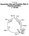
- a restriction site and function map of plasmid pGAG1317 is presented in Figure 17 of the accompanying drawings.
- Plasmid pAg932 can be conventionally isolated from E . coli K12 DH5/pAg932, a strain deposited with and made part of the permanent stock culture collection of the Northern Regional Research Laboratory (NRRL), Peoria, Illinois on November 20, 1987.
- a culture of E . coli K12 DH5/pAg932 can be obtained from the NRRL under the accession number NRRL B-18266.
- a restriction site and function map of plasmid pAg932 is presented in Figure 14 of the accompanying drawings.
- plasmid pAg1338 can be isolated from E . coli K12 DH5/pAg1338, also deposited and made part of the permanent stock culture collection of the NRRL on November 20, 1987.
- a culture of E . coli K12 DH5/pAg1338 can be obtained from the NRRL under the accession number NRRL B-18265.
- a restriction site and function map of plasmid pAg1338 is presented in Figure 15 of the accompanying drawings.
- Plasmid pGEMTM-4 is publicly available and may be purchased from Promega Biotech, 2800 South Fish Hatchery Road, Madison, WI 53711.
- a restriction site and function map of plasmid pGEMTM-4 is presented in Figure 16 of the accompanying drawings.
- Plasmid pGAG1317 comprises both the coding sequence of preproKSA and also additional sequences which comprise the 5′ and 3′ untranslated regions of preproKSA. These additional sequences are: and wherein A is deoxyadenyl, G is deoxyguanyl, C is deoxycytidyl, and T is thymidyl, at the 5′ and 3′ ends, respectfully, of the coding strand of the nascent prepro-KSA coding sequence. Due to the complementary nature of DNA base-pairing, the sequence of one strand of a double-stranded DNA molecule is sufficient to determine the sequence of the opposing strand.
- a variety of recombinant DNA expression vectors comprising the KSA encoding DNA have been constructed.
- the present vectors are of two types: those designed to transform eukaryotic, especially mammalian host cells; and those designed to transform E . coli .
- the eukaryotic or mammalian vectors exemplified herein can also transform E . coli , but the eukaryotic promoter present on these plasmids for transcription of KSA encoding DNA functions inefficiently in E . coli .
- the present DNA compounds which encode nascent KSA are especially preferred for the construction of vectors for transformation and expression of KSA in mammalian and other eukaryotic cells.
- Many mammalian host cells possess the necessary cellular machinery for the recognition and proper processing of signal (pre) peptide present on the amino-terminus of KSA.
- Some mammalian host cells also provide the post-translational modifications, such as glycosylation, that are observed in KSA present on the surface of adenocarcinoma cells.
- a wide variety of vectors exist for the transformation of eukaryotic host cells, and the specific vector exemplified below is in no way intended to limit the scope of the present invention.
- the BK enhancer-type vector of the present invention comprises a BK enhancer-adenovirus late promoter cassette plus a hygromycin resistance conferring gene and a murine dihydrofolate reductase (dhfr) gene.
- the use of the BK virus enhancer in conjunction with the adenovirus late promoter significantly increases transcription of a recombinant gene in eukaryotic host cells.
- the hygromycin resistance-conferring gene is present as a selectable marker for use in eukaryotic host cells.
- the murine dihydrofolate reductase gene under appropriate conditions, is amplified in the host chromosome.
- dhfr gene is a selectable marker in dhfr-negative cells and can be used to increase the copy number of a DNA segment by exposing the host cell to increasing levels of methotrexate.
- Plasmid pLPChd may be used to construct a eukaryotic expression vector for expression of the novel KSA structural gene of the present invention.
- Plasmid pLPChd contains the dhfr gene, the Adenovirus type-2 promoter and the BK virus enhancer.
- the BK virus which contains the BK virus enhancer, can be purchased or readily isolated in large quantities as described in Example 13.
- the BK virus is also available from the American Type Culture Collection under the accession number ATCC VR-837.
- Plasmid pdBPV-MMTneo comprises the replicon and ⁇ -lactamase gene from plasmid pBR322, the mouse metallothionein promoter positioned to drive expression of a structural gene that encodes a neomycin resistance-conferring enzyme, and about 8 kb of bovine papilloma virus (BPV) DNA.
- Plasmid pdBPV-MMTneo can be digested with restriction enzyme Bam HI to generate two fragments: the ⁇ 8 kb fragment that comprises the BPV DNA and an ⁇ 7 kb fragment that comprises the other sequences described above.
- BK virus has only one Bam HI restriction site, and plasmids pBKneo1 and pBKneo2 were constructed by ligating the ⁇ 7 kb Bam HI restriction fragment of plasmid pdBPV-MMTneo to Bam HI-linearized BK virus DNA.
- Plasmid pBKneo1 contains an ⁇ 2.1 kb Sal I- Hin dIII restriction fragment, whereas plasmid pBKneo2 contains an ⁇ 1.0 kb restriction fragment.
- Plasmids pBKneo1 and pBKneo2 each comprise the entire genome of the BK virus, including the enhancer sequence, and thus serve as useful starting materials for the expression vector of the present invention.
- Expression vector, plasmid pBLcat comprises the BK enhancer sequence in tandem with the human adenovirus-type-2 late promoter positioned to drive expression of the chloramphenicol acetyltransferase enzyme (CAT).
- Plasmid pSV2cat serves as a convenient source of the CAT gene and can be obtained from the ATCC under the accession number ATCC 37155.
- Human adenovirus-type-2 DNA is commercially available and can also be obtained from the ATCC under the accession number ATCC VR-2.
- Illustrative plasmid pBLcat was constructed by ligating the ⁇ 0.32 kb late-promoter-containing Acc I- Pvu II restriction fragment of human adenovirus-type-2 DNA to blunt-ended Bcl I linkers that attached only to the Pvu II end of the Acc I- Pvu II restriction fragment. The resulting fragment was then ligated to the ⁇ 4.51 kb Acc I- Stu I restriction fragment of plasmid pSV2cat to yield intermediate plasmid pLPcat.
- the desired plasmid pBLcat was constructed from plasmid pLPcat by ligating the origin of replication and enhancer-containing ⁇ 1.28 kb Acc I- Pvu II restriction fragment of BK virus DNA to the ⁇ 4.81 kb Acc I- Stu I restriction fragment of plasmid pLPcat.
- the construction of plasmid pBLcat is further described in Example 15.
- Plasmid pL133 was next constructed from plasmids pHC7, pSV2gpt and pSV2- ⁇ -globin.
- Plasmid pHC7 comprises a DNA sequence which encodes human protein C.
- Plasmid pHC7 can be isolated from E . coli K12 RR1/pHC7 which is available from the NRRL (deposit date, January 29, 1985) under accession number NRRL B-15926. Plasmid pHC7 was cut with restriction enzyme Ban I and the ⁇ 1.25 kb restriction fragment was isolated. Linkers were added, and the fragment was then cut with restriction enzymes Apa I and Hin dIII, then the desired ⁇ 1.23 kb restriction fragment was isolated.
- Plasmid pHC7 was next cut with restriction enzyme Pst I, the ⁇ 0.88 kb restriction fragment was isolated, linkers were added, the fragment was re-cut with restriction enzymes Apa I and Bgl II and the ⁇ 0.19 kb Apa I- Bgl II restriction fragment was isolated.
- Plasmid pSV2gpt (ATCC 37145) was digested with restriction enzymes Hin dIII and Bgl II and the ⁇ 5.1 kb fragment was isolated.
- Plasmid pSV2-HPC8 was then cut with restriction enzymes Hin dIII and Sal I, and the ⁇ 0.29 kb restriction fragment was isolated. Likewise, plasmid pSV2-HPC8 was also cut with restriction enzymes Bgl II and Sal I, and the ⁇ 1.15 kb restriction fragment was isolated. Plasmid pSV2- ⁇ -globin (NRRL B-15928, deposited January 29, 1985) was cut with restriction enzymes Bgl II and Hin dIII and the ⁇ 4.2 kb restriction fragment was isolated. These three fragments were then ligated together to form plasmid pL133. A detailed description of the construction of plasmid pL133 is found in Example 16.
- Plasmid pL133 was digested with restriction enzyme Hin dIII, then treated with alkaline phosphatase. Plasmid pBLcat was also cut with restriction enzyme Hin dIII and the ⁇ 0.87 kb restriction fragment was isolated. This fragment was ligated into the Hin dIII cut, phosphatased plasmid pL133 vector to form plasmid pLPC. Because the Hin dIII fragment of plasmid pBLcat can be inserted into plasmid pL133 in two orientations, it should be noted that pLPC is the plasmid wherein the proper orientation provides an ⁇ 1.0 kb Nde I- Stu I fragment.
- Plasmid pLPC like plasmid pL133, comprises the enhancer, early and late promoters, T-antigen-binding sites, and origin of replication of SV40.
- a detailed protocol for the construction of plasmid pLPC is provided in Example 17.
- the SV40 elements present on plasmid pLPC are situated closely together and difficult to delineate.
- the binding of T antigen to the T-antigen-binding sites, which is necessary for SV40 replication, is known to enhance transcription from the SV40 late promoter and surprisingly has a similar effect on the BK late promoter.
- plasmid pLPC nor plasmid pL133 are stably maintained as episomal (extrachromosomal) elements in the presence of SV40 T antigen, but rather, the two plasmids must integrate into the chromosomal DNA of the host cell to be stably maintained.
- the overall structure of the BK enhancer region is quite similar to that of SV40, for the BK enhancer, origin of replication, early and late promoters, and the BK analogue of the T-antigen-binding sites are all closely situated and difficult to delineate on the BK viral DNA.
- a plasmid that comprises the BK origin of replication and T-antigen-binding sites does not replicate to an extent that proves lethal and is stably maintained as an episomal element in the host cell.
- the T-antigen-driven replication can be used to increase the copy number of a vector comprising the BK origin of replication so that when selective pressure is applied more copies of the plasmid integrate into the host cell's chromosomal DNA.
- BK replication is also stimulated by SV40 T antigen.
- Episomal maintenance of a recombinant DNA expression vector is not always preferred over integration into the host cell chromosome.
- plasmid pLPC due to the absence of a selectable marker that functions in eukaryotic cells, the identification of stable, eukaryotic transformants of plasmid pLPC is difficult, unless plasmid pLPC is cotransformed with another plasmid that does comprise a selectable marker. Consequently, plasmid pLPC has been modified to produce derivative plasmids that are selectable in eukaryotic host cells.
- Plasmid pSV2hyg can be obtained from the Northern Regional Research Laboratory (NRRL), Peoria, IL 61640, under the accession number NRRL B-18039 (deposit date, February 11, 1986). Plasmid pSV2hyg was digested with restriction enzyme Bam HI, and the ⁇ 2.5 kb Bam HI restriction fragment, which comprises the entire hygromycin resistance-conferring gene, was isolated, treated with Klenow enzyme (the large fragment produced upon subtilisin cleavage of E .
- Bam HI restriction enzyme
- Plasmids pLPChyg1 and pLPChyg2 differ only with respect to the orientation of the hygromycin resistance-conferring fragment. Plasmid pLPChyg1 contains an ⁇ 5.0 kb Hin dIII fragment whereas plasmid pLPChyg2 contains an ⁇ 1.0 kb fragment.
- the construction protocol for plasmids pLPChyg1 and pLPChyg2 is described in Example 18.
- Plasmid pBW32 which contains the murine dihydrofolate reductase (dhfr) gene, was constructed next.
- Plasmid pTPA102 (NRRL B-15834, deposited on August 10, 1984) was cut with restriction enzyme Tth 111I and the ⁇ 4.4 kb restriction fragment was isolated. This fragment was treated with Klenow, linkers were added, then the fragment was cut with restriction enzymes Hin dIII and Bam HI to yield an ⁇ 2.0 kb restriction fragment.
- Plasmid pRC was then constructed by ligating the ⁇ 288 bp Cla I- Eco RI restriction fragment of pTPA102 into Cla I- Eco RI cut vector pKC7.
- Plasmid pKC7 can be obtained from the ATCC under the accession number ATCC 37084. Plasmid pRC was digested with restriction enzymes Bam HI and Hin dIII, then ligated to the ⁇ 2.0 kb restriction fragment of plasmid pTPA102, formed above, to yield plasmid pTPA103. The construction protocol for plasmid pTPA103 is described in Example 19A.
- Plasmid pTPA103 was cut with restriction enzyme Bgl II, treated with Klenow, and the Nde I linkers were added. This mixture was then ligated to form plasmid pTPA103der Nde I. Plasmid pTPA103der Nde I was cut with restriction enzyme Ava II, and the ⁇ 1.4 kb fragment was isolated. This fragment was treated with Klenow, then, after the addition of Hpa I linkers, was cut with restriction enzyme Eco RI. The ⁇ 770 bp fragment, containing trp PO and the amino terminus of TPA, was ligated into Eco RI- Sma I digested vector pUC19, to form pUC19TPAFE.
- Plasmid pUC19TPAFE was partially digested with restriction enzyme Hpa I, then totally cut with restriction enzyme Bam HI. The resultant ⁇ 3.42 kb Hpa I- Bam HI restriction fragment was then ligated to the ⁇ 1.015 Sca I- Bam HI fragment derived from plasmid pTPA103 to form plasmid pBW25.
- the construction protocol for plasmid pBW25 is described in Example 19B.
- Plasmid pBW25 was cut with restriction enzymes Hin dIII and Eco RI and the resultant ⁇ 810 bp fragment was ligated into Hin dIII- Eco RI cut phage M13mp8 (New England Biolabs) to form phage pM8BW26.
- An in vitro mutagenesis reaction was then performed on phage pM8BW26 (deleting DNA coding for amino acid residues) to form phage pM8BW27.
- Phage pM8BW27 was cut with restriction enzymes Eco RI and Nde I and the ⁇ 560 bp restriction fragment was isolated.
- a synthetic Nde I- Xba I linker of ⁇ 48 bp was synthesized.
- Plasmid pTPA103 was cut with restriction enzymes Eco RI and Bam HI and the ⁇ 689 bp fragment was isolated.
- Plasmid pL110 (constructed in Example 9) was partially digested with restriction enzyme Bam HI, then totally cut with Xba I and the ⁇ 6.0 kb fragment was isolated.
- This ⁇ 6.0 kb vector fragment, the ⁇ 689 bp fragment of plasmid pTPA103, the ⁇ 560 bp fragment of phage pM8BW27, and the ⁇ 48 bp linker were all then ligated together to form plasmid pBW28.
- the construction protocol of plasmid pBW28 is described in Example 19C.
- Plasmid pTPA301 was next formed by ligating the ⁇ 2.0 kb Hin dIII- Bgl II fragment of plasmid pTPA103 to the ⁇ 4.2 kb Hin dIII- Bgl II fragment of plasmid pSV2- ⁇ -globin. Plasmid pSV2-dhfr (ATCC 37146) was cut with restriction enzyme Pvu II. Following the addition of Bam HI linkers, the ⁇ 1.9 kb dhfr gene-containing fragment was ligated into Bam HI cut, phosphatased plasmid pTPA301 to form plasmid pTPA303.
- Plasmid pTPA301 was cut with restriction enzymes Eco RI and Bgl II to yield an ⁇ 2.7 kb fragment.
- Plasmid pTPA303 was cut with restriction enzymes Hin dIII and Eco RI to yield the ⁇ 2340 bp dhfr gene containing fragment.
- Plasmid pTPA303 was cut with restriction enzymes Hin dIII and Sst I to yield an ⁇ 1.7 kb fragment.
- Plasmid pBW28 was cut with restriction enzymes Xho II and Sst I to yield an ⁇ 680 bp fragment.
- Plasmid pLPChyg1 contains two Eco RI restriction enzyme recognition sites, one in the hygromycin resistance-conferring gene and one in the plasmid pBR322-derived sequences.
- Plasmid pLPChd1 has been designated plasmid pLPChd.
- a restriction site and function map of plasmid pLPChd is presented in Figure 20 of the accompanying drawings. The construction of plasmids pLPChd1 and pLPChd2, which differ only with respect to the orientation of the dhfr gene-containing DNA segment, is described in Example 20.
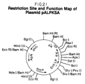
- Plasmid pALPKSA is a vector of the present invention derived from plasmid pGAG1317 and plasmid pLPChd.
- the ⁇ 1200 base pair Bss HII- Hinc II fragment of plasmid pGAG1317 is treated with Klenow to fill in the 5′ overhang. This fragment represents the entire KSA coding region.
- This fragment is then ligated into the Bcl I-digested, purified vector pLPChd. This essentially substitutes the protein C structural gene of plasmid pLPChd with the KSA structural gene of plasmid pGAG1317 to form preferred expression vector pALPKSA.
- a more detailed description of the construction of plasmid pALPKSA is provided in Example 21.
- a restriction site and function map of plasmid pALPKSA is presented in Figure 21 of the accompanying drawings.
- the present invention is in no way limited to the use of the particular eukaryotic promoters exemplified herein.
- Other promoters such as the SV40 late promoter or promoters from eukaryotic genes, such as for example, the estrogen-inducible chicken ovalbumin gene, the interferon genes, the glucocorticoid-inducible tyrosine aminotransferase gene, the thymidine kinase gene, the major early adenovirus gene, and the SV40 early promoter, can be readily isolated and modified for use on recombinant DNA expression vectors designed to produce KSA in eukaryotic host cells.
- Eukaryotic promoters can also be used in tandem to drive expression of KSA.
- retroviruses that infect a wide range of eukaryotic host cells.
- Long terminal repeats in the retrovirus DNA often encode promoter activity and can be used, in place of the BK enhancer-adenovirus late promoter described above, to drive expression of KSA.
- the vector pALPKSA can be transformed into and expressed in a variety of eukaryotic, especially mammalian host cells.
- Plasmid pALPKSA also comprises sequences that allow for replication in E . coli , as it is usually more efficient to prepare plasmid DNA in E . coli than in other host cells.
- Expression of KSA occurs in host cells in which the particular promoter associated with the nascent KSA structural gene functions. Skilled artisans will understand that a variety of eukaryotic host cells can be used to express KSA using the BK enhancer-adenovirus late promoter, so long as the host cell expresses an immediate-early gene product of a large DNA virus.
- the immediate-early gene product can be introduced into host cells by many means, such as transformation with a plasmid or other vector, virtually any eukaryotic cell can be used in the present method.
- Human cells are preferred host cells in the method of the present invention, because human cells are the natural host for BK virus and may contain cellular factors that serve to stimulate the BK enhancer. While human kidney cells are especially preferred as host cells, the adenovirus 5-transformed embryonic cell line 293, which expresses the E1A gene product, is most preferred and is available from the American Type Culture Collection in Rockville, Maryland, under the accession number ATCC CRL 15753.
- the present DNA compounds can also be expressed in prokaryotic host cells such as, for example, E . coli , Bacillus and Streptomyces . Since prokaryotic host cells usually do not glycosylate, and often do not properly fold, mammalian proteins made from recombinant genes, a variety of novel KSA derivatives can be produced by expressing the present KSA-encoding DNA in prokaryotic host cells.
- the novel KSA derivatives expressed in prokaryotic host cells may show varying degrees of reactivity with monoclonal antibody KS1/4 and can be used to determine the folding and post-translational modification requirements for specific antibody/antigen interactions.
- novel KSA derivatives may also be used to create novel, heretofore unknown antibodies which react only to specific portions of KSA.
- Skilled artisans will readily understand that the ability of an antibody to recognize certain portions of an antigen is essential when using competitive assays for diagnosis or therapy.
- the DNA encoding the eukaryotic signal peptide (prepeptide) and the eukaryotic propeptide was removed.
- prepeptide the eukaryotic signal peptide
- the present invention is not limited or dependent on any theory of mode of action, it is believed that the first 21 amino acid residues at the amino-terminus of nascent preproKSA act as a signal peptide (prepeptide).
- the present invention is not limited to the use of a particular eukaryotic signal peptide for expression of KSA in eukaryotic host cells.
- the next 60 amino acid residues at the amino-terminus of nascent prepro-KSA act as a propeptide.
- the removal of the prepropeptide forms a molecule which has a nascent chain that is substantially the same as the nascent chain of the KSA found on the cell surface of UCLA-P3 cells.
- prokaryotes do not efficiently process eukaryotic signal peptides; therefore it is somewhat inefficient to express the signal peptide-encoding portion of the nascent KSA structural gene in prokaryotes.
- the present invention also comprises the fusion of a prokaryotic signal peptide-encoding DNA to the KSA-encoding DNA of the present invention for expression and secretion of KSA in prokaryotes.
- amino acid residues 1-21 of nascent KSA which may encode a "signal" (prepeptide) for extracellular secretion of a portion of KSA, are not present in the nascent KSA found on the surface of adenocarcinoma cells.
- Residues 22-81 of nascent KSA which comprise a propeptide of KSA, are also removed during the processing of the protein and are believed to be responsible for the correct folding and modification of the molecule.
- Residues 82-265 of nascent KSA are encoded in the prokaryotic expression vector exemplified below, but residues 266-314 are not; those residues comprise the cytoplasmic domain and transmembrane region.
- the present invention is not limited to the expression of a particular KSA derivative.
- the present DNA compounds are readily modified to delete certain portions encoding various amino acid residues of the KSA.
- restriction enzyme digestion or site-directed mutagenesis upon the DNA compounds of the present invention will yield an almost limitless group of molecules which will encode for KSA derivatives. Such manipulations are within the scope of this invention, and can be performed given the detailed sequences disclosed herein.
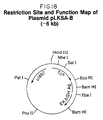
- Plasmid pLKSA is a plasmid of the present invention designed to express amino acid residues 82-265 of the KSA in E . coli .
- Plasmid pLKSA was constructed from plasmid pGAG1317 and plasmid pL110C. Plasmid pGAG1317 was described and disclosed earlier. A brief description of the construction of plasmid pL110C is provided below and a detailed description is provided in Examples 1-10. A restriction site and function map of plasmid pL110C is presented in Figure 13 of the accompanying drawings.
- Plasmid pKC283 was first obtained from E . coli K12 BE1201/pKC283, deposited on August 3, 1984. This culture may be obtained from the NRRL under accession number NRRL B-15830. Plasmid pKC283 comprises a hybrid lpp -pL promoter of bacteriophage ⁇ . This plasmid is obtained in E . coli K12 BE1201 cells because these cells comprise a temperature sensitive cI repressor integrated into the cellular DNA. A detailed description of the isolation of plasmid pKC283 is presented in Example 1. A restriction site and function map of plasmid pKC283 is presented in Figure 1 of the accompanying drawings.
- plasmid pKC283 was excised by first digesting the plasmid with restriction enzyme Pvu II. Specific DNA linkers were then added to the digested DNA to convert the Pvu II sites into a single Xho I site, which created plasmid pKC283PX.
- a detailed description of the isolation of plasmid pKC283PX is presented in Example 2.
- a restriction site and function map of plasmid pKC283PX is presented in Figure 2 of the accompanying drawings.
- plasmid pKC283PX is transformed into E . coli K12 MO( ⁇ +).
- E . coli K12 MO( ⁇ +) deposited on August 14, 1985, is available from the NRRL under the accession number NRRL B-15993.
- Plasmid pKC283PX was next digested with restriction enzymes Bgl II and Xho I. After the vector was purified, DNA linkers with Bgl II and Xho I ends were ligated into the vector to form plasmid pKC283-L. The Bgl II- Xho I linker also contained an Xba I site.
- a detailed description of the construction of plasmid pKC283-L is presented in Example 4.
- a restriction site and function map of plasmid pKC283-L is presented in Figure 3 of the accompanying drawings.
- plasmid pKC283-L The Xho I site of plasmid pKC283-L was next converted into a Bam HI site. This was accomplished by a total digestion of plasmid pKC283-L with restriction enzyme Xho I, followed by treatment with Klenow, then addition of Bam HI linkers, to form plasmid pKC283-LB.
- a detailed description of the construction of plasmid pKC283-LB is presented in Example 5.
- a restriction site and function map of plasmid pKC283-LB is presented in Figure 4 of the accompanying drawings.
- Plasmid pKC283PRS was then digested with restriction enzymes Pst I and Sph I and the ⁇ 0.85 kb Pst I- Sph I restriction fragment was isolated.
- plasmid pKC283-LB was digested with restriction enzymes Pst I and Sph I and the ⁇ 3.0 kb fragment was isolated.
- the ⁇ 0.85 kb Pst I- Sph I fragment of pKC283PRS was then ligated into the ⁇ 3.0 kb Pst I- Sph I vector fragment of pKC283-LB to form plasmid pL32.
- a detailed description of the construction of plasmid pL32 is presented in Example 6.
- a restriction site and function map of plasmid pL32 is presented in Figure 6 of the accompanying drawings.
- Plasmid pNM789 is obtained from the NRRL in E . coli K12 RV308/pNM789 under the accession number B-18216.
- a restriction site and function map of plasmid pNM789 is presented in Figure 7 of the accompanying drawings. Plasmid pNM789 was partially digested with restriction enzyme Pvu II, fully digested with restriction enzyme Bam HI, then treated with alkaline phosphatase. Next, a new Pvu II- Bam HI linker was ligated into the digested, phosphatased vector pNM789 to form plasmid 120. A detailed description of the construction of plasmid 120 is presented in Example 7. A restriction site and function map of plasmid 120 is presented in Figure 8 of the accompanying drawings.
- Plasmid 120 was then totally digested with restriction enzymes Xba I and Bam HI and the ⁇ 0.6 kb Xba I- Bam HI EK-BGH-encoding restriction fragment was isolated.
- Plasmid pL32 was also digested with restriction enzymes Xba I and Bam HI and the ⁇ 3.9 kb vector fragment was isolated.
- the ⁇ 0.6 kb Xba I- Bam HI fragment of plasmid 120 was then ligated into the ⁇ 3.9 kb vector fragment of plasmid pL32 to form plasmid pL47.
- a detailed description of the construction of plasmid pL47 is presented in Example 7.
- a restriction site and function map of plasmid pL47 is presented in Figure 9 of the accompanying drawings.
- Plasmid pPR12 comprises the temperature-sensitive pL repressor gene cI857 and the plasmid pBR322 tetracycline resistance-conferring gene. Plasmid pPR12 is disclosed and claimed in U.S. Patent No. 4,436,815, issued 13 March, 1984. A restriction site and function map of plasmid pPR12 is presented in Figure 10 of the accompanying drawings. The Eco RI site was removed from plasmid pPR12 by first totally digesting the plasmid with restriction enzyme Eco RI, followed by treatment with Klenow. The vector was then recircularized by ligation to form plasmid pBR12 ⁇ R1.
- Plasmid pPR12 ⁇ R1 was then digested with restriction enzyme Ava I and treated with Klenow.
- the Ava I-digested, Klenow treated pPR12 ⁇ R1 was next ligated to Eco RI linkers, cut with restriction enzyme Eco RI, then recircularized to form plasmid pPR12AR1.
- a detailed description of the construction of plasmid pPR12AR1 is presented in Example 8.
- a restriction site and function map of plasmid pPR12AR1 is presented in Figure 11 of the accompanying drawings.
- Plasmid pL47 was digested with restriction enzymes Pst I and Bam HI and the ⁇ 2.7 kb Pst I- Bam HI restriction fragment was isolated.
- plasmid pL47 was digested with restriction enzymes Eco RI and Bam HI and the ⁇ 1.03 kb Eco RI- Bam HI fragment was isolated.
- Plasmid pL110 was cut with restriction enzyme Nde I, treated with Klenow, then recircularized to form plasmid pL110A. Plasmid pL110 was also cut with restriction enzymes Hin dIII and Eco RI and the tetracycline resistance-conferring fragment was isolated. This fragment was ligated to the ⁇ 7.25 kb Hin dIII to Eco RI restriction fragment of phage m13mp18 to form phage M13Tc3. Phage m13mp18 may be purchased from New England Biolabs.
- Single-stranded phage M13Tc3 was then isolated and an in vitro mutagenesis reaction was performed to change the nucleotide C to an A in the Bam HI site of the tetracycline gene. This does not alter the amino acid composition of the tetracycline resistance-conferring protein but eliminates the Bam HI site.
- the newly mutagenized plasmid was transformed and a replicative form which lacked the Bam HI site was selected and designated plasmid pL110B.
- Plasmid pL110B was cut with restriction enzymes Nhe I and Sal I, then the tetracycline resistance-conferring fragment was isolated. Plasmid pL110A was likewise cut with restriction enzymes Nhe I and Sal I, then the large vector fragment was isolated. The tetracycline-resistance conferring fragment of plasmid pL110B was then ligated into the Nhe I- Sal I cut vector fragment of pL110A to form plasmid pL110C. A detailed description of the construction of pL110C is presented in Example 10. A detailed construction protocol and restriction site and function map of plasmid pL110C is presented in Figure 13 of the accompanying drawings.
- Plasmid pGAG1317 was cut with restriction enzyme Apa I, then treated with alkaline phosphatase.
- Linkers were ligated onto the molecule which comprise the DNA that encodes the first six amino acid residues of KSA plus an added methionine residue. These linkers also comprise an Xba I restriction site at the 5′ end.
- the DNA was then digested with Xba I and Eco RI, and the ⁇ 500 kb Xba I- Eco RI restriction fragment was isolated.
- Plasmid pL110C was digested with Eco RI and Xba I and the large vector fragment was isolated.
- plasmid pLKSA-B The ⁇ 500 kb Eco RI- Xba I fragment of pGAG1317 was then ligated into the Eco RI- Xba I vector fragment of pL110C to form plasmid pLKSA-B.
- a detailed description of the construction of plasmid pLKSA-B is presented in Example 12.
- a restriction site and function map of plasmid pLKSA-B is presented in Figure 18 of the accompanying drawings.
- Plasmid pLKSA-B was digested with restriction enzyme Eco RI, then dephosphorylated with alkaline phosphatase.
- Eco RI- Bam HI linkers were then ligated onto the Eco RI-cut molecule. These linkers also comprise DNA encoding the eight amino acid residues between the Eco RI site and the transmembrane region of KSA plus a transcriptional "stop" codon.
- This plasmid was then cut with restriction enzyme Xba I and the Xba I- Bam HI KSA-encoding fragment was isolated.
- pL110C was cut with restriction enzymes Xba I and Bam HI and the large vector fragment was isolated.
- the KSA-encoding Xba I- Bam HI restriction fragment of pLKSA-B was then ligated into the Xba I- Bam HI vector fragment of plasmid pL110C to form plasmid pLKSA.
- a detailed description of the construction of plasmid pLKSA is presented in Example 12.
- a restriction site and function map of plasmid pLKSA is presented in Figure 19 of the accompanying drawings.
- Plasmid pLKSA comprises the tetracycline resistance-conferring gene, the temperature sensitive cI857 repressor gene, the hybrid pL/ lpp promoter system and amino acid residues 82-265 of the nascent prepro KSA.
- Amino acid residues 82-265 of the nascent prepro KSA comprise the nascent amino acid antigen structure that is found on the cell surface of adenocarcinoma cells such as UCLA-P3 cells.
- Expression of KSA in E . coli is in no way limited to the use of a particular promoter, since the choice of a specific promoter is not critical to the operability of the present invention.
- Promoters which can be substituted for the previously exemplified pL promoter include, but are not limited to, the E . coli lactose ( lac ), the E . coli trp , bacteriophage ⁇ P L O L , and bacteriophage ⁇ P R O R promoters.
- one or more promoters can be used in tandem, such as, for example, the trp and lac promoters, or hybrid promoters, such as the tac promoter, can be used to drive expression of the KSA structural gene. All of the aforementioned promoters have been previously characterized, are well known in the art, and can be constructed either synthetically or from known plasmids.
- the present invention is not limited to the use of any given replicon-containing plasmid for expression of KSA in E . coli .
- Many replicons such as those from pBR322, pBR328, pACYC184, and the like, are known in the art and are suitable for the construction of recombinant DNA cloning and expression vectors designed to drive expression of the KSA-encoding DNA compounds of the present invention.
- selectable markers exist, both for eukaryotic and prokaryotic host cells, that are suitable for use on a recombinant DNA cloning or expression vector comprising a DNA compound (or sequence) of the present invention.
- DNA sequences and plasmids are possible.
- the degeneracy of the genetic code allows for the substitution of nucleotides throughout polypeptide coding regions as well as for the substitution of the translational stop signals for the translational stop signal specifically exemplified.
- sequences can be deduced from the now-known amino acid or DNA sequence of KSA and can be constructed by following conventional synthetic procedures. Such synthetic methods can be carried out in substantial accordance with the procedures of Itakura et al ., 1977 Science 198:1056 and Crea et al ., 1978, Proc. Nat. Acad. Sci. USA 75:5765.
- genes and linkers can be synthesized either by using a Systec 1450A DNA synthesizer (Systec Inc., 3816 Chandler Drive, Minneapolis, MN) or an ABS 380A DNA synthesizer (Applied Biosystems, Inc., 850 Lincoln Center Drive, Foster City, CA 94404).
- Systec 1450A DNA synthesizer Systec Inc., 3816 Chandler Drive, Minneapolis, MN
- ABS 380A DNA synthesizer Applied Biosystems, Inc., 850 Lincoln Center Drive, Foster City, CA 94404.
- Many other DNA synthesizing instruments are known in the art and can be used to make synthetic DNA fragments. Therefore, the present invention is in no way limited to the DNA sequences and plasmids specifically exemplified.
- the prokaryotic expression vectors and method of this invention can be applied to a wide range of host organisms, especially Gram-negative prokaryotic organisms such as Escherichia coli , E . coli K12, E . coli K12 RV308, E . coli K12 HB101, E . coli K12 C600, E . coli K12 RR1, E . coli K12 RR1 ⁇ M15, E . coli K12 MM294, E . coli K12 DH5, and the like. Although all of the embodiments of the present invention are useful, some of the vectors and transformants are preferred. A preferred transformant is E . coli K12 RV308/pLKSA.
- the expression vectors of this invention are used to transform either eukaryotic or prokaryotic host cells, such that a polypeptide with nascent chain KSA structure is expressed by the host cell. If the host cell is transformed with a vector comprising a promoter that functions in the host cell and drives transcription of the nascent KSA structural gene, and if the host cell possesses the cellular machinery with which to process the signal peptide, mature KSA can be found on the surface of such cells. Under other expression conditions, such as when plasmid pLKSA is in E . coli RV308, the KSA must be isolated from the host cell.
- KSA produced by recombinant methodology will have a profound effect upon the diagnosis, prognosis, treatment and study of cancers of epithelial origin. Furthermore, because KSA is also expressed on a subset of normal human epithelial cells, the amino acid and nucleotide sequences disclosed will be useful in understanding the role of such cell surface antigens in normal tissue differentiation, development and non-malignant disease states.
- the KSA gene (or subfragments thereof) can be used to probe DNA libraries derived from a wide range of cell types, to find other related genes or variants thereof. These new antigen genes can then be used to construct novel antibodies in a further attempt to combat cancer.
- Monoclonal antibody KS 1/4 has been shown to be an effective agent for the diagnosis, prognosis and treatment of cancer by Bumol in Reisfeld, R.A. and Sell, S. eds. Monoclonal Antibodies and Cancer Therapy. New York: Alan R. Liss, Inc., 1985, 257-259. Spearman et al ., 1987, J. Pharmacol. and Exp. Therapeutics 241:695-703, the teaching of which is herein incorporated by reference, disclosed the use of a monoclonal antibody-vinca alkaloid conjugate in the localization and treatment of tumors. This KS 1/4-DAVLB (4-desacetylvinblastine) conjugate was also responsible for tumor growth suppression as disclosed by Bumol et al .
- KSA will be useful in modifying the affinity of KS 1/4.
- X-Ray crystallographic analysis will demonstrate which amino acid residues of the antigen appear in close proximity to certain amino acid residues of KS 1/4.
- KS 1/4 can then be modified to provide negative residues near positive residues on the antigen.
- Such "engineered” antibodies will then display increased affinity to cell surface KSA in cancer patients.
- these protein engineering techniques can be used to create low affinity KS1/4 derivatives also.
- recombinant KSA and its derivatives can be isolated from any cell in which they are expressed using solubilization procedures that are well known in the art.
- Kahan in “Methods of Cancer Research,” Vol. IX, Busch, ed., p. 283-338 (Academic Press, New York, 1973) describes a variety of extraction techniques, as does Graham, in “New Techniques in Biophysics and Cell Biology,” Vol. 2, Pain et al ., eds., pp. 1-42 (Wiley, London 1975).
- the antigen fractions can then be used to create novel antibodies according to the teaching of Köhler and Milstein, 1975, Nature 256:495-497 or Goldenberg, U.S. Patent No. 4,444,744.
- These KS 1/4 "sister” antibodies which can be isolated due to their specific reactivity to recombinant KSA or UCLA-P3 cells, are also useful for the diagnosis and treatment of disease states.
- Recombinant KSA and its derivatives can also be used to raise polyclonal antibodies which react with tumor cells. Occasionally, such polyclonal antibodies will demonstrate an increased reactivity to tumor surface markers.
- Polyclonal antibodies are raised by methods which are well known in the art. An animal is challenged with the antigen or any subunit thereof, then, following an appropriate amount of time in which the animal's immune system produces antibodies against the antigen, the animal is bled. The antibodies are then isolated according to well known techniques such as those disclosed in "Immunodiagnosis of Cancer,” Herberman et al ., Eds. (Marcel Dekker, Inc., New York and Basel, 1979) and “Tumor Markers,” Sell, Ed. (Humana Press, Clifton, N.J., 1980). These antibodies are then used in immunological assays to test the presence of adenocarcinoma cells in tissue.
- Recombinant KSA and its derivatives can also be prepared to offer large quantities of defined antigen for the standardization of analytical methodology. For example, functional immunoassays can be developed to monitor fermentation and production of KS1/4 antibody or newly developed derivatives thereof. Affinity chromatography, using recombinant KSA, will greatly simplify the purification of monoclonal antibody KS1/4. The recombinant KSA and its derivatives can also be used in the purification, formulation analysis and stability studies of various monoclonal antibody based products. Recombinant KSA can also be used for development of potential anti-adenocarcinoma vaccines.
- Lyophils of E . coli K12 BE1201/pKC283, deposited on August 3, 1984, are obtained from the Northern Regional Research Laboratory, Peoria, Illinois 61604, under the accession number NRRL B-15830.
- the lyophils are decanted into tubes containing 10 ml LB medium (10 g Bacto-tryptone, 5 g Bacto-yeast extract, and 10 g NaCl per liter; pH is adjusted to 7.5) and incubated two hours at 32°C, at which time the cultures are made 50 »g/ml in ampicillin and then incubated at 32°C overnight.
- 10 ml LB medium 10 g Bacto-tryptone, 5 g Bacto-yeast extract, and 10 g NaCl per liter; pH is adjusted to 7.5
- coli K12 BE1201/pKC283 cells were cultured at 32°C, because the cells comprise a temperature-sensitive cI repressor gene integrated into the cellular DNA.
- the temperature of incubation is 37°C.
- a small portion of the overnight culture is placed on LB-agar (LB medium with 15 g/l Bacto-agar) plates containing 50 »g/ml ampicillin in a manner so as to obtain a single colony isolate of E . coli K12 BE1201/pKC283.
- the single colony obtained was inoculated into 10 ml of LB medium containing 50 »g/ml ampicillin and incubated overnight at 32°C with vigorous shaking.
- the 10 ml overnight culture was inoculated into 500 ml LB medium containing 50 »g/ml ampicillin and incubated at 32°C with vigorous shaking until the culture reached stationary phase.
- the cells were harvested by centrifugation at 4000 g for 10 minutes at 4°C, and the supernatant was discarded.
- the cell pellet was washed in 100 ml of ice-cold STE buffer (0.l M NaCl; 10 mM Tris-HC1, pH 7.8; and 1 mM EDTA). After washing, the cell pellet was resuspended in 10 ml of Solution 1 (50 mM glucose; 25 mM Tris-HC1, pH 8.0; and 10 mM EDTA) containing 5 mg/ml lysozyme and left at room temperature for 10 minutes. Twenty ml of Solution 2 (0.2 N NaOH and 1% SDS) were then added to the lysozyme-treated cells, and the solution was gently mixed by inversion. The mixture was incubated on ice for 10 minutes.
- the lysed cell mixture was centrifuged in a Beckman SW27 (or its equivalent) at 20,000 rpm for 20 minutes at 4°C.
- the cell DNA and debris formed a pellet on the bottom of the tube.
- About 36 ml of supernatant were recovered, and 0.6 volumes of isopropanol were added, mixed, and the resulting solution left at room temperature for 15 minutes.
- the plasmid DNA was collected by centrifugation at 12,000 g for 30 minutes at room temperature. The supernatant was discarded, and the DNA pellet was washed with 70% ethanol at room temperature. The ethanol wash was decanted, and the pellet was dried in a vacuum desiccator.
- the pellet was then resuspended in 8 ml of TE buffer (10 mM Tris-HC1, pH 8.0, and 1 mM EDTA).
- CsCl Eight grams were added to the DNA solution. About 0.8 ml of a 10 mg/ml solution of ethidium bromide in water were added for each 10 ml of CsCl-DNA solution. The final density of the solution was about 1.55 g/ml, and the ethidium bromide concentraton was about 600 »g/ml.
- the solution was transferred to a Beckman Type 50 centrifuge tube, filled to the top with paraffin oil, sealed, and centrifuged at 45,000 rpm for 24 hours at 20°C. After centrifugation, two bands of DNA were visible in ordinary light. After removing the cap from the tube, the lower DNA band was removed by using a syringe with a #21 hypodermic needle inserted through the side of the centrifuge tube.
- the ethidium bromide was removed by several extractions with water-saturated 1-butanol.
- the CsCl was removed by dialysis against TE buffer. After extractions with buffered phenol and then chloroform, the DNA was precipitated, washed with 70% ethanol, and dried.
- About 1 mg of plasmid pKC283 was obtained and stored at 4°C in TE buffer at a concentration of about 1 »g/»l.
- a restriction site and function map of plasmid pKC283 is presented in Figure 1 of the accompanying drawings.
- Restriction enzyme reactions described herein were routinely terminated by phenol and then chloroform extractions, which were followed by precipitation of the DNA, an ethanol wash, and resuspension of the DNA in TE buffer. After terminating the Pvu II digestion as described above, the Pvu II-digested plasmid pKC283 DNA was precipitated and then resuspended in 5 »l of TE buffer.
- the reaction mixture was adjusted to have the composition of high-salt buffer (0.1 M NaCl; 0.05 M Tris-HC1, pH 7.5; 10.0 mM MgCl2; and 1 mM DTT). About 10 »l (100 units) of restriction enzyme Xho I were added to the mixture, and the resulting reaction was incubated at 37°C for 2 hours.
- high-salt buffer 0.1 M NaCl; 0.05 M Tris-HC1, pH 7.5; 10.0 mM MgCl2; and 1 mM DTT.
- the reaction was terminated, and the Xho I-digested DNA was precipitated, resuspended, and ligated as described above, except that no Xho I linkers were added to the ligation mixture.
- the ligated DNA constituted the desired plasmid pKC283PX.
- a restriction site and function map of plasmid pKC283PX is presented in Figure 2 of the accompanying drawings.
- E . coli K12 MO( ⁇ +) deposited on August 14, 1985, can be obtained from the Northern Regional Research Laboratories in lyophylized form under the accession number NRRL B-15993.
- E . coli K12 MO( ⁇ +) comprises the wild-type lambda pL cI repressor gene, so that transcription from the hybrid pL- lpp promoter of the present invention does not occur in E . coli K12 MO( ⁇ +) cells.
- the lyophils are reconstituted, single colonies of MO( ⁇ +) are isolated, and a 10 ml overnight culture of the MO( ⁇ +)cells is prepared in substantial accordance with the procedure of Example 1, except that the temperature of incubation is 37°C and no ampicillin is used in the growth media.
- Fifty »l of the overnight culture were used to inoculate 5 ml of LB media which also contained 10 mM MgSO4 and 10 mM MgCl2.
- the culture was incubated at 37°C overnight with vigorous shaking.
- the culture was diluted to 200 ml with LB media containing 10 mM MgSO4 and 10 mM MgCl2.
- the diluted culture was incubated at 37°C with vigorous shaking until the absorbance at 550 nm (A550) was about 0.5, which indicated a cell density of about 1 x 10⁸ cells/ml.
- the culture was cooled for ten minutes in an ice-water bath, and the cells were then collected by centrifugation at 4000 g for 10 minutes at 4°C.
- the cell pellet was resuspended in 100 ml of cold 10 mM MgSO4 and then immediately re-pelleted by centrifugation.
- the cell pellet was resuspended in 100 ml of 30 mM CaCl2 and incubated on ice for 20 minutes.
- the cells were again collected by centrifugation and resuspended in 10 ml of 30 mM CaCl2. A one-half ml aliquot of the cells was added to the ligated DNA prepared in Example 2; the DNA had been made 30 mM in CaCl2.
- the cell-DNA mixture was incubated on ice for one hour, heat-shocked at 42°C for 90 seconds, and then chilled on ice for about two minutes.
- the cell-DNA mixture was diluted into 10 ml of LB media in 125 ml flasks and incubated at 37°C for one hour. One hundred »l aliquots were plated on LB-agar plates containing ampicillin and incubated at 37°C until colonies appeared.
- Plasmid DNA isolation was performed on a smaller scale in accordance with the procedure of Example 1, but the CsC1 gradient step was omitted until the desired E . coli K12 MO( ⁇ +)/pKC283PX transformants were identified.
- a restriction site and function map of plasmid pKC283PX is presented in Figure 2 of the accompanying drawings.
- a DNA linker with single-stranded DNA ends characteristic of Bgl II and Xho I restriction enzyme cleavage was synthesized and kinased.
- the linker was kinased in substantial accordance with the procedure of Example 2.
- the DNA linker had the following structure:
- the linker depicted above was synthesized from single-stranded deoxyoligonucleotides by procedures well known in the art.
- the single-stranded deoxyoligonucleotides can be synthesized with commercially available instruments, such as the 380A DNA Synthesizer marketed by Applied Biosystems (850 Lincoln Centre Drive, Foster City, CA 94404), which utilizes phosphoramidite chemistry. Other procedures for synthesizing DNA are also known in the art.
- the linker and Bgl II- Xho I-digested plasmid pKC283PX were ligated in substantial accordance with the procedure of Example 2.
- the ligated DNA constituted the desired plasmid pKC283-L.
- a restriction site and function map of plasmid pKC283-L is presented in Figure 3 of the accompanying drawings.
- the plasmid pKC283-L DNA was used to transform E . coli K12 MO( ⁇ +) and the resulting E . coli K12 MO( ⁇ +)/pKC283-L transformants were identified in substantial accordance with the procedure of Example 3.
- plasmid pKC283-L DNA prepared in substantial accordance with the procedures of Example 1, were dissolved in 20 »l 10X high-salt buffer, 20 »l 1 mg/ml BSA, 5 »l ( ⁇ 50 units) restriction enzyme Xho I, and 155 »l of H20, and the resulting reaction was incubated at 37°C for two hours.
- the Xho I-digested plasmid pKC283-L DNA was then precipitated from the reaction mixture by the addition of three volumes of 95% ethanol and one-tenth volume of 3 M sodium acetate, incubation in a dry ice-ethanol bath for five minutes, and centrifugation.
- the resulting DNA pellet was washed with 70% ethanol, dried, and resuspended in 2 »l 10X nick-translation buffer (0.5 M Tris-HCl, pH 7.2; 0.1 M MgSO4; and 1 mM DTT), 1 »l of a solution 2 mM in each of the deoxynucleotide triphosphates, 15 »l of H20, 1 »l ( ⁇ 6 units as defined by P-L Biochemicals) of Klenow, which is the large fragment of E . coli DNA polymerase I, and 1 »l of 1 mg/ml BSA.
- the resulting reaction was incubated at 25°C for 30 minutes; the reaction was stopped by incubating the solution at 70°C for five minutes.
- Bam HI linkers (5′-CGGGATCCCG-3′) were kinased and ligated to the Xho I-digested, Klenow-treated plasmid pKC283-L DNA in substantial accordance with the procedure of Example 2. After the ligation reaction, the DNA was digested with about 100 units of Bam HI for about 2 hours at 37°C in high-salt buffer. After the Bam HI digestion, the DNA was prepared for ligation in substantial accordance with the procedure of Example 2.
- plasmid pKC283PX About 10 »g of plasmid pKC283PX were digested with restriction enzyme Sal I in high-salt buffer, treated with Klenow, and ligated to Eco RI linkers (5′-GAGGAATTCCTC-3′) in substantial accordance with the procedure of Example 5, with the exception of the starting plasmid, restriction enzymes, and linkers used.
- Eco RI which results in the excision of ⁇ 2.1 kb of DNA
- the ⁇ 4.0 kb Eco RI restriction fragment was circularized by ligation to yield plasmid pKC283PRS.
- the ligated DNA was used to transform E . coli K12 MO( ⁇ +) in substantial accordance with the procedure of Example 3. After the E .
- Plasmid pKC283PRS DNA was prepared in substantial accordance with the procedure of Example 1.
- a restriction site and function map of plasmid pKC283PRS is presented in Figure 5 of the accompanying drawings.
- plasmid pKC283PRS About 10 »g of plasmid pKC283PRS were digested in 200 »l of high-salt buffer with about 50 units each of restriction enzymes Pst I and Sph I. After incubating the reaction at 37°C for about 2 hours, the reaction mixture was electrophoresed on a 0.6% low-gelling-temperature agarose (FMC Corporation, Marine Colloids Division, Rockland, Maine 04841) gel for 2-3 hours at ⁇ 130 V and ⁇ 75 mA in Tris-Acetate buffer.
- FMC Corporation Low-gelling-temperature agarose
- the gel was stained in a dilute solution of ethidium bromide, and the band of DNA constituting the ⁇ 0.85 kb Pst I- Sph I restriction fragment, which was visualized with long-wave UV light, was cut from the gel in a small segment.
- the volume of the segment was determined by weight and density of the segment, and an equal volume of 10 mM Tris-HCl, pH 7.6, was added to the tube containing the segment. The segment was then melted by incubation at 72°C. About 1 ug of the ⁇ 0.85 kb Pst I- Sph I restriction fragment of plasmid pKC283PRS was obtained in a volume of about 100 »l.
- plasmid pKC283-LB was digested with restriction enzymes Pst I and Sph I, and the resulting ⁇ 3.0 kb restriction fragment was isolated by agarose gel electrophoresis and prepared for ligation.
- Plasmid pL32 was transformed into E . coli K12 MO( ⁇ +) cells in substantial accordance with the procedure of Example 3. Plasmid pL32 DNA was prepared from the E . coli K12 MO( ⁇ +)/pL32 transformants in substantial accordance with the procedure of Example 1.
- plasmid pL32 may be constructed by carrying out the Sal I- Eco RI excision and ligation of the first paragraph of this Example upon plasmid pKC283-LB.
- E. coli K12 RV308/pNM789 deposited on May 4, 1987, can be obtained from the Northern Regional Research Laboratories in lyophilized form under the accession number NRRL B-18216.
- a restriction site and function map of pNM789 is presented in Figure 7 of the accompanying drawings. Plasmid DNA is extracted from the culture in substantial accordance with the teaching of Example 1, except that the temperature of incubation is 37°C. Ten micrograms of pNM789 are suspended in 200 »l Pvu II buffer (50 mM Tris-HCl (pH 7.5), 60 mM NaCl and 6mM MgCl2). One unit of Pvu II is added and the reaction mix is incubated for 5 minutes at 37°C.
- the enzyme is inactivated by heating 10 minutes at 65°C. 30 »l of 10X Bam HI buffer (200 mM Tris-HCl (pH 8.0), 1M NaCl and 70 mM MgCl2), 70 »l H2O and 10 units of Bam HI are next added and the reaction is incubated for 1 hour at 37°C. This is followed by the addition of 5 units of alkaline phosphatase and incubation for 1 hour at 65°C. The DNA fragments are separated on a 1 percent agarose gel, and a DNA fragment ( Figure 8) the size of a single cut fragment is purified.
- 10X Bam HI buffer 200 mM Tris-HCl (pH 8.0), 1M NaCl and 70 mM MgCl2)
- 70 »l H2O 10 units of Bam HI
- a DNA linker with a blunt end and a Bam HI end is synthesized in substantial accordance with the teaching of Example 4.
- This linker (shown at 118 in Figure 8) has the following structure: The linker is kinased and ligated into the Bam HI- Pvu II digested plasmid pNM789 in substantial accordance with the teaching of Example 2. This ligation mixture is used to transform E . coli K12 RV308 (NRRL B-15624, deposited on September 28, 1983) cells and plasmid isolation is performed upon these transformants in substantial accordance with the teaching of Example 3. Several plasmids are selected which contain the appropriate size Pvu II fragment (494bp) and Xba I- Bam HI fragment (628bp).
- plasmid 120 This intermediate plasmid is designated plasmid 120.
- a schematic outline of this procedure and a restriction site and function map of plasmid 120 is presented in Figure 8 of the accompanying drawings.
- Plasmid pL32 was also digested with restriction enzymes Xba I and Bam HI, and the ⁇ 3.9 kb restriction fragment was isolated and prepared for ligation.
- the ⁇ 3.9 kb Xba I- Bam HI restriction fragment of plasmid pL32 was ligated to the ⁇ 0.6 kb Xba I- Bam HI restriction fragment of plasmid 120 in substantial accordance with the procedure of Example 2 to yield plasmid pL47.
- a restriction site and function map of plasmid pL47 is presented in Figure 9 of the accompanying drawings.
- Plasmid pL47 was transformed into E . coli K12 MO( ⁇ +) in substantial accordance with the procedure of Example 3, and the E . coli K12 MO( ⁇ +)/pL47 transformants were identified.
- Plasmid pL47 DNA was prepared from the transformants in substantial accordance with the procedures of Example 1.
- Plasmid pPR12 comprises the temperature-sensitive pL repressor gene cI857 and the plasmid pBR322 tetracycline resistance-conferring gene. Plasmid pPR12 is disclosed and claimed in U.S. Patent #4,436,815, issued 13 March 1984. A restriction site and function map of plasmid pPR12 is presented in Figure 10 of the accompanying drawings.
- plasmid pPR12 About 10 »g of plasmid pPR12 were digested with about 50 units of restriction enzyme Eco RI in 200 »l of high-salt buffer at 37°C for two hours.
- the Eco RI-digested plasmid pPR12 DNA was precipitated and treated with Klenow in substantial accordance with the procedure of Example 5.
- the Eco RI-digested, Klenow-treated plasmid pPR12 DNA was recircularized by ligation in substantial accordance with the procedure of Example 2.
- the ligated DNA which constituted the desired plasmid pPR12 ⁇ R1, was used to transform E .
- E coli K12 RV308 in substantial accordance with the procedure of Example 3, except that selection was based on tetracycline (5 ug/ml) resistance, not ampicillin resistance.
- E . coli K12 RV308 is available from the NRRL under the accession number NRRL B-15624. After the E . coli K12 RV308/pPR12 ⁇ R1 transformants were identified plasmid pPR12 ⁇ R1 DNA was prepared from the transformants in substantial accordance with the procedure of Example 1.
- plasmid pPR12 ⁇ R1 About 10 »g of plasmid pPR12 ⁇ R1 were digested with about 50 units of restriction enzyme Ava I in 200 »l of medium-salt buffer at 37°C for 2 hours.
- the Ava I-digested plasmid pPR12 ⁇ R1 DNA was precipitated and treated with Klenow in substantial accordance with the procedure of Example 5.
- the Ava I-digested, Klenow-treated plasmid pPR12 ⁇ R1 DNA was ligated to Eco Rl linkers (5′-GAGGAATTCCTC-3′) in substantial accordance with the procedure of Example 2. After the linker ligation, the DNA was precipitated and then resuspended in about 200 »l of high-salt buffer containing about 50 units of restriction enzyme Eco R1.
- the resulting reaction was incubated at 37°C for about 2 hours.
- the reaction mixture was loaded onto an agarose gel, and the ⁇ 5.1 kb Eco R1 restriction fragment was purified in substantial accordance with the procedure of Example 6.
- the ⁇ 5.1 kb Eco R1 restriction fragment was recircularized by ligation in substantial accordance with the procedure of Example 2.
- the ligated DNA constituted the desired plasmid pPR12AR1.
- the plasmid pPR12AR1 DNA was transformed into E . coli K12 RV308 in substantial accordance with the procedure of Example 3, except that selection was based on tetracycline resistance, not ampicillin resistance. After identifying the E .
- Plasmid pPR12AR1 DNA was prepared in substantial accordance with the procedure of Example 1.
- a restriction site and function map of plasmid pPR12AR1 is presented in Figure 11 of the accompanying drawings.
- plasmid pPR12AR1 DNA About 10 »g of plasmid pPR12AR1 DNA were suspended in about 200 ml of high-salt buffer containing about 50 units each of restriction enzymes Pst I and Eco RI, and the digestion reaction was incubated at 37°C for about 2 hours. The reaction mixture was then loaded onto an agarose gel, and the ⁇ 2.9 kb Pst I- Eco R1 restriction fragment of plasmid pPR12AR1 was isolated and prepared for ligation in substantial accordance with the procedure of Example 6.
- plasmid pL47 About 10 ug of plasmid pL47 were digested with restriction enzymes Pst I and Bam HI in 200 ul of high-salt buffer at 37°C for two hours.
- the Pst I- Bam HI-digested DNA was loaded onto an agarose gel, and the ⁇ 2.7 kb Pst I- Bam HI restriction fragment that comprised the origin of replication and a portion of the ampicillin resistance-conferring gene was isolated and prepared for ligation in substantial accordance with the procedure of Example 6.
- Plasmid pGAG1317 was isolated in substantial accordance with the teaching of Example 1. About 10 »l of plasmid pGAG1317 were digested in substantial accordance with the teaching of Example 2, except restriction enzyme Apa I and 10X Apa I buffer (60mM Tris-HCl (pH 7.4), 60mM NaCl and 60mM MgCl2) were used.
- a DNA linker with an Apa I end and an Xba I end was synthesized in substantial accordance with the teaching of Example 4.
- This linker has the following structure: The linker was kinased and ligated into the Apa I digested plasmid pGAG1317 in substantial accordance with the teaching of Example 2. The resulting ligation reaction was incubated at 4°C overnight. After the ligation reaction, the reaction mixture was adjusted to have the composition of Xba I buffer (6mM Tris-HCl (pH 7.4), 100mM NaCl and 6mM MgCl2). About 10 »l (100 units) of restriction enzyme Xba I were added to the mixture, and the resulting reaction was incubated at 37°C for 2 hours.
- Example 11A The reaction was terminated, and the Xba I-digested DNA was precipitated, resuspended and digested with restriction enzyme Eco RI in substantial accordance with the teaching of Example 11C. After 2 hours at 37°C, the reaction mixture was precipitated and ran through a polyacrylamide gel, then the ⁇ 500 bp Xba I- Eco RI fragment was isolated, eluted and purified in substantial accordance with the teaching of Example 11A.
- plasmid pLKSA-B A restriction site and function map of plasmid pLKSA-B is presented in Figure 18 of the accompanying drawings.
- linkers have the following structure: 5′-AATTCTCAATGCAGGGTCTAAAATAAG-3′ After a one hour incubation at 37°C, the reaction mixture was placed at 90°C for 10 minutes to heat-kill the kinase. About 0.1 »g of the Eco RI digested, phosphatased pLKSA-B were added to the reaction mix along with the unphosphorylated complementary strand of the Eco RI- Bam HI linkers. This strand has the structure: 3′-GAGTTACGTCCCAGATTTTATTCCTAG-5′ The reaction mixture was allowed to slowly cool to room temperature, thereby allowing all complementary strands to anneal.
- plasmid pL110C About 1 »g of plasmid pL110C was digested with restriction enzymes Xba I and Bam HI in high-salt buffer in substantial accordance with the teaching of Example 2. The DNA was isolated and the large vector fragment was purified from an agarose gel in substantial accordance with the teaching of Example 11B. The Xba I- Eco RI/ Bam HI fragment of pLKSA-B was next ligated into the Xba I- Bam HI digested pL110C. The resultant plasmid was designated pLKSA. A restriction site and function map of plasmid pLKSA is presented in Figure 19 of the accompanying drawings.
- Plasmid pLKSA was transformed into E . coli RV308 in substantial accordance with the teaching of Examples 2 and 3.
- a single colony was grown overnight in 50 mls of LB broth plus 15 »g/ml tetracycline at 30°C. The temperature was then shifted to 42°C and the culture was allowed to shake in the incubator for three more hours. The cells were then pelleted and resuspended in 4 parts LB broth and 1 part lysis mix. Lysis mix is 1 mM EDTA, 0.17 mg/ml lysozyme and 6 »g/ml DNA in water. The culture was left on ice for 1 hour, then alternatively frozen at -70°C and thawed at 37°C.
- BK virus is obtained from the American Type Culture Collection under the accession number ATCC VR-837. The virus is delivered in freeze-dried form and resuspended in Hank's balanced salts (Gibco, 3175 Staley Road, Grand Island, NY 14072) to a titer of about 105 plaque-forming units (pfu)/ml.
- the host of choice for the preparation of BK virus DNA is primary human embryonic kidney (PHEK) cells, which can be obtained from Flow Laboratories, Inc., 7655 Old Springhouse Road, McLean, VA 22101, under catalogue number 0-100 or from M.A. Bioproducts under catalogue number 70-151.
- PHEK primary human embryonic kidney
- cytopathogenic effect varies from cell line to cell line and from virus to virus but usually consists of cells rounding up, clumping, and sloughing off the culture disk.
- the virus is released from the cells by three freeze-thaw cycles, and the cellular debris is removed by centrifugation at 5000Xg.
- the virus in 1 liter of supernatant fluid is precipitated and collected by the addition of 100 g of PEG-6000, incubation of the solution for 24 hours at 4°C, and centrifugation at 5000Xg for 20 minutes.
- the virus suspension is layered onto a 15 ml solution of saturated KBr in a tube, which is centrifuged at 75,000Xg for 3 hours.
- SDS Sodium dodecyl sulfate
- pronase is added to a concentration of 100 »g/ml
- the solution is incubated at 37°C for 2 hours.
- Cesium chloride is then added to the solution to a density of 1.56 g/ml
- ethidium bromide is added to the solution to a final concentration of 100 »g/ml.
- the solution is centrifuged in a Sorvall (DuPont Inst. Products, Biomedical Division, Newton, CT 06470) 865 rotor or similar vertical rotor at 260,000Xg for 24 hours.
- the solution of BK virus DNA is then dialyzed against TE buffer until the 260 nm/280 nm absorbance ratio of the DNA is between 1.75 and 1.90.
- the DNA is precipitated by adjusting the NaCl concentration to 0.15 M, adding two volumes of ethanol, incubating the solution at -70°C for at least 2 hours, and centrifuging the solution at 12,000Xg for 10 minutes.
- the resulting pellet of BK virus DNA is suspended in TE buffer at a concentration of 1 mg/ml.
- E . coli K12 HB101/pdBPV-MMTneo cells are obtained in lyophil form from the American Type Culture Collection under the accession number ATCC 37224.
- the lyophilized cells are plated on L-agar plates containing 100 »g/ml ampicillin and incubated at 37°C to obtain single colony isolates.
- L broth (10 g tryptone, 10 g NaCl, and 5 g yeast extract per liter) containing 50 »g/ml ampicillin was inoculated with a colony of E . coli K12 HB101/pdBPV-MMTneo and incubated in an airshaker at 37°C until the O.D.590 was ⁇ 1 absorbance unit, at which time 150 mg of chloramphenicol were added to the culture. The incubation was continued for about 16 hours; the chloramphenicol addition inhibits protein synthesis, and thus inhibits further cell division, but allows plasmid replication to continue.
- Plasmid DNA was then isolated from this culture in substantial accordance with the teaching of Example 1 and the ⁇ 1 mg of plasmid pdBPV-MMTneo DNA obtained by this procedure was suspended in 1 ml of TE buffer and stored at -20°C.
- Plasmid pBKneo1 contains an ⁇ 2.1 kb Sal I- Hin dIII restriction fragment.
- E . coli K12 HB101 cells deposited on September 28, 1983, are available in lyophilized form from the Northern Regional Research Laboratory under the accession number NRRL B-15626.
- E . coli K12 HB101 cells were cultured, made competent for transformation, and transformed with the ligated DNA prepared above in substantial accordance with the procedure of Example 3. The transformed cells were plated on L-agar plates containing 100 »g/ml ampicillin.
- E . coli K12 HB101/pBKneo1 and E . coli K12/pBKneo2 transformants were identified by their ampicillin-resistant phenotype and by restriction enzyme analysis of their plasmid DNA.
- plasmid pL133 DNA was dissolved in 2 »l of 10X Hin dIII buffer and 16 »l of H2O. About 1 »l ( ⁇ 10 units) of restriction enzyme Hin dIII was added to the solution of DNA, and the resulting reaction was incubated at 37°C for 2 hours. The DNA was then diluted to 100 »l with TE buffer and treated with calf-intestinal alkaline phosphatase in substantial accordance with the procedure in Example 12. The Hin dIII-digested plasmid pL133 DNA was extracted twice with phenol and once with chloroform, precipitated with ethanol, and resuspended in 10 »l of TE buffer.
- the ligated DNA was used to transform E . coli K12 HB101 in substantial accordance with the procedure of Example 3.
- the transformed cells were plated on L agar containing ampicillin, and the plasmid DNA of the ampicillin-resistant transformants was examined by restriction enzyme analysis to identify the E . coli K12 HB101/pLPC transformants.
- the ⁇ 0.87 kb Hin dIII restriction fragment that encodes the BK enhancer and Ad2 late promoter could insert into Hin dIII-digested plasmid pL133 in one of two orientations, only the construction which contains an ⁇ 1.0 kb Nde I- Stu I fragment yields pLPC.
- E . coli K12 RR1/pSV2hyg cells deposited February 11, 1986, are obtained from the Northern Regional Research Laboratory under the accession number NRRL B-18039. Plasmid pSV2hyg DNA is obtained from the cells in substantial accordance with the procedure of Example 1.
- the Klenow-treated, Bam HI-digested plasmid pSV2hyg DNA was extracted once with phenol and once with chloroform and then precipitated with ethanol. About 2 »g of the desired fragment were obtained and suspended in 5 »l of TE buffer.
- Nde I- Stu I-digested plasmid pLPC DNA was precipitated with ethanol, collected by centrifugation, and resuspended in 5 »l of 10X Klenow buffer and 40 »l of H2O. About 5 »l ( ⁇ 25 units) of Klenow enzyme were added to the solution of DNA, and the resulting reaction was incubated at 16°C for 30 minutes. After the Klenow reaction, the reaction mixture was loaded onto an agarose gel, and the ⁇ 5.82 kb Nde I- Stu I restriction fragment was isolated from the gel. About 5 »g of the desired fragment were obtained and suspended in 5 »l of TE buffer.
- the ligated DNA constituted the desired plasmids pLPChyg1 and pLPChyg2, which differ only with respect to the orientation of the ⁇ 2.5 kb Klenow-treated, Bam HI restriction fragment of plasmid pSV2hyg.
- the ligated DNA was used to transform E . coli K12 HB101 in substantial accordance with the procedure of Example 3.
- the desired E . coli K12 HB101/pLPChyg1 and E . coli K12 HB101/pLPChyg2 transformants were plated on L agar containing ampicillin and identified by restriction enzyme analysis of their plasmid DNA.
- the reaction was incubated at 16°C for 30 minutes; then, the reaction mixture was loaded onto an agarose gel and electrophoresed until the digestion products were clearly separated.
- the ⁇ 1.9 kb Klenow-treated, Bam HI restriction fragment of plasmid pBW32 that comprises the dhfr gene was isolated from the gel and prepared for ligation in substantial accordance with the procedure of Example 15A. About 4 »g of the desired fragment were obtained and suspended in 5 »l of TE buffer.
- Plasmid pLPChyg1 has two Eco RI restriction sites, one of which is within the coding sequence of the hygromycin resistance-conferring (HmR) gene, and it was desired to insert the dhfr-gene-containing restriction fragment into the Eco RI site of plasmid pLPChyg1 that is not in the HmR gene.
- the partially- Eco RI-digested plasmid pLPChyg1 DNA was loaded onto an agarose gel and electrophoresed until the singly-cut plasmid pLPChyg1 DNA was separated from uncut plasmid DNA and the other digestion products. The singly-cut DNA was isolated from the gel and prepared for ligation in substantial accordance with the procedure of Example 15A.
- the ligated DNA was used to transform E . coli K12 HB101 cells made competent for transformation in substantial accordance with the procedure of Example 3.
- the transformed cells were plated onto L agar containing 100 »g/ml ampicillin, and the ampicillin-resistant transformants were analyzed by restriction enzyme analysis of their plasmid DNA to identify the E . coli K12 HB101/pLPChd1 and E . coli K12 HB101/pLPChd2 transformants.
- plasmid pLPChd1 has been designated plasmid pLChd.
- a restriction site and function map of plasmid pLPChd is presented in Figure 20 of the accompanying drawings. Plasmid pLPChd1 and plasmid pLPChd2 DNA were isolated from the appropriate transformants in substantial accordance with the procedure of Example 1.
- the Hinc II- BssH II cut, filled-in, ⁇ 1200 bp fragment of pGAG1317 is then ligated into the Bcl I cut vector of pLPChd in substantial accordance with the teaching of Example 2.
- the resultant plasmid is then transformed into E . coli K12 RV308 in substantial accordance with the teaching of Example 3.
- the ligation yields two plasmids which differ only in the orientation of the antigen.
- Restriction mapping discloses the desired plasmid pALPKSA, which contains the antigen fragment driven by the Late Promoter of pLPChd.
- a restriction site and function map of plasmid pALPKSA is presented in Figure 21 of the accompanying drawings.
- the expression vector pALPKSA contains the BK enhancer described in U.S. Patent Application No. 07/129,028, Attorney Docket X-6606A, filed December 4, 1987, the teaching of which is incorporated herein by reference.
- the BK enhancer stimulates gene expression in the presence of the E1A gene product.
- 293 cells constitutively express the E1A gene product, 293 cells are the preferred host for the eukaryotic expression vectors of the present invention.
- 293 cells are human embryonic kidney cells transformed with adenovirus type 5 (note that any particular type of adenovirus can be used to supply the E1A gene product in the method of the present invention) and are available from the ATCC under the accession number CRL 1573.
- the expression vectors of the present invention function in a wide variety of host cells, even if the E1A gene product is not present.
- the E1A gene product can be introduced into a non-E1A-producing cell line either by transformation with a vector that comprises the E1A gene, or with sheared adenovirus DNA, or by infection with adenovirus.
- 293 cells are obtained from the ATCC under the accession number CRL 1573 in a 25 mm2 flask containing a confluent monolayer of about 5.5 X 10⁶ cells in Eagle's Minimum Essential Medium with 10% heat-inactivated horse serum. The flask is incubated at 37°C; medium is changed twice weekly.
- the cells are subcultured by removing the medium, rinsing with Hank's Balanced Salts solution (Gibco), adding 0.25% trypsin for 1-2 minutes, rinsing with fresh medium, aspirating, and dispensing into new flasks at a subcultivation ratio of 1:5 or 1:10.
- Hank's Balanced Salts solution Gibco
- 2X DNA-CaCl2 solution containing 40 »g/ml DNA and 250mM CaCl2.
- 2X HBS is prepared containing 280mM NaCl, 50mM Hepes, and 1.5mM sodium phosphate, with the pH adjusted to 7.05-7.15.
- the 2X DNA-CaCl2 solution is added dropwise to an equal volume of sterile 2X HBS.
- a one ml sterile plastic pipette with a cotton plug is inserted into the mixing tube that contains the 2X HBS, and bubbles are introduced by blowing while the DNA is being added.
- the calcium-phosphate-DNA precipitate is allowed to form without agitation for 30-45 minutes at room temperature.
- the transformation procedure utilizes a mixture of plasmids: an expression vector that lacks a selectable marker; and an expression vector that comprises a selectable marker that functions in eukaryotic cells. This co-transformation technique allows for the identification of cells that comprise both of the transforming plasmids.
- hygromycin is added to the growth medium to a final concentration of about 200 to 400 »g/ml.
- the cells are then incubated at 37°C for 2-4 weeks with medium changes at 3 to 4 day intervals.
- the resulting hygromycin-resistant colonies are transferred to individual culture flasks for characterization.
- the selection of neomycin (G418 is also used in place of neomycin)-resistant colonies is performed in substantial accordance with the selection procedure for hygromycin-resistant cells, except that neomycin is added to a final concentration of 400 »g/ml rather than hygromycin.
- 293 cells are dhfr positive, so 293 transformants that contain plasmids comprising the dhfr gene are not selected solely on the basis of the dhfr-positive phenotype, which is the ability to grow in media that lacks hypoxanthine and thymine.
- Cell lines that do lack a functional dhfr gene and are transformed with dhfr-containing plasmids can be selected for on the basis of the dhfr+ phenotype.
- dihydrofolate reductase (dhfr) gene as a selectable marker for introducing a gene or plasmid into a dhfr-deficient cell line and the subsequent use of methotrexate to amplify the copy number of the plasmid has been well established in the literature.
- dhfr dihydrofolate reductase
- amplifiable markers such as metallothionein genes, adenosine deaminase genes, or members of the multigene resistance family, exemplified by P-glycoprotein, can be utilized.
Landscapes
- Health & Medical Sciences (AREA)
- Chemical & Material Sciences (AREA)
- Organic Chemistry (AREA)
- Life Sciences & Earth Sciences (AREA)
- Proteomics, Peptides & Aminoacids (AREA)
- Gastroenterology & Hepatology (AREA)
- Biophysics (AREA)
- General Health & Medical Sciences (AREA)
- Genetics & Genomics (AREA)
- Medicinal Chemistry (AREA)
- Biochemistry (AREA)
- Molecular Biology (AREA)
- Immunology (AREA)
- Cell Biology (AREA)
- Oncology (AREA)
- Toxicology (AREA)
- Zoology (AREA)
- Preparation Of Compounds By Using Micro-Organisms (AREA)
- Micro-Organisms Or Cultivation Processes Thereof (AREA)
Applications Claiming Priority (4)
| Application Number | Priority Date | Filing Date | Title |
|---|---|---|---|
| US15025288A | 1988-01-29 | 1988-01-29 | |
| US150252 | 1988-01-29 | ||
| US18456988A | 1988-04-21 | 1988-04-21 | |
| US184569 | 1988-04-21 |
Publications (3)
| Publication Number | Publication Date |
|---|---|
| EP0326423A2 EP0326423A2 (en) | 1989-08-02 |
| EP0326423A3 EP0326423A3 (en) | 1989-10-18 |
| EP0326423B1 true EP0326423B1 (en) | 1995-05-24 |
Family
ID=26847467
Family Applications (1)
| Application Number | Title | Priority Date | Filing Date |
|---|---|---|---|
| EP89300836A Expired - Lifetime EP0326423B1 (en) | 1988-01-29 | 1989-01-27 | Vectors, compounds and methods for expression of a hum adenocarcinoma antigen |
Country Status (7)
| Country | Link |
|---|---|
| US (1) | US5348887A (da) |
| EP (1) | EP0326423B1 (da) |
| JP (1) | JP2774298B2 (da) |
| CA (1) | CA1340221C (da) |
| DE (1) | DE68922757T2 (da) |
| DK (1) | DK35189A (da) |
| IL (1) | IL89102A0 (da) |
Families Citing this family (15)
| Publication number | Priority date | Publication date | Assignee | Title |
|---|---|---|---|---|
| EP0967277A3 (en) | 1988-09-06 | 2003-10-15 | Xoma Corporation | Production of chimeric mouse-human antibodies with specificity to human tumor antigens |
| US20050196400A1 (en) * | 1991-05-06 | 2005-09-08 | Xoma Technology Ltd. | Production of chimeric mouse-human antibodies with specificity to human tumor antigens |
| CA2120507A1 (en) | 1991-10-18 | 1993-04-29 | Alban J. Linnenbach | Soluble variants of type 1 membrane proteins, and methods of using them |
| WO1997015597A1 (en) * | 1995-10-25 | 1997-05-01 | Centocor B.V. | HUMAN EPITHELIAL ANTIGEN Ep-CAM DERIVED PEPTIDES AND THEIR USE |
| WO1997046589A2 (en) * | 1996-06-07 | 1997-12-11 | Neorx Corporation | Humanized antibodies that bind to the same antigen as bound by antibody nr-lu-13, and their use in pretargeting methods |
| ATE395930T1 (de) | 1999-10-22 | 2008-06-15 | Aventis Pasteur | Verfahren zur erregung und/oder verstärkung der immunantwort gegen tumorantigene |
| US20020177552A1 (en) * | 2000-06-09 | 2002-11-28 | Corixa Corporation | Compositions and methods for the therapy and diagnosis of colon cancer |
| WO2003050268A2 (en) * | 2001-12-12 | 2003-06-19 | Aventis Pasteur Limited | Enhancement of the immune response using cd36-binding domain |
| US6747123B2 (en) * | 2002-03-15 | 2004-06-08 | Lucent Technologies Inc. | Organosilicate materials with mesoscopic structures |
| EP1864691B1 (en) | 2002-04-09 | 2011-07-20 | Sanofi Pasteur Limited | Modified CEA nucleic acid and expression vectors |
| AU2004280608B2 (en) * | 2002-04-09 | 2012-04-05 | Sanofi Pasteur, Inc. | Modified CEA/B7 vector |
| WO2004093808A2 (en) * | 2003-04-22 | 2004-11-04 | Maxygen, Inc. | Novel tumor-associated antigens |
| US20090156519A1 (en) * | 2003-12-23 | 2009-06-18 | Aventis Pasteur, Inc. | Modified KSA and Uses Thereof |
| AU2009223727B2 (en) | 2008-03-14 | 2014-09-11 | Sanofi Pasteur Biologics, Llc | Replication-defective flavivirus vaccines and vaccine vectors |
| WO2014140938A2 (en) | 2013-03-14 | 2014-09-18 | Centre Hospitalier Universitaire Vaudois | Immunological methods |
Family Cites Families (3)
| Publication number | Priority date | Publication date | Assignee | Title |
|---|---|---|---|---|
| IL76877A (en) * | 1984-11-02 | 1991-11-21 | Oncogen | Diagnostic method for the determination of human non-small cell lung carcinomas employing novel monoclonal antibodies and compositions containing said antibodies |
| WO1986006494A2 (fr) * | 1985-05-02 | 1986-11-06 | Pasteur Institut | Moyens pour le diagnostic in vitro de cellules malignesorigi naires du tube digestif |
| EP0246709A1 (en) * | 1986-05-20 | 1987-11-25 | Stichting Katholieke Universiteit | Recombinant DNA and cDNA, mRNA, protein, antibodies, and a method of detecting tumor cells |
-
1989
- 1989-01-27 IL IL89102A patent/IL89102A0/xx unknown
- 1989-01-27 EP EP89300836A patent/EP0326423B1/en not_active Expired - Lifetime
- 1989-01-27 CA CA000589328A patent/CA1340221C/en not_active Expired - Lifetime
- 1989-01-27 DE DE68922757T patent/DE68922757T2/de not_active Expired - Lifetime
- 1989-01-27 DK DK035189A patent/DK35189A/da not_active Application Discontinuation
- 1989-01-30 JP JP1020652A patent/JP2774298B2/ja not_active Expired - Lifetime
-
1992
- 1992-11-13 US US07/976,301 patent/US5348887A/en not_active Expired - Lifetime
Non-Patent Citations (5)
| Title |
|---|
| BIOSIS DATABASE, abstract no. 35107401; J. STRNAD et al.: "Cloning and expression of complementary DNA clones encoding a major human adenocarcinoma-epithelial cell surface antigen"; & J. Cell. Biochem. vol. 0, no. 12, part E, page 134, 1988 * |
| BIOSIS DATABASE, abstract no. 84109633; N. BEAUCHEMIN et al.: "Isolation and characterization of full-length functional complementary DNA clones for human carcinoembryionic antigen"; & Mol. Cell. Biol., vol. 7, no. 9, 1987, pages 3221-3230 * |
| BIOSIS DATABASE, abstract no. 87061041; J.R. SPORTSMAN et al.: "Isolation and characterization of the human adenocarcinoma-associated glycoprotein GP40"; & Biotechnol. Apppl. Biochem. 1988, vol. 10, no. 6, pages 536-544 * |
| BIOSIS DATABASE, abstract no. 87061385; J. STRNAD et al.: "Molecular cloning and characterization of a human adenocarcinoma.epithelial cell surface antigen complementary DNA"; & Cancer Research, vol. 49, no. 2, 1989, pages 314-317 * |
| CANCER RESEARCH, vol. 44, February 1984, pages 681-687, Philadelphia, US; N.M. VARKI et al.: "Antigens Associated with a Human Lung Adenocarcinoma Defined by Monoclonal Antibodies" pages 683, 684, figure 1 * |
Also Published As
| Publication number | Publication date |
|---|---|
| EP0326423A2 (en) | 1989-08-02 |
| DE68922757D1 (de) | 1995-06-29 |
| IL89102A0 (en) | 1989-08-15 |
| DK35189A (da) | 1989-07-31 |
| CA1340221C (en) | 1998-12-15 |
| EP0326423A3 (en) | 1989-10-18 |
| DK35189D0 (da) | 1989-01-27 |
| JP2774298B2 (ja) | 1998-07-09 |
| DE68922757T2 (de) | 1995-11-16 |
| JPH025867A (ja) | 1990-01-10 |
| US5348887A (en) | 1994-09-20 |
Similar Documents
| Publication | Publication Date | Title |
|---|---|---|
| EP0338767A2 (en) | Novel recombinant and chimeric antibodies directed against a human adenocarcinoma antigen | |
| EP0326423B1 (en) | Vectors, compounds and methods for expression of a hum adenocarcinoma antigen | |
| DE3885419T2 (de) | Für die direkte Expression von aktiviertem menschlichen Protein-C verwendbare Vektoren und Zusammensetzungen. | |
| KR0157983B1 (ko) | 아미드화 효소 발현계 | |
| US5326700A (en) | DNA sequences encoding t-PA derivatives with cleavable sites | |
| EP0245949B1 (en) | A method of using eukaryotic expression vectors comprising the bk virus enhancer | |
| IE75344B1 (en) | Purification of recombinant human interleukin-3 | |
| EP0363127B1 (en) | Improvements in or relating to eukaryotic expression | |
| EP0586094A1 (en) | Soluble LDL receptor and gene | |
| Rudert et al. | pLEF, a novel vector for expression of glutathione S-transferase fusion proteins in mammalian cells | |
| Yang et al. | The expression and characterization of human recombinant proinsulin-like growth factor II and a mutant that is defective in the O-glycosylation of its E domain | |
| EP0699764B1 (en) | A method of using eukaryotic expression vectors comprising the BK virus enhancer | |
| JP2581668B2 (ja) | ヒト正常細胞由来のヒト組織プラスミノ−ゲン活性化因子をコ−ドする新しいdna配列とそれを含むベクタ−及び細胞 | |
| EP0247843B1 (en) | Human protein s, a plasma protein regulator of hemostasis | |
| JP2837348B2 (ja) | ヒト正常細胞由来のヒト組織プラスミノーゲン活性化因子の製造方法 | |
| HK1021862A (en) | A method of using eukaryotic expression vectors comprising the bk virus enhancer | |
| HK1021863A (en) | A method of using eukaryotic expression vectors comprising the bk virus enhancer | |
| CA2083529A1 (en) | Plasmid phky334, an expression vector for ek-bgh and host cells transformed therewith | |
| JPH0692439B2 (ja) | インタ−ロイキン2レセプタ−及びその製造法 | |
| HK1009605B (en) | A method of using eukaryotic expression vectors comprising the bk virus enhancer |
Legal Events
| Date | Code | Title | Description |
|---|---|---|---|
| PUAI | Public reference made under article 153(3) epc to a published international application that has entered the european phase |
Free format text: ORIGINAL CODE: 0009012 |
|
| 17P | Request for examination filed |
Effective date: 19890220 |
|
| AK | Designated contracting states |
Kind code of ref document: A2 Designated state(s): BE CH DE FR GB IT LI NL SE |
|
| PUAL | Search report despatched |
Free format text: ORIGINAL CODE: 0009013 |
|
| AK | Designated contracting states |
Kind code of ref document: A3 Designated state(s): BE CH DE FR GB IT LI NL SE |
|
| 17Q | First examination report despatched |
Effective date: 19930426 |
|
| GRAA | (expected) grant |
Free format text: ORIGINAL CODE: 0009210 |
|
| AK | Designated contracting states |
Kind code of ref document: B1 Designated state(s): BE CH DE FR GB IT LI NL SE |
|
| REF | Corresponds to: |
Ref document number: 68922757 Country of ref document: DE Date of ref document: 19950629 |
|
| ET | Fr: translation filed | ||
| ITF | It: translation for a ep patent filed | ||
| PGFP | Annual fee paid to national office [announced via postgrant information from national office to epo] |
Ref country code: SE Payment date: 19951227 Year of fee payment: 8 |
|
| PLBE | No opposition filed within time limit |
Free format text: ORIGINAL CODE: 0009261 |
|
| STAA | Information on the status of an ep patent application or granted ep patent |
Free format text: STATUS: NO OPPOSITION FILED WITHIN TIME LIMIT |
|
| 26N | No opposition filed | ||
| PG25 | Lapsed in a contracting state [announced via postgrant information from national office to epo] |
Ref country code: SE Effective date: 19970128 |
|
| EUG | Se: european patent has lapsed |
Ref document number: 89300836.7 |
|
| REG | Reference to a national code |
Ref country code: GB Ref legal event code: IF02 |
|
| REG | Reference to a national code |
Ref country code: CH Ref legal event code: PFA Owner name: ELI LILLY AND COMPANY Free format text: ELI LILLY AND COMPANY#LILLY CORPORATE CENTER#INDIANAPOLIS/IN (US) -TRANSFER TO- ELI LILLY AND COMPANY#LILLY CORPORATE CENTER#INDIANAPOLIS/IN (US) |
|
| PGFP | Annual fee paid to national office [announced via postgrant information from national office to epo] |
Ref country code: GB Payment date: 20071212 Year of fee payment: 20 Ref country code: CH Payment date: 20080108 Year of fee payment: 20 |
|
| PGFP | Annual fee paid to national office [announced via postgrant information from national office to epo] |
Ref country code: DE Payment date: 20080131 Year of fee payment: 20 Ref country code: NL Payment date: 20071219 Year of fee payment: 20 Ref country code: IT Payment date: 20080115 Year of fee payment: 20 |
|
| PGFP | Annual fee paid to national office [announced via postgrant information from national office to epo] |
Ref country code: FR Payment date: 20080107 Year of fee payment: 20 |
|
| PGFP | Annual fee paid to national office [announced via postgrant information from national office to epo] |
Ref country code: BE Payment date: 20080201 Year of fee payment: 20 |
|
| REG | Reference to a national code |
Ref country code: CH Ref legal event code: PL |
|
| REG | Reference to a national code |
Ref country code: GB Ref legal event code: PE20 Expiry date: 20090126 |
|
| NLV7 | Nl: ceased due to reaching the maximum lifetime of a patent |
Effective date: 20090127 |
|
| PG25 | Lapsed in a contracting state [announced via postgrant information from national office to epo] |
Ref country code: NL Free format text: LAPSE BECAUSE OF EXPIRATION OF PROTECTION Effective date: 20090127 |
|
| PG25 | Lapsed in a contracting state [announced via postgrant information from national office to epo] |
Ref country code: GB Free format text: LAPSE BECAUSE OF EXPIRATION OF PROTECTION Effective date: 20090126 |