EP0344767A1 - Vecteurs de clonage - Google Patents
Vecteurs de clonage Download PDFInfo
- Publication number
- EP0344767A1 EP0344767A1 EP89109923A EP89109923A EP0344767A1 EP 0344767 A1 EP0344767 A1 EP 0344767A1 EP 89109923 A EP89109923 A EP 89109923A EP 89109923 A EP89109923 A EP 89109923A EP 0344767 A1 EP0344767 A1 EP 0344767A1
- Authority
- EP
- European Patent Office
- Prior art keywords
- micromonospora
- mixture
- microorganism
- minutes
- dna fragment
- Prior art date
- Legal status (The legal status is an assumption and is not a legal conclusion. Google has not performed a legal analysis and makes no representation as to the accuracy of the status listed.)
- Withdrawn
Links
Images


Classifications
-
- C—CHEMISTRY; METALLURGY
- C12—BIOCHEMISTRY; BEER; SPIRITS; WINE; VINEGAR; MICROBIOLOGY; ENZYMOLOGY; MUTATION OR GENETIC ENGINEERING
- C12N—MICROORGANISMS OR ENZYMES; COMPOSITIONS THEREOF; PROPAGATING, PRESERVING, OR MAINTAINING MICROORGANISMS; MUTATION OR GENETIC ENGINEERING; CULTURE MEDIA
- C12N15/00—Mutation or genetic engineering; DNA or RNA concerning genetic engineering, vectors, e.g. plasmids, or their isolation, preparation or purification; Use of hosts therefor
- C12N15/09—Recombinant DNA-technology
- C12N15/63—Introduction of foreign genetic material using vectors; Vectors; Use of hosts therefor; Regulation of expression
- C12N15/74—Vectors or expression systems specially adapted for prokaryotic hosts other than E. coli, e.g. Lactobacillus, Micromonospora
- C12N15/76—Vectors or expression systems specially adapted for prokaryotic hosts other than E. coli, e.g. Lactobacillus, Micromonospora for Actinomyces; for Streptomyces
Definitions
- the present invention relates to cloning vectors capable of autonomous replication in a microorganism of the genus Micromonospora .
- the present invention provides vectors useful in application of recombinant DNA technology to a microorganism of the genus Micromonospora .
- Actinomycetes other than microorganisms of the genus Streptomyces particularly microorganisms of the genus Micromonospora , are known as microorganisms producing such antibiotics as fortimicin, sagamicin and gentamicins.
- Clarification of biosynthetic systems of these substances is acquiring great importance for producing these substances.
- vectors capable of autonomous replication in a microorganism of the genus Micromonospora and having a transformation efficiency equivalent to that of the genetic recombination system for the genus Streptomyces the recombinant DNA technology has not been applied to microorganisms of the genus Micromonospora yet.
- development of host-vector systems for microorganisms of the genus Micromonospora has been desied.
- plasmids capable of autonomous replication in microorganisms of the genus Micromonospora have been found and isolated from Micromonospora olivasterospora ATCC 31010 and Micromonospora olivasterospora 3629-4 (FERM BP-1614).
- the present inventors have developed cloning vectors for microorganisms of the genus Micromonospora by utilizing the capability of these plasmids to autonomously replicate in these microorganisms and incorporating appropriate vector-markers into the plasmids.
- the present invention provides plasmids capable of autonomous replication in a microorganism of the genus Micromonospora or both of a microorganism of the genus Micromonospora and a microorganism of Escherichia coli .
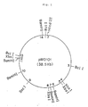
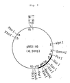
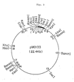
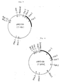
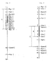
- Figs. 1 to 7 are restriction enzyme cleavage maps for pMO101, pMO116, pMO133, pMO126, pMO136, pMO217 and pMED101, respectively.
- Figs. 8 and 9 are restriction enzyme cleavage maps for DNA fragments containing neomycin-resistance genes derived from Micromonospora sp. MK-50. In the drawings, the neomycin-resistance genes exist in the region indicated by a bold line.
- Part A in Fig. 8 is the DNA fragment inserted into pMO116, pMO133 and pMO217.
- Part B in Fig. 9 is the DNA fragment inserted into pMO126 and
- Part C is the DNA fragment inserted into pMO136 and pMED101.
- the numbers in the drawings represent the following kb values, respectively.
- Fig. 10 A and B are restriction enzyme cleavage maps for pAN 25-10 and pAN 58-10, respectively.
- Plasmid pMO101 is a circular DNA having a molecular weight of about 25 megadaltons. Numbers of sites sensitive to various restriction enzymes are shown in Table 1. pMO101 has no sites sensitive to Eco RI and Bgl II. Table 1 Cleavage Characteristics of pMO101 with Restriction Enzymes Restriction enzyme No.
- Plasmid pMO201 is revealed to be a plasmid of 40 kilobases (kb) or more by agarose gel electrophoresis and shows a distinct band below the band of chromosomal DNA by ethidium bromide-cesium chloride density-gradient centrifugation, and thus seems to have a closed circular DNA structure. However, its digestion with various restriction enzymes yields a large number of ladder-DNA bands by agarose gel electrophoresis. This fact suggests that pMO201 is a mixture of two or more plasmids of considerably large molecular size. Thus, the structure of pMO201 has not been determined. Nevertheless, various vectors can be constructed by utilizing the DNA parts of the plasmid which harbor the function of autonomous replication in a microorganism of the genus Micromonospora .
- the above-mentioned plasmids have no vector-markers necessary for detecting them in microorganisms.
- a cloning vector having an appropriate vector-marker for detecting the vector is desirable.
- Such cloning vectors can be constructed by utilizing the ability of the above plasmids to autonomously replicate in a microorganism of the genus Micromonospora and incorporating an appropriate vector-marker into the plasmids.
- any DNA fragment can be used so long as it contains a gene which can be expressed in a microorganism of the genus Micromonospora .
- a DNA fragment containing a neomycin-resistance gene and derived from Micromonospora sp. MK-50 (FERM BP-1613) can be mentioned.
- Preferred examples are a DNA fragment of about 3 kb obtained by digestion with restriction enzyme Bgl II and which is characterized in that numbers of the sites sensitive to restriction enzymes are 1 for Kpn I, 3 for Bam HI, 2 for Pst I, 2 for Sac I, 1 for Cla I, 1 for Xba I, 1 for Xho I and 1 for Bcl I; a DNA fragment of about 3 kb obtained by digestion with restriction enzyme Bam HI and which is characterized in that numbers of the sites sensitive to restriction enzymes are 1 for Pst I, 1 for Xho I, 1 for Sac I and 1 for Sph I; and a DNA fragment of about 1.5 kb obtained by digestion with restriction enzymes Bam HI/ Sac I and which is characterized in that numbers of the sites sensitive to restriction enzymes are 1 for Pst I and 1 for Xho I.
- a cloning vector which is capable of autonomous replication and is detectable in a microorganism of the genus Micromonospora can be constructed by digesting a plasmid capable of autonomous replication in a microorganism of the genus Micromonospora at such restriction enzyme-cleavage sites that the ability to autonomously replicate is not impaired and by introducing appropriate vector markers into the sites.
- cloning vectors can be constructed by recombining DNA fragments obtained from plasmid pMO101 or pMO201 and harboring the function of autonomous replication in a microorganism of the genus Micromonospora with DNA fragments derived from Micromonospora sp. MK-50 and containing a neomycin-resistance gene.
- Examples of such cloning vectors include pMO116, pMO126, pMO133, pMO136 and pMO217.
- Plasmids pMO116, pMO126, pMO133 and pMO136 are cloning vectors constructed by utilizing the ability of pMO101 to autonomously replicate in a microorganism of the genus Micromonospora and by recombining the DNA fragments of pMO101 with the DNA fragments derived from Micromonospora sp. MK-50 and containing a neomycin-resistance gene. Numbers of the sites sensitive to various restriction enzymes in the respective plasmids are shown in Tables 2 to 5.
- pMO116 has no sites sensitive to Eco RI and Hind III
- pMO126 has no sites sensitive to Eco RI, Hind III, Bgl II, Cla I and Kpn I
- pMO133 has no sites sensitive to Eco RI, Hind III and Bgl II
- pMO136 has no sites sensitive to Eco RI, Hind III, Bcl I and Cla I.
- Table 2 Cleavage Characteristics of pMO116 with Restriction Enzymes Restriction enzyme No.
- Plasmid pMO217 is a cloning vector constructed by utilizing the ability of pMO201 to autonomously replicate in a microorganism of the genus Micromonospora and by incorporating a Bgl II-cleaved DNA fragement of about 3 kb derived from Micromonospora sp. MK-50 and containing a neomycin-resistance gene. Numbers of the sites sensitive to various restriction enzymes are shown in Table 6. pMO217 has no sites sensitive to Eco RI and Hind III. Table 6 Cleavage Characteristics of pMO217 with Restriction Enzymes Restriction enzyme No.
- the present invention provides shuttle vectors capable of autonomous replication in both of a microorganism of the genus Micromonospora and a microorganism of Escherichia coli .
- the shuttle vectors can be constructed by recombining the above-mentioned plasmid capable of autonomous replication in a microorganism of the genus Micromonospora with a plasmid capable of autonomous replication in a microorganism of Escherichia coli .
- the use of such shuttle vectors enables amplification of a gene derived from actinomycetes by using a microorganism of Escherichia coli instead of actinomycetes of slow growth, whereby analysis of genes and in vitro mutagenesis can be readily carried out.
- the plasmids capable of autonomous replication in a microorganism of Escherichia coli include, for example, pUC19 [Gene 33 , 103 (1985)], pBR322 [Gene 22 , 277 (1983)], and pACYC184 [J. Bacteriol, 134 , 1141 (1978)].
- shuttle vectors is plasmid pMED101 constructed by recombining plasmid pMO136 capable of autonomous replication in a microorganism of the genus Micromonospora with plasmid pUC19 capable of autonomous replication in a microorganism of Escherichia coli .
- pMED101 has sites sensitive to various restriction enzymes as shown in Table 7, and has no sites sensitive to Eco RI, Bcl I and Cla I.
- Table 7 Cleavage Characteristics of pMED101 with Restriction Enzymes Restriction enzyme No. of cleavage sites Length of DNA fragments formed by cleavage (kb) Hind III 1 7.5 Bam HI 1 7.5 Bgl II 1 7.5 Pst I 2 0.8, 6.7 Sac I 1 7.5 Xba I 2 3.5, 4.0 Xho I 2 3.5, 4.0 Kpn I 1 7.5 Mlu I 1 7.5 Sph I 1 7.5
- the plasmids of the present invention can autonomously replicate in a microorganism of the genus Micromonospora , and therefore, microorganisms of the genus Micromonospora , specifically the following species, can be used as host microorganisms.
- Micromonospora echinospora Micromonospora purpurea
- Micromonospora sagamiensis Micromonospora grisea
- Micromonospora ticansis Micromonospora rhodorangea Micromonospora rosaria
- a mutant strain released from restriction systems is more useful as a host microorganism.
- Such mutant strain can be obtained according to the procedure of Lomovskaya, et al. [Microbiological Review 44 206 (1980)].
- Table 8 Plasmid Strain Accession Number (Deposition date) pMO116 Micromonospora olivasterospora MH-116 FERM BP-1615 (12/12/'87) pMO126 Micromonospora olivasterospora MH-126 FERM BP-1616 (12/12/'87) pMO133 Micromonospora olivasterospora MH-133 FERM BP-1617 (12/12/'87) pMO217 Micromonospora olivasterospora MH-217 FERM BP-1618 (12/12/'87) pMED101 Escherichia coli TD-MED101E FERM BP-1770 (2/27/'88)
- the cloning vectors of the present invention are useful for the cloning and expression of genes in a host microorganism belonging to the genus Micromonospora .
- a gene responsible for the biosynthesis of an antibiotic is inserted into the cloning vector of the present invention and the recombinant vector is introduced into a microorganism belonging to the genus Micromonospora and capable of producing the antibiotic, productivity of the antibiotic by the transformant is greatly improved.
- Micromonospora olivasterospora ATCC 31010 was inoculated into 4 ml of SK No. 2c medium [20 g of Stabirose K (Matsutani Kagaku Kogyo), 5 g of glucose, 5 g of yeast extract (Daigo Eiyo Kagaku), 5 g of peptone (Kyokuto Seiyaku Kogyo), 3 g of meat extract (Kyokuto Seiyaku Kogyo), 0.2 g of KH2PO4, 0.6 g of MgSO4 ⁇ 7H2O, 1 g of CaCO3 and deionized water to make up to 1 l, adjusted to pH 7.6 and autoclaved at 120°C for 20 minutes] and subjected to shaking culture at 30°C for 2 to 3 days. Then, the whole culture was inoculated into 500 ml of SK No. 2 medium having the same composition as SK No. 2c medium except that CaCO3 is not contained, and subjected to shaking culture
- the culture obtained in (1) was centrifuged at 8,000 rpm at 4°C for 10 minutes to recover the cells.
- the cells were suspended in 40 - 50 ml of a lytic solution [10.3% sucrose, 25 mM tris(hydroxymethyl)aminomethane (hereinafter abbreviated to Tris) - HCl buffer (pH 8.0), and 25 mM disodium ethylenediamine tetraacetate (EDTA)] containing 5 mg/ml egg white lysozyme (Seikagaku Kogyo) and incubated at 37°C for 30 minutes.
- Tris tris(hydroxymethyl)aminomethane
- EDTA disodium ethylenediamine tetraacetate
- the upper layer obtained by centrifugation of the resulting mixture at 8,000 rpm at 4°C for 10 minutes was mixed with 3 M sodium acetate solution (1/10 volume of the upper layer) and then isopropyl alcohol containing 2% Triton x 100 (Nakarai Chemicals, Ltd.) (equal to volume of the upper layer) was added thereto. After being thoroughly mixed, the mixture was allowed to stand at room temperature for 30 minutes and again centrifuged at 8,000 rpm at 4°C for 10 minutes.
- the resulting pellets were dissolved in 20 ml of TE buffer [10 mM Tris-HCl buffer (pH 8.0) and 1 mM EDTA] and to the solution was added ribonuclease (Ribonuclease A, type IA, made by Sigma) which was heat-treated in advance at 80°C for 10 minutes to a final concentration of 20 ⁇ g/ml. After incubation at 37°C for 30 minutes, 2 ml of 3 M sodium acetate and 20 ml of isopropyl alcohol were added thereto, and the mixture was allowed to stand at room temperature for 30 minutes.
- TE buffer 10 mM Tris-HCl buffer (pH 8.0) and 1 mM EDTA
- the mixture was then subjected to centrifugation at 8,000 rpm at 4°C for 5 minutes to obtain pellets, which were dissolved again in TE buffer.
- Ethidium bromide was added to the solution to a final concentration of 0.75 mg/ml and 8 ml of the resulting solution was admixed with 8.00 g of cesium chloride.
- the resulting solution was centrifuged at 105,000 xg at 18°C for 40 hours. After this centrifugation, the circular DNA was detected as a specific band showing fluorescence under a ultraviolet lamp.
- the band was taken out with an injection needle and shaken several times together with isoamyl alcohol (Nakarai Chemicals, Ltd.) saturated with TE buffer to remove ethidium bromide, and then admixed with 3 M sodium acetate and isopropyl alcohol in a ratio of 10:1:10 (v/v/v).
- the mixture was allowed to stand on ice for 30 minutes.
- pellets were recovered therefrom by centrifugation at 12,000 rpm at 4°C for 5 minutes, washed with cold 70% ethanol and dried in vacuo
- the dried pellets were dissolved in 0.3 ml of TE buffer and kept in a freeze at -20°C. Through these operations, 40 to 50 ⁇ g of pMO101 plasmid DNA was obtained.
- pMO201 plasmid DNA was obtained from Micromonospora olivasterospor a 3629-4 (FERM BP-1614) by the foregoing precedure.
- Micromonospora sp. MK-50 was inoculated into 4 ml of SK No. 2c medium and subjected to shaking culture at 30°C for 3 days. Then, the whole culture was inoculated into 30 ml of SK No. 2 medium and subjected to shaking culture at 30°C for 2 days. The cells were collected by centrifugation at 12,000 rpm at 4°C for 10 minutes and washed twice with 20 ml of 10.3% sucrose solution.
- the cells were suspended in 6 ml of TS buffer [50 mM Tris-HCl buffer (pH 8.0) containing 10.3% sucrose and 25 mM EDTA] and admixed with 50 mg of egg white lysozyme, followed by incubation at 37°C for one hour.
- TS buffer 50 mM Tris-HCl buffer (pH 8.0) containing 10.3% sucrose and 25 mM EDTA
- 0.6 ml of proteinase K made by Sigma, adjusted to a concentration of 2 mg/ml with TS buffer
- 3.6 ml of 3.3% sodium dodecylsulfate solution was incubated at 37°C for one hour for bacteriolysis.
- the mixture was heated at 50°C for 30 minutes and cooled by dipping in a water bath.
- plenol-chloroform mixture After addition of 10 ml of plenol-chloroform mixture, the mixture was gently stirred for 5 minutes and centrifuged at 12,000 rpm at 4°C for 15 minutes. The upper layer was taken out and ribonuclease was added thereto to a final concentration of 20 ⁇ g/ml, followed by incubation at 37°C for 45 minutes. Then, a 5 M sodium chloride solution (1/10 volume of the upper layer) and polyethylene glycol 600 (Nakarai Chemicals, Ltd.) (1/4 volume of the upper layer) were added thereto, followed by gentle mixing.
- the mixture was centrifuged at 5,000 rpm at 4°C for 5 minutes, and the resulting pellets were dissolved in 5 - 10 ml of TE buffer.
- To the solution were added 3 M sodium acetate (1/10 volume of the solution), 66 mM MgCl2 solution (1/30 volume of the solution) and then cold 99% ethanol (2.2 times volume of the solution).
- the mixture was gently stirred and then centrifuged at 5,000 rpm at 4°C for 5 minutes to obtain pellets.
- the pellets were washed twice with cold 70% ethanol and dissolved in 4 ml of TE buffer, whereby a DNA solution having a concentration of about 450 ⁇ g/ml was obtained.
- the chromosomal DNA of Micromonospora sp. MK-50 obtained in (1) was partially digested with restriction enzyme Sau3 Al (Boehringer Mannheim) to make DNA fragments having a length of 4 - 6 kb as the main component. Then, 2 ⁇ g of vector pIJ702 [J. Gen. Microbiol. 129 , 2703-2714 (1983)] digested with restriction enzyme Bgl II (Boehringer Mannheim) and treated with alkaline phosphatase (for molecular biology, made by Boehringer Mannheim) was added to 100 ⁇ l of a solution containing 2 ⁇ g of the DNA mentioned above, which had been agitated with a phenol-chloroform mixture to inactivate the enzyme.
- Sau3 Al Boehringer Mannheim
- the dried pellets were dissolved in 25 ⁇ l of ligation buffer [66 mM Tris-HCl buffer (pH 7.6) containing 6.6 mM MgCl2, 10 mM dithiothreitol (Nakarai Chemicals, Ltd.) and 0.8 mM adenosine triphosphate (Sigma)], and one unit of T4 ligase (Boehringer Mannheim) was added to the solution. The mixture was incubated at 15°C overnight.
- ligation buffer 66 mM Tris-HCl buffer (pH 7.6) containing 6.6 mM MgCl2, 10 mM dithiothreitol (Nakarai Chemicals, Ltd.) and 0.8 mM adenosine triphosphate (Sigma)
- P-medium prepared by mixing a solution containing 10.3 g of sucrose, 25 mg of K2SO4, 203 mg of MgCl2 ⁇ 6H2O and 0.2 ml of trace element solution and made up to 90 ml with deionized water, which was sterilized in an autoclave, with 10 ml of 0.25 M TES [N-tris(hydroxymethyl)methyl-2-aminoethanesulfonic acid] buffer (pH 7.2), 0.5 ml of 5 M CaCl2 and 1 ml of 0.5% KH2PO4, separately sterilized in an autoclave.
- TES N-tris(hydroxymethyl)methyl-2-aminoethanesulfonic acid
- the mixture was then gently admixed with 25 ⁇ l of a protoplast suspension obtained from Streptomyces lividans TK 23 strain according to a conventional procedure [Current Topics in Microbiology and Immunology 96 69-95 (1985)], and the resulting mixture was allowed to stand at room temperature for one minute. Then, 500 ⁇ l of T-medium [a mixture of 75% by volume of P-medium and 25% by volume of polyethyleneglycol 1000 (Nakarai Chemicals, Ltd.)] was added thereto, and the mixture was stirred and diluted 10 times with P-medium.
- T-medium a mixture of 75% by volume of P-medium and 25% by volume of polyethyleneglycol 1000 (Nakarai Chemicals, Ltd.)
- the diluted mixture (100 ⁇ l) was spread on 12 plates of R5 regeneration agar plate medium prepared in the following manner: a solution (pH 7.2) containing 103 g of sucrose, 0.25 g of K2SO4, 10.12 g of MgCl2 ⁇ 6H2O, 10 g of glucose, 0.1 g of casamino acids (Difco Laboratories), 5 g of yeast extract (Difco Laboratories), 2 ml of a trace element solution, 5.7 g of TES and 2.5 g of sodium hydroxide and made up to 1 l with deionized water was divided into five equal parts; each part was mixed with 4.4 g of Bactoagar (Difco Laboratories) and sterilized at 120°C for 20 minutes; to each part were added 3 ml of 20% sodium l-glutaminate, 3 ml of 2% NaNO3, 2 ml of 0.5% KH2PO4 and 0.6 ml of 5M CaCl2, followed by rapid mixing; and the
- Replica plating was made on YEME medium [prepared by sterilizing in an autoclave the medium containing 3 g of yeast extract (Difco Laboratories), 5 g of Bactopeptone (Difco Laboratories), 3 g of malt extract (Difco Laboratories), 10 g of glucose and 20 g of Bactoagar in 1 l of deionized water (pH 7.2)] containing 25 ⁇ g/ml neomycin B (sulfate, made by Sigma) with velvet cloth, and culturing was further conducted at 30°C for 4 days.
- yeast extract Difco Laboratories
- Bactopeptone Difco Laboratories
- malt extract Difco Laboratories
- 10 g of glucose and 20 g of Bactoagar in 1 l of deionized water (pH 7.2) containing 25 ⁇ g/ml neomycin B (sulfate, made by Sigma) with velvet cloth, and culturing was further conducted at 30°C
- the colonies grown on the YEME plates were considered to contain recombinant plasmids harboring neomycin-resistance genes, and the plasmids were recovered in the same manner as in Example 1. It was found as the result of digestion with various restriction enzymes and analysis by agarose gel electrophoresis that two different genes expressing neomycin resistance were inserted in the obtained two plasmids, pNMR-1 and pNMR-10.
- pMO101 (3.8 ⁇ g) obtained in Example 1 was added to 30 ⁇ l of M buffer [Manual for Genetic Engineering (Cold Spring Harbor Laboratory), p.228 (1980)] containing 8 units of restriction enzyme Bcl I (Boehringer Mannheim) and incubated at 50°C for 2 hours. To the mixture were successively added 60 ⁇ l of deionized water, 10 ⁇ l of 1 M Tris-HCl buffer (pH 8.6) and 1.5 units of alkaline phosphatase (for molecular biology), and the mixture was incubated at 37°C for 45 minutes.
- M buffer Manual for Genetic Engineering (Cold Spring Harbor Laboratory), p.228 (1980)
- Bcl I Boehringer Mannheim
- the regenerated colonies which were neomycin-resistant were transferred into ATCC S-5 medium [a solution (pH 7.2) containing 10 g of Stabirose K, 1 g of yeast extract (Daigo Eiyo Kagaku), 1 g of meat extract (Kyokuto Seiyaku), 2 g of Bactotriptose (Difco Laboratories), 100 mg of FeSO4 ⁇ 2H2O and 20 g of Bactoagar and made up to 1 l with deionized water, which was sterilized at 120°C for 20 minutes] containing neomycin B at a concentration of 2.5 ⁇ g/ml, and cultured at 30°C for 5 days. Plasmids carried by the microorganisms grown on the plates were recovered in the same manner as in Example 1, whereby two plasmids, pMO116 and pMO133, were obtained.
- Restriction enzymes Bam HI and Bgl II (4 units each) were added to 30 ⁇ l of M buffer containing 1 ⁇ g of pMO116, followed by incubation at 37°C for 2.5 hours. To the mixture were added 60 ⁇ l of deionized water, 10 ⁇ l of 1 M Tris-HCl buffer (pH 8.6) and one unit of alkaline phosphatase (for molecular biology), and the mixture was incubated at 37°C for 45 minutes. Then, 100 ⁇ l of a phenol-chloroform mixture was added thereto, and the mixture was vigorously stirred and centrifuged at 12,000 rpm at 15°C for 5 minutes.
- the resulting supernatant was admixed with 0.5 ⁇ g of Bam HI-digested DNA fragment of 3 kb obtained from pNMR-10, 3 M sodium acetate (1/10 volume of the supernatant) and isopropyl alcohol (equal to volume of the supernatant), and the mixture was allowed to stand on ice for 30 minutes. Subsequent steps were carried out in the same manner as in Example 3, and a plasmid was isolated from the regenerated strain of Micromonospora olivasterospor a ATCC 21819 which was neomycin-resistant, whereby plasmid pMO126 was obtained.
- Restriction enzymes Bam HI and Sac I (4 units each, made by Boehringer Mannheim) were added to 30 ⁇ l of M buffer containing 1 ⁇ g of pMO116, followed by incubation at 37°C for 2.5 hours. To the mixture were added 60 ⁇ l of deionized water, 10 ⁇ l of 1 M Tris-HCl buffer (pH 8.6) and one unit of alkaline phosphatase (for molecular biology), and the mixture was incubated at 37°C for 45 minutes. Then, 100 ⁇ l of a phenol-chloroform mixture was added thereto, and the mixture was vigorously shaken for 30 seconds and centrifuged at 12,000 rpm at 15°C for 5 minutes.
- the resulting supernatant was admixed with about 0.5 ⁇ g of Bam HI/ Sac I-digested DNA fragment of 1.5 kb obtained from pNMR-10, 3 M sodium acetate (1/10 volume of the supernatant) and isopropyl alcohol (equal to volume of the supernatant), and the mixture was allowed to stand on ice for 30 minutes. Subsequent steps were carried out in the same manner as in Example 3, and a plasmid was isolated from the regenerated strain of Micromonospora olivasterospora ATCC 21819 which was neomycin-resistant, whereby plasmid pMO136 was obtained.
- pMO201 was added to 30 ⁇ l of M buffer containing 4 units of restriction enzyme Bgl II (Boehringer Mannheim), followed by incubation at 37°C for 2.5 hours.
- Bgl II restriction enzyme Bgl II
- To the mixtrue were added 60 ⁇ l of deionized water, 10 ⁇ l of 1 M Tris-HCl buffer (pH 8.6) and one unit of alkaline phophatase (for molecular biology), and the mixture was incubated at 37°C for 45 minutes. Then, 100 ⁇ l of a phenol-chloroform mixture was added thereto, and the mixture was vigorously stirred for 30 seconds and centrifuged at 12,000 rpm at 18°C for 5 minutes.
- the resulting supernatant was admixed with 0.5 ⁇ g of Bgl II-digested DNA fragment of 3 kb derived from pNMR-1, 3 M sodium acetate (1/10 volume of the supernatant) and isopropyl alcohol (equal to volume of the supernatant), and the mixture was allowed to stand on ice for 30 minutes. Subsequent steps were carried out in the same manner as in Example 3, and a plasmid was isolated from the regenerated strain of Micromonospora olivasterospora ATCC 21819 which was neomycin-resistant, whereby plasmid pMO217 was obtained.
- pMO136 (2 ⁇ g) obtained in Example 5 was added to 30 ⁇ l of M buffer containing 2 units of restriction enzyme Bgl II and 2 units of restriction enzyme Sac I (both restriction enzymes were made by Boehringer Mannheim), followed by incubation at 37°C for 2 hours.
- the mixture was made up to 100 ⁇ l with deionized water, and an equal amount of a phenol-chloroform mixture was added thereto.
- the mixture was vigorously stirred for 30 seconds and then centrifuged at 12,000 rpm at 18°C for 5 minutes.
- the resulting supernatant was admixed with 3 M sodium acetate (1/10 volume of the supernatant) and isopropyl alcohol (equal to volume of the supernatant), and the mixture was allowed to stand on ice for 30 minutes.
- the pellets obtained by centrifugation of the mixture at 12,000 rpm at 4°C for 5 minutes were washed with cold 70% ethanol and dried in vacuo .
- Plasmid pUC19 (3.5 ⁇ g) was added to 50 ⁇ l of M buffer containing 5 units of restriction enzyme Aat II (Toyobo), followed by incubation at 37°C for 2 hours. The mixture was made up to 100 ⁇ l with deionized water, and an equal amount of a phenol-chloroform mixture was added thereto. The mixture was vigorously stirred and then centrifuged at 12,000 rpm at 18°C for 5 minutes. The resulting upper layer was admixed with 3 M sodium acetate (1/10 volume of the upper layer) and isopropyl alcohol (equal to volume of the upper layer). The mixture was allowed to stand on ice for 30 minutes and centrifuged at 12,000 rpm at 4°C for 5 minutes.
- Aat II Toyobo
- the resulting pellets were washed with cold 70% ethanol and dried in vacuo .
- the dried pellets were redissolved in 20 ⁇ l of the buffer for T4 polymerase [a solution containing 0.033 M Tris-HCl buffer (pH 7.9), 0.066 M potassium acetate, 0.01 M magnesium acetate, 0.5 mM dithiothreitol, 0.1 mM dATP (deoxyadenosine triphosphate, made by Sigma), 0.1 mM dGTP (deoxyguanosine triphosphate, made by Sigma), 0.1 mM dCTP (deoxycytidine triphosphate, made by Sigma) and 0.1 mM dTTP (deoxythimidine triphosphate, made by Sigma)], and 5 units of T4 DNA polymerase (Takara Shuzo) was added to the solution.
- the mixture was subjected to incubation at 37°C for 5 minutes and then made up to 100 ⁇ l with deionized water. After addition of an equal amount of a phenol-chloroform mixture, the mixture was vigorously stirred and centrifuged at 12,000 rpm at 18°C for 5 minutes. The resulting upper layer was admixed with 3 M sodium acetate (1/10 volume of the upper layer) and isopropyl alcohol (equal to volume of the upper layer). The mixture was allowed to stand on ice for 30 minutes and then centrifuged at 12,000 rpm at 4°C for 5 minutes. The resulting pellets were washed with cold 70% ethanol and dried in vacuo
- the dried pellets were dissolved in ligation buffer together with 1.2 ⁇ g of Bgl II linker d (pC-A-G-A-T-C-T-G, made by Takara Shuzo), and one unit of T4 ligase (Boehringer Mannheim) was added to the solution.
- the mixture was subjected to incubation at 15°C overnight and then made up to 100 ⁇ l with deionized water. After addition of an equal amount of a phenol-chloroform mixture, the mixture was vigorously stirred and centrifuged at 12,000 rpm at 18°C for 5 minutes.
- the resulting upper layer was admixed with 3 M sodium acetate (1/10 volume of the upper layer) and isopropyl alcohol (equal to volume of the upper layer). The mixture was allowed to stand on ice for 30 minutes and then centrifuged at 12,000 rpm at 4°C for 5 minutes. The resulting pellets were washed with cold 70% ethanol and dried in vacuo .
- the mixture was made up to 100 ⁇ l with deionized water, and 10 ⁇ l of 1 M Tris-HCl buffer (pH 8.6) and 1.5 units of alkaline phosphatase (for molecular biology) were successively added thereto. The mixture was subjected to incubation at 37°C for one hour.
- the desired DNA fragment digested with Bgl II and Sac I was cut out of the agarose gel and electrically eluted in a dialysis tube.
- a phenol-chloroform mixture (equal to volume of the eluate), and the mixture was vigorously stirred for 30 seconds and centrifuged at 12,000 rpm at 18°C for 5 minutes.
- the resulting upper layer was admixed with 3 M sodium acetate (1/10 volume of the upper layer) and isopropyl alcohol (equal to volume of the upper layer). The mixture was allowed to stand on ice for 30 minutes and centrifuged at 12,000 rpm at 4°C for 5 minutes. The resulting pellets were washed with cold 70% ethanol and dried in vacuo .
- the Bgl II- Sac I DNA fragment (about 5.3 kb) derived from pMO136 and obtained in (1) and the Bgl II- Sac I fragment (about 2.2 kb) derived from pUC19 and obtained in (3) were dissolved in 30 ⁇ l of ligation buffer and one unit of T4 ligase (Boehringer Mannheim) was added to the solution. The mixture was incubated at 15°C overnight. To the mixture were added 3 M sodium acetate (1/10 volume of the mixture) and isopropyl alcohol (equal to volume of the mixture). The mixture was allowed to stand on ice for 30 minutes and then centrifuged at 12,000 rpm at 4°C for 5 minutes. The resulting pellets were washed with cold 70% ethanol, dried in vacuo and suspended in 10 ⁇ l of TE buffer.
- Competent cells of Escherichia coli K-12:HB101 strain 100 ⁇ l prepared according to a conventional procedure [Bolivar, et al.: Gene 2 : 75-93 (1977)] was mixed with the in vitro recombinant DNA (about 1.0 ⁇ g) obtained in (4), and the mixture was subjected to incubation on ice for 20 minutes and then at 37°C for 5 minutes.
- L-medium a solution (pH 7.0) containing 10 g of Bactotryptone (Difco Labratories), 5 g of yeast extract (Difco Laboratories) and 5 g of NaCl and made up to 1 l with deionized water, which was sterilized at 120°C for 20 minutes], followed by incubation at 37°C for one hour.
- a part of the mixture was appropriately diluted and spread on L-medium agar plate [L-medium containing 1.5% Bactoagar (Difco Laboratories)] containing 50 ⁇ g/ml ampicillin.
- Colonies which grew on the plates by acquiring ampicillin-resistance were selected and further cultured in L-medium containing 50 ⁇ g/ml ampicillin at 37°C overnight. Plasmids carried by the cells were isolated in the same manner as in Example 1, whereby pMED101 was obtained.
- Plasmids pMO101, pMO116, pMO126, pMO133, pMO136, pMO217 and pMED101 obtained in Examples 1 to 7 were respectively analyzed by digestion with various restriction enzymes and agarose gel electrophoresis to prepare restriction enzyme cleavage maps shown in Figs. 1 to 7.
- neomycin-resistance genes derived from Micromonospora sp. MK-50 and cloned on pNMR-1 and pNMR-10 were likewise analyzed to prepare restriction enzyme cleavage maps for the DNA fragments containing the neomycin-resistance genes (see Figs. 8 and 9).
- Micromonospora olivasterospora ATCC 21819 strain was transformed with 1 ⁇ g each of pMO116, pMO133 and pMO217 prepared above, individually in the same manner as in Example 3. Numbers of the transformed cells obtained with pMO116, pMO133 and pMO217 were 5.0 x 10⁶, 1.2 x 10⁷, and 1.1 x 10⁷, respectively, and thus very high transformation efficiencies were obtained.
- Example 7 To about 1.0 ⁇ g of pMED101 obtained in Example 7 was added P-medium to make up to 100 ⁇ l, and 50 ⁇ l of a protoplast suspension of Micromonospora olivasterospora ATCC 21819 was quickly added thereto. The mixture was allowed to stand at room temperature for one minute and then 600 ⁇ l of T-medium was added with gentle mixing. The mixture was diluted 10 times with P-medium, and 100 ⁇ l portions of the diluted mixture were spread on 24 plates of regeneration agar plate medium RM-1. By culturing on the agar medium, the protoplasts were regenerated as normal hypha cells.
- the transformants grown on the plates by acquiring neomycin resistnace were cultured in SK No. 2 medium and the plasmids carried by the transformants were investigated in the same manner as in Example 1. It was found that all of these transformants contained pMED101. In this test, 132 transformed cells were obtained with 1 ⁇ g of pMED101.
- the whole chromosomal DNA of Micromonospora olivasterospora ATCC 21819 which produces fortimicin A was extracted from the strain by the method described in Example 2 (1).
- the chromosomal DNA was partially digested with restriction enzyme Sau3 AI (Boehringer Mannheim) to make DNA fragments having a length of 6 - 8 kb by sucrose density gradient centrifugation.
- a density gradient of 40 - 10% sucrose solution was prepared in six sterilized Hitachi 40 PA tubes (Nissei Sangyo) by gently packing equal amounts of 20 mM Tris-HCl buffer solutions (pH 8.0) containing 1 M NaCl, 5 mM EDTA and, respectively, 40, 30, 20 and 10% sucrose to make up to 38 ml. Then, the solution containing 200 ⁇ g of the DNA fragments mentioned above was heated at 65°C for 15 minutes and cooled. The solution was gently put on the sucrose solution in the tube and the tube was set in Hitachi SRP 28 Rotor (Nissei Sangyo) and subjected to centrifugation at 26,000 rpm at 2°C for 24 hours.
- the supernatant in each tube was gently put into 30 Eppendorf tubes and fractions containing DNA fragments of 6 - 8 kb were collected by agarose gel electrophoresis. To the fractions was added TE buffer (4 times volume of the fractions), and then twice as much cold ethanol as the mixture by volume was added. The precipitated DNA (total amount: 82 ⁇ g) was recovered by centrifugation at 12,000 rpm for 5 minutes.
- the ligation product obtained in (1) was subjected to precipitation with isopropyl alcohol, and the precipitate was dissolved in 20 ⁇ l of TE buffer, followed by addition of 80 ⁇ l of P-medium.
- the mixture was allowed to stand at room temperature for one minute, and 600 ⁇ l to T-medium was added thereto.
- the mixture was diluted 10 times with P-medium, and 100 ⁇ l portions of the diluted solution were spread on 80 plates for regeneration, followed by incubation at 30°C for 6 days.
- 2.5 ml of the soft agar medium having the same composition as that used in Example 2 (2) and containing 40 ⁇ g/ml (final concentration) neomycin B was put on the plate, followed by incubation at 30°C for 3 weeks.
- neomycin-resistant transformants were inoculated on plates of an agar medium for the production of fortimicin A containing 2.5 ⁇ g/ml neomycin B, which was prepared in the following manner: a solution (pH 7.1) containing 1 g of maltose, 1 g of yeast extract (Difco Laboratories), 1 g of peptone (Kyokuto Seiyaku), 0.6 g of meat extract (Kyokuto Seiyaku), 4.55 g of TES, 40 mg of KH2PO4, 120 mg of MgSO4 ⁇ 7H2O and 20 g of Bactoagar (Difco Laboratories) and made up to 1 l with deionized water was sterilized at 120°C for 20 minutes; and 20 ml portions of the solution were poured into the plates and solidified. Culturing was carried out at 30°C for 6 days.
- a soft agar medium pH 7.5
- Escherichia coli cells which was prepared in the following manner: a solution containing 2.5 g of yeast extract (Difco Laboratories), 3 g of beef extract (Difco Laboratories), 6 g of Bacto-triptone (Difco Laboratories), 1 g of glucose, 12.9 g of Tris and 10 g of Bactoagar (Difco Laboratories) and made up to 1 l with deionized water was sterilized at 120°C for 20 minutes; and Escherichia coli was inoculated into the solution kept at 42°C.
- Plasmids were recovered from these transformants by the method described in Example 1 and named pAN 25-10 and pAN 58-10. Restriction enzyme cleavage maps for the plasmids were made and illustrated in Fig. 10, A and B. On the DNA fragments inserted into these plasmids, there exist genes coding for the enzymes relating to the fortimicin biosynthesis, i.e. the enzyme at step 3 wherein the intermediate compound Fu-10 is produced from scyllo-inosamine and glucosamine and the enzyme at step 11 wherein the intermediate compound KR is produced from the intermediate compound KH.
- Plasmids pAN 25-10 and pAN 58-10 were incorporated into the fortimicin-producing strain G-2 [Journal of Antibiotics 37 , 1670 (1984)] derived from Micromonospora olivasterospora ATCC 21819 by the method described in Example 3.
- the G-2 strain and the thus obtained transformants were cultured by the following method. That is, the first seed culture was carried out using 4 ml of SK No. 2 medium containing 0.25 ⁇ g/ml neomycin B at 30°C for 3 days with shaking. For the second seed culture, 1 ml of the first seed culture was inoculated into 10 ml of the same medium as the first seed medium and cultured with shaking at 30°C for 24 hours. Then, 3 ml of the culture was inoculated into 30 ml of a liquid medium for the production of fortimicin A (pH 7.0) containing 7.5 ⁇ g of neomycin B in a 300-ml flask.
- fortimicin A pH 7.0
- Composition of the medium are as follows: 40 g/l maltose, 5 g/l glucose, 10 g/l peptone (Kyokuto Seiyaku), 15 g/l soybean meal (Kyokuto Seiyaku), 1 g/l KH2PO4, 3 g/l K2HPO4, 3 g/l MgSO4 ⁇ 7H2O, 2 g/l NaCl, 10 mg/l FeSO4 ⁇ 7H2O, 10 mg/l NiCl2 ⁇ 6H2O, 10 mg/l ZnSO4 ⁇ 7H2O, 10 mg/l CuSO4 ⁇ 5H2O, 50 mg/l MnSO4 ⁇ 4-5H2O, 1 mg/l COCl2, 10 mg/l D-calcium pantothenate and 50 g/l dry yeast (Oriental Yeast Industry).
- a solution of sugar components and a solution of the other components were separately sterilized at 120°C for 20 minutes and combined. Culturing was carried out on a rotary shaker at 30°C for 7 days. Then, 2 ml of the culture was recovered and 0.3 ml of 2N H2SO4 was added. The mixture was shaken at 28°C for 30 minutes and then subjected to centrifugation at room temperature at 3,000 rpm for 20 minutes. Precoated plate No. 6721 (Merck) was spotted with 2 ⁇ l of the supernatant and development was carried out using the lower layer of chloroform : methanol : aqueous ammonia (1:1:1).
- results show that the insertion of the recombinant vectors into the host cells increases the productivity of fortimicin A and that the plasmids of the present invention are useful for the cloning and expression of genes in a microorganism of the genus Micromonospora .
Landscapes
- Health & Medical Sciences (AREA)
- Genetics & Genomics (AREA)
- Life Sciences & Earth Sciences (AREA)
- Engineering & Computer Science (AREA)
- Wood Science & Technology (AREA)
- Biomedical Technology (AREA)
- Organic Chemistry (AREA)
- Biotechnology (AREA)
- General Engineering & Computer Science (AREA)
- Chemical & Material Sciences (AREA)
- Zoology (AREA)
- Bioinformatics & Cheminformatics (AREA)
- Biophysics (AREA)
- Microbiology (AREA)
- Plant Pathology (AREA)
- Molecular Biology (AREA)
- Biochemistry (AREA)
- General Health & Medical Sciences (AREA)
- Physics & Mathematics (AREA)
- Micro-Organisms Or Cultivation Processes Thereof (AREA)
- Saccharide Compounds (AREA)
Applications Claiming Priority (2)
| Application Number | Priority Date | Filing Date | Title |
|---|---|---|---|
| JP63136225A JPH01304887A (ja) | 1988-06-02 | 1988-06-02 | クローニングベクター |
| JP136225/88 | 1988-06-02 |
Publications (1)
| Publication Number | Publication Date |
|---|---|
| EP0344767A1 true EP0344767A1 (fr) | 1989-12-06 |
Family
ID=15170217
Family Applications (1)
| Application Number | Title | Priority Date | Filing Date |
|---|---|---|---|
| EP89109923A Withdrawn EP0344767A1 (fr) | 1988-06-02 | 1989-06-01 | Vecteurs de clonage |
Country Status (2)
| Country | Link |
|---|---|
| EP (1) | EP0344767A1 (fr) |
| JP (1) | JPH01304887A (fr) |
Cited By (1)
| Publication number | Priority date | Publication date | Assignee | Title |
|---|---|---|---|---|
| WO2001075116A3 (fr) * | 2000-04-04 | 2002-05-10 | Schering Corp | Acides nucleiques isoles de plasmide de micromonospora rosaria pmr2 et vecteurs produits a partir de ceux-ci |
Citations (1)
| Publication number | Priority date | Publication date | Assignee | Title |
|---|---|---|---|---|
| EP0064350A1 (fr) * | 1981-04-28 | 1982-11-10 | Takeda Chemical Industries, Ltd. | Plasmide et sa production |
Family Cites Families (2)
| Publication number | Priority date | Publication date | Assignee | Title |
|---|---|---|---|---|
| JPS6047684A (ja) * | 1983-08-23 | 1985-03-15 | Tanabe Seiyaku Co Ltd | プラスミドおよびその製法 |
| JPS647760A (en) * | 1987-06-30 | 1989-01-11 | Canon Kk | Image reproducer |
-
1988
- 1988-06-02 JP JP63136225A patent/JPH01304887A/ja active Pending
-
1989
- 1989-06-01 EP EP89109923A patent/EP0344767A1/fr not_active Withdrawn
Patent Citations (1)
| Publication number | Priority date | Publication date | Assignee | Title |
|---|---|---|---|---|
| EP0064350A1 (fr) * | 1981-04-28 | 1982-11-10 | Takeda Chemical Industries, Ltd. | Plasmide et sa production |
Non-Patent Citations (3)
| Title |
|---|
| CHEMICAL ABSTRACTS, vol. 103, no. 3, 22nd July 1985, page 157, abstract no. 17787e, Columbus, Ohio, US; & JP-A-60 47 684 (TANABE SEIYAKU CO.) 15-03-1985 * |
| CHEMICAL ABSTRACTS, vol. 105, no. 11, 15th September 1986, page 157, abstract no. 92152p, Columbus, Ohio, US; T. OSHIDA et al.: "Isolation and characterization of plasmids from Micromonospora zionensis and Micromonospora rosaria", & PLASMID 1986, 16(1), 74-6 * |
| CHEMICAL ABSTRACTS, vol. 108, no. 25, 20th June 1988, page 309, abstract no. 218857e, Columbus, Ohio, US; P. MATSUSHIMA et al.: "Genetic transformation of Micromonospora rosaria by the Streptomyces plasmid pIJ702", & J. ANTIBIOT. 1988, 41(4), 583-5 * |
Cited By (2)
| Publication number | Priority date | Publication date | Assignee | Title |
|---|---|---|---|---|
| WO2001075116A3 (fr) * | 2000-04-04 | 2002-05-10 | Schering Corp | Acides nucleiques isoles de plasmide de micromonospora rosaria pmr2 et vecteurs produits a partir de ceux-ci |
| US6569668B2 (en) | 2000-04-04 | 2003-05-27 | Schering Corporation | Isolated nucleic acids from Micromonospora rosaria plasmid pMR2 and vectors made therefrom |
Also Published As
| Publication number | Publication date |
|---|---|
| JPH01304887A (ja) | 1989-12-08 |
Similar Documents
| Publication | Publication Date | Title |
|---|---|---|
| CA1188236A (fr) | Plasmides | |
| Wada et al. | Participation of Escherichia coli K-12 groE gene products in the synthesis of cellular DNA and RNA | |
| EP0093611B2 (fr) | Plasmide composé | |
| Matsushima et al. | Highly transformable mutants of Streptomyces fradiae defective in several restriction systems | |
| EP0118367B1 (fr) | Vecteur pVE1 pour le clonage de DNA recombinant, mutants de délétion et mutants hybrydes et dérivés recombinants de ceux-ci, produits et procédés | |
| Dally et al. | A genetic comparison of strains of Zymomonas mobilis by analysis of plasmid DNA | |
| US4717666A (en) | Cloned streptomycete lividans excretable β-galactosidase gene | |
| EP0032238A2 (fr) | Bactériophage recombinant pour clonage hétérologue de microorganismes du genre bacillus et procédé pour sa production | |
| US4703009A (en) | RDNA cloning vector pVE1, deletion and hybrid mutants and recombinant derivatives thereof products and processes | |
| EP0213898B1 (fr) | Système hôte-vecteur | |
| EP0353938B1 (fr) | Procédé de production de neuraminidase | |
| EP0354641B1 (fr) | L'ADN recombinante codant pour une 4''-0-isovaléryl acylase | |
| EP0344767A1 (fr) | Vecteurs de clonage | |
| US4518698A (en) | Plasmid and production thereof | |
| KOBAYASHI et al. | Isolation and characterization of a pock-forming plasmid pTA4001 from Streptomyces lavendulae | |
| EP0124076A2 (fr) | Méthode de production de plasmides recombinants, micro-organismes transformés par ces plasmides et alpha-amylase thermostabile | |
| EP0374771B1 (fr) | Méthode de production de l'endonucléase de restriction PvuI | |
| US4478937A (en) | Plasmid and production thereof | |
| EP0240105B1 (fr) | Gène codant pour la biotine synthétase et son utilisation | |
| EP0162725B1 (fr) | Vecteur plasmidique chimère | |
| EP0082637A2 (fr) | Microorganismes produisant la L-histidine | |
| MATSUNO et al. | Transformation of Bacillus subtilis, antifungal-antibiotic iturin producers with isolated antibiotic resistance plasmids | |
| EP0038156A2 (fr) | Un plasmide et sa préparation microbiologique | |
| KR100814313B1 (ko) | 대장균의 염색체 재조합에 사용되는 플라스미드 | |
| EP0138337A1 (fr) | Plasmides et leur préparation et utilisation, et micro-organismes régénérés à protoplastes contenant tels plasmides |
Legal Events
| Date | Code | Title | Description |
|---|---|---|---|
| PUAI | Public reference made under article 153(3) epc to a published international application that has entered the european phase |
Free format text: ORIGINAL CODE: 0009012 |
|
| AK | Designated contracting states |
Kind code of ref document: A1 Designated state(s): AT BE CH DE ES FR GB GR IT LI LU NL SE |
|
| 17P | Request for examination filed |
Effective date: 19900404 |
|
| STAA | Information on the status of an ep patent application or granted ep patent |
Free format text: STATUS: THE APPLICATION IS DEEMED TO BE WITHDRAWN |
|
| 18D | Application deemed to be withdrawn |
Effective date: 19930105 |