EP0355849A2 - Méthode et appareil de mesure automatisée de fluorescence - Google Patents
Méthode et appareil de mesure automatisée de fluorescence Download PDFInfo
- Publication number
- EP0355849A2 EP0355849A2 EP89115739A EP89115739A EP0355849A2 EP 0355849 A2 EP0355849 A2 EP 0355849A2 EP 89115739 A EP89115739 A EP 89115739A EP 89115739 A EP89115739 A EP 89115739A EP 0355849 A2 EP0355849 A2 EP 0355849A2
- Authority
- EP
- European Patent Office
- Prior art keywords
- fluorescence
- reaction
- reference sample
- measured
- reagent
- Prior art date
- Legal status (The legal status is an assumption and is not a legal conclusion. Google has not performed a legal analysis and makes no representation as to the accuracy of the status listed.)
- Granted
Links
- 238000005259 measurement Methods 0.000 title claims abstract description 38
- 238000000034 method Methods 0.000 title claims description 18
- 238000006243 chemical reaction Methods 0.000 claims abstract description 121
- 239000003153 chemical reaction reagent Substances 0.000 claims abstract description 83
- 239000013074 reference sample Substances 0.000 claims abstract description 62
- 239000000523 sample Substances 0.000 claims abstract description 59
- 238000012360 testing method Methods 0.000 claims abstract description 33
- 239000000126 substance Substances 0.000 claims abstract description 23
- 239000000427 antigen Substances 0.000 claims abstract description 17
- 102000036639 antigens Human genes 0.000 claims abstract description 17
- 108091007433 antigens Proteins 0.000 claims abstract description 17
- LOUPRKONTZGTKE-UHFFFAOYSA-N cinchonine Natural products C1C(C(C2)C=C)CCN2C1C(O)C1=CC=NC2=CC=C(OC)C=C21 LOUPRKONTZGTKE-UHFFFAOYSA-N 0.000 claims abstract description 15
- AKYHKWQPZHDOBW-UHFFFAOYSA-N (5-ethenyl-1-azabicyclo[2.2.2]octan-7-yl)-(6-methoxyquinolin-4-yl)methanol Chemical compound OS(O)(=O)=O.C1C(C(C2)C=C)CCN2C1C(O)C1=CC=NC2=CC=C(OC)C=C21 AKYHKWQPZHDOBW-UHFFFAOYSA-N 0.000 claims abstract description 13
- 239000001576 FEMA 2977 Substances 0.000 claims abstract description 13
- 229960003110 quinine sulfate Drugs 0.000 claims abstract description 13
- 102000004190 Enzymes Human genes 0.000 claims abstract description 10
- 108090000790 Enzymes Proteins 0.000 claims abstract description 10
- 238000005070 sampling Methods 0.000 claims abstract description 10
- 239000007790 solid phase Substances 0.000 claims abstract description 9
- 239000000243 solution Substances 0.000 claims description 46
- 238000005375 photometry Methods 0.000 claims description 11
- 239000000758 substrate Substances 0.000 claims description 10
- PYWVYCXTNDRMGF-UHFFFAOYSA-N rhodamine B Chemical compound [Cl-].C=12C=CC(=[N+](CC)CC)C=C2OC2=CC(N(CC)CC)=CC=C2C=1C1=CC=CC=C1C(O)=O PYWVYCXTNDRMGF-UHFFFAOYSA-N 0.000 claims description 7
- 229940043267 rhodamine b Drugs 0.000 claims description 7
- 239000002502 liposome Substances 0.000 claims description 6
- XLYOFNOQVPJJNP-UHFFFAOYSA-N water Substances O XLYOFNOQVPJJNP-UHFFFAOYSA-N 0.000 claims description 6
- 238000012921 fluorescence analysis Methods 0.000 claims 8
- 239000012490 blank solution Substances 0.000 claims 3
- 230000001678 irradiating effect Effects 0.000 claims 1
- 238000002372 labelling Methods 0.000 claims 1
- 230000008520 organization Effects 0.000 claims 1
- 238000012812 general test Methods 0.000 abstract description 16
- 238000006911 enzymatic reaction Methods 0.000 abstract description 4
- 238000004458 analytical method Methods 0.000 description 35
- 239000011324 bead Substances 0.000 description 16
- 230000007246 mechanism Effects 0.000 description 14
- 210000002966 serum Anatomy 0.000 description 12
- 230000003287 optical effect Effects 0.000 description 10
- 238000012937 correction Methods 0.000 description 8
- 230000036046 immunoreaction Effects 0.000 description 8
- 230000015556 catabolic process Effects 0.000 description 7
- 238000006731 degradation reaction Methods 0.000 description 7
- 239000007788 liquid Substances 0.000 description 5
- 238000005406 washing Methods 0.000 description 5
- HSHNITRMYYLLCV-UHFFFAOYSA-N 4-methylumbelliferone Chemical compound C1=C(O)C=CC2=C1OC(=O)C=C2C HSHNITRMYYLLCV-UHFFFAOYSA-N 0.000 description 4
- LOUPRKONTZGTKE-WZBLMQSHSA-N Quinine Chemical compound C([C@H]([C@H](C1)C=C)C2)C[N@@]1[C@@H]2[C@H](O)C1=CC=NC2=CC=C(OC)C=C21 LOUPRKONTZGTKE-WZBLMQSHSA-N 0.000 description 4
- 238000012545 processing Methods 0.000 description 4
- 235000001258 Cinchona calisaya Nutrition 0.000 description 2
- XUIIKFGFIJCVMT-GFCCVEGCSA-N D-thyroxine Chemical compound IC1=CC(C[C@@H](N)C(O)=O)=CC(I)=C1OC1=CC(I)=C(O)C(I)=C1 XUIIKFGFIJCVMT-GFCCVEGCSA-N 0.000 description 2
- 239000012472 biological sample Substances 0.000 description 2
- 239000007853 buffer solution Substances 0.000 description 2
- 238000007599 discharging Methods 0.000 description 2
- MHMNJMPURVTYEJ-UHFFFAOYSA-N fluorescein-5-isothiocyanate Chemical compound O1C(=O)C2=CC(N=C=S)=CC=C2C21C1=CC=C(O)C=C1OC1=CC(O)=CC=C21 MHMNJMPURVTYEJ-UHFFFAOYSA-N 0.000 description 2
- 230000006870 function Effects 0.000 description 2
- 229960000948 quinine Drugs 0.000 description 2
- 230000004936 stimulating effect Effects 0.000 description 2
- 210000001685 thyroid gland Anatomy 0.000 description 2
- 229940034208 thyroxine Drugs 0.000 description 2
- XUIIKFGFIJCVMT-UHFFFAOYSA-N thyroxine-binding globulin Natural products IC1=CC(CC([NH3+])C([O-])=O)=CC(I)=C1OC1=CC(I)=C(O)C(I)=C1 XUIIKFGFIJCVMT-UHFFFAOYSA-N 0.000 description 2
- BCHIXGBGRHLSBE-UHFFFAOYSA-N (4-methyl-2-oxochromen-7-yl) dihydrogen phosphate Chemical compound C1=C(OP(O)(O)=O)C=CC2=C1OC(=O)C=C2C BCHIXGBGRHLSBE-UHFFFAOYSA-N 0.000 description 1
- 102000004160 Phosphoric Monoester Hydrolases Human genes 0.000 description 1
- 108090000608 Phosphoric Monoester Hydrolases Proteins 0.000 description 1
- 230000005856 abnormality Effects 0.000 description 1
- -1 acryl Chemical group 0.000 description 1
- 239000003513 alkali Substances 0.000 description 1
- 239000002585 base Substances 0.000 description 1
- 210000004369 blood Anatomy 0.000 description 1
- 239000008280 blood Substances 0.000 description 1
- 238000010276 construction Methods 0.000 description 1
- 238000001816 cooling Methods 0.000 description 1
- 230000006735 deficit Effects 0.000 description 1
- 238000001514 detection method Methods 0.000 description 1
- 230000004907 flux Effects 0.000 description 1
- 239000011521 glass Substances 0.000 description 1
- 230000001900 immune effect Effects 0.000 description 1
- 230000036039 immunity Effects 0.000 description 1
- 238000003018 immunoassay Methods 0.000 description 1
- 238000009413 insulation Methods 0.000 description 1
- 239000003550 marker Substances 0.000 description 1
- 238000000691 measurement method Methods 0.000 description 1
- 239000011295 pitch Substances 0.000 description 1
- 238000003127 radioimmunoassay Methods 0.000 description 1
- 238000011084 recovery Methods 0.000 description 1
- 239000012088 reference solution Substances 0.000 description 1
- 239000011347 resin Substances 0.000 description 1
- 229920005989 resin Polymers 0.000 description 1
- 239000012488 sample solution Substances 0.000 description 1
- 239000004094 surface-active agent Substances 0.000 description 1
- 239000012780 transparent material Substances 0.000 description 1
Images


Classifications
-
- G—PHYSICS
- G01—MEASURING; TESTING
- G01N—INVESTIGATING OR ANALYSING MATERIALS BY DETERMINING THEIR CHEMICAL OR PHYSICAL PROPERTIES
- G01N35/00—Automatic analysis not limited to methods or materials provided for in any single one of groups G01N1/00 - G01N33/00; Handling materials therefor
- G01N35/02—Automatic analysis not limited to methods or materials provided for in any single one of groups G01N1/00 - G01N33/00; Handling materials therefor using a plurality of sample containers moved by a conveyor system past one or more treatment or analysis stations
- G01N35/025—Automatic analysis not limited to methods or materials provided for in any single one of groups G01N1/00 - G01N33/00; Handling materials therefor using a plurality of sample containers moved by a conveyor system past one or more treatment or analysis stations having a carousel or turntable for reaction cells or cuvettes
-
- G—PHYSICS
- G01—MEASURING; TESTING
- G01N—INVESTIGATING OR ANALYSING MATERIALS BY DETERMINING THEIR CHEMICAL OR PHYSICAL PROPERTIES
- G01N21/00—Investigating or analysing materials by the use of optical means, i.e. using sub-millimetre waves, infrared, visible or ultraviolet light
- G01N21/62—Systems in which the material investigated is excited whereby it emits light or causes a change in wavelength of the incident light
- G01N21/63—Systems in which the material investigated is excited whereby it emits light or causes a change in wavelength of the incident light optically excited
- G01N21/64—Fluorescence; Phosphorescence
- G01N21/6428—Measuring fluorescence of fluorescent products of reactions or of fluorochrome labelled reactive substances, e.g. measuring quenching effects, using measuring "optrodes"
-
- G—PHYSICS
- G01—MEASURING; TESTING
- G01N—INVESTIGATING OR ANALYSING MATERIALS BY DETERMINING THEIR CHEMICAL OR PHYSICAL PROPERTIES
- G01N33/00—Investigating or analysing materials by specific methods not covered by groups G01N1/00 - G01N31/00
- G01N33/48—Biological material, e.g. blood, urine; Haemocytometers
- G01N33/50—Chemical analysis of biological material, e.g. blood, urine; Testing involving biospecific ligand binding methods; Immunological testing
- G01N33/74—Chemical analysis of biological material, e.g. blood, urine; Testing involving biospecific ligand binding methods; Immunological testing involving hormones or other non-cytokine intercellular protein regulatory factors such as growth factors, including receptors to hormones and growth factors
- G01N33/76—Human chorionic gonadotropin including luteinising hormone, follicle stimulating hormone, thyroid stimulating hormone or their receptors
-
- G—PHYSICS
- G01—MEASURING; TESTING
- G01N—INVESTIGATING OR ANALYSING MATERIALS BY DETERMINING THEIR CHEMICAL OR PHYSICAL PROPERTIES
- G01N33/00—Investigating or analysing materials by specific methods not covered by groups G01N1/00 - G01N31/00
- G01N33/48—Biological material, e.g. blood, urine; Haemocytometers
- G01N33/50—Chemical analysis of biological material, e.g. blood, urine; Testing involving biospecific ligand binding methods; Immunological testing
- G01N33/74—Chemical analysis of biological material, e.g. blood, urine; Testing involving biospecific ligand binding methods; Immunological testing involving hormones or other non-cytokine intercellular protein regulatory factors such as growth factors, including receptors to hormones and growth factors
- G01N33/78—Thyroid gland hormones, e.g. T3, T4, TBH, TBG or their receptors
-
- G—PHYSICS
- G01—MEASURING; TESTING
- G01N—INVESTIGATING OR ANALYSING MATERIALS BY DETERMINING THEIR CHEMICAL OR PHYSICAL PROPERTIES
- G01N35/00—Automatic analysis not limited to methods or materials provided for in any single one of groups G01N1/00 - G01N33/00; Handling materials therefor
- G01N35/02—Automatic analysis not limited to methods or materials provided for in any single one of groups G01N1/00 - G01N33/00; Handling materials therefor using a plurality of sample containers moved by a conveyor system past one or more treatment or analysis stations
- G01N35/04—Details of the conveyor system
- G01N2035/0439—Rotary sample carriers, i.e. carousels
- G01N2035/0446—Combinations of the above
- G01N2035/0448—Combinations of the above composed of interchangeable ring elements
-
- G—PHYSICS
- G01—MEASURING; TESTING
- G01N—INVESTIGATING OR ANALYSING MATERIALS BY DETERMINING THEIR CHEMICAL OR PHYSICAL PROPERTIES
- G01N21/00—Investigating or analysing materials by the use of optical means, i.e. using sub-millimetre waves, infrared, visible or ultraviolet light
- G01N21/17—Systems in which incident light is modified in accordance with the properties of the material investigated
- G01N21/25—Colour; Spectral properties, i.e. comparison of effect of material on the light at two or more different wavelengths or wavelength bands
- G01N21/27—Colour; Spectral properties, i.e. comparison of effect of material on the light at two or more different wavelengths or wavelength bands using photo-electric detection ; circuits for computing concentration
- G01N21/274—Calibration, base line adjustment, drift correction
-
- G—PHYSICS
- G01—MEASURING; TESTING
- G01N—INVESTIGATING OR ANALYSING MATERIALS BY DETERMINING THEIR CHEMICAL OR PHYSICAL PROPERTIES
- G01N2201/00—Features of devices classified in G01N21/00
- G01N2201/04—Batch operation; multisample devices
- G01N2201/0415—Carrusel, sequential
-
- Y—GENERAL TAGGING OF NEW TECHNOLOGICAL DEVELOPMENTS; GENERAL TAGGING OF CROSS-SECTIONAL TECHNOLOGIES SPANNING OVER SEVERAL SECTIONS OF THE IPC; TECHNICAL SUBJECTS COVERED BY FORMER USPC CROSS-REFERENCE ART COLLECTIONS [XRACs] AND DIGESTS
- Y10—TECHNICAL SUBJECTS COVERED BY FORMER USPC
- Y10S—TECHNICAL SUBJECTS COVERED BY FORMER USPC CROSS-REFERENCE ART COLLECTIONS [XRACs] AND DIGESTS
- Y10S435/00—Chemistry: molecular biology and microbiology
- Y10S435/968—High energy substrates, e.g. fluorescent, chemiluminescent, radioactive
-
- Y—GENERAL TAGGING OF NEW TECHNOLOGICAL DEVELOPMENTS; GENERAL TAGGING OF CROSS-SECTIONAL TECHNOLOGIES SPANNING OVER SEVERAL SECTIONS OF THE IPC; TECHNICAL SUBJECTS COVERED BY FORMER USPC CROSS-REFERENCE ART COLLECTIONS [XRACs] AND DIGESTS
- Y10—TECHNICAL SUBJECTS COVERED BY FORMER USPC
- Y10S—TECHNICAL SUBJECTS COVERED BY FORMER USPC CROSS-REFERENCE ART COLLECTIONS [XRACs] AND DIGESTS
- Y10S436/00—Chemistry: analytical and immunological testing
- Y10S436/80—Fluorescent dyes, e.g. rhodamine
-
- Y—GENERAL TAGGING OF NEW TECHNOLOGICAL DEVELOPMENTS; GENERAL TAGGING OF CROSS-SECTIONAL TECHNOLOGIES SPANNING OVER SEVERAL SECTIONS OF THE IPC; TECHNICAL SUBJECTS COVERED BY FORMER USPC CROSS-REFERENCE ART COLLECTIONS [XRACs] AND DIGESTS
- Y10—TECHNICAL SUBJECTS COVERED BY FORMER USPC
- Y10S—TECHNICAL SUBJECTS COVERED BY FORMER USPC CROSS-REFERENCE ART COLLECTIONS [XRACs] AND DIGESTS
- Y10S436/00—Chemistry: analytical and immunological testing
- Y10S436/805—Optical property
-
- Y—GENERAL TAGGING OF NEW TECHNOLOGICAL DEVELOPMENTS; GENERAL TAGGING OF CROSS-SECTIONAL TECHNOLOGIES SPANNING OVER SEVERAL SECTIONS OF THE IPC; TECHNICAL SUBJECTS COVERED BY FORMER USPC CROSS-REFERENCE ART COLLECTIONS [XRACs] AND DIGESTS
- Y10—TECHNICAL SUBJECTS COVERED BY FORMER USPC
- Y10T—TECHNICAL SUBJECTS COVERED BY FORMER US CLASSIFICATION
- Y10T436/00—Chemistry: analytical and immunological testing
- Y10T436/10—Composition for standardization, calibration, simulation, stabilization, preparation or preservation; processes of use in preparation for chemical testing
-
- Y—GENERAL TAGGING OF NEW TECHNOLOGICAL DEVELOPMENTS; GENERAL TAGGING OF CROSS-SECTIONAL TECHNOLOGIES SPANNING OVER SEVERAL SECTIONS OF THE IPC; TECHNICAL SUBJECTS COVERED BY FORMER USPC CROSS-REFERENCE ART COLLECTIONS [XRACs] AND DIGESTS
- Y10—TECHNICAL SUBJECTS COVERED BY FORMER USPC
- Y10T—TECHNICAL SUBJECTS COVERED BY FORMER US CLASSIFICATION
- Y10T436/00—Chemistry: analytical and immunological testing
- Y10T436/11—Automated chemical analysis
- Y10T436/115831—Condition or time responsive
Definitions
- This invention relates to method and apparatus for automatic measurement of fluorescence and more particularly to analytical method and apparatus applicable to measurement of a fluorescent substance created by adding a latently fluorescent reagent to a sample to be tested.
- An enzyme immunoassay method is disclosed in, for example, JP-A-62-50662.
- a complex of antigen, antibody and enzyme is created on the surface of a bead and the intensity of fluorescence caused by an enzyme reaction, which develops when a substrate is brought into contact with the complex, is measured.
- JP-A-62-50662 uses an ordinary fluorophotometer to measure reaction solutions and fails to refer to an apparatus using an automated reaction system and a fluorophotometer in combination.
- U.S. Patent No. 4,043,756 discloses photometry of light absorbed by reaction solutions of samples to be tested which are sucked in flow cells. Before carrying out measurement of test samples, a reaction solution of a reference sample and a reagent blank are first measured with a view of correcting a standard curve, and a drift variation of the analyzer per se is corrected using a corrected standard curve.
- an immunoanalyzer When designing an immunoanalyzer by incorporating a fluorophotometer into an automatic analyzer, it is of significance to ensure high stability of the detection system and high reliability of results of measurement in a practiced immunoanalyzer.
- quantitative measurement of a small amount of antigens or antibodies in a biological sample is needed and abnormality in performance of the optical system for fluorophotometry leads to impairment of analysis of small amounts. Further, degradation of reagent is a factor of increasing measurement error in the analysis of small amounts.
- a reference sample used in this literature is a reaction solution prepared by reacting the same element as a target element in a test sample in the same way. In immunoanalysis, however, it is difficult to obtain a chemically stable solution of a substance which may be used as a reference sample in reactions.
- An object of this invention is to provide method and apparatus which can decide automatically whether an analyzer capable of measuring fluorescence is maintained at proper performance.
- Another object of this invention is to provide method and apparatus which can produce highly accurate results of fluorophotometry of a target element in a sample to be tested.
- Still another object of this invention is to provide method and apparatus which can improve stability of measurement when quantitative measurement of concentration of antigens or antibodies in a test sample is effected indirectly by measuring fluorescence stemming from a fluorescent substance in a reaction solution which results from immunoreaction.
- fluorescent substances are manifested corresponding to the amount of target elements in a test sample contained in a reaction container, and the intensity of fluorescence stemming from a reaction solution in the reaction container is measured by means of a fluorophotometer.
- the lower limit of magnitude of measured signals necessary for the analysis of small amounts is precedently set through an input unit and stored in a memory unit.
- a fluorescent reference sample used as a fluorescent reference sample is a solution of a chemically stable substance such as for example quinine sulfate.
- a sample to be tested (general test sample) and the reference sample are pipetted into corresponding reaction containers in a movable train of reaction containers. While a latently fluorescent reagent is added to the reaction container for test sample, no latently fluorescent reagent is added to the reaction container for fluorescent reference sample so that fluorescence stemming from the solution itself may be measured. In this manner, the intensity of fluorescence which is substantially constant can be obtained reiteratively as far as the performance of the optical system remains constant.
- the intensity of fluorescence from the reference sample is measured and a measured value is compared with a setting value stored precedently in a memory unit.
- the intensity of fluorescence from the reference sample is below the setting value, indicating that the optical system is placed in a state which does not meet the condition for the analysis of small amounts
- the subsequent pipetting of the test sample by means of a pipetter is discontinued to prevent invalid measurement.
- the operator checks the light source and photoelectric detectors and replaces degraded components with new ones, as necessary, to enable the optical system to recover necessary functions.
- the pipetter is allowed to start sampling the general samples and a reaction disk is also allowed to start normal operation. Subsequently, test samples which have already been contained in several reaction containers on the reaction disk may be brought into the analysis processing under programmed control without being discarded.
- the upper limit of values of fluorophotometry of a reagent blank is set precedently and stored.
- an alarm indicative of degradation of reagent is delivered, thus urging the operator to exchange the reagent with new one.
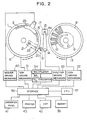
- An immunoanalysis apparatus to which the invention is applied may be constructed as shown in Fig. 2.
- a plurality of reaction containers 2 are held in array on a rotary circular reaction disk 1 along its circumferential edge and the reaction disk 1 is rotated intermittently by means of a disk driving mechanism 31.
- the reaction container 2 suitable for fluorophotometry is made of a transparent material such as glass or acryl resin.
- a fluorophotometer 19 has a photometric position 20 which lies inside a reaction thermostatic tank 6.
- the photometer 19 is of a multi-wavelength photometric type having a plurality of detectors and it faces the reaction container 2 so that when a reaction container 2 is at the photometric position 20 on the reaction disk 1, flux of light 22 emitted from as light source lamp 21 transmits through that reaction container 2. Exciting light, monochromatic light of a predetermined wavelength, is irradiated on the solution in the reaction container from above or below the reaction container, and fluorescence emitted from the solution transmits through the side surface of the container and monochromatic light of a predetermined wavelength is selected and detected by a photomultiplier tube serving as the photoelectric detector.
- a plurality of reagent cups 12 needed for a plurality of kinds of analysis items are held in array on a reagent disk 9 which is rotatable clockwise or counterclockwise.
- Each of the reagent cups 12 has a plurality of (for example, three) reagent solution storage chambers and individual chambers store a first reagent, a second reagent and as necessary a third reagent which correspond to specified analysis items, respectively.
- the reagent disk 9 and a sample disk 8 are rotated about a center shaft 10 by a number of pitches designated by a controller and this rotational operation is accomplished by a disk driving mechanism 32.
- a sample container 11 containing a target test sample and a required reagent cup 12 on the reagent disk 9 can be positioned and stopped at positions where the requisite test sample and reagent are sucked.
- Sample cups 11 are maintained at a predetermined temperature inside an air thermoconstant tank.
- the reagent cups 12 are maintained at a low temperature of 10°C or less by means of a cooling tank 13.
- the reference sample or reference solution is formed of a liquid or solution containing a substance which is able to be excited to fluorescence by itself and for example, may be quinine sulfate, rhodamine B or water.
- one of the reagent cups 12 container a quinine sulfate solution at a predetermined concentration.
- An arm 14a can be moved vertically (orthogonally to the sheet of drawing) and rotated about a shaft 14 by means of a pipetter driving mechanism 33.
- the arm 14a is mounted with a liquid charge/discharge nozzle or pipetting probe 15 which communicates with a cylinder in a pipetter driving mechanism 33 through a liquid flow path.
- the probe 15 can be moved vertically at any one of the sample suction position on the sample disk 8, reagent suction position on the reagent disk 9, liquid discharge position 16 on the reaction disk 1 and probe washing position 17.
- the probe 15 may be operated to pipette a predetermined amount of sample from a sample cup 11 to a reaction container at a discharge position 16 and during the second cycle of rotation of the reaction disk 1, the probe 15 may be operated to pipette a predetermined amount of reagent from a reagent cup 12 to the reaction container at that discharge position 16.
- the probe 15 may be operated to pipette a test sample and a first reagent during the first cycle of rotation of the reaction disk and to pipette a second reagent and as necessary a third reagent during the second and ensuing cycle of rotation of the reaction disk.
- solid phases attached with antibodies (or antigens) corresponding to individual analysis items i.e., kinds of antigens or antibodies are stored precedently in reaction containers.
- antibodies or antigens
- the surface of the bead is coated in advance with, for example, specified antibodies.
- a sample sucked into the probe 15 from a sample cup 11 is discharged into that reaction container.
- the interior and exterior of the probe 15 are washed by means of a washing unit also designated by reference numeral 17.
- the probe 15 sucks a reagent solution for immunoreaction from a selected reagent cup 12 and adds the reagent to the same reaction container 2 still staying at that discharge position 16.
- the immunoreaction reagent solution contains antibodies labeled by enzyme.
- the reaction disk 1 is advanced clockwise by one step. Similar sampling operations are sequentially undertaken by the probe 15, serving as a pipetting mechanism, for the following reaction containers.
- the reaction container containing the test sample added with the immunoreaction reagent (that is, a first reagent)
- an antigen standing for a target element in the sample couples to an antibody attached to the bead and the antibody labeled by enzyme couples to the coupled antigen.
- immunocomplexes are created on the bead through antigen/antibody reaction.
- the reaction container containing the immunocomplex reaches a region of a washer 25 having a plurality of nozzles, a liquid containing part of the reagent which has not been reacted is discharged from the reaction container and accordingly the bead having the immunocomplex remains in the reaction container.
- a cleansing solution is charged into the reaction container, and the cleansing solution is then discharged after cleansing, leaving the bead having the immunocomplex in the reaction container.
- the above bead washing operation is repeated plural times.
- the washer 25 is operated for vertical movement and charging/discharging by means of a washer driving mechanism 40.
- the probe 15 is operated to suck a latently fluorescent reagent standing for a second reagent from a specified reagent cup 12 and add the second reagent to the bead.
- a latently fluorescent reagent a solution may be used containing a substrate which is able to turn into a fluorescent substance.
- the substrate reacts with the enzyme attached to the solid phase so as to turn into the fluorescent substance.
- exciting light is irradiated on the container by means of the fluorophotometer 19 to measure the intensity of fluorescence stemming from the reaction solution in the container.
- Output signals produced from a plurality of photodetectors of the photometer 19 are applied to a multiplexer 34 at which only a signal based on requisite monochromatic light is selected.
- the selected signal is converted by an analog/digital converter 35 into a digital signal which in turn is stored into a memory unit 38 such as a random access memory or a floppy disk through an interface 36 under the control of a central processing unit (CPU) 37.
- CPU central processing unit
- the lower limit for deciding the performance of the optical system and the upper limit for deciding degradation of the reagent are set in the analyzer.
- a setting value for deciding the performance of the optical system and an acceptable upper limit value for deciding the reagent degradation are inputted by the operator who is manipulating the keyboard of an operation panel 41 and stored in the memory unit 38 under the control of the CPU 37.
- T4 thyroxine
- TSH thyroid stimulating horumon
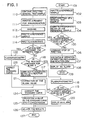
- step 101 the operator pushes the start key on the operation panel 41 of the analyzer to start analysis operation.
- the arm 14 a of the pipetting mechanism is first operated to pipette a predetermined amount of quinine sulfate solution, standing for the fluorescent reference sample, from a reagent cup 12 containing the quinine sulfate into the first reaction container (step 102). Thereafter, operation for pipetting a general test sample from a sample cup 11 corresponding to the following reaction container 2 is started (step 103).
- the pipetting mechanism is controlled by the controller such that neither a latently fluorescent reagent containing a substrate nor an immunoreaction start reagent is added to the reaction container filled with the quinine sulfate.
- fluorescence stemming from the reference sample is measured by means of the fluorophotometer 19 (step 104).
- the photometry uses an exciting wavelength of 380 nm to measure a fluorescent wavelength of 460nm. The same exciting wavelength as above is applied to any analysis items of general test sample.
- step 105 the controller decides whether a measured value of the intensity of fluorescence stemming from the reference sample exceeds a predetermined setting value. If the results of comparison indicate that the measured value of the fluorescence emitted from the reference sample is less than the setting value, the procedure proceeds to step 106. At that time, the controller issues a command for stopping operation to individual mechanisms including the pipetting mechanism 33 and reaction disk 1 and as a result the analysis operation of the analysis apparatus is discontinued (step 107) and at the same time an alarm indicative of unsuitability of the optical system for the analysis of small amounts is displayed on the screen of the CRT 42 (step 108). In this case, the operation of the analysis apparatus once ends (step 109). Observing the alarm display, the operator checks the fluorophotometer 19 and exchanges the light source lamp or the photoelectric detectors ,if degraded, with new ones to recover the performance of the analyzer.
- step 110 the pipetting mechanism 33 continues pipetting general test samples (step 110). No sample is pipetted in a reaction container for blank measurement but a latently fluorescent reagent is added to the blank measurement reaction container by the same amount as that added to the general test sample.
- a sample solution is pipetted, by an amount of 50 ⁇ l, from a sample cup 11 to a reaction container for general test sample and thereafter an immunoreaction reagent is 200 ⁇ l pipetted in that reaction container (step 111).
- the immunoreaction reagent a solution containing an antibody for each analysis item labeled by enzyme is used.
- an antigen/antibody reaction proceeds under the condition of heat insulation at 37°C to create antigen/antibody reaction complexes on a bead.
- the reaction container reaches the region of the washer 25 thirty minutes after start of the reaction and at the region, part of the solution containing substances which have not been reacted is discharged from the reaction container, a cleansing solution is 500 ⁇ l charged into the reaction container and the used cleansing solution is finally discharged from the reaction container (step 112).
- the above bead washing operation using the cleansing solution is repeated three times.
- the pipetting mechanism 14 operates to pipette the reference sample solution and the latently fluorescent reagent into corresponding reaction containers for reference sample and reagent blank which are disposed at predetermined intervals in the train of reaction containers (step 113).
- the pipetting mechanism 14 then pipettes a latently fluorescent reagent into the reaction container for general test sample washed in the step 112.
- a latently fluorescent reagent a substrate and a buffer solution are added by 50 ⁇ l and 200 ⁇ l, respectively, (step 114) and an enzyme reaction proceeds for 30 minutes at 37°C.
- a solution of 4-methylumbelliferyl phosphate is used as the substrate and alkali phosphatase is used as the enzyme to proceed with the enzyme reaction.
- 4-methylumbelliferone is created which is a fluorescent substance.
- step 115 it is decided whether the reaction container positioned at the photometry position 20 is for the reference sample and similarly, it is decided in step 116 whether the reaction container reaching the photometry position is for the reagent blank.
- the intensity of fluorescence stemming therefrom is measured 15 minutes after the step 114 (step 117).
- the amount of 4-methylumbelliferone created in the reaction solution depends on the amount of antigen/antibody reaction complexes on the bead.
- step 122 At which the intensity of fluorescence stemming from the quinine sulfate solution is measured.
- a correction coefficient for a standard curve stored in advance is calculated on the basis of a measured value of the intensity of fluorescence stemming from the reference sample (step 123) and used for correcting the standard curve (step 124)
- step 118 the procedure branches from step 116 to step 118, at which the intensity of fluorescence stemming from the reagent blank that is unaffected by the antigen/antibody reaction complex is measured, and the fluorescence intensity is decided as to whether to be greater than an acceptable value stored in advance (step 119). If a value measured for the reagent blank exceeds the acceptable value, an alarm indicative of degradation of the latently fluorescent reagent is displayed on the screen of the CRT 42 (step 120). This alarm is printed out of a printer 43 upon delivery of results of analysis in step 126.
- the fluorophotometry of the reaction solution of general test sample follows and the measured value of the intensity of fluorescence stemming from the reaction solution is corrected on the basis of a reagent blank value (step 121) and then used for being compared with the standard curve. Even if the reagent blank value is decided to be greater than the acceptable value, the analysis apparatus may be allowed not to be discontinued but to continue analyzing general test samples, provided that the operator fulfils exchange of the substrate solution in accordance with the alarm display.
- step 125 element concentrating corresponding to the measured values for the general test samples are calculated from the standard curve corrected on the basis of the measured value for the reference sample and the measured value for reagent blank, and in step 126 analysis values of individual test samples are delivered.
- the ratio between a photometrical value measured for the reference sample when preparing a standard curve and a photometrical value measured for the reference sample immediately before conducting photometry of the test sample is used to correct the standard curve itself but alternatively a value measured for the test sample may first be corrected by the correction coefficient and then applied to the initial standard curve for the purpose of calculating concentration of analysis elements.
- an average value B of the corrected values in Table 1 is 391.4 and an average value B′ of the corrected values in Table 2 is 391.9, demonstrating that even when the measurement is not carried out on the same line, the influence of drift of the analysis apparatus can be removed to ensure highly reproducible measurement.
- the immunoanalysis apparatus shown in Fig. 2 is also used.
- a rhodamine B solution is used and a cup containing the reference sample is disposed on the reagent disk 9.
- the analyzer is operated in accordance with a flow similar to that shown in Fig. 1 to measure T4 and TSH.
- No latently fluorescent reagent is added to the rhodamine B solution and the intensity of fluorescent stemming from the rhodamine B itself is measured.
- the wavelength of exciting light for the reference sample is 550 nm and the wavelength of fluorescence excited from the reference sample is 590 nm.
- the immunoanalysis apparatus shown in Fig. 2 is also used.
- water is employed in this embodiment.
- the analysis apparatus is operated in accordance with a flow similar to that shown in Fig. 1 to measure T4 and TSH.
- addition of any latently fluorescent reagent is not effected, either, and the intensity of fluorescence stemming from water itself is measured.
- the wavelength of exciting light for the water serving as the reference sample is 380 nm and the wavelength of fluorescence excited from the reference sample is 470 nm.
- FIG. 2 A fourth embodiment of the invention will now be described.
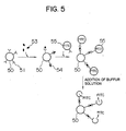
- the substrate but liposomes sensitized by antibodies are used as the latently fluorescent reagent.
- TSH as a measurement item.
- the analysis apparatus shown in Fig. 2 is also used and a reference sample of quinine sulfate and a solution of dispersed liposomes each having sealed-in FITC serving as fluorescence marker are set on the reagent disk 9.
- Anti-TSH antibodies are attached to the surface of the liposome.
- the same anti-TSH antibodies 51 as above are attached to the surface of a head to contained in a reaction container 2.
- Ten samples of control blood serum are set on the sample disk 8.
- An example of a standard curve prepared in advance is shown in Fig. 6.
- the operation of the analysis apparatus also follows the steps 101 to 113 in Fig. 1.
- step 105 the value measured for the reference sample of quinine sulfate is decided to be greater than the setting value.
- antigens 53 (TSH) in the blood serum sample couple to the antibodies 51 on the bead 50 to create antigen/antibody reaction complexes 54.
- TSH antigens 53
- a reagent containing liposomes 55 sensitized by antibodies is added.
- the washer 25 operates to remove part of reagent and part of sample which have not been reacted in the reaction container and after washing, a buffer solution containing a surfactant is 500 ⁇ l added in the reaction container. This ruptures the enclosure of the lipsome 55 and the sealed-in FITC flows out. The reaction container containing this reaction solution is positioned at the photometry position 20 and fluorophotometry is then carried out.
- the value measured for test samples is corrected using the measured values for the reference sample and reagent blank in a way shown in the flow chart of Fig. 1.
- the correction coefficient for standard curve based on the fluorophotometry of the reference sample was 165.4/175.4.
- the average value of fluorescence intensity of measured values for 10 analyzed TSH samples was 270.2 and the corrected average value was 254.8.
Landscapes
- Health & Medical Sciences (AREA)
- Life Sciences & Earth Sciences (AREA)
- Chemical & Material Sciences (AREA)
- Immunology (AREA)
- Molecular Biology (AREA)
- Engineering & Computer Science (AREA)
- Physics & Mathematics (AREA)
- General Health & Medical Sciences (AREA)
- Biomedical Technology (AREA)
- Biochemistry (AREA)
- Urology & Nephrology (AREA)
- General Physics & Mathematics (AREA)
- Pathology (AREA)
- Endocrinology (AREA)
- Hematology (AREA)
- Analytical Chemistry (AREA)
- Cell Biology (AREA)
- Biotechnology (AREA)
- Chemical Kinetics & Catalysis (AREA)
- Microbiology (AREA)
- Food Science & Technology (AREA)
- Medicinal Chemistry (AREA)
- Nuclear Medicine, Radiotherapy & Molecular Imaging (AREA)
- Optics & Photonics (AREA)
- Reproductive Health (AREA)
- Investigating, Analyzing Materials By Fluorescence Or Luminescence (AREA)
- Automatic Analysis And Handling Materials Therefor (AREA)
- Investigating Or Analysing Materials By The Use Of Chemical Reactions (AREA)
Applications Claiming Priority (2)
| Application Number | Priority Date | Filing Date | Title |
|---|---|---|---|
| JP63210741A JP2656564B2 (ja) | 1988-08-26 | 1988-08-26 | 免疫分析方法 |
| JP210741/88 | 1988-08-26 |
Publications (3)
| Publication Number | Publication Date |
|---|---|
| EP0355849A2 true EP0355849A2 (fr) | 1990-02-28 |
| EP0355849A3 EP0355849A3 (fr) | 1991-07-03 |
| EP0355849B1 EP0355849B1 (fr) | 1994-06-15 |
Family
ID=16594346
Family Applications (1)
| Application Number | Title | Priority Date | Filing Date |
|---|---|---|---|
| EP89115739A Expired - Lifetime EP0355849B1 (fr) | 1988-08-26 | 1989-08-25 | Méthode et appareil de mesure automatisée de fluorescence |
Country Status (4)
| Country | Link |
|---|---|
| US (1) | US5324635A (fr) |
| EP (1) | EP0355849B1 (fr) |
| JP (1) | JP2656564B2 (fr) |
| DE (1) | DE68916132T2 (fr) |
Cited By (10)
| Publication number | Priority date | Publication date | Assignee | Title |
|---|---|---|---|---|
| EP0517092A1 (fr) * | 1991-06-03 | 1992-12-09 | Abbott Laboratories | Trousse de réactifs pour immunoessais |
| DE4309529A1 (de) * | 1992-03-24 | 1993-09-30 | Hitachi Ltd | Analytisches Verfahren und Vorrichtung |
| US5376313A (en) * | 1992-03-27 | 1994-12-27 | Abbott Laboratories | Injection molding a plastic assay cuvette having low birefringence |
| WO2001092882A1 (fr) * | 2000-06-02 | 2001-12-06 | Cis Bio International | Procede permettant de detecter un liquide dans un melange |
| EP1748291B1 (fr) * | 2005-07-25 | 2010-06-09 | Ushiodenki Kabushiki Kaisha | Appareil de mesure de micropuces |
| WO2011012657A1 (fr) * | 2009-07-29 | 2011-02-03 | F. Hoffmann-La Roche Ag | Analyseur automatique |
| EP2333563A1 (fr) * | 2009-12-14 | 2011-06-15 | Roche Diagnostics GmbH | Analyseur comportant un appareil pour fournir des réactifs |
| WO2014145619A1 (fr) * | 2013-03-15 | 2014-09-18 | Hycor Biomedical, Inc. | Dispositif et procédés associés de réalisation de mesures de luminescence et de fluorescence d'un échantillon |
| EP2072999A4 (fr) * | 2006-10-13 | 2015-07-22 | Beckman Coulter Inc | Procédé d'identification d'anomalie et appareil d'analyse |
| EP2779900B1 (fr) * | 2011-11-14 | 2015-09-16 | Roche Diagnostics GmbH | Instrument d'analyse pour le dépistage d'au moins un analyte dans un échantillon |
Families Citing this family (43)
| Publication number | Priority date | Publication date | Assignee | Title |
|---|---|---|---|---|
| US5507410A (en) | 1992-03-27 | 1996-04-16 | Abbott Laboratories | Meia cartridge feeder |
| CA2129368C (fr) * | 1992-03-27 | 2002-01-22 | Linda S. Schmidt | Procede de verification de methodologies d'essai pour methodes analytiques |
| US6190617B1 (en) | 1992-03-27 | 2001-02-20 | Abbott Laboratories | Sample container segment assembly |
| US5578494A (en) | 1992-03-27 | 1996-11-26 | Abbott Laboratories | Cap actuator for opening and closing a container |
| US5605665A (en) | 1992-03-27 | 1997-02-25 | Abbott Laboratories | Reaction vessel |
| US5627522A (en) | 1992-03-27 | 1997-05-06 | Abbott Laboratories | Automated liquid level sensing system |
| US5610069A (en) | 1992-03-27 | 1997-03-11 | Abbott Laboratories | Apparatus and method for washing clinical apparatus |
| US5960160A (en) | 1992-03-27 | 1999-09-28 | Abbott Laboratories | Liquid heater assembly with a pair temperature controlled electric heating elements and a coiled tube therebetween |
| US5646049A (en) | 1992-03-27 | 1997-07-08 | Abbott Laboratories | Scheduling operation of an automated analytical system |
| US5536471A (en) | 1992-03-27 | 1996-07-16 | Abbott Laboratories | Syringe with bubble flushing |
| US5575978A (en) | 1992-03-27 | 1996-11-19 | Abbott Laboratories | Sample container segment assembly |
| US5635364A (en) | 1992-03-27 | 1997-06-03 | Abbott Laboratories | Assay verification control for an automated analytical system |
| USD349861S (en) | 1992-07-20 | 1994-08-23 | Abbott Laboratories | Automated analytical instrument |
| US5577137A (en) * | 1995-02-22 | 1996-11-19 | American Research Corporation Of Virginia | Optical chemical sensor and method using same employing a multiplicity of fluorophores contained in the free volume of a polymeric optical waveguide or in pores of a ceramic waveguide |
| JP3063564B2 (ja) * | 1995-03-17 | 2000-07-12 | 株式会社日立製作所 | 自動分析装置 |
| US5874216A (en) * | 1996-02-23 | 1999-02-23 | Ensys Environmental Products, Inc. | Indirect label assay device for detecting small molecules and method of use thereof |
| EP0914608A1 (fr) * | 1996-05-09 | 1999-05-12 | 3-Dimensional Pharmaceuticals, Inc. | Procede d'analyse et appareil a changement thermique et a microplaque pour l'optimisation de la mise au point de ligands et de la chimie des proteines a variables multiples |
| US6063591A (en) * | 1997-05-14 | 2000-05-16 | 3M Innovative Properties Company | System for measuring the efficacy of a sterilization cycle |
| US6025189A (en) * | 1997-05-14 | 2000-02-15 | 3M Innovative Properties Company | Apparatus for reading a plurality of biological indicators |
| US5863790A (en) * | 1997-05-14 | 1999-01-26 | Minnesota Mining And Manfacturing Company | Biological sterility indicator |
| WO1999001737A2 (fr) * | 1997-06-10 | 1999-01-14 | Calspan Corporation | Detection des matieres d'un agent chimique par utilisation d'un polymere sorbant et sonde a fluorescence |
| WO1999004628A1 (fr) * | 1997-07-28 | 1999-02-04 | Dermatolazer Technologies Ltd. | Methode de traitement de pathogenes basee sur la phototherapie et composition utile dans ce procede |
| US6043880A (en) * | 1997-09-15 | 2000-03-28 | Becton Dickinson And Company | Automated optical reader for nucleic acid assays |
| US6597450B1 (en) | 1997-09-15 | 2003-07-22 | Becton, Dickinson And Company | Automated Optical Reader for Nucleic Acid Assays |
| ATE335498T1 (de) | 1997-11-12 | 2006-09-15 | Johnson & Johnson Pharm Res | Methode mit hoher durchsatzrate zur funktionellen klassifizierung von in einem genomischen versuchsansatz identifizierten proteinen |
| US6211526B1 (en) * | 1998-09-30 | 2001-04-03 | The United States Of America As Represented By The Secretary Of The Navy | Marking of materials using luminescent and optically stimulable glasses |
| US6569631B1 (en) | 1998-11-12 | 2003-05-27 | 3-Dimensional Pharmaceuticals, Inc. | Microplate thermal shift assay for ligand development using 5-(4″dimethylaminophenyl)-2-(4′-phenyl)oxazole derivative fluorescent dyes |
| US6300638B1 (en) | 1998-11-12 | 2001-10-09 | Calspan Srl Corporation | Modular probe for total internal reflection fluorescence spectroscopy |
| US6838680B2 (en) * | 1999-05-12 | 2005-01-04 | Aclara Biosciences, Inc. | Multiplexed fluorescent detection in microfluidic devices |
| US6323495B1 (en) | 1999-09-24 | 2001-11-27 | Umm Electronics, Inc. | Method and apparatus for the determination of phase delay in a lifetime fluorometer without the use of lifetime standards |
| US20020110843A1 (en) * | 2000-05-12 | 2002-08-15 | Dumas David P. | Compositions and methods for epitope mapping |
| US7115232B2 (en) * | 2001-07-13 | 2006-10-03 | Hudson Gordon S | Fluorescence validation microplate and method of use |
| DE10200499A1 (de) * | 2002-01-03 | 2003-07-10 | Zeiss Carl Jena Gmbh | Verfahren und/oder Anordnung zur Identifikation von fluoreszierenden, lumineszierenden und/oder absorbierenden Substanzen auf und/oder in Probenträgern |
| EP1506413B1 (fr) * | 2002-05-17 | 2016-07-06 | Becton Dickinson and Company | Systeme automatise destine a isoler, amplifier et detecter une sequence d'acides nucleiques cibles |
| JP2004219218A (ja) * | 2003-01-14 | 2004-08-05 | Fuji Photo Film Co Ltd | 自動分析装置 |
| US20050112587A1 (en) * | 2003-11-25 | 2005-05-26 | Sherrill James V. | Analyzing biological probes |
| CN103884698B (zh) | 2004-06-07 | 2017-04-12 | 先锋生物科技股份有限公司 | 用于微流体器件的光学透镜系统和方法 |
| JP5813914B2 (ja) * | 2009-04-27 | 2015-11-17 | 株式会社東芝 | 自動分析装置 |
| CA2815951A1 (fr) | 2010-11-03 | 2012-05-10 | Reametrix Inc. | Systeme de mesure pour la detection de fluorescence, et procede associe |
| AU2013202804A1 (en) | 2012-06-14 | 2014-01-16 | Gen-Probe Incorporated | Use of a fluorescent material to detect failure or deteriorated performance of a fluorometer |
| CN103616358B (zh) * | 2013-11-30 | 2015-12-09 | 广州蓝勃生物科技有限公司 | 环保型多项目全自动固相荧光检测系统 |
| JP6166008B2 (ja) * | 2015-03-31 | 2017-07-26 | シスメックス株式会社 | 免疫測定装置 |
| KR102818381B1 (ko) * | 2022-03-21 | 2025-06-10 | 주식회사 앱솔로지 | 자동 면역분석 시스템 |
Family Cites Families (19)
| Publication number | Priority date | Publication date | Assignee | Title |
|---|---|---|---|---|
| FR1588298A (fr) * | 1968-10-08 | 1970-04-10 | ||
| US3973129A (en) * | 1975-01-10 | 1976-08-03 | Bell Telephone Laboratories, Incorporated | Fluorimetric apparatus and method for analysis of body fluid |
| US4043756A (en) * | 1976-12-29 | 1977-08-23 | Hycel, Inc. | Calibration in an automatic chemical testing apparatus |
| US4271123A (en) * | 1979-10-22 | 1981-06-02 | Bio-Rad Laboratories, Inc. | Automated system for performing fluorescent immunoassays |
| US4372745A (en) * | 1979-12-19 | 1983-02-08 | Electro-Nucleonics, Inc. | Chemical luminescence amplification substrate system for immunochemistry involving microencapsulated fluorescer |
| JPS56147068A (en) * | 1980-04-16 | 1981-11-14 | Olympus Optical Co Ltd | Automatic analyzer |
| JPS5719646A (en) * | 1980-07-09 | 1982-02-01 | Olympus Optical Co Ltd | Calibration method for electrolyte measurement in automatic biochemical analyzer with builtin type flame light photometer |
| JPS5924258A (ja) * | 1982-07-30 | 1984-02-07 | Shimadzu Corp | 自動生化学分析装置 |
| JPS59182347A (ja) * | 1983-03-31 | 1984-10-17 | Shimadzu Corp | 自動分析装置 |
| US4536655A (en) * | 1983-08-29 | 1985-08-20 | Axonics, Inc. | Fluorometer having an improved optical system |
| JPS6073465A (ja) * | 1983-09-30 | 1985-04-25 | Shimadzu Corp | 自動分析装置 |
| JPH0664069B2 (ja) * | 1984-11-14 | 1994-08-22 | オリンパス光学工業株式会社 | 免疫学的自動分析方法 |
| JPH0660901B2 (ja) * | 1985-08-30 | 1994-08-10 | 東ソー株式会社 | 酵素免疫測定法 |
| JPS62144071A (ja) * | 1985-12-18 | 1987-06-27 | Hitachi Ltd | 自動化学分析装置 |
| JPS6314289A (ja) * | 1986-07-03 | 1988-01-21 | Nec Corp | デ−タ収集処理装置 |
| JPS63101758A (ja) * | 1986-10-20 | 1988-05-06 | Toshiba Corp | 自動化学分析装置 |
| US4886761A (en) * | 1987-03-26 | 1989-12-12 | Yellowstone Diagnostics Corporation | Polysilicon binding assay support and methods |
| JPS63206660A (ja) * | 1987-02-24 | 1988-08-25 | Toshiba Corp | 自動化学分析装置 |
| JPS646867A (en) * | 1987-06-30 | 1989-01-11 | Konishiroku Photo Ind | Chemical analyzer |
-
1988
- 1988-08-26 JP JP63210741A patent/JP2656564B2/ja not_active Expired - Fee Related
-
1989
- 1989-08-24 US US07/397,853 patent/US5324635A/en not_active Expired - Lifetime
- 1989-08-25 DE DE68916132T patent/DE68916132T2/de not_active Expired - Fee Related
- 1989-08-25 EP EP89115739A patent/EP0355849B1/fr not_active Expired - Lifetime
Cited By (29)
| Publication number | Priority date | Publication date | Assignee | Title |
|---|---|---|---|---|
| US5294404A (en) * | 1991-06-03 | 1994-03-15 | Abbott Laboratories | Reagent pack for immunoassays |
| EP0517092A1 (fr) * | 1991-06-03 | 1992-12-09 | Abbott Laboratories | Trousse de réactifs pour immunoessais |
| DE4309529A1 (de) * | 1992-03-24 | 1993-09-30 | Hitachi Ltd | Analytisches Verfahren und Vorrichtung |
| US5376313A (en) * | 1992-03-27 | 1994-12-27 | Abbott Laboratories | Injection molding a plastic assay cuvette having low birefringence |
| WO2001092882A1 (fr) * | 2000-06-02 | 2001-12-06 | Cis Bio International | Procede permettant de detecter un liquide dans un melange |
| FR2809817A1 (fr) * | 2000-06-02 | 2001-12-07 | Cis Bio Int | Procede de detection de presence d'un liquide dans un melange |
| US6952259B2 (en) | 2000-06-02 | 2005-10-04 | Cis Bio International | Method of detecting the presence of a liquid in a mix |
| EP1748291B1 (fr) * | 2005-07-25 | 2010-06-09 | Ushiodenki Kabushiki Kaisha | Appareil de mesure de micropuces |
| EP2072999A4 (fr) * | 2006-10-13 | 2015-07-22 | Beckman Coulter Inc | Procédé d'identification d'anomalie et appareil d'analyse |
| WO2011012657A1 (fr) * | 2009-07-29 | 2011-02-03 | F. Hoffmann-La Roche Ag | Analyseur automatique |
| US8916096B2 (en) | 2009-07-29 | 2014-12-23 | Hitachi High-Technologies Corporation | Automatic analyzer |
| EP2333563A1 (fr) * | 2009-12-14 | 2011-06-15 | Roche Diagnostics GmbH | Analyseur comportant un appareil pour fournir des réactifs |
| EP2779900B1 (fr) * | 2011-11-14 | 2015-09-16 | Roche Diagnostics GmbH | Instrument d'analyse pour le dépistage d'au moins un analyte dans un échantillon |
| US9611504B2 (en) | 2011-11-14 | 2017-04-04 | Roche Diabetes Care, Inc. | Methods of measuring analytes that include a test element quality measurement based upon intrinsic luminescence of a test chemical of the test element |
| CN108445242A (zh) * | 2013-03-15 | 2018-08-24 | Hycor生物医学有限责任公司 | 进行样本的冷光和荧光测量的装置和相关方法 |
| US10739262B2 (en) | 2013-03-15 | 2020-08-11 | Hycor Biomedical, Llc | Automated immunoanalyzer system for performing diagnostic assays for autoimmune and infectious diseases |
| US9651550B2 (en) | 2013-03-15 | 2017-05-16 | Hycor Biomedical, Llc | Automated immunoanalyzer system for performing diagnostic assays for autoimmune and infectious diseases |
| US9658225B2 (en) | 2013-03-15 | 2017-05-23 | Hycor Biomedical, Llc | Automated immunoanalyzer system for performing diagnostic assays for allergies and autoimmune diseases |
| US9658226B2 (en) | 2013-03-15 | 2017-05-23 | Hycor Biomedical, Llc | Automated immunoanalyzer system for performing diagnostic assays for autoimmune and infectious diseases |
| US9753033B2 (en) | 2013-03-15 | 2017-09-05 | Hycor Biomedical, Llc | Device and associated methods for performing luminescence and fluorescence measurements of a sample |
| WO2014145619A1 (fr) * | 2013-03-15 | 2014-09-18 | Hycor Biomedical, Inc. | Dispositif et procédés associés de réalisation de mesures de luminescence et de fluorescence d'un échantillon |
| US10732110B2 (en) | 2013-03-15 | 2020-08-04 | Hycor Biomedical, Llc | Automated immunoanalyzer system for performing diagnostic assays for autoimmune and infectious diseases |
| US9766233B2 (en) | 2013-03-15 | 2017-09-19 | Hycor Biomedical, Llc | Device and associated methods for performing luminescence and fluorescence measurements of a sample |
| US10732111B2 (en) | 2013-03-15 | 2020-08-04 | Hycor Biomedical, Llc | Automated immunoanalyzer system for performing diagnostic assays for allergies and autoimmune diseases |
| US9075055B2 (en) | 2013-03-15 | 2015-07-07 | Hycor Biomedical, Inc. | Device and associated methods for performing luminescence and fluorescence measurements of a sample |
| US10955346B2 (en) | 2013-03-15 | 2021-03-23 | Hycor Biomedical, Llc | Device and associated methods for performing luminescence and fluorescence measurements of a sample |
| US11204323B2 (en) | 2013-03-15 | 2021-12-21 | Hycor Biomedical, Llc | Device and associated methods for performing luminescence and fluorescence measurements of a sample |
| EP3933384A1 (fr) * | 2013-03-15 | 2022-01-05 | Hycor Biomedical, LLC | Dispositif et procédé associé de réalisation de mesures de luminescence et de fluorescence d'un échantillon |
| CN108445242B (zh) * | 2013-03-15 | 2022-03-04 | Hycor生物医学有限责任公司 | 进行样本的冷光和荧光测量的装置和相关方法 |
Also Published As
| Publication number | Publication date |
|---|---|
| DE68916132T2 (de) | 1994-12-08 |
| JP2656564B2 (ja) | 1997-09-24 |
| EP0355849A3 (fr) | 1991-07-03 |
| EP0355849B1 (fr) | 1994-06-15 |
| DE68916132D1 (de) | 1994-07-21 |
| JPH0259671A (ja) | 1990-02-28 |
| US5324635A (en) | 1994-06-28 |
Similar Documents
| Publication | Publication Date | Title |
|---|---|---|
| EP0355849B1 (fr) | Méthode et appareil de mesure automatisée de fluorescence | |
| JP3453573B2 (ja) | 自動連続ランダム・アクセス分析システムおよびその構成要素 | |
| US5316726A (en) | Automated immunoassay analyzer with pictorial display of assay information | |
| JP3425567B2 (ja) | 自動連続ランダム・アクセス分析システム | |
| JP3600227B2 (ja) | 自動連続ランダムアクセス分析システムおよびその構成要素 | |
| JP3439466B2 (ja) | 自動連続ランダム・アクセス分析システム及びその構成要素 | |
| EP0198513B1 (fr) | Procédé analytique et dispositif pour la détermination de fluorescence ou phosphorescence | |
| US5380487A (en) | Device for automatic chemical analysis | |
| AU2004260068B2 (en) | Automated multi-detector analyzer | |
| US4816418A (en) | Method and apparatus for performing automated, multi-sequential immunoassays | |
| JP2950698B2 (ja) | 洗浄機能付き自動分析装置 | |
| EP0521636B1 (fr) | Procédé pour l'essay d'activité enzymatique et leur dispositif | |
| JP3739547B2 (ja) | 自動分析装置 | |
| JPH05273118A (ja) | 被検試料液の濃度又は成分の分析方法及び分析装置 | |
| JPH0554067B2 (fr) | ||
| JPH06167503A (ja) | 免疫自動分析装置 | |
| JPH0421140B2 (fr) | ||
| JPH0575977B2 (fr) |
Legal Events
| Date | Code | Title | Description |
|---|---|---|---|
| PUAI | Public reference made under article 153(3) epc to a published international application that has entered the european phase |
Free format text: ORIGINAL CODE: 0009012 |
|
| 17P | Request for examination filed |
Effective date: 19890825 |
|
| AK | Designated contracting states |
Kind code of ref document: A2 Designated state(s): DE FR GB |
|
| PUAL | Search report despatched |
Free format text: ORIGINAL CODE: 0009013 |
|
| AK | Designated contracting states |
Kind code of ref document: A3 Designated state(s): DE FR GB |
|
| 17Q | First examination report despatched |
Effective date: 19931008 |
|
| GRAA | (expected) grant |
Free format text: ORIGINAL CODE: 0009210 |
|
| AK | Designated contracting states |
Kind code of ref document: B1 Designated state(s): DE FR |
|
| REF | Corresponds to: |
Ref document number: 68916132 Country of ref document: DE Date of ref document: 19940721 |
|
| ET | Fr: translation filed | ||
| PLBE | No opposition filed within time limit |
Free format text: ORIGINAL CODE: 0009261 |
|
| STAA | Information on the status of an ep patent application or granted ep patent |
Free format text: STATUS: NO OPPOSITION FILED WITHIN TIME LIMIT |
|
| 26N | No opposition filed | ||
| PGFP | Annual fee paid to national office [announced via postgrant information from national office to epo] |
Ref country code: FR Payment date: 19970618 Year of fee payment: 9 |
|
| PGFP | Annual fee paid to national office [announced via postgrant information from national office to epo] |
Ref country code: DE Payment date: 19970930 Year of fee payment: 9 |
|
| PG25 | Lapsed in a contracting state [announced via postgrant information from national office to epo] |
Ref country code: FR Free format text: LAPSE BECAUSE OF NON-PAYMENT OF DUE FEES Effective date: 19990430 |
|
| PG25 | Lapsed in a contracting state [announced via postgrant information from national office to epo] |
Ref country code: DE Free format text: LAPSE BECAUSE OF NON-PAYMENT OF DUE FEES Effective date: 19990601 |
|
| REG | Reference to a national code |
Ref country code: FR Ref legal event code: ST |