EP0362077A1 - Bestimmungsverfahren für anionische Sulfatid-Polyoside und Zusammensetzung dafür - Google Patents
Bestimmungsverfahren für anionische Sulfatid-Polyoside und Zusammensetzung dafür Download PDFInfo
- Publication number
- EP0362077A1 EP0362077A1 EP89402691A EP89402691A EP0362077A1 EP 0362077 A1 EP0362077 A1 EP 0362077A1 EP 89402691 A EP89402691 A EP 89402691A EP 89402691 A EP89402691 A EP 89402691A EP 0362077 A1 EP0362077 A1 EP 0362077A1
- Authority
- EP
- European Patent Office
- Prior art keywords
- conjugate
- enzyme
- revealing
- gag
- polycation
- Prior art date
- Legal status (The legal status is an assumption and is not a legal conclusion. Google has not performed a legal analysis and makes no representation as to the accuracy of the status listed.)
- Withdrawn
Links
Images

Classifications
-
- G—PHYSICS
- G01—MEASURING; TESTING
- G01N—INVESTIGATING OR ANALYSING MATERIALS BY DETERMINING THEIR CHEMICAL OR PHYSICAL PROPERTIES
- G01N33/00—Investigating or analysing materials by specific methods not covered by groups G01N1/00 - G01N31/00
- G01N33/48—Biological material, e.g. blood, urine; Haemocytometers
- G01N33/50—Chemical analysis of biological material, e.g. blood, urine; Testing involving biospecific ligand binding methods; Immunological testing
- G01N33/86—Chemical analysis of biological material, e.g. blood, urine; Testing involving biospecific ligand binding methods; Immunological testing involving blood coagulating time or factors, or their receptors
-
- G—PHYSICS
- G01—MEASURING; TESTING
- G01N—INVESTIGATING OR ANALYSING MATERIALS BY DETERMINING THEIR CHEMICAL OR PHYSICAL PROPERTIES
- G01N2400/00—Assays, e.g. immunoassays or enzyme assays, involving carbohydrates
- G01N2400/10—Polysaccharides, i.e. having more than five saccharide radicals attached to each other by glycosidic linkages; Derivatives thereof, e.g. ethers, esters
- G01N2400/38—Heteroglycans, i.e. polysaccharides having more than one sugar residue in the main chain in either alternating or less regular sequence, e.g. gluco- or galactomannans, Konjac gum, Locust bean gum or Guar gum
- G01N2400/40—Glycosaminoglycans, i.e. GAG or mucopolysaccharides, e.g. chondroitin sulfate, dermatan sulfate, hyaluronic acid, heparin, heparan sulfate, and related sulfated polysaccharides
Definitions
- the invention relates to a new method for assaying sulfated anionic polysaccharides, using in the assay principle the chemical structure of these compounds, and not their biological activities.
- the invention also relates to a ready-to-use box for implementing this method.
- the sulfated anionic polysaccharides such as glycosaminoglycans and in particular heparin, whose anticoagulant activity is well known, are assayed according to methods based on their biological activities, in particular their activity on various coagulation factors. This is how heparin is measured by its activity on activated factor X or activated factor II or even according to more global methods based on the coagulation time, such as the time of Cephalin Kaolin or TCK or else the method described in the French Pharmacopoeia.
- heparin is a very heterogeneous product, both in terms of the length of its chains and their charge rate, and that it was possible to fractionate them. and to fragment them in order to develop products with more specific activity. It has also been demonstrated that heparin, in addition to its well-known activities on coagulation, acts as a regulator in various biological systems and that fragments lacking activity on coagulation nevertheless retain their regulatory activity. It therefore became necessary to develop a new dosing method allowing the quantity of product contained in a liquid, and in particular in biological media, to be evaluated without resorting to biological activity.
- the invention proposes to remedy these drawbacks and proposes a new method which allows the quantitative determination of the sulfated polysaccharides contained in a liquid, in particular in a biological medium, in particular plasma.
- This assay is based on the strong anionic character of this type of product and allows the assay of any sulfated polysaccharide, whatever its structure or type of activity, provided that it has a total amount of anionic charges of at least at least two, preferably at least three, per disaccharide unit.
- Products of this type are represented by heparin and its fractions, fragments and derivatives, pentosan polysulfate, dextran sulfate and certain heparan sulfates; on the other hand, hyaluronic acid, dermatan sulphate, chondroitin sulphates and weakly sulphated heparans cannot be assayed by this method.
- the new process of the invention is based on the determination, by means of an anionic conjugate revealing, of the cationic sites which have remained free on a surface covered with a polycation, after the latter has been brought into contact with the liquid containing the polysaccharide.
- anionic sulphated to be assayed the revealing conjugate being chosen in such a way that its charge density allows it to be fixed on the sites which remain vacant on the polycation covering the surface, without however displacing the polysaccharide to be assayed which is previously fixed therein, and the quantity of the product to be dosed being determined by reference to a calibration curve established simultaneously on the same plate.
- the result is read on a calibration curve which directly indicates the amount of anionic polysaccharide sulfated per milliliter of the biological medium.
- the polycation used is a basic polymer chosen from the group consisting of protamine sulfate, polybrene, or a polyamino acid such as poly-arginine or poly-L-lysine.
- a poly-L-lysine is used, the average molecular weight of which is between 6000 and 9000 daltons. This polycation has the advantage of giving very good reproducibility in the assays.
- the revealing anionic conjugate consists of a glycosaminoglycan covalently linked to a polypeptide, preferably an enzyme. It could also be linked to an organic molecule containing aromatic nuclei capable of being labeled for example with iodine or with phosphorus, for example tyrosine labeled with iodine 125.
- GAG glycosaminoglycan
- GAG-P any glycosaminoglycan-polypeptide, each conjugate being designated by the glycosaminoglycan GAG and the polypeptide P concerned, for example GAG-enzyme, GAG-peroxidase , GAG-phosphatase, GAG-albumin, Heparin-P, Heparin-enzyme, Heparin-peroxidase, Heparin-phosphatase, Oligosaccharide-CHO-peroxidase (OS-CHO-peroxidase) and so on.
- OS-CHO-peroxidase Oligosaccharide-CHO-peroxidase
- the GAG used for the preparation of the conjugate must be sufficiently anionic to go to settle on the sites left vacant, but must not be more anionic than the product to be dosed, which it would then take the place of.
- the choice of GAG to be used for the preparation of the conjugate is therefore to be made as a function of the charge density of the anionic sulfated polysaccharide which it is desired to assay.
- the GAG used for the preparation of the GAG-P revealing conjugate can be any GAG or one of its fractions or fragments.
- the GAG used is preferably heparin or one of its fragments. These indeed have the capacity to attach, by their anionic charges, to the sites left vacant on the polycation after the product to be tested has been brought into contact with the latter.
- a mixture of GAGs which is particularly suitable for the preparation of GAG-P is represented by a mixture of heparin fragments consisting of chains essentially having from 4 to 10 disaccharide units.
- an enzyme is used as the polypeptide for the preparation of the revealing conjugate GAG-P; the latter is chosen from those which are easy to prepare and stable and which can be dosed easily, for example by the action of a substrate and the release of a colored product which is assayed by optical density; horseradish peroxidase and phosphatase are examples of such products.
- the GAG-enzyme conjugate is diluted with another GAG-P conjugate, preferably with a GAG-albumin, the GAG being identical for the two conjugates.
- the precision of the dosage is inversely proportional to the specific activity of the GAG-enzyme and, if the latter is too high, it is difficult to define operating conditions giving satisfactory precision.
- Use is therefore preferably made in order to be able to dose with a precision of the order of ⁇ g / ml to a mixture of conjugates, the final specific activity of the mixture being established so as to have good dosing precision under the operating conditions.
- the GAG-enzyme and GAG-albumin mixture has an enzymatic activity which is in our operating conditions between 1 u.DO and 2 u.DO, the most favorable specific activity being 1.5 u.DO.
- the conjugate formed by a GAG represented by a fragment of heparin prepared by depolymerization with nitrous acid and having an average molecular weight of 2,000 daltons in particular the product designated OS 4-10- CHO, the preparation of which is described in the paragraph "PREPARATION I a)" below, on the one hand, and as an enzyme horseradish peroxidase, constituted a revealer of choice for assaying in biological media various anionic sulfated polysaccharides , such as heparin and its fragments such as those sold under the name of Fraxiparine (R) (Choay, France), Fragmin (R) (Kabi, Sweden), Lovenox (R) (Pharmuka, France), or even as the pentosan polysulfate sold under the name of Hemoclar (R) (Midy, France), or the dextran sulfate and its derivatives, or the soluble functionalized dextrans described in patent application EP
- the polycation is adsorbed on the wells of a microtiter plate in basic medium, by incubation for 24 to 72 hours, preferably 48 hours, at a temperature between 0 ° and + 20 ° C, preferably + 4 ° C.
- the revealing anionic conjugate of step (b) consists of an anionic polysaccharide covalently linked to a product chosen from the group consisting of a enzyme and an organic molecule containing aromatic nuclei capable of being labeled with iodine or phosphorus.
- a product chosen from the group consisting of a enzyme and an organic molecule containing aromatic nuclei capable of being labeled with iodine or phosphorus.
- an enzyme is used which avoids using labeled products.
- step (b) the mixture is incubated for at least 2 hours at a temperature between 0 ° and + 20 ° C, preferably + 4 ° C.
- the anionic conjugate revealing of step (b) consists of an anionic polysaccharide, in particular a GAG, covalently linked to an enzyme and during step (d) uses an enzymatic assay method, the enzyme of the revealing conjugate being assayable for example by action on a substrate and release of a colored product which is assayed by optical density.
- the various washes are carried out using isotonic chlorinated solute (NaCl 9 P. 1000).
- the basic medium used for dissolving the polycation is a tris-phosphate buffer or a bicarbonate buffer at a pH between 9 and 10; preferably using a bicarbonate buffer with an ionic strength of between 0.05 M and 0.2 M, in particular 0.1 M, pH 9.6.
- the polycation is dissolved in the basic medium at a concentration of 100 ⁇ g / ml and 250 ⁇ g of this solution are placed in each well which is left to incubate for 48 hours.
- the assay will be all the more sensitive as the polycation concentration will be lower, but the range of response will be all the wider as the concentration will be higher, the sensitivity being inversely proportional to the polycation concentration.
- a concentration of 100 ⁇ g / ml is used as indicated above for assaying a GAG present in the plasma at a concentration of between 0.1 and 0.5 ⁇ g / ml.
- the results obtained are particularly satisfactory under the conditions indicated above when the sulfated anionic polysaccharide to be assayed is a regular fragment of heparin, that is to say a fragment consisting of disaccharides trisulphated, and whose average molecular weight is between 5,000 and 6,000 daltons.
- 200 ⁇ l of the liquid containing the sulfated polyanion to be assayed, this latter as well as the liquid used for carrying out the preparation are deposited in each well, after washing for elimination of the residual polycation, the calibration curve being added with a protease inhibitor which makes it possible both to comply with the polycation and, when the liquid is a biological medium, in particular plasma, to prevent the formation of thrombin and therefore of a clot which would disrupt the dosage.
- Benzamidine is preferably used as protease inhibitor, which is added at a concentration of between 0.0025 M and 0.1 M, preferably 0.0125 M, and the incubation is allowed to take place for at least 2 hours between 0 ° and + 20 ° C, preferably + 4 ° C.
- the mixture of revealing conjugates uses peroxidase as an enzyme
- the mixture comprises quantities of GAG-enzyme and of GAG-albumin expressed in equivalent of pure product of the order of 3.4 ⁇ g / ml of peroxidase and 7, 7 ⁇ g / ml of albumin. Under these conditions, preferably 200 ⁇ l per well of the developer mixture is used.
- the substrate used to reveal the amount of enzyme fixed in the wells consists of a mixture of orthophenylene diamine and peroxide hydrogen.
- a mixture of orthophenylene diamine and peroxide hydrogen 100 ⁇ l of a mixture containing 0.5 mg of orthophenylene diamine in a ml of 0.1 M sodium citrate / HCl buffer, pH 3.5 and 0.5 ⁇ l of peroxide are used for each well. hydrogen. Under these conditions, incubation is carried out for approximately 10 minutes at ambient temperature (approximately 20 to 25 ° C.), then the reaction is stopped by the addition of 1N sulfuric acid, advantageously 100 ⁇ l per well.
- the optical density is read at 492 nm, either directly at the bottom of the well, or by transferring into a spectrophotometer tank.
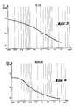
- the calibration curve in FIG. 1 is obtained, which gives the ordinate adsorption at 492 nm and the log of the concentration in ⁇ g / ml on the abscissa. Reading the optical density of the sample to be assayed, by referring to the calibration curve, to deduce the concentration of the product to be assayed in one ml of the biological medium.
- the revealing conjugate is a GAG-phosphatase
- the assay substrate is 4-nitrophenylphosphate, the reading of the optical density taking place at 405 nm.
- the invention also relates to a box intended for the implementation of the method according to the invention.
- This box contains: - Microtiter plate, - Polycation, - Developer conjugate consisting of a polyanion conjugated to a developer selected from the group consisting of an enzyme and an organic molecule capable of being labeled, - Substrate of dosage chosen according to the revealing conjugate.
- the box contains: - Microtiter plate, - Polycation chosen from the group consisting of protamine sulfate, polybrene, poly-arginine and poly-L-lysine, - lyophilized GAG-enzyme, - Substrate of the chosen enzyme, generator of a dye.
- the box comprises: - Microtiter plate, - Polycation, - lyophilized GAG-enzyme, the enzyme being chosen from the group consisting of horseradish peroxidase and phosphatase, - lyophilized GAG-albumin, - Substrate of the chosen enzyme, generator of a dye.
- the box comprises a multiple of each of the following elements: - Microtiter plate: 1, - Poly-L-lysine: 25 ml to 100 ⁇ g / ml, - lyophilized horseradish GAG-peroxidase: 68 ⁇ g, - GAG-albumin: 54 ⁇ g, - Orthophenylene diamine: 10 ml (5 mg).
- the box according to the present invention is accompanied by a method of use as defined above.
- the microtitration plates are covered with polycation and ready for use.
- the GAG-P conjugates in particular GAG-enzyme and GAG-albumin, belong to the family of N-polysaccharides-polypeptides of formula I below: (Oside - Z ′ - CH2 - NH) n P ′ (I) in which: - P ′ is the n-valent radical of a polypeptide P linked to the n NH groups originating from n free amino groups originally present in the molecule of P; - Z ′ is the terminal reducing saccharide unit of the Oside-Z polysaccharide in its aldehyde form, either natural or originating from nitrous depolymerization, deprived of the aldehyde group of Z; - Oside is the remainder of the Oside-Z ′ polysaccharide; - n is an integer from 3 to 30; or, where appropriate, one of its pharmaceutically acceptable salts.
- nitrous depolymerization certain methods of depolymerization of polysaccharides, in particular depolymerization by the action of nitrous acid, hereinafter referred to as "nitrous depolymerization", lead to the transformation of the osidic units.
- nitrous depolymerization the glycosidic units of heparin are transformed, by nitrous depolymerization, into 2,5-anhydromannosidic units according to Scheme (B) said 2,5-anhydromannoside unit having an aldehyde group.
- the nitrous depolymerization technique made it possible to prepare low molecular weight heparins having at the end of the majority of the species the group, 2,5-anhydromannose, therefore an aldehyde group.
- polypeptide designated "P" is schematically represented by the following formula II: P ° (NH2) m (II) in which P ° is the m-valent radical of the polypeptide P to which its m free NH2 functional groups are linked, said m NH2 functional groups being the terminal amino group and the (m-1) amino groups in the epsilon position of the lysines contained in the polypeptide.
- n the n-valent radical linked to the amino groups which have reacted with the aldehyde group of Z.
- radical P ′ m-valent ou n-valent is the residue of polypeptide P with its m or n free bonds as illustrated above and the bivalent structures Ia ⁇ , Ib ⁇ , Ic ⁇ and Id ⁇ below can be explained by themselves.
- the polysaccharide is schematically represented by the formula Oside-Z (III) Z being the terminal reducing saccharide unit of said polysaccharide in its aldehyde form either natural or originating from a nitrous depolymerization and Oside being the remainder of the chain of said polysaccharide.
- the monovalent radical Oside-Z′- represents the polysaccharide Oside-Z as defined above, deprived of the aldehyde group of Z.
- Oside- represents the radical of an Oside-Z (III) polysaccharide, also called “polysaccharide”, originating from biocompatible natural substances, without aglycone residues, said polysaccharide being deprived of a saccharide unit reducing terminal (Z).
- III polysaccharide
- biocompatible designates the property of natural polysaccharides, having a molecular weight preferably less than 100,000, in particular between 1,200 and 50,000, and their derivatives or analogs whose constituents are found naturally in the organism of mammals and which, therefore, can be easily metabolized and eliminated.
- the polysaccharide originating from biocompatible natural substances is a glycosaminoglycan as defined by RW Jeanloz, Arthritis and Rheumatism, 1960, 3 , 233-237 and by B. Casu, Advances in Carbohydrate Chemistry and Biochemistry, 1985, 43 , 51 -134.
- Oside represents the radical of an Oside-Z glycosaminoglycan chosen from heparin, heparin fragments, chondroitin sulfate 4-S, chondroitin sulfate 6-S, l heparan sulfate and dermatan sulfate, deprived of the reducing terminal unit Z.
- the N-polyosyl-polypeptide comes from a glycosaminoglycan of the heparinic type Oside-Z whose motif Z represents the reducing terminal motif of a heparin chain having undergone nitrous depolymerization. It is represented by the following formula (Ia): which corresponds to formula (I), the grouping between the dotted lines being the radical Z ′ and R1 being H or SO3 ⁇ .
- Oside denotes, according to a preferred aspect of the present invention, the radical originating from a heparin, represented by the formula in which R1 represents hydrogen or an SO3 ⁇ group, R2 represents an acetyl group or an SO3 ⁇ group and p is from 4 to 24.
- the value of p, in the radical (Ia ′), is such that it represents the number of disaccharide units of heparin fragments originating from a nitrous depolymerization of natural heparin, in mixtures having advantageously an average molecular mass of between 2000 and 8000 daltons, advantageously between 2000 and 6500 daltons, preferably around 2000 to 2500 daltons.
- the N-polyosyl-polypeptide comes from an Oside-Z polysaccharide whose motif Z represents the natural reducing terminal motif of a heparin-type glycosaminoglycan. It is represented by the following formula (Ib): which corresponds to formula (I), the grouping between the dotted lines being the radical Z ′, R1 being H or SO3 ⁇ , R2 being H, SO3 ⁇ or acetyl and Oside being the remainder of the heparinic chain.
- R2 preferably represents SO3 ⁇ or acetyl and Oside preferably represents the radical (Ia ′), in which p is from 10 to 100, the value of p being such that it represents the number of disaccharide units that form mixtures of chains present in natural heparin.
- the N-polyosyl-polypeptide comes from an Oside-Z polysaccharide whose motif Z represents the natural reducing terminal motif of a glycosaminoglycan of the chondroitin or dermatan sulfate type. It is represented by the following formula (Ic): which corresponds to formula (I), the grouping between the dotted lines being the radical Z ′, one of the R1 being H and the other SO3 ⁇ and Oside being the rest of the chain of chondroitin sulfate or dermatan sulfate.
- the radical Oside is represented by the formula (Ic ′) where one of R1 represents hydrogen and the other an SO3 ⁇ group and p is from 10 to 100, the value of p being such that it represents the number of disaccharide units which form the mixtures of chains present in chondroitin sulfate or dermatan sulfate.
- the N-polyosyl-polypeptide comes from an Oside-Z polysaccharide which has undergone depolymerization under conditions such that the chain has a number of odd units of which the reducing Z unit represents a uronic acid , D-glucuronic or L-iduronic acid. It is then represented by the following formula (Id): which corresponds to formula (I), the grouping between the dotted lines being the radical Z ′, R1 being H or SO ⁇ 3, and Oside being the remainder of the polysaccharide chain.
- the radical Z ′ can be better represented by the following respective bivalent structures: in which R1 and R2 are as defined above and the stereoconfiguration is that of the reducing terminal unit Z.
- P ′ is advantageously the n-valent radical of an enzymatic protein, in particular an enzyme capable of acting on a substrate by releasing a colored product, the latter being able to be assayed by optical density.
- an enzyme capable of acting on a substrate by releasing a colored product the latter being able to be assayed by optical density.
- Horseradish peroxidase and phosphatase are examples of such enzymes. Very satisfactory and reproducible results have been obtained with horseradish peroxidase.
- N-polyosyl-polypeptide used as GAG-enzyme for the implementation of the present invention corresponds to the formula (Ia) above, in which P ′ is the residue of a molecule of horseradish peroxidase and n is 3.
- the free amine units of the enzyme P are covalently grafted onto the chains of the GAG, each comprising only one aldehyde group capable of reacting with an amine motif.
- the natural biocompatible Oside-Z polysaccharides used as starting materials III are glycosaminoglycans as defined above (GAGs), in particular substances consisting of chains in which uronic acids alternate (D-glucuronic acid or L acid -iduronic) and amino sugars, the latter possibly being glucosamines in the case of glycosaminoglycans of the heparin type, or galactosamines in the case of glycosaminoglycans of the chondroitin sulfate type, namely chondroitin sulfate 4-S, chondroitin sulfate 6-S and dermatan sulfate, the carboxyl groups of the osidic units of said GAGs can be salified or esterified, the hydroxyl groups being variously substituted by functional groups, preferably sulfate groups, and the amino groups being substituted either by sulfate groups , or by acetyl groups.
- GAGs glycosa
- Such GAGs are made up of a mixture of heterogeneous chains with regard to their molecular weights, which can vary from 1,800 to 50,000 daltons, and with regard to the position and the number of their functional groups.
- the starting polysaccharide III is a glycosaminoglycan chosen from heparin, heparin fragments, chondroitin sulfate 4-S, chondroitin sulfate 6-S, heparan sulfate and dermatan sulfate.
- Particularly preferred starting compounds III are heparin-type glycosaminoglycans, that is to say heparin, heparan sulfate and their derivatives obtained either by fractionation from heparin or from heparan sulfate, or by hemi-synthesis or synthesis.
- Such polysaccharides have been widely described in the literature. They may be natural products obtained by fractionation or synthesis, such as those described in French patents 2,440,376 and 2,461,719 or in EP 84,999 and 113,599; they may also be products obtained by depolymerization such as those described in European patents 37,319, 40,144 and 181,252.
- mixtures of heparin fragments obtained by depolymerization are used nitreuse having a molecular weight of less than 10,000 daltons, in particular those having an average molecular weight of between 2,000 and 2,500 daltons. It is also advantageous to use heparin fragments which are homogeneous with regard to their molecular mass or oligosaccharides obtained by synthesis which are homogeneous both in terms of their molecular mass and the position and number of their functional groups.
- Oside-Z is the mixture of heparin fragments obtained by nitrous depolymerization and described in EP 37,319; it is designated "CY 222-CHO".
- heparin derivatives lacking an antithrombin III (AT III) binding site that is to say that the heparin chains have been fractionated so as to eliminate the oligosaccharide chains comprising the binding site to AT III using, for example, an affinity chromatography on Sepharose-AT III resin or ion exchange chromatography, as described in E. Sache et al., Thromb. Res., 1982, 25 , 443-458, or that these sites were destroyed for example by depolymerization with periodic acid and exhaustive beta-elimination.
- antithrombin III or AT III is a plasma coagulation inhibitor, active on various plasma proteases and whose activity is greatly increased by heparin acting as a co-factor.
- the coupling between one of the amino motifs of the P enzyme and the Oside-Z GAG chain takes place at the terminal motif of the Oside-Z chain, the latter having or not undergone a prior operation of fragmentation by depolymerization.
- the amount of polysaccharide chains to be used must be in a large excess compared to the peptide chains in a ratio of 100 to 2000 moles of Oxide-Z per 1 mole of the peptide substance P.
- This excess is advantageously from 500 to 1500 moles of Oside-Z for 1 mole of the peptide substance P.
- the reaction medium is an aqueous medium which must be devoid of primary or secondary amines capable of interfering with the coupling reaction between Oside-Z and the polypeptide P.
- the aqueous medium is a buffer such as a phosphate or bicarbonate buffer or any other buffer capable of being adjusted to the appropriate pH by the addition of a mineral acid or an alkali hydroxide.
- a water-miscible organic solvent such as ethanol, acetone, dimethylformamide can be added.
- a 0.05 M phosphate buffer is added with sodium chloride.
- polysorbate 80 derivatives of sorbitan monooleate or tween
- this surfactant making it possible to avoid the adsorption of the peptide substance P on the surfaces.
- the pH of the reaction medium is chosen so that the amine is in protonated form, it can vary from 6 to 9, it is however advantageously from 7 to 8, preferably close to neutral.
- the reaction temperature can vary depending on the peptide substance used, it is advantageously between + 2 ° C and + 35 ° C, the reaction being continued for 2 to 15 days.
- the operation is carried out at low temperature, for example at + 4 ° C., especially when the peptide substance is an enzyme, a large number of the latter being thermolabile, which is in particular the case of thrombolytic enzymes.
- the lower the temperature the slower the kinetics of the reaction.
- the duration of the reaction will therefore vary according to the temperature chosen.
- the reaction is allowed to proceed with stirring at + 4 ° C for 2 to 8 days if the polysaccharide used has an aldehyde group originating from a nitrous depolymerization.
- the peptide substance used is a thrombolytic enzyme such as urokinase, pro-urokinase or tissue plasminogen activator, or even when it is an enzyme such as peroxidase or phosphatase capable of being easily dosed by action on a substrate, maintaining the reaction at + 4 ° C. for a period of 3 to 5 days makes it possible to obtain a satisfactory grafting yield.
- a thrombolytic enzyme such as urokinase, pro-urokinase or tissue plasminogen activator
- an enzyme such as peroxidase or phosphatase capable of being easily dosed by action on a substrate
- the Schiff polybase of formula (IV) thus obtained can be isolated according to known techniques, for example by precipitation with an appropriate salt, such as ammonium sulfate, and subjected to reduction with a cyanoborohydride, such as sodium cyanoborohydride.
- an appropriate salt such as ammonium sulfate
- a cyanoborohydride such as sodium cyanoborohydride.
- the reduction can also be done directly in the reaction medium, without isolating the Schiff poly-base.
- This operation can be repeated after recovery of the precipitate by again adding the latter of ammonium sulphate under the same conditions.
- a permeation gel is carried out using a gel chosen according to the nature and size of the polypeptide.
- the filtration gel is advantageously carried out on a column containing an ultrogel type gel, for example that sold by the company IBF (France) under the name of Ultrogel (R) AcA 44 or that sold by Pharmacia under the name of Sephacryl (R) S 200.
- the precipitate obtained in the previous step is dissolved in a buffer suitable for the peptide substance P used, the solution is then deposited on the column which has been previously equilibrated using the same buffer .
- the gel filtration is followed by means of a spectrophotometer at 280 nm and the protein peak is recovered. It contains the entire product, which is then characterized and measured.
- Oside represents the radical of an Oside-Z glycosaminoglycan chosen from heparin, heparin fragments, chondroitin sulfate 4-S, chondroitin sulfate 6-S , heparan sulfate and dermatan sulfate, deprived of the reducing terminal unit Z.
- the Schiff polybases originating from an Oside-Z polysaccharide whose motif Z represents the reducing terminal motif of a glycosaminoglycan chain nitrous depolymerized heparin. It is represented by the following formula (IVa): which corresponds to formula (IV), the grouping between the dotted lines being the radical Z ′ and R1 being H or SO3 ⁇ .
- the Schiff polybase of the invention corresponds to the formula (IVa) above, in which P ′ is a peroxidase molecule, in particular horseradish peroxidase or phosphatase, and n is equal or less than 6, preferably 3.
- Horseradish peroxidase can be purchased commercially. These are, for example, peroxidase sold by SERVA, France. In the example which follows, peroxidase SERVA Lot 18077 was used, the activity of which is 1.126 u / mg.
- the heparin fragment was prepared as follows: 500 g of injectable heparin, in the form of sodium salt, are dissolved in 4500 ml of demineralized water, at a temperature of 18 ° C.
- the YW / USP ratio of the heparin used is close to 1, these titles having a value of the order of 160-170.
- the solution obtained is stirred vigorously, and its pH is lowered to 2.5 by addition of concentrated hydrochloric acid. 15 g of sodium nitrite dissolved in 300 ml of water are then added. The pH of the reaction is adjusted to 2.5 with concentrated hydrochloric acid and the total volume of the solution is brought to 5000 ml. The reaction is allowed to proceed for 45 minutes and then the absence of residual nitrous ions in the reaction solution is checked, for example by means of indicator paper impregnated with starch potassium iodide (development of a blue-purplish coloration in presence of NO2 ⁇ ions).
- reaction is allowed to continue until the nitrous ions have completely disappeared in the absence of reaction with iodinated starch paper, by carrying out controls every 3 or 4 minutes.
- reaction products are recovered by adding 10 liters of ethanol. After standing for 48 hours, the product is decanted and the supernatant is removed.
- the product recovered in the previous step is deposited on an Ultrogel (R) AcA 44 column (IBF, France) of dimension 2.6 x 100 cm. The operation is followed by a spectrophotometer at 280 nm and the protein peak is recovered.
- the coupling rate of the product is studied by assaying the proteins by the Coomassie Blue method (SM Read and DH Northcote, Ann. Biochem., 1981, 116 , 53-64) and OS 4-10 CHO by the method with carbazole (determination of uronic acids, R. Bitter and H. Muir, Ann. Biochem., 1962, 4 , 330-334).
- GAG-albumin according to formula (I) in which P is albumin and Z-Oside is OS 4-10-CHO.
- bovine albumin in solution at 30 mg are dissolved in 2.0 ml of 0.05 M phosphate buffer, 0.5 M NaCl, pH 7. 60 mg of sodium cyanoborohydride in 1 ml are added of the same buffer and 800 mg of OS 4-10-CHO in 1 ml of the same buffer. The pH is adjusted to 7 with 2N sodium hydroxide and allowed to react with stirring for 7 days. There is an important insoluble matter.
- the product recovered in the previous step is deposited on an Ultrogel (R) AcA 44 column (IBF, France) of dimension 2.6 x 100 cm. The operation is followed by a spectrophotometer at 280 nm and the protein peak is recovered.
- the coupling rate of the product is studied by measuring the proteins by the Coomassie Blue method and the OS 4-10-CHO by the carbazol method. The results are calculated for an average PM of 2,000 daltons for OS 4-10-CHO and 65,000 daltons for albumin.
- the wells are then emptied by aspiration, then washed with 2 times 300 ⁇ l of isotonic chlorinated solute (NaCl 9 p. 1000). The plates are ready for dosing.
- 200 ⁇ l of the sample solution to be assayed are introduced into each well and, in parallel, a calibration curve is established by placing 200 ⁇ l of the same biological medium in a suitable number of wells to which a known quantity of the sulfated polyanion which has been added is added. wish to dose, i.e. 0.0 -0.25 - 0.5 - 1 - 2.5 and 5 ⁇ g.
- Benzamidine was added to all the biological media at a concentration of 0.0125 M.
- the revealing mixture is prepared from the conjugates GAG-peroxidase and GAG-albumin in the proportion, expressed in equivalent of pure product, of 3.4 ⁇ g / ml of peroxidase for 7.7 ⁇ g / ml of albumin, in solution in solute NaCl 9 p. 1000.
- the calibration curve is given in Figure 1. It gives the optical density on the ordinate and the log of the concentration expressed in ⁇ g / ml on the abscissa.
- the IC 1772 product was prepared as follows.
- 10 g of heparin injectable from pig mucus, in the form of a sodium salt, titrating 157 uI / mg in the Codex assay and 155 u / mg in the anti-factor Xa assay of Yin et al. are dissolved in 250 ml of demineralized water at 4 ° C.
- the pH of the solution is adjusted to 5.0 with concentrated hydrochloric acid.
- 10 g of sodium metaperiodate (NaIO4, MW: 213.89) dissolved in 250 ml of demineralized water are added with moderate stirring.
- the pH of the whole is adjusted to 5.0 with concentrated hydrochloric acid.
- the solution is left in the dark for 24 hours in a cold room at 4 ° C.
- reaction solution is then distributed in 3 NOJAX 40 R dialysis hoses (porosity from 3 to 4000 Da) and subjected to dialysis for 15 hours against running demineralized water.
- the whole is left to stand for 3 hours, then centrifuged at 2500 rpm for 20 minutes.
- the precipitate is collected, resuspended in 200 ml of pure ethanol, ground with Ultra-Turrax R and finally recovered by filtration on a sintered buchner.
- the precipitate formed is collected by centrifugation for 20 minutes at 2500 rpm. It is resuspended in 150 ml of pure ethanol, ground with Ultra-Turrax, recovered by filtration on a sintered buchner, washed with 300 ml of pure ethanol and finally dried under vacuum at 40 ° C for 24 hours.
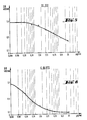
- FIGS. 1 to 6 represent the calibration curves obtained with the products A to F in order.
- FIGS. 1 to 6 It appears from FIGS. 1 to 6 that, based on the assay method described in the present invention, a variation in the optical density is obtained which is a function of the amount of product present in the plasma.
- This variation in optical density therefore makes it possible to evaluate, in a given sample, for example the plasma of an animal or of a patient treated with one of the products (strong sulfated polyanions), the content of this plasma in the product under consideration.
Landscapes
- Health & Medical Sciences (AREA)
- Life Sciences & Earth Sciences (AREA)
- Hematology (AREA)
- Engineering & Computer Science (AREA)
- Molecular Biology (AREA)
- Biomedical Technology (AREA)
- Chemical & Material Sciences (AREA)
- Immunology (AREA)
- Urology & Nephrology (AREA)
- Biotechnology (AREA)
- Biochemistry (AREA)
- Cell Biology (AREA)
- Food Science & Technology (AREA)
- Medicinal Chemistry (AREA)
- Physics & Mathematics (AREA)
- Analytical Chemistry (AREA)
- Microbiology (AREA)
- General Health & Medical Sciences (AREA)
- General Physics & Mathematics (AREA)
- Pathology (AREA)
- Measuring Or Testing Involving Enzymes Or Micro-Organisms (AREA)
- Investigating Or Analysing Biological Materials (AREA)
- Polysaccharides And Polysaccharide Derivatives (AREA)
Applications Claiming Priority (2)
| Application Number | Priority Date | Filing Date | Title |
|---|---|---|---|
| FR8812765A FR2636967B1 (fr) | 1988-09-29 | 1988-09-29 | Procede de dosage de polyosides anioniques sulfates, et coffret pour la mise en oeuvre de ce procede |
| FR8812765 | 1988-09-29 |
Publications (1)
| Publication Number | Publication Date |
|---|---|
| EP0362077A1 true EP0362077A1 (de) | 1990-04-04 |
Family
ID=9370542
Family Applications (1)
| Application Number | Title | Priority Date | Filing Date |
|---|---|---|---|
| EP89402691A Withdrawn EP0362077A1 (de) | 1988-09-29 | 1989-09-29 | Bestimmungsverfahren für anionische Sulfatid-Polyoside und Zusammensetzung dafür |
Country Status (11)
| Country | Link |
|---|---|
| EP (1) | EP0362077A1 (de) |
| JP (1) | JPH02150297A (de) |
| AU (1) | AU631144B2 (de) |
| DK (1) | DK482189A (de) |
| FI (1) | FI894632A7 (de) |
| FR (1) | FR2636967B1 (de) |
| IL (1) | IL91841A0 (de) |
| NO (1) | NO893877L (de) |
| NZ (1) | NZ230832A (de) |
| PT (1) | PT91849A (de) |
| ZA (1) | ZA897427B (de) |
Cited By (1)
| Publication number | Priority date | Publication date | Assignee | Title |
|---|---|---|---|---|
| WO1993015406A1 (en) * | 1992-01-30 | 1993-08-05 | Imperial College Of Science, Technology & Medecine | Methods and apparatus for assay of sulphated polysaccharides |
Citations (2)
| Publication number | Priority date | Publication date | Assignee | Title |
|---|---|---|---|---|
| EP0084999A1 (de) * | 1982-01-15 | 1983-08-03 | D.R.O.P.I.C. (Société Civile) | Verfahren zur Herstellung von organischen Oligosacchariden übereinstimmend mit Fragmenten von natürlichen Muco-Polysacchariden, die erhaltenen Oligosaccharide und ihre biologische Verwendung |
| WO1987007183A1 (en) * | 1986-05-28 | 1987-12-03 | New York University | Specific immunoassay for heparin |
-
1988
- 1988-09-29 FR FR8812765A patent/FR2636967B1/fr not_active Expired - Lifetime
-
1989
- 1989-09-29 ZA ZA897427A patent/ZA897427B/xx unknown
- 1989-09-29 FI FI894632A patent/FI894632A7/fi not_active Application Discontinuation
- 1989-09-29 JP JP1255092A patent/JPH02150297A/ja active Pending
- 1989-09-29 PT PT91849A patent/PT91849A/pt unknown
- 1989-09-29 DK DK482189A patent/DK482189A/da not_active Application Discontinuation
- 1989-09-29 EP EP89402691A patent/EP0362077A1/de not_active Withdrawn
- 1989-09-29 NZ NZ230832A patent/NZ230832A/en unknown
- 1989-09-29 IL IL91841A patent/IL91841A0/xx not_active IP Right Cessation
- 1989-09-29 NO NO89893877A patent/NO893877L/no unknown
- 1989-09-29 AU AU42419/89A patent/AU631144B2/en not_active Ceased
Patent Citations (2)
| Publication number | Priority date | Publication date | Assignee | Title |
|---|---|---|---|---|
| EP0084999A1 (de) * | 1982-01-15 | 1983-08-03 | D.R.O.P.I.C. (Société Civile) | Verfahren zur Herstellung von organischen Oligosacchariden übereinstimmend mit Fragmenten von natürlichen Muco-Polysacchariden, die erhaltenen Oligosaccharide und ihre biologische Verwendung |
| WO1987007183A1 (en) * | 1986-05-28 | 1987-12-03 | New York University | Specific immunoassay for heparin |
Non-Patent Citations (1)
| Title |
|---|
| THROMBOSIS AND HAEMOSTASIS, vol. 54, no. 3, 1985, pages 630-634, F.K. Schattauer Verlag GmbH, Stuttgart, DE; J. DAWES et al.: "The measurement of heparin and other therapeutic sulphated polysaccharides in plasma, serum and urine" * |
Cited By (1)
| Publication number | Priority date | Publication date | Assignee | Title |
|---|---|---|---|---|
| WO1993015406A1 (en) * | 1992-01-30 | 1993-08-05 | Imperial College Of Science, Technology & Medecine | Methods and apparatus for assay of sulphated polysaccharides |
Also Published As
| Publication number | Publication date |
|---|---|
| NO893877L (no) | 1990-03-30 |
| FI894632A0 (fi) | 1989-09-29 |
| DK482189A (da) | 1990-03-30 |
| PT91849A (pt) | 1990-03-30 |
| JPH02150297A (ja) | 1990-06-08 |
| FR2636967A1 (fr) | 1990-03-30 |
| FR2636967B1 (fr) | 1990-12-07 |
| NO893877D0 (no) | 1989-09-29 |
| ZA897427B (en) | 1990-07-25 |
| FI894632L (fi) | 1990-03-30 |
| AU4241989A (en) | 1990-04-05 |
| NZ230832A (en) | 1992-05-26 |
| IL91841A0 (en) | 1990-06-10 |
| AU631144B2 (en) | 1992-11-19 |
| FI894632A7 (fi) | 1990-03-30 |
| DK482189D0 (da) | 1989-09-29 |
Similar Documents
| Publication | Publication Date | Title |
|---|---|---|
| EP0040144B1 (de) | Sulfatisierte Polysaccharide, Verfahren zu ihrer Herstellung und ihre Verwendung als Arzneimittel | |
| EP0287477B1 (de) | Heparine mit niedrigem Molekulargewicht und regelmässiger Struktur, ihre Herstellung und biologische Verwendungen | |
| EP0214879B1 (de) | Verfahren zur Sulphatierung von Glykosaminoglykanen, daraus hergestellte Glykosaminoglykane und ihre biologischen Verwendungen | |
| EP0090100B1 (de) | Unlösliche Polymere mit antikoagulierenden Eigenschaften dieselben enthaltende Gegenstände und Verfahren zu ihrer Herstellung | |
| EP0027089B1 (de) | Oligosaccharidfraktionen und Oligosaccharide mit biologischen Eigenschaften, Verfahren zu ihrer Herstellung und ihre Verwendungen als Arzneimittel | |
| EP0544592B1 (de) | Hochmolekulare, N,O-sulfatierte Heparosane; Verfahren zu deren Herstellung und diese enthaltende Arzneimittel | |
| EP0428182B1 (de) | Derivate von Dextran mit antikoagulierenden und antikomplementären Eigenschaften, ihre Herstellung und ihre biologischen Anwendungen | |
| LU87957A1 (fr) | Melanges de polysaccharides de bas poids moleculaires procede de preparation et utilisation | |
| EP0403377A1 (de) | Sulfatierte Polysaccharide, Antikoagulierungs- und Antikomplementärmittel, hergestellt aus Fukanen aus braunen Algen, und Verfahren zu deren Herstellung | |
| CA2678168A1 (fr) | Heparines de bas poids moleculaire comprenant au moins une liaison covalente avec la biotine ou un derive de la biotine, leur procede de preparation et leur utilisation | |
| WO2010023374A1 (fr) | Polysaccharides a activite antithrombotique comprenant une liaison covalente avec une chaine amine | |
| EP0165134B1 (de) | Oligosaccharide, ihre Herstellung durch Synthese und ihre biologische Verwendung | |
| FR2634485A1 (fr) | Glycosaminoglycanes selectivement o-acyles, leur preparation et compositions pharmaceutiques les contenant | |
| CA2468759C (fr) | Procede de sulfonation de composes comprenant des groupements hydroxyles (oh) libres ou des amines primaires ou secondaires | |
| EP0362077A1 (de) | Bestimmungsverfahren für anionische Sulfatid-Polyoside und Zusammensetzung dafür | |
| CA2533555A1 (fr) | Melanges d'oligosaccharides derives d'heparine, leur preparation et les compositions pharmaceutiques les contenant | |
| EP0344068B1 (de) | N-Polyosyl-Polypeptide | |
| EP0138632A2 (de) | Komplexe, welche Oligosaccharide enthalten, ihre Herstellung und ihre biologische und biochemische Verwendung | |
| Cholakis et al. | Department of Chemical Engineering & Applied Chemistry University of Toronto, Toronto, Ontario, M5S 1A4 |
Legal Events
| Date | Code | Title | Description |
|---|---|---|---|
| PUAI | Public reference made under article 153(3) epc to a published international application that has entered the european phase |
Free format text: ORIGINAL CODE: 0009012 |
|
| AK | Designated contracting states |
Kind code of ref document: A1 Designated state(s): AT BE CH DE ES GB GR IT LI LU NL SE |
|
| 17P | Request for examination filed |
Effective date: 19901002 |
|
| RAP1 | Party data changed (applicant data changed or rights of an application transferred) |
Owner name: ELF SANOFI |
|
| 17Q | First examination report despatched |
Effective date: 19920518 |
|
| STAA | Information on the status of an ep patent application or granted ep patent |
Free format text: STATUS: THE APPLICATION HAS BEEN WITHDRAWN |
|
| 18W | Application withdrawn |
Withdrawal date: 19930320 |