EP0370637A1 - Methoden und Gerät zur Messung der Konzentration einer Gaskomponente im ausgeatmeten Gas - Google Patents
Methoden und Gerät zur Messung der Konzentration einer Gaskomponente im ausgeatmeten Gas Download PDFInfo
- Publication number
- EP0370637A1 EP0370637A1 EP89311186A EP89311186A EP0370637A1 EP 0370637 A1 EP0370637 A1 EP 0370637A1 EP 89311186 A EP89311186 A EP 89311186A EP 89311186 A EP89311186 A EP 89311186A EP 0370637 A1 EP0370637 A1 EP 0370637A1
- Authority
- EP
- European Patent Office
- Prior art keywords
- concentration
- gas
- carbon dioxide
- gases
- breathing
- Prior art date
- Legal status (The legal status is an assumption and is not a legal conclusion. Google has not performed a legal analysis and makes no representation as to the accuracy of the status listed.)
- Granted
Links
Images


Classifications
-
- A—HUMAN NECESSITIES
- A61—MEDICAL OR VETERINARY SCIENCE; HYGIENE
- A61B—DIAGNOSIS; SURGERY; IDENTIFICATION
- A61B6/00—Apparatus or devices for radiation diagnosis; Apparatus or devices for radiation diagnosis combined with radiation therapy equipment
- A61B6/02—Arrangements for diagnosis sequentially in different planes; Stereoscopic radiation diagnosis
- A61B6/03—Computed tomography [CT]
- A61B6/032—Transmission computed tomography [CT]
-
- A—HUMAN NECESSITIES
- A61—MEDICAL OR VETERINARY SCIENCE; HYGIENE
- A61B—DIAGNOSIS; SURGERY; IDENTIFICATION
- A61B5/00—Measuring for diagnostic purposes; Identification of persons
- A61B5/02—Detecting, measuring or recording for evaluating the cardiovascular system, e.g. pulse, heart rate, blood pressure or blood flow
- A61B5/026—Measuring blood flow
- A61B5/0275—Measuring blood flow using tracers, e.g. dye dilution
-
- A—HUMAN NECESSITIES
- A61—MEDICAL OR VETERINARY SCIENCE; HYGIENE
- A61B—DIAGNOSIS; SURGERY; IDENTIFICATION
- A61B5/00—Measuring for diagnostic purposes; Identification of persons
- A61B5/08—Measuring devices for evaluating the respiratory organs
- A61B5/0813—Measurement of pulmonary parameters by tracers, e.g. radioactive tracers
-
- A—HUMAN NECESSITIES
- A61—MEDICAL OR VETERINARY SCIENCE; HYGIENE
- A61B—DIAGNOSIS; SURGERY; IDENTIFICATION
- A61B6/00—Apparatus or devices for radiation diagnosis; Apparatus or devices for radiation diagnosis combined with radiation therapy equipment
- A61B6/48—Diagnostic techniques
- A61B6/481—Diagnostic techniques involving the use of contrast agents
-
- G—PHYSICS
- G06—COMPUTING OR CALCULATING; COUNTING
- G06T—IMAGE DATA PROCESSING OR GENERATION, IN GENERAL
- G06T5/00—Image enhancement or restoration
- G06T5/50—Image enhancement or restoration using two or more images, e.g. averaging or subtraction
-
- A—HUMAN NECESSITIES
- A61—MEDICAL OR VETERINARY SCIENCE; HYGIENE
- A61B—DIAGNOSIS; SURGERY; IDENTIFICATION
- A61B6/00—Apparatus or devices for radiation diagnosis; Apparatus or devices for radiation diagnosis combined with radiation therapy equipment
- A61B6/50—Apparatus or devices for radiation diagnosis; Apparatus or devices for radiation diagnosis combined with radiation therapy equipment specially adapted for specific body parts; specially adapted for specific clinical applications
- A61B6/507—Apparatus or devices for radiation diagnosis; Apparatus or devices for radiation diagnosis combined with radiation therapy equipment specially adapted for specific body parts; specially adapted for specific clinical applications for determination of haemodynamic parameters, e.g. perfusion CT
Definitions
- This invention relates to methods of and apparatus for measuring the concentration of a gaseous component of exhaled gases.
- the invention finds particular application in the art of medical diagnostic imaging in conjunction with measuring xenon concentration in end-tidal gases. However, it is to be appreciated that the invention will also find application in conjunction with measuring concentrations of other gases.
- the xenon concentration in the end-tidal gas can be measured by placing a thermoconductivity detector in the exhalation line, as disclosed in US-A-4,622,976 which describes the measurement of xenon concentration, carbon dioxide concentration, or the like. Because the thermoconductivity detector will indicate xenon concentration continuously, it was necessary to determine which reading represents the end-tidal gases.
- a chamber or reservoir was formed in the exhaled gas line leading from the breathing mask to hold exhaled gas.
- a mechanical system determined when the patient started to inhale fresh gas from a supply line. The thermoconductivity of the gas retained in the exhale chamber was measured.
- Such a technique is disclosed in US-A-4,535,780.
- One of the problems of this technique is that the apparatus is expensive.
- Another problem is that the mechanical means for determining the changeover point between inhaling and exhaling tends to be relatively unreliable.
- exhaled gases intermix in the chamber diluting the end-tidal gases with earlier exhaled gases.
- Another technique for determining the end-tidal gas was to monitor the xenon concentration continuously and assume that the xenon concentration minima were attributable to the end-tidal gases.
- the minimum xenon concentrations did not correspond to end-tidal gas.
- the magnitudes of the minima increased sufficiently close to the breathing gas xenon concentration that they were difficult to identify.
- the end-tidal gas minimum values became substantially indistinguishable from the exhaled xenon concentration in the rest of the exhaled gas after the first minute of a five to seven minute xenon protocol.
- a method of measuring the concentration of a preselected gaseous component, other than carbon dioxide, of end-tidal gases comprising: measuring concentrations of a first component of exhaled gas, other than said preselected gaseous component, during at least exhalation portions of a respiratory cycle; comparing changes in the concentration of said first component with preselected criteria; and measuring the concentration of the preselected gaseous component in response to the comparison.
- a method of measuring partition coefficient and blood flow values of a patient disposed in a diagnostic scanner comprising: (a) providing the patient with breathing gases; (b) introducing an enhancement gas into the breathing gases such that a concentration of the enhancement gas in the patient's blood increases with time; (c) monitoring breathing gases exhaled by the patient for carbon dioxide concentration; (d) comparing the measured carbon dioxide concentration with preselected characteristics indicative of carbon dioxide concentration changes in end-tidal gases; (e) in response to determining that the carbon dioxide concentration characteristic indicative of end-tidal gases has occurred, measuring the enhancement gas concentration in the exhaled breathing gases; (f) repeating steps (c) to (e) over a plurality of respiratory cycles to generate a blood enhancement gas concentration curve; (g) also over the plurality of respiratory cycles, generating a plurality of diagnostic images in which the enhancement gas concentration changes from image to image, each diagnostic image including a plurality of pixel values; and (h) calculating partition coefficient and blood flow values from
- an apparatus for measuring the concentration of an enhancement gas of end-tidal gases comprising: breathing mask means through which a patient inhales breathing gas and exhales exhalation gases; breathing gas supply means for supplying breathing gas to the mask means; enhancement gas introduction means for selectively introducing an enhancement gas into the breathing gas; and enhancement gas analyzing means for measuring enhancement gas concentration, the enhancement gas analyzing means being operatively connected with the mask means to receive at least exhalation gases therefrom, characterised in that said apparatus further includes: carbon dioxide analyzing means for measuring carbon dioxide concentration, the carbon dioxide analyzing means being operatively connected with the mask means to receive at least exhalation gases therefrom; carbon dioxide concentration comparing means for comparing the measured carbon dioxide concentration with characteristics of a carbon dioxide concentration curve indicative of end-tidal gases; and end-tidal enhancement gas concentration means operatively connected with the enhancement gas analyzing means and the comparing means for providing a record of end-tidal enhancement gas concentrations.
- a gas supply means A supplies breathing gas to a medical diagnostic CT scanner B.
- a processing system or means C processes data from the CT scanner and the breathing gas supply system to produce images and other diagnostic information.
- the breathing gas supply means A includes a breathing air or oxygen supply 10 and a xenon gas supply 12.
- the xenon gas supply means provides a mixture of 80% xenon and 20% oxygen to guarantee that the patient receives at least 20% oxygen even during a malfunction.
- a first blower 14 supplies recirculated breathing gases along with added xenon for mixture with additional oxygen from the oxygen supply means 10. Most of the mixture passes to a breathing bag 16 with the exception of a small fraction that is pumped by a pump 18 through a xenon detector 20.
- the xenon detector 20 determines the concentration of xenon in the gaseous breathing mixture and controls a xenon control valve 22 to maintain the xenon concentration substantially constant. Typically, the xenon concentration is selected to be about 30%.
- a carbon dioxide absorber 30 removes carbon dioxide from the breathing gases that are supplied to an outlet port check valve 32.
- the patient draws the breathing gases through the outlet check valve 32.
- the exhaled gases are returned through an exhaust gas path or tube 36 and a return check valve 38.
- the exhaust gases passing through the return check valve 38 are returned to the blower 14 and recirculated through the system.
- the exhaust gas analysis is performed periodically, e.g. every 0.25 seconds, on gases passing through a small diameter line 40 extending parallel to the exhaust gas path to reduce the dilution effects.
- the exhaust gases tend to tumble and mix which dilutes end-tidal gases with other exhaust gases.
- the narrow diameter line inhibits the swirling and countercurrent flow patterns that tend to intermix the gases and enables a more nearly instantaneous reading of the gases to be made.
- the gases drawn through the small diameter line by a pump or blower 42 are analyzed by a xenon detector 44 and a carbon dioxide detector 46.
- the xenon detector measures the xenon concentration of the exhaled gases and the carbon dioxide detector measures the carbon dioxide concentration of the exhaled gases.
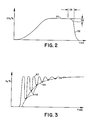
- the concentration of carbon dioxide gas increases generally exponentially and logarithmically to a plateau 50.
- the carbon dioxide concentration remains relatively constant through a significant portion of the exhalation portion of the respiratory cycle.
- the patient inhales CO2 free gas through check valve 32 and the CO2 concentration drops rapidly 52.
- a carbon dioxide level comparing means 54 compares the carbon dioxide level with preselected characteristics, specifically, a preselected drop 56 from the plateau 50.
- the comparing means 54 triggers the selection of a previously taken end-tidal xenon concentration reading by the xenon detector 11 a fixed, preselected time earlier 58, e.g. a 0.5 sec.
- the concentration of xenon gas peaks 60 at the xenon concentration in the breathing air.
- the xenon concentration reaches a minimum value 62 at the end-tidal portion of each breath.
- the blood xenon concentration and the corresponding end-tidal xenon concentration increase along a blood xenon absorption curve 64.
- the concentration of xenon in the patient's blood approaches the concentration of xenon in the breathing gas. After a half dozen or a dozen breaths, the differences in height between the xenon concentration peaks and valleys becomes so small that the random noise fluctuations cannot reliably be differentiated from the true xenon concentration minimum.
- the end-tidal xenon concentrations 62 are conveyed to a blood xenon concentration curve memory means 66.
- a blood xenon concentration display means 68 is connected with the blood xenon concentration memory means to provide the physician with a continuous indication of blood xenon concentrations.
- the output of this memory means may also be displayed as a graph of xenon or blood xenon concentration vs. time.
- the CT medical diagnostic scanner B of the preferred embodiment includes an x-ray source 70 for projecting a fan beam of radiation through an image region 72.
- the patient is supported on a patient table 74 with tissue to be examined, typically the head or brain tissue, disposed in the image region.
- An array of x-ray detectors 76 receives the fan beam of radiation from the x-ray source including the radiation which has traversed the portion of the patient from the image region.
- An image reconstruction means 78 reconstructs CT image representations from the x-ray data collected by the radiation detectors as the radiation source is rotated about the region of interest.
- the processing circuitry C includes a reference image means 80 for storing an image representation of the tissue of interest of the patient without xenon gas absorbed in the patient's blood.
- a reference image means 80 for storing an image representation of the tissue of interest of the patient without xenon gas absorbed in the patient's blood.
- several additional CT images are taken, each being stored temporarily in an image memory 82.
- Each image can be considered as the sum of a patient tissue image and a xenon image.
- a subtraction means 84 subtracts the reference image from each subsequent image to produce a difference image indicative of xenon concentrations. More specifically, each difference image includes an array of pixel values, each pixel value corresponding to a preselected volume cell (voxel) or subregion of the portion of the patient disposed in the image region.
- a partition coefficient and blood flow rate determining means 90 utilizes known relationships, such as the relationships set forth in the Kety equation, to determine a partition coefficient and blood flow rate for each image voxel. These calculations are based on the blood xenon concentration curve stored in the blood xenon concentration in memory 66 and the increase in xenon concentration in subsequent corresponding pixels of the difference image.
- the partition coefficient and blood flow values are determined on a pixel by pixel basis from a look-up table means 92.
- the look-up table means is addressed by the relative times t1, t2, t n of the CT scans, corresponding pixel values of a plurality of CT scans as stored in difference scan memories 94, and by blood xenon concentration curve parameters.
- a blood xenon concentration parameter determining means 96 determines conformity of the actual blood xenon concentration curve to preselected concentration curve characteristics, such as saturation concentration, slope, exponential time constants, and the like.
- the look-up table means includes an array of look-up tables. The blood xenon concentration curve characteristics determine which look-up table(s) conforms best to the patient blood xenon concentration curve. The selected look-up table(s) is then addressed by the corresponding pixel values from each image in the difference image memory means 94. The relative times at which the images were collected in the preferred embodiment are preset. However, if the times are to be variable, these times are also utilized to select among a larger plurality of look-up tables.
- Digital look-up tables can only be addressed by preselected addresses. The actual pixel values may fall between these addresses. Each of the two closest addresses are accessed and an interpolating means 98 performs a weighted interpolation of the retrieved partition coefficient and flow values. Analogously, the absorption curve characteristics may fall between two or more look-up tables of the array. An interpolation means 98 performs a weighted interpretation of the partition coefficient and flow values retrieved by the closest difference image pixel values and the closest time or xenon curve addressed look-up tables.
- the procedure is repeated for each pixel of the images to build a partition coefficient image for storage in a partition coefficient image memory 100 and a flow image for storage in a flow image memory means 102.
- the partition coefficient and flow images may be selectively displayed on a display means 96.
- other parameters may also be displayed, such as a confidence or fit value which is indicative of how closely each partition coefficient and flow value conforms with the normal statistical deviation from the actual value, i.e. reliability or confidence that one may have in each pixel value.
Landscapes
- Health & Medical Sciences (AREA)
- Life Sciences & Earth Sciences (AREA)
- Engineering & Computer Science (AREA)
- Medical Informatics (AREA)
- Physics & Mathematics (AREA)
- General Health & Medical Sciences (AREA)
- Veterinary Medicine (AREA)
- Biophysics (AREA)
- Biomedical Technology (AREA)
- Heart & Thoracic Surgery (AREA)
- Pathology (AREA)
- Molecular Biology (AREA)
- Surgery (AREA)
- Animal Behavior & Ethology (AREA)
- Public Health (AREA)
- Pulmonology (AREA)
- Optics & Photonics (AREA)
- Theoretical Computer Science (AREA)
- High Energy & Nuclear Physics (AREA)
- Nuclear Medicine, Radiotherapy & Molecular Imaging (AREA)
- Physiology (AREA)
- Radiology & Medical Imaging (AREA)
- General Physics & Mathematics (AREA)
- Hematology (AREA)
- Cardiology (AREA)
- Measurement Of The Respiration, Hearing Ability, Form, And Blood Characteristics Of Living Organisms (AREA)
- Investigating Or Analysing Biological Materials (AREA)
- Measuring Pulse, Heart Rate, Blood Pressure Or Blood Flow (AREA)
Applications Claiming Priority (2)
| Application Number | Priority Date | Filing Date | Title |
|---|---|---|---|
| US07/275,784 US5140981A (en) | 1986-11-24 | 1988-11-23 | End-tidal gas detection |
| US275784 | 2006-01-29 |
Publications (2)
| Publication Number | Publication Date |
|---|---|
| EP0370637A1 true EP0370637A1 (de) | 1990-05-30 |
| EP0370637B1 EP0370637B1 (de) | 1994-05-25 |
Family
ID=23053784
Family Applications (1)
| Application Number | Title | Priority Date | Filing Date |
|---|---|---|---|
| EP89311186A Expired - Lifetime EP0370637B1 (de) | 1988-11-23 | 1989-10-30 | Methoden und Gerät zur Messung der Konzentration einer Gaskomponente im ausgeatmeten Gas |
Country Status (4)
| Country | Link |
|---|---|
| US (1) | US5140981A (de) |
| EP (1) | EP0370637B1 (de) |
| JP (1) | JP2802655B2 (de) |
| DE (1) | DE68915512T2 (de) |
Cited By (5)
| Publication number | Priority date | Publication date | Assignee | Title |
|---|---|---|---|---|
| AU655158B2 (en) * | 1989-09-11 | 1994-12-08 | Nellcor Incorporated | Agent gas analyzer and method of use |
| WO1997020591A1 (de) * | 1995-12-06 | 1997-06-12 | Messer Griesheim Gmbh | Analytische konfiguration zur überwachung von xenon-haltigem narkosegas |
| US6197323B1 (en) | 1997-03-10 | 2001-03-06 | Michael Georgieff | Medicinal preparation containing a lipophilic inert gas |
| RU2210394C2 (ru) * | 2001-09-03 | 2003-08-20 | Сухолитко Валентин Афанасьевич | Способ респираторной поддержки оператора во время работы |
| US20120310059A1 (en) * | 2011-05-31 | 2012-12-06 | Nellcor Puritan Bennett Llc | Clinical Acceptance Tool |
Families Citing this family (6)
| Publication number | Priority date | Publication date | Assignee | Title |
|---|---|---|---|---|
| SE9302477L (sv) * | 1993-07-22 | 1995-01-23 | Siemens Elema Ab | Apparat för tillförsel av andningsgas |
| US5533512A (en) * | 1994-03-18 | 1996-07-09 | Ohmeda Inc. | Method and apparatus for detection of venous air emboli |
| US6076005A (en) * | 1998-02-25 | 2000-06-13 | St. Jude Children's Research Hospital | Respiration responsive gating means and apparatus and methods using the same |
| AU2003238288A1 (en) * | 2003-06-19 | 2005-02-04 | Everest Biomedical Instruments | Breath end-tidal gas monitor |
| JP5351583B2 (ja) * | 2009-03-30 | 2013-11-27 | 日本光電工業株式会社 | 呼吸波形解析装置 |
| RU2016111654A (ru) * | 2013-08-30 | 2017-10-05 | Кэпниа, Инк. | Система измерения углекислого газа у новорожденных |
Citations (3)
| Publication number | Priority date | Publication date | Assignee | Title |
|---|---|---|---|---|
| US3890959A (en) * | 1971-03-22 | 1975-06-24 | Univ New York | Method for determining discrete localized blood flow in a large volume of tissue |
| DE3522113A1 (de) * | 1984-06-20 | 1986-01-02 | Kabushiki Kaisha Toshiba, Kawasaki, Kanagawa | Abbildungsgeraet fuer die rechnergestuetzte tomographie |
| US4779621A (en) * | 1987-02-19 | 1988-10-25 | Picker International, Inc. | Xenon calibration phantom |
Family Cites Families (3)
| Publication number | Priority date | Publication date | Assignee | Title |
|---|---|---|---|---|
| US4535780A (en) * | 1984-11-07 | 1985-08-20 | General Electric Company | Apparatus for measuring xenon concentration in xenon cerebral blood-flow studies |
| US4622976A (en) * | 1985-04-15 | 1986-11-18 | Enhancer R & D | Method and apparatus for conducting xenon inhalation studies |
| US4793357A (en) * | 1986-11-24 | 1988-12-27 | Picker International, Inc. | CT blood flow mapping with xenon gas enhancement |
-
1988
- 1988-11-23 US US07/275,784 patent/US5140981A/en not_active Expired - Lifetime
-
1989
- 1989-10-30 DE DE68915512T patent/DE68915512T2/de not_active Expired - Fee Related
- 1989-10-30 EP EP89311186A patent/EP0370637B1/de not_active Expired - Lifetime
- 1989-11-13 JP JP1292528A patent/JP2802655B2/ja not_active Expired - Fee Related
Patent Citations (3)
| Publication number | Priority date | Publication date | Assignee | Title |
|---|---|---|---|---|
| US3890959A (en) * | 1971-03-22 | 1975-06-24 | Univ New York | Method for determining discrete localized blood flow in a large volume of tissue |
| DE3522113A1 (de) * | 1984-06-20 | 1986-01-02 | Kabushiki Kaisha Toshiba, Kawasaki, Kanagawa | Abbildungsgeraet fuer die rechnergestuetzte tomographie |
| US4779621A (en) * | 1987-02-19 | 1988-10-25 | Picker International, Inc. | Xenon calibration phantom |
Cited By (8)
| Publication number | Priority date | Publication date | Assignee | Title |
|---|---|---|---|---|
| AU655158B2 (en) * | 1989-09-11 | 1994-12-08 | Nellcor Incorporated | Agent gas analyzer and method of use |
| WO1997020591A1 (de) * | 1995-12-06 | 1997-06-12 | Messer Griesheim Gmbh | Analytische konfiguration zur überwachung von xenon-haltigem narkosegas |
| US6197323B1 (en) | 1997-03-10 | 2001-03-06 | Michael Georgieff | Medicinal preparation containing a lipophilic inert gas |
| US6328708B1 (en) | 1997-03-10 | 2001-12-11 | Michael Georgieff | Device for controlled anaesthesia, analgesia and /or sedation |
| US6511453B2 (en) | 1997-03-10 | 2003-01-28 | Michael Georgieff | Device for controlled anaesthesia, analgesia and/or sedation |
| RU2210394C2 (ru) * | 2001-09-03 | 2003-08-20 | Сухолитко Валентин Афанасьевич | Способ респираторной поддержки оператора во время работы |
| US20120310059A1 (en) * | 2011-05-31 | 2012-12-06 | Nellcor Puritan Bennett Llc | Clinical Acceptance Tool |
| US8608657B2 (en) * | 2011-05-31 | 2013-12-17 | Covidien Lp | Clinical acceptance tool |
Also Published As
| Publication number | Publication date |
|---|---|
| JP2802655B2 (ja) | 1998-09-24 |
| DE68915512T2 (de) | 1994-09-08 |
| US5140981A (en) | 1992-08-25 |
| DE68915512D1 (de) | 1994-06-30 |
| JPH02184757A (ja) | 1990-07-19 |
| EP0370637B1 (de) | 1994-05-25 |
Similar Documents
| Publication | Publication Date | Title |
|---|---|---|
| US4622976A (en) | Method and apparatus for conducting xenon inhalation studies | |
| Gold et al. | Pulmonary function testing | |
| JP2688453B2 (ja) | 動脈血液中co2監視と閉ループ制奥装置 | |
| US5957128A (en) | Method and device for determination of the functional residual capacity (FRC) | |
| US5361771A (en) | Portable pulmonary function testing device and method | |
| EP0370637B1 (de) | Methoden und Gerät zur Messung der Konzentration einer Gaskomponente im ausgeatmeten Gas | |
| US20060129055A1 (en) | Systems and methods for accurately, substantially noninvasivelydetermining pulmonary capillary blood flow or cardiac output | |
| US6302851B1 (en) | Method and apparatus for determining a pulmonary function parameter for gas exchange | |
| US5024230A (en) | Dual flow/lambda display for xenon enhanced tomography | |
| US4793358A (en) | Apparatus for measuring local cerebral blood flow | |
| Green et al. | A simple measurement of phase IV (" closing volume") using a critical orifice helium analyzer. | |
| US4930510A (en) | Parameterization for CT blood flow mapping with xenon gas enhancement | |
| JPH0785B2 (ja) | 肺換気機能診断装置 | |
| US4779621A (en) | Xenon calibration phantom | |
| Byrne et al. | Experimental and clinical assessment of lung injury by measurement of extravascular lung water and transcapillary protein flux in ARDS: a review of current techniques | |
| US4535780A (en) | Apparatus for measuring xenon concentration in xenon cerebral blood-flow studies | |
| Williams et al. | Methods of studying lobar and segmental function of the lung in man | |
| JPH0622495B2 (ja) | 局所脳血流測定用ct装置 | |
| US6176838B1 (en) | Method and apparatus for measuring hepatic blood flow amount | |
| FI104042B (fi) | Menetelmä keuhkojen perfuusion mittaamiseksi | |
| US20150202400A1 (en) | Accelerated closed-circuit wash-in of tracer gas using bolus injection in lung function testing | |
| Bickel et al. | An analog computer program for cardiac output in humans using mass spectrometer analysis of expired air | |
| Cotes | Lung volumes | |
| JP4680293B2 (ja) | キセノンct装置、動脈血速度定数決定方法及び血流量算出方法 | |
| Ishii et al. | Quantitative assessment of ventilation-perfusion mismatch by radioxenon imaging of the lung |
Legal Events
| Date | Code | Title | Description |
|---|---|---|---|
| PUAI | Public reference made under article 153(3) epc to a published international application that has entered the european phase |
Free format text: ORIGINAL CODE: 0009012 |
|
| AK | Designated contracting states |
Kind code of ref document: A1 Designated state(s): DE FR GB NL |
|
| 17P | Request for examination filed |
Effective date: 19900618 |
|
| 17Q | First examination report despatched |
Effective date: 19930208 |
|
| GRAA | (expected) grant |
Free format text: ORIGINAL CODE: 0009210 |
|
| AK | Designated contracting states |
Kind code of ref document: B1 Designated state(s): DE FR GB NL |
|
| ET | Fr: translation filed | ||
| REF | Corresponds to: |
Ref document number: 68915512 Country of ref document: DE Date of ref document: 19940630 |
|
| PLBE | No opposition filed within time limit |
Free format text: ORIGINAL CODE: 0009261 |
|
| STAA | Information on the status of an ep patent application or granted ep patent |
Free format text: STATUS: NO OPPOSITION FILED WITHIN TIME LIMIT |
|
| 26N | No opposition filed | ||
| PGFP | Annual fee paid to national office [announced via postgrant information from national office to epo] |
Ref country code: GB Payment date: 19960924 Year of fee payment: 8 |
|
| PG25 | Lapsed in a contracting state [announced via postgrant information from national office to epo] |
Ref country code: GB Free format text: LAPSE BECAUSE OF NON-PAYMENT OF DUE FEES Effective date: 19971030 |
|
| GBPC | Gb: european patent ceased through non-payment of renewal fee |
Effective date: 19971030 |
|
| PGFP | Annual fee paid to national office [announced via postgrant information from national office to epo] |
Ref country code: NL Payment date: 20020919 Year of fee payment: 14 |
|
| PGFP | Annual fee paid to national office [announced via postgrant information from national office to epo] |
Ref country code: DE Payment date: 20031215 Year of fee payment: 15 |
|
| REG | Reference to a national code |
Ref country code: FR Ref legal event code: CD |
|
| PG25 | Lapsed in a contracting state [announced via postgrant information from national office to epo] |
Ref country code: NL Free format text: LAPSE BECAUSE OF NON-PAYMENT OF DUE FEES Effective date: 20040501 |
|
| NLV4 | Nl: lapsed or anulled due to non-payment of the annual fee |
Effective date: 20040501 |
|
| REG | Reference to a national code |
Ref country code: FR Ref legal event code: TP |
|
| PGFP | Annual fee paid to national office [announced via postgrant information from national office to epo] |
Ref country code: FR Payment date: 20041027 Year of fee payment: 16 |
|
| PG25 | Lapsed in a contracting state [announced via postgrant information from national office to epo] |
Ref country code: DE Free format text: LAPSE BECAUSE OF NON-PAYMENT OF DUE FEES Effective date: 20050503 |
|
| PG25 | Lapsed in a contracting state [announced via postgrant information from national office to epo] |
Ref country code: FR Free format text: LAPSE BECAUSE OF NON-PAYMENT OF DUE FEES Effective date: 20060630 |
|
| REG | Reference to a national code |
Ref country code: FR Ref legal event code: ST Effective date: 20060630 |