EP0374778A2 - Verfahren zur Proteinimmobilisierung an einer Festphase, so hergestellte Protein tragende Festphase sowie deren Verwendung - Google Patents
Verfahren zur Proteinimmobilisierung an einer Festphase, so hergestellte Protein tragende Festphase sowie deren Verwendung Download PDFInfo
- Publication number
- EP0374778A2 EP0374778A2 EP89123321A EP89123321A EP0374778A2 EP 0374778 A2 EP0374778 A2 EP 0374778A2 EP 89123321 A EP89123321 A EP 89123321A EP 89123321 A EP89123321 A EP 89123321A EP 0374778 A2 EP0374778 A2 EP 0374778A2
- Authority
- EP
- European Patent Office
- Prior art keywords
- protein
- solid phase
- solution
- immobilized
- proteins
- Prior art date
- Legal status (The legal status is an assumption and is not a legal conclusion. Google has not performed a legal analysis and makes no representation as to the accuracy of the status listed.)
- Granted
Links
Images

Classifications
-
- G—PHYSICS
- G01—MEASURING; TESTING
- G01N—INVESTIGATING OR ANALYSING MATERIALS BY DETERMINING THEIR CHEMICAL OR PHYSICAL PROPERTIES
- G01N33/00—Investigating or analysing materials by specific methods not covered by groups G01N1/00 - G01N31/00
- G01N33/48—Biological material, e.g. blood, urine; Haemocytometers
- G01N33/50—Chemical analysis of biological material, e.g. blood, urine; Testing involving biospecific ligand binding methods; Immunological testing
- G01N33/53—Immunoassay; Biospecific binding assay; Materials therefor
- G01N33/543—Immunoassay; Biospecific binding assay; Materials therefor with an insoluble carrier for immobilising immunochemicals
-
- Y—GENERAL TAGGING OF NEW TECHNOLOGICAL DEVELOPMENTS; GENERAL TAGGING OF CROSS-SECTIONAL TECHNOLOGIES SPANNING OVER SEVERAL SECTIONS OF THE IPC; TECHNICAL SUBJECTS COVERED BY FORMER USPC CROSS-REFERENCE ART COLLECTIONS [XRACs] AND DIGESTS
- Y10—TECHNICAL SUBJECTS COVERED BY FORMER USPC
- Y10S—TECHNICAL SUBJECTS COVERED BY FORMER USPC CROSS-REFERENCE ART COLLECTIONS [XRACs] AND DIGESTS
- Y10S530/00—Chemistry: natural resins or derivatives; peptides or proteins; lignins or reaction products thereof
- Y10S530/81—Carrier - bound or immobilized peptides or proteins and the preparation thereof, e.g. biological cell or cell fragment as carrier
- Y10S530/812—Peptides or proteins is immobilized on, or in, an organic carrier
-
- Y—GENERAL TAGGING OF NEW TECHNOLOGICAL DEVELOPMENTS; GENERAL TAGGING OF CROSS-SECTIONAL TECHNOLOGIES SPANNING OVER SEVERAL SECTIONS OF THE IPC; TECHNICAL SUBJECTS COVERED BY FORMER USPC CROSS-REFERENCE ART COLLECTIONS [XRACs] AND DIGESTS
- Y10—TECHNICAL SUBJECTS COVERED BY FORMER USPC
- Y10S—TECHNICAL SUBJECTS COVERED BY FORMER USPC CROSS-REFERENCE ART COLLECTIONS [XRACs] AND DIGESTS
- Y10S530/00—Chemistry: natural resins or derivatives; peptides or proteins; lignins or reaction products thereof
- Y10S530/81—Carrier - bound or immobilized peptides or proteins and the preparation thereof, e.g. biological cell or cell fragment as carrier
- Y10S530/812—Peptides or proteins is immobilized on, or in, an organic carrier
- Y10S530/813—Carrier is a saccharide
-
- Y—GENERAL TAGGING OF NEW TECHNOLOGICAL DEVELOPMENTS; GENERAL TAGGING OF CROSS-SECTIONAL TECHNOLOGIES SPANNING OVER SEVERAL SECTIONS OF THE IPC; TECHNICAL SUBJECTS COVERED BY FORMER USPC CROSS-REFERENCE ART COLLECTIONS [XRACs] AND DIGESTS
- Y10—TECHNICAL SUBJECTS COVERED BY FORMER USPC
- Y10S—TECHNICAL SUBJECTS COVERED BY FORMER USPC CROSS-REFERENCE ART COLLECTIONS [XRACs] AND DIGESTS
- Y10S530/00—Chemistry: natural resins or derivatives; peptides or proteins; lignins or reaction products thereof
- Y10S530/81—Carrier - bound or immobilized peptides or proteins and the preparation thereof, e.g. biological cell or cell fragment as carrier
- Y10S530/812—Peptides or proteins is immobilized on, or in, an organic carrier
- Y10S530/813—Carrier is a saccharide
- Y10S530/814—Cellulose or derivatives thereof
-
- Y—GENERAL TAGGING OF NEW TECHNOLOGICAL DEVELOPMENTS; GENERAL TAGGING OF CROSS-SECTIONAL TECHNOLOGIES SPANNING OVER SEVERAL SECTIONS OF THE IPC; TECHNICAL SUBJECTS COVERED BY FORMER USPC CROSS-REFERENCE ART COLLECTIONS [XRACs] AND DIGESTS
- Y10—TECHNICAL SUBJECTS COVERED BY FORMER USPC
- Y10S—TECHNICAL SUBJECTS COVERED BY FORMER USPC CROSS-REFERENCE ART COLLECTIONS [XRACs] AND DIGESTS
- Y10S530/00—Chemistry: natural resins or derivatives; peptides or proteins; lignins or reaction products thereof
- Y10S530/81—Carrier - bound or immobilized peptides or proteins and the preparation thereof, e.g. biological cell or cell fragment as carrier
- Y10S530/812—Peptides or proteins is immobilized on, or in, an organic carrier
- Y10S530/815—Carrier is a synthetic polymer
-
- Y—GENERAL TAGGING OF NEW TECHNOLOGICAL DEVELOPMENTS; GENERAL TAGGING OF CROSS-SECTIONAL TECHNOLOGIES SPANNING OVER SEVERAL SECTIONS OF THE IPC; TECHNICAL SUBJECTS COVERED BY FORMER USPC CROSS-REFERENCE ART COLLECTIONS [XRACs] AND DIGESTS
- Y10—TECHNICAL SUBJECTS COVERED BY FORMER USPC
- Y10S—TECHNICAL SUBJECTS COVERED BY FORMER USPC CROSS-REFERENCE ART COLLECTIONS [XRACs] AND DIGESTS
- Y10S530/00—Chemistry: natural resins or derivatives; peptides or proteins; lignins or reaction products thereof
- Y10S530/81—Carrier - bound or immobilized peptides or proteins and the preparation thereof, e.g. biological cell or cell fragment as carrier
- Y10S530/812—Peptides or proteins is immobilized on, or in, an organic carrier
- Y10S530/815—Carrier is a synthetic polymer
- Y10S530/817—Entrapped within the carrier, e.g. gel, hollow fibre
Definitions
- the invention relates to a method for protein immobilization on a solid phase, a protein-carrying solid phase produced in this way and the use thereof.
- Proteins immobilized on solid phases are of great importance in various fields of technology.
- the use of immobilized proteins in biotechnology for example in bioreactors or in biochemical production, e.g. B. for affinity chromatography.
- Immobilized proteins have become particularly important for analysis. Bioaffine reactions play an important role, particularly in the detection methods of parameters relevant to medical diagnostics in body fluids, such as blood, plasma, serum, urine, saliva, liquor, etc., ie those reactions in which a certain substance is specifically bound to a protein.
- Known substance / protein pairs that enter into such bioaffine reactions are in general form, for. B. antigen / antibody, carbohydrate / lectin, biotin / avidin etc. If the protein is immobilized on a solid phase, corresponding substances present in a sample that are specifically bindable to the protein can be bound to it and removed from the sample and qualitatively and / or be determined quantitatively.
- heterogeneous immunoassays were developed for the detection of a partner of the antigen / antibody pair.
- Many variants are known to the person skilled in the art. Common to all is that a partner of the antigen / antibody pair is bound to a solid phase and with this immobilized partner, at least part of the corresponding binding partner is separated from the rest of the sample. The amount of the separated binding partner or the part of the binding partner that has not been separated but remains in the sample is then determined on the solid phase or in the rest of the sample.
- a number of methods are known from the prior art for the immobilization of proteins on solid phases.
- a protein can be fixed to a solid phase by chemical or physical forces.
- Methods for producing covalent bonds between a solid support material and proteins to be bound to it have long been known.
- EP-A-0 274 911 describes the use of chemically reactive plastic membranes which are able to covalently bind proteins directly.
- this method for producing protein-carrying solid phases requires a long contact time between the membrane and the protein to be bound in order to allow the chemical reaction to proceed to completion.
- active sites that are not saturated with protein must be coated with an inert protein in a second subsequent step, so that there are no longer any free membrane active sites that could negatively influence the later use of the protein-laden solid phase.
- GB-A-1 505 400 proposes crosslinking an immunologically active protein and then adsorbing it onto polystyrene latex particles.
- the adsorption is carried out in the latex emulsion. After part of the protein has bound to the latex particles, these are centrifuged off and washed several times.
- the protein-bearing polystyrene particles are stored in a buffered aqueous solution as a suspension and used for separation reactions.
- EP-A-0 122 209 describes a method for binding biological macromolecules to solid phases, which consists in polymerizing the macromolecules to be fixed, for several hours together with hydrophobic carrier materials, such as e.g. B. incubate polystyrene and wash several times after binding some of the polymerized macromolecules to the carrier material before use or storage.
- hydrophobic carrier materials such as e.g. B. incubate polystyrene and wash several times after binding some of the polymerized macromolecules to the carrier material before use or storage.
- EP-A-0 269 092 relates to a method which is said to improve liability compared to the two aforementioned.
- the protein to be fixed is covalently bound to a hydrophobic carrier protein and this complex is adsorbed onto a hydrophobic solid phase, with the use of the hydrophobic interactions between solid phase and carrier protein being used to achieve a particularly advantageous fixation.
- the present invention is based on the object of firmly binding proteins to support materials which are essentially insoluble in water by means of a process which is simple and fast, even on an industrial scale, in order to obtain protein-carrying solid phases which have a wide range of uses offer the binding and possible separation of specifically bindable substances from liquid samples.
- the method according to the invention for immobilizing a protein on a solid phase is characterized in that the protein to be fixed is aggregated, contacted in a liquid with a hydrophilic solid phase and the solid phase is dried after the contact has taken place.
- proteins that can be chemically or physically aggregated to higher molecular weights can be immobilized using the method according to the invention.
- Chemical aggregation is understood to mean any increase in molecular weight that is achieved by means of chemical agents.
- proteins. B. be homopolymerized by adding carbodiimides.
- linkers can also be linked or crosslinked, for example, by multifunctional molecules, so-called linkers.
- the range of chemical linkers opens up the possibility of high variability of the aggregates e.g. B. with regard to the accessibility of certain protein sites, so-called epitopes, for substances specifically bindable with the protein or with regard to the adhesiveness of the protein aggregate to the solid phase.
- linkers such as. B. disuccinidyl suberate, S-acetyl-mercapto-succinic anhydride or maleinimido-hexanoyl-hydroxysuccinimide have been found to be advantageous.
- Disuccinidyl s appreciatedate for the aggregation of human albumin is particularly suitable for the present invention.
- Physical aggregation is understood to mean any increase in molecular weight that is achieved without chemical agents.
- proteins such as e.g. B. thermally aggregate albumin and so increase the molecular weight (EP-A-0 269 092).
- Thermally aggregated albumin is a particularly well immobilizable protein in the sense of the present invention.
- the protein aggregates produced prior to their application to solid phases such. B. should be lyophilized for storage, it may be advisable, as is often the case with the lyophilization of protein solutions, to add a stabilizer to this before the lyophilization, which increases the shelf life of the lyophilized protein aggregate and its solubility when reconstituted with water or buffer.
- the type and amount of these substances are depending on the type of protein. In general, these substances, if any, added to the protein aggregate solution must not have any negative influence on the immobilization process, in particular the ability of the protein aggregate to bind to the solid phase. Suitable substances are e.g. B.
- Sucrose has proven to be particularly advantageous, especially in the case of human albumin crosslinked with disuccinidyl suberate.
- a typical concentration range for the stabilizers and / or solubilizers which may be added to the protein aggregate solution to be lyophilized is 2 to 20% by weight.
- the preferred concentration is between 4 and 10% by weight.
- the concentration of stabilizer and / or solubilizer must not be so high that the Binding ability of the protein aggregate to the solid phase is adversely affected.
- the concentration of sucrose in the protein aggregate solution should be less than 2% by weight.
- proteins not only naturally occurring proteins isolated from natural sources can be immobilized by the method according to the invention, but also synthetically produced proteins.
- proteins which can be immobilized particularly well according to the invention are albumin, immunoglobulins, transferrin and collagen.
- Proteins which contain a non-protein part, such as, for example, biotin or a carbohydrate, can also be fixed to a solid phase according to the invention. In this way, non-proteins can also be immobilized on solid phases according to the invention if they are previously bound to a protein which can act as an anchor on the solid phase for the non-protein part.
- proteins can of course also be bound to another protein acting as an anchor on the solid phase.
- the strength of the immobilization of the protein according to the invention on the solid phase depends on various factors: on the one hand on the particular protein to be fixed, on the other hand on the type of carrier material. With regard to the protein, it has been shown that the adhesion to the solid phase increases with increasing molecular weight. No general minimum size for achieving a certain bond strength can be specified. It depends on the type of protein to be bound in each case and is easy for the person skilled in the art to determine. For albumin, for example, an aggregate of 4 to 5 albumin units has already proven to be well immobilizable in the sense of the invention, an aggregate of at least 10 albumin units is particularly advantageous.
- Preferred solid phases in the sense of the invention are all those materials which are essentially insoluble in water and which are more hydrophilic than the aggregated protein to be immobilized. Suitable materials are e.g. As polyester, sulphite pulp, cellulose, linters, nitrocellulose, cellulose acetate or solid phases based on nylon. They can be in any form, such as powder, granules, fibers, nonwovens or foils. Nonwovens based on cellulose are particularly preferred as solid phases in the sense of the invention.
- the aggregated protein is contacted in a liquid with the solid phase.
- the aggregated protein is in dissolved form, preferably in an aqueous liquid, for example a buffer solution. If nonwovens are used as solid phases, it has proven particularly advantageous to soak them with a solution of the aggregated protein by immersing them in the solution and saturating them therein with the solution.
- phosphate or HEPES buffers with a pH between about 6 and about 9, preferably between about 7 and about 8, have proven to be particularly suitable as solvents for human albumin.
- a hydrophilic solid phase with a residual moisture content of less than 7% at 20 ° C. and 50% relative humidity is generally referred to as dry in the sense of the invention.
- the residual moisture content, from which one can speak of "dry" depends in a special case on the selected solid phase. So z. B. cellulosic nonwovens with residual moisture contents of less than 7%, mixed nonwovens of cellulose and polyester fibers in a weight ratio of 50:50 of less than 5% as dry.
- the drying of protein-bearing nonwovens is advantageously carried out at temperatures between 30 and 80 ° C according to the inventive method. If the drying times are not decisive for the choice of the method, the drying can also be carried out at room temperature, e.g. B. be carried out in an air stream.
- the particular advantage of the method according to the invention lies in its simplicity. It is sufficient to immerse the hydrophilic carrier material, in particular hydrophilic nonwovens, in a solution of the aggregated protein to be fixed in order to saturate it with liquid. After impregnation, the carrier material is removed from the protein solution and dried. After drying, the protein aggregate is firmly bound to the solid phase. In this way it is possible to achieve a homogeneous loading density of the solid phase with protein, the amount of immobilized protein being precisely known.
- the amount of protein that can be bound to a solid phase is much higher than can be explained by adsorption.
- no protein can be eluted from the solid phase, without drying, ie. H. if a simple adsorption of the protein aggregate on the solid phase was made possible, the majority of the adsorbed protein can be eluted again by washing.
- a washing step is therefore unnecessary if the concentration of the protein solution is chosen so that the entire protein is immobilized after drying. This protein concentration depends on the type of carrier material. The limit concentration, which is just completely immobilized, can easily be determined by simple experiments.
- composition of the impregnation solution can be freely selected in a wide range without adversely affecting the binding properties of the protein to be immobilized.
- the type and concentration of the buffer, pH, ionic strength and, if appropriate, stabilizers and / or solubilizers to be added are predominantly selected according to aspects which relate solely to the properties of the particular protein aggregate in solution. This choice usually has no influence on the strength of the binding of the immobilized protein aggregate.
- Solid phases produced by the process according to the invention are very suitable for binding substances which are specifically bindable with the immobilized protein and, if appropriate, removing them from liquids.
- a wide variety of applications are conceivable, such as. B. for biochemical production as a carrier of immobilized proteins in bioreactors or for affinity chromatography for the enrichment and / or separation of specifically bindable substances.
- Protein-bearing solid phases produced according to the invention are particularly preferably used for analytical determinations of ingredients in liquid samples. They are very particularly preferred for bioaffine determinations of ingredients in body fluids, such as. B. blood, plasma, serum, urine, cerebrospinal fluid, saliva, etc., as described in the introduction.
- the protein-carrying solid phases according to the invention are particularly suitable for heterogeneous immunoassays, in particular for those which are generally carrier-bound, e.g. B. be carried out on a test strip.
- HSA human serum albumin
- DSS disuccinidyl suberate
- pHSA poly human serum albumin
- HSA high molecular weight fraction
- the washing centrifugates for a) and b) are combined in each case and 300 mU anti-HSA-IgG- ⁇ -galactosidase conjugate (160 ⁇ l) are added to each and shaken for 10 minutes. Then 200 ⁇ l are transferred to microtiter plate holes, the wall of which is loaded with HSA. After incubation for one hour, the mixture is washed with phosphate-buffered physiological saline, developed with o-nitrophenylgalactoside and, after 10 minutes, the absorbance is measured at 405 nm. The quantitative determination of the pHSA eluted from the fleece is carried out using a calibration curve.
- the amount of pHSA with which each piece of fleece was contacted was 3.75 ⁇ g. After elution, the following amounts were determined in the combined washing centrifugates: method pHSA in the eluate Eluate based on watering solution a) 0.012 ⁇ g 0.3% b) 2.64 ⁇ g 71%
- the protein immobilized according to the invention is bound to the solid phase much more firmly than the merely adsorbed protein, which for the most part can be washed off the solid phase again.
- 3 g of rabbit IgG are dissolved in 300 ml of potassium phosphate buffer, 100 mM, pH 8.5 and with a solution of 58 mg of N-tert-butyloxycarbonyl-triiodothyronine-N'-hydroxysuccinimide ester (BOC-T3-N ' -hydroxysuccinimide ester) in 30 ml of dioxane.
- BOC-T3-N ' -hydroxysuccinimide ester N-tert-butyloxycarbonyl-triiodothyronine-N'-hydroxysuccinimide ester
- the protein solution is dialyzed against a 200-fold volume of 20 mM potassium phosphate buffer, pH 7.8 and adjusted to a concentration of 50 mg / ml via ultrafiltration (Amicon YM 100 R membrane).
- disuccinidyl suberate concentration 10 mg disuccinidyl suberate / ml dioxane
- dialysis is carried out against 500 times the volume of potassium phosphate buffer, 50 mM, pH 7.2 and the fraction eluted on Superose 6 R in the exclusion volume is separated off and stabilized with sucrose (concentration 6 mg sucrose / mg IgG) and lyophilized.
- a fleece made of 60% sulfite pulp and 40% linters is soaked with crosslinked rabbit IgG-T3 conjugate from Example 3 a) in a concentration of 250 ⁇ g / ml in 50 mM potassium phosphate buffer, pH 7.2 and after complete Saturation dried at 50 ° C for 60 minutes in a forced air cabinet. 50 ⁇ l each of sheep anti-T3-IgG- ⁇ -galactosidase conjugate (prepared analogously to the methods described in J.
- Immunoassay 4 (1983) 209-327) (120 mU in phosphate-buffered saline, 5 g / l bovine serum albumin) Incubated for 5 minutes with 50 ⁇ l of a standard T3 series and then pipetted onto the pieces of nonwoven fabric (8 ⁇ 8 mm, thickness 0.5 mm) of the T3 matrix prepared above. After an incubation time of 5 minutes, the fleece pieces are centrifuged out and the ⁇ -galactosidase activity of the filtrate is determined at 408 mM chlorphenol red galactoside solution (produced according to EP-A 0 146 866).
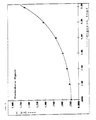
- the standard curve shown in FIG. 1 is obtained, which can be used for testing solutions with an unknown T3 content.
- disuccinidyl suberate solution concentration 10 mg disuccinidyl suberate / ml dioxane
- dialysis is carried out against 500 times the volume of potassium phosphate buffer 50 mM, pH 7.2, the fraction eluted on Superose 6 R in the exclusion volume is separated off, stabilized with sucrose (concentration 6 mg sucrose / mg IgG) and lyophilized.
- a fleece made of 60% sulfite pulp and 40% linters is soaked with crosslinked rabbit-IgG-digoxigenin conjugate from Example 4 a) (250 ⁇ g / ml in 50 mM potassium phosphate buffer, pH 7.2) and saturated Dried at 50 ° C for 60 minutes in a forced air cabinet.
- each of sheep anti-digoxin-IgG- ⁇ -galactosidase conjugate (prepared analogously to the methods described in J. Immunoassay 4 (1983) 209-327) (120 mU in phosphate-buffered saline, 5 g / l bovine serum albumin ) are incubated for 5 minutes with 50 ⁇ l of a standard Digoxin series and then onto the pieces of fleece (8 x 8 mm, thickness 0.5 mm) of the Digoxin matrix prepared above pipetted.
- the nonwoven pieces are spun out in a centrifuge and the ⁇ -galactosidase activity of the centrifugate is determined with 40 mM chlorophenol red galactoside solution (produced according to EP-A 0 146 866) at 578 nm.
- the standard curve of FIG. 2 is obtained, by means of which the unknown digoxin content in a solution can be determined.
- a fleece produced under 1. is agitated for 15 minutes in 1 ml of 50 mM potassium phosphate buffer, pH 7.0. The supernatant is transferred to a 1 ml Enzymun plastic tube (Boehringer Mannheim GmbH, Mannheim, Federal Republic of Germany), which is coated on the inside with 1 ⁇ g / ml tBSA-Biotin 1: 1 (Boehringer Mannheim GmbH, Mannheim, Federal Republic of Germany), and one Incubated for one hour. After washing twice with water, 1 ml of a peroxidase-biotin conjugate solution (200 mU / ml, Boehringer Mannheim GmbH, Mannheim, Federal Republic of Germany) is added to the tube and incubated for 30 minutes.
- a peroxidase-biotin conjugate solution 200 mU / ml, Boehringer Mannheim GmbH, Mannheim, Federal Republic of Germany
- 2,2′-azino-di- [3-ethylbenzthiazoline sulfonate (6)] is added to the tube and the reaction solution is measured at 405 nm.
- the system is calibrated with a standard range of solutions of known tBSA-SA concentration.
Landscapes
- Health & Medical Sciences (AREA)
- Immunology (AREA)
- Life Sciences & Earth Sciences (AREA)
- Engineering & Computer Science (AREA)
- Molecular Biology (AREA)
- Biomedical Technology (AREA)
- Chemical & Material Sciences (AREA)
- Hematology (AREA)
- Urology & Nephrology (AREA)
- Biotechnology (AREA)
- Biochemistry (AREA)
- Cell Biology (AREA)
- Food Science & Technology (AREA)
- Medicinal Chemistry (AREA)
- Physics & Mathematics (AREA)
- Analytical Chemistry (AREA)
- Microbiology (AREA)
- General Health & Medical Sciences (AREA)
- General Physics & Mathematics (AREA)
- Pathology (AREA)
- Peptides Or Proteins (AREA)
- Immobilizing And Processing Of Enzymes And Microorganisms (AREA)
- Investigating Or Analysing Biological Materials (AREA)
Abstract
Description
- Die Erfindung betrifft ein Verfahren zur Proteinimmobilisierung an einer Festphase, eine so hergestellte Protein tragende Festphase sowie deren Verwendung.
- An Festphasen immobilisierte Proteine besitzen eine große Bedeutung in verschiedenen Gebieten der Technik. Genannt seien zum Beispiel der Einsatz immobilisierter Proteine in der Biotechnologie, beispielsweise in Bioreaktoren oder in der biochemischen Produktion, z. B. für Affinitätschromatographien.
- Eine besondere Bedeutung haben immobilisierte Proteine für die Analytik erlangt. Insbesondere bei Nachweisverfahren von für die medizinische Diagnostik relevanten Parametern in Körperflüssigkeiten, wie Blut, Plasma, Serum, Urin, Speichel, Liquor etc. spielen bioaffine Reaktionen eine wesentliche Rolle, d. h. solche Reaktionen, bei denen eine bestimmte Substanz spezifisch an ein Protein gebunden wird. Bekannte Substanz/Protein-Paare, die solche bioaffinen Reaktionen eingehen, sind in allgemeiner Form z. B. Antigen/Antikörper, Kohlehydrat/Lectin, Biotin/Avidin etc. Ist das Protein an eine Festphase immobilisiert, können in einer Probe vorliegende entsprechende Substanzen, die mit dem Protein spezifisch bindefähig sind, daran gebunden und aus der Probe entfernt und qualitativ und/oder quantitativ bestimmt werden.
- Beispielsweise wurde für den Nachweis eines Partners des Antigen/Antikörper-Paares das Prinzip des heterogenen Immunoassays entwickelt. Dem Fachmann sind viele Varianten bekannt. Allen gemeinsam ist, daß ein Partner des Antigen/Antikörper-Paares an eine Festphase gebunden ist und mit diesem immobilisierten Partner eine Abtrennung zumindest eines Teils des entsprechenden Bindungspartners aus der restlichen Probe durchgeführt wird. Die Menge des abgetrennten Bindungspartners bzw. des Teils des nicht abgetrennten, sondern in der Probe verbliebenen Bindungspartners wird dann an der Festphase bzw. in der restlichen Probe ermittelt.
- Für die Immobilisierung von Proteinen an Festphasen sind aus dem Stand der Technik eine Reihe von Verfahren bekannt. So kann die Fixierung eines Proteins an eine Festphase durch chemische oder physikalische Kräfte erfolgen. Seit langem sind Methoden zur Herstellung kovalenter Bindungen zwischen einem festen Trägermaterial und daran zu bindenden Proteinen bekannt. Beispielsweise beschreibt EP-A-0 274 911 den Einsatz chemisch reaktiver Kunststoffmembranen, die Proteine direkt kovalent zu binden vermögen. Dieses Verfahren zur Herstellung Protein tragender Festphasen erfordert jedoch eine lange Kontaktzeit zwischen Membran und zu bindendem Protein, um die chemische Reaktion vollständig ablaufen zu lassen. Außerdem müssen nicht mit Protein abgesättigte Aktivstellen in einem zweiten anschließenden Schritt mit einem Inertprotein belegt werden, damit keine freien Membran-Aktivstellen mehr vorhanden sind, die die spätere Verwendung der Proteinbeladenen Festphase negativ beeinflussen könnten.
- Bekannte Verfahren, bei denen reaktive Gruppen der Festphase mit einem bifunktionellen Linker gekuppelt werden, an dessen verbleibende freie Funktion dann das zu fixierende Protein kovalent gebunden wird, erfordern noch mehr Zeit und noch mehr Verfahrensschritte. Mit jedem Verfahrensschritt aber steigen auch das Risiko der Produktion von Fehlchargen und die Produktionskosten.
- Ein Problem der nicht-kovalenten Fixierung von Protein an eine Festphase ist dessen schwächere Bindung. So können Proteine, die aus einer Lösung an eine Festphase adsorbiert wurden, relativ leicht wieder von dieser abgelöst werden. Vorschläge zur Beseitigung dieses Problems sind bekannt. In DE-A-34 46 636 sind z. B. Verfahren zur Fixierung eines immunreaktiven Stoffes auf einem porösen Trägermaterial beschrieben. Hierbei wird zur Vermeidung des Haftfestigkeitsproblems am Trägermaterial die Fixierung durch Ablaufenlassen einer Immunreaktion zwischen den beiden Partnern einer Immunreaktion, also zwischen Antikörper und Antigen oder Hapten, erreicht. Es wird so ein Immunkomplex-Netz gebildet, das das zu bindende Protein (Antikörper oder Antigen) enthält und an die Festphase bindet. Der Nachteil dieses Fixierungsverfahrens besteht darin, daß außer dem zu bindenden Protein noch ein weiterer teurer Einsatzstoff (Antikörper oder Antigen) benötigt wird. Außerdem muß die Ansatzführung sehr exakt durchgeführt werden, um eine optimale Bindung an die Festphase zu erreichen.
- In GB-A-1 505 400 wird vorgeschlagen, ein immunologisch wirksames Protein zu vernetzen und dann an Polystyrol-Latexpartikel zu adsorbieren. Die Adsorption wird in der Latexemulsion durchgeführt. Nach der Bindung eines Teils des Proteins an die Latexpartikel werden diese abzentrifugiert und mehrmals gewaschen. Die Protein tragenden Polystyrol-Partikel werden in gepufferter wässriger Lösung als Suspension aufbewahrt und für Abtrennreaktionen eingesetzt.
- EP-A-0 122 209 beschreibt ein Verfahren zur Bindung biologischer Makromoleküle an Festphasen, das darin besteht, die zu fixierenden Makromoleküle zu polymerisieren, mehrere Stunden zusammen mit hydrophoben Trägermaterialien, wie z. B. Polystyrol zu inkubieren und nach der Bindung eines Teils der polymerisierten Makromoleküle an daß Trägermaterial dieses vor dem Einsatz bzw. der Lagerung mehrmals zu waschen.
- Beide vorgenannten Verfahren führen nicht zu einer zufriedenstellenden Haftung von Proteinen, insbesondere von spezifisch bindefähigen, d. h. bioaffinen Substanzen am Träger. Außerdem sind für die Proteinfixierung an den Träger langwierige und umständliche Inkubations- und Waschschritte notwendig. EP-A-0 269 092 betrifft ein Verfahren, das gegenüber den beiden vorgenannten die Haftung verbessern soll. Hierzu wird das zu fixierende Protein kovalent an ein hydrophobes Trägerprotein gebunden und dieser Komplex an eine hydrophobe Festphase adsorbiert, wobei durch Ausnutzung der hydrophoben Wechselwirkungen zwischen Festphase und Trägerprotein eine besonders vorteilhafte Fixierung erreicht werden soll.
- Sämtlichen drei letztgenannten Schriften ist gemeinsam, daß ausschließlich hydrophobe Trägermaterialien zur nicht-kovalenten Fixierung von Proteinen eingesetzt werden. Dies schränkt die Auswahl an Trägermaterialien erheblich ein. Vor allem für die Anwendung Protein tragender Festphasen in wässrigen Flüssigkeiten, wie es biologische Flüssigkeiten alle sind, sind hydrophile Materialien wegen der besseren Benetzbarkeit oft vorzuziehen.
- Der vorliegenden Erfindung liegt ausgehend von diesem Stand der Technik die Aufgabe zugrunde, Proteine mittels eines auch im technischen Maßstab einfachen und schnellen Verfahrens fest an in Wasser im wesentlichen unlösliche Trägermaterialien zu binden, um so Protein tragende Festphasen zu erhalten, die breite Verwendungsmöglichkeiten zum Einsatz bei der Bindung und eventuellen Abtrennung spezifisch bindefähiger Substanzen aus flüssigen Proben bieten.
- Diese Aufgabe wird durch die in den Ansprüchen gekennzeichnete Erfindung gelöst. So ist das erfindungsgemäße Verfahren zur Immobilisierung eines Proteins an einer Festphase dadurch gekennzeichnet, daß das zu fixierende Protein aggregiert, in einer Flüssigkeit mit einer hydrophilen Festphase kontaktiert und die Festphase nach erfolgtem Kontakt getrocknet wird.
- Mittels des erfindungsgemaßen Verfahrens können prinzipiell alle Proteine immobilisiert werden, die chemisch oder physikalisch zu höheren Molekulargewichten aggregiert werden können. Unter chemischer Aggregation ist hierbei jede Molekulargewichtserhöhung zu verstehen, die mittels chemischer Agentien erzielt wird. So können Proteine z. B. durch Zusatz von Carbodiimiden homopolymerisiert werden. Sie können aber beispielsweise auch durch mehrfunktionelle Moleküle, sogenannte Linker, miteinander verknüpft oder vernetzt werden. Die Palette chemischer Linker eröffnet die Möglichkeit hoher Variabilität der Aggregate z. B. hinsichtlich der Zugänglichkeit bestimmter Proteinstellen, sogenannte Epitope, für mit dem Protein spezifisch bindefähige Substanzen oder hinsichtlich der Haftungsfähigkeit des Proteinaggregats an der Festphase. Die Methode, Proteine mittels Linker zu vernetzen, ist dem Fachmann aus dem Stand der Technik bekannt. Beispielsweise beschreiben GB-A-1 505 400, EP-A-0 122 209 und EP-A-0 269 092 solche Verfahren. Für das erfindungsgemäße Verfahren haben sich Linker, wie z. B. Disuccinidyl-suberat, S-Acetyl-mercapto-bernsteinsäureanhydrid oder Maleinimido-hexanoyl-hydroxysuccinimid als vorteilhaft erwiesen. Besonders gut geeignet für die vorliegende Erfindung ist Disuccinidyl-süberat zur Aggregation von Humanalbumin.
- Unter physikalischer Aggregation ist jede Molekulargewichtserhöhung zu verstehen, die ohne chemische Agentien erzielt wird. Beispielsweise ist es bekannt, Proteine, wie z. B. Albumin thermisch zu aggregieren und so das Molekulargewicht zu erhöhen (EP-A-0 269 092). Thermisch aggregiertes Albumin ist ein im Sinne der vorliegenden Erfindung besonders gut immobilisierbares Protein.
- Wenn die hergestellten Proteinaggregate vor ihrem Aufbringen auf Festphasen z. B. zur Lagerung lyophilisiert werden sollen, empfiehlt es sich gegebenenfalls, wie oft bei der Lyophilisation von Proteinlösungen, diesen vor der Lyophilisation einen Stabilisator zuzusetzen, der die Lagerfähigkeit des lyophilisierten Proteinaggregats und dessen Löslichkeit bei der Rekonstitution mit Wasser oder Puffer erhöht. Art und Menge dieser Substanzen sind von der Art des jeweiligen Proteins abhängig. Generell dürfen diese gegebenenfalls der Proteinaggregat-Lösung zugegebenen Stoffe keinen negativen Einfluß auf das Immobilisierungsverfahren, insbesondere die Bindefähigkeit des Proteinaggregats an die Festphase ausüben. Geeignete Stoffe sind z. B. Saccharose, Trehalose, Mannose, Dextrane und ähnliche Kohlenhydrate, aber auch Proteine, wie Crotein C, Kollagen oder Rinderserum-albumin. Saccharose hat sich als besonders vorteilhaft erwiesen, insbesondere im Falle von mit Disuccinidyl-suberat vernetztem Humanalbumin. Ein typischer Konzentrationsbereich für der zu lyophilisierenden Proteinaggregat-Lösung gegebenenfalls zugesetzte Stabilisatoren und/oder Lösungsvermittler ist 2 bis 20 Gew.-%. Für den vorgenannten Fall der Saccharose-Zugabe zu einer Lösung von mit Disuccinidyl-suberat vernetztem Humanalbumin liegt die vorzugsweise Konzentration zwischen 4 und 10 Gew.-%. Bei Zusatz von Stabilisatoren und/oder Lösungsvermittlern zu der zu lyophilisierenden Proteinaggregat-Lösung ist darauf zu achten, daß bei Verwendung des rekonstituierten Lyophilisats zur Beladung einer Festphase im Sinne der Erfindung die Konzentration an Stabilisator und/oder Lösungsvermittler nicht so hoch sein darf, daß die Bindefähigkeit des Proteinaggregats an die Festphase negativ beeinflußt wird. Beispielsweise sollte im Falle der Immobilisierung von mit Disuccinidyl-suberat vernetztem Humanalbumin die Konzentration an Saccharose in der Proteinaggregat-Lösung weniger als 2 Gew.-% betragen.
- Als Proteine sind nach dem erfindungsgemäßen Verfahren nicht nur natürlich vorkommende und aus natürlichen Quellen isolierte Proteine immobilisierbar, sondern auch synthetisch hergestellte. Beispiele für Proteine, die erfindungsgemäß besonders gut immobilisierbar sind, sind Albumin, Immunglobuline, Transferrin und Collagen. Ebenso sind erfindungsgemäß auch solche Proteine an eine Festphase fixierbar, die einen nicht-Proteinteil, wie beispielsweise Biotin oder ein Kohlehydrat gebunden enthalten. Auf diese Art und Weise sind auch Nichtproteine an Festphasen erfindungsgemäß zu immobilisieren, wenn sie zuvor an ein Protein gebunden werden, das für den Nichtproteinteil als Anker an der Festphase wirken kann.
- Auf gleiche Weise können natürlich auch Proteine an ein anderes als Anker an der Festphase wirkendes Protein gebunden sein.
- Die Festigkeit der erfindungsgemäßen Immobilisierung des Proteins an die Festphase ist abhängig von verschiedenen Faktoren: Einerseits von dem jeweiligen zu fixierenden Protein, andererseits von der Art des Trägermaterials. Bezüglich des Proteins hat es sich gezeigt, daß die Haftung an der Festphase mit zunehmendem Molekulargewicht zunimmt. Es läßt sich keine generelle Mindestgröße zur Erzielung einer bestimmten Bindefestigkeit angeben. Sie hängt von der Art des jeweils zu bindenden Proteins ab und ist für den Fachmann leicht ermittelbar. Für Albumin hat sich beispielsweise ein Aggregat aus 4 bis 5 Albumineinheiten bereits als gut immobilisierbar im Sinne der Erfindung erwiesen, ein Aggregat aus mindestens 10 Albumineinheiten ist besonders vorteilhaft.
- Bevorzugte Festphasen im Sinne der Erfindung sind alle diejenigen in Wasser im wesentlichen unlöslichen Materialien, die hydrophiler sind als das zu immobilisierende aggregierte Protein. Geeignete Materialien sind z. B. Polyester, Sulphitzellstoff, Zellwolle, Linters, Nitrocellulose, Celluloseacetat oder Festphasen auf Nylonbasis. Sie können in beliebiger Form vorliegen, wie beispielsweise Pulver, Körnchen, Fasern, Vliesen oder Folien. Besonders bevorzugt als Festphasen im Sinne der Erfindung sind Vliese auf Cellulose-Basis.
- Gemäß dem Verfahren der vorliegenden Erfindung wird das aggregierte Protein in einer Flüssigkeit mit der Festphase kontaktiert. Das aggregierte Protein liegt dabei in gelöster Form vor, vorzugsweise in einer wässrigen Flüssigkeit, beispielsweise einer Pufferlösung. Werden Vliese als Festphasen eingesetzt, hat es sich als besonders vorteilhaft erwiesen, diese mit einer Lösung des aggregierten Proteins zu tränken, indem sie in die Lösung getaucht und darin mit der Lösung gesättigt werden.
- Als Pufferlösungen können dabei all diejenigen eingesetzt werden, die dem Fachmann als das jeweilige Protein und dessen biologische Aktivität nicht schädigend bekannt sind. Beispielsweise haben sich für Humanalbumin Phosphat- oder HEPES-Puffer mit einem pH-Wert zwischen ca. 6 und ca. 9, vorzugsweise zwischen ca. 7 und ca. 8 als besonders geeignet als Lösungsmittel erwiesen.
- Zur Erzielung einer guten Haftung des aggregierten Proteins an der Festphase ist es wichtig, daß nach der Kontaktierung des Proteinaggregats mit der Festphase die das Protein tragende Festphase getrocknet wird. Unter Trocknung werden hierbei alle speziellen Bemühungen verstanden, Flüssigkeit weitest möglich zu entfernen. Als trocken im Sinne der Erfindung wird im allgemeinen eine hydrophile Festphase mit einem Restfeuchtegehalt von weniger als 7 % bei 20 °C und 50 % relativer Feuchte bezeichnet. Der Restfeuchtegehalt, ab dem von "trocken" geredet werden kann, ist im speziellen Fall abhängig von der ausgewählten Festphase. So sind z. B. Cellulose-Vliese bei Restfeuchtegehalten von weniger als 7 %, Mischvliese aus Cellulose und Polyesterfasern im Gewichtsverhältnis 50:50 von weniger als 5 % als trocken anzusprechen. Um solche Trocknungsgrade bei hydrophilen Festphasen in wirtschaftlich vertretbaren Trocknungszeiten zu erreichen, muß in der Regel Wärme zugeführt werden. So wird nach dem erfindungsgemäßen Verfahren die Trocknung Protein-tragender Vliese vorteilhafterweise bei Temperaturen zwischen 30 und 80 °C durchgeführt. Sind die Trocknungszeiten nicht entscheidend für die Wahl des Verfahrens kann die Trocknung auch bei Raumtemperatur z. B. im Luftstrom durchgeführt werden.
- Es wurde überraschenderweise festgestellt, daß die Erreichung eines so geringen Restfeuchtegehaltes der das Protein tragenden Festphase entscheidend für die ausgezeichnete Haftung des Proteins am Träger ist. Höhere Restfeuchtegehalte führen zu einer schwächeren Fixierung des Proteins an der Festphase.
- Der besondere Vorteil des erfindungsgemäßen Verfahrens liegt in seiner Einfachheit. Es reicht aus, das hydrophile Trägermaterial, insbesondere hydrophile Vliese, in eine Lösung des zu fixierenden aggregierten Proteins zu tauchen, um es mit Flüssigkeit zu sättigen. Nach erfolgter Tränkung wird das Trägermaterial aus der Proteinlösung genommen und getrocknet. Nach dem Trocknen ist das Proteinaggregat fest an die Festphase gebunden. Auf diese Art und Weise ist es möglich, eine homogene Beladungsdichte der Festphase mit Protein zu erreichen, wobei die Menge an immobilisiertem Protein genau bekannt ist.
- Die Menge an Protein, die so an eine Festphase gebunden werden kann, ist wesentlich höher als es durch Adsorption erklärbar ist. So kann dann, wenn nach Durchführung des erfindungsgemäßen Verfahrens nach der Trocknung durch einen zusätzlichen Waschschritt kein Protein von der Festphase eluierbar ist, ohne Trocknung, d. h. wenn eine einfache Adsorption des Proteinaggregats an die Festphase ermöglicht wurde, der Großteil des adsorbierten Proteins durch Waschung wieder eluiert werden. Nach dem erfindungsgemäßen Verfahren erübrigt sich deshalb ein Waschschritt, wenn die Konzentration der Proteinlösung so gewählt wird, daß das gesamte Protein nach der Trocknung immobilisiert ist. Diese Proteinkonzentration ist von der Art des jeweiligen Trägermaterials abhängig. Die Grenzkonzentration, die gerade noch vollständig immobilisiert wird, kann durch einfache Versuche leicht ermittelt werden.
- Ein weiterer Vorteil des erfindungsgemäßen Verfahrens liegt darin, daß die Zusammensetzung der Tränklösung in weiten Bereichen frei wählbar ist, ohne die Bindungseigenschaften des zu immobilisierenden Proteins nachteilig zu beeinflussen. So können z. B. Art und Konzentration des Puffers, pH, Ionenstärke und gegebenenfalls zuzusetzende Stabilisatoren und/oder Lösungsvermittler überwiegend nach Gesichtspunkten gewählt werden, die allein die Eigenschaften des jeweiligen gelösten Proteinaggregats betreffen. Auf die Festigkeit der Bindung des immobilisierten Proteinaggregats hat diese Wahl meist keinen Einfluß.
- Nach dem erfindungsgemäßen Verfahren hergestellte Festphasen eignen sich sehr gut für die Bindung von mit dem immobilisierten Protein spezifisch bindefähigen Substanzen und gegebenenfalls deren Entfernung aus Flüssigkeiten. Es sind die unterschiedlichsten Anwendungsbereiche denkbar, wie z. B. für die biochemische Produktion als Träger immobilisierter Proteine in Bioreaktoren oder für die Affinitätschromatographie zur Anreicherung und/oder Abtrennung von spezifisch bindefähigen Substanzen.
- Besonders bevorzugt werden erfindungsgemäß hergestellte Proteintragende Festphasen für analytische Bestimmungen von Inhaltsstoffen in flüssigen Proben eingesetzt. Ganz besonders bevorzugt werden sie für bioaffine Bestimmungen von Inhaltsstoffen in Körperflüssigkeiten, wie z. B. Blut, Plasma, Serum, Urin, Liquor, Speichel etc., wie sie einleitend beschrieben wurden. Für heterogene Immunoassays eignen sich die erfindungsgemäßen Proteintragenden Festphasen besonders gut, insbesondere für solche, die insgesamt trägergebunden, z. B. auf einem Teststreifen, durchgeführt werden.
- Im folgenden wird die Erfindung anhand einiger Beispiele näher erläutert.
- 1,5 g HSA werden in 30 ml Kaliumphosphat-Puffer, 200 mM, pH = 8,0 vorgelegt und innerhalb 2 Stunden mit 2,5 ml einer Lösung aus 50 mg DSS/ml Dioxan versetzt. Nach Ablauf der Vernetzungsreaktion wird gegen das 500fache Volumen Kaliumphosphat-Puffer, 20 mM, pH 7,2 dialysiert. Die hochmolekulare Fraktion (pHSA) mit einem Molekulargewicht von mehr als 650 000 Dalton wird an Superose 6R (Pharmacia, Freiburg, Bundesrepublik Deutschland) über Gelfiltration abgetrennt und nach Zugabe von 6 mg Saccharose/mg Protein lyophilisiert.
- 6 x 6 mm² große und 0,5 mm dicke Vliesstücke aus 50 % Polyester/50 % Linters werden mit 15 µl einer Lösung von 250 mg/l pHSA aus Beispiel 1 in 10 mM Natriumphosphat-Puffer pH 7,5 getränkt und
- a) 30 Minuten bei 50 °C getrocknet und dreimal mit 25 µl Natriumphosphat-Puffer (10 mM, pH 7,5) jeweils in der Zentrifuge gewaschen und anschließend ausgeschleudert bzw.
- b) nach 10 Minuten in einer Zentrifuge ausgeschleudert und zweimal mit je 25 µl Natriumphosphat-Puffer (10 mM, pH 7,5) gewaschen.
- Die Waschzentrifugate für a) und b) werden jeweils vereint und jeweils mit 300 mU anti-HSA-IgG-ß-Galactosidasekonjugat (160 µl) versetzt und 10 Minuten geschüttelt. Dann werden je 200 µl in Mikrotiterplatten-Löcher transferiert, deren Wand mit HSA beladen ist. Nach einstündiger Inkubation wird mit Phosphat-gepufferter physiologischer Kochsalzlösung gewaschen, mit o-Nitrophenylgalactosid entwickelt und nach 10 Minuten die Extinktion bei 405 nm vermessen. Die quantitative Bestimmung des aus dem Vlies eluierten pHSA erfolgt über eine mitgeführte Eichkurve.
- Die pHSA-Menge mit der jedes Vliesstück kontaktiert wurde, betrug 3,75 µg. Nach der Elution wurden folgende Mengen in den vereinten Waschzentrifugaten ermittelt:
Verfahren pHSA im Eluat Eluat bezogen auf Tränklösung a) 0,012 µg 0,3 % b) 2,64 µg 71 % - Das erfindungsgemäß immobilisierte Protein ist aufgrund des Trocknungsschrittes wesentlich fester an die Festphase gebunden als das lediglich adsorbierte Protein, das zum größten Teil wieder von der Festphase abgewaschen werden kann.
- 3 g Kaninchen-IgG werden in 300 ml Kaliumphosphat-Puffer, 100 mM, pH 8,5 gelöst und mit einer Lösung aus 58 mg N-tert.-butyloxycarbonyl-trijodthyronin-N′-hydroxysuccinimid-ester (BOC-T3-N′-hydroxysuccinimid-ester) in 30 ml Dioxan versetzt.
- Nach zwei Stunden Reaktionszeit wird die Proteinlösung gegen das 200fache Volumen 20 mM Kaliumphosphat-Puffer, pH 7,8 dialysiert und über Ultrafiltration (Amicon YM 100R-Membran) auf eine Konzentration von 50 mg/ml eingestellt.
- Zur Vernetzung werden dem Ansatz langsam 35 ml Disuccinidyl-Suberat (Konzentration 10 mg Disuccinidyl-Suberat/ml Dioxan) zugegeben. Nach Beendigung der Vernetzungsreaktion wird gegen das 500fache Volumen Kaliumphosphat-Puffer, 50 mM, pH 7,2 dialysiert und die an Superose 6R im Ausschlußvolumen eluierte Fraktion abgetrennt sowie mit Saccharose (Konzentration 6 mg Saccharose/mg IgG) stabilisiert und lyophilisiert.
- Ein Vlies aus 60 % Sulfit-Zellstoff und 40 % Linters wird mit vernetztem Kaninchen-IgG-T3-Konjugat aus Beispiel 3 a) in einer Konzentration von 250 µg/ml in 50 mM Kaliumphosphat-Puffer, pH 7,2 getränkt und nach vollständiger Sättigung bei 50 °C 60 Minuten lang in einem Umluftschrank getrocknet. Jeweils 50 µl Schaf-anti-T3-IgG-ß-Galactosidase-Konjugat (hergestellt analog in J. Immunoassay 4 (1983) 209 - 327 beschriebenen Verfahren) (120 mU in Phosphatgepufferter physiologischer Kochsalzlösung, 5 g/l Rinderserum-Albumin) werden 5 Minuten lang mit 50 µl einer T3-Standardreihe inkubiert und anschließend auf vorstehend hergestellte Vliesstücke (8 x 8 mm, Dicke 0,5 mm) der T3-Matrix pipettiert. Nach 5 Minuten Inkubationszeit werden die Vliesstücke auszentrifugiert und die ß-Galactosidaseaktivität des Filtrates mit 40 mM Chlorphenolrotgalactosid-Lösung (hergestellt nach EP-A 0 146 866) bei 578 nm bestimmt.
- Es wird die in Figur 1 dargestellte Standardkurve erhalten, die für die Prüfung von Lösungen mit unbekanntem T3-Gehalt herangezogen werden kann.
- a) 3 g Kaninchen-IgG werden in 300 ml Kaliumphosphat-Puffer, 100 mM, pH 8,5, gelöst und mit einer Lösung aus 58 mg Digoxigenin-(3-succinidyl)-N-hydroxysuccinimid in 30 ml Dioxan versetzt. Nach 2 Stunden Reaktionszeit wird die Proteinlösung gegen das 200fache Volumen 20 mM Kaliumphosphat-Puffer pH 7,0 dialysiert und über Ultrafiltration (Amicon YM 100R-Membran) auf eine Konzentration von 50 mg/ml eingestellt.
- Zur Vernetzung werden dem Ansatz langsam 35 ml Disuccinidyl-Suberat-Lösung (Konzentration 10 mg Disuccinidyl-Suberat/ml Dioxan) zugegeben. Nach Beendigung der Vernetzungsreaktion wird gegen das 500fache Volumen Kaliumphosphat-Puffer 50 mM, pH 7,2 dialysiert, die an Superose 6R im Ausschlußvolumen eluierte Fraktion abgetrennt, mit Saccharose (Konzentration 6 mg Saccharose/mg IgG) stabilisiert und lyophilisiert.
- Ein Vlies aus 60 % Sulfit-Zellstoff und 40 % Linters wird mit vernetztem Kaninchen-IgG-Digoxigenin-Konjugat aus Beispiel 4 a) (250 µg/ml in 50 mM Kaliumphosphat-Puffer, pH 7,2) bis zur Sättigung getr1nkt und bei 50 °C 60 Minuten lang in einem Umluftschrank getrocknet.
- Jeweils 50 µl Schaf-anti-Digoxin-IgG-ß-Galactosidase-Konjugat (hergestellt analog in J. Immunoassay 4 (1983) 209 - 327 beschriebenen Verfahren) (120 mU in Phosphat-gepufferter physiologischer Kochsalzlösung, 5 g/l Rinderserum-Albumin) werden 5 Minuten lang mit 50µl einer Digoxin-Standardreihe inkubiert und anschließend auf die vorstehend hergestellten Vliesstücke (8 x 8 mm, Dicke 0,5 mm) der Digoxin-Matrix pipettiert. Nach 5 Minuten Inkubationszeit werden die Vliesstücke jeweils in einer Zentrifuge ausgeschleudert und die ß-Galactosidaseaktivität des Zentrifugates mit 40 mM Chlorphenolrotgalactosid-Lösung (hergestellt nach EP-A 0 146 866) bei 578 nm bestimmt.
- Es wird die Standardkurve der Figur 2 erhalten, mittels der der unbekannte Digoxingehalt in einer Lösung bestimmt werden kann.
- 8 mm x 8 mm große Quadrate aus Papier (80 % Polyester / 20 % Cellulose / 20 % (bezogen auf Fasern) Etadurin) werden mit 42 µl einer Lösung von 0,5 µg/µl tBSA-SA (Boehringer Mannheim GmbH, Mannheim, Bundesrepublik Deutschland) in 50 mMol/l Kaliumphosphatpuffer, pH 7,0 imprägniert. Anschließend wird 30 Minuten bei 70 °C getrocknet.
- Ein unter 1. hergestelltes Vlies wird 15 Minuten lang in 1 ml 50 mM Kaliumphosphatpuffer, pH 7,0 bewegt. Der Überstand wird in ein 1 ml Enzymun-Plastikröhrchen (Boehringer Mannheim GmbH, Mannheim, Bundesrepublik Deutschland), das innen mit 1 µg/ml tBSA-Biotin 1:1 (Boehringer Mannheim GmbH, Mannheim, Bundesrepublik Deutschland) beschichtet ist, überführt und eine Stunde lang inkubiert. Nach zweimaligem Waschen mit Wasser wird 1 ml einer Peroxidase-Biotin-Konjugatlösung (200 mU/ml, Boehringer Mannheim GmbH, Mannheim, Bundesrepublik Deutschland) in das Röhrchen gegeben und 30 Minuten inkubiert. Nach zweimaligem Waschen mit Wasser wird 2,2′-Azino-di-[3-ethylbenzthiazolinsulfonat(6)] in das Röhrchen gegeben und die Reaktionslösung bei 405 nm vermessen. Das System wird mit einer Standardreihe von Lösungen bekannter tBSA-SA-Konzentration kalibriert.
- Bei Verwendung des nach 1. hergestellten imprägnierten Papiers werden 18 ng tBSA-SA, das entspricht 0,09 % desorbiert. Wird bei dem Imprägnierverfahren nach 1. der Trockenschritt weggelassen, werden 560 ng tBSA-SA, das sind 2,7 % desorbiert. Hieraus folgt, daß das erfindungsgemäß immobilisierte Protein aufgrund des Trocknungsschrittes wesentlich fester an die Festphase gebunden ist als das lediglich adsorbierte Protein.
Claims (7)
Priority Applications (1)
| Application Number | Priority Date | Filing Date | Title |
|---|---|---|---|
| AT89123321T ATE103936T1 (de) | 1988-12-19 | 1989-12-16 | Verfahren zur proteinimmobilisierung an einer festphase, so hergestellte protein tragende festphase sowie deren verwendung. |
Applications Claiming Priority (2)
| Application Number | Priority Date | Filing Date | Title |
|---|---|---|---|
| DE3842700 | 1988-12-19 | ||
| DE3842700A DE3842700A1 (de) | 1988-12-19 | 1988-12-19 | Verfahren zur proteinimmobilisierung an einer festphase, so hergestellte protein tragende festphase sowie deren verwendung |
Publications (3)
| Publication Number | Publication Date |
|---|---|
| EP0374778A2 true EP0374778A2 (de) | 1990-06-27 |
| EP0374778A3 EP0374778A3 (de) | 1991-10-09 |
| EP0374778B1 EP0374778B1 (de) | 1994-04-06 |
Family
ID=6369520
Family Applications (1)
| Application Number | Title | Priority Date | Filing Date |
|---|---|---|---|
| EP89123321A Expired - Lifetime EP0374778B1 (de) | 1988-12-19 | 1989-12-16 | Verfahren zur Proteinimmobilisierung an einer Festphase, so hergestellte Protein tragende Festphase sowie deren Verwendung |
Country Status (8)
| Country | Link |
|---|---|
| US (1) | US5310885A (de) |
| EP (1) | EP0374778B1 (de) |
| JP (1) | JPH087213B2 (de) |
| AT (1) | ATE103936T1 (de) |
| AU (1) | AU607088B2 (de) |
| CA (1) | CA2005926C (de) |
| DE (2) | DE3842700A1 (de) |
| ES (1) | ES2051977T3 (de) |
Cited By (1)
| Publication number | Priority date | Publication date | Assignee | Title |
|---|---|---|---|---|
| WO1998058259A1 (en) * | 1997-06-19 | 1998-12-23 | Savyon Diagnostics Ltd. | Stabilization of polypeptides for use in immunoassay procedures |
Families Citing this family (50)
| Publication number | Priority date | Publication date | Assignee | Title |
|---|---|---|---|---|
| US5512169A (en) * | 1992-12-30 | 1996-04-30 | Dow Corning Corporation | Liquid column packing materials |
| US5599660A (en) * | 1993-01-19 | 1997-02-04 | Pharmacia Biotech Inc. | Method and preparation for sequential delivery of wax-embedded, inactivated biological and chemical reagents |
| KR100393499B1 (ko) * | 1995-03-14 | 2003-10-17 | 킴벌리-클라크 월드와이드, 인크. | 습윤성제품 |
| AU6157898A (en) | 1997-02-06 | 1998-08-26 | E. Heller & Company | Small volume (in vitro) analyte sensor |
| US8346337B2 (en) | 1998-04-30 | 2013-01-01 | Abbott Diabetes Care Inc. | Analyte monitoring device and methods of use |
| US8974386B2 (en) | 1998-04-30 | 2015-03-10 | Abbott Diabetes Care Inc. | Analyte monitoring device and methods of use |
| US6949816B2 (en) | 2003-04-21 | 2005-09-27 | Motorola, Inc. | Semiconductor component having first surface area for electrically coupling to a semiconductor chip and second surface area for electrically coupling to a substrate, and method of manufacturing same |
| US6175752B1 (en) | 1998-04-30 | 2001-01-16 | Therasense, Inc. | Analyte monitoring device and methods of use |
| US9066695B2 (en) | 1998-04-30 | 2015-06-30 | Abbott Diabetes Care Inc. | Analyte monitoring device and methods of use |
| US8465425B2 (en) | 1998-04-30 | 2013-06-18 | Abbott Diabetes Care Inc. | Analyte monitoring device and methods of use |
| US8480580B2 (en) | 1998-04-30 | 2013-07-09 | Abbott Diabetes Care Inc. | Analyte monitoring device and methods of use |
| US8688188B2 (en) | 1998-04-30 | 2014-04-01 | Abbott Diabetes Care Inc. | Analyte monitoring device and methods of use |
| US6251260B1 (en) | 1998-08-24 | 2001-06-26 | Therasense, Inc. | Potentiometric sensors for analytic determination |
| US6591125B1 (en) | 2000-06-27 | 2003-07-08 | Therasense, Inc. | Small volume in vitro analyte sensor with diffusible or non-leachable redox mediator |
| US6338790B1 (en) | 1998-10-08 | 2002-01-15 | Therasense, Inc. | Small volume in vitro analyte sensor with diffusible or non-leachable redox mediator |
| KR100307294B1 (ko) * | 1998-12-29 | 2001-12-17 | 김영환 | 반도체장치의제조방법 |
| US6654625B1 (en) | 1999-06-18 | 2003-11-25 | Therasense, Inc. | Mass transport limited in vivo analyte sensor |
| US6616819B1 (en) | 1999-11-04 | 2003-09-09 | Therasense, Inc. | Small volume in vitro analyte sensor and methods |
| GB0016836D0 (en) * | 2000-07-07 | 2000-08-30 | Lee Helen | Improved dipstick assays (1) |
| US6528325B1 (en) * | 2000-10-13 | 2003-03-04 | Dexall Biomedical Labs, Inc. | Method for the visual detection of specific antibodies in human serum by the use of lateral flow assays |
| US6560471B1 (en) | 2001-01-02 | 2003-05-06 | Therasense, Inc. | Analyte monitoring device and methods of use |
| AU2002309528A1 (en) | 2001-04-02 | 2002-10-15 | Therasense, Inc. | Blood glucose tracking apparatus and methods |
| US8771183B2 (en) | 2004-02-17 | 2014-07-08 | Abbott Diabetes Care Inc. | Method and system for providing data communication in continuous glucose monitoring and management system |
| AU2003303597A1 (en) | 2002-12-31 | 2004-07-29 | Therasense, Inc. | Continuous glucose monitoring system and methods of use |
| US7587287B2 (en) | 2003-04-04 | 2009-09-08 | Abbott Diabetes Care Inc. | Method and system for transferring analyte test data |
| FR2855177B1 (fr) * | 2003-05-23 | 2005-07-08 | Raymond Robert | Composition contenant des acides nucleiques lyophilises en presence de collagene et de disaccharides |
| US8066639B2 (en) | 2003-06-10 | 2011-11-29 | Abbott Diabetes Care Inc. | Glucose measuring device for use in personal area network |
| US7393453B2 (en) * | 2004-06-23 | 2008-07-01 | Kham Lin | Method for analysis of perchlorate |
| US8112240B2 (en) | 2005-04-29 | 2012-02-07 | Abbott Diabetes Care Inc. | Method and apparatus for providing leak detection in data monitoring and management systems |
| US7766829B2 (en) | 2005-11-04 | 2010-08-03 | Abbott Diabetes Care Inc. | Method and system for providing basal profile modification in analyte monitoring and management systems |
| US7885698B2 (en) | 2006-02-28 | 2011-02-08 | Abbott Diabetes Care Inc. | Method and system for providing continuous calibration of implantable analyte sensors |
| US8226891B2 (en) | 2006-03-31 | 2012-07-24 | Abbott Diabetes Care Inc. | Analyte monitoring devices and methods therefor |
| US7620438B2 (en) | 2006-03-31 | 2009-11-17 | Abbott Diabetes Care Inc. | Method and system for powering an electronic device |
| US20080071158A1 (en) | 2006-06-07 | 2008-03-20 | Abbott Diabetes Care, Inc. | Analyte monitoring system and method |
| US8732188B2 (en) | 2007-02-18 | 2014-05-20 | Abbott Diabetes Care Inc. | Method and system for providing contextual based medication dosage determination |
| US8930203B2 (en) | 2007-02-18 | 2015-01-06 | Abbott Diabetes Care Inc. | Multi-function analyte test device and methods therefor |
| US8123686B2 (en) | 2007-03-01 | 2012-02-28 | Abbott Diabetes Care Inc. | Method and apparatus for providing rolling data in communication systems |
| US8461985B2 (en) | 2007-05-08 | 2013-06-11 | Abbott Diabetes Care Inc. | Analyte monitoring system and methods |
| US8665091B2 (en) | 2007-05-08 | 2014-03-04 | Abbott Diabetes Care Inc. | Method and device for determining elapsed sensor life |
| US7928850B2 (en) | 2007-05-08 | 2011-04-19 | Abbott Diabetes Care Inc. | Analyte monitoring system and methods |
| US8456301B2 (en) | 2007-05-08 | 2013-06-04 | Abbott Diabetes Care Inc. | Analyte monitoring system and methods |
| US8103456B2 (en) | 2009-01-29 | 2012-01-24 | Abbott Diabetes Care Inc. | Method and device for early signal attenuation detection using blood glucose measurements |
| US20100213057A1 (en) | 2009-02-26 | 2010-08-26 | Benjamin Feldman | Self-Powered Analyte Sensor |
| US9226701B2 (en) | 2009-04-28 | 2016-01-05 | Abbott Diabetes Care Inc. | Error detection in critical repeating data in a wireless sensor system |
| WO2010138856A1 (en) | 2009-05-29 | 2010-12-02 | Abbott Diabetes Care Inc. | Medical device antenna systems having external antenna configurations |
| EP2473099A4 (de) | 2009-08-31 | 2015-01-14 | Abbott Diabetes Care Inc | Analytüberwachungssystem und -verfahren zur leistungs- und rauschverwaltung |
| WO2011026147A1 (en) | 2009-08-31 | 2011-03-03 | Abbott Diabetes Care Inc. | Analyte signal processing device and methods |
| EP2482720A4 (de) | 2009-09-29 | 2014-04-23 | Abbott Diabetes Care Inc | Verfahren und vorrichtung zur bereitstellung einer benachrichtigungsfunktion in analytüberwachungssystemen |
| EP2775918B1 (de) | 2011-11-07 | 2020-02-12 | Abbott Diabetes Care Inc. | Analytüberwachungsvorrichtung und -verfahren |
| US9968306B2 (en) | 2012-09-17 | 2018-05-15 | Abbott Diabetes Care Inc. | Methods and apparatuses for providing adverse condition notification with enhanced wireless communication range in analyte monitoring systems |
Family Cites Families (17)
| Publication number | Priority date | Publication date | Assignee | Title |
|---|---|---|---|---|
| US4004979A (en) * | 1968-03-29 | 1977-01-25 | Agence Nationale De Valorisation De La Recherche (Anvar) | Preparation of active proteins cross-linked to inactive proteins |
| US4081329A (en) * | 1972-12-08 | 1978-03-28 | Boehringer Mannheim Gmbh | Preparation of carrier-bound macromolecular compounds |
| BE821053A (fr) * | 1974-08-01 | 1975-04-14 | Produit pour diagnostic par determination immunochimique | |
| CA1056746A (en) * | 1974-09-06 | 1979-06-19 | Chung J. Lai | Biologically active membrane material |
| JPS6057127B2 (ja) * | 1976-09-28 | 1985-12-13 | 富士写真フイルム株式会社 | 磁気記録媒体 |
| US4338398A (en) * | 1979-03-20 | 1982-07-06 | Kabushiki Kaisha Hayashibara Seibutsu Kagaku Kenkyujo | Immobilization of starch degrading enzymes |
| US4410634A (en) * | 1979-10-26 | 1983-10-18 | Dynasciences Corporation | Method of passively adsorbing immuno-reactive haptens to solid phases |
| US4461829A (en) * | 1981-09-14 | 1984-07-24 | Miles Laboratories, Inc. | Homogeneous specific binding assay element and lyophilization production method |
| JPS5875063A (ja) * | 1981-10-30 | 1983-05-06 | Japan Synthetic Rubber Co Ltd | 免疫学的診断試薬の製造方法 |
| FR2543972B1 (fr) * | 1983-04-06 | 1985-12-27 | Immunotech Sa | Procede de fixation de macromolecules biologiques sur des supports |
| JPS59224565A (ja) * | 1983-04-28 | 1984-12-17 | Morinaga Milk Ind Co Ltd | 抗原検出用試薬 |
| IL75828A (en) * | 1985-07-17 | 1991-06-10 | Univ Ramot | Immobilization by biologically active proteins |
| US4742158A (en) * | 1986-04-25 | 1988-05-03 | Merck & Co., Inc. | Purification of hepatitis pre-S antigens by polymerized serum albumin affinity binding |
| US4794090A (en) * | 1986-09-26 | 1988-12-27 | W. R. Grace & Co.-Conn. | Immobilization support for biologicals |
| DE3640412A1 (de) * | 1986-11-26 | 1988-06-09 | Boehringer Mannheim Gmbh | Verfahren zur bestimmung einer spezifisch bindefaehigen substanz |
| DE3725504A1 (de) * | 1987-07-31 | 1989-02-09 | Thraenhart Olaf Priv Doz Dr | Verfahren und vorrichtung zum nachweis von antikoerpern und antigenen |
| WO1990004178A1 (en) * | 1988-10-11 | 1990-04-19 | Coulter Corporation | Immunoreactant carriers having a novel biocompatible intermediate coating and process of making same |
-
1988
- 1988-12-19 DE DE3842700A patent/DE3842700A1/de not_active Withdrawn
-
1989
- 1989-12-12 AU AU46165/89A patent/AU607088B2/en not_active Ceased
- 1989-12-16 AT AT89123321T patent/ATE103936T1/de not_active IP Right Cessation
- 1989-12-16 EP EP89123321A patent/EP0374778B1/de not_active Expired - Lifetime
- 1989-12-16 ES ES89123321T patent/ES2051977T3/es not_active Expired - Lifetime
- 1989-12-16 DE DE89123321T patent/DE58907404D1/de not_active Expired - Lifetime
- 1989-12-19 JP JP1327506A patent/JPH087213B2/ja not_active Expired - Lifetime
- 1989-12-19 CA CA002005926A patent/CA2005926C/en not_active Expired - Lifetime
-
1992
- 1992-05-19 US US07/887,239 patent/US5310885A/en not_active Expired - Lifetime
Cited By (1)
| Publication number | Priority date | Publication date | Assignee | Title |
|---|---|---|---|---|
| WO1998058259A1 (en) * | 1997-06-19 | 1998-12-23 | Savyon Diagnostics Ltd. | Stabilization of polypeptides for use in immunoassay procedures |
Also Published As
| Publication number | Publication date |
|---|---|
| AU4616589A (en) | 1990-06-21 |
| DE58907404D1 (de) | 1994-05-11 |
| EP0374778A3 (de) | 1991-10-09 |
| JPH02262600A (ja) | 1990-10-25 |
| CA2005926C (en) | 1999-11-16 |
| ES2051977T3 (es) | 1994-07-01 |
| JPH087213B2 (ja) | 1996-01-29 |
| US5310885A (en) | 1994-05-10 |
| CA2005926A1 (en) | 1990-06-19 |
| ATE103936T1 (de) | 1994-04-15 |
| AU607088B2 (en) | 1991-02-21 |
| EP0374778B1 (de) | 1994-04-06 |
| DE3842700A1 (de) | 1990-06-21 |
Similar Documents
| Publication | Publication Date | Title |
|---|---|---|
| EP0374778B1 (de) | Verfahren zur Proteinimmobilisierung an einer Festphase, so hergestellte Protein tragende Festphase sowie deren Verwendung | |
| DE3105768C2 (de) | Verwendung eines Trägers für die Immobilisierung von bioaktiven Materialien | |
| EP0269092B1 (de) | Verfahren zur Bestimmung einer spezifisch bindefähigen Substanz | |
| EP0353570B1 (de) | Mit Reagenz ablösbar imprägnierte Trägermatrix | |
| DE3882058T2 (de) | Immunoreaktives Reagens, Verfahren zu seiner Herstellung und seine Verwendung zur Bestimmung einer immunoreaktiven Spezies. | |
| EP0235526A2 (de) | Aktivierte Polymerfestkörper und Verfahren zu ihrer Herstellung | |
| DE2953449A1 (de) | Verfahren und ausruestung fuer die enzym-immunoassay von biologischen substanzen | |
| DE2450523B2 (de) | Verfahren zum immunologischen nachweis von proteinen | |
| DE3715485A1 (de) | Stabilisierung von ss-galactosidase | |
| EP0752426A2 (de) | Synthetische Kalibratoren für den Einsatz in Immunoassays, bestehend aus Analyten oder Teilsequenzen davon, die an inerte Trägermoleküle konjugiert sind | |
| EP0226182B1 (de) | Reagenzpapier für die immunologische Analyse und Verfahren zu seiner Herstellung | |
| EP0185372B1 (de) | Verfahren zur Herstellung eines immunreaktiven porösen Trägermaterials | |
| DE69619257T2 (de) | Die konjugation von liganden an immobilisierte proteine in einem organischen lösungsmittel | |
| DE3346795A1 (de) | Enzymimmunoassay und entsprechendes diagnostikum | |
| DE3889479T2 (de) | Mit Nieder-pI-Protein beschichtete Membranstruktur und Verfahren zu ihrer Herstellung und Verwendung. | |
| DE2163318C3 (de) | Verfahren zur Herstellung von zur passiven Agglutination geeigneten Tra gerteilchen aus mit Formaldehyd be handelten Mikroorganismen | |
| DE3909456A1 (de) | Verbessertes verfahren zur herstellung immobilisierter antikoerper | |
| WO2020174032A1 (de) | Verfahren zur funktionalisierung einer oberfläche, erzeugnis mit einer nach dem verfahren funktionalisierten oberfläche und verwendungen derselben | |
| EP1386160B1 (de) | Verfahren zum nachweis von blutzell-antigenen und gegen diese gerichtete antikörper | |
| DE2755689A1 (de) | Verfahren zur bestimmung der anwesenheit einer komponente einer immunchemischen reaktion und diagnostisches immunchemisches testmaterial zur durchfuehrung des verfahrens | |
| DE4218257A1 (de) | Verfahren zur immunchemischen Bestimmung eines Analyten | |
| EP0134025B1 (de) | Makromolekulare Massen mit chemisch aktiven Füllstoffen, Verfahren zu ihrer Herstellung und ihre Verwendung | |
| EP0249877A2 (de) | Verfahren zur Herstellung eines immunreaktiven Trägermaterials für die heterogene immunologische Analyse | |
| EP0322742A2 (de) | Verfahren zur Herstellung einer haptenisierten Polysaccharidmatrix | |
| EP0312907B1 (de) | Immunreaktives Trägermaterial |
Legal Events
| Date | Code | Title | Description |
|---|---|---|---|
| PUAI | Public reference made under article 153(3) epc to a published international application that has entered the european phase |
Free format text: ORIGINAL CODE: 0009012 |
|
| 17P | Request for examination filed |
Effective date: 19891216 |
|
| AK | Designated contracting states |
Kind code of ref document: A2 Designated state(s): AT BE CH DE ES FR GB GR IT LI LU NL SE |
|
| PUAL | Search report despatched |
Free format text: ORIGINAL CODE: 0009013 |
|
| AK | Designated contracting states |
Kind code of ref document: A3 Designated state(s): AT BE CH DE ES FR GB GR IT LI LU NL SE |
|
| 17Q | First examination report despatched |
Effective date: 19930608 |
|
| GRAA | (expected) grant |
Free format text: ORIGINAL CODE: 0009210 |
|
| AK | Designated contracting states |
Kind code of ref document: B1 Designated state(s): AT BE CH DE ES FR GB GR IT LI LU NL SE |
|
| REF | Corresponds to: |
Ref document number: 103936 Country of ref document: AT Date of ref document: 19940415 Kind code of ref document: T |
|
| REF | Corresponds to: |
Ref document number: 58907404 Country of ref document: DE Date of ref document: 19940511 |
|
| ITF | It: translation for a ep patent filed | ||
| GBT | Gb: translation of ep patent filed (gb section 77(6)(a)/1977) |
Effective date: 19940519 |
|
| ET | Fr: translation filed | ||
| REG | Reference to a national code |
Ref country code: ES Ref legal event code: FG2A Ref document number: 2051977 Country of ref document: ES Kind code of ref document: T3 |
|
| REG | Reference to a national code |
Ref country code: GR Ref legal event code: FG4A Free format text: 3011782 |
|
| EAL | Se: european patent in force in sweden |
Ref document number: 89123321.5 |
|
| PLBE | No opposition filed within time limit |
Free format text: ORIGINAL CODE: 0009261 |
|
| STAA | Information on the status of an ep patent application or granted ep patent |
Free format text: STATUS: NO OPPOSITION FILED WITHIN TIME LIMIT |
|
| 26N | No opposition filed | ||
| PGFP | Annual fee paid to national office [announced via postgrant information from national office to epo] |
Ref country code: LU Payment date: 20001201 Year of fee payment: 12 |
|
| PGFP | Annual fee paid to national office [announced via postgrant information from national office to epo] |
Ref country code: SE Payment date: 20001206 Year of fee payment: 12 |
|
| PGFP | Annual fee paid to national office [announced via postgrant information from national office to epo] |
Ref country code: GR Payment date: 20001221 Year of fee payment: 12 |
|
| PGFP | Annual fee paid to national office [announced via postgrant information from national office to epo] |
Ref country code: NL Payment date: 20001231 Year of fee payment: 12 |
|
| PGFP | Annual fee paid to national office [announced via postgrant information from national office to epo] |
Ref country code: BE Payment date: 20010315 Year of fee payment: 12 |
|
| PG25 | Lapsed in a contracting state [announced via postgrant information from national office to epo] |
Ref country code: LU Free format text: LAPSE BECAUSE OF NON-PAYMENT OF DUE FEES Effective date: 20011216 |
|
| PG25 | Lapsed in a contracting state [announced via postgrant information from national office to epo] |
Ref country code: SE Free format text: LAPSE BECAUSE OF NON-PAYMENT OF DUE FEES Effective date: 20011217 |
|
| PG25 | Lapsed in a contracting state [announced via postgrant information from national office to epo] |
Ref country code: GR Free format text: LAPSE BECAUSE OF NON-PAYMENT OF DUE FEES Effective date: 20011231 Ref country code: BE Free format text: LAPSE BECAUSE OF NON-PAYMENT OF DUE FEES Effective date: 20011231 |
|
| REG | Reference to a national code |
Ref country code: GB Ref legal event code: IF02 |
|
| BERE | Be: lapsed |
Owner name: BOEHRINGER MANNHEIM G.M.B.H. Effective date: 20011231 |
|
| PG25 | Lapsed in a contracting state [announced via postgrant information from national office to epo] |
Ref country code: NL Free format text: LAPSE BECAUSE OF NON-PAYMENT OF DUE FEES Effective date: 20020701 |
|
| EUG | Se: european patent has lapsed |
Ref document number: 89123321.5 |
|
| NLV4 | Nl: lapsed or anulled due to non-payment of the annual fee |
Effective date: 20020701 |
|
| REG | Reference to a national code |
Ref country code: CH Ref legal event code: PFA Owner name: BOEHRINGER MANNHEIM GMBH Free format text: BOEHRINGER MANNHEIM GMBH#SANDHOFERSTRASSE 112-132 POSTFACH 31 01 20#D-68298 MANNHEIM (DE) -TRANSFER TO- BOEHRINGER MANNHEIM GMBH#SANDHOFERSTRASSE 112-132 POSTFACH 31 01 20#D-68298 MANNHEIM (DE) |
|
| PGFP | Annual fee paid to national office [announced via postgrant information from national office to epo] |
Ref country code: CH Payment date: 20081028 Year of fee payment: 20 |
|
| PGFP | Annual fee paid to national office [announced via postgrant information from national office to epo] |
Ref country code: AT Payment date: 20081110 Year of fee payment: 20 Ref country code: ES Payment date: 20081217 Year of fee payment: 20 |
|
| PGFP | Annual fee paid to national office [announced via postgrant information from national office to epo] |
Ref country code: IT Payment date: 20081212 Year of fee payment: 20 |
|
| PGFP | Annual fee paid to national office [announced via postgrant information from national office to epo] |
Ref country code: FR Payment date: 20081205 Year of fee payment: 20 |
|
| PGFP | Annual fee paid to national office [announced via postgrant information from national office to epo] |
Ref country code: DE Payment date: 20081230 Year of fee payment: 20 |
|
| PGFP | Annual fee paid to national office [announced via postgrant information from national office to epo] |
Ref country code: GB Payment date: 20081110 Year of fee payment: 20 |
|
| REG | Reference to a national code |
Ref country code: CH Ref legal event code: PL |
|
| REG | Reference to a national code |
Ref country code: GB Ref legal event code: PE20 Expiry date: 20091215 |
|
| REG | Reference to a national code |
Ref country code: ES Ref legal event code: FD2A Effective date: 20091217 |
|
| PG25 | Lapsed in a contracting state [announced via postgrant information from national office to epo] |
Ref country code: ES Free format text: LAPSE BECAUSE OF EXPIRATION OF PROTECTION Effective date: 20091217 Ref country code: GB Free format text: LAPSE BECAUSE OF EXPIRATION OF PROTECTION Effective date: 20091215 |