EP0374844A1 - Méthode et appareil de mesure des caractéristiques d'une substance par dispersion de lumière - Google Patents
Méthode et appareil de mesure des caractéristiques d'une substance par dispersion de lumière Download PDFInfo
- Publication number
- EP0374844A1 EP0374844A1 EP89123472A EP89123472A EP0374844A1 EP 0374844 A1 EP0374844 A1 EP 0374844A1 EP 89123472 A EP89123472 A EP 89123472A EP 89123472 A EP89123472 A EP 89123472A EP 0374844 A1 EP0374844 A1 EP 0374844A1
- Authority
- EP
- European Patent Office
- Prior art keywords
- light
- substance
- light receiving
- point
- points
- Prior art date
- Legal status (The legal status is an assumption and is not a legal conclusion. Google has not performed a legal analysis and makes no representation as to the accuracy of the status listed.)
- Granted
Links
- 239000000126 substance Substances 0.000 title claims abstract description 57
- 238000000034 method Methods 0.000 title claims description 23
- 238000000149 argon plasma sintering Methods 0.000 title claims description 15
- 230000003287 optical effect Effects 0.000 claims abstract description 47
- 238000009792 diffusion process Methods 0.000 claims abstract description 34
- 230000001678 irradiating effect Effects 0.000 claims description 9
- 238000012545 processing Methods 0.000 claims description 8
- 238000005259 measurement Methods 0.000 abstract description 31
- 230000009102 absorption Effects 0.000 description 28
- 238000010521 absorption reaction Methods 0.000 description 28
- 239000000835 fiber Substances 0.000 description 12
- QVGXLLKOCUKJST-UHFFFAOYSA-N atomic oxygen Chemical compound [O] QVGXLLKOCUKJST-UHFFFAOYSA-N 0.000 description 10
- 229910052760 oxygen Inorganic materials 0.000 description 10
- 239000001301 oxygen Substances 0.000 description 10
- 210000001519 tissue Anatomy 0.000 description 8
- 239000008280 blood Substances 0.000 description 7
- 210000004369 blood Anatomy 0.000 description 7
- 108010054147 Hemoglobins Proteins 0.000 description 4
- 102000001554 Hemoglobins Human genes 0.000 description 4
- 208000032843 Hemorrhage Diseases 0.000 description 4
- 208000034158 bleeding Diseases 0.000 description 4
- 231100000319 bleeding Toxicity 0.000 description 4
- 230000000740 bleeding effect Effects 0.000 description 4
- 238000000862 absorption spectrum Methods 0.000 description 3
- 238000012937 correction Methods 0.000 description 3
- 238000001514 detection method Methods 0.000 description 3
- 230000031700 light absorption Effects 0.000 description 3
- 239000002131 composite material Substances 0.000 description 2
- 230000004907 flux Effects 0.000 description 2
- 239000004065 semiconductor Substances 0.000 description 2
- CURLTUGMZLYLDI-UHFFFAOYSA-N Carbon dioxide Chemical compound O=C=O CURLTUGMZLYLDI-UHFFFAOYSA-N 0.000 description 1
- UGFAIRIUMAVXCW-UHFFFAOYSA-N Carbon monoxide Chemical compound [O+]#[C-] UGFAIRIUMAVXCW-UHFFFAOYSA-N 0.000 description 1
- 108010064719 Oxyhemoglobins Proteins 0.000 description 1
- 208000027418 Wounds and injury Diseases 0.000 description 1
- 210000004204 blood vessel Anatomy 0.000 description 1
- 229910002091 carbon monoxide Inorganic materials 0.000 description 1
- 230000006378 damage Effects 0.000 description 1
- 230000007547 defect Effects 0.000 description 1
- 230000001419 dependent effect Effects 0.000 description 1
- 201000010099 disease Diseases 0.000 description 1
- 208000037265 diseases, disorders, signs and symptoms Diseases 0.000 description 1
- 238000009826 distribution Methods 0.000 description 1
- 235000013399 edible fruits Nutrition 0.000 description 1
- 230000003203 everyday effect Effects 0.000 description 1
- 235000013305 food Nutrition 0.000 description 1
- 230000014509 gene expression Effects 0.000 description 1
- 238000005286 illumination Methods 0.000 description 1
- 208000014674 injury Diseases 0.000 description 1
- 235000013372 meat Nutrition 0.000 description 1
- 230000004060 metabolic process Effects 0.000 description 1
- 238000012986 modification Methods 0.000 description 1
- 230000004048 modification Effects 0.000 description 1
- 210000000056 organ Anatomy 0.000 description 1
- 238000007254 oxidation reaction Methods 0.000 description 1
- 235000014102 seafood Nutrition 0.000 description 1
- 230000035945 sensitivity Effects 0.000 description 1
- 230000003595 spectral effect Effects 0.000 description 1
- 210000004003 subcutaneous fat Anatomy 0.000 description 1
Images




Classifications
-
- A—HUMAN NECESSITIES
- A61—MEDICAL OR VETERINARY SCIENCE; HYGIENE
- A61B—DIAGNOSIS; SURGERY; IDENTIFICATION
- A61B5/00—Measuring for diagnostic purposes; Identification of persons
- A61B5/145—Measuring characteristics of blood in vivo, e.g. gas concentration or pH-value ; Measuring characteristics of body fluids or tissues, e.g. interstitial fluid or cerebral tissue
- A61B5/1455—Measuring characteristics of blood in vivo, e.g. gas concentration or pH-value ; Measuring characteristics of body fluids or tissues, e.g. interstitial fluid or cerebral tissue using optical sensors, e.g. spectral photometrical oximeters
- A61B5/14551—Measuring characteristics of blood in vivo, e.g. gas concentration or pH-value ; Measuring characteristics of body fluids or tissues, e.g. interstitial fluid or cerebral tissue using optical sensors, e.g. spectral photometrical oximeters for measuring blood gases
-
- G—PHYSICS
- G01—MEASURING; TESTING
- G01N—INVESTIGATING OR ANALYSING MATERIALS BY DETERMINING THEIR CHEMICAL OR PHYSICAL PROPERTIES
- G01N21/00—Investigating or analysing materials by the use of optical means, i.e. using sub-millimetre waves, infrared, visible or ultraviolet light
- G01N21/17—Systems in which incident light is modified in accordance with the properties of the material investigated
- G01N21/47—Scattering, i.e. diffuse reflection
- G01N21/49—Scattering, i.e. diffuse reflection within a body or fluid
-
- A—HUMAN NECESSITIES
- A61—MEDICAL OR VETERINARY SCIENCE; HYGIENE
- A61B—DIAGNOSIS; SURGERY; IDENTIFICATION
- A61B2562/00—Details of sensors; Constructional details of sensor housings or probes; Accessories for sensors
- A61B2562/02—Details of sensors specially adapted for in-vivo measurements
- A61B2562/0233—Special features of optical sensors or probes classified in A61B5/00
- A61B2562/0242—Special features of optical sensors or probes classified in A61B5/00 for varying or adjusting the optical path length in the tissue
Definitions
- the present invention relates to a method of and the apparatus for measuring the inside information of substance with the use of light scattering, in which light is irradiated from the surface of substance into the inside thereof and, after diffused inside of the substance, the light is received at one point on the substance surface, thereby to measure variations of the light intensity according to the nature of the inside of the substance.
- Hb hemoglobin
- the molecular absorption coefficients of HbO2 and Hb at each wavelength differ from each other.
- the composite molecular absorption coefficient of HbO2 and Hb of the living body at each wavelength is regarded as a value ranged between the molecular absorption coefficient of HbO2 and the molecular absorption coefficient of Hb.
- an approximate component ratio of HbO2 to Hb may be presumed. For example, when this value is in the vicinity of the molecular absorption coefficient of HbO2, it means that HbO2 is a dominant component. On the other hand, when this value is in the vicinity of the molecular absorption coefficient of Hb, it means that Hb is a dominant component.
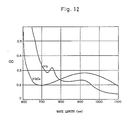
- the light absorption spectra of HbO2 and Hb present an isobestic point in the vicinity of 800 nm as shown in Fig. 12.
- the molecular absorption coefficient of HbO2 is greater than the molecular absorption coefficient of Hb, while in the area of wavelength shorter than 800 nm, the molecular absorption coefficient of Hb is greater than the molecular absorption coefficient of HbO2.
- the component ratio of HbO2 to Hb i.e., the degree of oxygen saturation of blood may be acquired regardless of the amount of measured blood.
- the blood condition may be acquired with better precision (See Japanese Patent Publication No. 11614/1983).
- the end surfaces of two condensing fibers respectively come in contact with two points of the surface of a living body, and the reflected light or transmitted light of the light irradiated into the living body from one condensing fiber is received by the other condensing fiber.
- the light thus received is detected and converted, by a photosensor, into an electric signal, which is then subjected to a processing.
- This method presents the following problems.
- the method of measuring the inside information of substance with the use of light scattering in accordance with the present invention comprises the steps of: successively irradiating lights at a plurality of irradiation points which are disposed substantially in the same direction with respect to a light receiving point on the surface of substance, the substantial optical diffusion paths from the irradiation points to the light receiving point being different in length from one another; receiving the irradiated lights at the light receiving point; executing a predetermined calculation based on the difference of light reception data corresponding to the respective irradiations; and measuring the inside information of the substance.
- the apparatus for measuring the inside information of substance with the use of light scattering comprises: light irradiating means for irradiating lights at a plurality of irradiation points disposed substantially in the same direction with respect to a light receiving point on the surface of substance, the substantial optical diffusion paths from the irradiation points to the light receiving point being different in length from one another; light receiving means for receiving the irradiated lights at the light receiving point; and data processing means for executing a predetermined calculation based on the difference of light reception data, thereby to supply the inside information of the substance.
- the positional relationship of the light receiving point and the irradiation points may be reverse. More specifically, light may be irradiated from an irradiation point on the surface of substance, and the light may be received at a plurality of light receiving points disposed substantially in the same direction with respect to the irradiation point, the substantial optical diffusion paths from the irradiation point to the light receiving points being different in length from one another.
- the term "substantial optical diffusion path” refers to the locus of the center points or average points of the energy distributions of a luminous flux when a number of lights (photons) advance from a fixed point in substance or on the surface thereof and reach another fixed point while scattering and diffusing in the substance.
- the expression “the substantial optical diffusion paths being different in length from one another” refers the state where the substantial optical diffusion paths between the irradiation points and the light receiving point are different in length from one another, whether the physical distances therebetween are the same, or different from one another.
- lights are successively irradiated at a plurality of irradiation points disposed substantially in the same direction with respect to the light receiving point on the surface of substance, the substantial optical diffusion paths to the light receiving point being different in length from one another, and the irradiated lights are received at the light receiving point.
- the light reception data at the light receiving point are different from one another due to the difference of the substantial optical diffusion paths between the irradiation points and the light receiving point.
- the substantial optical diffusion paths between the irradiation points and the light receiving points are common for the most part thereof, but they become different in length in the vicinity of the irradiation points, since the irradiation points are disposed substantially in the same direction with respect to the light receiving point. Accordingly, by obtaining the difference of light reception data, there may be measured the inside information of the substance based on the difference in length of the optical diffusion paths. In other words, the inside information of the substance in the most part of the optical diffusion paths are cancelled by one another and only the inside information of the substance in the vicinity of the irradiation points may be taken out and measured.
- the method of and apparatus for measuring the inside information of substance with the use of light scattering of the present invention may measure the inside information of substance with the use of the difference of light reception data generated by the difference in length of the substantial optical diffusion paths between a single light receiving point and a plurality of irradiation points, or between a plurality of light receiving points and a single irradiation point. Accordingly, there may be obtained reliable measured data of inside information with good reproducibility even though measuring conditions of the substance such as the parts to be measured are different from one another.
- Fig. 1 is a view illustrating a basic embodiment of the present invention.
- Fig. 1 there are disposed projectors 2a, 2b and a light receiver 3 for receiving the lights passing through a living body after irradiated from the projectors 2a, 2b, the pro jectors 2a, 2b and the receiver 3 facing the interface 1 of tissue of a living body.
- the irradiation points A, B of the projectors 2a, 2b are disposed substantially in the same direction with respect to the light receiving point C of the light receiver 3 such that the irradiation point B than the irradiation point A, is closer to the light receiving point C.
- the distances between the light receiving point C and each of the irradiation points A, B are properly set such that the sensitivities of the lights received by the light receiver 3 are detected and influences of the reflected light from the interface 1 of the living body tissue are disregarded. More specifically, if the light receiving point C is excessively separated from the irradiation points A, B, the intensities of the lights entering into the light receiver 3 from the projectors 2a, 2b are weakened to deteriorate the measurement SN ratio.
- the directivities of the projectors 2a, 2b and the light receiver 3 are set as narrow as possible. However, if the directivities are too narrow, a major portion of the lights irradiated from the projectors 2a, 2b enters into the inside of the living body. This prevents a sufficient amount of light from reaching the light receiver 3.
- the distance AB is shorter than the distance AC between the light receiving point C and the irradiation point A or the distance BC between the light receiving point C and the irradiation point B. If the distance AB is substantially equal to or longer than the distance AC or the distance BC, the result of measurement of the inside information of a living body between the irradiation points A and B is apt to contain an error, as will be discussed later, and no reproducibility of measurements may be assured.
- the projector 2a comprises (i) a projector body 22a incorporating a light source such as a semiconductor laser, an LED or the like, and (ii) a light guiding member (for example, lens) 21a for guiding the light projected from the light source.
- the projector 2b comprises a projector body 22b and a light guiding member 21b.
- the light receiver 3 comprises a light receiver body 32 incorporating a photodetector such as a phototransistor or the like, and a light guiding member 31 for guiding the light received from the surface 1, to the photodetector.
- condensing fibers or the like may be used as the light guiding members 21a, 21b and 31.
- the light guiding members 21a, 21b, 31 face the interface 1 of the living body tissue with gaps provided therebetween (See Fig. 2).
- the light guiding members 21a, 21b, 31 may come in contact with the interface 1 of the living body tissue.
- the illumination lights enter into the inside of the interface 1. Portions of these lights are transmitted in the inside of the living body, and reach the light receiver 3. More specifically, that portion of the light projected from the projector 2a which may be detected at the light receiving point C, advances while scattering and diffusing inside of the living body.
- the optical diffusion path where this light advances while diffusing, is shown by a hatched area Ra in Fig. 2.
- a path pa represents an optical path where a major light portion passes, i.e., an optical path connecting the centers of the entire luminous flux which diffusively advances (which path pa is substantially an optical diffusion path and is hereinafter simply referred to an optical diffusion path).
- the irradiation points A, B of the projectors 2a, 2b are disposed substantially in the same direction with respect to the light receiving point C of the light receiver 3, and the distance AB is shorter than the distance AC or the distance BC. Accordingly, the optical diffusion path pa of the light which reaches the light receiver 3 after projected from the projector 2a, is mostly identical with the optical diffusion path pb of the light which reaches the light receiver 3 after projected from the projector 2b. However, as compared with the optical diffusion path pb , the optical diffusion path pa additionally has a path corresponding to the distance AB.
- the light which is projected from the projector 2a and reaches the light receiver 3 additionally contains the inside information of the living body between the irradiation points A and B, particularly the inside information in the vicinity of the surface of the living body. Accordingly, when the light which is projected from the projector 2a and reaches the light receiver 3, is detected and entered into the data processing circuit 5, and the light which is projected from the projector 2b and reaches the light receiver 3, is detected and entered into the data processing circuit 5, and a predetermined calculation is then made based on the difference between both detection signals, the inside information data of the living body between the irradiation points A, B may be obtained.
- Fig. 3 is a double logarithmic graph illustivelyrating the relationship between a light reception amount (the number of counts by a photomultiplier) P and the distance L between a projector and a light receiver at a predetermined wavelength ⁇ 1.
- the light reception amount P presents an expansion in the graph. It is considered that such an expansion is caused by the influence of light reflected from the tissue surface, or the like.
- the graph presents a substantially linear line. Accordingly, two points L1 and L2 exceeding the predetermined distance L0 are selected and light reception amounts P11 and P21 at these points are measured.
- the ratios a1/a2 and a3/a2 may be obtained.
- the Hb concentration cr and the HbO2 concentration co may be obtained from the equations (16) and (17). Then, the degree of oxygen saturation may be obtained.
- the light irradiated from the irradiation point expands as if diffusing while repeatedly reflected and refracted, and reaches the light receiving point. It is therefore considered that the actual optical path length is longer than the apparent optical path length. Further, since the living body presents different absorptivities at different wavelengths, the optical paths differ in length dependent on wavelengths.
- the present invention is not limited to the embodiment above-mentioned.
- lights are successively irradiated at a plurality of irradiation points on the surface of substance and the irradiated lights are received at one light receiving point.
- arrangement may be made such that light is irradiated at one point on the surface of substance and the light is received at a plurality of light receiving points.
- Fig. 4 shows such an arrangement in which light irradiated by a single projector 2 is incident upon light receivers 3a, 3b at the same time.
- detection signals of the light receivers 3a, 3b are subjected to the calculation processing as above-mentioned, there may be taken data containing the inside information of a living body between the light receiving points A and B.
- the present invention may provide apparatus for measuring the inside information of a part of a living body 1 between the opposite surfaces 1a and 1b thereof.
- projectors 2a, 2b (of which light guiding members are incorporated in the casings of the projectors 2a, 2b) are disposed as facing the surface 1a of a living body, and a light receiver 3 (of which light guiding member is incorporated in the casing of the light receiver 3) is disposed as facing the opposite surface 1b of the living body, the light receiver 3 being adapted to receive the lights which have passed through the living body after irradiated from the projectors 2a, 2b.
- the distance AB between the irradiation points A and B of the projectors 2a and 2b is much smaller than the thickness of the living body.
- the optical diffusion path pa and the optical diffusion path pb considerably overlap each other in the inside of the living body and, in particular, in the vicinity of the light receiver 3.
- the optical diffusion paths pa , pb less overlap each other.
- This embodiment is advantageous in that the information of an inner part of substance with respect to the surface thereof is obtained and the influence of light reflected and scattered from the surface is completely disregarded. Accordingly, the directivities of the projectors 2a, 2b and the light receiver 3 may be sharpened and the light amount may be increased. Thus, a thick sample may be measured with good reproducibility.
- the degree of oxygen saturation of hemoglobin in blood is measured.
- the present invention is not limited to such an application. That is, other substance may be measured with the wavelength suitably changed. For example, there may be measured the concentration of dissolved carbon monoxide, carbon dioxide or the like.
- the embodiments above-mentioned show the arrangements in which two irradiation points or two light receiving points are disposed. However, the present invention is not limited to such arrangements, but may be applied to an arrangement having three or more measuring points to improve the measurement precision.
- a living body is measured.
- the present inveniton may be applied to measurement of the inside information (type, disease or the like) of food such as fruit, meat, seafood, etc., or plant such as seeds, saplings, etc. It should therefore be understood that a variety of modifications of the present invention may be made without departing from the scope thereof.
- a semiconductor laser having an output of 0.8 mW and a wavelength of 780 nm (isobestic point)
- a photomultiplier having a condensing fiber.
- the light irradiation portion of the projector was secured to the surface of an arm of a human being, and the tip of the condensing fiber was applied to another portion of the same arm.
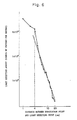
- Fig. 6 is a graph in which the light reception amounts (the counted number of photons per second) weres plotted while moving the tip of the condensing fiber. It is understood from Fig. 6 that, when the distance between the irradiation point and the light receiving point is not greater than about 4 cm, the light reception amount presents an expansion which means that the influence of light reflected from the arm surface is exerted. It is therefore required that the distance between the irradiation point and the light receiving point is at least 3 cm or more and, preferably, 4 cm or more. If the distance above-mentioned is excessively longer, the light reception amount is reduced and the influence of noise or the like of the apparatus is exerted. It is therefore preferable to set such a distance less than about 6 cm.
- Example 2 Two small-size LEDs applied to the same arm of a human being were alternately lighted at predetermined intervals.
- a photomultiplier having a condensing fiber was used as the light receiver, and the tip of the condensing fiber was secured as separated by about 4 cm from the contact points of the small-size LEDs. Data were taken while the distance between two LEDs was changed from 0.3 cm to 1 cm. When the distance between the LEDs was about 1 cm, the reproducibility of measurement was lowered under the influence of a variety of factors such as capillary vessels or the like existing between the LED irradiation points. When the distance was in a range from about 0.3 to about 0.5 cm, data excellent in reproducibility were obtained.
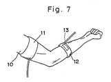
- an arm 10 was placed on a table in a quiet manner, a tourniquet 11 attached to a tonometer was put on the upper arm portion and an arm band 12 incorporating a measuring device 13 (to be discussed later) was put on and secured to the front arm portion with the measuring unit of the measuring device 13 applied to the inner side of the arm 10. Care was taken such that the venous blood vessels of the arm 10 do not exist between the projector and the light receiver.
- the measuring device 13 included one light receiver and nine LEDs.
- the LEDs were disposed as separated by about 0.5 cm from one another, and as separated by about 4 cm from the light receiver. Each LED was lighted for 10 msec. and came ON in order. Each lightening cycle was equal to about 100 msec. After repetition of 50 lightening cycles, i.e., 5 seconds, one data was obtained. There were applied a method of obtaining the molecular absorption coefficients based on the logarithm of the light reception amount ratio above-mentioned, and a method of obtaining the molecular absorption coefficients based on the difference between the light reception amounts. Data were respectively obtained by these methods.
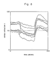
- Figs. 8 and 9 show the results of the measurements above-mentioned.
- Fig. 8 is a graph showing the results of the measurements made by the method of obtaining the molecular absorption coefficients based on the logarithm of the light reception amount ratio
- Fig. 9 is a graph showing the results of the measurements made by the method of obtaining the molecular absorption coefficients based on the difference of the light reception amounts.
- Each of Figs. 8 and 9 shows eight turned lines which respectively correspond to the data of 8 persons.
- the degree of oxygen saturation is once lowered after the bleeding has been arrested (after 50 se conds), and then abruptly increased after the arrest of bleeding has been released (after 125 seconds). Such a trend commonly appears on the data of all the persons even though there are some differences among the individuals. Accordingly, it may be considered that the degrees of oxygen saturation were measured with good reproducibility.
- Measurements were made on the same person at the same measuring point thereof (the arm inner side) with no bleeding arrested, twice every day at the same times (10 o'clock and 16 o'clock) in the morning and in the afternoon, with the use of the measuring device 13 mentioned earlier. Such measurements were made for 6 days. At each measurement, 30 data were obtained, of which 20 data were plotted.
- Figs. 10 and 11 The measurement results are shown in Figs. 10 and 11, in each of which total 12 turned lines respectively show the data of two measurements of six days. It is apparent from Figs. 10 and 11 that the degrees of oxygen saturation measured in the morning are substantially the same, and the degrees of oxygen saturation measured in the afternoon are also substantially the same. Further, it is apparent from Figs. 10 and 11 that the degrees of oxygen saturation in the morning are considerably different from those in the afternoon. Thus, the variations of the measurement results are small to prove the reliability of the data.
Landscapes
- Physics & Mathematics (AREA)
- Health & Medical Sciences (AREA)
- Life Sciences & Earth Sciences (AREA)
- Pathology (AREA)
- General Health & Medical Sciences (AREA)
- Surgery (AREA)
- Spectroscopy & Molecular Physics (AREA)
- Engineering & Computer Science (AREA)
- Biomedical Technology (AREA)
- Heart & Thoracic Surgery (AREA)
- Medical Informatics (AREA)
- Molecular Biology (AREA)
- Optics & Photonics (AREA)
- Animal Behavior & Ethology (AREA)
- Biophysics (AREA)
- Public Health (AREA)
- Veterinary Medicine (AREA)
- Chemical & Material Sciences (AREA)
- Analytical Chemistry (AREA)
- Biochemistry (AREA)
- General Physics & Mathematics (AREA)
- Immunology (AREA)
- Measurement Of The Respiration, Hearing Ability, Form, And Blood Characteristics Of Living Organisms (AREA)
- Investigating Or Analysing Materials By Optical Means (AREA)
- Measuring And Recording Apparatus For Diagnosis (AREA)
Applications Claiming Priority (2)
| Application Number | Priority Date | Filing Date | Title |
|---|---|---|---|
| JP320083/88 | 1988-12-19 | ||
| JP63320083A JPH06103257B2 (ja) | 1988-12-19 | 1988-12-19 | 光散乱を用いた物質の吸光係数測定方法および装置 |
Publications (2)
| Publication Number | Publication Date |
|---|---|
| EP0374844A1 true EP0374844A1 (fr) | 1990-06-27 |
| EP0374844B1 EP0374844B1 (fr) | 1995-08-23 |
Family
ID=18117530
Family Applications (1)
| Application Number | Title | Priority Date | Filing Date |
|---|---|---|---|
| EP89123472A Expired - Lifetime EP0374844B1 (fr) | 1988-12-19 | 1989-12-19 | Méthode et appareil de mesure des caractéristiques d'une substance par dispersion de lumière |
Country Status (4)
| Country | Link |
|---|---|
| US (1) | US5057695A (fr) |
| EP (1) | EP0374844B1 (fr) |
| JP (1) | JPH06103257B2 (fr) |
| DE (1) | DE68923941T2 (fr) |
Cited By (11)
| Publication number | Priority date | Publication date | Assignee | Title |
|---|---|---|---|---|
| WO1995012349A1 (fr) * | 1993-11-05 | 1995-05-11 | Aarnoudse, Jan, Gerard | Mesure optique non invasive in vivo des proprietes d'un constituant de l'organisme humain ou animal |
| WO1996022728A1 (fr) * | 1995-01-27 | 1996-08-01 | Johnson & Johnson Medical Inc. | Capteur medical non invasif |
| WO1996028085A1 (fr) * | 1995-03-14 | 1996-09-19 | Nellcor Puritan Bennett Incorporated | Oxymetrie de couche isolee par impulsions |
| DE19640807A1 (de) * | 1996-10-02 | 1997-09-18 | Siemens Ag | Verfahren und Vorrichtung zur nichtinvasiven optischen Erfassung der Sauerstoffversorgung eines Patienten |
| EP0796057A4 (fr) * | 1994-12-02 | 1999-11-03 | Non Invasive Technology Inc | Techniques optiques d'examen de tissus biologiques |
| US6223069B1 (en) * | 1996-08-29 | 2001-04-24 | Pulsion Medical Systems Ag | Process and device for non-invasively determining cerebral blood flow by near-infrared spectroscopy |
| DE4303047B4 (de) * | 1993-02-03 | 2004-03-25 | Bilz, Dietrich, Dr. | Verfahren zur Untersuchung mehrdimensionaler inhomogener Strukturen |
| EP1598003A3 (fr) * | 1998-08-13 | 2006-03-01 | Whitland Research Limited | Dispositif optique |
| US7774037B2 (en) | 1998-07-04 | 2010-08-10 | Whitland Research Limited | Non-invasive measurement of blood analytes |
| US8483789B2 (en) | 2005-09-13 | 2013-07-09 | Edwards Lifesciences Corporation | Continuous spectroscopic measurement of total hemoglobin |
| WO2015165885A1 (fr) * | 2014-04-28 | 2015-11-05 | Sintef Tto As | Mesure de propriétés d'un matériau organique |
Families Citing this family (132)
| Publication number | Priority date | Publication date | Assignee | Title |
|---|---|---|---|---|
| US5873821A (en) * | 1992-05-18 | 1999-02-23 | Non-Invasive Technology, Inc. | Lateralization spectrophotometer |
| US5782755A (en) * | 1993-11-15 | 1998-07-21 | Non-Invasive Technology, Inc. | Monitoring one or more solutes in a biological system using optical techniques |
| US5792051A (en) * | 1988-12-21 | 1998-08-11 | Non-Invasive Technology, Inc. | Optical probe for non-invasive monitoring of neural activity |
| DE69029152T2 (de) * | 1990-02-15 | 1997-03-06 | Hewlett Packard Gmbh | Verfahren zur nichtinvasiven Messung der Sauerstoffsättigung |
| US5419321A (en) * | 1990-05-17 | 1995-05-30 | Johnson & Johnson Professional Products Limited | Non-invasive medical sensor |
| US6246894B1 (en) | 1993-02-01 | 2001-06-12 | In-Line Diagnostics Corporation | System and method for measuring blood urea nitrogen, blood osmolarity, plasma free hemoglobin and tissue water content |
| US6681128B2 (en) | 1990-10-06 | 2004-01-20 | Hema Metrics, Inc. | System for noninvasive hematocrit monitoring |
| US6725072B2 (en) | 1990-10-06 | 2004-04-20 | Hema Metrics, Inc. | Sensor for transcutaneous measurement of vascular access blood flow |
| US6266546B1 (en) | 1990-10-06 | 2001-07-24 | In-Line Diagnostics Corporation | System for noninvasive hematocrit monitoring |
| US7142307B1 (en) * | 1991-03-01 | 2006-11-28 | Stark Edward W | Method and apparatus for optical interactance and transmittance measurements |
| US5632272A (en) * | 1991-03-07 | 1997-05-27 | Masimo Corporation | Signal processing apparatus |
| US5490505A (en) * | 1991-03-07 | 1996-02-13 | Masimo Corporation | Signal processing apparatus |
| EP1357481A3 (fr) | 1991-03-07 | 2005-04-27 | Masimo Corporation | Appareil et procédé de traitement de signal |
| MX9702434A (es) | 1991-03-07 | 1998-05-31 | Masimo Corp | Aparato de procesamiento de señales. |
| US6549795B1 (en) | 1991-05-16 | 2003-04-15 | Non-Invasive Technology, Inc. | Spectrophotometer for tissue examination |
| DE4218321A1 (de) * | 1991-12-09 | 1993-06-17 | Siemens Ag | Diagnostikanlage |
| US5764209A (en) * | 1992-03-16 | 1998-06-09 | Photon Dynamics, Inc. | Flat panel display inspection system |
| US6785568B2 (en) | 1992-05-18 | 2004-08-31 | Non-Invasive Technology Inc. | Transcranial examination of the brain |
| US5954053A (en) * | 1995-06-06 | 1999-09-21 | Non-Invasive Technology, Inc. | Detection of brain hematoma |
| US5762609A (en) * | 1992-09-14 | 1998-06-09 | Sextant Medical Corporation | Device and method for analysis of surgical tissue interventions |
| US5772597A (en) * | 1992-09-14 | 1998-06-30 | Sextant Medical Corporation | Surgical tool end effector |
| IL107396A (en) * | 1992-11-09 | 1997-02-18 | Boehringer Mannheim Gmbh | Method and apparatus for analytical determination of glucose in a biological matrix |
| AU7080594A (en) * | 1993-05-28 | 1994-12-20 | Somanetics Corporation | Method and apparatus for spectrophotometric cerebral oximetry |
| JP3433498B2 (ja) * | 1993-06-02 | 2003-08-04 | 浜松ホトニクス株式会社 | 散乱吸収体の内部情報計測方法及び装置 |
| CA2174719C (fr) | 1993-08-24 | 2005-07-26 | Mark R. Robinson | Systeme de mesure, non invasif, precis et robuste, pour substance a analyser |
| US7376453B1 (en) | 1993-10-06 | 2008-05-20 | Masimo Corporation | Signal processing apparatus |
| ZA948393B (en) * | 1993-11-01 | 1995-06-26 | Polartechnics Ltd | Method and apparatus for tissue type recognition |
| DE4337570A1 (de) * | 1993-11-04 | 1995-05-11 | Boehringer Mannheim Gmbh | Verfahren zur Analyse von Glucose in einer biologischen Matrix |
| US6493565B1 (en) | 1993-11-15 | 2002-12-10 | Non-Invasive Technology, Inc. | Examination of biological tissue by monitoring one or more solutes |
| JP3433508B2 (ja) * | 1993-12-01 | 2003-08-04 | 浜松ホトニクス株式会社 | 散乱吸収体計測方法及び散乱吸収体計測装置 |
| US5497769A (en) * | 1993-12-16 | 1996-03-12 | I.S.S. (Usa) Inc. | Photosensor with multiple light sources |
| US5492118A (en) | 1993-12-16 | 1996-02-20 | Board Of Trustees Of The University Of Illinois | Determining material concentrations in tissues |
| US5632273A (en) * | 1994-02-04 | 1997-05-27 | Hamamatsu Photonics K.K. | Method and means for measurement of biochemical components |
| US5490506A (en) * | 1994-03-28 | 1996-02-13 | Colin Corporation | Peripheral blood flow evaluating apparatus |
| DE19512478C2 (de) * | 1994-08-10 | 2001-05-31 | Bernreuter Peter | Verfahren zur Bestimmung der arteriellen Sauerstoffsättigung |
| US8019400B2 (en) | 1994-10-07 | 2011-09-13 | Masimo Corporation | Signal processing apparatus |
| EP1905352B1 (fr) | 1994-10-07 | 2014-07-16 | Masimo Corporation | Méthode de traitement de signal |
| US5513642A (en) * | 1994-10-12 | 1996-05-07 | Rensselaer Polytechnic Institute | Reflectance sensor system |
| US5697367A (en) * | 1994-10-14 | 1997-12-16 | Somanetics Corporation | Specially grounded sensor for clinical spectrophotometric procedures |
| US6957094B2 (en) * | 1994-12-02 | 2005-10-18 | Non-Invasive Technology, Inc. | Examination of scattering properties of biological tissue |
| DE4445683A1 (de) * | 1994-12-21 | 1996-06-27 | Boehringer Mannheim Gmbh | Verfahren zur Untersuchung eines streuenden Mediums mit intensitätsmoduliertem Licht |
| US5853364A (en) * | 1995-08-07 | 1998-12-29 | Nellcor Puritan Bennett, Inc. | Method and apparatus for estimating physiological parameters using model-based adaptive filtering |
| DE19543020A1 (de) * | 1995-11-18 | 1997-05-22 | Boehringer Mannheim Gmbh | Verfahren und Vorrichtung zur Bestimmung von analytischen Daten über das Innere einer streuenden Matrix |
| WO1998007364A1 (fr) | 1996-08-16 | 1998-02-26 | Roche Diagnostics Gmbh | Systeme de controle pour la prise reguliere d'un medicament |
| US6117099A (en) | 1996-10-23 | 2000-09-12 | In-Line Diagnostics Corporation | System and method for noninvasive hemodynamic measurements in hemodialysis shunts |
| US5891024A (en) * | 1997-04-09 | 1999-04-06 | Ohmeda Inc. | Two stage calibration and analyte measurement scheme for spectrophotomeric analysis |
| US6002952A (en) | 1997-04-14 | 1999-12-14 | Masimo Corporation | Signal processing apparatus and method |
| WO1999063883A1 (fr) | 1998-06-11 | 1999-12-16 | S.P.O. Medical Equipment Ltd. | Dispositif de detection de stress physiologique et procede correspondant |
| IL121079A0 (en) | 1997-06-15 | 1997-11-20 | Spo Medical Equipment Ltd | Physiological stress detector device and method |
| US6804543B2 (en) | 1998-02-05 | 2004-10-12 | Hema Metrics, Inc. | Sensor for transcutaneous measurement of vascular access blood flow |
| JP4733264B2 (ja) * | 1998-02-11 | 2011-07-27 | ノン−インヴェイシヴ テクノロジイ,インク. | 胸部腫瘍の検出、画像形成および特徴表示 |
| WO1999040841A1 (fr) * | 1998-02-11 | 1999-08-19 | Non-Invasive Technology, Inc. | Visualisation et caracterisation du tissu cerebral |
| US20070167704A1 (en) * | 1998-02-13 | 2007-07-19 | Britton Chance | Transabdominal examination, monitoring and imaging of tissue |
| JP2002502654A (ja) * | 1998-02-13 | 2002-01-29 | ノン−インヴェイシヴ テクノロジイ,インク. | 組織の腹部横断検査、監視および画像形成 |
| US6078833A (en) * | 1998-03-25 | 2000-06-20 | I.S.S. (Usa) Inc. | Self referencing photosensor |
| US6526298B1 (en) | 1998-05-18 | 2003-02-25 | Abbott Laboratories | Method for the non-invasive determination of analytes in a selected volume of tissue |
| US6662031B1 (en) | 1998-05-18 | 2003-12-09 | Abbott Laboratoies | Method and device for the noninvasive determination of hemoglobin and hematocrit |
| US6241663B1 (en) | 1998-05-18 | 2001-06-05 | Abbott Laboratories | Method for improving non-invasive determination of the concentration of analytes in a biological sample |
| US6662030B2 (en) | 1998-05-18 | 2003-12-09 | Abbott Laboratories | Non-invasive sensor having controllable temperature feature |
| US7043287B1 (en) | 1998-05-18 | 2006-05-09 | Abbott Laboratories | Method for modulating light penetration depth in tissue and diagnostic applications using same |
| US6353226B1 (en) | 1998-11-23 | 2002-03-05 | Abbott Laboratories | Non-invasive sensor capable of determining optical parameters in a sample having multiple layers |
| US6615061B1 (en) | 1998-11-23 | 2003-09-02 | Abbott Laboratories | Optical sensor having a selectable sampling distance for determination of analytes |
| US7047054B2 (en) * | 1999-03-12 | 2006-05-16 | Cas Medical Systems, Inc. | Laser diode optical transducer assembly for non-invasive spectrophotometric blood oxygenation monitoring |
| US6175751B1 (en) * | 1999-03-16 | 2001-01-16 | Allen Maizes | Apparatus and method for sensing oxygen levels in a fetus |
| US6587704B1 (en) * | 1999-06-16 | 2003-07-01 | Orsense Ltd. | Method for non-invasive optical measurements of blood parameters |
| US7840257B2 (en) * | 2003-01-04 | 2010-11-23 | Non Invasive Technology, Inc. | Examination of biological tissue using non-contact optical probes |
| US7904139B2 (en) | 1999-08-26 | 2011-03-08 | Non-Invasive Technology Inc. | Optical examination of biological tissue using non-contact irradiation and detection |
| EP2308374B1 (fr) | 1999-12-22 | 2012-07-18 | Orsense Ltd. | Procédé de mesures optiques pour définir plusieurs paramètres dans le sang d'un patient |
| US6577884B1 (en) | 2000-06-19 | 2003-06-10 | The General Hospital Corporation | Detection of stroke events using diffuse optical tomagraphy |
| DE60111531T2 (de) * | 2000-07-21 | 2006-05-11 | Eidgenössische Technische Hochschule Zürich | Sonde und vorichtung zur messung zerebraler hämodynamik und sauerstoffsättigung |
| CA2417917C (fr) * | 2000-08-04 | 2014-10-14 | Photonify Technologies, Inc. | Systemes et procedes permettant d'obtenir des informations concernant des chromophores presents dans des milieux physiologiques |
| US6746407B2 (en) * | 2000-12-29 | 2004-06-08 | Hema Metrics, Inc. | Method of measuring transcutaneous access blood flow |
| US7657292B2 (en) * | 2001-03-16 | 2010-02-02 | Nellcor Puritan Bennett Llc | Method for evaluating extracellular water concentration in tissue |
| US7239902B2 (en) * | 2001-03-16 | 2007-07-03 | Nellor Puritan Bennett Incorporated | Device and method for monitoring body fluid and electrolyte disorders |
| US6591122B2 (en) | 2001-03-16 | 2003-07-08 | Nellcor Puritan Bennett Incorporated | Device and method for monitoring body fluid and electrolyte disorders |
| US8135448B2 (en) * | 2001-03-16 | 2012-03-13 | Nellcor Puritan Bennett Llc | Systems and methods to assess one or more body fluid metrics |
| US6606509B2 (en) | 2001-03-16 | 2003-08-12 | Nellcor Puritan Bennett Incorporated | Method and apparatus for improving the accuracy of noninvasive hematocrit measurements |
| US7167734B2 (en) | 2001-04-13 | 2007-01-23 | Abbott Laboratories | Method for optical measurements of tissue to determine disease state or concentration of an analyte |
| JP2003202287A (ja) * | 2002-01-08 | 2003-07-18 | Hamamatsu Photonics Kk | 散乱吸収体測定方法及び装置 |
| CN1268286C (zh) * | 2002-01-25 | 2006-08-09 | 松下电器产业株式会社 | 光学式生物信息测量方法以及光学式生物信息测量装置 |
| US7072701B2 (en) | 2002-07-26 | 2006-07-04 | Cas Medical Systems, Inc. | Method for spectrophotometric blood oxygenation monitoring |
| US20040092829A1 (en) * | 2002-11-07 | 2004-05-13 | Simon Furnish | Spectroscope with modified field-of-view |
| US7336358B2 (en) * | 2003-02-24 | 2008-02-26 | August Ninth Analyses, Inc. | Method and apparatus for determining particle size and composition of mixtures |
| US6993372B2 (en) * | 2003-06-03 | 2006-01-31 | Orsense Ltd. | Method and system for use in non-invasive optical measurements of blood parameters |
| US7426410B2 (en) * | 2003-06-06 | 2008-09-16 | Infraredx, Inc. | Spectroscopy of deeply-scattered light |
| US7194293B2 (en) | 2004-03-08 | 2007-03-20 | Nellcor Puritan Bennett Incorporated | Selection of ensemble averaging weights for a pulse oximeter based on signal quality metrics |
| US7277741B2 (en) | 2004-03-09 | 2007-10-02 | Nellcor Puritan Bennett Incorporated | Pulse oximetry motion artifact rejection using near infrared absorption by water |
| US7313425B2 (en) * | 2004-07-08 | 2007-12-25 | Orsense Ltd. | Device and method for non-invasive optical measurements |
| CN100522059C (zh) | 2004-10-15 | 2009-08-05 | 日本国长崎县政府 | 血糖值的非侵入测量装置 |
| US8055321B2 (en) | 2005-03-14 | 2011-11-08 | Peter Bernreuter | Tissue oximetry apparatus and method |
| US7865223B1 (en) | 2005-03-14 | 2011-01-04 | Peter Bernreuter | In vivo blood spectrometry |
| US7616303B2 (en) * | 2005-04-25 | 2009-11-10 | University Of Massachusetts | Systems and methods for correcting optical reflectance measurements |
| JP4647447B2 (ja) * | 2005-09-20 | 2011-03-09 | 富士フイルム株式会社 | 試料分析装置 |
| JP4662831B2 (ja) * | 2005-09-20 | 2011-03-30 | 富士フイルム株式会社 | 試料分析装置 |
| KR100792259B1 (ko) * | 2006-05-19 | 2008-01-07 | 삼성전자주식회사 | 휴대용 체지방 측정 장치 및 상기 장치에 장착되는 광센서모듈 |
| US8255025B2 (en) | 2006-06-09 | 2012-08-28 | Nellcor Puritan Bennett Llc | Bronchial or tracheal tissular water content sensor and system |
| US8396524B2 (en) * | 2006-09-27 | 2013-03-12 | Covidien Lp | Medical sensor and technique for using the same |
| US8180419B2 (en) * | 2006-09-27 | 2012-05-15 | Nellcor Puritan Bennett Llc | Tissue hydration estimation by spectral absorption bandwidth measurement |
| US7643858B2 (en) | 2006-09-28 | 2010-01-05 | Nellcor Puritan Bennett Llc | System and method for detection of brain edema using spectrophotometry |
| US8116852B2 (en) * | 2006-09-29 | 2012-02-14 | Nellcor Puritan Bennett Llc | System and method for detection of skin wounds and compartment syndromes |
| US8280469B2 (en) | 2007-03-09 | 2012-10-02 | Nellcor Puritan Bennett Llc | Method for detection of aberrant tissue spectra |
| US8175665B2 (en) * | 2007-03-09 | 2012-05-08 | Nellcor Puritan Bennett Llc | Method and apparatus for spectroscopic tissue analyte measurement |
| US8109882B2 (en) | 2007-03-09 | 2012-02-07 | Nellcor Puritan Bennett Llc | System and method for venous pulsation detection using near infrared wavelengths |
| US8357090B2 (en) * | 2007-03-09 | 2013-01-22 | Covidien Lp | Method and apparatus for estimating water reserves |
| US8690864B2 (en) | 2007-03-09 | 2014-04-08 | Covidien Lp | System and method for controlling tissue treatment |
| US8346327B2 (en) * | 2007-03-09 | 2013-01-01 | Covidien Lp | Method for identification of sensor site by local skin spectrum data |
| US20080221416A1 (en) * | 2007-03-09 | 2008-09-11 | Nellcor Puritan Bennett Llc | System and method for detection of macular degeneration using spectrophotometry |
| US20080221411A1 (en) * | 2007-03-09 | 2008-09-11 | Nellcor Puritan Bennett Llc | System and method for tissue hydration estimation |
| KR101506177B1 (ko) * | 2008-01-10 | 2015-03-26 | 삼성전자주식회사 | 생체 신호 측정 센서 및 그의 제조 방법 |
| JP5531271B2 (ja) * | 2008-05-10 | 2014-06-25 | 独立行政法人国立高等専門学校機構 | 二枚貝の検査方法及び検査装置 |
| US8406865B2 (en) | 2008-09-30 | 2013-03-26 | Covidien Lp | Bioimpedance system and sensor and technique for using the same |
| WO2010056973A1 (fr) | 2008-11-14 | 2010-05-20 | Nonin Medical, Inc. | Sélection du chemin de capteur optique |
| CN102883658B (zh) * | 2009-11-19 | 2016-06-22 | 调节成像公司 | 用于使用结构化照明经由单元件检测来分析浑浊介质的方法和设备 |
| US9433352B2 (en) * | 2011-02-23 | 2016-09-06 | National University Corporation Shizuoka University | Optical measuring device |
| US20150018646A1 (en) | 2013-07-12 | 2015-01-15 | Sandeep Gulati | Dynamic sample mapping noninvasive analyzer apparatus and method of use thereof |
| US9351672B2 (en) | 2012-07-16 | 2016-05-31 | Timothy Ruchti | Multiplexed pathlength resolved noninvasive analyzer apparatus with stacked filters and method of use thereof |
| US9351671B2 (en) | 2012-07-16 | 2016-05-31 | Timothy Ruchti | Multiplexed pathlength resolved noninvasive analyzer apparatus and method of use thereof |
| US9585604B2 (en) | 2012-07-16 | 2017-03-07 | Zyomed Corp. | Multiplexed pathlength resolved noninvasive analyzer apparatus with dynamic optical paths and method of use thereof |
| JP6148836B2 (ja) * | 2012-09-12 | 2017-06-14 | 東京都下水道サービス株式会社 | ガス濃度測定装置 |
| EP3401678B1 (fr) | 2012-11-07 | 2020-04-08 | Modulated Imaging Inc. | Imagerie modulée efficace |
| JP2015039542A (ja) * | 2013-08-22 | 2015-03-02 | セイコーエプソン株式会社 | 脈波測定装置 |
| US11147518B1 (en) | 2013-10-07 | 2021-10-19 | Masimo Corporation | Regional oximetry signal processor |
| US10010276B2 (en) * | 2013-10-07 | 2018-07-03 | Masimo Corporation | Regional oximetry user interface |
| EP4434457A3 (fr) | 2014-01-07 | 2024-12-18 | Opsolution GmbH | Dispositif et procédé de détermination d'une concentration dans un échantillon |
| JP6630061B2 (ja) * | 2014-05-28 | 2020-01-15 | 天津先陽科技発展有限公司 | 拡散スペクトルデータの処理方法及び処理装置 |
| WO2016054079A1 (fr) | 2014-09-29 | 2016-04-07 | Zyomed Corp. | Systèmes et procédés pour la détection et la mesure du glucose sanguin du sang et d'autres analytes à l'aide du calcul de collision |
| EP3258297B1 (fr) * | 2014-12-08 | 2025-04-16 | Gummi-Welz GmbH u. Co. KG Gummi-Kunststofftechnik-Schaumstoffe | Système de grille lumineuse |
| WO2016103323A1 (fr) * | 2014-12-22 | 2016-06-30 | 株式会社日立製作所 | Dispositif pour mesure optique de corps vivant, dispositif d'analyse et procédé d'analyse |
| JP2017051340A (ja) * | 2015-09-08 | 2017-03-16 | 株式会社東芝 | センサ及びセンサシステム |
| US9554738B1 (en) | 2016-03-30 | 2017-01-31 | Zyomed Corp. | Spectroscopic tomography systems and methods for noninvasive detection and measurement of analytes using collision computing |
| JP2017209413A (ja) * | 2016-05-27 | 2017-11-30 | ソニーモバイルコミュニケーションズ株式会社 | 制御装置、検出装置、および制御方法 |
| IT201700094946A1 (it) * | 2017-08-22 | 2019-02-22 | Bosch Gmbh Robert | Procedimento per l’analisi di un oggetto e dispositivo di analisi previsto per l’analisi di un oggetto |
Citations (4)
| Publication number | Priority date | Publication date | Assignee | Title |
|---|---|---|---|---|
| US3638640A (en) * | 1967-11-01 | 1972-02-01 | Robert F Shaw | Oximeter and method for in vivo determination of oxygen saturation in blood using three or more different wavelengths |
| US4223680A (en) * | 1977-06-28 | 1980-09-23 | Duke University, Inc. | Method and apparatus for monitoring metabolism in body organs in vivo |
| EP0104772A2 (fr) * | 1982-09-02 | 1984-04-04 | Nellcor Incorporated | Capteur optique étalonné pour oxymétrie |
| EP0286142A2 (fr) * | 1987-04-09 | 1988-10-12 | Sumitomo Electric Industries, Ltd. | Oxymètre à réflexion |
Family Cites Families (7)
| Publication number | Priority date | Publication date | Assignee | Title |
|---|---|---|---|---|
| US4621643A (en) * | 1982-09-02 | 1986-11-11 | Nellcor Incorporated | Calibrated optical oximeter probe |
| JPS6072542A (ja) * | 1983-09-28 | 1985-04-24 | 株式会社島津製作所 | 光線ct装置 |
| JPS62124443A (ja) * | 1985-11-26 | 1987-06-05 | Hamamatsu Photonics Kk | 光により物体内部の情報を得る装置 |
| US4714080A (en) * | 1986-10-06 | 1987-12-22 | Nippon Colin Co., Ltd. | Method and apparatus for noninvasive monitoring of arterial blood oxygen saturation |
| DE3723881A1 (de) * | 1987-07-18 | 1989-01-26 | Nicolay Gmbh | Verfahren zum ermitteln der sauerstoffsaettigung des blutes eines lebenden organismus und elektronische schaltung sowie vorrichtung zum durchfuehren dieses verfahrens |
| US4819752A (en) * | 1987-10-02 | 1989-04-11 | Datascope Corp. | Blood constituent measuring device and method |
| US4846183A (en) * | 1987-12-02 | 1989-07-11 | The Boc Group, Inc. | Blood parameter monitoring apparatus and methods |
-
1988
- 1988-12-19 JP JP63320083A patent/JPH06103257B2/ja not_active Expired - Fee Related
-
1989
- 1989-12-15 US US07/450,980 patent/US5057695A/en not_active Expired - Lifetime
- 1989-12-19 DE DE68923941T patent/DE68923941T2/de not_active Expired - Fee Related
- 1989-12-19 EP EP89123472A patent/EP0374844B1/fr not_active Expired - Lifetime
Patent Citations (4)
| Publication number | Priority date | Publication date | Assignee | Title |
|---|---|---|---|---|
| US3638640A (en) * | 1967-11-01 | 1972-02-01 | Robert F Shaw | Oximeter and method for in vivo determination of oxygen saturation in blood using three or more different wavelengths |
| US4223680A (en) * | 1977-06-28 | 1980-09-23 | Duke University, Inc. | Method and apparatus for monitoring metabolism in body organs in vivo |
| EP0104772A2 (fr) * | 1982-09-02 | 1984-04-04 | Nellcor Incorporated | Capteur optique étalonné pour oxymétrie |
| EP0286142A2 (fr) * | 1987-04-09 | 1988-10-12 | Sumitomo Electric Industries, Ltd. | Oxymètre à réflexion |
Cited By (15)
| Publication number | Priority date | Publication date | Assignee | Title |
|---|---|---|---|---|
| DE4303047B4 (de) * | 1993-02-03 | 2004-03-25 | Bilz, Dietrich, Dr. | Verfahren zur Untersuchung mehrdimensionaler inhomogener Strukturen |
| WO1995012349A1 (fr) * | 1993-11-05 | 1995-05-11 | Aarnoudse, Jan, Gerard | Mesure optique non invasive in vivo des proprietes d'un constituant de l'organisme humain ou animal |
| EP0796057A4 (fr) * | 1994-12-02 | 1999-11-03 | Non Invasive Technology Inc | Techniques optiques d'examen de tissus biologiques |
| WO1996022728A1 (fr) * | 1995-01-27 | 1996-08-01 | Johnson & Johnson Medical Inc. | Capteur medical non invasif |
| WO1996028085A1 (fr) * | 1995-03-14 | 1996-09-19 | Nellcor Puritan Bennett Incorporated | Oxymetrie de couche isolee par impulsions |
| US6223069B1 (en) * | 1996-08-29 | 2001-04-24 | Pulsion Medical Systems Ag | Process and device for non-invasively determining cerebral blood flow by near-infrared spectroscopy |
| DE19640807A1 (de) * | 1996-10-02 | 1997-09-18 | Siemens Ag | Verfahren und Vorrichtung zur nichtinvasiven optischen Erfassung der Sauerstoffversorgung eines Patienten |
| US7774037B2 (en) | 1998-07-04 | 2010-08-10 | Whitland Research Limited | Non-invasive measurement of blood analytes |
| EP1598003A3 (fr) * | 1998-08-13 | 2006-03-01 | Whitland Research Limited | Dispositif optique |
| US7221970B2 (en) | 1998-08-13 | 2007-05-22 | Edwards Lifesciences Corporation | Optical device |
| US7236813B2 (en) | 1998-08-13 | 2007-06-26 | Edwards Lifesciences Corporation | Optical device |
| US8483789B2 (en) | 2005-09-13 | 2013-07-09 | Edwards Lifesciences Corporation | Continuous spectroscopic measurement of total hemoglobin |
| WO2015165885A1 (fr) * | 2014-04-28 | 2015-11-05 | Sintef Tto As | Mesure de propriétés d'un matériau organique |
| CN106233121A (zh) * | 2014-04-28 | 2016-12-14 | 辛特福特图有限公司 | 有机材料的性质的测量 |
| CN106233121B (zh) * | 2014-04-28 | 2019-05-21 | 辛特福特图有限公司 | 有机材料的性质的测量 |
Also Published As
| Publication number | Publication date |
|---|---|
| EP0374844B1 (fr) | 1995-08-23 |
| US5057695A (en) | 1991-10-15 |
| JPH06103257B2 (ja) | 1994-12-14 |
| DE68923941D1 (de) | 1995-09-28 |
| JPH02163634A (ja) | 1990-06-22 |
| DE68923941T2 (de) | 1996-03-07 |
Similar Documents
| Publication | Publication Date | Title |
|---|---|---|
| US5057695A (en) | Method of and apparatus for measuring the inside information of substance with the use of light scattering | |
| US6026314A (en) | Method and device for noninvasive measurements of concentrations of blood components | |
| US5529065A (en) | Method for measuring scattering medium and apparatus for the same | |
| US6097975A (en) | Apparatus and method for noninvasive glucose measurement | |
| US5517987A (en) | Method for measuring internal information in scattering medium and apparatus for the same | |
| US5222496A (en) | Infrared glucose sensor | |
| EP1478265B1 (fr) | Spectraphotometrie a impulsion active | |
| US7328052B2 (en) | Near infrared risk assessment of diseases | |
| US5222495A (en) | Non-invasive blood analysis by near infrared absorption measurements using two closely spaced wavelengths | |
| US5803908A (en) | System for noninvasive hematocrit monitoring | |
| CN100522059C (zh) | 血糖值的非侵入测量装置 | |
| US4836206A (en) | Method and device for determining viability of intact teeth | |
| JP2539707B2 (ja) | 吸光スペクトルの補正方法およびその方法を用いた光拡散物質の分光測定装置 | |
| US5640957A (en) | Ultraviolet radiation protection evaluator | |
| US20020038079A1 (en) | System for noninvasive hematocrit monitoring | |
| US20050267346A1 (en) | Non-invasive blood component measurement system | |
| CA2383725A1 (fr) | Procede de determination d'analytes au moyen d'un spectre en proche infrarouge, d'un spectre visible adjacent et de longueurs d'ondes distinctes de spectre en proche infrarouge | |
| JP2004230000A (ja) | 血中吸光物質濃度測定装置 | |
| EP0693900A1 (fr) | Systeme et procede de surveillance non invasive de l'hematocrite | |
| JP2004290544A (ja) | 血液分析装置 | |
| US5246004A (en) | Infrared cholesterol sensor | |
| US9888855B2 (en) | Reflection detection type measurement apparatus and method for skin autofluorescence | |
| US20150201840A1 (en) | Reflection detection type measurement apparatus for skin autofluorescence | |
| US20090177052A1 (en) | Methods of using raman spectral information in determining analyte concentrations | |
| CA2123152A1 (fr) | Determination non invasive de la concentration d'analytes par radiation non continue |
Legal Events
| Date | Code | Title | Description |
|---|---|---|---|
| PUAI | Public reference made under article 153(3) epc to a published international application that has entered the european phase |
Free format text: ORIGINAL CODE: 0009012 |
|
| AK | Designated contracting states |
Kind code of ref document: A1 Designated state(s): DE FR GB |
|
| 17P | Request for examination filed |
Effective date: 19901112 |
|
| 17Q | First examination report despatched |
Effective date: 19930210 |
|
| RBV | Designated contracting states (corrected) |
Designated state(s): DE GB |
|
| GRAA | (expected) grant |
Free format text: ORIGINAL CODE: 0009210 |
|
| AK | Designated contracting states |
Kind code of ref document: B1 Designated state(s): DE GB |
|
| REF | Corresponds to: |
Ref document number: 68923941 Country of ref document: DE Date of ref document: 19950928 |
|
| PLBE | No opposition filed within time limit |
Free format text: ORIGINAL CODE: 0009261 |
|
| STAA | Information on the status of an ep patent application or granted ep patent |
Free format text: STATUS: NO OPPOSITION FILED WITHIN TIME LIMIT |
|
| 26N | No opposition filed | ||
| REG | Reference to a national code |
Ref country code: GB Ref legal event code: IF02 |
|
| PGFP | Annual fee paid to national office [announced via postgrant information from national office to epo] |
Ref country code: GB Payment date: 20031202 Year of fee payment: 15 |
|
| PGFP | Annual fee paid to national office [announced via postgrant information from national office to epo] |
Ref country code: DE Payment date: 20031222 Year of fee payment: 15 |
|
| PG25 | Lapsed in a contracting state [announced via postgrant information from national office to epo] |
Ref country code: GB Free format text: LAPSE BECAUSE OF NON-PAYMENT OF DUE FEES Effective date: 20041219 |
|
| PG25 | Lapsed in a contracting state [announced via postgrant information from national office to epo] |
Ref country code: DE Free format text: LAPSE BECAUSE OF NON-PAYMENT OF DUE FEES Effective date: 20050701 |
|
| GBPC | Gb: european patent ceased through non-payment of renewal fee |
Effective date: 20041219 |