EP0380966A1 - Vorrichtung zur Informationsverwaltung, insbesondere für Bilderspeicher- und Bildkommunikationssysteme - Google Patents
Vorrichtung zur Informationsverwaltung, insbesondere für Bilderspeicher- und Bildkommunikationssysteme Download PDFInfo
- Publication number
- EP0380966A1 EP0380966A1 EP90100928A EP90100928A EP0380966A1 EP 0380966 A1 EP0380966 A1 EP 0380966A1 EP 90100928 A EP90100928 A EP 90100928A EP 90100928 A EP90100928 A EP 90100928A EP 0380966 A1 EP0380966 A1 EP 0380966A1
- Authority
- EP
- European Patent Office
- Prior art keywords
- data
- folder
- patient
- uid
- mass storage
- Prior art date
- Legal status (The legal status is an assumption and is not a legal conclusion. Google has not performed a legal analysis and makes no representation as to the accuracy of the status listed.)
- Withdrawn
Links
- 238000000034 method Methods 0.000 claims description 6
- 238000003384 imaging method Methods 0.000 description 7
- 238000007726 management method Methods 0.000 description 5
- 210000004556 brain Anatomy 0.000 description 1
- 230000006835 compression Effects 0.000 description 1
- 238000007906 compression Methods 0.000 description 1
- 238000002591 computed tomography Methods 0.000 description 1
- 238000013500 data storage Methods 0.000 description 1
- 239000010432 diamond Substances 0.000 description 1
- 230000014759 maintenance of location Effects 0.000 description 1
- 238000009206 nuclear medicine Methods 0.000 description 1
- 230000003287 optical effect Effects 0.000 description 1
- 210000004789 organ system Anatomy 0.000 description 1
- 238000010926 purge Methods 0.000 description 1
- 230000009466 transformation Effects 0.000 description 1
- 238000000844 transformation Methods 0.000 description 1
- 238000002604 ultrasonography Methods 0.000 description 1
Images




Classifications
-
- G—PHYSICS
- G16—INFORMATION AND COMMUNICATION TECHNOLOGY [ICT] SPECIALLY ADAPTED FOR SPECIFIC APPLICATION FIELDS
- G16H—HEALTHCARE INFORMATICS, i.e. INFORMATION AND COMMUNICATION TECHNOLOGY [ICT] SPECIALLY ADAPTED FOR THE HANDLING OR PROCESSING OF MEDICAL OR HEALTHCARE DATA
- G16H30/00—ICT specially adapted for the handling or processing of medical images
- G16H30/20—ICT specially adapted for the handling or processing of medical images for handling medical images, e.g. DICOM, HL7 or PACS
Definitions
- the invention relates to information management, and more particularly relates to information management for medical applications. In its most immediate sense, the invention relates to an information management system for use in a PACS (Picture Archive and Communications System).
- PACS Picture Archive and Communications System
- a hospital or other clinical facility usually utilizes a plurality of imaging modalities.
- a hospital may own X-ray equipment, a magnetic resonance imager, a CT scanner, ultrasound imagers and scintillation cameras. These different modalities are used individually and in combination, so that a patient may, during a particular hospital admission, undergo repeated CT scans, a magnetic resonance study, a nuclear medicine study using a scintillation camera, etc.
- the volume of digital information produced by a fully digital radiology department has been estimated. Based on several hundred thousand images per year, information storage requirements can mount to several Terabytes per year. (A Terabyte equals 1012 bytes.)
- One object of the invention is to provide an information management system which can quickly locate information of interest, even where large quantities (on the order of Terabytes) of information are involved.
- Another object is to provide such a system which can be implemented in a PACS.
- a further object is to provide such a system which can be easily used by doctors and hospital administrators in a clinical environment.
- Still another object is to provide such a system which can satisfy, in a cost-effective matter, legal requirements applicable to the retention of medical information.
- An additional object is to provide such a system which can cope with the open-ended growth which can be expected of data in a PACS.
- an information management system separates the storage of the ultimate data from the storage and manipulation of the pointers which reference that data.
- the data itself (referred to hereinafter as the "data objects") may be stored on one or more mass storage devices but is excluded from a unique database server which has the function of manipulating pointers.
- each data object (e.g. each image, each graph, each field of text, where the invention is implemented in a PACS) is uniquely identified by a data object identifier and, as stored in a mass storage unit or units, the data objects are keyed by data object identifiers.
- the data object identifiers are manipulated in a single database server which only manipulates index data, and the data object identifiers are keyed using a set of data elements (hereinafter referred to as "data object descriptions") which are examined when individual data objects, or groups of data objects, are retrieved.
- data object identifiers are, for example, patient name, modality type, date, etc.
- data is compressed and decompressed in such a manner as to remove data from the database server when the data need not be kept immediately available.
- This is accomplished by using a data structure in which data is stored in compressed form as nested data objects and stored in expanded form as database entities.
- the nesting structure is particularly adapted to the needs of physicians and hospital personnel.
- data is put into a single data format which can reference data objects themselves, or which can reference references to data objects.
- a data object contains pointers to other data objects
- the pointer-containing data object is called a "folder".
- the various folders are designed to have particular utility in a PACS.
- a folder When a folder must be kept immediately available (as where it relates to a patient who is currently hospitalized and whose records must be accessed) it is stored in the database server. When the folder need not be immediately available, it is stored in the form of nested data objects in the mass storage device(s).
- Fig. 1 shows a PACS as it might be installed in a hospital radiology department.
- Image data is acquired by e.g. a magnetic resonance unit 2, a scintillation camera 4 or an X-ray unit 6. This data is stored as described below.
- a physician or administrator wishes to view the medical records of a patient, including text, images or both, the records may be brought up on either of the workstations 8 and 10 and updated as desired.
- Fig. 1 is schematic and neither the number and nature of imaging modalities nor the number and nature of workstations is a part of the invention.
- An active radiology department will produce Terabytes of information per year. In accordance with the invention, most of this is stored on one or more mass storage devices 12. The rest is stored on a single database server 14. The manner in which this is accomplished is described below.
- the data produced by an imaging modality is always stored in the form of data objects on the mass storage device(s) 12.
- references to this data may be stored either on the mass storage device(s) 12 or on the database server 14.
- a basic building block is the data object format (see Fig. 2). This contains, in addition to other information: a unique data object identifier, or UID; a data object description, or DESC; and non-DESC elements.
- the UID is advantageously in the form defined by a Siemens Aktiengesellschaft extension to ACR-NEMA Digital Imaging and Communications Standard 300-1985.
- the extension, denominated Standard Product Interconnect (SPI) for Compatibility of Digital Imaging serves to identify the location of the data involved, the equipment which generates the data, and the date and time that the data was created.
- SPI Standard Product Interconnect
- the UID is part of a dated "pointer".
- the pointer is made up of the UID plus a system address which together uniquely identify the location of an information-containing data object.
- the DESC is a set of data elements which are used to select data objects or folders (described below).
- DESC elements are patient name, imaging modality and organ system imaged. When, for example, a physician wishes to see an image of a patient's brain, an eventual prerequisite to finding such an image is a search to find data objects which relate to that patient's name.
- Another example of a DESC is a "type" element; this identifies whether the data object is a folder (described below) or not.
- the non-DESC elements can be either a) image data, graph data, text etc. or b) UID's.
- a data object might, for example, be an image of a cross-section through a patient's heart, and in this case the non-DESC elements would be the information in that image.
- the non-DESC elements might be the UID's of other data objects.
- a meta-data object in data object format may, in non-DESC elements, identify the UID's of all data which relates to a single examination of a patient, so that one UID identifies the location of the relevant image, another UID identifies the location of the radiologist's report, a third identifies the location of the patient's electrocardiogram, etc. If its non-DESC elements are UID's, a meta-data object in the form of a data object is called a "folder".
- the preferred embodiment uses a structure which is designed to have particular utility in a radiology department. All of the ultimate data objects which relate to a single examination of the patient are contained in, and are therefore connected by, references in an Examination Folder. All of the examinations which relate to a single hospital admission are contained in, and are therefore connected by, references in a Hospitalization Folder.
- the patient's medical history is contained in a History Folder.
- the patient's demographic information is contained in a Demographics Folder, and the patient's medical history, hospitalization history and demographics information are contained in, and are therefore connected by, a Master Jacket Folder.
- the Master Jacket Folder is linked to a Patient Entity which identifies the patient's Master Jacket Folder even after the Master Jacket Folder has been archived (see below).
- Fig. 3 is a schematic illustration of the data structure used in the preferred embodiment of the invention.
- diamonds indicate relationships and unshaded rectangles indicate data. Shaded rectangles indicate folders, as described above.
- the Reference Folder is a user-defined folder which allows the user to associate information of interest.
- the data object format In the preferred embodiment, all information is stored in a single format, which is called the data object format. If the information stored is produced by an imaging modality or by a user, the information is called a data object. If, on the other hand, the information stored is information about connections between data objects, the information is called a folder. Folders are in the same format as data objects; the only differences reside in the types of information stored in them.
- the data in the database as a whole may conveniently be expanded and compressed by transformations between folders in data object format and database entities in the database server 14. This is because there is an isomorphism between folder data objects and database entities in the database server 14.
- data objects are always stored on the mass storage device(s) 12.
- Folders on the other hand, may be stored on the database server 14 or on the mass storage device(s) 12, depending on how accessible they need to be.
- the master jacket and all the other folders to which it relates are archived in nested data object format and only the Patient Entity remains in the database server 14, to identify the location of the Master Jacket Folder.
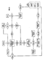
- Fig. 6 when for example a patient has not been hospitalized within the last year, only the Patient Entity resides in the database server 14. All other information relating to the patient is stored in the mass storage device(s) 12, perhaps on optical media.
- each Master Jacket Folder contains information representing the last discharge date of the patient and the date that the patient's records were last accessed, it is possible to automatically purge data from the database server 14 and to store it on the mass storage device(s) 12. Further, if for example a patient is readmitted to hospital after a long period of health, the relevant folders can be retrieved from the mass storage device(s) 12 and read into the database server 14, for rapid access to all patient records.
- Fig. 7 illustrates how the above-described data phases relate to each other. As data ages, more and more of it is moved from the database server 14 to the mass storage device(s) 12, and less and less folders reside in the database server 14. However, when necessary, all the folders can be retrieved into the database server 14.
- the user can define and redefine the criteria by which folders are moved from the database server 14 to the mass storage device(s) 12. For example, the above-mentioned one year period may be shortened or lengthened. Normally, folders which have been moved to the mass storage device(s) 12 will remain there until the patient has been readmitted or until they are needed for some other reason.
Landscapes
- Health & Medical Sciences (AREA)
- Nuclear Medicine, Radiotherapy & Molecular Imaging (AREA)
- Radiology & Medical Imaging (AREA)
- Engineering & Computer Science (AREA)
- Epidemiology (AREA)
- General Health & Medical Sciences (AREA)
- Medical Informatics (AREA)
- Primary Health Care (AREA)
- Public Health (AREA)
- Medical Treatment And Welfare Office Work (AREA)
- Measuring And Recording Apparatus For Diagnosis (AREA)
- Information Retrieval, Db Structures And Fs Structures Therefor (AREA)
Applications Claiming Priority (2)
| Application Number | Priority Date | Filing Date | Title |
|---|---|---|---|
| US30386189A | 1989-01-30 | 1989-01-30 | |
| US303861 | 1989-01-30 |
Publications (1)
| Publication Number | Publication Date |
|---|---|
| EP0380966A1 true EP0380966A1 (de) | 1990-08-08 |
Family
ID=23174031
Family Applications (1)
| Application Number | Title | Priority Date | Filing Date |
|---|---|---|---|
| EP90100928A Withdrawn EP0380966A1 (de) | 1989-01-30 | 1990-01-17 | Vorrichtung zur Informationsverwaltung, insbesondere für Bilderspeicher- und Bildkommunikationssysteme |
Country Status (2)
| Country | Link |
|---|---|
| EP (1) | EP0380966A1 (de) |
| JP (1) | JPH02245877A (de) |
Cited By (4)
| Publication number | Priority date | Publication date | Assignee | Title |
|---|---|---|---|---|
| EP0487110A3 (en) * | 1990-11-22 | 1993-09-29 | Kabushiki Kaisha Toshiba | Computer-aided diagnosis system for medical use |
| FR2727221A1 (fr) * | 1994-11-23 | 1996-05-24 | Kodak Pathe | Procede de communication d'informations numeriques et systeme pour la mise en oeuvre du procede |
| KR100296574B1 (ko) * | 1996-11-08 | 2001-10-24 | 포만 제프리 엘 | 착탈식대용량저장매체에아카이브를생성하기위한방법및아카이브서버 |
| US6574629B1 (en) | 1998-12-23 | 2003-06-03 | Agfa Corporation | Picture archiving and communication system |
Citations (3)
| Publication number | Priority date | Publication date | Assignee | Title |
|---|---|---|---|---|
| US3872448A (en) * | 1972-12-11 | 1975-03-18 | Community Health Computing Inc | Hospital data processing system |
| GB2157036A (en) * | 1984-03-20 | 1985-10-16 | Olympus Optical Co | Image retrieval and registration system |
| EP0206565A2 (de) * | 1985-06-17 | 1986-12-30 | Coats Viyella plc | Einzelhandelssysteme |
-
1990
- 1990-01-17 EP EP90100928A patent/EP0380966A1/de not_active Withdrawn
- 1990-01-26 JP JP2017623A patent/JPH02245877A/ja active Pending
Patent Citations (3)
| Publication number | Priority date | Publication date | Assignee | Title |
|---|---|---|---|---|
| US3872448A (en) * | 1972-12-11 | 1975-03-18 | Community Health Computing Inc | Hospital data processing system |
| GB2157036A (en) * | 1984-03-20 | 1985-10-16 | Olympus Optical Co | Image retrieval and registration system |
| EP0206565A2 (de) * | 1985-06-17 | 1986-12-30 | Coats Viyella plc | Einzelhandelssysteme |
Non-Patent Citations (3)
| Title |
|---|
| BIOMEDIZINISCHE TECHNIK, vol. 31, no. 6, June 1986, pages 143-149; H.U. LEMKE et al.: "Work stations for computer-graphic display in medical imaging" * |
| COMPUTERS IN CARDIOLOGY, Linkoping, 8th - 11th September 1985, pages 519-522, IEEE, New York, US; R.W. BROWER et al.: "Database management for cardiac catheterisation laboratories in the Netherlands" * |
| PROCEEDINGS PRIP 82, PATTERN RECOGNITION AND IMAGE PROCESSING, Las Vegas, 14th - 17th June 1982, pages 294-299, IEEE, New York, US; S.J. DWYER III et al.: "A diagnostic digital imaging management system" * |
Cited By (5)
| Publication number | Priority date | Publication date | Assignee | Title |
|---|---|---|---|---|
| EP0487110A3 (en) * | 1990-11-22 | 1993-09-29 | Kabushiki Kaisha Toshiba | Computer-aided diagnosis system for medical use |
| FR2727221A1 (fr) * | 1994-11-23 | 1996-05-24 | Kodak Pathe | Procede de communication d'informations numeriques et systeme pour la mise en oeuvre du procede |
| WO1996016372A1 (en) * | 1994-11-23 | 1996-05-30 | Kodak-Pathe | Method of communicating digital data and a system for implementing the method |
| KR100296574B1 (ko) * | 1996-11-08 | 2001-10-24 | 포만 제프리 엘 | 착탈식대용량저장매체에아카이브를생성하기위한방법및아카이브서버 |
| US6574629B1 (en) | 1998-12-23 | 2003-06-03 | Agfa Corporation | Picture archiving and communication system |
Also Published As
| Publication number | Publication date |
|---|---|
| JPH02245877A (ja) | 1990-10-01 |
Similar Documents
| Publication | Publication Date | Title |
|---|---|---|
| US4672683A (en) | Image retrieval and registration system | |
| US7634121B2 (en) | Method and system for rule-based comparison study matching to customize a hanging protocol | |
| US20020023067A1 (en) | Integrating a primary record viewing system with a different secondary record viewing system | |
| JPH0567160A (ja) | データベース共用ネツトワークにおける資料識別情報の利用方法 | |
| US20050108060A1 (en) | Medical image information management system | |
| US9934539B2 (en) | Timeline for multi-image viewer | |
| US20180301216A1 (en) | Automatic layout apparatus, automatic layout method, and automatic layout program | |
| JPH1097582A (ja) | 医療情報システム | |
| KR20170046115A (ko) | 의료 영상과 관련된 장치들 간에 송수신되는 의료 데이터를 생성하는 방법 및 장치. | |
| KR101555197B1 (ko) | 의료 영상을 관리하는 방법 및 이를 위한 장치 | |
| JP2003319933A (ja) | 画像表示システム | |
| JPH09259250A (ja) | 医用画像管理方法 | |
| JPH02245877A (ja) | 情報管理システムおよびpacs内のデータ蓄積方法 | |
| JPH065538B2 (ja) | 大量デジタル画像ファイル方式 | |
| JP2001101320A (ja) | 医療情報システム | |
| JPH1091710A (ja) | 医用画像配送システム | |
| JP2000132628A (ja) | 画像管理システム及びその画像管理方法、画像制御装置、画像表示装置並びに記憶媒体 | |
| Anderson et al. | Implementation of a diagnostic display and image manipulation node | |
| Ivanov et al. | Design and implementation of a regional PACS | |
| JP2001005825A (ja) | 医療画像サーバ装置および画像検索方法 | |
| JPS6129971A (ja) | 医用画像の検索方法 | |
| JPH0283780A (ja) | 医用情報データベースシステム | |
| JP2003281279A (ja) | 医療画像表示システム | |
| Prior et al. | Information management for data retrieval in a PACS | |
| JPH0528233A (ja) | 画像保存システム |
Legal Events
| Date | Code | Title | Description |
|---|---|---|---|
| PUAI | Public reference made under article 153(3) epc to a published international application that has entered the european phase |
Free format text: ORIGINAL CODE: 0009012 |
|
| AK | Designated contracting states |
Kind code of ref document: A1 Designated state(s): DE FR GB |
|
| 17P | Request for examination filed |
Effective date: 19900828 |
|
| STAA | Information on the status of an ep patent application or granted ep patent |
Free format text: STATUS: THE APPLICATION HAS BEEN WITHDRAWN |
|
| 17Q | First examination report despatched |
Effective date: 19930402 |
|
| 18W | Application withdrawn |
Withdrawal date: 19930504 |