EP0387777A2 - Procédé pour la détection de la FGF et pour le diagnostic de tumeurs - Google Patents
Procédé pour la détection de la FGF et pour le diagnostic de tumeurs Download PDFInfo
- Publication number
- EP0387777A2 EP0387777A2 EP19900104706 EP90104706A EP0387777A2 EP 0387777 A2 EP0387777 A2 EP 0387777A2 EP 19900104706 EP19900104706 EP 19900104706 EP 90104706 A EP90104706 A EP 90104706A EP 0387777 A2 EP0387777 A2 EP 0387777A2
- Authority
- EP
- European Patent Office
- Prior art keywords
- fgf
- antibody
- amino acid
- tumor
- examples
- Prior art date
- Legal status (The legal status is an assumption and is not a legal conclusion. Google has not performed a legal analysis and makes no representation as to the accuracy of the status listed.)
- Withdrawn
Links
Images


Classifications
-
- G—PHYSICS
- G01—MEASURING; TESTING
- G01N—INVESTIGATING OR ANALYSING MATERIALS BY DETERMINING THEIR CHEMICAL OR PHYSICAL PROPERTIES
- G01N33/00—Investigating or analysing materials by specific methods not covered by groups G01N1/00 - G01N31/00
- G01N33/48—Biological material, e.g. blood, urine; Haemocytometers
- G01N33/50—Chemical analysis of biological material, e.g. blood, urine; Testing involving biospecific ligand binding methods; Immunological testing
- G01N33/74—Chemical analysis of biological material, e.g. blood, urine; Testing involving biospecific ligand binding methods; Immunological testing involving hormones or other non-cytokine intercellular protein regulatory factors such as growth factors, including receptors to hormones and growth factors
-
- G—PHYSICS
- G01—MEASURING; TESTING
- G01N—INVESTIGATING OR ANALYSING MATERIALS BY DETERMINING THEIR CHEMICAL OR PHYSICAL PROPERTIES
- G01N33/00—Investigating or analysing materials by specific methods not covered by groups G01N1/00 - G01N31/00
- G01N33/48—Biological material, e.g. blood, urine; Haemocytometers
- G01N33/50—Chemical analysis of biological material, e.g. blood, urine; Testing involving biospecific ligand binding methods; Immunological testing
- G01N33/53—Immunoassay; Biospecific binding assay; Materials therefor
- G01N33/575—Immunoassay; Biospecific binding assay; Materials therefor for cancer
- G01N33/5758—Immunoassay; Biospecific binding assay; Materials therefor for cancer involving compounds serving as markers for tumours, cancers or neoplasias, e.g. cellular determinants, receptors, heat shock/stress proteins, A-protein, oligosaccharides or metabolites
-
- G—PHYSICS
- G01—MEASURING; TESTING
- G01N—INVESTIGATING OR ANALYSING MATERIALS BY DETERMINING THEIR CHEMICAL OR PHYSICAL PROPERTIES
- G01N2333/00—Assays involving biological materials from specific organisms or of a specific nature
- G01N2333/435—Assays involving biological materials from specific organisms or of a specific nature from animals; from humans
- G01N2333/475—Assays involving growth factors
- G01N2333/50—Fibroblast growth factors [FGF]
Definitions
- the present invention relates to a method for detecting and/or measuring fibroblast growth factor (hereinafter also abbreviated FGF) using a sandwich technique, and to a method for diagnosis of tumor.
- FGF fibroblast growth factor
- FGF FGF-like growth factor
- basic FGF which has a basic isoelectric point
- acidic FGF which has an acidic isoelectric point
- the entire amino acid sequence of both types is known [F. Esch et al., Proc. Natl. Acad. Sci. USA, 85 , 6507(1985); K.A. Thomas et al., Proc Natl. Acad. Sci. USA, 82 , 6409 (1985)].
- FGF exhibits a growth promoting activity on 3T3 cells and mesoderm-derived cells, including vascular endothelial cells, in vitro and an angiogenic activity in vivo [D. Gospodarowicz et al., Endocrine Reviews, 8 , 95(1987)].
- the angiogenic activity of FGF in combination with the cell growth promoting activity, can be used in therapeutic drugs for wounds and burns, and preventive/therapeutic drugs for thrombosis, arteriosclerosis and other diseases.
- FGF is found in nature in only extremely small quantities; it has been very difficult, due to various limitations, to detect natural FGF from human tissues. In addition, no method has yet been developed which permits easy quantitative determination of FGF.
- the present inventors conducted various investigations with the aim of finding a practical means of FGF measurement, and found that FGF can be measured to high sensitivity by using heparin as a ligand labeled with a labeling agent in the sandwich technique wherein two ligands are used.
- the present inventors made further investigations based on this finding, in developing the present invention.
- the present invention involves (1) a method of detecting and/or measuring FGF by a sandwich technique using an antibody coupled to a carrier, a sample and heparin labeled with a labeling agent, and (2) use of an antibody coupled to a carrier and heparin labeled with a labeling agent for diagnosing tumor by subjecting a sample to a sandwich method.
- the method of detecting and measuring the present invention may be based on radioimmunoassay (hereinafter also abbreviated RIA) or enzyme immunoassay (hereinafter also abbreviated EIA).
- RIA radioimmunoassay
- EIA enzyme immunoassay
- the FGF may be any FGF, as long as it is derived from warm-blooded animals, and may also be an FGF mutein.
- FGF includes its mutein, unless otherwise stated.
- FGF acidic FGF
- bFGF basic FGF
- the FGF may be of natural derivation, or may be produced by gene engineering technology.
- mammalian-derived aFGF examples include bovine aFGF [K.A. Thomas et al., Proc. Natl. Acad. Sci. USA, 81 , 357(1984)] and human aFGF (G. Gimenez-Gallego et al., Biochem. Biophys. Res. Commun., 138 , 611 (1986)].
- mammalian-derived bFGF examples include bovine bFGF [Proc. Natl. Acad. Sci. USA, 82 , 6507(1985)] and human bFGF [European Patent Publication No. 237966; European Molecular Biology Organization (EMBO) Journal, 5 ,2523(1986)].
- the above-mentioned mutein refers to a peptide or protein with a mutated amino acid sequence; the mutation includes amino acid addition, constituent amino acid deletion and amino acid replacement by other amino acid.
- amino acid addition examples include addition of at least one amino acid.
- constituent amino acid deletion examples include a lack of at least one FGF-constituent amino acid.
- amino acid replacement by other amino acid examples include replacement of at least one FGF-constituent amino acid by other amino acid.
- the "at least one amino acid" in the mutein resulting from the addition of at least one amino acid to FGF does not include methionine derived from the initiation codon or signal peptide used for peptide expression.
- amino acids may be added, as long as the properties of FGF are maintained.
- preferred amino acids to be added include partial or entire amino acid sequences of proteins recognized as homologous with FGF and having similar activities.
- any number of constituent amino acids may be lacking, as long as the properties of FGF are maintained.
- any number of FGF-constituent amino acids may be so replaced, as long as the properties of FGF are maintained.
- constituent amino acids to be replaced examples include cysteine and others; cysteine however is preferred.
- constituent amino acids to be replaced, other than cysteine include aspartic acid, arginine, glycine and valine.
- the replacement amino acid be e.g. a neutral amino acid.
- examples include glycine, valine, alanine, leucine, isoleucine, tyrosine, phenylalanine, histidine, tryptophane, serine, threonine and methionine; serine and threonine however are preferred.
- the constituent amino acid to be replaced is not cysteine, it is necessary to choose a replacement amino acid different from the constituent amino acid to be replaced, in such properties as hydrophilicity, hydro phobicity or electric charge.
- a replacement amino acid different from the constituent amino acid to be replaced, in such properties as hydrophilicity, hydro phobicity or electric charge.
- the amino acid to be replaced is aspartic acid
- examples of the replacement amino acid include asparagine, threonine, valine, phenylalanine, and arginine; asparagine and arginine however are preferred.
- amino acid to be replaced is arginine
- replacement amino acid examples of the replacement amino acid are glutamine, threonine, leucine, phenylalanine and aspartic acid; glutamine however is preferred.
- the constituent amino acid to be replaced is glycine
- examples of the replacement amino acid include threonine, leucine, phenylalanine, serine, glutamic acid and arginine; threonine however is preferred.
- the replacement amino acid When the constituent amino acid to be replaced is serine, examples of the replacement amino acid include methionine, alanine, leucine, cysteine, glutamine, arginine, and aspartic acid; methionine however is preferred.
- the replacement amino acid When the constituent amino acid to be replaced is valine, examples of the replacement amino acid include serine, leucine, proline, glycine, lysine, and aspartic acid; serine however is preferred.
- the preferred constituent amino acids to be replaced are aspartic acid, arginine, glycine, serine, and valine.
- the preferred replacement amino acids are asparagine, glutamine, arginine, threonine, methionine, serine, and leucine.
- the constituent amino acid cysteine should ideally be replaced by serine.
- two or more constituent amino acids may be replaced simultaneously; it is preferable that 2 or 3 constituent amino acids be replaced.
- the mutein of the present invention may result from a combination of 2 or 3 of the above additions, eliminations and replacements.
- Site-directed mutagenesis may be employed to produce said mutein. This technology is widely known and described by R.F. Lather and J.P. Lecoq in “Genetic Engineering", Academic Press (1983), pp. 31-50. Oligonucleotide-directed mutagenesis is described by M. Smith and S. Gillam in “Genetic Engineering: Principles and Methods", Prenam Press (1981), vol. 3, pp 1-32.
- the phage DNA carrying the mutated gene is then isolated and inserted into a plasmid.
- the plasmid thus prepared is used to transform an appropriate host; the transformant thus obtained is cultivated in medium to produce the desired mutein.
- the method of the present invention is based on the principle of the sandwich technique, a noncompetitive method of immunochemical measurement.
- Examples of carriers useful for the method of the present invention include gel grains such as agarose gels [eg. Sepharose 4B, Sepharose 6B (produced by Pharmacia Fine Chemical, Sweden)], dextran gels [e.g. Sephadex G-75, Sephadex G-100, Sephadex G-200 (produced by Pharmacia Fine Chemical, Sweden)] and polyacrylamide gels [e.g. Biogel P-30, Biogel P-60, Biogel P-100 (produced by Bio-Rad Laboratories, USA)]; cellulose grains such as Avicel (produced by Asahi Chemical Industries, Japan) and ion exchange cellulose (e.g.
- agarose gels eg. Sepharose 4B, Sepharose 6B (produced by Pharmacia Fine Chemical, Sweden)
- dextran gels e.g. Sephadex G-75, Sephadex G-100, Sephadex G-200 (produced by Pharmacia Fine Chemical, Sweden)
- polyacrylamide gels e.g. Biogel P-30,
- diethylaminoethyl cellulose, carboxymethyl cellulose physical adsorbents such as glass (e.g. glass balls, glass rods, aminoalkyl glass balls, aminoalkyl glass rods), silicon pieces, styrene resins (e.g. polystyrene balls, polystyrene grains), and immunoassay plates (e.g. plated produced by Nunc, Sweden); and ion exchange resins such as weakly acidic cation exchange resins [eg. Amberlite IRC-50 (produced by Rohm & Haas, USA), Zeo-Karb 226 (produced by Permutit AG, West Germany)] and weakly alkaline anion exchange resins [eg. Amberlite IR-4B, Dowex 3 (produced by Dow Chemical, USA)].
- glass e.g. glass balls, glass rods, aminoalkyl glass balls, aminoalkyl glass rods
- silicon pieces e.g. polystyrene resins (e.g. poly
- the antibody against FGF used as a conjugate for the method of the present invention, may be polyclonal or monoclonal.
- the polyclonal antibody against FGF can be produced by inoculating FGF as antigen to a warm-blooded animal to produce an anti-FGF antibody, which is then collected.
- Any FGF can be used as the antigen, as long as it possesses biological or immunological activities similar to those of natural FGF; examples include FGF species produced by gene engineering technology and their fragments comprising a partial amino acid sequence essential to their biological or immunological activities. These FGF species produced by gene engineering technology may have additional Met at the polypeptide amino terminal, and may also be a mixture of an FGF and another FGF having additional Met at the amino terminal.
- the above fragments can be produced by known routine methods of peptide synthesis, which may be based on a solid phase method or liquid phase method.
- methods of peptide synthesis include the methods described in "The Peptides", vol. 1 (1966), edited by Schröder and Lubke, Academic Press, New York, USA, "Peptide Synthesis (in Japanese)", edited by Izumiya et al., Maruzen (1975), and “Fundamentals and Experiments of Peptide Synthesis (in Japanese)", edited by Izumiya et al., Maruzen (1985).
- fragments may be produced by cleaving FGF using appropriate enzymes.
- appropriate enzymes include the method described in "Biochemical Experiments Course 1, Protein Chemistry II (in Japanese)", edited by the Japanese Biochemical Society, Tokyo Kagaku Dojin (1976), pp. 255-332.
- FGF as the antigen is coupled with carrier protein.
- carrier protein examples include bovine tyroglobulin, bovine serum albumin, bovine gamma globulin, hemocyamin, and Freud's complete adjuvant (produced by Difco Laboratories).
- Antigen FGF and carrier protein can be coupled by a routine method.
- Examples of coupling reagents include glutaraldehyde and water-soluble carbodiimide.
- the antigen FGF and the carrier protein should be coupled in a ratio of about 1 to 1 to about 1 to 10. Good results are often obtained when reaction is carried out at a nearly neutral pH level, ideally pH 6 to 8.
- Reaction time is generally 1 to 12 hr, preferably 2 to 6 hr.
- the complex thus formed may be dialyzed against water at 0 to 18°C by a routine method, after which it may be frozen or lyophilized for storage.
- the immunogen thus produced is inoculated to a warm-blooded animal.
- warm-blooded animals which can be used to produce the above antibody include mammals (e.g. rabbits, sheep, goats, rats, mice, guinea pigs, bovines, horses, swine) and birds (e.g. chickens, doves, ducks, geese, quails).
- the immunogen is inoculated to a warm-blooded animal in an amount such that efficient antibody production will occur; for example, antibodies are often produced when 1 mg of the immunogen, in an emulsion in 1 ml of a physiological saline solution containing Freund's complete adjuvant, is subcutaneously inoculated to a rabbit at the back and hind leg paw 5 times at 4-week intervals.
- the antibody thus formed in the warm-blooded animal is, for example, collected as follows: when a rabbit is used, blood is usually collected via ear vein between 7 and 12 days after the final inoculation, then centrifuged to yield a serum.
- the antiserum thus obtained is subjected to affinity chromatography using a carrier coupled with an antigen peptide; the fraction adsorbed to the column is recovered to separate a purified polyclonal antibody.
- This monoclonal antibody is produced by immunizing a mammal with the antigen polypeptide or protein complex, fusing spleen cells excised from the animal and homo- or heterogenic lymphoid cells to yield a hybridoma, which is then cloned, inoculating the cloned hybridoma to a mammal to produce and accumulate a monoclonal antibody, which is then collected.
- Immunization of e.g. mice can be achieved via any route, such as subcutaneous, intraperitoneal, intravenous, intramuscular, and intracutaneous administration; mainly subcutaneous, intraperitoneal, or intravenous (preferably subcutaneous) injection is recommended.
- Immunization interval immunization quantity etc. can also be widely varied so that various modes are possible; for example, in a method often used, immunization is conducted 2 to 6 times at 2-week intervals and spleen cells are excised and used 1 to 5 days, preferably 2 to 4 days after final immunization. It is desirable that the immunization quantity be over 0.1 ⁇ g, preferably 10 to 300 ⁇ g per mouse in each immunization, in terms of peptide. It is also desirable that partial blood sampling be conducted before spleen excision, to confirm increased blood antibody level, and that a cell fusion experiment be conducted using spleen cells.
- lymphoid cell line such as an appropriate allo- or heterogenic (preferably allogenic) myeloma with a marker for hypoxanthine-guanine-phosphoribosyl transferase deficiency (HGPRT ⁇ ) or thymidine kinase deficiency (TK ⁇ ) [e.g. P3-X63-Ag 8UI (Ichimori et al., Journal of Immunological Method, 80 , 55 (1985)]. Fusion can, for example, be achieved in accordance with the method of Köller and Milstein [Nature, 256 , 495 (1975)].
- myeloma cells and spleen cells in a ratio of about 1 to 5, are suspended in a medium prepared by mixing Iskov medium and Ham F-12 medium in a 1 to 1 ratio (hereinafter referred to as IH medium), followed by cell fusion reaction in the presence of a fusogen such as Sendai virus or polyethylene glycol (PEG).
- a fusogen such as Sendai virus or polyethylene glycol (PEG).
- DMSO dimethylsulfoxide
- PEG with a degree of polymerization of 1000 to 6000 is normally used at a concentration of 10 to 80% for 0.5 to 30 minutes: efficient fusion is achieved, for example, by treating PEG 6000 at 35 to 55% for 4 to 10 minutes.
- the fused cells can be selectively proliferated using hypoxanthine-aminopterin-thymidine medium (HAT medium; Nature, 256 , 495 (1975)) etc.
- HAT medium hypoxanthine-aminopterin-thymidine medium
- the culture supernatant of the proliferated cells can be screened for the desired antibody production; antibody titer screening can be conducted as follows: The culture supernatant is first examined for the production of antibody against the immunogen peptide by radioimmunoassay (RIA) or enzyme immunoassay (EIA). Various modifications are possible for these methods. An example of a preferred method of measurement based on EIA is described below.
- a carrier such as cellulose beads, is coupled with e.g. rabbit anti-mouse immunoglobulin antibody by a routine method; the subject culture supernatant or mouse serum is added, followed by reaction at constant temperature (about 4 to 40°C; the same applies below) for a given time.
- reaction product is then thoroughly washed; an enzyme-labeled peptide (enzyme and peptide are coupled by a routine method, then purified) is added, followed by reaction at constant temperature for a given time.
- an enzyme substrate is added, followed by reaction at a constant temperature for a given time; the formed coloring substance is then measured by optical absorbance, fluorescence intensity etc.
- the hybridoma thus cloned is proliferated in liquid medium.
- the desired monoclonal antibody can be obtained from the culture broth obtained by cultivating the hybridoma in a liquid medium, such as a medium prepared by adding about 0.1 ⁇ 40% bovine serum to RPMI-1640 medium [Moore, G.E. et al., Journal of American Medical Association, 199 , 549 (1967)] for 2 to 10 days, preferably 3 to 5 days.
- the antibody can also be obtained by intraperitoneally inoculating the hybridoma to a mammal for cell proliferation, and collecting the ascites fluid.
- mice when mice are used, for example 1 ⁇ 104 to 1 ⁇ 107, preferably 5 ⁇ 105 to 2 ⁇ 106 hybridoma cells are intraperitoneally inoculated to each mouse of BALB/c or other lines, previously inoculated with mineral oil etc.; ascites fluid is then collected 7 to 20 days, preferably 10 to 14 days after inoculation.
- the antibody produced and accumulated in the ascites fluid can easily be isolated in the form of pure immunoglobulin by e.g. ammonium sulfate fractionation and DEAE-cellulose column chromatography.
- the antibody molecule may be IgG, or its fragment [e.g. F(ab′)2, Fab′, Fab ⁇ , or Fab]. It is preferable however that the antibody molecule is coupled directly with a labeling agent be Fab′.
- an antibody to the carrier
- known routine methods can be used. Examples of usable methods include the bromocyan method and glutaraldehyde method, both described in "Taisha,” vol. 8(1971), p. 696.
- physical adsorption onto the carrier's surface may be employed.
- the antibody is immobilized onto a carrier such as a 96-well plastic plate (e.g. Immunoplate, produced by Nunc, Denmark), at 0.1 to 10 ⁇ g/well, glass beads and plastic beads. Immobilization is achieved by an overnight reaction (at about 4°C), or at room temperature for 0.5 to 4 hours, in the case of plastic carriers.
- immobilization is achieved, for example, by the method described in Proc. Natl. Acad. Sci. USA, vol. 80, pp. 3513-3516 (1983).
- Various commercially available plates for antibody immobilization e.g. the Immunoplate, produced by Nunc, mentioned above
- Nunc mentioned above
- heparins used for the method of the present invention include N-sulfuric acid-, N-acetyl-, and O-sulfuric acid-substituted products of polysaccharides comprising D-glucosamine, D-glucuronic acid and L-iduronic acid.
- labeling agents for RIA examples include 3H, 125I and other radioisotopes.
- labeling agents for EIA include enzymes, fluorescent substances and luminescent substances; the use of enzyme is recommended. Stable enzymes with high specific activity are preferred; peroxidase, alkaline phosphatase, ⁇ -D-galactosidase, glucose oxidase etc. can be used; peroxidase is preferred. Peroxidase of various derivations can be used; examples include peroxidase derived from horseradish, pinapple, fig, sweet potato, kidney bean, corn etc.; horseradish peroxidase (HRP) is preferred.
- HRP horseradish peroxidase
- fluorescent substances examples include FITC (fluorescein isothiocyanate), rhodamine, and lanthanide chelate.
- luminescent substances include acridinium esters.
- a heparin molecule which is highly reactive on peptide or protein is first chosen. This coupling can, for example, be achieved with the method described by A.D. Cardin et al. [Thrombosis Research, 34 , 541 (1984)], which uses low density lipoprotein (LDL)-AffiGel-10.
- LDL low density lipoprotein
- HRH highly reactive heparin
- labeling agents include 125I. Before coupling 125I to HRH, it is necessary to maximize the specific activity for labeling.
- This can, for example, be achieved by introducing a hydroxyphenyl group to HRH.
- This particular maximization can, for example, be achieved by the method using the Bolton-Hunter reagent, described by N. Hirose et al. [Analytical Biochemistry, 156 , 320 (1986)].
- the subject sample to be measured includes: urine, serum, plasma, cerebrospinal fluid and other body fluids, or cell or bacterial extracts or their supernatants.
- EIA for the measuring method of the present invention, when using peroxidase as the labeling agent, is described in detail below; however, such use of peroxidase is not to be construed as a limitation on the present invention.
- the method of the present invention offers high measurement sensitivity, thus permitting detection and measurement of trace amounts of blood FGF.
- the method of detecting and measuring FGF using the present invention is also applicable to diagnosis of tumors. Since tumor cells are believed to release large amounts of FGF, the blood, urine, ascites, pleural effusion, cerebrospinal fluid, external effusion, gastric juice, cystic fluid and other body fluids of tumor patients should contain more FGF than do those of normal humans. Therefore, if a large amount of FGF is detected in the subject sample obtained from a patient, the patient's disease can be primarily diagnosed as a tumor.
- the method of tumor diagnosis according to the present invention is characterized by the detection and measurement of a fibroblast growth factor (FGF) by the sandwich technique (using a carrier-coupled antibody, a subject sample and heparin coupled with a labeling agent).
- FGF fibroblast growth factor
- target tumors include: brain tumor, lung tumor, digestive tract tumor, liver tumor, pancreas tumor, gallbladder tumor, bile duct tumor, kidney tumor, urinary bladder tumor, adrenal tumor, rhabdomyoma, rhabdomyosarcoma, leiomyosarcoma, endocrine neoplasia tumor, parathyroid tumor, and parathyroid cancer.
- Detection and measurement can be achieved in the same manner as the above-mentioned methods.
- the presence of a tumor can be determined. This, as indicated above, would occur when a greater amount of FGF is present in the preselected sample than in the "normal" control.
- the sample should contain at least double the amount of FGF, more preferably, it should contain at least three times the amount of FGF than the "normal" control contains.
- the FGF-detecting and/or measuring method of the present invention permits measurement of FGF to high sensitivity, thus the present method functions well as a means of detecting and measuring FGF in body fluids or cells.
- cyanogen bromide-activated Sepharose 4B (Pharmacia) was transferred to a glass filter and sufficiently expanded with 20 ml of 0.001 N HCl, after which it was thoroughly washed with 400 ml of the same aqueous hydrochloric acid.
- 2 ml of a solution of 0.1 M sodium bicarbonate and 0.5 M sodium chloride was added to 100 ⁇ l of a solution of the peptide [Tyr11] human bFGF (1-11) Nle12 (protein content, 40 mg/ml); this mixed solution was adjusted to pH 8.4 with a solution of 0.1 M sodium hydroxide.
- the gel was then washed with sequential additions of a 0.1 M acetate buffer solution containing 1 M sodium chloride (pH 4.0) and a 0.1 M borate buffer solution containing 1 M sodium chloride (pH 8.0), each in 100 ml amounts.
- the gel was then suspended in PBS and stored in a column at 4°C.
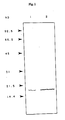
- the heparin-coupled protein in the culture supernatant of bovine corneal endothelial cells (BCE cell) and recombinant bFGF was subjected to SDS-PAGE, followed by western blotting; a specific band (lane 1) was detected at the position corresponding to bFGF (lane 2) (see Fig. 1).
- Heparin was labeled with 125I in accordance with the method described in Analytical Biochemistry, 156 , 320-325 (1986), which is as follows:
- Heparin was purified using a column of low density lipoprotein (LDL)- AffiGel-10 in order to obtain HRH (highly reactive heparin), which was then reacted with Bolton-Hunter reagent to introduce a hydroxyphenyl group.
- Iodization with 125I (1 mCi) was conducted in a test tube containing 15 ⁇ g of solidified iodogen, cooled with ice for 15 minutes. Unreacted 125I was removed by dialysis against distilled water, to obtain purified 125I-heparin.
- the anti-[Thr11] human bFGF (1-11) Nle12 rabbit antibody (3 ⁇ g/ml) was added to the plate at 100 or 200 ⁇ l per well. The plate was left at 4°C overnight. After discarding the solution, the plate was washed with PBS; 300 ⁇ l of 1% BSA (bovine serum albumin)/PBS/0.04% NaN3 was added to each well; the plate was left at 4°C, for at least one night, to facilitate blocking.
- BSA bovine serum albumin
- the sample After discarding the solution in the plate, the sample, appropriately diluted and adjusted to an NaCl concentration of 0.5 M, was added to the plate and left at 4°C overnight.
- each well was washed with 0.5 M NaCl/PBS; the 125I-heparin described in (1) above was added to the plate at 10 to 20 ⁇ 104 cpm per well; the plate was left at 4°C overnight. After thorough washing with 0.5 M NaCl/PBS, each well was cut out using scissors; the count from each well was taken using a gamma counter.
- Fig. 3 The results shown in Fig. 3 have revealed that 5%, 10%, 20%, 30%, or 50% fetal calf serum (FCS) hardly affects the quantitative determination of blood bFGF.
- FCS fetal calf serum
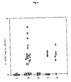
- Fig. 4 reveals that the blood bFGF concentration is evidently high in these patients.
Landscapes
- Health & Medical Sciences (AREA)
- Life Sciences & Earth Sciences (AREA)
- Engineering & Computer Science (AREA)
- Molecular Biology (AREA)
- Immunology (AREA)
- Biomedical Technology (AREA)
- Chemical & Material Sciences (AREA)
- Hematology (AREA)
- Urology & Nephrology (AREA)
- Food Science & Technology (AREA)
- Biochemistry (AREA)
- Cell Biology (AREA)
- Biotechnology (AREA)
- Medicinal Chemistry (AREA)
- Physics & Mathematics (AREA)
- Analytical Chemistry (AREA)
- Microbiology (AREA)
- General Health & Medical Sciences (AREA)
- General Physics & Mathematics (AREA)
- Pathology (AREA)
- Endocrinology (AREA)
- Investigating Or Analysing Biological Materials (AREA)
- Medicines Containing Antibodies Or Antigens For Use As Internal Diagnostic Agents (AREA)
- Peptides Or Proteins (AREA)
- Preparation Of Compounds By Using Micro-Organisms (AREA)
Applications Claiming Priority (2)
| Application Number | Priority Date | Filing Date | Title |
|---|---|---|---|
| JP6437189 | 1989-03-15 | ||
| JP64371/89 | 1989-03-15 |
Publications (2)
| Publication Number | Publication Date |
|---|---|
| EP0387777A2 true EP0387777A2 (fr) | 1990-09-19 |
| EP0387777A3 EP0387777A3 (fr) | 1992-01-02 |
Family
ID=13256360
Family Applications (1)
| Application Number | Title | Priority Date | Filing Date |
|---|---|---|---|
| EP19900104706 Withdrawn EP0387777A3 (fr) | 1989-03-15 | 1990-03-13 | Procédé pour la détection de la FGF et pour le diagnostic de tumeurs |
Country Status (1)
| Country | Link |
|---|---|
| EP (1) | EP0387777A3 (fr) |
Cited By (2)
| Publication number | Priority date | Publication date | Assignee | Title |
|---|---|---|---|---|
| EP0646182B1 (fr) * | 1992-06-18 | 1996-08-28 | The Whittier Institute For Diabetes And Endocrinology | Procede de detection des maladies neoplasiques |
| FR2976587A1 (fr) * | 2011-06-20 | 2012-12-21 | Basf Beauty Care Solutions France Sas | Methode de dosage in vitro par technique immunologique |
Family Cites Families (1)
| Publication number | Priority date | Publication date | Assignee | Title |
|---|---|---|---|---|
| EP0288687B1 (fr) * | 1987-03-03 | 1994-12-28 | Takeda Chemical Industries, Ltd. | Anticorps monoclonal, hybridome, leur production et leur utilisation |
-
1990
- 1990-03-13 EP EP19900104706 patent/EP0387777A3/fr not_active Withdrawn
Cited By (5)
| Publication number | Priority date | Publication date | Assignee | Title |
|---|---|---|---|---|
| EP0646182B1 (fr) * | 1992-06-18 | 1996-08-28 | The Whittier Institute For Diabetes And Endocrinology | Procede de detection des maladies neoplasiques |
| US5670323A (en) * | 1992-06-18 | 1997-09-23 | The Whittier Institute For Diabetes And Endocrinology | Process for the detection of malignant melanoma |
| FR2976587A1 (fr) * | 2011-06-20 | 2012-12-21 | Basf Beauty Care Solutions France Sas | Methode de dosage in vitro par technique immunologique |
| WO2012175454A3 (fr) * | 2011-06-20 | 2013-05-16 | Basf Beauty Care Solutions France S.A.S. | Méthode de dosage in vitro par technique immunologique |
| US9146237B2 (en) | 2011-06-20 | 2015-09-29 | Basf Beauty Care Solutions France S.A.S. | In vitro assay method using immunological technique |
Also Published As
| Publication number | Publication date |
|---|---|
| EP0387777A3 (fr) | 1992-01-02 |
Similar Documents
| Publication | Publication Date | Title |
|---|---|---|
| CA1340977C (fr) | Recepteur proteique capteur, ainsi que son anticorps | |
| US4446122A (en) | Purified human prostate antigen | |
| UA74129C2 (uk) | Виділене антитіло або фрагмент антитіла, що є специфічним до онкоембріонального домену ed-b фібронектину (fn) та таким, що безпосередньо з ним зв'язується | |
| US5185432A (en) | Monoclonal antibodies and antigen for human non-small cell lung carcinoma and other certain human carcinomas | |
| EP0735370A1 (fr) | Méthode d'essai de MAP-kinase | |
| US4882275A (en) | Method of purifying endothelial cell growth factors using immobilized heparin | |
| CA2611048A1 (fr) | Reactifs et dosages diagnostiques permettant de detecter la proteine psp94 | |
| US5187062A (en) | Method for detecting and measuring FGF | |
| EP0387777A2 (fr) | Procédé pour la détection de la FGF et pour le diagnostic de tumeurs | |
| JP3057292B2 (ja) | モノクローナル抗体、ハイブリドーマ、それらの製造法および用途 | |
| WO1981001849A1 (fr) | Antigene purifie de prostate humain | |
| US5437995A (en) | Monoclonal antibody against an acidic FGF protein and hybridoma for its production | |
| EP0817842A1 (fr) | Anticorps contre la betacelluline humaine et ses applications | |
| CN1046737C (zh) | 同种异体和异基因蛋白的抗体,它们在诊断和治疗中的应用及其检测方法 | |
| JPH0315758A (ja) | 線維芽細胞増殖因子の検出・測定法および腫瘍の診断法 | |
| US6306608B1 (en) | Anti-human LECT2 antibody, cells producing the same, and method and kit for assaying the same | |
| AU593063B2 (en) | Purified human angiogenic factor, method for its preparation and pharmaceutical preparations | |
| US6617432B1 (en) | Nuclear matrix proteins polynucleotide sequences encoding them and their use | |
| JP3268147B2 (ja) | モノクローナル抗体 | |
| EP0507948A1 (fr) | Anticorps monoclonal, hybridomes, leur production et leur utilisation | |
| JPH06125784A (ja) | モノクローナル抗体,ハイブリドーマ,その製造法および用途 | |
| JP3107248B2 (ja) | 抗体および免疫化学的測定法 | |
| JPH0746105B2 (ja) | インタ−フエロン−γの免疫化学的測定法および測定用試薬 | |
| JP2002000273A (ja) | プロテインcインヒビターと肝細胞増殖因子アクチベーターとの複合体およびその測定方法並びに臓器障害の検出方法 | |
| JPH08169898A (ja) | ペプチド及びモノクローナル抗体 |
Legal Events
| Date | Code | Title | Description |
|---|---|---|---|
| PUAI | Public reference made under article 153(3) epc to a published international application that has entered the european phase |
Free format text: ORIGINAL CODE: 0009012 |
|
| AK | Designated contracting states |
Kind code of ref document: A2 Designated state(s): AT BE CH DE DK ES FR GB GR IT LI LU NL SE |
|
| PUAL | Search report despatched |
Free format text: ORIGINAL CODE: 0009013 |
|
| AK | Designated contracting states |
Kind code of ref document: A3 Designated state(s): AT BE CH DE DK ES FR GB GR IT LI LU NL SE |
|
| RAP3 | Party data changed (applicant data changed or rights of an application transferred) |
Owner name: TAKEDA CHEMICAL INDUSTRIES, LTD. |
|
| 17P | Request for examination filed |
Effective date: 19920522 |
|
| 17Q | First examination report despatched |
Effective date: 19940221 |
|
| STAA | Information on the status of an ep patent application or granted ep patent |
Free format text: STATUS: THE APPLICATION IS DEEMED TO BE WITHDRAWN |
|
| 18D | Application deemed to be withdrawn |
Effective date: 19940705 |