EP0412557A1 - Wachstumsfaktor aus parenchymalen Lebenszellen, dafür kodierendes Gen, Verfahren zur Herstellung dieses Faktors und Transformanten - Google Patents
Wachstumsfaktor aus parenchymalen Lebenszellen, dafür kodierendes Gen, Verfahren zur Herstellung dieses Faktors und Transformanten Download PDFInfo
- Publication number
- EP0412557A1 EP0412557A1 EP90115397A EP90115397A EP0412557A1 EP 0412557 A1 EP0412557 A1 EP 0412557A1 EP 90115397 A EP90115397 A EP 90115397A EP 90115397 A EP90115397 A EP 90115397A EP 0412557 A1 EP0412557 A1 EP 0412557A1
- Authority
- EP
- European Patent Office
- Prior art keywords
- growth factor
- cell growth
- hhgf
- hepatic parenchymal
- amino acid
- Prior art date
- Legal status (The legal status is an assumption and is not a legal conclusion. Google has not performed a legal analysis and makes no representation as to the accuracy of the status listed.)
- Granted
Links
Images



Classifications
-
- C—CHEMISTRY; METALLURGY
- C12—BIOCHEMISTRY; BEER; SPIRITS; WINE; VINEGAR; MICROBIOLOGY; ENZYMOLOGY; MUTATION OR GENETIC ENGINEERING
- C12N—MICROORGANISMS OR ENZYMES; COMPOSITIONS THEREOF; PROPAGATING, PRESERVING, OR MAINTAINING MICROORGANISMS; MUTATION OR GENETIC ENGINEERING; CULTURE MEDIA
- C12N15/00—Mutation or genetic engineering; DNA or RNA concerning genetic engineering, vectors, e.g. plasmids, or their isolation, preparation or purification; Use of hosts therefor
- C12N15/09—Recombinant DNA-technology
- C12N15/11—DNA or RNA fragments; Modified forms thereof; Non-coding nucleic acids having a biological activity
-
- C—CHEMISTRY; METALLURGY
- C07—ORGANIC CHEMISTRY
- C07K—PEPTIDES
- C07K14/00—Peptides having more than 20 amino acids; Gastrins; Somatostatins; Melanotropins; Derivatives thereof
- C07K14/435—Peptides having more than 20 amino acids; Gastrins; Somatostatins; Melanotropins; Derivatives thereof from animals; from humans
- C07K14/475—Growth factors; Growth regulators
- C07K14/4753—Hepatocyte growth factor; Scatter factor; Tumor cytotoxic factor II
-
- C—CHEMISTRY; METALLURGY
- C12—BIOCHEMISTRY; BEER; SPIRITS; WINE; VINEGAR; MICROBIOLOGY; ENZYMOLOGY; MUTATION OR GENETIC ENGINEERING
- C12N—MICROORGANISMS OR ENZYMES; COMPOSITIONS THEREOF; PROPAGATING, PRESERVING, OR MAINTAINING MICROORGANISMS; MUTATION OR GENETIC ENGINEERING; CULTURE MEDIA
- C12N5/00—Undifferentiated human, animal or plant cells, e.g. cell lines; Tissues; Cultivation or maintenance thereof; Culture media therefor
- C12N5/10—Cells modified by introduction of foreign genetic material
- C12N5/12—Fused cells, e.g. hybridomas
- C12N5/16—Animal cells
-
- A—HUMAN NECESSITIES
- A61—MEDICAL OR VETERINARY SCIENCE; HYGIENE
- A61K—PREPARATIONS FOR MEDICAL, DENTAL OR TOILETRY PURPOSES
- A61K38/00—Medicinal preparations containing peptides
Definitions
- This invention relates to a hepatic parenchymal cell growth factor obtained by the recombinant DNA technology; to a gene encoding it; to a transformant carrying an expression vector which comprises at least a promoter sequence required for the expression of a protein, a sequence encoding a signal peptide, a DNA sequence encoding human hepatic parenchymal cell growth factor, and a terminator sequence; and to a process for the production of human hepatic parenchymal cell growth factor by culturing the transformant.
- the liver is the most highly differentiated and largest adenogenous organ in a living body. It exhibits various important functions such as treatment (metabolism), storage, detoxication, decomposition, excretion and the like of various nutritive substances (carbohydrates, proteins, lipids, vitamins, hormones and the like), and especially it plays an important role in the intermediate metabolism of a living body.
- hepatic parenchymal cells which are controlled by various hormones in a living body and may show remarkably active proliferation in certain cases.
- hepatic parenchymal cells which are controlled by various hormones in a living body and may show remarkably active proliferation in certain cases.
- hepatic parenchymal cells which are controlled by various hormones in a living body and may show remarkably active proliferation in certain cases.
- patients suffering from hepatic carcinoma have been treated by partial hepatectomy followed by regeneration.
- This human hepatic parenchymal cell growth factor (human hepatocyte growth factor; to be referred to as "hHGF” hereinafter) had a molecular weight of approximately 76,000 to 92,000 as estimated by SDS-PAGE under non-reducing conditions, but SDS-PAGE under reducing conditions revealed two bands at molecular weights of 56,000 to 65,000 and 32,000 to 35,000.
- Nakamura et al. reported rat platelet derived factor having similar activity ( Biochem. Biophys. Res. Commun ., 122, 1450 (1984)), and estimated its molecular weight to be approximately 27,000 by SDS-PAGE ( Proc. Natl. Acad. Sci. USA , 83, 6489 (1986)).
- the factor was a protein having a molecular weight of 82,000, which consisted of two polypeptides having molecular weights of 69,000 and 34,000 ( FEBS Letters , 224, 311 (1987)).
- hHGF hHGF-like growth factor
- isolation and purification of a large amount of hHGF from plasma of patients with fulminant hepatitis are not so easy in view of labor, time and economy, and stable isolation of only hHGF from sera in which various infectious agents exist is extremely difficult to achieve. Because of these reasons, stable and large scale isolation and purification of hHGF from plasma of patients with fulminant hepatitis have not been attempted.
- the present inventors have conducted intensive studies for the purpose of obtaining a large amount of hHGF by the recombinant DNA technology and succeeded for the first time in cloning a gene coding for hHGF, which is useful for such a purpose. Further, the present inventors have constructed a new expression vector containing the gene, enabling the expression of hHGF. Thus, the present invention has been attained.
- Another object is to provide a gene coding for such a hepatic parenchymal cell growth factor.
- Still another object is to provide an expression vector containing the gene.
- a further object of the present invention is to provide a transformant, for example, an animal cell, carrying the expression vector, which is capable of producing the hepatic parenchymal cell growth factor.
- a still further object is to provide a process for producing the hepatic parenchymal cell growth factor by the recombinant DNA technology.
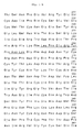
- human hepatocyte growth factor represented by the amino acid sequence as shown in Fig. 1, which contains a signal sequence; hHGF represented by the amino acid sequence ranging from the 30th glutamic acid residue (Glu) to the last serine residue (Ser) in Fig. 1; and hHGF represented by the amino acid sequence ranging from the 32nd glutamine residue (Gln) to the last serine residue (Ser) in Fig. 1.
- the present invention also comprises modifications of hHGF as mentioned above which may have at least one addition, deletion or replacement of an amino acid, provided that these modifications are functionally equivalent to naturally occurring hHGF.
- a gene coding for the hHGF represented by any of said amino acid sequences is also provided; the gene represented by the base sequence as shown in Fig. 2, which codes for hHGF containing a signal sequence; the gene represented by the base sequence ranging from the 88th base, guanine (G), to the last base (G) in Fig. 2; and the gene represented by the base sequence ranging from the 94th base, cytosine (C), to the last base (G) in Fig. 2.
- the present invention also provides a process for producing hHGF represented by the amino acid sequence of Fig. 1 which comprises transforming a host cell with an expression vector containing a gene coding for the hHGF, and culturing the resulting transformant. Further, a transformant capable of producing the hHGF is provided according to the present invention.
- the gene (cDNA) coding for the hHGF of the present invention may have the base sequence as shown in Fig. 2, in which, however, only the base sequence of a single-stranded DNA is described while complementary base sequences are obmitted as a matter of convenience.
- the gene may be used to express hHGF having the amino acid sequence as shown in Fig. 1 by the recombinant DNA technology.
- a protein translated from the corresponding mRNA coding for the hHGF contains a signal sequence.
- This signal sequence will be cleaved off when the protein is secreted from host cells, thus resulting in the production of hHGF having the amino acid sequence which ranges from the 30th glutamic acid residue (Glu) or the 32nd glutamine residue (Gln) to the last amino acid residue of the sequence represented by Fig. 1.
- Glu glutamic acid residue
- Gln 32nd glutamine residue
- an hHGF-encoding gene having the base sequence which ranges from the 88th G or the 94th C to the last base of the sequence represented by Fig. 2, may be used after ligating the gene with an ATG codon of a vector DNA.
- a DNA fragment of a gene coding for hHGF of the present invention may be obtained by the following procedures:
- hHGF may be purified from plasma of patients with fulminant hepatitis.
- the purified hHGF will be dissociated into two polypeptides by the breakage of disulfide bonds under reducing conditions.
- the larger polypeptide having a molecular weight of 56,000 to 65,000 is called "H chain", and the smaller one having a molecular weight of 32,000 to 35,000 "L chain”.
- hHGF The purified hHGF is reduced and the thiol groups of cystein residues formed are carboxymethylated followed by subjecting to reverse-phase high-pressure liquid chromatography to isolate the H and L chains.
- hHGF may be electrophoresed under a reducing condition on a gel, from which each of the H and L chains is extracted.
- the N-terminal amino acid sequences of the both chains may then be determined by analyzing the chains by Applied Biosystems gas phase protein sequencer.
- hHGF itself, of after separating it into H and L chains, may be hydrolyzed by an appropriate proteolytic enzyme, such as Achromobacter Protease I (lysyl endopeptidase).
- Achromobacter Protease I lysyl endopeptidase
- the resulting peptide fragments may be isolated by reverse-phase high-pressure liquid chromatography.
- Each peptide may be analyzed as described above to determine an internal amino acid sequence of the polypeptide.
- DNA base sequences may be deduced to select a sequence suitable for the preparation of an oligonucleotide, for example, the one as shown in Examples described hereinbelow.
- a sequence suitable for the preparation of an oligonucleotide for example, the one as shown in Examples described hereinbelow.
- Such an suitable oligonucleotide is synthesized and used as a probe.
- a cDNA library to be screened for a gene coding for hHGF may be any one derived from human liver, spleen or placenta and is commercially available from Clontech Laboratories, Inc. A placental cDNA library is particularly preferred. Further, cDNA library may also be prepared in a usual manner from a cell line or tissue material in which hHGF is expressed.
- E. coli is infected with lambda phage which contains such a cDNA and cultured, in accordance with the method of Maniatis (Molecular Cloning, A Laboratory Manual, pages 56 to 73, Cold Spring Harbor Laboratories, 1982). Plaques thus formed are then subjected to a selection process using as a probe an oligonucleotide prepared above based on the base sequence deduced from a portion of the amino acid sequence of hHGF, according to the plaque hybridization method (Molecular Cloning, A Laboratory Manual, pages 320 to 328, Cold Spring Harbor Laboratory, 1982). Thus, several different lambda phage clones may be easily obtained, each clone having both the base sequence used as the probe and a base sequence corresponding to the other region of the amino acid sequence of the desired hHGF.
- the positive plaques in the screening process are selected and the phages are grown according to the method of Maniatis (Molecular Cloning, A Laboratory Manual, pages 76 to 79, Cold Spring Harbor Laboratory, 1982).
- DNA is purified by the glycerol gradient method and digested with an appropriate enzyme, such as Eco RI.
- the resulting cDNA is then subcloned into a plasmid vector, such as pUC18 and pUC19, or a single stranded phage, such as M13mp18 and M13mp19.
- the base sequence of a desired cDNA segment may be determined according to the dideoxy chain termination method of Sanger et al . (Proc. Natl. Acad. Sci.
- the obtained cDNA sub-clones are analyzed to determine the base sequences, which may code for different portions of hHGF and be combined (Fig. 2).
- the whole base sequence of a gene coding for the entire amino acid sequence of hHGF as shown in Fig. 1 may be determined.
- hHGF hHGF-containing cells
- expressed hHGF must possess the biological activity to proliferate hepatic parenchymal cells.
- hHGF naturally occurring hHGF is a glycoprotein
- hHGF contains a number of cyteine residues
- the positions of disulfide bonds formed between thiol groups in the cysteine residues and the higher order sturcture play an important role in maintaining the activity of hHGF.
- animal cells for example, CHO, COS, and mouse L, C127 and FM3A cells
- host cells for example, animal cells, for example, CHO, COS, and mouse L, C127 and FM3A cells
- microorganisms such as yeast, eg., Saccharomyces cerevisiae , and Escherichia coli , eg., E . coli YA-21
- yeast eg., Saccharomyces cerevisiae
- Escherichia coli eg., E . coli YA-21
- a gene coding for immature hHGF containing a signal sequence i.e., such a gene that also contains 1 to 87, or 1 to 93 nucleotides of the DNA sequence as shown in Fig. 2
- a gene coding for immature hHGF containing a signal sequence i.e., such a gene that also contains 1 to 87, or 1 to 93 nucleotides of the
- An expression vector which may be used in the present invention contains a DNA fragment coding for at least a portion of the amino acid sequence of the hHGF protein downstream from the promoter of the vector. It may be contemplated herein to use various promoters, including SV40 promoter, the promoters of apolipoprotein E and Al genes, of heat shock protein gene, and of metallothionein gene, HSV TK promoter, adenovirus promoter, and retrovirus LTR. In the present invention, however, SV40 promoter or the promoter of metallothionein gene is preferred.
- a DNA fragment coding for immature hHGF containing a signal sequence is inserted into a vector downstream from its promoter in the direction of transcription. It is possible to insert a combination of two or three such hHGF DNA fragments. Also, it may be possible to prepare such a unit that comprises an hHGF DNA fragment, 5′ upstream of which a promoter is linked to, and to insert two or three such units into a vector in tandem along the direction of transcription.
- a polyadenylation signal should be present downstream from the hHGF gene in the expression vector.
- a polyadenylation signal may be derived from SV40 DNA, beta-globin gene, or metallothionein gene.
- a selective marker when an animal cell such as CHO cell is transformed with the expression vector.
- a selective marker gene may be inserted into the expression vector downstream from the polyadenylation signal along or against the direction of transcription, otherwise another plasmid containing a selective marker gene must be co-transformed to obtain a transformant.
- selective markers may include DHFR gene providing methotrexate resistance (J. Mol. Biol., 159, 601 (1982)); Neo gene providing G-418 antibiotic resistance (J. Mol. Appl. Genet., 1, 327 (1982)); E . coli derived Ecogpt gene providing mycophenolic acid resistance (Proc. Natl. Acad. Sci.
- the selective marker gene has a promoter, for example, SV40 promoter, 5′ upstream thereof and a polyadenylation signal 3′ downstream therefrom.
- a selective marker gene when such a selective marker gene is not inserted into the expression vector, another vector or plasmid which contains a marker enabling the selection of a transformant may be co-transformed into a host cell together with the expression vector containing hHGF gene.
- vectors may include pSV2neo (J. Mol. Appl. Genet., 1, 327 (1982)); pMBG (Nature, 294, 228 (1981)); pSV2gpt (Proc. Natl. Acad. Sci. USA, 78, 2072 (1981)); and pAd-D26-1 (J. Mol. Biol., 159, 601 (1982)).
- a transformant may be easily selected on the basis of the phenotype of the selective marker used.
- those cells containing the desired hHGF protein gene may be repeatedly subjected to co-transformation using a different selective marker. This may preferably increase about 20 fold the amount of protein expressed.
- Introduction of the expression vector into animal cells may be conducted by the calcium phosphate method (Virology, 52, 456 (1973)) or the electroporation method (J. Membr. Biol., 10, 279 (1972)).
- the calcium phosphate method is commonly used.
- Animal cells thus transformed may be cultured in conventional manners by suspension or adhesion culture.
- MEM or RPMI1640 may be used as a culture medium and the culture may be carried out in the absence of presence of 5-10% serum and in the presence of an appropriate amount of insulin, dexamethasone or transferrin.
- the animal cells producing the hHGF protein will secrete the produced hHGF protein into culture media.
- the hHGF protein can be purified and isolated from the suprenatant of the culture. Specifically, the supernatant may be subjected to a combination of various chromatographic operations on S-Sepharose, heparin-Sepharose, hydroxyapatite, and/or sulfated cerulophain to purify and isolate hHGF protein.
- hHGF(prehHGF) having the amino acid sequence starting from Met as shown in Fig. 1 is first expressed in host cells.
- the hHGF (prehHGf) is then hydrolyzed between the 31st Gly and the 32nd Gln in the host cells; thus, the signal peptide of 31 amino acids is cleaved off.
- the N-terminal Gln is then deaminated to be converted into pyroglutamic acid.
- hHGF having pyroglutamic acid residue at its N-terminus is secreted.
- the peptide chain from the N-terminal pyroglutamic acid to the 494th Arg constitutes the heavy (H) chain while the remaining peptide from the 495th Val to the last Ser the light (L) chain.
- the hHGF protein having biological activities can be produced abundantly, stably and easily by introducing, into a host cell, an expression vector into which the hHGF gene of the present invention has been inserted. Such production of hHGF has not been attained prior to the present invention.
- hHGF, hHGF-like substances or hHGF-containing fused proteins may be used in the treatment of hepatic diseases as hepatic regeneration-enhancing, hepatic function-improving, hepatitis-curing, hepatic cirrhosis-suppressing agents.
- a 50 ⁇ g portion of the purified hHGF preparation was dissolved in 100 ⁇ l of 50 mM Tris-HCl buffer (pH 9) containing 5M urea, and the resulting solution was mixed with an amount of Achromobacter Protease I equivalent to 1/200 of hHGF in molar ratio, followed by incubation at 37°C for 6 hours.
- the resulting peptide mixture was subjected to reduction and carboxymethylation in convention manners. Each peptide was then separated and isolated by means of reverse-phase high-pressure liquid chromatography using Bakerbond WP Octyl Column (J. T. Baker).
- oligonucleotides TH23 consisting of 17 bases (5′-T-G-T/C/A/G-A-A/G-A/G-A/G-T-C-T/C-T-C-C-A-T-A/G-T-T-3′) and 24 oligonucleotides TH24 consisting of 17 bases (5′-G-G-T/C-T-C-C-C-A-A/G-A-A-A/G/T-A-T-A/G-T-G-3′) were prepared.
- each synthetic oligonucleotide was labeled with 32P in a conventional manner using polynucleotide kinase in a reaction solution (50 mM Tris-HCl (pH 7.6), 10 mM magnesium chloride, 10 mM mercaptoethyl alcohol, 100 ⁇ M [ ⁇ 32P] ATP and substrate DNA). Unnecessary mononucleotides in these labeled samples were removed by conventional DEAE cellulose column chromatography.
- ⁇ phage cDNA library originated from 34-week-old human placenta was screened according to the manufacturer's specifications. E. coli Y-1090 strain was infected with phages of 1,000,000 clones and cultured at 42°C overnight on an NZY soft agar medium using five Petri dishes (24.5 cm x 24.5 cm), each dish containing 200,000 clones (NZY medium; 1% NZ-Amine, 0.5% yeast extracts and 0.5% sodium chloride, adjusted to pH 7.5 and supplemented with 0.25% magnesium chloride, and NZY soft agar medium; NZY medium supplemented with agar powder to its final concentration of 0.7% and autoclaved).
- Phage particles grown on one dish were transferred on two nylon membranes and each membrane was put on a filter paper impregnated with 0.1 M sodium hydroxide and 1.5 M sodium chloride. After standing still for 2 minutes on the filter paper, moisture of the nylon membrane was removed by using another dry filter paper. The thus dried membrane was then placed on another filter paper impregnated with 2 x SSCP - 0.2 M Tris-HCl (pH 7.4), stood still on the filter paper and then air-dried on another dry filter paper. These procedures were repeated again.
- 2 x SSCP means double concentration of SSCP solution and similar way of expressions are used hereinafter (10 x SSCP; 1.2 M sodium chloride, 150 mM sodium citrate, 130 mM potassium dihydrogenphosphate and 1 mM EDTA and pH 7.2).
- nylon membranes were incubated for 36 hours in a hybridization solution containing the 32P-labeled synthetic oligonucleotide probe prepared in [1] above [3 x SSC, 10 x Denhalt, 50 ⁇ g/ml of salmon sperm DNA, 1 M sodium chloride, 1% SDS, 250 ⁇ g/ml of salmon sperm DNA and 100,000 c.p.m./ml of 32P-labeled probe DNA per each synthesized probe].
- the incubation temperature was calculated by regarding A or T as 2°C and G or C as 4°C and totaling these values of each probe (42°C in the case of the TH23 probe and 46°C in the case of the TH24 probe).
- coli Y-1090 cells were infected with the thus extracted phage particles, and an appropriate amount of the infected cells was cultured on the soft agar medium in a Petri dish (9 cm in diameter).
- the plaque hybridization was carried out as previously described. By repeating a series of these procedures, each clone corresponding to the positive autoradiographic signal was isolated.
- DNA fragments were extracted from the ⁇ phage clones and subcloned into plasmid vectors pUC18 and pUC19 and single strand phage vectors M13mp18 and M13mp19 as follows.
- Resulting pellet was suspended in 6 ml of buffer A [0.5% NP40, 36 mM calcium chloride, 30 mM Tris-HCl (pH 7.5), 50 mM magnesium chloride, 125 mM potassium chloride, 0.5 mM EDTA, 0.25% deoxycholic acid and 0.6 mM mercaptoethanol].
- the suspension was then mixed with 100 ⁇ l of 10 mg/ml Deoxyribonuclease I and 10 ⁇ l of 10 mg/ml Ribonuclease A and incubated at 30°C for 30 minutes in order to hydrolyze E. coli -originated nucleic acids.
- reaction mixture was mixed with an equal volume of chloroform and stirred thoroughly followed by centrifugation 3,000 rpm for 10 minutes (model LC-06; rotor, TS-7; Tomy Seiko Co., Ltd.) to obtain supernatant fluid.
- a double-layer glycerol solution was prepared in a centrifugal tube for ultracentrifugation (rotor, RPS40T; Hitachi Ltd.) by by firstly chargingthe tube with 1 ml of a 40% glycerol solution [0.5% NP40, 30 mM Tris-HCl (pH 7.5), 125 mM potassium chloride, 0.5 mM EDTA, 0.6 mM mercaptoethanol and 40% glycerol] and then by placing thereon 3 ml of a 10% glycerol solution [0.5% NP40, 30 mM Tris-HCl (pH 7.5), 125 mM potassium chloride, 0.5 mM EDTA, 0.6 mM mercaptoethanol and 10% glycerol].
- a 40% glycerol solution [0.5% NP40, 30 mM Tris-HCl (pH 7.5), 125 mM potassium chloride, 0.5 mM EDTA, 0.6
- phage particles recovered as the pellet in the tube were suspended in 0.4 ml of 40 mM Tris-HCl (pH 7.5), 10 mM EDTA and 2% SDS and the suspension was incubated at 55°C for 1 hour in the presence of 4 ⁇ l of 10 mg/ml Proteinase K. Thereafter, the resulting solution was transferred into an Eppendorf tube, and the phage DNA was extracted with an equal volume of phenol/chloroform and recovered by ethanol precipitation. In this way, 200 ⁇ g of the phage DNA was obtained.
- the phage DNA was digested with restriction enzyme Eco RI in a conventional manner and the digests were analyzed by agarose gel electrophoresis. Three Eco RI fragments of 0.2 kb, 0.85 kb and 0.72 kb in size were obtained from the clone, ⁇ hHGF21. On the other hand, cDNA fragments were obtained by recovering the insert cDNA fragments from the agarose gel by conventional methods.
- Fig. 3 shows the scheme used for the preparation of an hHGF expressino plasmid.
- plasmid pUCHGF-1 DNA was prepared, which comprised a Bam HI- Kpn I fragment containing hHGF cDNA (Biochem. Biophys. Res. Commun., 163(2), 967-973 (1989)).
- the Bam HI- Kpn I fragment of about 2.3 kb in size extended from the Bam HI site at 27 base upstream of the initiation codon ATG to the Kpn I site at 8 base upstream of the stop codon TAG.
- the plasmid DNA (10 ug) was digested with Kpn I restriction enzyme in a conventional manner.
- the resulting DNA fragment was extracted with phenol/chloroform, purified by ethanol precipitation, and dissolved in 10 ⁇ l water.
- the thus modified plasmid was used to transform E . coli in a conventional manner. From the resulting transformants, plasmid DNA was prepared in accordance with the conventional method described in "Molecular Cloning", Cold Spring Harbor Laboratory, p.93 (1982).
- the plasmid DNA (10 ug) was digested with Bam HI restriction enzyme in a conventional manner.
- the dresulting reaction mixture was subjected to 1.0% agarose gel electrophoresis to isolate the hHGF DNA fragments containing ATG initiation and TGA stop codons from concomitant undesirable DNA fragments.
- Bam HI- Bam HI DNA fragment of about 2.3 kb coding for hHGF was prepared according to Maniatis et al . method described in "Molecular Cloning", Cold Spring Harbor Laboratory, p.164 (1982). The DNA fragment was treated with T4 DNA polymerase in a conventional manner to form blunt ends at both termini thereof. The blunt-ended DNA fragment was extrated with phenol/chloroform, purified by ethanol precipitation, and dissolved in 10 ⁇ l water.
- 0.05 ug of expression vector pKCR as described in Proc. Natl. Acad. Sci., 78, 1527 (1981) was digested with Sma I restriction enzyme to form blunt ends in a conventional manner, extracted with phenol/chloroform, and purified by ethanol precipitation.
- the vector DNA was dissolved in 400 ⁇ l of 50 mM Tris-HCl (pH 8), 1 mM magnesium chloride, and dephosphorylated with 1 unit of bacterial alkaline phosphatase (TOYOBO, BAP-101) at 65°C for 30 minutes.
- the DNA fragment was then extracted with phenol/chloroform, purified by ethanol precipitation, and dissolved in 10 ul water.
- the vector pKCR DNA fragment (0.01 ug) prepared above was ligated to the blunt-ended Bam HI hHGF cDNA fragment (0.1 ug) in the presence of T4 DNA ligase (TOYOBO, LGA-101) in 20 ul of 66 mM Tris-HCl (pH 7.6), 6.6 mM magnesium chloride, 10 mM dithiothreitol, 66 uM ATP at 14°C for 12 hours.
- T4 DNA ligase TOYOBO, LGA-101
- the resulting reaction mixture (10 ul) was used to transform E . coli HB 101 (Takara Shuzo) according to the specification.
- the transformants were cultured on a medium containing 50 ug/ml ampicillin. Several tens of ampicillin resistant strains were obtained.
- Plasmids from these strains were analyzed in accordance with Maniatis et al . method described in "Molecular Cloning", Cold Spring Harbor Laboratory, 86-96 (1982). Thus, plasmid pKCRHGF-2 was obtained, in which two hHGF genes had been incorporated in tandem into the Sma I cleavage site present between the promoter and polyadenylation signal in the expression vector pKCR.
- Fig. 4 The structure of the plasmid pKCRHGF-2 is shown in Fig. 4.
- plasmid pSV2neo DNA J. Appl. Genet., 1, 327 (1982)
- plasmid pAd-D26-1 DNA J. Molec. Biol., 159, 601 (1982)
- CHO cells were cultured to semi-confluent in ERDF medium (Kyokuto Seiyaku, Japan) containing 10% FCS (fetal calf serum) in a Petri dish of 9 cm in diameter. The medium was removed from the dish and there was added dropwise a DNA solution, which had previously been prepared in the following manner:
- This DNA solution was added to the semi-confluent cells in Petri dish and the cells were allowed to stand at room temperature for 30 minutes. Then, 9 ml of ERDF medium containing 10% FCS was added to the dish followed by culturing in the presence of 5% CO2 at 37°C for 4 to 5 hours.
- the media was removed from the dish and the cells were washed with 5 ml of 1xTBS++ solution (25 mM Tris-HCl, pH 7.5, 140 mM sodium chloride, 5 mM potassium chloride, 0.6 mM disodium hydrogenphosphate, 0.08 mM calcium chloride, 0.08 mM magnesium chloride).
- 1xTBS++ solution 25 mM Tris-HCl, pH 7.5, 140 mM sodium chloride, 5 mM potassium chloride, 0.6 mM disodium hydrogenphosphate, 0.08 mM calcium chloride, 0.08 mM magnesium chloride.
- 5 ml of 1xTBS++ solution containing 20% glycerol was added to the cells.
- the cells were allowed to stand at room temperature for 1 to 2 minutes. After removing the supernatant, the cells were again washed with 5 ml of 1xTBS++ solution. Thereafter, 10 ml of ERDF medium containing 10% FCS was added to the Petri dish followed by
- the medium was removed from the dish and the cells were washed with 5 ml of 1xTBS++ solution. Then, 2 ml of trypsin-EDTA solution (Sigma) was added to the cells and the mixture was allowed to stand at room temperature for 30 seconds. The trypsin-EDTA solution was then removed from the dish. After 5 minutes, 10 ml of ERDF medium containing 10% FCS was added to the dish to strip off the cells. The cultured cells of one Petri dish of 9 cm in diameter were divided into 10 portions and each portion was placed in a Petri dish of 9 cm in diameter. G418 sulphate (GENETICIN, GIBCO) was added to each dish to 200 ug/ml and the cells were further cultured.
- G418 sulphate GENETICIN, GIBCO
- G418-resistant cells were isolated and distributed in wells of a 24-well culture plate, each well of 3.1 cm2 containing 1 ml of ERDF medium + 10% FCS, followed by further culturing for approximately 7 days.
- the medium was replaced by FCS-free ERDF medium and the culture was continued for further 72 hours. Then, 2 ml of the medium was recovered from each well and concentrated to 50 ⁇ l by Centricon (Millipore). About 15 ⁇ l of the sample was subjected to electrophoresis on SDS-polyacrylamide gel.
- hHGF protein was isolated from each well and quantitative measurement of hHGF protein was performed by enzyme immunoassay. The amount of hHGF expressed was determined in B-1, B-27 and B-102 cells, which produced a significantly lare amount of protein.
- Plasmid DNAs of the expression vector pKCRHGF-2 prepared in (I) above and pMBG coding for mycophenolic acid resistance as a marker for selecting transformants were recovered from recombinant E . coli strains containing the respective plasmid and purified, in accordance with the aforementioned Maniatis et al . method.
- the hHGF-expressing cells were cultured to semi-confluent in ERDF medium containing 10% FCS in a Petri dish of 9 cm in diameter. The medium was then removed from the dish and there was added dropwise the DNA solution, which had previously been prepared in the same manner as in Example 2 (II) except that 10 ug of pKCRHGF-2 plasmid DNA and 1 ug of pMBG plasmid DNA were used.
- This DNA solution was added to the semi-confluent cells in Petri dish and the cells were allowed to stand at room temperature for 30 minutes. Then, 9 ml of ERDF medium containing 10% FCS was added to the dish followed by culturing in the presence of 5% CO2 at 37°C for 4 to 5 hours.
- the medium was removed from the dish and the cells were washed with 5 ml of 1xTBS++ solution. After removing the 1xTBS++ solution, 5 ml of 1xTBS++ solution containing 20% glycerol was added to the cells. The cells were allowed to stand at room temperature for 1 to 2 minutes. After removing the supernatant, the cells were again washed with 5 ml of 1xTBS++ solution. Thereafter, 10 ml of ERDF medium containing 10% FCS was added to the Petri dish followed by culturing in the presence of 5% CO2 at 37°C.
- the medium was removed from the dish and the cells were washed with 5 ml of 1xTBS++ solution. Then, 2 ml of trypsin-EDTA solution (Sigma) was added to the cells and the mixture was allowed to stand at room temperature for 30 seconds. The trypsin-EDTA solution was then removed from the dish. After 5 minutes, 10 ml of alpha-MEM(-) medium containing 10% FCS was added to the dish to strip off the cells. The cultured cells of one Petri dish of 9 cm in diameter were divided into 10 portions and each portion was placed in a Petri dish of 9 cm in diameter. Mycophenolic acid (Sigma) and xanthine (Sigma) were added to each dish to 1 ug/ml and 250 ug/ml, respectively, and the cells were further cultured.
- trypsin-EDTA solution Sigma
- mycophenolic acid-resistant cells were isolated and distributed in wells of a 24-well culture plate, each well of 3.1 cm2 containing 1 ml of ERDF medium + 10% FCS, followed by further culturing for approximately 7 days.
- the medium was replaced by FCS-free ERDF medium and the culture was continued for further 72 hours. Then, 2 ml of the medium was recovered from each well and concentrated to 50 ul by Centricon (Millipore). About 15 ul of the sample was subjected to electrophoresis on SDS-polyacrylamide gel.
- the hHGF producing cell BD-24 prepared in Example 3 was cultured in ERDF medium containing 10% FCS. The supernatant (500 ml) was adsorbed on a column filled with 10 ml S-Sepharose Fast FlowTM (Pharmacia). The proteins were eluted using 10 mM sodium phosphate-containing buffer (pH 7.5) with increasing concentrations of sodium chloride therein. Recombinant hHGF protein was eluted with approximately 0.7 M sodium chloride.
- This hHGF fraction was analyzed by SDS-polyacrylamide gel electrophoresis yielding a broad band with molecular weights of about 76,000 to 92,000 under non-reducing condition and on the other hand a broad band of about 60,000 to 65,000 and a weak band of about 56,000 under reducing condition, which corresponded to H chain of hHGF protein, further giving two bands with molecular weights of about 32,000 to 35,000, which corresponded to L chain of hHGF protein.
- These band multiplicity and broadness may arise from heterogeneity of glycosylated sugar chains on hHGF protein.
- the buffer of the purified hHGF protein solution was substitued by 0.1 M aqueous ammonium bicarbonate solution. This solution was mixed with 1/50 volume of Staphylococcus aureus V8 protease (Miles Laboratory) and incubated at 37°C overnight to yield a peptide mixture. This mixture was subjected to reverse phase high pressure liquid chromatography using C8 column (Bakerbond, 4.6 x 250 mm) while increasing acetonitrile concentration from 0% to 60% at a rate of 1% per minute.
- composition shown in Table substantially coincides with the theoretical composition of a peptide extending from the 32nd glutamine to the 41st glutamic acid, as calculated from the first methionine, in the amino acid sequence (Fig. 1) deduced from the base sequence of cDNA coding for hHGF (Fig. 2).
- This peptide was analyzed by fast atom bombardment massspectroscopy (NIHON DENSHI, Japan, model HX-100). A peak was found at mass 1321 indicating that the peptide had a molecular weight of 1320. Since the theoretical molecular weight of the peptide extending from the 32nd glutamine to the 41st glutamic acid is 1337 of the amino acid sequence shown in Fig. 1, it can be concluded that the amino terminal glutamine of this peptide is converted by deamination into pyroglutamic acid.
- the N terminal amino acid of the secreted hHGF protein is found to be pyroglutamic acid derived from the 32nd amind acid glutamine in the amino acid sequence shown in Fig. 1.
Landscapes
- Health & Medical Sciences (AREA)
- Life Sciences & Earth Sciences (AREA)
- Chemical & Material Sciences (AREA)
- Genetics & Genomics (AREA)
- Organic Chemistry (AREA)
- Engineering & Computer Science (AREA)
- Zoology (AREA)
- Biomedical Technology (AREA)
- Wood Science & Technology (AREA)
- General Health & Medical Sciences (AREA)
- Bioinformatics & Cheminformatics (AREA)
- Molecular Biology (AREA)
- Biotechnology (AREA)
- Biochemistry (AREA)
- Gastroenterology & Hepatology (AREA)
- Biophysics (AREA)
- General Engineering & Computer Science (AREA)
- Proteomics, Peptides & Aminoacids (AREA)
- Medicinal Chemistry (AREA)
- Microbiology (AREA)
- Toxicology (AREA)
- Physics & Mathematics (AREA)
- Cell Biology (AREA)
- Plant Pathology (AREA)
- Preparation Of Compounds By Using Micro-Organisms (AREA)
- Micro-Organisms Or Cultivation Processes Thereof (AREA)
- Peptides Or Proteins (AREA)
- Crystals, And After-Treatments Of Crystals (AREA)
- Soft Magnetic Materials (AREA)
Applications Claiming Priority (6)
| Application Number | Priority Date | Filing Date | Title |
|---|---|---|---|
| JP1209449A JP2577091B2 (ja) | 1989-08-11 | 1989-08-11 | 肝実質細胞増殖因子及びそれをコードする遺伝子 |
| JP209449/89 | 1989-08-11 | ||
| JP2088592A JP3036782B2 (ja) | 1990-04-03 | 1990-04-03 | ヒト肝実質細胞増殖因子の生産方法および該因子を産生する形質転換体 |
| JP88592/90 | 1990-04-03 | ||
| JP200898/90 | 1990-07-27 | ||
| JP2200898A JP2963163B2 (ja) | 1990-07-27 | 1990-07-27 | ヒト肝実質細胞増殖因子 |
Publications (2)
| Publication Number | Publication Date |
|---|---|
| EP0412557A1 true EP0412557A1 (de) | 1991-02-13 |
| EP0412557B1 EP0412557B1 (de) | 1997-04-23 |
Family
ID=27305852
Family Applications (1)
| Application Number | Title | Priority Date | Filing Date |
|---|---|---|---|
| EP90115397A Expired - Lifetime EP0412557B1 (de) | 1989-08-11 | 1990-08-10 | Wachstumsfaktor aus parenchymalen Lebenszellen, dafür kodierendes Gen, Verfahren zur Herstellung dieses Faktors und Transformanten |
Country Status (7)
| Country | Link |
|---|---|
| US (1) | US5500354A (de) |
| EP (1) | EP0412557B1 (de) |
| KR (1) | KR960006122B1 (de) |
| AT (1) | ATE152124T1 (de) |
| CA (1) | CA2022752C (de) |
| DE (2) | DE412557T1 (de) |
| HU (1) | HUT58798A (de) |
Cited By (16)
| Publication number | Priority date | Publication date | Assignee | Title |
|---|---|---|---|---|
| WO1991012272A1 (en) * | 1990-02-16 | 1991-08-22 | Imperial Cancer Research Technology Limited | Protein factor |
| EP0456188A1 (de) * | 1990-05-09 | 1991-11-13 | Toshikazu Nakamura | Arzneimittel gegen Leberzirrhose |
| EP0461560A1 (de) * | 1990-06-11 | 1991-12-18 | Toshikazu Nakamura | Rekombinanter menschlicher Hepatozytwachstumsfaktor und Verfahren zu seiner Herstellung |
| EP0462549A1 (de) * | 1990-06-19 | 1991-12-27 | NAKAMURA, Toshikazu | Verfahren zur Herstellung eines therapeutischen Mittels für Nierenkrankheiten |
| EP0517182A1 (de) * | 1991-06-03 | 1992-12-09 | Mitsubishi Chemical Corporation | Hepatozytwachstumsfaktor enthaltende Mittel |
| EP0492614A3 (en) * | 1990-12-28 | 1993-02-10 | Toshikazu Nakamura | Epitheliocyte growth accelerator |
| EP0550296A3 (en) * | 1991-11-28 | 1993-12-29 | Terumo Corp | Vascular endothelial cells growth factor |
| US5316921A (en) * | 1992-05-18 | 1994-05-31 | Genentech, Inc. | Single-chain hepatocyte growth factor variants |
| US5328837A (en) * | 1992-05-18 | 1994-07-12 | Genentech, Inc. | Hepatocyte growth factor protease domain variants |
| EP0539590A4 (de) * | 1990-07-13 | 1995-05-24 | ||
| US5466593A (en) * | 1992-11-05 | 1995-11-14 | Mitsubishi Kasei Corporation | Hepatocyte growth factor activating protease and gene encoding the protease |
| US5547856A (en) * | 1992-05-18 | 1996-08-20 | Genentech, Inc. | Hepatocyte growth factor variants |
| US5554509A (en) * | 1993-01-26 | 1996-09-10 | Clonit Spa | Nucleotide probes and methods for determining TaqI polymorphisms in the human Apo(a) gene |
| EP0691347A4 (de) * | 1993-03-23 | 1997-07-30 | Taisho Pharmaceutical Co Ltd | Wachstumssubstanz für leberparenchymzellen |
| WO2001098347A1 (en) * | 2000-06-22 | 2001-12-27 | Nippon Zenyaku Kogyo Ltd. | Canine liver cell growth factor |
| KR20020097319A (ko) * | 2001-06-20 | 2002-12-31 | 윤재승 | 사람 간세포 성장인자 코딩 벡터, 형질전환 균주 및이로부터 발현된 재조합 단백질 |
Families Citing this family (8)
| Publication number | Priority date | Publication date | Assignee | Title |
|---|---|---|---|---|
| ATE513042T1 (de) * | 1998-02-05 | 2011-07-15 | Glaxosmithkline Biolog Sa | Tumorassoziierte antigenderivate der mage-familie,nukleinsäuresequenzen, die dafür kodieren, zur herstellung von fusionsproteinen und zusammensetzungen zur impfung |
| KR100562824B1 (ko) | 2002-03-20 | 2006-03-23 | 주식회사 바이로메드 | 유전자 발현효율이 높으며 간세포 성장인자의 두 가지이형체를 동시에 발현하는 하이브리드 간세포 성장인자유전자 |
| US20090202606A1 (en) * | 2008-01-25 | 2009-08-13 | Viromed Co., Ltd. | Treatment and Prevention of Cardiac Conditions Using Two or More Isoforms of Hepatocyte Growth Factor |
| KR101290322B1 (ko) * | 2008-04-09 | 2013-07-26 | 주식회사 바이로메드 | 플라스미드 dna의 발현 증강용 동결건조 dna 제제 |
| JPWO2012144535A1 (ja) | 2011-04-18 | 2014-07-28 | 国立大学法人京都大学 | 急性肝不全抑制剤及びその薬効評価法 |
| JP6240337B2 (ja) | 2013-10-22 | 2017-11-29 | バイロメッド カンパニー リミテッド | 肝細胞増殖因子の2つ以上のアイソフォームを利用した筋萎縮性側索硬化症の予防又は治療用組成物 |
| KR101808192B1 (ko) * | 2016-08-26 | 2018-01-18 | 아미코젠주식회사 | 7-아미노세팔로스포란산의 고농도 생산 재조합 아크레모니움 크리소제눔 균주의 제조방법 및 이 방법으로 제조된 균주 |
| AU2019305221B2 (en) | 2018-07-19 | 2024-11-07 | Helixmith Co., Ltd. | Lyophilized pharmaceutical compositions for naked DNA gene therapy |
-
1990
- 1990-08-07 CA CA002022752A patent/CA2022752C/en not_active Expired - Lifetime
- 1990-08-10 EP EP90115397A patent/EP0412557B1/de not_active Expired - Lifetime
- 1990-08-10 DE DE199090115397T patent/DE412557T1/de active Pending
- 1990-08-10 DE DE69030539T patent/DE69030539T2/de not_active Expired - Lifetime
- 1990-08-10 AT AT90115397T patent/ATE152124T1/de not_active IP Right Cessation
- 1990-08-10 HU HU904957A patent/HUT58798A/hu unknown
- 1990-08-11 KR KR1019900012415A patent/KR960006122B1/ko not_active Expired - Lifetime
-
1993
- 1993-07-09 US US08/089,417 patent/US5500354A/en not_active Expired - Lifetime
Non-Patent Citations (4)
| Title |
|---|
| BIOCHEMICAL AND BIOPHYSICAL RESEARCH COMMUNICATIONS, vol. 163, no. 2, 15th September 1989, pages 967-973, Academic Press, Inc.; K. MIYAZAWA et al.: "Molecular cloning and sequence analysis of cDNA for human hepatocyte growth factor" * |
| CANCER RESEARCH, vol. 49, 15th June 1989, pages 3314-3320; R. Zarnegar et al.: "Purification and biological characterization of human hepatopoietin A, a polypeptide growth factor for hepatocytes" * |
| J. CLIN. INVEST., vol. 81, February 1988, pages 414-419; E. GOHDA et al.: "Purification and partial characterization of hepatocyte growth factor from plasma of a patient with fulminant hepatic failure" * |
| NATURE, vol. 342, 23rd November 1989, pages 440-443; T. NAKAMURA et al.: "Molecular cloning and expression of human hepatocyte growth factor" * |
Cited By (23)
| Publication number | Priority date | Publication date | Assignee | Title |
|---|---|---|---|---|
| WO1991012272A1 (en) * | 1990-02-16 | 1991-08-22 | Imperial Cancer Research Technology Limited | Protein factor |
| EP0456188A1 (de) * | 1990-05-09 | 1991-11-13 | Toshikazu Nakamura | Arzneimittel gegen Leberzirrhose |
| EP0461560A1 (de) * | 1990-06-11 | 1991-12-18 | Toshikazu Nakamura | Rekombinanter menschlicher Hepatozytwachstumsfaktor und Verfahren zu seiner Herstellung |
| EP0859009A3 (de) * | 1990-06-11 | 1998-08-26 | Toshikazu Nakamura | Rekombinanter menschlicher Hepatozytwachstumsfaktor und Verfahren zu seiner Herstellung |
| EP0462549A1 (de) * | 1990-06-19 | 1991-12-27 | NAKAMURA, Toshikazu | Verfahren zur Herstellung eines therapeutischen Mittels für Nierenkrankheiten |
| EP0539590A4 (de) * | 1990-07-13 | 1995-05-24 | ||
| EP0492614A3 (en) * | 1990-12-28 | 1993-02-10 | Toshikazu Nakamura | Epitheliocyte growth accelerator |
| US5342831A (en) * | 1990-12-28 | 1994-08-30 | Toyo Boseki Kabushiki Kaisha | Epitheliocyte growth accelerator |
| EP0517182A1 (de) * | 1991-06-03 | 1992-12-09 | Mitsubishi Chemical Corporation | Hepatozytwachstumsfaktor enthaltende Mittel |
| EP0550296A3 (en) * | 1991-11-28 | 1993-12-29 | Terumo Corp | Vascular endothelial cells growth factor |
| US5328837A (en) * | 1992-05-18 | 1994-07-12 | Genentech, Inc. | Hepatocyte growth factor protease domain variants |
| US5316921A (en) * | 1992-05-18 | 1994-05-31 | Genentech, Inc. | Single-chain hepatocyte growth factor variants |
| US5547856A (en) * | 1992-05-18 | 1996-08-20 | Genentech, Inc. | Hepatocyte growth factor variants |
| US5879910A (en) * | 1992-05-18 | 1999-03-09 | Genetech, Inc. | Hepatocyte growth factor protease domain variants |
| US5580963A (en) * | 1992-05-18 | 1996-12-03 | Genentech, Inc. | Single-chain hepatocyte growth factor variants |
| US5466593A (en) * | 1992-11-05 | 1995-11-14 | Mitsubishi Kasei Corporation | Hepatocyte growth factor activating protease and gene encoding the protease |
| US5677164A (en) * | 1992-11-05 | 1997-10-14 | Mitsubishi Chemical Corporation | Hepatocyte growth factor activating protease and gene encoding the protease |
| US5554509A (en) * | 1993-01-26 | 1996-09-10 | Clonit Spa | Nucleotide probes and methods for determining TaqI polymorphisms in the human Apo(a) gene |
| EP0691347A4 (de) * | 1993-03-23 | 1997-07-30 | Taisho Pharmaceutical Co Ltd | Wachstumssubstanz für leberparenchymzellen |
| US5807711A (en) * | 1993-03-23 | 1998-09-15 | Taisho Pharmaceutical Co., Ltd. | Parenchymal hepatocyte growth factors |
| WO2001098347A1 (en) * | 2000-06-22 | 2001-12-27 | Nippon Zenyaku Kogyo Ltd. | Canine liver cell growth factor |
| US7129064B2 (en) | 2000-06-22 | 2006-10-31 | Nippon Zenyaku Kogyo Ltd. | Canine hepatocyte growth factor |
| KR20020097319A (ko) * | 2001-06-20 | 2002-12-31 | 윤재승 | 사람 간세포 성장인자 코딩 벡터, 형질전환 균주 및이로부터 발현된 재조합 단백질 |
Also Published As
| Publication number | Publication date |
|---|---|
| HUT58798A (en) | 1992-03-30 |
| KR910004803A (ko) | 1991-03-29 |
| CA2022752C (en) | 1998-07-07 |
| DE69030539T2 (de) | 1997-09-11 |
| DE412557T1 (de) | 1991-07-04 |
| DE69030539D1 (de) | 1997-05-28 |
| ATE152124T1 (de) | 1997-05-15 |
| US5500354A (en) | 1996-03-19 |
| KR960006122B1 (ko) | 1996-05-09 |
| EP0412557B1 (de) | 1997-04-23 |
| CA2022752A1 (en) | 1991-02-12 |
| HU904957D0 (en) | 1991-01-28 |
Similar Documents
| Publication | Publication Date | Title |
|---|---|---|
| EP0412557B1 (de) | Wachstumsfaktor aus parenchymalen Lebenszellen, dafür kodierendes Gen, Verfahren zur Herstellung dieses Faktors und Transformanten | |
| AU650893B2 (en) | O-glycosylated alpha-2 interferon | |
| AU646014B2 (en) | Novel platelet-derived growth factor B chain analogs and method for homogeneous production thereof | |
| EP0775711A1 (de) | Peptide mit antithrombotischer aktivität und verfahren zu ihrer herstellung | |
| US5972886A (en) | Megakaryocyte differentiation factor | |
| EP0221761A2 (de) | DNS-Sequenzen, rekombinante DNS-Moleküle und Verfahren zur Herstellung von "mullerian inhibiting substance"-ähnlichen Polypeptiden | |
| US5328836A (en) | Plasmids containing DNA encoding the amino acid sequence of TCF-II and use thereof | |
| WO2007122975A1 (ja) | Hgf前駆体蛋白質改変体及びその活性型蛋白質 | |
| CA2213512A1 (en) | Method for producing bioactive fusion protein | |
| JP3921271B2 (ja) | グルクロン酸転移酵素をコードするdna | |
| WO2000071716A2 (en) | Vascular endothelial growth factor dimers | |
| DE69324547T2 (de) | Protease, die menschlichen Hepatozytwachtumsfaktor umsetzt, und dafür kodierendes Gen | |
| JP2577091B2 (ja) | 肝実質細胞増殖因子及びそれをコードする遺伝子 | |
| JP2706704B2 (ja) | 組換ヒト肝実質細胞増殖因子 | |
| US20020165358A1 (en) | TCF mutant | |
| US5916770A (en) | Macrophage stimulating protein variant and method for producing the same | |
| US5427780A (en) | Composition comprising Mullerian inhibiting substance-like polypeptides | |
| EP0759467A2 (de) | Inhibitor des Hepatozytwachstumsfaktoraktivators | |
| JP2980889B2 (ja) | 肝実質細胞増殖因子 | |
| JP2980888B2 (ja) | 肝実質細胞増殖因子をコードする遺伝子 | |
| JP3610395B2 (ja) | 成熟肝実質細胞増殖因子 | |
| JP2859577B2 (ja) | 肝実質細胞増殖因子 | |
| JP2963163B2 (ja) | ヒト肝実質細胞増殖因子 | |
| US6280968B1 (en) | Human PEC-60-like protein and DNA encoding the same | |
| JP3292873B2 (ja) | 組換肝実質細胞増殖因子 |
Legal Events
| Date | Code | Title | Description |
|---|---|---|---|
| PUAI | Public reference made under article 153(3) epc to a published international application that has entered the european phase |
Free format text: ORIGINAL CODE: 0009012 |
|
| AK | Designated contracting states |
Kind code of ref document: A1 Designated state(s): AT CH DE FR GB IT LI LU NL SE |
|
| 17P | Request for examination filed |
Effective date: 19901224 |
|
| ITCL | It: translation for ep claims filed |
Representative=s name: BARZANO' E ZANARDO MILANO S.P.A. |
|
| ITCL | It: translation for ep claims filed |
Representative=s name: ING. A. GIAMBROCONO & C. S.R.L. |
|
| TCAT | At: translation of patent claims filed | ||
| TCNL | Nl: translation of patent claims filed | ||
| EL | Fr: translation of claims filed | ||
| DET | De: translation of patent claims | ||
| 17Q | First examination report despatched |
Effective date: 19931026 |
|
| EL1 | Fr: translation or corrected translation of claims filed | ||
| RAP1 | Party data changed (applicant data changed or rights of an application transferred) |
Owner name: MITSUBISHI CHEMICAL CORPORATION |
|
| GRAG | Despatch of communication of intention to grant |
Free format text: ORIGINAL CODE: EPIDOS AGRA |
|
| GRAH | Despatch of communication of intention to grant a patent |
Free format text: ORIGINAL CODE: EPIDOS IGRA |
|
| GRAH | Despatch of communication of intention to grant a patent |
Free format text: ORIGINAL CODE: EPIDOS IGRA |
|
| GRAA | (expected) grant |
Free format text: ORIGINAL CODE: 0009210 |
|
| AK | Designated contracting states |
Kind code of ref document: B1 Designated state(s): AT CH DE FR GB IT LI LU NL SE |
|
| PG25 | Lapsed in a contracting state [announced via postgrant information from national office to epo] |
Ref country code: AT Effective date: 19970423 Ref country code: CH Effective date: 19970423 Ref country code: LI Effective date: 19970423 Ref country code: NL Effective date: 19970423 |
|
| REF | Corresponds to: |
Ref document number: 152124 Country of ref document: AT Date of ref document: 19970515 Kind code of ref document: T |
|
| REG | Reference to a national code |
Ref country code: CH Ref legal event code: NV Representative=s name: ROTTMANN, ZIMMERMANN + PARTNER AG Ref country code: CH Ref legal event code: EP |
|
| ITF | It: translation for a ep patent filed | ||
| REF | Corresponds to: |
Ref document number: 69030539 Country of ref document: DE Date of ref document: 19970528 |
|
| ET | Fr: translation filed | ||
| PG25 | Lapsed in a contracting state [announced via postgrant information from national office to epo] |
Ref country code: LU Free format text: LAPSE BECAUSE OF NON-PAYMENT OF DUE FEES Effective date: 19970810 |
|
| NLV1 | Nl: lapsed or annulled due to failure to fulfill the requirements of art. 29p and 29m of the patents act | ||
| REG | Reference to a national code |
Ref country code: CH Ref legal event code: PL |
|
| PLBQ | Unpublished change to opponent data |
Free format text: ORIGINAL CODE: EPIDOS OPPO |
|
| PLBI | Opposition filed |
Free format text: ORIGINAL CODE: 0009260 |
|
| PLBF | Reply of patent proprietor to notice(s) of opposition |
Free format text: ORIGINAL CODE: EPIDOS OBSO |
|
| 26 | Opposition filed |
Opponent name: SNOW BRAND MILK PRODUCTS CO., LTD. Effective date: 19980123 |
|
| PLBF | Reply of patent proprietor to notice(s) of opposition |
Free format text: ORIGINAL CODE: EPIDOS OBSO |
|
| PLBF | Reply of patent proprietor to notice(s) of opposition |
Free format text: ORIGINAL CODE: EPIDOS OBSO |
|
| PLBF | Reply of patent proprietor to notice(s) of opposition |
Free format text: ORIGINAL CODE: EPIDOS OBSO |
|
| PLBO | Opposition rejected |
Free format text: ORIGINAL CODE: EPIDOS REJO |
|
| APAC | Appeal dossier modified |
Free format text: ORIGINAL CODE: EPIDOS NOAPO |
|
| APAE | Appeal reference modified |
Free format text: ORIGINAL CODE: EPIDOS REFNO |
|
| APAC | Appeal dossier modified |
Free format text: ORIGINAL CODE: EPIDOS NOAPO |
|
| REG | Reference to a national code |
Ref country code: GB Ref legal event code: IF02 |
|
| APBU | Appeal procedure closed |
Free format text: ORIGINAL CODE: EPIDOSNNOA9O |
|
| PLBN | Opposition rejected |
Free format text: ORIGINAL CODE: 0009273 |
|
| STAA | Information on the status of an ep patent application or granted ep patent |
Free format text: STATUS: OPPOSITION REJECTED |
|
| 27O | Opposition rejected |
Effective date: 20030203 |
|
| APAH | Appeal reference modified |
Free format text: ORIGINAL CODE: EPIDOSCREFNO |
|
| PGFP | Annual fee paid to national office [announced via postgrant information from national office to epo] |
Ref country code: FR Payment date: 20090814 Year of fee payment: 20 |
|
| PGFP | Annual fee paid to national office [announced via postgrant information from national office to epo] |
Ref country code: SE Payment date: 20090806 Year of fee payment: 20 Ref country code: DE Payment date: 20090806 Year of fee payment: 20 Ref country code: GB Payment date: 20090805 Year of fee payment: 20 |
|
| PGFP | Annual fee paid to national office [announced via postgrant information from national office to epo] |
Ref country code: IT Payment date: 20090813 Year of fee payment: 20 |
|
| REG | Reference to a national code |
Ref country code: GB Ref legal event code: PE20 Expiry date: 20100809 |
|
| EUG | Se: european patent has lapsed | ||
| PG25 | Lapsed in a contracting state [announced via postgrant information from national office to epo] |
Ref country code: GB Free format text: LAPSE BECAUSE OF EXPIRATION OF PROTECTION Effective date: 20100809 |
|
| PG25 | Lapsed in a contracting state [announced via postgrant information from national office to epo] |
Ref country code: DE Free format text: LAPSE BECAUSE OF EXPIRATION OF PROTECTION Effective date: 20100810 |