EP0415321B1 - Für Oligodendrocyten cytotoxischer Faktor - Google Patents
Für Oligodendrocyten cytotoxischer Faktor Download PDFInfo
- Publication number
- EP0415321B1 EP0415321B1 EP90116388A EP90116388A EP0415321B1 EP 0415321 B1 EP0415321 B1 EP 0415321B1 EP 90116388 A EP90116388 A EP 90116388A EP 90116388 A EP90116388 A EP 90116388A EP 0415321 B1 EP0415321 B1 EP 0415321B1
- Authority
- EP
- European Patent Office
- Prior art keywords
- cells
- nerves
- oligodendrocytes
- cultures
- pharmaceutical composition
- Prior art date
- Legal status (The legal status is an assumption and is not a legal conclusion. Google has not performed a legal analysis and makes no representation as to the accuracy of the status listed.)
- Expired - Lifetime
Links
Images


Classifications
-
- A—HUMAN NECESSITIES
- A61—MEDICAL OR VETERINARY SCIENCE; HYGIENE
- A61K—PREPARATIONS FOR MEDICAL, DENTAL OR TOILETRY PURPOSES
- A61K35/00—Medicinal preparations containing materials or reaction products thereof with undetermined constitution
- A61K35/56—Materials from animals other than mammals
- A61K35/60—Fish, e.g. seahorses; Fish eggs
-
- C—CHEMISTRY; METALLURGY
- C12—BIOCHEMISTRY; BEER; SPIRITS; WINE; VINEGAR; MICROBIOLOGY; ENZYMOLOGY; MUTATION OR GENETIC ENGINEERING
- C12N—MICROORGANISMS OR ENZYMES; COMPOSITIONS THEREOF; PROPAGATING, PRESERVING, OR MAINTAINING MICROORGANISMS; MUTATION OR GENETIC ENGINEERING; CULTURE MEDIA
- C12N9/00—Enzymes; Proenzymes; Compositions thereof; Processes for preparing, activating, inhibiting, separating or purifying enzymes
- C12N9/10—Transferases (2.)
- C12N9/1025—Acyltransferases (2.3)
- C12N9/104—Aminoacyltransferases (2.3.2)
- C12N9/1044—Protein-glutamine gamma-glutamyltransferase (2.3.2.13), i.e. transglutaminase or factor XIII
-
- A—HUMAN NECESSITIES
- A61—MEDICAL OR VETERINARY SCIENCE; HYGIENE
- A61P—SPECIFIC THERAPEUTIC ACTIVITY OF CHEMICAL COMPOUNDS OR MEDICINAL PREPARATIONS
- A61P25/00—Drugs for disorders of the nervous system
-
- A—HUMAN NECESSITIES
- A61—MEDICAL OR VETERINARY SCIENCE; HYGIENE
- A61P—SPECIFIC THERAPEUTIC ACTIVITY OF CHEMICAL COMPOUNDS OR MEDICINAL PREPARATIONS
- A61P43/00—Drugs for specific purposes, not provided for in groups A61P1/00-A61P41/00
-
- C—CHEMISTRY; METALLURGY
- C07—ORGANIC CHEMISTRY
- C07K—PEPTIDES
- C07K14/00—Peptides having more than 20 amino acids; Gastrins; Somatostatins; Melanotropins; Derivatives thereof
- C07K14/435—Peptides having more than 20 amino acids; Gastrins; Somatostatins; Melanotropins; Derivatives thereof from animals; from humans
- C07K14/475—Growth factors; Growth regulators
-
- C—CHEMISTRY; METALLURGY
- C07—ORGANIC CHEMISTRY
- C07K—PEPTIDES
- C07K16/00—Immunoglobulins [IG], e.g. monoclonal or polyclonal antibodies
- C07K16/18—Immunoglobulins [IG], e.g. monoclonal or polyclonal antibodies against material from animals or humans
- C07K16/22—Immunoglobulins [IG], e.g. monoclonal or polyclonal antibodies against material from animals or humans against growth factors ; against growth regulators
-
- A—HUMAN NECESSITIES
- A61—MEDICAL OR VETERINARY SCIENCE; HYGIENE
- A61K—PREPARATIONS FOR MEDICAL, DENTAL OR TOILETRY PURPOSES
- A61K38/00—Medicinal preparations containing peptides
Definitions
- the present invention relates to an oligodendrocyte inhibitory/cytotoxic factor capable of causing a reduction in the number of process-bearing oligodendrocytes.
- the new factor is useful for enhancing regeneration of injured nerves of the central nervous system in mammals.
- the optic nerve is a favorable model for studying the role of glial cells in axonal regeneration, as neuronal cell bodies are absent and the nerve is accessible for lesion.
- the cellular components of the rat optic nerve are the macroglia (astrocytes and oligodendrocytes and microglia (amoeboid and ramified microglia).
- the astrocytes are divided into two types, which can be distinguished by antigenic markers: the protoplasmic or type-1 astrocytes are Ran2+, GFAP+, and A2B5-, while the fibrous or type-2 astrocytes are Ran2-, GFAP+, and A2B5+ (Miller and Raff, 1984; Raff and Miller, 1984)).
- the type-2 astrocytes and oligodendrocytes have a common progenitor cell (the O-2A progenitor), which has the phenotype Ran2-, GFAP-, and A2B5+.
- the oligodendrocyte/type-2 astrocyte lineage (arising from the O-2A progenitor) is specialized for myelination of axons: the oligodendrocytes produce the myelin and the type-2 astrocytes contribute to the structure of the nodes of Ranvier.
- the O-2A progenitor cells can be made to differentiate in vitro into either oligodendrocytes or type-2 astrocytes depending on the culture medium.
- the type-1 astrocytes have a different progenitor cell.
- the nonneuronal cells contribute to the CNS environment and they have been implicated in the failure of CNS regeneration in mammals. These cells include astrocytes, which hypertrophy to form fibrous scars in response to lesion and oligodendroglia, which are inhibitory to axonal growth.
- the astrocytic scar is largely composed of type-1 astrocytes and has been considered to prevent growth by forming a physical barrier. The time course of scar formation is long, however, and it seems unlikely that a barrier formed by the scar would prevent regenerative axonal growth in the immediate posttraumatic period.
- Type-1 astrocytes in rat optic nerve have been shown to express laminin, a molecule implicated in support of axonal outgrowth, prenatally, during the development of optic nerve.
- Mammalian adult brain type-1 astrocytes normally do not express laminin, except only transiently following an injury to the brain. In contrast, laminin is continuously expressed in regenerative fish optic nerve. In in vitro preparations, axons grow in close contact with type-1 astrocytes.
- Mature oligodendrocytes are now believed to be nonpermissive for axonal growth. Growing axons will avoid contacting mature oligodendrocytes, in vitro. During development, most axonal growth in the optic nerve takes place before birth, before any oligodendrocytes have differentiated. It seems, therefore that axonal regeneration in mammals is hindered by both the presence of mature oligodendrocytes, which are nonpermissive to axonal growth, and the type-1 reactive astrocytes, which are lacking the supportive element(s), in contrast to the fish optic nerve.
- the invention relates to an oligodendrocyte cytotoxic factor, herein designated OCF, from lower non-mammalian vertebrates having the following characteristics:
- the invention also relates to a method of isolation of OCF from different sources, such as from regenerating fish optic nerves, and to its purification.
- the invention also relates to the use of OCF for the preparation of pharmaceutical compositions useful for regeneration of nerves of the CNS in mammals, and to the pharmaceutical compositions thus obtained.
- the figures show:
- the novel oligodendrocyte cytotoxic factor (OCF) of the invention can be obtained from regenerating injured nerves of lower vertebrates, such as fish. It is present in the conditioned medium of regenerating fish optic nerves and can be isolated therefrom and further purified. It can also be isolated from macrophages, a much more available source, or any other suitable cellular source. It is isolated from the conditioned medium of said cellular sources and then purified.
- OCF oligodendrocyte cytotoxic factor
- conditioned medium refers to a medium conditioned by regenerating fish optic nerves prepared by incubating segments of fish optic nerves, removed 8 days after crush, in a serum-free medium for 1.5- 3 h at room temperature, collected and filtered.
- the CM obtained is free of any tissue.
- serum-free media examples include Dulbecco's modified Eagle's medium (DMEM; 4 optic nerves per 300 ⁇ l medium), L-15 Leibowitz medium, etc.
- the cytotoxic activity of OCF is measured by its ability to reduce the number of mature oligodendrocytes in rat brain cultures.
- the number of oligodendrocytes is determined by using antibodies directed to galactocerebroside (Galc), which label mature oligodendrocytes. Reduction in the number of Galc positive cells indicates reduction in the number of mature oligodendrocytes.
- Galc galactocerebroside
- the cytotoxic activity of OCF is specific to oligodendrocyte lineage. Thus, it inhibits differentiation of progenitors of the oligodendrocyte lineage to mature oligodendrocytes. It has no cytotoxic effect on other cells, such as type-1 astrocytes or fibroblast cells.
- OCF was found to be present in the CM of regenerating injured fish optic nerves, but not in the CM of non-injured fish optic nerves. It is also not present in the CM of injured mammal optic nerves.
- the OCF derived from regenerating injured nerves of fish reduces the number of mature oligodendrocytes in cultures of mammalian nerves, such as cultures of rat nerves.
- OCF is water soluble and heat-sensitive, losing its activity at 56°C after 30 minutes. At 100°C, OCF loses its activity after 10 minutes.
- OCF is sensitive to proteases, losing activity upon digestion with trypsin.
- the factor of the invention is obtainable from the CM of regenerating fish optic nerves and then purified by conventional biochemical procedures, such as ion exchange chromatography, size exclusion chromatography, HPLC, etc.
- OCF a factor derived from the environment of these injured nerves.
- OCF inhibits maturation of oligodendrocytes from their progenitors and causes a reduction in the number of process-bearing mature oligodendrocytes when added to a mammalian system.
- OCF was found to be active in rat cultures. Therefore, this factor may provide a way to avoid the repressive or nonpermissive effect of mature oligodendrocytes on regeneration in the adult mammalian CNS.
- Factors like OCF are not needed during development, as developing axons no not encounter fully differentiated myelin-producing oligodendrocytes, which are known to have a nonpermissive effect on axonal growth.
- the observed effect of the substances derived from regenerating fish optic nerves on the number of cultured mammalian oligodendrocytes implies that the axonal growth in injured rabbit optic nerves treated with CM of regenerating fish optic nerves (Lavie et al., 1987) might be due to the possible in vivo effect ot these components in modulating the population of oligodendrocytes, in addition to their effect on the properties of astrocytes (Cohen and Schwartz, 1989).
- the observed inhibitory effect of OCF is not mediated via PDGF or CNTF, which indirectly cause a reduction in the number of mature oligodendrocytes (Noble et al., 1988; Lillien et al., 1988).
- Normal fish optic nerves can provide, in vitro, a permissive environment for mammalian CNS neurons to send out neurites. It is possible that the normal fish optic nerves are free of myelin-associated neuronal growth inhibitors, observed in mammals, or they have them but at a low level. If the fish have no or a low level of axonal growth inhibitors, it would imply that fish and mammals differ in their oligodendrocytes, or that inhibitory/cytotoxic factor(s) are regulating the expression of these myelin-associated axonal growth inhibitors. Such a possibility is compatible with the low levels of the inhibitory/cytotoxic factors found in the intact fish optic nerves.
- the fish visual system is unusual in that there is a continuous addition of cells throughout life and, thus, the optic nerve always contains a contingent of growing optic axons. Therefore, there is a possibility that a mechanism to grow in an adult environment is also present in the noninjured fish optic nerve, but to a much lesser degree. It is suggestive that mammals either miss the immediate posttraumatic accessibility to such factors at the appropriate time, or that they do not have these factors at all.
- the cytotoxic effect found in the regenerating fish optic nerves might depend directly or indirectly upon macrophages, activated resident microglial cells, or activated astrocytes. If the macrophages rapidly invade the injured optic nerve of fish, as they do in regenerating peripheral nerves of mammals, they could be responsible for the observed increased activity in CM-R relative to CM-N, and for contributing to the elimination of the nonpermissive mature oligodendrocytes. This would create an environment permissive for growth while neurons are still in an injury-induced mode of growth.
- Monoclonal antibodies to OCF can be prepared by procedures well known in the art. Animals are immunized with a crude, enriched or purified preparation of OCF and the spleen and/or lymph node cells of the immunized animal are fused with suitable myeloma cells. Screening for positive hybridoma supernatants is performed with the bioassay for OCF described herein.
- the monoclonal antibodies to OCF can be used for purification of OCF by affinity chromatography according to well-known procedures.
- One of the ways of using the factor of the invention to induce regeneration of injured nerves of the CNS in mammals consists in implanting a preparation of the factor in the target organ.
- a longitudinal implant of the active soluble factor may be used throughout the nerve, to increase accessibility to the applied substance:minipump for continuous supply of the factor.
- Enzymatic digestion with trypsin was carried out as follows: Trypsin-linked to agarose was equilibrated with PBS and incubated for 4 h at 37°C with CM. The solution was centrifuged (1000 x g, 5 min); at the end of the incubation, the recovered supernatant was tested in vitro in the cultures described below. CM were heat treated at 100°C for 10 min.
- Rats 250-350 g were anesthetized with Rompum (10 mg/kg, intraperitoneally) and Vetalar (50 mg/kg, intraperitoneally).
- a lateral canthotomy was performed under a binocular operating microscope and the conjunctiva was incised laterally to the cornea, after separation of the retractor bulbi muscles.
- the optic nerve was identified and exposed near the eyeball by blunt dissection.
- the dura was left intact.
- the nerve was crushed using a calibrated cross-action forceps, 1 mm distal to the eye for a period of 30 sec.
- the canthotomy was sutured and the animal allowed to recover. At the appropriate time, the animals were reanesthetized and the optic nerves were excised.
- the digestion was terminated by addition of an equal volume of solution of soybean trypsin inhibitor (5000 U/ml, Sigma), bovine pancreas DNAse 1 (74 U/ml, Sigma), bovine serum albumin (BSA, fraction V, 3 mg/ml, Sigma), followed by centrifugation.
- the supernatant was replaced with Raff's modification (Raff et al., 1983) of Bottenstein's and Sato's defined medium (Bottenstein and Sato, 1978), followed by trituration of the tissue.
- Fifty ⁇ l of the resulting cell suspension was plated onto each poly-L-lysine (PLL, 20 ⁇ l/ml) coated coverslip (cells from one optic nerve were seeded on 3 coverslips).
- CM from regenerating fish optic nerves (12 ⁇ m/ml) were added to experimental samples 48 h later.
- the cells were examined after additional 48 h, by indirect immunofluorescence labeling.
- Glial cells were prepared from cerebral cortex of newborn Sprague-Dawley rats (McCarthy and DeVellis, 1980). Cells were plated into PLL-coated flasks (85 mm2, Nunc) or onto PLL-coated coverslips (105 cells/coverslip), for analysis of mixed glial cell cultures. The cells were grown in DMEM supplemented with 5% fetal bovine serum (FBS, Sigma), that was changed every 2 days.
- FBS fetal bovine serum
- the flask containing mixed populations of glial cells of newborn rat brains was shaken overnight, the nonadherent cells were plated onto PLL-coated coverslips at approximately 104 cells/coverslip, unless otherwise specified.
- the cells were allowed to adhere for 30 min and were then fed with Raff's modification of Bottenstein and Sato's defined medium, in order to encourage oligodendrocyte development.
- Cells were treated at different time periods after seeding and were subjected to indirect-immunofluorescence staining or ELISA, usually after 48 h, unless otherwise specified.
- A2B5 mouse IgM monoclonal antibodies (hybridoma supernatant, diluted 1:1), which label mature type-2 astrocytes and perinatal progenitors of both oligodendrocyte and type-2 astrocyte (O-2A perinatal progenitors) (Eisenbarth et al., 1979; Raff et al., 1983); O4 mouse IgM monoclonal antibodies (concentrated hybridoma supernatant, diluted 1:100), which label immature oligodendrocytes (Sommer and Shachner, 1981) and O-2A adult progenitors (ffrench-Constant and Raff, 1986a); mouse antigalactocerebroside (Galc) monoclonal antibodies (hybridoma supernatant, diluted 1:10), which label mature oligodendrocytes (Raff et al., 1978); rabbit anti-glial fibrillary acidic protein (Galc) monoclonal antibodies (hy
- Rhodamine- or fluorescein-conjugated second antibodies were purchased from different sources: goat anti-mouse IgM (diluted 1:50, Jackson Immunoresearch Laboratories); goat anti-mouse IgG3 (diluted 1:50, Serotec); swine anti-rabbit IgG (diluted 1:50, Dakopatts).
- Immunolabeling of surface antigens was carried out first by incubating each of the required antibodies with the cells in 50 ⁇ l volume at 37°C for 30 min. Cells were then washed several times with Hank's balanced salt solution containing 2% heat-inactivated FBS, and further incubated for 30 min in the appropriate rhodamine- or fluorescein-conjugated second antibodies diluted in DMEM. The cells were washed and fixed in methanol at -20°C, for 10 min. For intracellular antigens (GFAP), cells were first fixed and then immunolabeled.
- GFAP intracellular antigens
- cultures were double labeled simultaneously with A2B5 and Galc antibodies, followed by anti-mouse IgM (rhodamine) and anti-mouse IgG3 (fluorescein).
- coverslips were washed, mounted in a drop of glycerol containint 22 mM of 1,4-diazobicyclo(2,2)octaine (Sigma), to prevent fading, sealed and examined in a Zeiss Universal microscope equipped with phase contrast, epifluorescence (for rhodamine and fluorescein detection) optics and camera. The concrete number of all immunolabeled cells were counted in all coverslips.
- CM derived from either regenerating nerves or intact nerves were added at the indicated concentrations. Forty-eight hours later the cells were examined by incubating with Galc antibodies at 37°C for 30 min, followed by horseradish peroxidase conjugated to goat anti-mouse antibodies (BioMakor, Israel), for additional 30 min at 37°C.
- Determination of the amount of bound antibodies was obtained by washing the cells and adding 100 ⁇ l of substrate [2,2-azino-di(3-ethylbenzthiazoline)sulphate, Sigma], supplemented with 0.003% H2O2 to each well. Absorption was recorded in a Titertech Multiskan MMC at 405 nm, with a reference wavelength of 630 nm.
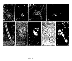
- This example shows that media conditioned by regenerating fish optic nerves containing OCF cause reduction in process-bearing oligodendrocytes in cultures of dissociated injured adult rat optic nerves.
- Fig. 1a,b shows process-bearing 04 positive cells in cultures derived from adult rat optic nerves, which had been injured 3 days prior to their removal.
- CM conditioned media
- CM-R regenerating fish optic nerves
- Fig. 1c-g the number of 04 positive cells that appeared at 96 h in vitro
- Fig. 1a,b The 04 positive cells which remained in the CM-R treated cultures had few processes and were therefore, identified as immature oligodendrocytes (Fig. 1c-g).
- CM-R CM of fish regenerating nerves
- This example shows that the media conditioned by regenerating fish optic nerves containing OCF affect maturation of Galc positive cells in newborn rat cultures of oligodendrocytes.
- Newborn rat brain oligodendrocytes and their perinatal progenitors were obtained in the following set of experiments by the method of McCarthy and DeVellis, 1980.
- the oligodendrocytes were seeded on poly-L-lysine coated coverslips.
- CM of regenerating fish optic nerves (CM-R) were added to these cultures (12 ⁇ g/ml) enriched with newborn rat brain oligodendrocytes either at the time of seeding or 24 h and 48 h later. In all cases, the same number of oligodendrocytes were seeded as in control untreated cultures.
- CM-R cause a reduction in the number of rat brain oligodendrocytes Type of * cells Hours ** of post-treatment % of control*** (mean ⁇ SD) Number of experiments (n) Oligodendrocytes 24 40.3 ⁇ 6.4 3 Oligodendrocytes 48 14.4 ⁇ 10.4 12 Mixed glial cells 48 25.7 ⁇ 4.7 4 * In these experiments, the number of counted oligodendrocytes (Galc positive cells) in control cultures per coverslip was between 350-1700 cells. ** CM-R at 12 ⁇ g protein.ml was added. *** The percentage of inhibition was calculated in each experiment by taking the number of counted cells in control, nontreated cultures, as 100%.
- CM-R oligodendrocytes
- Table II The inhibitory/cytotoxic effect of CM-R on oligodendrocytes is heat and trypsin labile Treatment Number of Galc positive cells (mean ⁇ SEM) Control 1427 ⁇ 247 CM-R 361 ⁇ 17 * CM-R (boiled) 1474 ⁇ 70 ** CM-R (trypsin treated) 1087 ⁇ 10 **
- rat brain oligodendrocytes were cultured as in Fig. 2.
- CM-R boiled or trypsin treated, 20 ⁇ g/ml
- the number of Galc positive process-bearing cells was determined by counting immunofluorescent cells. The results of one experiment, carried out in triplicate, are given. These results were reproduced in two additional experiments.
- * p-value 0.025. ** No significant differences were observed between control cultures and cultures treated with boiled or trypsinized CM-R.
- This experiment shows that the inhibitory/cytotoxic activity of OCF is associated with regeneration in the fish optic nerves.
- CM-R non-injured normal fish optic nerves
- oligodendrocytes Differentiation of oligodendrocytes has recently been shown to be regulated by platelet-derived growth factor (PDGF) (Noble et al., 1988, Raff et al., 1988 and Richardson et al 1988). Addition of PDGF to cultures of oligodendrocyte progenitors transiently delays their differentiation, due to its mitogen activity.
- PDGF platelet-derived growth factor
- CM-R CM of regenerating fish optic nerves
- Ciliary neurotrophic factor is another factor which has recently been shown to be effective in O-2A cell lineage by inducing type-2 astrocyte differentiation and thereby indirectly causing a reduction in the number of mature Galc positive oligodentrocytes Lillien et al., 1988). Since the CM-R did not cause an increase in the number of cells stained with A2B5 antibodies, and thus did not increase either progenitors or type-2 astrocytes, it is unlikely that the CM-R effect is mediated via CNTF.
- This experiment shows the specificity of the inhibitory/cytotoxic effect of OCF on oligodendrocytes by media conditioned with regenerating fish optic nerves.
- CM-R In order to examine whether the observed inhibitory/cytotoxic effect of the CM is unique to oligodendrocytes, we applied the CM-R to a mixed population of astrocytes and oligodendrocytes.
- Figure 6 shows that the observed inhibitory effect is not due to a non-specific toxic effect but is rather selective to oligodendrocytes.
- CM-R and CM-N Such conditions are the most favorable for differentiation of oligodendrocytes seeded onto the monolayer by the astrocytes.
- the inhibitory effect of the CM-R was pronounced, whereas the CM-N had only a marginally inhibitory activity at 72 and 96 h in vitro (Fig. 6b).
- CM-R glial fibrillary acidic protein
- DMEM Dulbecco's modified Eagle's medium
- Comparison between the cell populations obtained after dissociation of nerves which were kept in organ culture and those of the regularly crushed nerves (i.e., OC-3 and PC-3 in rat and OC-2 and PC-2 in fish) might indicate whether the postinjury behaviour of these cells is intrinsic to the optic nerve, or whether it is affected by some external factor, such as the blood monocytes. Any such external factors would be absent in the organ cultures, since the optic nerve is taken out at the time of injury.
- goldfish optic nerve cells were grown either in L-15 Leibowitz medium supplemented with 1-2% FCS, or in L-15 medium supplemented with the same additives used in Raff's modification of Bottenstein and Sato defined medium, plus 1-2% FCS.
- Organ culture of fish optic nerves was performed in L-15 medium supplemented with the same additives used in Raff's modification of Bottenstein and Sato defined medium, plus 1-2% FCS.
- CM Medium conditioned by regenerating goldfish optic nerves
- the macrophages in the fish cultures included blood-derived as well as resident macrophages.
- the two types of macrophages were distinguishable by morphological criteria: the blood-derived macrophages were typically circular and their vesicles were also arranged in a circular manner; the resident macrophages, on the other hand, were irregular in shape and their vesicles were distributed randomly through the cytoplasm.
- cells which were pronounced of the blood-derived macrophages (Fig. 7) were abundant in cultures of optic nerves excised several days after a crush injury, but were rare in cultures of optic nerves organ cultured after the excision.
- the cells which were considered as resident macrophages appeared in many forms, but could be divided into two major groups: those with a straight, fibroblast-like appearance (Fig. 8F and Fig. 9E), and those which were rounder and thicker, and which sometimes possessed processes (Fig. 9A-D).
- Fig. 9F-G the resident macrophages
- Fig. 9H-I the oligodendrocytes
- the nature of this contact seems to be cell-engulfing; oligodendrocytes seem to be engulfed by both macrophage types.
- These resident macrophages were also nonspecific esterase positive (Fig. 8D-G).
- CM regenerating fish optic nerves
- This experiment shows the sensitivity of the OCF of the invention to treatment at 56°C.
- Galc positive cells in cultures of newborn rat brain cells enriched for oligodendrocytes is carried out by ELISA as described in Experimental Procedures. As can be seen from Table VI, at 56°C there is a reduction in the number of Galc positive cells as evidenced by the reduction in the measured optical density.
- Table VI Temperature sensitivity of the OCF comprised in the conditioned medium (CM) as measured by ELISA Concentration a Average optical density ⁇ SD Room Temperature 56°C b - 0.752 ⁇ 0.076 c - CM(200 ⁇ g/ml) 0.556 ⁇ 0.008 0.738 ⁇ 0.040 CM(20 ⁇ g/ml) 0.646 ⁇ 0.029 0.709 ⁇ 0.016 a Final concentration of the CM b The culture is heated at 56°C for 30 min. c Experiment done in 4 trials, the mean of which is given.
- This experiment shows the effect of conditioned medium of fish blood macrophage on oligodendrocytes.
- a macrophage-rich culture was prepared as follows. Blood was collected from fish into PBS containing heparin, and then layered over Hypaque-Ficoll gradient made by mixing 70.6 ml of 12% w/v Ficoll (Sigma) with 29.4 ml of 32.8 w/v sodium diatriozate (Sigma). Ten ml of blood were layered over 5 ml of Hypaque-Ficoll in a 15 ml polystyrene tube. The tubes were centrifuged at 1000 g for 25 min at 10°C. The buffy coat containing lymphocytes and monocytes were removed and washed once with PBS. Cells were collected and adjusted to 20 x 106 cells/ml. After 1 h at 16°C, the cells were washed 3 times with PBS to remove loosely adherent and non-adherent cells. Then L-15 medium was added to the cells.
- Medium conditioned by macrophage-rich culture was prepared by incubating the cells in a serum-free medium for 8 h or in medium containing lipopolysaccharide. This conditioned medium was added to oligodendrocyte cultures and the number of oligcdendrocytes was determined by counting immunofluorescent Galc positive cells, as described in Experimental Procedures. In the first experiment, the number of Galc-positive cells counted in the presence of the conditioned medium of the macrophage-rich culture was 420 in comparison to 839 in the control. In the second experiment, there were 24 Galc-positive cells in comparison to 62 in the control.
- the oligodendrocyte cytotoxic factor OCF of the invention is useful as active ingredient of pharmaceutical compositions to be used for regeneration of nerves of the central nervous system. Preferably, it will be administered locally to the target organ.
- the OCF is used in a quantity and purity sufficient to facilitate regeneration of CNS axons in mammals, particularly humans.
- the OCF is administered in any manner which is calculated to bring the factor to the vicinity of the injured axons to be regenerated.
- the OCF is injected in a pharmaceutically acceptable liquid carrier directly to the site.
- an implant bearing the OCF may be surgically inserted.
- Such an implant may consist of any material, such as nitrocellulose, which will absorb OCF like a sponge and slowly release it at site of implantation. Other means of delivery will be apparent to those skilled in this art, and are intended to be comprehended within the scope of the present invention.
- the amount of the OCF to be administered to any given patient depends on a variety of factors, such as the injury being treated, the site of injured axons it is wished to regenerate and the condition of the patient. Typically, however, the OCF is administered at a dose of about 100 units, as a single injection or as multiple injections around the injured site, or the OCF is soaked onto nitrocellulose or any other adsorbable carrier. Precise dosages will be empirically determined.
- the OCF is preferably administered as soon as possible after the injury being treated. Thus, it is preferably used for acute injury rather than chronic injury. It will be more difficult to facilitate regeneration in accordance with the present invention the longer a period of degeneration has existed.
- irradiation of the injury site with low energy laser preferably He-Ne laser (5 min/day, 35 mW) can delay the post-traumatic process of the degeneration and thereby delay scar formation (Assia et al., Brain Res., 476 , 205-212 (1988).
- the OCF may be formulated with any pharmaceutically acceptable carrier or excipient.
- the OCF may be presented as an aqueous solution, for example as a sterile aqueous solution.
- a solution or powder containing OCF may be stabilized by means of a stabilizing agent.
- the composition is administered to the site of the injury in a quantity effective for facilitation of the regeneration of injured axons.
Landscapes
- Health & Medical Sciences (AREA)
- Chemical & Material Sciences (AREA)
- Life Sciences & Earth Sciences (AREA)
- Organic Chemistry (AREA)
- General Health & Medical Sciences (AREA)
- Medicinal Chemistry (AREA)
- Genetics & Genomics (AREA)
- Engineering & Computer Science (AREA)
- Bioinformatics & Cheminformatics (AREA)
- Biochemistry (AREA)
- Pharmacology & Pharmacy (AREA)
- Animal Behavior & Ethology (AREA)
- Molecular Biology (AREA)
- Public Health (AREA)
- Veterinary Medicine (AREA)
- Zoology (AREA)
- General Chemical & Material Sciences (AREA)
- Proteomics, Peptides & Aminoacids (AREA)
- Wood Science & Technology (AREA)
- Biomedical Technology (AREA)
- Nuclear Medicine, Radiotherapy & Molecular Imaging (AREA)
- Chemical Kinetics & Catalysis (AREA)
- Biophysics (AREA)
- Gastroenterology & Hepatology (AREA)
- Epidemiology (AREA)
- Neurosurgery (AREA)
- Immunology (AREA)
- Biotechnology (AREA)
- Microbiology (AREA)
- Neurology (AREA)
- General Engineering & Computer Science (AREA)
- Marine Sciences & Fisheries (AREA)
- Toxicology (AREA)
- Medicines Containing Material From Animals Or Micro-Organisms (AREA)
- Pharmaceuticals Containing Other Organic And Inorganic Compounds (AREA)
- Medicines That Contain Protein Lipid Enzymes And Other Medicines (AREA)
- Peptides Or Proteins (AREA)
- Medicines Containing Antibodies Or Antigens For Use As Internal Diagnostic Agents (AREA)
- Preparation Of Compounds By Using Micro-Organisms (AREA)
- Micro-Organisms Or Cultivation Processes Thereof (AREA)
Claims (10)
- Für Oligodendrocyten cytotoxischer Faktor aus niederen Wirbeltieren, die keine Säugetiere sind, mit den folgenden Eigenschaften:i. er ist selektiv toxisch für Oligodendrocyten, nicht jedoch für andere Zellen, wie z.B. Typ-1-Astrocyten und Fibroblastenzellen;ii. er verringert die Zahl der reifen Oligodendrocyten in Kulturen von verletzten Nerven von niederen Wirbeltieren und Säugetieren;iii. er hemmt die Differenzierung von Stammzellen der Oligodendrocyten-Familie zu reifen Oligodendrocyten;iv. er ist wasserlöslich;v. er ist hitzeempfindlich, wobei er seine Aktivität bei Erhitzen auf 56°C verliert;vi. er ist empfindlich gegenüber Proteasen, wobei er seine Aktivität bei Spaltung durch Trypsin verliert; undvii. er liegt in den konditionierten Medien von sich regenerierenden verletzten Nerven von niederen Wirbeltieren, die keine Säugetiere sind, wie z.B. Fischen, vor und kann daraus gereinigt werden, dagegen liegt er weder in den konditionierten Medien von intakten Nerven von niederen Wirbeltieren noch in den konditionierten Medien von verletzten oder intakten Nerven von Säugetieren vor.
- Arzneimittel, das als Wirkstoff den für Oligodendrocyten cytotoxischen Faktor nach Anspruch 1 und einen pharmazeutisch verträglichen Träger umfaßt.
- Arzneimittel nach Anspruch 2 für die Induktion der Regeneration von Nerven von Säugetieren.
- Arzneimittel nach Anspruch 2 für die Induktion der Regeneration von verletzten Nerven des Zentralnervensystems von Säugetieren.
- Arzneimittel nach Anspruch 2, wobei das Arzneimittel so beschaffen ist, daß es an der Stelle der Verletzung in Säugetieren verabreicht werden kann.
- Arzneimittel nach Anspruch 5, wobei die Verabreichung durch eine direkte Injektion an die Stelle der Verletzung in Säugetieren durchgeführt wird.
- Arzneimittel nach Anspruch 5, wobei ein Implantat, das den OCF enthält, an der Stelle der Verletzung eingepflanzt wird.
- Arzneimittel nach einem der Ansprüche 3 bis 7, wobei das Säugetier ein Mensch ist.
- Verfahren zur Herstellung eines Faktors nach Anspruch 1, umfassend die Auftrennung von konditionierten Medien von sich regenerierenden Nerven von niederen Wirbeltieren, die keine Säugetiere sind, mittels Trennverfahren, vorzugsweise Chromatographie.
- Verfahren zur Herstellung eines Arzneimittels nach einem der Ansprüche 2 bis 8, umfassend die Kombination des für Oligodendrocyten cytotoxischen Faktors nach Anspruch 1 und eines pharmazeutisch verträglichen Trägers.
Applications Claiming Priority (2)
| Application Number | Priority Date | Filing Date | Title |
|---|---|---|---|
| IL91459 | 1989-08-28 | ||
| IL91459A IL91459A0 (en) | 1989-08-28 | 1989-08-28 | Oligodendrocyte inhibitory factor |
Publications (2)
| Publication Number | Publication Date |
|---|---|
| EP0415321A1 EP0415321A1 (de) | 1991-03-06 |
| EP0415321B1 true EP0415321B1 (de) | 1995-11-08 |
Family
ID=11060333
Family Applications (1)
| Application Number | Title | Priority Date | Filing Date |
|---|---|---|---|
| EP90116388A Expired - Lifetime EP0415321B1 (de) | 1989-08-28 | 1990-08-27 | Für Oligodendrocyten cytotoxischer Faktor |
Country Status (16)
| Country | Link |
|---|---|
| EP (1) | EP0415321B1 (de) |
| JP (1) | JPH0411896A (de) |
| KR (1) | KR910004200A (de) |
| AT (1) | ATE130008T1 (de) |
| CA (1) | CA2024030A1 (de) |
| DE (1) | DE69023433T2 (de) |
| DK (1) | DK0415321T3 (de) |
| ES (1) | ES2081884T3 (de) |
| FI (1) | FI904229A7 (de) |
| GR (1) | GR3018534T3 (de) |
| HU (1) | HUT56117A (de) |
| IE (1) | IE903108A1 (de) |
| IL (1) | IL91459A0 (de) |
| NZ (1) | NZ235059A (de) |
| PT (1) | PT95123A (de) |
| ZA (1) | ZA906853B (de) |
Families Citing this family (5)
| Publication number | Priority date | Publication date | Assignee | Title |
|---|---|---|---|---|
| IL97365A0 (en) * | 1991-02-27 | 1992-05-25 | Yeda Res & Dev | Pharmaceutical compositions comprising a lymphokine |
| IE911318A1 (en) * | 1990-04-23 | 1991-10-23 | Yeda Res & Dev | Use of a tumor necrosis factor for facilitating nerve¹regeneration |
| FI941451L (fi) * | 1992-07-30 | 1994-05-11 | Yeda Resarch And Debelopment C | Entsymaattisesti valmistettu dimeerinen IL-2 ja sen valmistuksessa käytettävä, hermoista johdettu transglutaminaasi |
| US6267955B1 (en) | 1995-09-15 | 2001-07-31 | Yeda Research And Development Co. Ltd. | Mononuclear phagocytes and their use to promote axonal regeneration |
| US5800812A (en) * | 1995-09-15 | 1998-09-01 | Yeda Research And Development Co. Ltd. | Methods of use of mononuclear phagocytes to promote axonal regeneration |
Family Cites Families (2)
| Publication number | Priority date | Publication date | Assignee | Title |
|---|---|---|---|---|
| IL71440A0 (en) * | 1984-04-04 | 1984-07-31 | Univ Ramot | Composition for nerve regeneration |
| IL87349A (en) * | 1988-08-04 | 1994-06-24 | Michal Schwartz | Water-soluble factor that activates glial cells, its preparation and pharmaceutical compositions comprising it |
-
1989
- 1989-08-28 IL IL91459A patent/IL91459A0/xx unknown
-
1990
- 1990-08-27 CA CA002024030A patent/CA2024030A1/en not_active Abandoned
- 1990-08-27 NZ NZ235059A patent/NZ235059A/xx unknown
- 1990-08-27 DK DK90116388.1T patent/DK0415321T3/da active
- 1990-08-27 EP EP90116388A patent/EP0415321B1/de not_active Expired - Lifetime
- 1990-08-27 AT AT90116388T patent/ATE130008T1/de not_active IP Right Cessation
- 1990-08-27 IE IE310890A patent/IE903108A1/en unknown
- 1990-08-27 ES ES90116388T patent/ES2081884T3/es not_active Expired - Lifetime
- 1990-08-27 FI FI904229A patent/FI904229A7/fi not_active Application Discontinuation
- 1990-08-27 DE DE69023433T patent/DE69023433T2/de not_active Expired - Fee Related
- 1990-08-28 PT PT95123A patent/PT95123A/pt not_active Application Discontinuation
- 1990-08-28 ZA ZA906853A patent/ZA906853B/xx unknown
- 1990-08-28 JP JP2226435A patent/JPH0411896A/ja active Pending
- 1990-08-28 HU HU905507A patent/HUT56117A/hu unknown
- 1990-08-28 KR KR1019900013324A patent/KR910004200A/ko not_active Ceased
-
1995
- 1995-12-27 GR GR950403675T patent/GR3018534T3/el unknown
Non-Patent Citations (1)
| Title |
|---|
| Neuron 1 (1988) 85-96 * |
Also Published As
| Publication number | Publication date |
|---|---|
| DK0415321T3 (da) | 1995-12-27 |
| KR910004200A (ko) | 1991-03-28 |
| ZA906853B (en) | 1991-06-26 |
| DE69023433D1 (de) | 1995-12-14 |
| ES2081884T3 (es) | 1996-03-16 |
| FI904229A7 (fi) | 1991-03-01 |
| NZ235059A (en) | 1992-09-25 |
| FI904229A0 (fi) | 1990-08-27 |
| HU905507D0 (en) | 1991-02-28 |
| PT95123A (pt) | 1991-05-22 |
| JPH0411896A (ja) | 1992-01-16 |
| IE903108A1 (en) | 1991-03-13 |
| IL91459A0 (en) | 1990-04-29 |
| GR3018534T3 (en) | 1996-03-31 |
| HUT56117A (en) | 1991-07-29 |
| EP0415321A1 (de) | 1991-03-06 |
| AU6137290A (en) | 1991-02-28 |
| ATE130008T1 (de) | 1995-11-15 |
| AU642897B2 (en) | 1993-11-04 |
| CA2024030A1 (en) | 1991-03-01 |
| DE69023433T2 (de) | 1996-04-04 |
Similar Documents
| Publication | Publication Date | Title |
|---|---|---|
| Wilby et al. | A glial cell line-derived neurotrophic factor-secreting clone of the Schwann cell line SCTM41 enhances survival and fiber outgrowth from embryonic nigral neurons grafted to the striatum and to the lesioned substantia nigra | |
| Lunn et al. | Absence of Wallerian degeneration does not hinder regeneration in peripheral nerve | |
| US5202120A (en) | Methods of reducing glial scar formation and promoting axon and blood vessel growth and/or regeneration through the use of activated immature astrocytes | |
| Yamamoto et al. | Protective effect of NGF atelocollagen mini-pellet on the hippocampal delayed neuronal death in gerbils | |
| Goméz et al. | Transplantation of olfactory ensheathing cells fails to promote significant axonal regeneration from dorsal roots into the rat cervical cord | |
| US5753491A (en) | Use of neuro-derived fetal cell lines for transplantation therapy | |
| Haile et al. | Culturing of glial and neuronal cells on polysialic acid | |
| Sivron et al. | Presence of growth inhibitors in fish optic nerve myelin: postinjury changes | |
| Bolin et al. | Isolation of activated adult Schwann cells and a spontaneously immortal Schwann cell clone | |
| Barami et al. | Cellular transplantation and spinal cord injury | |
| Cohen et al. | Oligodendrocyte cytotoxic factor associated with fish optic nerve regeneration: implications for mammalian CNS regeneration | |
| Nothias et al. | Homotypic fetal transplants into an experimental model of spinal cord neurodegeneration | |
| Hadani et al. | Substances originating from the optic nerve of neonatal rabbit induce regeneration-associated response in the injured optic nerve of adult rabbit. | |
| Sivron et al. | Glial response to axonal injury: in vitro manifestation and implication for regeneration | |
| NZ337813A (en) | Mononuclear phagocytes and their use to promote axonal regeneration | |
| EP0415321B1 (de) | Für Oligodendrocyten cytotoxischer Faktor | |
| Zong et al. | Nanoparticles carrying neurotrophin-3-modified Schwann cells promote repair of sciatic nerve defects | |
| Neuberger et al. | Distribution of fibroblast growth factor in cultured dorsal root ganglion neurons and Schwann cells. II. Redistribution after neural injury | |
| EP1059931B1 (de) | Verwendung von cd137 zur förderung der proliferation peripherer monocyten | |
| US5962404A (en) | Enzymatically-produced oligodendrocyte cytotoxic dimeric IL-2 factor | |
| AU647883B2 (en) | Pharmaceutical compositions comprising interleukin-2 | |
| Ishikawa et al. | Long-term cultured astrocytes inhibit myelin formation, but not axonal growth in the co-cultured nerve tissue | |
| Schwartz et al. | Overcoming the inability of the injured mammalian central nervous system axons to grow into their degenerating environment | |
| JPH06511393A (ja) | 酵素的に産生された2量体il−2とその調製のための神経由来のトランスグルタミナーゼ | |
| Schwartzª et al. | in Central Nervous System Regeneration |
Legal Events
| Date | Code | Title | Description |
|---|---|---|---|
| PUAI | Public reference made under article 153(3) epc to a published international application that has entered the european phase |
Free format text: ORIGINAL CODE: 0009012 |
|
| AK | Designated contracting states |
Kind code of ref document: A1 Designated state(s): AT BE CH DE DK ES FR GB GR IT LI LU NL SE |
|
| 17P | Request for examination filed |
Effective date: 19910809 |
|
| 17Q | First examination report despatched |
Effective date: 19920324 |
|
| GRAA | (expected) grant |
Free format text: ORIGINAL CODE: 0009210 |
|
| AK | Designated contracting states |
Kind code of ref document: B1 Designated state(s): AT BE CH DE DK ES FR GB GR IT LI LU NL SE |
|
| PG25 | Lapsed in a contracting state [announced via postgrant information from national office to epo] |
Ref country code: GR Free format text: LAPSE BECAUSE OF FAILURE TO SUBMIT A TRANSLATION OF THE DESCRIPTION OR TO PAY THE FEE WITHIN THE PRESCRIBED TIME-LIMIT Effective date: 19951108 |
|
| REF | Corresponds to: |
Ref document number: 130008 Country of ref document: AT Date of ref document: 19951115 Kind code of ref document: T |
|
| REF | Corresponds to: |
Ref document number: 69023433 Country of ref document: DE Date of ref document: 19951214 |
|
| REG | Reference to a national code |
Ref country code: DK Ref legal event code: T3 |
|
| ITF | It: translation for a ep patent filed | ||
| ET | Fr: translation filed | ||
| REG | Reference to a national code |
Ref country code: CH Ref legal event code: NV Representative=s name: ISLER & PEDRAZZINI AG PATENTANWAELTE |
|
| REG | Reference to a national code |
Ref country code: GR Ref legal event code: FG4A Free format text: 3018534 |
|
| REG | Reference to a national code |
Ref country code: ES Ref legal event code: FG2A Ref document number: 2081884 Country of ref document: ES Kind code of ref document: T3 |
|
| PGFP | Annual fee paid to national office [announced via postgrant information from national office to epo] |
Ref country code: GB Payment date: 19960809 Year of fee payment: 7 |
|
| PGFP | Annual fee paid to national office [announced via postgrant information from national office to epo] |
Ref country code: FR Payment date: 19960810 Year of fee payment: 7 |
|
| PGFP | Annual fee paid to national office [announced via postgrant information from national office to epo] |
Ref country code: SE Payment date: 19960822 Year of fee payment: 7 |
|
| PGFP | Annual fee paid to national office [announced via postgrant information from national office to epo] |
Ref country code: DK Payment date: 19960823 Year of fee payment: 7 Ref country code: CH Payment date: 19960823 Year of fee payment: 7 |
|
| PG25 | Lapsed in a contracting state [announced via postgrant information from national office to epo] |
Ref country code: AT Effective date: 19960827 |
|
| PGFP | Annual fee paid to national office [announced via postgrant information from national office to epo] |
Ref country code: ES Payment date: 19960830 Year of fee payment: 7 |
|
| PG25 | Lapsed in a contracting state [announced via postgrant information from national office to epo] |
Ref country code: LU Free format text: LAPSE BECAUSE OF NON-PAYMENT OF DUE FEES Effective date: 19960831 Ref country code: BE Effective date: 19960831 |
|
| PGFP | Annual fee paid to national office [announced via postgrant information from national office to epo] |
Ref country code: NL Payment date: 19960831 Year of fee payment: 7 |
|
| PLBE | No opposition filed within time limit |
Free format text: ORIGINAL CODE: 0009261 |
|
| STAA | Information on the status of an ep patent application or granted ep patent |
Free format text: STATUS: NO OPPOSITION FILED WITHIN TIME LIMIT |
|
| 26N | No opposition filed | ||
| PGFP | Annual fee paid to national office [announced via postgrant information from national office to epo] |
Ref country code: DE Payment date: 19961030 Year of fee payment: 7 |
|
| BERE | Be: lapsed |
Owner name: YEDA RESEARCH AND DEVELOPMENT CY LTD Effective date: 19960831 |
|
| REG | Reference to a national code |
Ref country code: GR Ref legal event code: MM2A Free format text: 3018534 |
|
| PG25 | Lapsed in a contracting state [announced via postgrant information from national office to epo] |
Ref country code: GB Free format text: LAPSE BECAUSE OF NON-PAYMENT OF DUE FEES Effective date: 19970827 Ref country code: DK Free format text: LAPSE BECAUSE OF NON-PAYMENT OF DUE FEES Effective date: 19970827 |
|
| REG | Reference to a national code |
Ref country code: DK Ref legal event code: EBP |
|
| PG25 | Lapsed in a contracting state [announced via postgrant information from national office to epo] |
Ref country code: SE Free format text: LAPSE BECAUSE OF NON-PAYMENT OF DUE FEES Effective date: 19970828 Ref country code: ES Free format text: LAPSE BECAUSE OF EXPIRATION OF PROTECTION Effective date: 19970828 |
|
| PG25 | Lapsed in a contracting state [announced via postgrant information from national office to epo] |
Ref country code: LI Free format text: LAPSE BECAUSE OF NON-PAYMENT OF DUE FEES Effective date: 19970831 Ref country code: CH Free format text: LAPSE BECAUSE OF NON-PAYMENT OF DUE FEES Effective date: 19970831 |
|
| PG25 | Lapsed in a contracting state [announced via postgrant information from national office to epo] |
Ref country code: NL Free format text: LAPSE BECAUSE OF NON-PAYMENT OF DUE FEES Effective date: 19980301 |
|
| REG | Reference to a national code |
Ref country code: CH Ref legal event code: PL |
|
| GBPC | Gb: european patent ceased through non-payment of renewal fee |
Effective date: 19970827 |
|
| PG25 | Lapsed in a contracting state [announced via postgrant information from national office to epo] |
Ref country code: FR Free format text: LAPSE BECAUSE OF NON-PAYMENT OF DUE FEES Effective date: 19980430 |
|
| PG25 | Lapsed in a contracting state [announced via postgrant information from national office to epo] |
Ref country code: DE Free format text: LAPSE BECAUSE OF NON-PAYMENT OF DUE FEES Effective date: 19980501 |
|
| EUG | Se: european patent has lapsed |
Ref document number: 90116388.1 |
|
| NLV4 | Nl: lapsed or anulled due to non-payment of the annual fee |
Effective date: 19980301 |
|
| REG | Reference to a national code |
Ref country code: FR Ref legal event code: ST |
|
| REG | Reference to a national code |
Ref country code: ES Ref legal event code: FD2A Effective date: 20010201 |
|
| PG25 | Lapsed in a contracting state [announced via postgrant information from national office to epo] |
Ref country code: IT Free format text: LAPSE BECAUSE OF NON-PAYMENT OF DUE FEES;WARNING: LAPSES OF ITALIAN PATENTS WITH EFFECTIVE DATE BEFORE 2007 MAY HAVE OCCURRED AT ANY TIME BEFORE 2007. THE CORRECT EFFECTIVE DATE MAY BE DIFFERENT FROM THE ONE RECORDED. Effective date: 20050827 |