EP0419367A1 - Verfahren zum Nachweis einer biologischen Substanz mit Hilfe von Liganden - Google Patents
Verfahren zum Nachweis einer biologischen Substanz mit Hilfe von Liganden Download PDFInfo
- Publication number
- EP0419367A1 EP0419367A1 EP90402611A EP90402611A EP0419367A1 EP 0419367 A1 EP0419367 A1 EP 0419367A1 EP 90402611 A EP90402611 A EP 90402611A EP 90402611 A EP90402611 A EP 90402611A EP 0419367 A1 EP0419367 A1 EP 0419367A1
- Authority
- EP
- European Patent Office
- Prior art keywords
- antibody
- biological substance
- ligand
- labeled
- antibodies
- Prior art date
- Legal status (The legal status is an assumption and is not a legal conclusion. Google has not performed a legal analysis and makes no representation as to the accuracy of the status listed.)
- Granted
Links
- 238000000034 method Methods 0.000 title claims abstract description 62
- 239000000126 substance Substances 0.000 title claims abstract description 51
- 239000003446 ligand Substances 0.000 title claims abstract description 31
- 239000000700 radioactive tracer Substances 0.000 claims abstract description 22
- 239000003795 chemical substances by application Substances 0.000 claims abstract description 17
- 230000000694 effects Effects 0.000 claims abstract description 15
- 239000007788 liquid Substances 0.000 claims abstract description 15
- 239000000427 antigen Substances 0.000 claims description 27
- 102000036639 antigens Human genes 0.000 claims description 26
- 108091007433 antigens Proteins 0.000 claims description 26
- 239000000523 sample Substances 0.000 claims description 21
- 238000011534 incubation Methods 0.000 claims description 11
- 239000007787 solid Substances 0.000 claims description 10
- 238000001514 detection method Methods 0.000 claims description 7
- 102000004169 proteins and genes Human genes 0.000 claims description 7
- 108090000623 proteins and genes Proteins 0.000 claims description 7
- 102000039446 nucleic acids Human genes 0.000 claims description 6
- 108020004707 nucleic acids Proteins 0.000 claims description 6
- 150000007523 nucleic acids Chemical class 0.000 claims description 6
- 239000000872 buffer Substances 0.000 claims description 3
- 241000894006 Bacteria Species 0.000 claims description 2
- 102000008857 Ferritin Human genes 0.000 claims description 2
- 238000008416 Ferritin Methods 0.000 claims description 2
- 108050000784 Ferritin Proteins 0.000 claims description 2
- 101800004937 Protein C Proteins 0.000 claims description 2
- 101800001700 Saposin-D Proteins 0.000 claims description 2
- 241000700605 Viruses Species 0.000 claims description 2
- 239000002253 acid Substances 0.000 claims description 2
- 238000003149 assay kit Methods 0.000 claims description 2
- 238000009396 hybridization Methods 0.000 claims description 2
- 239000007791 liquid phase Substances 0.000 claims description 2
- 244000045947 parasite Species 0.000 claims description 2
- 229960000856 protein c Drugs 0.000 claims description 2
- 102100036546 Salivary acidic proline-rich phosphoprotein 1/2 Human genes 0.000 claims 1
- 239000003153 chemical reaction reagent Substances 0.000 description 13
- 238000012360 testing method Methods 0.000 description 13
- 230000003287 optical effect Effects 0.000 description 9
- 239000000243 solution Substances 0.000 description 9
- 102000003992 Peroxidases Human genes 0.000 description 8
- 108040007629 peroxidase activity proteins Proteins 0.000 description 8
- 239000000203 mixture Substances 0.000 description 7
- 229940088597 hormone Drugs 0.000 description 6
- 239000005556 hormone Substances 0.000 description 6
- 210000004754 hybrid cell Anatomy 0.000 description 5
- 230000003053 immunization Effects 0.000 description 5
- 238000002649 immunization Methods 0.000 description 5
- 239000007790 solid phase Substances 0.000 description 5
- 102000002260 Alkaline Phosphatase Human genes 0.000 description 4
- 108020004774 Alkaline Phosphatase Proteins 0.000 description 4
- 241001465754 Metazoa Species 0.000 description 4
- 241000699670 Mus sp. Species 0.000 description 4
- 230000000890 antigenic effect Effects 0.000 description 4
- 238000002347 injection Methods 0.000 description 4
- 239000007924 injection Substances 0.000 description 4
- 230000003248 secreting effect Effects 0.000 description 4
- 238000001179 sorption measurement Methods 0.000 description 4
- 238000002965 ELISA Methods 0.000 description 3
- 238000003556 assay Methods 0.000 description 3
- 210000004027 cell Anatomy 0.000 description 3
- 230000007423 decrease Effects 0.000 description 3
- 238000002474 experimental method Methods 0.000 description 3
- 229910000162 sodium phosphate Inorganic materials 0.000 description 3
- 239000001488 sodium phosphate Substances 0.000 description 3
- RYFMWSXOAZQYPI-UHFFFAOYSA-K trisodium phosphate Chemical compound [Na+].[Na+].[Na+].[O-]P([O-])([O-])=O RYFMWSXOAZQYPI-UHFFFAOYSA-K 0.000 description 3
- XZKIHKMTEMTJQX-UHFFFAOYSA-N 4-Nitrophenyl Phosphate Chemical compound OP(O)(=O)OC1=CC=C([N+]([O-])=O)C=C1 XZKIHKMTEMTJQX-UHFFFAOYSA-N 0.000 description 2
- 102000004190 Enzymes Human genes 0.000 description 2
- 108090000790 Enzymes Proteins 0.000 description 2
- 102000012673 Follicle Stimulating Hormone Human genes 0.000 description 2
- 108010079345 Follicle Stimulating Hormone Proteins 0.000 description 2
- 108010001336 Horseradish Peroxidase Proteins 0.000 description 2
- 102000009151 Luteinizing Hormone Human genes 0.000 description 2
- 108010073521 Luteinizing Hormone Proteins 0.000 description 2
- 241000699666 Mus <mouse, genus> Species 0.000 description 2
- 239000004793 Polystyrene Substances 0.000 description 2
- QAOWNCQODCNURD-UHFFFAOYSA-N Sulfuric acid Chemical compound OS(O)(=O)=O QAOWNCQODCNURD-UHFFFAOYSA-N 0.000 description 2
- 238000007792 addition Methods 0.000 description 2
- 239000002671 adjuvant Substances 0.000 description 2
- 239000007864 aqueous solution Substances 0.000 description 2
- 150000001875 compounds Chemical class 0.000 description 2
- 238000007796 conventional method Methods 0.000 description 2
- 230000002255 enzymatic effect Effects 0.000 description 2
- 229940088598 enzyme Drugs 0.000 description 2
- 210000004408 hybridoma Anatomy 0.000 description 2
- 238000003018 immunoassay Methods 0.000 description 2
- 230000002163 immunogen Effects 0.000 description 2
- 230000002452 interceptive effect Effects 0.000 description 2
- 229920002223 polystyrene Polymers 0.000 description 2
- 230000002285 radioactive effect Effects 0.000 description 2
- 230000035945 sensitivity Effects 0.000 description 2
- 210000002966 serum Anatomy 0.000 description 2
- 239000000758 substrate Substances 0.000 description 2
- 238000005406 washing Methods 0.000 description 2
- GEYOCULIXLDCMW-UHFFFAOYSA-N 1,2-phenylenediamine Chemical compound NC1=CC=CC=C1N GEYOCULIXLDCMW-UHFFFAOYSA-N 0.000 description 1
- BFSVOASYOCHEOV-UHFFFAOYSA-N 2-diethylaminoethanol Chemical compound CCN(CC)CCO BFSVOASYOCHEOV-UHFFFAOYSA-N 0.000 description 1
- HRPVXLWXLXDGHG-UHFFFAOYSA-N Acrylamide Chemical compound NC(=O)C=C HRPVXLWXLXDGHG-UHFFFAOYSA-N 0.000 description 1
- 206010003445 Ascites Diseases 0.000 description 1
- 101710114816 Gene 41 protein Proteins 0.000 description 1
- 102000003886 Glycoproteins Human genes 0.000 description 1
- 108090000288 Glycoproteins Proteins 0.000 description 1
- 101000605534 Homo sapiens Prostate-specific antigen Proteins 0.000 description 1
- 241000713772 Human immunodeficiency virus 1 Species 0.000 description 1
- -1 IgE Proteins 0.000 description 1
- 206010035226 Plasma cell myeloma Diseases 0.000 description 1
- 102100038358 Prostate-specific antigen Human genes 0.000 description 1
- 102000017975 Protein C Human genes 0.000 description 1
- 102000011923 Thyrotropin Human genes 0.000 description 1
- 108010061174 Thyrotropin Proteins 0.000 description 1
- 241000223997 Toxoplasma gondii Species 0.000 description 1
- 241000251539 Vertebrata <Metazoa> Species 0.000 description 1
- 238000001042 affinity chromatography Methods 0.000 description 1
- 210000000628 antibody-producing cell Anatomy 0.000 description 1
- 239000011324 bead Substances 0.000 description 1
- 230000008033 biological extinction Effects 0.000 description 1
- 230000015572 biosynthetic process Effects 0.000 description 1
- 230000000903 blocking effect Effects 0.000 description 1
- 238000004587 chromatography analysis Methods 0.000 description 1
- 238000010367 cloning Methods 0.000 description 1
- 230000003247 decreasing effect Effects 0.000 description 1
- 230000007547 defect Effects 0.000 description 1
- 238000011161 development Methods 0.000 description 1
- 238000010790 dilution Methods 0.000 description 1
- 239000012895 dilution Substances 0.000 description 1
- KAKKHKRHCKCAGH-UHFFFAOYSA-L disodium;(4-nitrophenyl) phosphate;hexahydrate Chemical compound O.O.O.O.O.O.[Na+].[Na+].[O-][N+](=O)C1=CC=C(OP([O-])([O-])=O)C=C1 KAKKHKRHCKCAGH-UHFFFAOYSA-L 0.000 description 1
- 238000010828 elution Methods 0.000 description 1
- 239000012149 elution buffer Substances 0.000 description 1
- 230000004720 fertilization Effects 0.000 description 1
- 239000007850 fluorescent dye Substances 0.000 description 1
- 229940028334 follicle stimulating hormone Drugs 0.000 description 1
- 230000004927 fusion Effects 0.000 description 1
- 238000001502 gel electrophoresis Methods 0.000 description 1
- PCHJSUWPFVWCPO-UHFFFAOYSA-N gold Chemical compound [Au] PCHJSUWPFVWCPO-UHFFFAOYSA-N 0.000 description 1
- 239000010931 gold Substances 0.000 description 1
- 229910052737 gold Inorganic materials 0.000 description 1
- 230000001900 immune effect Effects 0.000 description 1
- 229940124452 immunizing agent Drugs 0.000 description 1
- 230000000984 immunochemical effect Effects 0.000 description 1
- 150000002500 ions Chemical class 0.000 description 1
- 238000002372 labelling Methods 0.000 description 1
- 239000004816 latex Substances 0.000 description 1
- 229920000126 latex Polymers 0.000 description 1
- 229940040129 luteinizing hormone Drugs 0.000 description 1
- 239000003550 marker Substances 0.000 description 1
- 239000011325 microbead Substances 0.000 description 1
- 238000002156 mixing Methods 0.000 description 1
- 201000000050 myeloid neoplasm Diseases 0.000 description 1
- 230000035935 pregnancy Effects 0.000 description 1
- 238000002360 preparation method Methods 0.000 description 1
- 238000011002 quantification Methods 0.000 description 1
- 238000010791 quenching Methods 0.000 description 1
- 230000000171 quenching effect Effects 0.000 description 1
- 230000000717 retained effect Effects 0.000 description 1
- 210000000952 spleen Anatomy 0.000 description 1
- 210000004989 spleen cell Anatomy 0.000 description 1
- 210000004988 splenocyte Anatomy 0.000 description 1
- 210000002700 urine Anatomy 0.000 description 1
Images


Classifications
-
- G—PHYSICS
- G01—MEASURING; TESTING
- G01N—INVESTIGATING OR ANALYSING MATERIALS BY DETERMINING THEIR CHEMICAL OR PHYSICAL PROPERTIES
- G01N33/00—Investigating or analysing materials by specific methods not covered by groups G01N1/00 - G01N31/00
- G01N33/48—Biological material, e.g. blood, urine; Haemocytometers
- G01N33/50—Chemical analysis of biological material, e.g. blood, urine; Testing involving biospecific ligand binding methods; Immunological testing
- G01N33/53—Immunoassay; Biospecific binding assay; Materials therefor
Definitions
- the subject of the present invention is a method for detecting and / or assaying a biological substance according to an affinity method, as well as a detection or assay kit or kit allowing the implementation of this method.
- the biological substance which can be considered here as an antigen
- the excess of antigens can lead to situations where the antigen binds on the one hand to the antibody 1 immobilized, and on the other hand on the labeled antibody 2, in solution, so that only a small proportion of the antibody 2 is fixed on the support.
- the solid phase is coated essentially with the antigen alone, while the antibody 2 carrying the tracer agent is present in solution in the form of a complex with the antigen.
- the detected signal tends to zero for very high concentrations of antigens.
- Another solution to this problem is to mix the labeled antibody with the same unlabeled antibody while increasing the overall concentration of the first and second antibodies.
- This method is effective, but it has the disadvantage that only part of the antibody 1-antigen-antibody 2 complexes are labeled, which leads to a reduction in the sensitivity of the development system.
- Patent applications GB-2161165, GB-2171999 and PCT No. 85/04422 describe the use, in the determination of a small antigen (of molecular mass less than 5000) or a hapten, of a first antibody capable of recognizing the (unique) antigenic determinant of the antigen or hapten, and a second antibody capable of specifically recognizing the combination of the antigen or hapten with the first antibody.
- These patent applications do not suggest the use of an analogous antibody system in the determination of antigens, of higher molecular weight, having several epitopes, in order to eliminate the risks of erroneous results due to an effect. zone.
- the method of the invention can therefore be used, in particular according to the simultaneous method, when the concentration of the antigen, in the sample examined, is likely to vary in significant proportions. It is then possible to totally or strongly eliminate the zone effect observed at high concentration of antigen.
- the method of the invention therefore makes it possible in particular to solve the problems posed by the drop or the extinction of the signal detected for high concentrations of antigen in an immunoassay with simultaneous incubation of the fixed antibody and of the labeled antibody.
- the method of the invention can be applied to systems other than those based on the antigen-antibody affinity, for example to nucleic probes, the determinations of which can also be distorted by a zone effect.
- the present invention therefore relates to a method for detecting and / or assaying a biological substance in a liquid containing it or capable of containing it, according to a method known per se, using two different ligands, said substance biological having at least two binding sites with different ligands, and the concentration of said biological substance in said liquid being capable of varying in proportions ranging from 1 to 100 or more, process in which a first ligand having an affinity is used for a site of said biological substance, and a second ligand, and in which one of the ligands is labeled with a tracer, characterized in that in order to avoid a zone effect, the second ligand is a ligand having a specific affinity for the complex formed by the first ligand attached by affinity to said biological substance.
- the biological substances to which the process of the invention applies have molecular weights greater than 5000, and generally greater than 10,000: this is the case in practice for nucleic acids (DNA or RNA) and also for proteins having several different epitopes. Most often, the biological substances to which the process of the invention applies have a molecular mass which can range from approximately 20,000 to 500,000.
- the biological substance is a nucleic acid
- the first ligand is a first nucleic probe (which can be fixed on a solid support), part of which is capable of hybridizing with a first sequence of said nucleic acid while that the other part of the first probe does not hybridize with the biological substance
- the second ligand is a second nucleic probe (labeled with a tracer agent), part of which is capable of hybridizing with a second sequence of said acid nucleic only if the other part of said second probe is capable of hybridizing with said other because of the first probe, it being understood that said first and second sequences of the biological substance are sufficiently close to allow the effective hybridization of said other parts .
- the tracer agent is for example a radioactive or enzymatic agent, or an antigen capable of being recognized by an antibody itself conjugated with an enzyme or a fluorescent or luminescent compound.
- the first and second ligands are antibodies, first antibody recognizing an antigenic site characteristic of the biological substance and the second antibody specifically recognizing the immune complex formed by the first antibody attached to the biological substance.
- the biological substance is in particular a protein.
- the proteins whose concentration is likely to vary greatly in the samples to be analyzed, mention will be made, by way of nonlimiting examples, of the hormone chorionogonadotropin (hCG), the FSH (follicle stimulating hormone), the LH (luteinizing hormone) , TSh (thyroid stimulating hormone), alpha-foeto protein, reactive protein C, IgE, ferritin, and various surface antigens of bacteria, viruses or parasites, for example the P30 antigen of toxoplasma gondii or the HIV1 glycoprotein Gp41.
- hCG hormone chorionogonadotropin
- FSH follicle stimulating hormone
- LH luteinizing hormone
- TSh thyroid stimulating hormone
- alpha-foeto protein reactive protein C
- IgE luteinizing hormone
- ferritin various surface antigens of bacteria, viruses or parasites
- various surface antigens of bacteria, viruses or parasites for example the P30 antigen of toxoplasma gondii or the HIV1
- the subject of the present invention is therefore in particular a method for detecting and / or assaying a biological substance in a liquid sample containing it or capable of containing it, in which a first antibody directed against said biological substance is used, and a second antibody, characterized in that the second antibody is an antibody directed against the immune complex formed by the first antibody and said biological substance.
- One of the two antibodies can be labeled with a tracer, and the other antibody can be attached to a solid phase.
- At least one of the two antibodies is a monoclonal antibody.
- the two antibodies are monoclonal antibodies.
- the second antibody with a third labeled antibody capable of recognizing an epitope, of the biological substance, different from the epitope recognized by the first antibody. Detection is thus improved, in particular for low concentrations of the biological substance.
- the liquid sample is brought into contact with said first antibody and with said second antibody, and after suitable incubation times, the liquid phase is separated from the solid support and the presence and / or the quantity of the biological substance is determined on the said liquid sample by determining the presence and / or the quantity of tracer agent fixed on the solid support.
- This process can in particular be implemented according to the simultaneous method, that is to say that the liquid sample is brought into contact with both the first and second antibodies simultaneously, without intermediate incubation, so as to perform only '' a single incubation.
- the detecton or assay method of the invention can be implemented in solution, that is to say without solid phase, using the fact that, in the antibody complex 1- antigen-antibody 2, the two antibodies are very close to each other, since the second antibody recognizes the binding zone between antibody 1 and the antigen.
- the liquid route that is to say without antibody attached to a solid phase, is characterized in that the two antibodies are each labeled with a tracer agent, and that the tracer agents for the two antibodies are interactive.
- the tracer agents for the two antibodies are interactive.
- one of the tracers is a fluorescent compound and the other tracer is a compound capable of quenching fluorescence.
- one of the antibodies is labeled with an enzyme that catalyzes a reaction involving a substance linked to the other antibody.
- the two markers together constitute a multi-enzymatic system capable of producing a light signal.
- Such interactive tracer agents are known; see for example "Immunoassays in the Clinical Laboratory", Nakumura, Dito, Tucker Editeurs, Alain R. Liss Inc. New York, 1978, pages 211 - 226.
- the first antibodies which can be used for implementing the process of the present invention are prepared according to the usual methods, by immunization of vertebrate animals with the biological substance considered.
- the antibodies obtained can be purified for example by affinity chromatography.
- the antibody-producing cells of the immunized animals are transformed into hybridomas, according to known methods, and then the hybrid cells producing the antibodies of interest are cloned.
- the animals are immunized according to the usual techniques with the biological substance-first antibody complex (the first antibody preferably being monoclonal). It has been found that animals thus immunized produce a certain proportion of antibodies which recognize the complex used for immunization, but which do not appreciably recognize either the biological substance alone, nor the first antibody alone.
- the second antibody is a monoclonal antibody obtained for example according to the hybridoma technique.
- the labeling of the antibodies with a tracer agent is carried out according to known techniques. Likewise, the disclosure of the marker is carried out in a known manner.
- the attachment of antibodies to a solid support is also known.
- the solid supports are, for example microtiter plates, tubes, polystyrene beads or bars, latex microbeads, etc.
- the fixing on the support is made by adsorption, by covalence, or by means of an antibody (itself fixed directly on the support) recognizing an isogenic antigenic determinant of the first antibody.
- the intermediate antibody of course comes from another animal species than that which served to obtain the first antibody.
- the subject of the invention is also a box or kit for detecting, according to the method described above, a biological substance, characterized in that it contains, in addition to buffers and possibly appropriate solid supports, at least said first and second ligands as defined above.
- the detection box or kit may also contain an agent for revealing the presence of the tracer agent.
- the zone effect at high concentrations is sufficiently weak not to give false negative results.
- This weak zone effect is likely to limit the possibilities of rigorous quantification at high concentrations of antigen. But the result found being in this case close to the maximum signal, it then suffices to dilute the antigen to concentrations (which can be determined in advance) where the signal is easily interpretable, in order to then be able to carry out, always according to the method of the invention, a correct dosage.
- Example 1 Obtaining antibodies and hCG detection tests.
- the hormone hCG has two subunits, alpha and beta. Only the beta subunit carries antigen specificity.
- the alpha subunit is common to several hormones (FSH, LH, TSH, ).
- Stage 1 Obtaining monoclonal antibodies directed against hCG.
- the antigen used is hCG marketed by DIOSYNTH (Netherlands), reference 8856 01, containing 3020 U / mg of dry weight. The purity of the preparation was checked by acrylamide gel electrophoresis.
- hCG 3000 ⁇ g of hCG.
- mice are immunized by injection intraperitoneally with 30 U hCG, by injection and by mouse. The first injection is given with complete Freund's adjuvant, the other injections 4-8 weeks apart with incomplete adjuvant. A final booster is given 3 days before fusion, with 30 U of HcC in physiological buffer, intravenously. After this booster, the spleen of the immunized mice is removed and the splenocytes are collected.
- hybrid cell lines secreting monoclonal antibodies recognizing hCC were obtained.
- the monoclonal antibodies were produced at the end of stage 1 on mouse ascites and purified by chromatography on an ion exchanger (DEAE Trisacryl, sold by IBF ref. 250-771), with elution with an aqueous solution of a gradient of sodium phosphate.
- the sodium phosphate concentration of the elution buffer is close to 15 mM.
- Stage 2 obtaining of monoclonal antibodies directed against the binding zone of an anti-hCG beta-chain monoclonal antibody.
- An immune complex is prepared with one of the monoclonal antibodies against the hCG beta chain, obtained in the previous stage (hereinafter called anti-beta antibodies) and hCG, by equimolar mixing.
- the immune complex solution thus obtained is used as an immunogen to immunize Balb C mice, according to the method described above.
- Hybrid cells are prepared as above and cells secreting antibodies are selected which recognize in ELISA the immune complex used for immunization.
- the clones producing the antibodies specific for the immune complex are selected, which do not recognize the hCG (that is to say they recognize neither the alpha chain nor the beta chain) and which also do not recognize the monoclonal anti-beta antibody used to prepare the immune complex used as an immunizing agent.
- anti-complex 1 anti-complex 2 and anti-complex 3, respectively.
- Stage 3 hCG detection test.
- the anti-beta antibody is fixed by adsorption on a microtiter plate.
- the second antibody used is the anti-complex 1 antibody conjugated with alkaline phosphatase, in the form of an aqueous solution containing sodium phosphate and horse serum.
- the tests are carried out on solutions at various concentrations of hCG (from 0 to 500 ⁇ g / ml).
- the reagents are incubated simultaneously for one hour. After washing the plate, the alkaline phosphatase substrate, that is to say the PNPP (para nitrophenyl phosphate) was added at a rate of 0.2 mg per well, and after one hour, the densities optics were measured.
- the alkaline phosphatase substrate that is to say the PNPP (para nitrophenyl phosphate) was added at a rate of 0.2 mg per well, and after one hour, the densities optics were measured.
- stage 3 Experiments similar to those described in Example 1, stage 3, were carried out using the anti-beta antibody as immobilized antibody and the anti-complex 1, labeled with alkaline phosphatase as the second antibody.
- the microtiter plate After incubation for one hour at 37 ° C., the microtiter plate is washed, then the OPD substrate (ortho-phenylene diamine) is added and the mixture is left to incubate for 5 minutes. Next, the optical densities at the wavelength 492 nm are read.
- OPD substrate ortho-phenylene diamine
- Example 1 The tests are carried out with the three anti-complex antibodies obtained in Example 1, stage 3, conjugated to peroxidase.
- the incubation protocol is as follows: - anti-beta antibody fixed on the microtiter plate by adsorption. - simultaneous incubation for one hour at 37 ° C. of the hCG antigen and of the anti-complex antibody labeled with peroxidase, or of the anti-alpha antibody labeled with peroxidase, or of a 75:25 mixture ( en masse) of anti-complex and anti-alpha. - After washing the plate, add the substrate (OPD). - After 15 minutes of incubation and blocking of the reaction by addition of a 1N solution of sulfuric acid, the optical densities are read at 492 nm.
- the hCG calibration range is from 0 to 200,000 U / l. It is prepared by dilutions from a standard standard at 10,000U / ml.
- anti-complex 1 and anti-complex 2 antibodies as well as with anti-complex 1 / anti-alpha and anti-complex 2 / anti-alpha mixtures, there is no drop in signal at high hCG concentration.
- the commercial reagent contains anti-alpha- and anti-beta-hCG monoclonal antibodies. With the antibodies of this commercial reagent, a standard "sandwich” test is therefore carried out.
- the correlation lines of the 60 sera are shown in Table 5, with y representing the results obtained with the anti-beta antibody fixed on the solid phase and the anti-alpha, anti-complex conjugates, or the anti-alpha / anti-complex, and x representing the results obtained with the commercial reagent.
- the proportions of the antibody mixtures are the same as in Example 4.
- Monoclonal anti-human IgE antibodies were obtained according to the experimental protocol described in Stage 1 of Example 1, then an immune complex was prepared with one of the anti-IgE monoclonal Ac obtained in the previous stage (hereinafter called -after anti-IgE Ab 1) and human IgE by equimolecular mixture.
- the immune complex solution thus obtained was used as an immunogen to immunize Balb C mice according to conventional techniques.
- Hybrid cells were prepared according to conventional techniques and gold selected cells secreting Ab which recognize in ELISA the immune complex used for immunization.
- we cloned the clones which produce specific Ac of the immune complex that is to say which recognize neither the IgE alone, nor the anti-IgE 1 Ac alone).
- anti-complex 1 ′ A specific monoclonal Ab (hereinafter called anti-complex 1 ′) was retained having a good specific affinity for the immune complex.
- This Ac was then purified as described in Stage 1 of Example 1 and was labeled either with horseradish peroxidase according to the Nakane method, or with a radioactive tracer (125I).
- the anti IgE-1 Ab is fixed by adsorption on a polystyrene tube.
- the second Ac is used an anti IgE-2 Ab, recognizing an epitope opposite to that recognized by the anti-IgE 1, the anti IgE 2 Ab having been previously marked with a radioactive tracer (125I).
- the reagents, IgE (50 ⁇ l) - Anti IgE 1 Ab - Anti IgE 2 Ac (200 ⁇ l) are incubated for hours with shaking.
- Tests are carried out by varying the concentration of IgE.
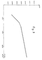
- the results are represented in the attached FIG. 2, on which the concentration (IU / ml; logarithmic scale) is plotted on the abscissa and the number of counts per minute (cpm) on the ordinate.
- the count obtained results in a "bell" curve presenting a maximum of signal for a concentration of approximately 7000 IU / ml of IgE. Beyond this value, the signal decreases when the Ag concentration increases. The zone effect results in an erroneous default signal from around 50,000 IU / ml.
- Assays were carried out on 15 sera whose values were between 0 and 1,000 IU / ml.
- the results obtained with a commercially available reagent (BIOMERIEUX ref. 69200) were compared with the reagents of the invention.
- the commercial reagent contains anti IgE 1 and anti IgE2 monoclonal Ab.
- the reagent of the invention contains monoclonal anti IgE 1 and anti-complex 1 ′ Ab.
Landscapes
- Health & Medical Sciences (AREA)
- Life Sciences & Earth Sciences (AREA)
- Immunology (AREA)
- Engineering & Computer Science (AREA)
- Molecular Biology (AREA)
- Biomedical Technology (AREA)
- Chemical & Material Sciences (AREA)
- Hematology (AREA)
- Urology & Nephrology (AREA)
- Biotechnology (AREA)
- Biochemistry (AREA)
- Cell Biology (AREA)
- Food Science & Technology (AREA)
- Medicinal Chemistry (AREA)
- Physics & Mathematics (AREA)
- Analytical Chemistry (AREA)
- Microbiology (AREA)
- General Health & Medical Sciences (AREA)
- General Physics & Mathematics (AREA)
- Pathology (AREA)
- Preparation Of Compounds By Using Micro-Organisms (AREA)
- Peptides Or Proteins (AREA)
- Investigating Or Analysing Biological Materials (AREA)
Applications Claiming Priority (2)
| Application Number | Priority Date | Filing Date | Title |
|---|---|---|---|
| FR8912354 | 1989-09-20 | ||
| FR8912354A FR2652163B1 (fr) | 1989-09-20 | 1989-09-20 | Procede de detection d'une substance biologique a l'aide de ligands. |
Publications (2)
| Publication Number | Publication Date |
|---|---|
| EP0419367A1 true EP0419367A1 (de) | 1991-03-27 |
| EP0419367B1 EP0419367B1 (de) | 1994-06-08 |
Family
ID=9385700
Family Applications (1)
| Application Number | Title | Priority Date | Filing Date |
|---|---|---|---|
| EP90402611A Revoked EP0419367B1 (de) | 1989-09-20 | 1990-09-20 | Verfahren zum Nachweis einer biologischen Substanz mit Hilfe von Liganden |
Country Status (4)
| Country | Link |
|---|---|
| EP (1) | EP0419367B1 (de) |
| DE (1) | DE69009644T2 (de) |
| ES (1) | ES2054297T3 (de) |
| FR (1) | FR2652163B1 (de) |
Cited By (4)
| Publication number | Priority date | Publication date | Assignee | Title |
|---|---|---|---|---|
| EP0475784A1 (de) * | 1990-09-14 | 1992-03-18 | Biosite Diagnostics Inc. | Antikörper für Ligand-Rezeptor Komplexe und ihre Nützlichkeit in Ligand-Rezeptor Bestimmungen |
| EP0918218A3 (de) * | 1997-11-21 | 2000-01-05 | Daiichi Pure Chemicals Co. Ltd. | Verfahren für die immunologische Untersuchnung |
| US6342396B1 (en) | 1997-01-30 | 2002-01-29 | Bio Merieux | Method for isolating, in particular for detecting or quantifying an analyte in a medium |
| EP2289943A4 (de) * | 2008-05-20 | 2011-09-14 | Otsuka Pharma Co Ltd | Zur unterscheidung der strukturänderung zwischen mit antikörper konjugiertem antikörper und nicht mit antikörper konjugiertem antikörper fähiger antikörper und verfahren zur herstellung des antikörpers |
Citations (4)
| Publication number | Priority date | Publication date | Assignee | Title |
|---|---|---|---|---|
| GB2161165A (en) * | 1984-06-12 | 1986-01-08 | Cambridge Patent Dev | Secondary antibodies |
| GB2171999A (en) * | 1985-03-04 | 1986-09-10 | Iq | Antibodies |
| GB2189810A (en) * | 1986-04-28 | 1987-11-04 | Antibody Technology Ltd | Antibodies their preparation and use and products containing them |
| US8504422B2 (en) * | 2010-05-24 | 2013-08-06 | Microsoft Corporation | Enhancing photo browsing through music and advertising |
Family Cites Families (2)
| Publication number | Priority date | Publication date | Assignee | Title |
|---|---|---|---|---|
| FR581817A (de) * | 1923-05-19 | 1924-12-06 | ||
| GB8408193D0 (en) * | 1984-03-30 | 1984-05-10 | Cambridge Patent Dev | Antibodies |
-
1989
- 1989-09-20 FR FR8912354A patent/FR2652163B1/fr not_active Expired - Fee Related
-
1990
- 1990-09-20 EP EP90402611A patent/EP0419367B1/de not_active Revoked
- 1990-09-20 ES ES90402611T patent/ES2054297T3/es not_active Expired - Lifetime
- 1990-09-20 DE DE1990609644 patent/DE69009644T2/de not_active Revoked
Patent Citations (4)
| Publication number | Priority date | Publication date | Assignee | Title |
|---|---|---|---|---|
| GB2161165A (en) * | 1984-06-12 | 1986-01-08 | Cambridge Patent Dev | Secondary antibodies |
| GB2171999A (en) * | 1985-03-04 | 1986-09-10 | Iq | Antibodies |
| GB2189810A (en) * | 1986-04-28 | 1987-11-04 | Antibody Technology Ltd | Antibodies their preparation and use and products containing them |
| US8504422B2 (en) * | 2010-05-24 | 2013-08-06 | Microsoft Corporation | Enhancing photo browsing through music and advertising |
Cited By (6)
| Publication number | Priority date | Publication date | Assignee | Title |
|---|---|---|---|---|
| EP0475784A1 (de) * | 1990-09-14 | 1992-03-18 | Biosite Diagnostics Inc. | Antikörper für Ligand-Rezeptor Komplexe und ihre Nützlichkeit in Ligand-Rezeptor Bestimmungen |
| US5480792A (en) * | 1990-09-14 | 1996-01-02 | Biosite Diagnostics, Inc. | Antibodies to complexes of ligand receptors and ligands and their utility in ligand-receptor assays |
| US5985579A (en) * | 1990-09-14 | 1999-11-16 | Biosite Diagnostics, Inc. | Antibodies to complexes of ligand receptors and ligands and their utility in ligand-receptor assays |
| US6342396B1 (en) | 1997-01-30 | 2002-01-29 | Bio Merieux | Method for isolating, in particular for detecting or quantifying an analyte in a medium |
| EP0918218A3 (de) * | 1997-11-21 | 2000-01-05 | Daiichi Pure Chemicals Co. Ltd. | Verfahren für die immunologische Untersuchnung |
| EP2289943A4 (de) * | 2008-05-20 | 2011-09-14 | Otsuka Pharma Co Ltd | Zur unterscheidung der strukturänderung zwischen mit antikörper konjugiertem antikörper und nicht mit antikörper konjugiertem antikörper fähiger antikörper und verfahren zur herstellung des antikörpers |
Also Published As
| Publication number | Publication date |
|---|---|
| FR2652163A1 (fr) | 1991-03-22 |
| FR2652163B1 (fr) | 1994-04-15 |
| DE69009644T2 (de) | 1994-09-22 |
| EP0419367B1 (de) | 1994-06-08 |
| ES2054297T3 (es) | 1994-08-01 |
| DE69009644D1 (de) | 1994-07-14 |
Similar Documents
| Publication | Publication Date | Title |
|---|---|---|
| US4536479A (en) | Use of anti-idiotype antibodies in immunoassays | |
| EP1527101B1 (de) | Antikörper, die spezifische für probnp(1-108) sind, und ihre verwendung zur diagnose von herzinfarkt | |
| CN111417856A (zh) | 抑制靶干扰作用的抗药物抗体测定法 | |
| EP0533719B1 (de) | Verfahren für den nachweis und/oder die bestimmung von hormonen | |
| JPS61502417A (ja) | ポリクロナ−ル抗体、製法と用途 | |
| JPWO2020067396A1 (ja) | 糖化ヘモグロビン(%)の測定方法 | |
| GB2098730A (en) | Immunolocalisation | |
| JPS60237363A (ja) | 免疫グロブリンの定量方法 | |
| EP0088974A2 (de) | Homogenes immunologisches Verfahren mit markiertem, monoclonalem Antianalyten | |
| EP0419367B1 (de) | Verfahren zum Nachweis einer biologischen Substanz mit Hilfe von Liganden | |
| JPH1090268A (ja) | 免疫学的粒子凝集反応方法 | |
| EP0148075A2 (de) | Anti-PGE spezifische monoklonale Antikörper, ihre Herstellung und ihre biologischen Verwendungen | |
| JPH02159562A (ja) | 抗原に対してクラス特異的抗体の測定法及びこのために好適な試薬 | |
| EP0164780B1 (de) | Verfahren zur immunologischen Bestimmung einer Substanz in einer flüssigen Probe | |
| US20060115907A1 (en) | Immune complex-specific antibodies for increased sensitivity in immunoassay array tests | |
| JP2649016B2 (ja) | 免疫反応干渉作用の除去方法 | |
| JP2515533B2 (ja) | 蛋白の定量方法 | |
| JPH0854392A (ja) | 分離工程のない特異的結合アッセイ、試験キットおよび乾式分析要素 | |
| JPH04135484A (ja) | 8―ハイドロキシ―2’―デオキシグアノシンのモノクローナル抗体及びその製造法並びにモノクローナル抗体を生産するハイブリッド細胞 | |
| JPWO2008078809A1 (ja) | 鳥類抗体を使用する免疫学的検出方法 | |
| JP3174402B2 (ja) | 競合法による免疫測定法および測定用キット | |
| EP0423333A1 (de) | Anti-endoserin-antikörper | |
| JPH04108397A (ja) | モノクローナル抗体及びそれを用いたtsh測定法 | |
| JP3360273B2 (ja) | サンドイッチ法による免疫測定法および測定用キット | |
| JPH0763758A (ja) | 免疫学的測定法 |
Legal Events
| Date | Code | Title | Description |
|---|---|---|---|
| PUAI | Public reference made under article 153(3) epc to a published international application that has entered the european phase |
Free format text: ORIGINAL CODE: 0009012 |
|
| AK | Designated contracting states |
Kind code of ref document: A1 Designated state(s): BE CH DE ES FR GB IT LI NL SE |
|
| 17P | Request for examination filed |
Effective date: 19910610 |
|
| 17Q | First examination report despatched |
Effective date: 19930622 |
|
| GRAA | (expected) grant |
Free format text: ORIGINAL CODE: 0009210 |
|
| AK | Designated contracting states |
Kind code of ref document: B1 Designated state(s): BE CH DE ES FR GB IT LI NL SE |
|
| GBT | Gb: translation of ep patent filed (gb section 77(6)(a)/1977) |
Effective date: 19940614 |
|
| REF | Corresponds to: |
Ref document number: 69009644 Country of ref document: DE Date of ref document: 19940714 |
|
| REG | Reference to a national code |
Ref country code: ES Ref legal event code: FG2A Ref document number: 2054297 Country of ref document: ES Kind code of ref document: T3 |
|
| ITF | It: translation for a ep patent filed | ||
| PG25 | Lapsed in a contracting state [announced via postgrant information from national office to epo] |
Ref country code: SE Effective date: 19940908 |
|
| PLBI | Opposition filed |
Free format text: ORIGINAL CODE: 0009260 |
|
| 26 | Opposition filed |
Opponent name: BEHRINGWERKE AG Effective date: 19950308 |
|
| NLR1 | Nl: opposition has been filed with the epo |
Opponent name: BEHRINGWERKE AG |
|
| PLBF | Reply of patent proprietor to notice(s) of opposition |
Free format text: ORIGINAL CODE: EPIDOS OBSO |
|
| PGFP | Annual fee paid to national office [announced via postgrant information from national office to epo] |
Ref country code: FR Payment date: 19960628 Year of fee payment: 7 |
|
| PGFP | Annual fee paid to national office [announced via postgrant information from national office to epo] |
Ref country code: BE Payment date: 19960829 Year of fee payment: 7 |
|
| PGFP | Annual fee paid to national office [announced via postgrant information from national office to epo] |
Ref country code: GB Payment date: 19960911 Year of fee payment: 7 Ref country code: ES Payment date: 19960911 Year of fee payment: 7 |
|
| PGFP | Annual fee paid to national office [announced via postgrant information from national office to epo] |
Ref country code: NL Payment date: 19960930 Year of fee payment: 7 |
|
| PGFP | Annual fee paid to national office [announced via postgrant information from national office to epo] |
Ref country code: CH Payment date: 19961001 Year of fee payment: 7 |
|
| PGFP | Annual fee paid to national office [announced via postgrant information from national office to epo] |
Ref country code: DE Payment date: 19961031 Year of fee payment: 7 |
|
| RDAH | Patent revoked |
Free format text: ORIGINAL CODE: EPIDOS REVO |
|
| RDAG | Patent revoked |
Free format text: ORIGINAL CODE: 0009271 |
|
| STAA | Information on the status of an ep patent application or granted ep patent |
Free format text: STATUS: PATENT REVOKED |
|
| REG | Reference to a national code |
Ref country code: CH Ref legal event code: PL |
|
| GBPR | Gb: patent revoked under art. 102 of the ep convention designating the uk as contracting state |
Free format text: 970217 |
|
| 27W | Patent revoked |
Effective date: 19970217 |
|
| NLR2 | Nl: decision of opposition |