EP0435245A2 - Kit de réaction - Google Patents
Kit de réaction Download PDFInfo
- Publication number
- EP0435245A2 EP0435245A2 EP90125480A EP90125480A EP0435245A2 EP 0435245 A2 EP0435245 A2 EP 0435245A2 EP 90125480 A EP90125480 A EP 90125480A EP 90125480 A EP90125480 A EP 90125480A EP 0435245 A2 EP0435245 A2 EP 0435245A2
- Authority
- EP
- European Patent Office
- Prior art keywords
- reaction vessel
- reaction
- vessel
- sample
- blood
- Prior art date
- Legal status (The legal status is an assumption and is not a legal conclusion. Google has not performed a legal analysis and makes no representation as to the accuracy of the status listed.)
- Granted
Links
Images

Classifications
-
- G—PHYSICS
- G01—MEASURING; TESTING
- G01N—INVESTIGATING OR ANALYSING MATERIALS BY DETERMINING THEIR CHEMICAL OR PHYSICAL PROPERTIES
- G01N33/00—Investigating or analysing materials by specific methods not covered by groups G01N1/00 - G01N31/00
- G01N33/48—Biological material, e.g. blood, urine; Haemocytometers
- G01N33/50—Chemical analysis of biological material, e.g. blood, urine; Testing involving biospecific ligand binding methods; Immunological testing
- G01N33/53—Immunoassay; Biospecific binding assay; Materials therefor
- G01N33/543—Immunoassay; Biospecific binding assay; Materials therefor with an insoluble carrier for immobilising immunochemicals
- G01N33/54366—Apparatus specially adapted for solid-phase testing
-
- B—PERFORMING OPERATIONS; TRANSPORTING
- B01—PHYSICAL OR CHEMICAL PROCESSES OR APPARATUS IN GENERAL
- B01L—CHEMICAL OR PHYSICAL LABORATORY APPARATUS FOR GENERAL USE
- B01L3/00—Containers or dishes for laboratory use, e.g. laboratory glassware; Droppers
- B01L3/50—Containers for the purpose of retaining a material to be analysed, e.g. test tubes
- B01L3/502—Containers for the purpose of retaining a material to be analysed, e.g. test tubes with fluid transport, e.g. in multi-compartment structures
- B01L3/5027—Containers for the purpose of retaining a material to be analysed, e.g. test tubes with fluid transport, e.g. in multi-compartment structures by integrated microfluidic structures, i.e. dimensions of channels and chambers are such that surface tension forces are important, e.g. lab-on-a-chip
-
- G—PHYSICS
- G01—MEASURING; TESTING
- G01N—INVESTIGATING OR ANALYSING MATERIALS BY DETERMINING THEIR CHEMICAL OR PHYSICAL PROPERTIES
- G01N33/00—Investigating or analysing materials by specific methods not covered by groups G01N1/00 - G01N31/00
- G01N33/48—Biological material, e.g. blood, urine; Haemocytometers
- G01N33/50—Chemical analysis of biological material, e.g. blood, urine; Testing involving biospecific ligand binding methods; Immunological testing
- G01N33/53—Immunoassay; Biospecific binding assay; Materials therefor
- G01N33/5302—Apparatus specially adapted for immunological test procedures
- G01N33/5304—Reaction vessels, e.g. agglutination plates
-
- B—PERFORMING OPERATIONS; TRANSPORTING
- B01—PHYSICAL OR CHEMICAL PROCESSES OR APPARATUS IN GENERAL
- B01L—CHEMICAL OR PHYSICAL LABORATORY APPARATUS FOR GENERAL USE
- B01L2200/00—Solutions for specific problems relating to chemical or physical laboratory apparatus
- B01L2200/02—Adapting objects or devices to another
- B01L2200/026—Fluid interfacing between devices or objects, e.g. connectors, inlet details
- B01L2200/027—Fluid interfacing between devices or objects, e.g. connectors, inlet details for microfluidic devices
-
- B—PERFORMING OPERATIONS; TRANSPORTING
- B01—PHYSICAL OR CHEMICAL PROCESSES OR APPARATUS IN GENERAL
- B01L—CHEMICAL OR PHYSICAL LABORATORY APPARATUS FOR GENERAL USE
- B01L2300/00—Additional constructional details
- B01L2300/08—Geometry, shape and general structure
- B01L2300/0809—Geometry, shape and general structure rectangular shaped
- B01L2300/0822—Slides
-
- B—PERFORMING OPERATIONS; TRANSPORTING
- B01—PHYSICAL OR CHEMICAL PROCESSES OR APPARATUS IN GENERAL
- B01L—CHEMICAL OR PHYSICAL LABORATORY APPARATUS FOR GENERAL USE
- B01L2400/00—Moving or stopping fluids
- B01L2400/04—Moving fluids with specific forces or mechanical means
- B01L2400/0403—Moving fluids with specific forces or mechanical means specific forces
- B01L2400/0406—Moving fluids with specific forces or mechanical means specific forces capillary forces
-
- B—PERFORMING OPERATIONS; TRANSPORTING
- B01—PHYSICAL OR CHEMICAL PROCESSES OR APPARATUS IN GENERAL
- B01L—CHEMICAL OR PHYSICAL LABORATORY APPARATUS FOR GENERAL USE
- B01L9/00—Supporting devices; Holding devices
- B01L9/52—Supports specially adapted for flat sample carriers, e.g. for plates, slides, chips
Definitions
- This invention concerns a reaction vessel kit for carrying out agglutination reactions, and in particular, a reaction vessel kit used for hemanalysis involving immunological antigen-antibody reactions.
- reaction vessels used for detections by using immunological agglutination reactions are, for example, of the type disclosed in US Patent No. 4,303,616. These reaction vessels are usually referred to by the generic name of microplates.
- One detection method using this kind of reaction vessle is particle agglutination method whereby antigens or antibodies in the sample are detected based on an immunological agglutination reaction.
- a specific marker particle is used and antigens or antibodies which conjugate specifically to the substance being measured are coated on the surface of the particle.
- a man-made marker particle on which the antibodies against for the virus are coated is used.
- the method is carried out using said reaction vessel as follows. First, said marker particles are mixed with the sample in the reaction vessel, an immunological reaction takes place with the antigens or antibodies in the sample, and the marker particles collect on one of the walls (for example the bottom) of the reaction vessel.
- the particles collected on the wall of the vessel however have a different distribution pattern depending on whether there was or was not an immunological reaction with the substance being measured in the sample. It is therefore possible to determine a positive or negative reaction for the substance from the distribution pattern of marker particles on the wall of the vessel.
- Another method, the mixed agglutination method was reported by A.S. Wiener and M.H. Herman. This method was subsequently improved in stages so that it could even determine blood group.
- To determine blood group for example, the following procedure is carried out using said reaction vessel. First, suitable quantities of a fixed concentration of red blood cells and a fixed dilution of serum are mixed in the reaction vessel, and allowed to stand for a certain time. As in the method described above, the distribution pattern of sedimented red blood cells is different according to whether there was or was not an immunological reaction between antigens on the red blood cells, and antibodies in the serum. It therefore possible to determine a positive or negative reaction from the distribution pattern of sedimented red blood cells.
- the distribution pattern obtained by these methods can be easily observed with the naked eye, but it may also be analyzed automatically by the methods disclosed in Unexamined Published Japanese Patent Application No. 54-78499. Further, in Examined Japanese Utility Model No. 61-39321, a method is disclosed whereby a particle agglutination pattern is formed on an optically flat focus plane, and the agglutination image is easily observed.
- the internal diameter of the vessel is made smaller so that the quantity of liquid becomes smaller, the time required to form the distribution pattern is shorter, but h e problem then arises that it is necessary to handle minute quantities of reagent.
- the quantity of liquid is no greater than 5 ul, it becomes extremely difficult technically to pipette reagents accurately with high reproducibility.
- the method described in Examined Japanese Utility Model No. 61-39321 is a flow type measurement method which uses a rolling pump to introduce a particle suspension into the reaction vessel.
- the number of samples or items which can be measured simultaneously by such flow type method is however limited, and it is impossible to simultaneously handle many samples over multiple items as in the method in which conventional microplate is used. Further, there is a large error in introducing measured quantities of sample by a rolling pump, and the method is not suited to the analysis of minute quantities of sample of the order of um.
- This invention aims to provide a reaction vessel for agglutination reactions, wherein an accurate distribution pattern of sedimented particles is formed in a short time, and the distribution pattern obtained can be observed accurately.
- kits comprising a reaction vessel having a reaction zone suitable for aspirating the sample into the interior of the reaction vessel by capillarity and a transparent plate with a flat surface in at least part of said reaction zone, and a reaction vessel supporting base to incline said vessel at a specified angle.
- Said reaction zone is generally a space formed by 2 transparent plates with flat surfaces, and 2 spacers inserted between these 2 plates at a specified distance from each other.
- Said reaction vessel supporting base inclines the reaction vessel at a specified angle and maintains the angle.
- the vessel is mounted on the base such that one of the sides into which a spacer has been inserted is in the lower position.
- the sample is introduced by means of capillarity into the reaction vessel from the opening of the reaction zone.
- the cross-sectional area of the reaction zone is therefore suitable for aspirating the sample into the interior of the vessel by capillarity. This cross-sectional area varies according to the sample being observed, but if the sample consists of blood components, it is preferably 0.2 - 5 mm2.
- the reaction zone of the vessel is enclosed by two plates and spacers which maintain these plates at a small separation apart.
- the sample is filled in the reaction zone.
- the liquid surface of the sample in the reaction zone is therefore constantly in contact with the inner wall of the zone, and is maintained flat. For this reason, there is no disturbance of the particle distribution pattern.
- the cross-sectional area of the reaction zone is such that the sample can be introduced into the zone by capillarity, and is extremely small. For this reason, the distance over which the sedimented particles move is much shorter, and the distribution pattern can be formed in a short time.
- sample is introduced by means of capillarity, and may for example be dropped in to the opening of the reaction zone.
- the quantity of sample solution in the reaction zone is always constant regardless of the quantity dropped in, and it is not therefore particularly essential to pipette small quantities accurately.
- Fig. 1A is a perspective view of the reaction vessel kit of this invention
- Fig. 1B is a plan view seen from direction X1 of the reaction vessel shown in Fig. 1A
- Fig. 1C is a sectional view of the reaction vessel shown in Fig. 1B.
- lower plate 2 and upper plate 4 which consist of transparent members, are arranged substantially parallel on either side of two spacers 3.
- the part 5 enclosed by lower plate 2, upper plate 4 and the two spacers is the reaction zone where agglutination reactions are carried out, and it is also a sample inlet channel for introducing the sample.
- the two sides where spacers 3 are not inserted constitute a sample inlet port 12 from which the sample is injected or exhausted.
- Reaction vessel support 1 is provided with an inclined surface at a specified angle with respect to the horizontal. Said reaction vessel is mounted on the inclined surface of this supporting base.
- the thickness of spacers 3, that is the distance between lower plate 2 and upper plate 4, is 0.05 - 1.0 mm, and that the distance between the two spacers 3 is 0.1 - 1.0 mm. Further, it is desirable that the length of the long direction of reaction zone 5, that is the width of upper plate 4, is 3 - 10 mm. It is moreover desirable that the slope of the inclined surface of the reaction vessel supporting base 1 is 10 - 60° with respect to the horizontal.
- This reaction vessel kit may be used in tests to verify the occurrence or non-occurrence of agglutination reactions.
- this kit is used to establish the occurrence of a reaction between blood corpuscles and antibodies in serum, the following procedure is carried out.
- a fixed concentration of a blood corpuscles suspension and a fixed dilution of serum are mixed in a test tube.
- the mixture is then aspirated by a pipette, and a suitable quantity is dropped to sample inlet port 12 of the reaction vessel.
- the mixture dropped spreads by capillarity throughout the reaction zone 5.
- the reaction vessel is allowed to stand undisturbed for a certain time, and blood corpuscles sediment out. Since the reaction vessel is inclined, the sedimented corpuscles move downwards along inclined lower plate 2 without collecting on lower plate 2. From the blood corpuscles distribution pattern formed in the lower part of the reaction zone, it is then possible to verify the occurrence or non-occurrence of a reaction.
- a positive pattern 10 is formed wherein the corpuscles are spread uniformly to the upper part of inclined lower plate 2 as shown in Fig. 2A.
- a negative pattern 9 is formed wherein the corpuscles are gathered in a line on the lowermost part of the vessel, as shown in Fig. 2B.
- the spacers 3 are rectangular.
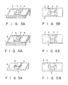
- the spacers need not however be rectangular, and may also be of the form of spacers 6 and 7 shown in Figs. 3A - 4B.
- the sample inlet port is enlarged, and introduction of aliquot parts of sample is easier.
- the vessel and its supporting base may be integrally molded.
- Fig. 5A and 5B show an integrated reaction vessel of this type.
- a tubular reaction zone 5 of such a cross-sectional area as to permit aspiration of the sample by capillarity is provided on the inclined surface of integrated reaction vessel 8, and sunken liquid troughs 9 are provided on both edges of reaction zone 5.
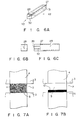
- the integrated reaction vessel may also be molded in a rod shape.
- a reaction zone 32 which is a throughhole having a specified angle with respect to the horizontal and a hollow 29 connected to this reaction zone 32.
- Reaction zone 32 has a cross-sectional area such that the sample can be aspirated by capillarity.
- this rod-shaped reaction vessel 27 is used in agglutination reaction tests, the following procedure is carried out. First, the end 30 of rod-shaped reaction vessel 27 is dipped in a previously prepared particle suspension, and the particle suspension is thereby introduced into reaction zone 32 from sample inlet port 28 by means of capillarity.
- reaction vessel 27 is removed from the suspension, and laid horizontally to allow the reaction to proceed undisturbed for 10 min. After the reaction, by observing the particle distribution pattern formed in reaction zone 32, it is possible to determine whether there was or was not an agglutination reaction.
- reaction zone 32 is connected to hollow 29 and hollow 29 is connected to the outside via opening 31, so unnecessary pressure does not act on reaction zone 32. Further, by applying a negative pressure from opening 31, sample or wash solution may be forcibly aspirated. Reaction zone 32 may also be washed by introducing wash solution from opening 31, or simply by applying a positive pressure to opening 31.
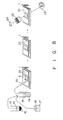
- a sample of blood is separated into a blood corpuscle component 15 and blood serum component 14 by centrifugation or another suitable method. If the blood was treated with an anticoagulant such as hepalin, the blood serum component 14 will be plasma.
- 2 ul of the blood corpuscle component 15 and 18 ul of a previously prepared solution 17 are mixed in a test tube 19, and prereacted together.
- This solution 17 consists of an anti-A serum dilution prepared by diluting standard anti-A serum (Orso Co.) to 1/30 with physiological saline.
- 5 ul of blood corpuscle suspension 18 are pipetted by means of pipette 20 into reaction zone 5 of reaction vessel 21.
- reaction vessel wherein the distance between lower plate 2 and upper plate 4 is 80 um, the distance between the two spacers 3 is 500 um and the length of the long direction of reaction zone 5 is 250 um, and a supporting base wherein the slope of the inclined surface is 45°, all the human red blood cells sediment in the lower part of the reaction zone in about 10 minutes.
- the reaction vessel is set on supporting base 1, and the base is left in an incubator at 36°C for about 10 minutes.
- the distance between the upper and lower plates of the reaction vessel and the distance between the two spacers are closely related to the time required to sediment the particles, and more specifically, should both be small in order to complete the sedimentation of particles in a short time. If however the distance between the two spacers is too small, the area where the particle distribution pattern is formed becomes smaller, and therefore the error of measurement increases. It is moreover necessary that the length of the long direction of the reaction zone is such that small quantities of sample do not dry out during the reaction, and such that the sample can spread by capillarity throughout the whole of the reaction zone.
- the reaction vessel 21 is transferred to the measurement area.
- the measurement area comprises a light source 23 and a microscope optical system 25 to observe the blood corpuscle distribution pattern magnified, and it also has a photo sensor 26 to image the magnified blood corpuscle distribution in the reaction zone.
- the occurrence or non-occurrence of an antigen-antibody reaction is determined by magnifying the reaction zone 5 of reaction vessel 21 by means of optical system 25, and observing the blood corpuscle distribution pattern.
- the image obtained by photo sensor 26 may be analyzed by a computer to determine the occurrence or non-occurrence of the reaction.
- the observed blood corpuscle distribution has a pattern of the type shown in Fig. 2A, it indicates that there are A antigens on the surface of the blood corpuscles in the sample, and that the blood corpuscles have agglutinated due to anti-A antibodies. If on the other hand the blood corpuscle distribution has a pattern of the type shown in Fig. 2B, it indicates that there are no antigens on the surface of the blood corpuscles in the sample, and that the cells moved freely down the sloping surface of the reaction vessel.
- the reactivity of the sample with respect to anti-B serum is measured in the same way, and from its measured reactivity with respect to anti-A serum and anti-B serum, the blood group of the sample can be determined. If the blood corpuscles in the sample showed an antigen-antibody reaction with respect to both anti-A serum and anti-B serum, the blood group of the sample is AB; if there was a reaction only with respect to anti-A serum, the blood group of the sample is A; if there was a reaction only with respect to anti-B serum, the blood group of the sample is B; while if there was no reaction to either serum, the blood group of the sample is O.
- the determination of blood group which previously required 60 minutes, can be accomplished in about 10 minutes using the reaction vessel kit of this invention.
- a sample of blood is separated into a blood corpuscle component 15 and blood serum component 14 by centrifugation or another suitable method. If the blood was treated with an anticoagulant such as hepalin, the blood serum component 14 will be plasma. After separation, 2 ul of the supernatant blood serum component 14 and 25 ul of a previously prepared solution 17 are mixed in a test tube 19, and prereacted together.
- This solution 17 consists of sensitized particles diluted to 1.0% (v/v) with physiological saline, organically synthesized peptides having an identical amino acid sequence to HIV antigen being coated on the surface of these sensitized particles.
- the sensitized particles may consist of synthetic particles of polystyrene or gelatin which have been modified chemically, animal red blood cells fixated by glutaraldehyde or the like, or similar particles.
- the antigen used to sensitize the particles may be an inactivated virus, or a recombinant protein produced by Escherichia coil or the like using gene manipulation techniques.
- the distribution of particles which have sedimented in zone 5 of reaction vessel 21 is observed using the same method as in Example 1. If the observed particle distribution gives a pattern of the type shown in Fig. 2A, it indicates that there are anti-HIV antibodies in the serum of the sample, and that the particles have agglutinated due to the anti-HIV antibodies. If on the other hand the particle distribution gives a pattern of the type shown in Fig. 2B, it indicates that there are no antibodies in the sample reacting to the surface antigens of the sensitized particles.
- an assay may be carried out for viruses or bacteria other than HIV such as HTLV-1, HB or gonococcus. Further, using antibody-sensitized particles, an assay for antigen may be carried out for HBs, drugs or cancer markers. Further, if particles of several different colors are made to conjugate respectively with different antibodies or antigens, and for example a color CCD camera is used as photo sensor 26, assays for several antibodies or antigens may be carried out in one operation.
- an assay can be thus be carried out with high sensitivity in 1/6 of the time conventional required. Moreover, the assay may be performed using less of the costly particle reagent than was previously required.
- an antiserum against for A antigens on the surface of blood corpuscles is coated in the reaction zone 5 of a reaction vessel kit as shown in Figs. 1A - 1C using the method described below.
- Standard anti-A serum (Orso Co.) is diluted to 1/10 with l0mM Tris-HCl buffer solution, pH 9, containing 0.15M NaCl, 5ul of the diluted solution is dropped to reaction zone 5 of lower plate 2. After dropping of the solution, the lower plate is incubated at 37°C for 1 hour while maintaining humid conditions so that the solution does not dry out. The reaction zone 5 is then washed by dropping 10 mM phosphate buffer solution, pH 7.2, containing 0.15M NaCl.
- reaction zone 5 After removing the wash solution by gently shaking lower plate 2, 10 ml of 10mM phosphate buffer solution, pH 7.2 containing 3% (w/v) of bovine serum albumin and 0.15 M NaCl are dropped to reaction zone 5, and the lower plate is incubated at room temperature while maintaining humid conditions for 1 hour. By this procedure, the area of the reaction zone which adsorbs blood corpuscles non-specifically is blocked. After incubation, reaction zone 5 is again washed with 10 mM phosphate buffer solution, pH 7.2, containing 0.15 M NaCl. If the lower plate is to be kept for a long period, 0.02% (w/v) NaN3 is added to the final wash solution, and the lower plate is kept at 4°C after washing.
- upper plate 4 is also treated so that non-specific conjugation with blood corpuscles does not occur. This is done by leaving the upper plate at room temperature for 1 hour in 10mM phosphate buffer solution, pH 7.2, containing 3% (w/v) bovine serum albumin and 0.15 M NaCl, and washing it with 10 mM phosphate buffer solution, pH 7.2, containing 0.15 M NaCl. If the upper plate is to be left for a long period, 0.02% (w/v) NaN3 is added to the final wash solution, and the upper plate is kept at 4°C as in the case of the lower plate.
- the reaction vessel is assembled using two spacers 3 which are fixed by means of double-sided adhesive tape or adhesive.
- the kit used here comprises a reaction vessel wherein the spacers are of thickness 80 um, the distance between the two spacers is 0.5 mm and the length of the long direction of the reaction zone is 5 mm, and a reaction vessel supporting base wherein the inclination angle is 45°.
- a sample of blood is separated into a blood corpuscle component 15 and blood serum component 14 by centrifugation or another suitable method. If the blood was treated with an anticoagulant such as hepalin, the blood serum component 14 will be plasma.
- 2 ul of the sedimented blood corpuscle component 15 and 98 ul of a solution 17 are mixed in a test tube 19 to prepare a 2% blood corpuscle suspension 18.
- solution 17 is physiological saline.
- This blood corpuscle suspension 18 is then introduced by means of a pipette 20 to reaction zone 5 of reaction vessel 21 wherein anti-A serum has been coated as described above. After pipetting, the vessel is set on supporting base 1 and incubated at about 36°C for 10 min, care being taken not to shake the vessel. The pipetting may be performed after setting of the reaction vessel 21 on the supporting base 1.
- the distribution pattern of particles sedimented in reaction zone 5 of reaction vessel 21 is observed as in Example 1. If there are A antigens on the surface of the blood corpuscles in the sample, the corpuscles conjugate with the anti-A serum that was coated in the reaction vessel, and the blood corpuscles are uniformly distributed practically all over reaction zone 5. The corpuscle distribution which appears in this case therefore gives a pattern of the type shown in Fig. 7A. If on the other hand there are no A antigens on the surface of the corpuscles in the sample, the blood corpuscles do not conjugate with the anti-A serum coated in the reaction vessel, and the corpuscles move freely down the inclined lower plate 2 of reaction vessel 21.
- the distribution of blood corpuscles gives a pattern of the type shown in Fig. 7B.
- a antigens on the surface of blood corpuscles in the sample may be determined in the same way by measuring the reactivity of the sample with respect to anti-B serum.
- the blood group of the sample can be determined. If the blood corpuscles in the sample showed an antigen-antibody reaction with respect to both anti-A serum and anti-B serum, the blood group of the sample is AB; if there was a reaction only with respect to anti-A serum, the blood group of the sample is A; if there was a reaction only with respect to anti-B serum, the blood group of the sample is B; while if there was no reaction to either serum, the blood group of the sample is O.
- the determination of blood group which previously required 60 minutes, can be accomplished in about 10 minutes using the reaction vessel kit of this invention.
- HIV antigens are coated in the reaction zone 5 of the reaction vessel.
- the HIV antigens coated in the reaction vessel may be chemically synthesized HIV virus surface antigens, HIV recombinant proteins produced by Escherichia coil, or the HIV virus itself.
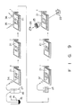
- a sample of blood is separated into a blood corpuscle component 15 and blood serum component 14 by centrifugation or another suitable method. If the blood was treated with an anticoagulant such as hepalin, the blood serum component 14 will be plasma. 2 ul of the serum component obtained was removed in a test-tube 19, then 18 ul of physiological saline was added and mixed with it. 5 ul of this diluted serum solution 27 was pipetted by means of pipette 34 to a reaction vessel 21 wherein HIV antigens had been coated. After pipetting, the reaction vessel was set on supporting base 1 and was incubated at 37°C for 2 min.
- an anticoagulant such as hepalin
- reaction zone 5 Following the reaction, physiological saline as wash solution was introduced by nozzle 35 into reaction zone 5 of reaction vessel 21, and waste liquid was aspirated simultaneously by nozzle 36 from the opposite side.
- the reaction zone 5 can be washed, and the serum component which did not react with the antigen coated in reaction vessel 21 can be washed out. It is desirable that wash solution remaining in reaction zone 5 is wiped off with filter paper, or eliminated by blowing in air from the nozzle.
- a particle reagent 33 to which goat anti-human antibodies had been coated was introduced by means of nozzle 37 to the reaction vessel, and the vessel was incubated at 37°C for 10 min.
- the anti-human antibodies used here may also be monoclonal antibodies derived from mouse or the like, or a substance such as protein A which binds to antibodies.
- the particles on which the antibodies are coated may be red blood cells fixated by glutaraldehyde or the like, or synthetic particles such as polystyrene.
- the particle distribution pattern is observed by the same method as that of Example 1. If there are antibodies against for HIV in the serum of the sample, they conjugate with the HIV antigens coated in the reaction vessel, and the particles to which anti-human antibodies have been coated conjugate with the antibodies which have conjugated with the antigens. The particle distribution is therefore a pattern of the type shown in Fig. 7A. If on the other hand there are no antibodies against for HIV in the serum of the sample, the particles move freely down the inclined lower plate of the reaction vessel without causing any reaction. The particle distribution is therefore a pattern of the type shown in Fig. 7B.
- reaction vessel kit Using this reaction vessel kit, a high sensitivity assay can be performed in a much shorter time than the 60 - 120 min previously required.
- antibodies against for viruses other than HIV such as HTLV-1 or HTLV-II or for bacteria may also be detected.
- the antibody usually has 2 - 10 antigen binding sites. Particles to which the same antigen has been coated as the antigen coated in the reaction vessel, may therefore also be used as particle reagent 33.
- an assay for antigen may also be carried out by the "sandwich” method wherein antibodies are coated on both the reaction vessel and the particles.
Landscapes
- Health & Medical Sciences (AREA)
- Chemical & Material Sciences (AREA)
- Immunology (AREA)
- Life Sciences & Earth Sciences (AREA)
- Engineering & Computer Science (AREA)
- Hematology (AREA)
- Urology & Nephrology (AREA)
- Molecular Biology (AREA)
- Biomedical Technology (AREA)
- General Health & Medical Sciences (AREA)
- Analytical Chemistry (AREA)
- Physics & Mathematics (AREA)
- Cell Biology (AREA)
- Food Science & Technology (AREA)
- Medicinal Chemistry (AREA)
- Biotechnology (AREA)
- Microbiology (AREA)
- Biochemistry (AREA)
- Chemical Kinetics & Catalysis (AREA)
- General Physics & Mathematics (AREA)
- Pathology (AREA)
- Dispersion Chemistry (AREA)
- Clinical Laboratory Science (AREA)
- Investigating Or Analysing Biological Materials (AREA)
- Automatic Analysis And Handling Materials Therefor (AREA)
- Sampling And Sample Adjustment (AREA)
Applications Claiming Priority (2)
| Application Number | Priority Date | Filing Date | Title |
|---|---|---|---|
| US45814389A | 1989-12-28 | 1989-12-28 | |
| US458143 | 1989-12-28 |
Publications (3)
| Publication Number | Publication Date |
|---|---|
| EP0435245A2 true EP0435245A2 (fr) | 1991-07-03 |
| EP0435245A3 EP0435245A3 (en) | 1992-02-26 |
| EP0435245B1 EP0435245B1 (fr) | 1995-05-10 |
Family
ID=23819546
Family Applications (1)
| Application Number | Title | Priority Date | Filing Date |
|---|---|---|---|
| EP19900125480 Expired - Lifetime EP0435245B1 (fr) | 1989-12-28 | 1990-12-24 | Kit de réaction |
Country Status (3)
| Country | Link |
|---|---|
| EP (1) | EP0435245B1 (fr) |
| JP (1) | JPH04208836A (fr) |
| DE (1) | DE69019326T2 (fr) |
Cited By (3)
| Publication number | Priority date | Publication date | Assignee | Title |
|---|---|---|---|---|
| EP1975618A1 (fr) * | 2007-03-30 | 2008-10-01 | Fujifilm Corporation | Procédé de détection d'échantillon et instrument |
| US8084002B2 (en) | 2006-05-08 | 2011-12-27 | Vivacta Ltd. | Chemical sensing device |
| CN110064458A (zh) * | 2019-03-15 | 2019-07-30 | 温州市中心医院 | 一种方便取用的血液储存装置 |
Families Citing this family (2)
| Publication number | Priority date | Publication date | Assignee | Title |
|---|---|---|---|---|
| JP3120453B2 (ja) * | 1997-06-19 | 2000-12-25 | トヨタ自動車株式会社 | 微小液滴の保持方法、反応方法 |
| EP2045015B1 (fr) * | 2007-10-01 | 2011-11-23 | Tecan Trading AG | Agencement de micro-cuvettes et son utilisation |
Family Cites Families (6)
| Publication number | Priority date | Publication date | Assignee | Title |
|---|---|---|---|---|
| US4323536A (en) * | 1980-02-06 | 1982-04-06 | Eastman Kodak Company | Multi-analyte test device |
| US4426451A (en) * | 1981-01-28 | 1984-01-17 | Eastman Kodak Company | Multi-zoned reaction vessel having pressure-actuatable control means between zones |
| JPS5821141A (ja) * | 1981-07-30 | 1983-02-07 | Olympus Optical Co Ltd | 粒子凝集反応判定方法および判定容器 |
| JPS6086468A (ja) * | 1983-10-18 | 1985-05-16 | Olympus Optical Co Ltd | 抗原抗体反応の判定方法 |
| US4596695A (en) * | 1984-09-10 | 1986-06-24 | Cottingham Hugh V | Agglutinographic reaction chamber |
| DE3889885T2 (de) * | 1987-12-23 | 1994-12-15 | Abbott Lab | Vorrichtung zur Agglutinierungsreaktion. |
-
1990
- 1990-12-24 DE DE1990619326 patent/DE69019326T2/de not_active Expired - Lifetime
- 1990-12-24 EP EP19900125480 patent/EP0435245B1/fr not_active Expired - Lifetime
- 1990-12-27 JP JP41897890A patent/JPH04208836A/ja not_active Withdrawn
Cited By (4)
| Publication number | Priority date | Publication date | Assignee | Title |
|---|---|---|---|---|
| US8084002B2 (en) | 2006-05-08 | 2011-12-27 | Vivacta Ltd. | Chemical sensing device |
| EP1975618A1 (fr) * | 2007-03-30 | 2008-10-01 | Fujifilm Corporation | Procédé de détection d'échantillon et instrument |
| CN110064458A (zh) * | 2019-03-15 | 2019-07-30 | 温州市中心医院 | 一种方便取用的血液储存装置 |
| CN110064458B (zh) * | 2019-03-15 | 2021-04-23 | 温州市中心医院 | 一种方便取用的血液储存装置 |
Also Published As
| Publication number | Publication date |
|---|---|
| JPH04208836A (ja) | 1992-07-30 |
| DE69019326D1 (de) | 1995-06-14 |
| EP0435245B1 (fr) | 1995-05-10 |
| DE69019326T2 (de) | 1995-11-09 |
| EP0435245A3 (en) | 1992-02-26 |
Similar Documents
| Publication | Publication Date | Title |
|---|---|---|
| US5188968A (en) | Method and reaction kit for agglutination detection | |
| US5066465A (en) | Reaction apparatus | |
| US5219763A (en) | Agglutination method for the determination of multiple ligands | |
| JP5479417B2 (ja) | 制御流れアッセイ装置及び方法 | |
| JP4351539B2 (ja) | 遠心力及び/又は毛管力によって流体を正確に移動し、操作する方法及び装置 | |
| US5503985A (en) | Disposable device for diagnostic assays | |
| US9766256B2 (en) | Magnetic particle tagged reagents and techniques | |
| EP1450159A2 (fr) | Procédé et appareil de détection de l'agglutinement des analyses | |
| KR20000071894A (ko) | 단백질칩 및 이를 이용한 다목적 자동화 진단시스템 | |
| JP2009098138A (ja) | 高感度免疫測定方法 | |
| EP0435245B1 (fr) | Kit de réaction | |
| JPH08201391A (ja) | マ−カ−粒子を用いた免疫学的測定方法 | |
| EP0413758A1 (fr) | Dispositif d'ecoulement capillaire et procede d'analyse a double capture | |
| EP0439917B1 (fr) | Appareil pour la détection et la mesure semiquantitative de composés | |
| JPH09229936A (ja) | 磁性粒子を用いる被検物質の測定方法並びに該方法に使用する担体及び測定器具 | |
| EP0327786A1 (fr) | Méthode et appareil pour l'exécution de réactions chimiques ou biochimiques en phases porteuses poreuses | |
| JPH08146001A (ja) | 免疫測定法及びその装置 | |
| EP3102953B1 (fr) | Épreuve directe de compatibilité | |
| JP2000131320A (ja) | 特異的結合物質固定粒子を用いる被検物質の測定方法 | |
| US20050130223A1 (en) | Method, system and kit for detecting an analyte in a sample | |
| JPH10132819A (ja) | 粒子を用いる被検物質の測定方法及び該方法に使用する測定器具 | |
| JPH03191864A (ja) | 間接凝集免疫測定方法及び装置 | |
| JP2009085703A (ja) | 高感度免疫測定方法 | |
| KR910001313B1 (ko) | 면역 분석 방법 및 장치 | |
| JPH01274066A (ja) | 酵素免疫測定法 |
Legal Events
| Date | Code | Title | Description |
|---|---|---|---|
| PUAI | Public reference made under article 153(3) epc to a published international application that has entered the european phase |
Free format text: ORIGINAL CODE: 0009012 |
|
| 17P | Request for examination filed |
Effective date: 19901224 |
|
| AK | Designated contracting states |
Kind code of ref document: A2 Designated state(s): DE FR GB |
|
| PUAL | Search report despatched |
Free format text: ORIGINAL CODE: 0009013 |
|
| AK | Designated contracting states |
Kind code of ref document: A3 Designated state(s): DE FR GB |
|
| 17Q | First examination report despatched |
Effective date: 19930315 |
|
| GRAA | (expected) grant |
Free format text: ORIGINAL CODE: 0009210 |
|
| AK | Designated contracting states |
Kind code of ref document: B1 Designated state(s): DE FR GB |
|
| ET | Fr: translation filed | ||
| REF | Corresponds to: |
Ref document number: 69019326 Country of ref document: DE Date of ref document: 19950614 |
|
| PLBE | No opposition filed within time limit |
Free format text: ORIGINAL CODE: 0009261 |
|
| STAA | Information on the status of an ep patent application or granted ep patent |
Free format text: STATUS: NO OPPOSITION FILED WITHIN TIME LIMIT |
|
| 26N | No opposition filed | ||
| REG | Reference to a national code |
Ref country code: GB Ref legal event code: IF02 |
|
| REG | Reference to a national code |
Ref country code: FR Ref legal event code: CD |
|
| PGFP | Annual fee paid to national office [announced via postgrant information from national office to epo] |
Ref country code: FR Payment date: 20100106 Year of fee payment: 20 Ref country code: GB Payment date: 20091229 Year of fee payment: 20 |
|
| PGFP | Annual fee paid to national office [announced via postgrant information from national office to epo] |
Ref country code: DE Payment date: 20091230 Year of fee payment: 20 |
|
| REG | Reference to a national code |
Ref country code: GB Ref legal event code: 732E Free format text: REGISTERED BETWEEN 20100812 AND 20100818 |
|
| REG | Reference to a national code |
Ref country code: GB Ref legal event code: PE20 Expiry date: 20101223 |
|
| PG25 | Lapsed in a contracting state [announced via postgrant information from national office to epo] |
Ref country code: GB Free format text: LAPSE BECAUSE OF EXPIRATION OF PROTECTION Effective date: 20101223 |
|
| PG25 | Lapsed in a contracting state [announced via postgrant information from national office to epo] |
Ref country code: DE Free format text: LAPSE BECAUSE OF EXPIRATION OF PROTECTION Effective date: 20101224 |