EP0443880B1 - Méthode d'analyse du virus de l'anémie infectieuse équine (EIA-Virus) - Google Patents
Méthode d'analyse du virus de l'anémie infectieuse équine (EIA-Virus) Download PDFInfo
- Publication number
- EP0443880B1 EP0443880B1 EP91301460A EP91301460A EP0443880B1 EP 0443880 B1 EP0443880 B1 EP 0443880B1 EP 91301460 A EP91301460 A EP 91301460A EP 91301460 A EP91301460 A EP 91301460A EP 0443880 B1 EP0443880 B1 EP 0443880B1
- Authority
- EP
- European Patent Office
- Prior art keywords
- peptide
- glutamic acid
- leucine
- glutamine
- conjugate
- Prior art date
- Legal status (The legal status is an assumption and is not a legal conclusion. Google has not performed a legal analysis and makes no representation as to the accuracy of the status listed.)
- Expired - Lifetime
Links
- 241000713730 Equine infectious anemia virus Species 0.000 title claims abstract description 17
- 238000003556 assay Methods 0.000 title claims description 26
- 108090000765 processed proteins & peptides Proteins 0.000 claims abstract description 129
- 239000000427 antigen Substances 0.000 claims abstract description 36
- 102000036639 antigens Human genes 0.000 claims abstract description 36
- 108091007433 antigens Proteins 0.000 claims abstract description 36
- 210000002966 serum Anatomy 0.000 claims abstract description 35
- 230000000890 antigenic effect Effects 0.000 claims abstract description 24
- 101710091045 Envelope protein Proteins 0.000 claims abstract description 10
- 101710188315 Protein X Proteins 0.000 claims abstract description 10
- 102100021696 Syncytin-1 Human genes 0.000 claims abstract 2
- 238000012360 testing method Methods 0.000 claims description 55
- 208000009724 equine infectious anemia Diseases 0.000 claims description 47
- 238000000034 method Methods 0.000 claims description 44
- 102000004190 Enzymes Human genes 0.000 claims description 26
- 108090000790 Enzymes Proteins 0.000 claims description 26
- 241000283073 Equus caballus Species 0.000 claims description 24
- 239000000243 solution Substances 0.000 claims description 23
- 230000003612 virological effect Effects 0.000 claims description 23
- 150000001413 amino acids Chemical group 0.000 claims description 21
- 235000013922 glutamic acid Nutrition 0.000 claims description 19
- 239000004220 glutamic acid Substances 0.000 claims description 19
- WHUUTDBJXJRKMK-UHFFFAOYSA-N Glutamic acid Natural products OC(=O)C(N)CCC(O)=O WHUUTDBJXJRKMK-UHFFFAOYSA-N 0.000 claims description 18
- 239000000758 substrate Substances 0.000 claims description 18
- 239000007790 solid phase Substances 0.000 claims description 17
- 229940024606 amino acid Drugs 0.000 claims description 15
- 235000001014 amino acid Nutrition 0.000 claims description 15
- ZDXPYRJPNDTMRX-UHFFFAOYSA-N glutamine Natural products OC(=O)C(N)CCC(N)=O ZDXPYRJPNDTMRX-UHFFFAOYSA-N 0.000 claims description 15
- 238000002835 absorbance Methods 0.000 claims description 14
- LOKCTEFSRHRXRJ-UHFFFAOYSA-I dipotassium trisodium dihydrogen phosphate hydrogen phosphate dichloride Chemical group P(=O)(O)(O)[O-].[K+].P(=O)(O)([O-])[O-].[Na+].[Na+].[Cl-].[K+].[Cl-].[Na+] LOKCTEFSRHRXRJ-UHFFFAOYSA-I 0.000 claims description 14
- 239000002953 phosphate buffered saline Substances 0.000 claims description 14
- ROHFNLRQFUQHCH-UHFFFAOYSA-N Leucine Natural products CC(C)CC(N)C(O)=O ROHFNLRQFUQHCH-UHFFFAOYSA-N 0.000 claims description 11
- AYFVYJQAPQTCCC-UHFFFAOYSA-N Threonine Natural products CC(O)C(N)C(O)=O AYFVYJQAPQTCCC-UHFFFAOYSA-N 0.000 claims description 10
- 239000004473 Threonine Substances 0.000 claims description 10
- 239000000203 mixture Substances 0.000 claims description 9
- 235000009697 arginine Nutrition 0.000 claims description 7
- 239000004475 Arginine Substances 0.000 claims description 6
- 108010001336 Horseradish Peroxidase Proteins 0.000 claims description 6
- ODKSFYDXXFIFQN-UHFFFAOYSA-N arginine Natural products OC(=O)C(N)CCCNC(N)=N ODKSFYDXXFIFQN-UHFFFAOYSA-N 0.000 claims description 6
- HNDVDQJCIGZPNO-UHFFFAOYSA-N histidine Natural products OC(=O)C(N)CC1=CN=CN1 HNDVDQJCIGZPNO-UHFFFAOYSA-N 0.000 claims description 6
- MHAJPDPJQMAIIY-UHFFFAOYSA-N Hydrogen peroxide Chemical compound OO MHAJPDPJQMAIIY-UHFFFAOYSA-N 0.000 claims description 5
- ROHFNLRQFUQHCH-YFKPBYRVSA-N L-leucine Chemical compound CC(C)C[C@H](N)C(O)=O ROHFNLRQFUQHCH-YFKPBYRVSA-N 0.000 claims description 5
- KZSNJWFQEVHDMF-UHFFFAOYSA-N Valine Natural products CC(C)C(N)C(O)=O KZSNJWFQEVHDMF-UHFFFAOYSA-N 0.000 claims description 5
- 235000004279 alanine Nutrition 0.000 claims description 5
- 239000004474 valine Substances 0.000 claims description 5
- GEYOCULIXLDCMW-UHFFFAOYSA-N 1,2-phenylenediamine Chemical compound NC1=CC=CC=C1N GEYOCULIXLDCMW-UHFFFAOYSA-N 0.000 claims description 4
- 230000008859 change Effects 0.000 claims description 4
- 230000002285 radioactive effect Effects 0.000 claims description 4
- 229920002554 vinyl polymer Polymers 0.000 claims description 4
- 102000002260 Alkaline Phosphatase Human genes 0.000 claims description 3
- 108020004774 Alkaline Phosphatase Proteins 0.000 claims description 3
- 239000003795 chemical substances by application Substances 0.000 claims description 3
- ZCYVEMRRCGMTRW-UHFFFAOYSA-N 7553-56-2 Chemical group [I] ZCYVEMRRCGMTRW-UHFFFAOYSA-N 0.000 claims description 2
- 241001494479 Pecora Species 0.000 claims description 2
- 229910052740 iodine Inorganic materials 0.000 claims description 2
- 239000011630 iodine Substances 0.000 claims description 2
- 238000002156 mixing Methods 0.000 claims description 2
- 238000010324 immunological assay Methods 0.000 claims 1
- 238000003018 immunoassay Methods 0.000 abstract description 28
- 125000003275 alpha amino acid group Chemical group 0.000 abstract description 18
- 238000001514 detection method Methods 0.000 abstract description 7
- 241000283086 Equidae Species 0.000 abstract description 2
- 238000002967 competitive immunoassay Methods 0.000 abstract 1
- 241000700605 Viruses Species 0.000 description 21
- 108090000623 proteins and genes Proteins 0.000 description 14
- 102000004196 processed proteins & peptides Human genes 0.000 description 13
- 235000018102 proteins Nutrition 0.000 description 13
- 102000004169 proteins and genes Human genes 0.000 description 13
- 229960002989 glutamic acid Drugs 0.000 description 12
- 238000002372 labelling Methods 0.000 description 11
- 102100034349 Integrase Human genes 0.000 description 8
- 238000011534 incubation Methods 0.000 description 7
- 102000009027 Albumins Human genes 0.000 description 6
- 108010088751 Albumins Proteins 0.000 description 6
- 238000006243 chemical reaction Methods 0.000 description 6
- 239000000872 buffer Substances 0.000 description 5
- 208000015181 infectious disease Diseases 0.000 description 5
- 102000013415 peroxidase activity proteins Human genes 0.000 description 5
- 108040007629 peroxidase activity proteins Proteins 0.000 description 5
- UIIMBOGNXHQVGW-UHFFFAOYSA-M Sodium bicarbonate Chemical compound [Na+].OC([O-])=O UIIMBOGNXHQVGW-UHFFFAOYSA-M 0.000 description 4
- 201000010099 disease Diseases 0.000 description 4
- 208000037265 diseases, disorders, signs and symptoms Diseases 0.000 description 4
- 239000000463 material Substances 0.000 description 4
- CDBYLPFSWZWCQE-UHFFFAOYSA-L sodium carbonate Substances [Na+].[Na+].[O-]C([O-])=O CDBYLPFSWZWCQE-UHFFFAOYSA-L 0.000 description 4
- 238000010998 test method Methods 0.000 description 4
- 229920001817 Agar Polymers 0.000 description 3
- WSFSSNUMVMOOMR-UHFFFAOYSA-N Formaldehyde Chemical compound O=C WSFSSNUMVMOOMR-UHFFFAOYSA-N 0.000 description 3
- 241000725303 Human immunodeficiency virus Species 0.000 description 3
- 241001465754 Metazoa Species 0.000 description 3
- QAOWNCQODCNURD-UHFFFAOYSA-N Sulfuric acid Chemical compound OS(O)(=O)=O QAOWNCQODCNURD-UHFFFAOYSA-N 0.000 description 3
- 230000003466 anti-cipated effect Effects 0.000 description 3
- 230000015572 biosynthetic process Effects 0.000 description 3
- 238000010790 dilution Methods 0.000 description 3
- 239000012895 dilution Substances 0.000 description 3
- 238000012986 modification Methods 0.000 description 3
- 230000004048 modification Effects 0.000 description 3
- 238000002360 preparation method Methods 0.000 description 3
- 238000000163 radioactive labelling Methods 0.000 description 3
- 235000011149 sulphuric acid Nutrition 0.000 description 3
- 101710132601 Capsid protein Proteins 0.000 description 2
- WHUUTDBJXJRKMK-VKHMYHEASA-N L-glutamic acid Chemical class OC(=O)[C@@H](N)CCC(O)=O WHUUTDBJXJRKMK-VKHMYHEASA-N 0.000 description 2
- PXIPVTKHYLBLMZ-UHFFFAOYSA-N Sodium azide Chemical compound [Na+].[N-]=[N+]=[N-] PXIPVTKHYLBLMZ-UHFFFAOYSA-N 0.000 description 2
- FAPWRFPIFSIZLT-UHFFFAOYSA-M Sodium chloride Chemical compound [Na+].[Cl-] FAPWRFPIFSIZLT-UHFFFAOYSA-M 0.000 description 2
- 208000036142 Viral infection Diseases 0.000 description 2
- 239000008272 agar Substances 0.000 description 2
- 238000004458 analytical method Methods 0.000 description 2
- 208000007502 anemia Diseases 0.000 description 2
- 238000007385 chemical modification Methods 0.000 description 2
- 239000011248 coating agent Substances 0.000 description 2
- 238000000576 coating method Methods 0.000 description 2
- 235000018417 cysteine Nutrition 0.000 description 2
- 238000011161 development Methods 0.000 description 2
- 239000007850 fluorescent dye Substances 0.000 description 2
- 238000001215 fluorescent labelling Methods 0.000 description 2
- 230000002209 hydrophobic effect Effects 0.000 description 2
- 238000002169 hydrotherapy Methods 0.000 description 2
- 150000002632 lipids Chemical class 0.000 description 2
- 235000018977 lysine Nutrition 0.000 description 2
- 238000010647 peptide synthesis reaction Methods 0.000 description 2
- 229910000030 sodium bicarbonate Inorganic materials 0.000 description 2
- 235000017557 sodium bicarbonate Nutrition 0.000 description 2
- 229910000029 sodium carbonate Inorganic materials 0.000 description 2
- 239000000126 substance Substances 0.000 description 2
- 238000003786 synthesis reaction Methods 0.000 description 2
- 230000009385 viral infection Effects 0.000 description 2
- 238000005406 washing Methods 0.000 description 2
- DGVVWUTYPXICAM-UHFFFAOYSA-N β‐Mercaptoethanol Chemical compound OCCS DGVVWUTYPXICAM-UHFFFAOYSA-N 0.000 description 2
- JQPFYXFVUKHERX-UHFFFAOYSA-N 2-hydroxy-2-cyclohexen-1-one Natural products OC1=CCCCC1=O JQPFYXFVUKHERX-UHFFFAOYSA-N 0.000 description 1
- DCXYFEDJOCDNAF-UHFFFAOYSA-N Asparagine Natural products OC(=O)C(N)CC(N)=O DCXYFEDJOCDNAF-UHFFFAOYSA-N 0.000 description 1
- BTBUEUYNUDRHOZ-UHFFFAOYSA-N Borate Chemical compound [O-]B([O-])[O-] BTBUEUYNUDRHOZ-UHFFFAOYSA-N 0.000 description 1
- 238000002965 ELISA Methods 0.000 description 1
- 108060003951 Immunoglobulin Proteins 0.000 description 1
- XUJNEKJLAYXESH-REOHCLBHSA-N L-Cysteine Chemical compound SC[C@H](N)C(O)=O XUJNEKJLAYXESH-REOHCLBHSA-N 0.000 description 1
- ODKSFYDXXFIFQN-BYPYZUCNSA-P L-argininium(2+) Chemical compound NC(=[NH2+])NCCC[C@H]([NH3+])C(O)=O ODKSFYDXXFIFQN-BYPYZUCNSA-P 0.000 description 1
- KDXKERNSBIXSRK-YFKPBYRVSA-N L-lysine Chemical compound NCCCC[C@H](N)C(O)=O KDXKERNSBIXSRK-YFKPBYRVSA-N 0.000 description 1
- KDXKERNSBIXSRK-UHFFFAOYSA-N Lysine Natural products NCCCCC(N)C(O)=O KDXKERNSBIXSRK-UHFFFAOYSA-N 0.000 description 1
- 239000004472 Lysine Substances 0.000 description 1
- 241000699670 Mus sp. Species 0.000 description 1
- 229910019142 PO4 Inorganic materials 0.000 description 1
- 102000004160 Phosphoric Monoester Hydrolases Human genes 0.000 description 1
- 108090000608 Phosphoric Monoester Hydrolases Proteins 0.000 description 1
- 125000000539 amino acid group Chemical group 0.000 description 1
- 230000027645 antigenic variation Effects 0.000 description 1
- 238000013459 approach Methods 0.000 description 1
- 150000001484 arginines Chemical class 0.000 description 1
- 235000009582 asparagine Nutrition 0.000 description 1
- 229960001230 asparagine Drugs 0.000 description 1
- 238000002820 assay format Methods 0.000 description 1
- 230000006399 behavior Effects 0.000 description 1
- 239000007853 buffer solution Substances 0.000 description 1
- 150000001718 carbodiimides Chemical class 0.000 description 1
- 239000003153 chemical reaction reagent Substances 0.000 description 1
- 230000002860 competitive effect Effects 0.000 description 1
- 238000012790 confirmation Methods 0.000 description 1
- 230000008878 coupling Effects 0.000 description 1
- 238000010168 coupling process Methods 0.000 description 1
- 238000005859 coupling reaction Methods 0.000 description 1
- 238000012258 culturing Methods 0.000 description 1
- OILAIQUEIWYQPH-UHFFFAOYSA-N cyclohexane-1,2-dione Chemical compound O=C1CCCCC1=O OILAIQUEIWYQPH-UHFFFAOYSA-N 0.000 description 1
- XUJNEKJLAYXESH-UHFFFAOYSA-N cysteine Natural products SCC(N)C(O)=O XUJNEKJLAYXESH-UHFFFAOYSA-N 0.000 description 1
- 150000001945 cysteines Chemical class 0.000 description 1
- 238000003745 diagnosis Methods 0.000 description 1
- 239000012153 distilled water Substances 0.000 description 1
- 230000000694 effects Effects 0.000 description 1
- 238000006911 enzymatic reaction Methods 0.000 description 1
- 238000002474 experimental method Methods 0.000 description 1
- 230000002349 favourable effect Effects 0.000 description 1
- 125000000524 functional group Chemical group 0.000 description 1
- 239000000499 gel Substances 0.000 description 1
- 238000012817 gel-diffusion technique Methods 0.000 description 1
- -1 glutamic acid amino acid Chemical class 0.000 description 1
- 150000002307 glutamic acids Chemical class 0.000 description 1
- 210000004408 hybridoma Anatomy 0.000 description 1
- 230000003053 immunization Effects 0.000 description 1
- 102000018358 immunoglobulin Human genes 0.000 description 1
- 238000011835 investigation Methods 0.000 description 1
- PNDPGZBMCMUPRI-UHFFFAOYSA-N iodine Chemical compound II PNDPGZBMCMUPRI-UHFFFAOYSA-N 0.000 description 1
- JDNTWHVOXJZDSN-UHFFFAOYSA-N iodoacetic acid Chemical compound OC(=O)CI JDNTWHVOXJZDSN-UHFFFAOYSA-N 0.000 description 1
- 238000002955 isolation Methods 0.000 description 1
- 238000011031 large-scale manufacturing process Methods 0.000 description 1
- 150000002669 lysines Chemical class 0.000 description 1
- KQSSATDQUYCRGS-UHFFFAOYSA-N methyl glycinate Chemical compound COC(=O)CN KQSSATDQUYCRGS-UHFFFAOYSA-N 0.000 description 1
- 231100000989 no adverse effect Toxicity 0.000 description 1
- NBIIXXVUZAFLBC-UHFFFAOYSA-K phosphate Chemical compound [O-]P([O-])([O-])=O NBIIXXVUZAFLBC-UHFFFAOYSA-K 0.000 description 1
- 239000010452 phosphate Substances 0.000 description 1
- 239000007981 phosphate-citrate buffer Substances 0.000 description 1
- 235000020004 porter Nutrition 0.000 description 1
- 238000000746 purification Methods 0.000 description 1
- 230000009467 reduction Effects 0.000 description 1
- 230000035945 sensitivity Effects 0.000 description 1
- 239000011780 sodium chloride Substances 0.000 description 1
- BEOOHQFXGBMRKU-UHFFFAOYSA-N sodium cyanoborohydride Chemical compound [Na+].[B-]C#N BEOOHQFXGBMRKU-UHFFFAOYSA-N 0.000 description 1
- 239000012064 sodium phosphate buffer Substances 0.000 description 1
- 241000894007 species Species 0.000 description 1
- 150000003573 thiols Chemical class 0.000 description 1
- 241001430294 unidentified retrovirus Species 0.000 description 1
- 238000011179 visual inspection Methods 0.000 description 1
- XLYOFNOQVPJJNP-UHFFFAOYSA-N water Chemical compound O XLYOFNOQVPJJNP-UHFFFAOYSA-N 0.000 description 1
Images

Classifications
-
- C—CHEMISTRY; METALLURGY
- C07—ORGANIC CHEMISTRY
- C07K—PEPTIDES
- C07K14/00—Peptides having more than 20 amino acids; Gastrins; Somatostatins; Melanotropins; Derivatives thereof
- C07K14/005—Peptides having more than 20 amino acids; Gastrins; Somatostatins; Melanotropins; Derivatives thereof from viruses
-
- C—CHEMISTRY; METALLURGY
- C12—BIOCHEMISTRY; BEER; SPIRITS; WINE; VINEGAR; MICROBIOLOGY; ENZYMOLOGY; MUTATION OR GENETIC ENGINEERING
- C12N—MICROORGANISMS OR ENZYMES; COMPOSITIONS THEREOF; PROPAGATING, PRESERVING, OR MAINTAINING MICROORGANISMS; MUTATION OR GENETIC ENGINEERING; CULTURE MEDIA
- C12N2740/00—Reverse transcribing RNA viruses
- C12N2740/00011—Details
- C12N2740/10011—Retroviridae
- C12N2740/15011—Lentivirus, not HIV, e.g. FIV, SIV
- C12N2740/15022—New viral proteins or individual genes, new structural or functional aspects of known viral proteins or genes
Definitions
- the present invention relates to a synthetic peptide and its use in an immunoassay for the detection of Equine Infectious Anemia (EIA) viral infection.
- EIA Equine Infectious Anemia
- Equine Infectious Anemia is a viral disease, commonly known as swamp fever, which primarily affects horses and ponies. There is no known cure for the disease. Diagnosis and isolation is the only way to control the disease.
- the accepted way to diagnose the presence of EIA has been to detect the presence of antibodies specific for the disease in the serum of affected animals using the Coggins or agar gel diffusion test described in U.S. Patent No. 3,929,982 and U.S. Patent No. 3,932,601.
- a prepared antigen is placed alongside the serum to be tested in an agar or gel medium. If EIA antibodies are present in the test serum, they will diffuse toward the antigen forming a precipitin line in the agar medium where they eventually meet.
- This methodology is inherently insensitive in that the EIA antigen may be contaminated with non-EIA antigens during its preparation Antibodies against non-EIA antigens may be present in the test serum and can react with the non-EIA antigens forming a variety of nonspecific precipitin lines. Even if the prepared EIA viral antigen can be purified, the Coggins test is labor intensive and demanding of considerable expertise in interpretation of results. The Coggins test procedure is also slow to yield results; it takes twenty-four to forty-eight hours for the formation of clearly visible precipitin lines.
- HIV human immunodeficiency virus
- a synthetic peptide comprising an amino acid sequence at least a portion of which corresponds to an antigenic site on the envelope protein portion of the equine infectious anemia virus, said amino acid sequence being one of leucine - leucine - lysine - glutamic acid - arginine - glutamine - glutamine - valine - glutamic acid - glutamic acid - threonine - phenylalanine - asparagine - leucine - isoleucine - glycine - cysteine - isoleucine - glutamic acid - arginine - threonine - histidine - valine - phenylalanine - cysteine (Referred to as SEQ ID No:2) or glutamine - threonine - histidine - alanine - aspartic acid - valine - glutamine - le
- the present invention also provides a method of detecting the presence of antibodies to the equine infectious anemia virus using as antigen the synthetic peptide of the present invention.
- a method of detecting the presence of antibodies to the equine infectious anemia virus in equine test sera comprising using as antigen a synthetic peptide having an amino acid sequence at least a portion of which corresponds to an antigenic site on an equine infectious anemia virus, wherein said peptide is one of SEQ ID No:2 having the amino acid sequence leucine - leucine - lysine - glutamic acid - arginine - glutamine - glutamine - valine - glutamic acid - glutamic acid - threonine - phenylalanine - asparagine - leucine -isoleucine - glycine - cysteine - isoleucine - glutamic acid - arginine - threonine - histidine - valine - phenylalanine - cysteine or SEQ ID No: 1 having the amino acid sequence leucine - leucine - ly
- the method comprises the steps of exposing a selected amount of the peptide to a selected amount of equine test serum for a first period of time sufficient to allow any equine infectious anemia viral antibodies present in the test serum to bind with the peptide, forming a peptide-antibody complex.
- the method further includes the steps of exposing the peptide-antibody complex to a selected amount of a labelled conjugate for a second period of time sufficient to allow the conjugate to bind with the peptide-antibody complex and detecting the presence of the labelled conjugate which bound with the peptide-antibody complex.
- the conjugate may be labeled with a radioactive element, such as Iodine, or may be magnetically labeled.
- the conjugate may also be an anti-horse antibody such as sheep anti-horse immunoglobulin which will recognize and bind to an equine infections anemia viral antibody, a purified equine infections anemia viral antigen or a synthetic peptide, such as the synthetic peptide of the present invention, having an amino acid sequence corresponding to an antigenic site on an equine infectious anemia virus.
- the conjugate is preferably labeled with an enzyme selected from the group consisting of horseradish peroxidase and alkaline phosphate.
- the step of detecting an enzyme labeled conjugate includes the additional step of adding a substrate, such as an orthophenylene-diamine/hydrogen peroxide solution, in an amount sufficient to react with the enzyme label to convert the substrate to a sufficient amount of a product to produce a color change that is visible to the naked eye.
- the method may further include the step of quantifying the amount of the enzyme-labeled conjugate bound to the peptide-antibody complex by measuring the absorbance of the product of the enzymatic reaction using a spectrophotometer.
- An alternate method of the present invention include the steps of exposing the peptide of the present invention to a solid phase support, such as a multi-well polyvinyl microtitre plate, for a first period of time sufficient to permit the peptide to bind to the support and subsequently removing unbound peptide.
- the alternate method further comprises the steps of mixing a selected amount of labeled anti-EIA antibody with a selected amount of equine test serum, adding the mixture to the solid phase support containing the peptide to expose the peptide to the mixture for a second period of time sufficient to permit the peptide to bind to the antibodies in the mixture, removing any unbound antibodies from the support, and detecting the amount of labeled anti-EIA antibody bound to the peptide.
- the amount of labeled anti-EIA antibody is less than the amount of antibody in the selected amount of test serum.
- the present invention provides a synthetic peptide for use as the antigen in an immunoassay for the detection antibodies in the sera of affected animals indicative of Equine Infectious Anemia (EIA) viral infection.
- the amino acid sequence of the peptide of the present invention comprises those sequences which correspond to antigenic sites located in the envelope protein portion of the equine infectious anemia virus. The two particular amino acid sequences of the present invention have been found to provide very good results.
- One which consists of the amino acids comprising residues 68-91 of the GP-45 protein, follows: glutamine - threonine - histidine - alanine - aspartic acid - valine - glutamine - leucine - leucine - lysine - glutamic acid - arginine - glutamine - glutamine - valine - glutamic acid - glutamic acid - threonine - phenylalanine - asparagine - leucine - isoleucine - glycine - cysteine.
- peptide SEQ ID No: 1 The other, which consists of the amino acids comprising residues 76-99 of the GP-45 protein, follows: leucine - leucine -lysine - glutamic-acid - arginine - glutamine - glutamine - valine - glutamic acid - glutamic acid - threonine - phenylalanine - asparagine - leucine - isoleucine - glycine - cysteine - isoleucine - glutamic acid - arginine - threonine - histidine - valine - phenylalanine - cysteine. (Referred to hereinafter as peptide SEQ ID No: 2)
- the synthetic peptide preferably peptide SEQ ID No: 2 is used as the antigen in an indirect double antibody immunoassay.
- the peptide is added to a solid phase support and incubated for a sufficient time to ensure that the peptide is bound to the support. Any peptide which does not bind is removed.
- a substance, known as a blocker, which will not react with EIA viral antibodies is added to the support.
- the equine test serum is added to the support and allowed to incubate for a period of time sufficient to permit any EIA antibodies present in the test serum to bind to the peptide. After incubation, unbound test serum antibodies are removed.
- Labeled conjugate is added which binds to the peptide-antibody complex. Following a time sufficient to allow such binding, any unbound labeled conjugate is removed. The amount of bound labeled conjugate is detected by means appropriate for the type of labeling employed. A high level of bound conjugate indicates a positive result, i.e., the presence of EIA viral antibodies. A low level of bound conjugate indicates a negative result.
- any multi-well microtitre plate such as a Costar ⁇ 96 well polyvinyl microtitre plate, will work well.
- the direct binding of equine antibodies present in the test serum to the solid phase support is likely to result in a false pcsitive reading.
- the blocker is used to fill any empty sites on the support which did not bind synthetic peptide. Any substance which will not react with EIA viral antibodies will function as a blocker.
- a conjugate is some species which will recognize and bind with the test serum EIA viral antibodies.
- the conjugate may be a second antibody, referred to as an anti-horse antibody for its ability to recognize and bind with any other horse antibody, including an EIA viral antibody.
- the conjugate may be purified equine infectious anemia viral antigen, or the synthetic peptide of the present invention which has an amino acid sequence corresponding to an antigenic site of an equine infectious anemia virus.
- the conjugate may be labeled using a variety of labeling means, including but not limited to: radiolabeling, enzyme labeling, fluorescent labeling, and magnetic labeling. Radioactive Iodine is a well known radiolabeling agent. If enzyme labeling is the labeling means chosen, the conjugate is labeled with an enzyme preferably selected from the group consisting of horseradish peroxidase and alkaline phosphatase. Other enzymes may be employed.
- the labeled conjugate is detected by adding an amount of a substrate which will recognize and react with the enzyme label to form a product that will produce a color change visible to the naked eye.
- the presence of color indicates a sufficient level of test serum antibodies to indicate infection.
- An absence of color is an indicator of a lack of infection, as the animal did not produce a significant number of antibodies to the virus.
- the labeled conjugate had few antibodies, if any, to bind with and was subsequently removed from the support.
- peroxidase substrates and phosphatase substrates which will react with horseradish peroxidase and alkaline phosphatase enzymes, respectively, to form a colored product.
- a preferred peroxidase substrate is an ortho-phenylenediamine/hydrogen peroxide solution.
- the intensity of the color of the product may be quantified using a spectrophotometer to read absorbance. However, measuring the absorbance is not necessary to obtain an accurate reading of the results of the assay.
- the assay was performed using only peptide SEQ ID No: 2 to test blind 131 samples in a series of four test runs.
- a peptide coated plate In order to prepare a peptide coated plate, the peptide was dissolved in a 0.1 M solution of sodium bicarbonate/sodium carbonate buffer, having a pH of 9, at a concentration of 5 ⁇ g/ml.
- This buffer known as the coating buffer, was prepared by dissolving 1.59 g of sodium carbonate, 2.93 g of sodium bicarbonate and 0.2 g of sodium azide in distilled water to make up a total volume of 1.0 liter. This buffer may be stored at room temperature for not more than two weeks.
- One hundred microliters of the peptide in buffer solution was added to each well of a 96 well polyvinyl microtitre plate. The plate was incubated in a humid chamber overnight, or approximately 18 hours.
- the peptide solution was removed by washing the plate with phosphate buffered saline (PBS).
- PBS phosphate buffered saline
- a solution of equine albumin (100 mg/ml) in PBS was added to the plate and was allowed to incubate for one hour at room temperature. Following incubation, the plate was washed with PBS and allowed to air dry. The resulting plate was peptide coated and any sites not containing peptide were covered with equine albumin blocker. A plate prepared in this manner will remain stable for at least six months with no loss of sensitivity.
- Equine test serum in a 1:10 dilution in 10% equine albumin in PBS was added directly to one of the wells of the peptide coated plate to expose the peptide to the test serum and was allowed to incubate at room temperature for 60 minutes to allow antibodies present in the sera to bind with the peptide to form a peptide-antibody complex. After incubation, the serum was removed by washing the peptide coated plate five times with PBS. Fifty microliters of horseradish peroxidase labeled sheep-anti-horse immunoglobin diluted in 10% equine albumin in PBS was added to the peptide coated plate and allowed to incubate for 60 minutes to allow the conjugate to bind with the peptide-antibody complex.
- a substrate solution was prepared by dissolving 40 mg of ortho-phenylenediamine in 10 ml of a phosphate-citrate buffer at pH 5.0 and adding 40 ⁇ l of 30% hydrogen peroxide.
- the substrate solution is light sensitive and must be made up freshly before use and must be used immediately.
- One hundred microliters of substrate solution was added to the plate and color development was allowed to proceed for 5-30 minutes.
- Fifty microliters of a solution of H2SO4 (2.5 M) was added to stop the reaction between the enzyme and the substrate. The color of the solution was observed.
- test Run 1 No. A492 nm Result No. A492 nm Result 1 0.866 + (+) 11 0.146 - (-) 2 0.104 - (-) 12 1.892 + (+) 3 1.232 + (+) 13 >2.0 - (-) 4 0.201 - (-) 14 0.210 - (-) 5 1.327 + (+) 15 1.256 + (+) 6 0.143 - (-) 16 0.168 - (-) 7 1.674 + (+) 17 >2.0 + (+) 8 1.562 + (+) 18 1.765 + (+) 9 0.211 - (-) 19 0.124 - (-) 10 1.982 + (+) 20 0.176 - (-) Test Run 2 No.
- A492 nm Result No. A492 nm Result 62 0.145 - (-) 82 0.122 - (-) 63 >2.0 + (+) 83 0.142 - (-) 64 0.118 - (-) 84 0.131 - (-) 65 1.678 + (+) 85 >2.0 + (+) 66 1.987 + (+) 86 >2.0 + (+) 67 0.121 - (-) 87 0.135 - (-) 68 0.133 - (-) 88 0.126 - (-) 69 >2.0 + (+) 89 1.769 + (+) 70 0.117 - (-) 90 0.126 - (-) 71 1.864 + (+) 91 >2.0 + (+) 72 0.128 - (-) 92 0.133 - (-) 73 0.122 - (-) 93 0.121 - (-) 74 0.118 - (-) 94 0.142 - (-) 75 1.893 + (+) 95 >2.0 + (+) 76 0.125 - (
- Test Run 4 The samples in Test Run 4 marked by asterisks gave low values of Absorbance at 492 nm, but were still well above the expected values of negative samples. These samples are designated as low positives.
- the supplier of the test samples The National Veterinary Scienes Lab of Ames, Iowa, currently does not count these two samples in certifying accuracy of test results, as many test laboratories were unable to detect a positive result using the approved Coggins test method.
- Lysines were modified by reaction with a solution containing 5 M formaldehyde and 100mM sodium cyanoborohydride, in 50mM sodium phosphate buffer, pH 7.0, for 2 hours.
- Glutamic Acids were modified by reaction with a 10 fold molar excess of glycine methyl ester and a 1 molar excess of carbodiimide.
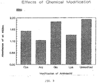
- Figure 3 shows that the modification of the glutamic acid amino acid does not result in more than a minor loss of antigenic activity, as measured by comparison of the Absorbance values obtained in assays using the unmodified peptide to those using the modified glutamic acid.
- glutamic acid does not play a critical role in the antigenic activity of the peptide. It may be modified or replaced during synthesis with no adverse effect upon the function of the peptide as antigen.
- modification of cysteine, lysine, or arginine does not result in a total loss of antigenic activity.
- the functional groups of these amino acids may be modified for use in coupling the peptide to an enzyme for labeling and to other solid phases. This observation suggests that the synthetic peptide may be used in a second type of assay, known as a sandwich immunoassay.
- the assay is carried out in the same manner as the indirect double antibody immunoassay.
- the labeled conjugate is a purified form of EIA viral antigen, rather than a second anti-horse antibody. The antigen will recognize and bind with any EIA antibodies present in the test serum.
- Labeled synthetic peptide may be used as the conjugate in this type of assay.
- a sandwich immunoassay may be performed in two steps: adding test serum and incubating and subsequently adding labeled conjugate and incubating. Alternatively, a sandwich immunoassay may be performed with one incubation wherein the test serum is mixed with labeled conjugate then added in one step to be exposed to the synthetic peptide.
- This example describes the use of the synthetic peptide in a two-step sandwich immunoassay.
- the assay was performed in a two step procedure, that is, with two incubations.
- a peptide SEQ ID No: 2 coated plate was prepared as; in Example 1. Fifty microliters of equine test serum was added to one well of the peptide coated plate and allowed to incubate for 5 minutes. The plate was then washed 5 times with PBS solution. Fifty microliters of an appropriate dilution, empiracally determined to be approximately 10 ⁇ g/ml, of the enzyme labeled peptide solution was added and allowed to incubate for 5 minutes. The plate was then washed 5 times with PBS solution. A peroxidase substrate was added and color was allowed to develop. Fifty microliters of a solution of H2SO4 (2.5 M) was added to stop the reaction between the enzyme and the substrate.
- the presence of color indicates the presence of antibodies directed against the EIA virus. Negative samples give little, if any, color. Color intensity may be quantified by using a spectrophotometer and reading Absorbance at 492 nm.
- a SEQ ID No: 2 peptide coated plate was prepared as in Example 1.
- An enzyme labeled peptide solution was prepared as in Example 2.
- Fifty microliters of equine test serum was added to 50 ⁇ l of the enzyme labeled peptide solution. This mixture was added to one well of the plate. The plate was allowed to incubate at room temperature for 5 minutes. The plate was then washed 5 times with PBS solution.
- a peroxidase substrate was added. The color was allowed to develop and 2.5 M H2SO4 was added in a quantity sufficient to stop the reaction between the enzyme and the substrate.
- Color intensity may be quantified by using a spectrophotometer and reading Absorbance at 492 nm.
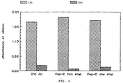
- Figure 4 shows a comparison of the three assay formats shown previously by Examples 1, 2, and 3.
- the indirect second antibody method is designated 2nd Ab; Pep-E two step and Pep-E one step refer to the enzyme labeled peptide in a sandwich assay utilizing either a single or double incubation.
- 2nd Ab The indirect second antibody method
- Pep-E two step and Pep-E one step refer to the enzyme labeled peptide in a sandwich assay utilizing either a single or double incubation.
- an indirect double antibody immunoassay or a sandwich immunoassay may be performed wherein a synthetic peptide is bound to a solid phase filter material. Any unbound test serum antibodies or unbound labeled conjugate will automatically pass through the filter material. It will be appreciated that use of a filter material as the solid phase support eliminates the need for incorporating the specific steps of removal of unbound materials from the assay.
- a competition immunoassay is contemplated.
- a synthetic peptide is bound to a solid phase support with unbound peptide subsequently being removed.
- a known amount of labeled conjugate in the form of second anti-EIA antibody in relatively small concentration is added to the test serum. This mixture is added to the solid phase support containing bound peptide.
- Both the labeled second antibodies and the EIA viral antibodies present in the test serum compete for the binding sites available on the synthetic peptide. All antibodies which do not bind to the peptide are removed and any labeled second antibody which does bind is determined by means appropriate for the type of label which is employed.
- test serum does not contain EIA viral antibodies.
- test serum contains EIA viral antibodies which take up the binding sites on the peptide so that labeled conjugate cannot bind in an appreciable amount. Therefore, the interpretation given the results of a competition immunoassay is the opposite of that given the results of an indirect or sandwich immunoassay.
- a high level of bound labeled conjugate is a negative result and a low level is a positive result.
- Immunoassays utilizing a synthetic peptide from this invention may be performed satisfactorily at any temperature within the range of about 4°C to about 45°C. It is anticipated that this wide range of temperatures will facilitate field-use of the assay. Either undiluted equine test sera or a dilution of this serum made in 10% equine albumin in phosphate buffered saline may be used in the assay. Incubation times for exposing the peptide to the equine test sera and for exposing the peptide antibody complex to the conjugate may range from about 5 minutes to about 60 minutes. A five minute assay is performed on undiluted test serum. Longer assay times of up to about 60 minutes are used for sera which has been diluted 1:10 or greater.
- a virus has both antigenic and non-antigenic regions.
- the envelope protein portion of the EIA virus was chosen for investigation because it is known that the envelope proteins of other viruses contain many regions which exhibit antigenic activity.
- the complete genome of the envelope protein portion of the EIA virus has been sequenced. See Rushlow, K. et al., Virology 155: 309-321 (1986).
- the amino acid sequence of the envelope protein can be predicted from this published gene sequence. The published amino acid sequence served as the starting point for experiments to determine the amino acid sequence of the synthetic peptide of the present invention.
- the envelope protein is cleaved by the virus into two protein portions, known as GP-45 and GP-90.
- GP-90 is the larger of the two proteins and is known to exhibit antigenic variation, (See Rushlow, et al.), making peptides within this protein unsuitable for use in an immunoassay.
- the GP-45 protein was investigated to determine its most likely antigenic sites.
- the first amino acid residue of the GP-45 protein begins a lipid spanning region which ends approximately at residue 29.
- the lipid spanning region is known to be hydrophobic and likely to be along the internal domain of the virus. Based on experience with other viruses, it has been found that hydrophilic regions are typically positioned adjacent to hydrophobic regions. Hydrophilic regions are likely to be external and thus potential antigenic sites.
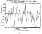
- Figure 1 shows a computer generated hydropathy profile plotting the relative hydrophilicity and hydrophobicity of each amino acid of the protein which was used to determine the most likely location of the hydrophilic regions along the GP-45 protein.
- Several well defined peaks are observed in the hydrophilic region.
- the amino acid sequence corresponding to each peak is hydrophilic and, therefore, likely to be antigenic.
- Three of these amino acid sequences were chosen for peptide synthesis. It is anticipated that additional peptides corresponding to various other hydrophilic regions of the protein may also be synthesized. From laboratory experience, it is known that peptides from 20-30 amino acids in length provide favorable immunogens and often work as antigens.
- amino acid sequences chosen for synthesis and analysis were those of peptides SEQ ID No: 1 and SEQ ID No: 2, described above, and a third peptide, consisting of the amino acids comprising residues 111-133 of the GP-45 protein.
- sequence of peptide SEQ ID No: 3 follows: histidine - leucine - asparagine - glutamic acid - serine - threonine - glutamine - tryptophan - aspartic acid - aspartic acid - tryptophan - valine - serine - lysine - methionine - glutamic acid - aspartic acid - leucine - asparagine - glutamine - glutamic acid - isoleucine - leucine. (Referred to hereinafter as SEQ ID No: 3.) Each of the three synthetic peptides was tested for antigenic activity by using each as the antigen in the following immunoassay to detect the presence of the EIA virus.
- the group 2 samples show little color development and low level absorbance and indicate a negative result. Based upon use of peptide SEQ ID No: 2 as antigen, it was predicted that samples 2, 3, 4, and 5 tested positive for EIA infection and samples 1, 6, 7, 8, 9 and 10 tested negative. These predictions were verified to be 100% correct upon confirmation by the supplier of the test samples. It is clear that peptide SEQ ID No: 2 allows for accurate and ready discrimination between positive and negative test samples.
- Test 1 No. A492 nm Result No. A492 nm Result 1 0.043 - (-) 6 0.033 - (-) 2 0.516 + (+) 7 0.041 - (-) 3 0.576 + (+) 8 0.048 - (-) 4 0.518 + (+) 9 0.033 - (-) 5 0.309 + (+) 10 0.037 - (-) 10 samples 4 positive 6 negative 100% correct
- Peptide SEQ ID No: 2 is preferred because it provides a clear distinction in color, visible to the naked eye, between positive and negative samples. This color distinction is not as striking when peptide SEQ ID No: 1 is used. However, it will be appreciated that the use of peptide SEQ ID No: 1 as antigen will give accurate results. A means for reading results more sophisticated than mere visual inspection, such as a reflective meter, must be employed.
- peptide SEQ ID No: 3 as antigen in the immunoassay does not appear to give accurate results. For example, it is known from the key provided by the supplier of the samples that sample 10 is negative and sample 5 is positive. Yet as shown in Figure 2, with peptide SEQ ID No: 3 antigen, sample 10 shows a substantially higher absorbance than does sample 5. Therefore, it is concluded that peptide SEQ ID No: 3 will not adequately function as antigen in an immunoassay for the detection of the EIA virus.
Landscapes
- Chemical & Material Sciences (AREA)
- Organic Chemistry (AREA)
- Health & Medical Sciences (AREA)
- Life Sciences & Earth Sciences (AREA)
- Molecular Biology (AREA)
- Biochemistry (AREA)
- Biophysics (AREA)
- Virology (AREA)
- General Health & Medical Sciences (AREA)
- Medicinal Chemistry (AREA)
- Genetics & Genomics (AREA)
- Proteomics, Peptides & Aminoacids (AREA)
- Gastroenterology & Hepatology (AREA)
- Peptides Or Proteins (AREA)
- Micro-Organisms Or Cultivation Processes Thereof (AREA)
- Measuring Or Testing Involving Enzymes Or Micro-Organisms (AREA)
- Medicines Containing Material From Animals Or Micro-Organisms (AREA)
Claims (27)
- Peptide de synthèse comprenant une séquence d'acides aminés dont une partie au moins correspond à un site antigénique sur la partie enveloppe protéique du virus de l'anémie infectieuse équine, ladite séquence d'acides aminés étant l'une ou l'autre des séquences suivantes :
leucine - leucine - lysine - acide glutamique - arginine - glutamine - glutamine - valine - acide glutamique - acide glutamique - thréonine - phénylalanine - asparagine - leucine - isoleucine - glycine - cystéine - isoleucine - acide glutamique - arginine - thréonine - histidine - valine - phénylalanine - cystéine
(appelée ci-après SEQ ID n° 2)
glutamine - thréonine - histidine - alanine - acide aspartique - valine - glutamine - leucine - leucine - lysine - acide glutamique - arginine - glutamine - glutamine - valine - acide glutamique - acide glutamique - thréonine - phénylalanine - asparagine - leucine - isoleucine - glycine - cystéine
(appelée ci-après SEQ ID n° 1). - Peptide selon la revendication 1, dans laquelle un ou plusieurs acides aminés de ladite séquence d'acides aminés sont chimiquement modifiés sans affecter de façon significative l'aptitude dudit peptide à agir en tant qu'antigène dans une analyse pour la détection de la présence d'anticorps contre le virus de l'anémie infectieuse équine.
- Méthode de détection de la présence d'anticorps contre le virus de l'anémie infectieuse équine dans un sérum de test équin comprenant l'utilisation comme antigène d'un peptide de synthèse ayant une séquence d'acides aminés dont une partie au moins correspond à un site antigénique sur le virus de l'anémie infectieuse équine, ledit peptide étant, soit le peptide SEQ ID n° 2 ayant la séquence d'acides aminés suivante :
leucine - leucine - lysine - acide glutamique - arginine - glutamine - glutamine - valine - acide glutamique - acide glutamique - thréonine - phénylalanine - asparagine - leucine - isoleucine - glycine - cystéine - isoleucine - acide glutamique - arginine - thréonine - histidine - valine - phénylalanine - cystéine,
soit le peptide SEQ ID n° 1 ayant la séquence d'acides aminés suivante :
glutamine - thréonine - histidine - alanine - acide aspartique - valine - glutamine - leucine - leucine - lysine - acide glutamique - arginine - glutamine - glutamine - valine - acide glutamique - acide glutamique - thréonine - phénylalanine - asparagine - leucine - isoleucine - glycine - cystéine. - Méthode selon la revendication 3, comprenant les étapes suivantes :(a) l'exposition d'une quantité choisie dudit peptide à une quantité choisie de sérum de test équin pendant un premier laps de temps suffisant pour permettre à tout anticorps du virus de l'anémie infectieuse équine présent dans le sérum de test de se lier audit peptide en formant un complexe peptide-anticorps ;(b) l'exposition dudit complexe peptide-anticorps à une quantité choisie d'un conjugué marqué pendant un second laps de temps suffisant pour permettre audit conjugué marqué de se lier au complexe peptide-anticorps ;(c) la détection de la présence dudit conjugué marqué qui s'est lié audit complexe peptide-anticorps.
- Méthode selon la revendication 4, dans laquelle ledit conjugué est un antigène purifié du virus de l'anémie infectieuse équine.
- Méthode selon la revendication 4, dans laquelle ledit conjugué est un peptide de synthèse ayant une séquence d'acides aminés qui correspond à un site antigénique sur un virus de l'anémie infectieuse équine.
- Méthode selon la revendication 4, dans laquelle ledit conjugué est un anticorps anti-cheval qui reconnaît - et se lie à - un anticorps du virus de l'anémie infectieuse équine.
- Méthode selon la revendication 7, dans laquelle ledit anticorps anti-cheval est l'immunoglobuline G anti-cheval du mouton.
- Méthode selon l'une quelconque des revendications 4 à 8, dans laquelle ledit conjugué est marqué par une enzyme choisie entre la peroxidase de raifort et une phosphatase alcaline.
- Méthode selon la revendication 9, dans laquelle l'étape de détection dudit conjugué marqué par une enzyme comprend l'étape supplémentaire d'addition d'un substrat en quantité suffisante pour réagir avec ledit enzyme de marquage, pour convertir ledit substrat en une quantité suffisante d'un produit provoquant un changement de couleur qui soit visible à l'oeil nu.
- Méthode selon la revendication 10, dans laquelle le substrat est une solution de peroxyde d'hydrogène / diamine d'orthophénylène.
- Méthode selon la revendication 10, comprenant en outre l'étape de détermination de la quantité de conjugué marqué par une enzyme qui s'est lié au complexe peptide-anticorps en mesurant l'absorbance dudit produit au moyen d'un spectrophotomètre.
- Méthode selon l'une quelconque des revendications 4 à 8, dans laquelle ledit conjugué est marqué par un élément radioactif.
- Méthode selon la revendication 13, dans laquelle ledit élément radioactif est l'iode.
- Méthode selon l'une quelconque des revendications 4 à 8, dans laquelle ledit conjugué est marqué par un procédé magnétique.
- Méthode selon l'une quelconque des revendications 4 à 15, dans laquelle ladite analyse immunologique est pratiquée à une température comprise dans la plage allant de 4°C environ et 45°C environ.
- Méthode selon la revendication 16, dans laquelle ladite température est la température de la pièce.
- Méthode selon l'une quelconque des revendications 4 à 17, dans laquelle chacun desdits premier et second laps de temps est compris entre 5 minutes environ et 60 minutes environ.
- Méthode selon l'une quelconque des revendications 4 à 18, dans laquelle la concentration dudit peptide est comprise entre 0,1 µg/ml et 200 µg/ml.
- Méthode selon la revendication 19, dans laquelle ladite concentration est de 0,5 µg/ml.
- Méthode selon l'une quelconque des revendications 4 à 20, comprenant en outre, après l'étape (b) et avant l'étape (c), l'étape de fixation dudit peptide sur un support en phase solide.
- Méthode selon la revendication 21, dans laquelle le support en phase solide est un plateau microtitre en polyvinyle à alvéoles multiples.
- Méthode selon la revendication 21, comprenant en outre les étapes suivantes :(a') l'enlèvement des anticorps du virus de l'anémie infectieuse équine dudit support, avant de procéder à l'étape (a) ; et(b') l'enlèvement du conjugué marqué non lié, avant de procéder à l'étape (c).
- Méthode selon la revendication 23, dans laquelle les étapes d'enlèvement sont effectuées au moyen d'un agent de rinçage.
- Méthode selon la revendication 24, dans laquelle ledit agent de rinçage est une solution saline tamponnée au phosphate dont le pH est compris entre 6 et 10.
- Méthode selon la revendication 3, comprenant les étapes suivantes :(a) l'exposition dudit peptide à un support en phase solide pendant un premier laps de temps suffisant pour permettre audit peptide de se lier au support ;(b) l'enlèvement du peptide non lié ;(c) le mélange d'une quantité choisie d'anticorps anti-EIA marqué avec une quantité choisie de sérum de test équin ;(d) l'addition dudit mélange au support en phase solide contenant ledit peptide pour exposer ledit peptide audit mélange pendant un second laps de temps suffisant pour permettre audit peptide de se lier aux anticorps présents dans ledit mélange ;(e) l'enlèvement dudit support de tous les anticorps dudit mélange qui ne se sont pas liés ;(f) la détermination de la quantité d'anticorps anti-EIA marqué qui s'est lié audit peptide.
- Méthode selon la revendication 26, dans laquelle la quantité choisie d'anticorps anti-EIA marqué est inférieure à la quantité d'anticorps dans la quantité choisie de sérum de test.
Applications Claiming Priority (3)
| Application Number | Priority Date | Filing Date | Title |
|---|---|---|---|
| US485338 | 1990-02-23 | ||
| US07/485,338 US5427907A (en) | 1990-02-23 | 1990-02-23 | Assay for equine infectious anemia virus |
| CA002108044A CA2108044A1 (fr) | 1990-02-23 | 1991-04-08 | Essai pour deceler le virus de l'anemie infectieuse des equides |
Publications (3)
| Publication Number | Publication Date |
|---|---|
| EP0443880A2 EP0443880A2 (fr) | 1991-08-28 |
| EP0443880A3 EP0443880A3 (en) | 1991-10-16 |
| EP0443880B1 true EP0443880B1 (fr) | 1996-05-15 |
Family
ID=25676719
Family Applications (1)
| Application Number | Title | Priority Date | Filing Date |
|---|---|---|---|
| EP91301460A Expired - Lifetime EP0443880B1 (fr) | 1990-02-23 | 1991-02-22 | Méthode d'analyse du virus de l'anémie infectieuse équine (EIA-Virus) |
Country Status (7)
| Country | Link |
|---|---|
| US (1) | US5427907A (fr) |
| EP (1) | EP0443880B1 (fr) |
| AT (1) | ATE138078T1 (fr) |
| CA (1) | CA2108044A1 (fr) |
| DE (1) | DE69119445T2 (fr) |
| ES (1) | ES2087242T3 (fr) |
| WO (1) | WO1992017494A1 (fr) |
Families Citing this family (5)
| Publication number | Priority date | Publication date | Assignee | Title |
|---|---|---|---|---|
| US5427907A (en) * | 1990-02-23 | 1995-06-27 | Virginia Commonwealth University | Assay for equine infectious anemia virus |
| US6596846B2 (en) * | 1996-12-18 | 2003-07-22 | Universidade Federal De Minas Gerais | Method and composition for the diagnosis of equine infectious anemia virus disease by using the recombinant capsid protein virus (p26) |
| BR9606273A (pt) * | 1996-12-18 | 1998-12-15 | Univ Minas Gerais | Processo para o teste imunoenzimático com proteína P26 recombinante do capsídio viral no diagnóstico da anemia infecciosa equina |
| AU5743398A (en) | 1997-12-30 | 1999-07-26 | Universidade Federal De Minas Gerais | The process for expression and production of recombinant protein gp90 derived from glicoprotein envelop of equine infectious anemia virus eiav |
| US6350574B1 (en) | 1998-09-23 | 2002-02-26 | Ronald C. Montelaro | Fluorescence polarization—based diagnostic assay for equine infectious anemia virus |
Family Cites Families (4)
| Publication number | Priority date | Publication date | Assignee | Title |
|---|---|---|---|---|
| US4806467A (en) * | 1985-10-21 | 1989-02-21 | Fermenta Animal Health Company | Method for the detection of equine infectious anemia and other retrovirus infections using a competitive enzyme-linked immunoabsorbent assay and reagents useful in the same |
| NO881151L (no) * | 1987-03-27 | 1988-09-28 | Syntello Ab | Syntetisk hiv-1-antigen. |
| CA1341285C (fr) * | 1988-02-12 | 2001-08-14 | Chang Yi Wang | Peptides synthetiques servant a la detection d'anticorps de la proteine de surface gp120 du virus hiv, destines au diagnostic du sida, ainsi que d'etats pre-sidatiques, ou aux fins de vaccins |
| US5427907A (en) * | 1990-02-23 | 1995-06-27 | Virginia Commonwealth University | Assay for equine infectious anemia virus |
-
1990
- 1990-02-23 US US07/485,338 patent/US5427907A/en not_active Expired - Lifetime
-
1991
- 1991-02-22 DE DE69119445T patent/DE69119445T2/de not_active Expired - Fee Related
- 1991-02-22 ES ES91301460T patent/ES2087242T3/es not_active Expired - Lifetime
- 1991-02-22 AT AT91301460T patent/ATE138078T1/de not_active IP Right Cessation
- 1991-02-22 EP EP91301460A patent/EP0443880B1/fr not_active Expired - Lifetime
- 1991-04-08 WO PCT/US1991/002287 patent/WO1992017494A1/fr not_active Ceased
- 1991-04-08 CA CA002108044A patent/CA2108044A1/fr not_active Abandoned
Also Published As
| Publication number | Publication date |
|---|---|
| DE69119445D1 (de) | 1996-06-20 |
| US5427907A (en) | 1995-06-27 |
| WO1992017494A1 (fr) | 1992-10-15 |
| DE69119445T2 (de) | 1996-09-26 |
| EP0443880A3 (en) | 1991-10-16 |
| ATE138078T1 (de) | 1996-06-15 |
| CA2108044A1 (fr) | 1992-10-15 |
| EP0443880A2 (fr) | 1991-08-28 |
| ES2087242T3 (es) | 1996-07-16 |
Similar Documents
| Publication | Publication Date | Title |
|---|---|---|
| Clark et al. | ELISA techniques | |
| JP2733059B2 (ja) | Htlv−▲iii▼抗体の診断用ペプチド並びにそれらの製造法及び用途 | |
| US4753873A (en) | Peptides for the diagnosis of HTLV-III antibodies, their preparation and use | |
| JP4130505B2 (ja) | D−アミノ酸からなるペプチドによる診断方法の干渉の排除 | |
| CA2210916A1 (fr) | Augmentation de la sensibilite du dosage immunochimique d'un analyte | |
| EP0443880B1 (fr) | Méthode d'analyse du virus de l'anémie infectieuse équine (EIA-Virus) | |
| EP0514509B1 (fr) | Composition comprenant des antigenes conjugues a une activite enzymatique, et destinee aux necessaires a usage prive et epidemiologique de diagnostic immunologique, notamment de la maladie de chagas, et procede de preparation de ladite composition | |
| WO1991013360A1 (fr) | NOUVEAUX PEPTIDES D'HIV p24, ANTIGENES DIAGNOSTIQUES ET METHODE D'IMMUNO-ANALYSE DISCRIMINANTE | |
| JPH08510063A (ja) | リコンビナントタンパクおよび合成ペプチド試薬を用いるhivイムノアッセイ | |
| CA1279818C (fr) | Test pour la detection des anticorps contre certains micro-organismes | |
| EP0313986B1 (fr) | Dosages immunologiques utilisant des antigènes produits dans des organismes hétérologues | |
| JPH06502723A (ja) | 抗体を検出する分析系の感度及び/又は特異性の決定方法 | |
| JP3889045B2 (ja) | Hivを検出するためのペプチド | |
| EP0951646B1 (fr) | DOSAGE IMMUNO-ENZYMATIQUE POUR DIAGNOSTIQUER L'ANEMIE INFECTIEUSE EQUINE AU MOYEN DE LA PROTEINE RECOMBINANTE (gp90) DERIVEE DU VIRUS DE L'ANEMIE INFECTIEUSE EQUINE | |
| Jin et al. | Use of α-N, N-bis [carboxymethyl] lysine-modified peroxidase in immunoassays | |
| EP0386713A2 (fr) | Essai immunologique des anticorps contre HIV | |
| US7026133B2 (en) | Method and composition for the diagnosis of equine infectious anemia virus disease by using the recombinant capsid protein virus (p26) | |
| AU7769391A (en) | Assay for equine infectious anemia virus | |
| AU729356B2 (en) | The immunoenzimatic assay for the diagnosis of equine infectious anemia virus disease by using recombinant protein (rGP90) derived from equine infectious anemia virus | |
| KR20000041180A (ko) | 신증후출혈열의 진단을 위한 합성 올리고펩티드 및 방법 | |
| EP1102067A1 (fr) | Methode et trousse de dosage d'anticorps anti-gad | |
| JPS63209600A (ja) | パ−オキシダ−ゼ標識による測定方法 | |
| JPS62860A (ja) | 微生物診断用テスト方法 | |
| JPWO2000073800A1 (ja) | 抗gad抗体測定方法およびキット | |
| JPH07280806A (ja) | 抗好中球細胞質抗体の測定方法 |
Legal Events
| Date | Code | Title | Description |
|---|---|---|---|
| PUAI | Public reference made under article 153(3) epc to a published international application that has entered the european phase |
Free format text: ORIGINAL CODE: 0009012 |
|
| PUAL | Search report despatched |
Free format text: ORIGINAL CODE: 0009013 |
|
| AK | Designated contracting states |
Kind code of ref document: A2 Designated state(s): AT DE ES FR GB |
|
| AK | Designated contracting states |
Kind code of ref document: A3 Designated state(s): AT DE ES FR GB |
|
| 17P | Request for examination filed |
Effective date: 19920306 |
|
| 17Q | First examination report despatched |
Effective date: 19950315 |
|
| GRAH | Despatch of communication of intention to grant a patent |
Free format text: ORIGINAL CODE: EPIDOS IGRA |
|
| GRAA | (expected) grant |
Free format text: ORIGINAL CODE: 0009210 |
|
| AK | Designated contracting states |
Kind code of ref document: B1 Designated state(s): AT DE ES FR GB |
|
| REF | Corresponds to: |
Ref document number: 138078 Country of ref document: AT Date of ref document: 19960615 Kind code of ref document: T |
|
| REG | Reference to a national code |
Ref country code: ES Ref legal event code: BA2A Ref document number: 2087242 Country of ref document: ES Kind code of ref document: T3 |
|
| REF | Corresponds to: |
Ref document number: 69119445 Country of ref document: DE Date of ref document: 19960620 |
|
| ET | Fr: translation filed | ||
| REG | Reference to a national code |
Ref country code: ES Ref legal event code: FG2A Ref document number: 2087242 Country of ref document: ES Kind code of ref document: T3 |
|
| PLBE | No opposition filed within time limit |
Free format text: ORIGINAL CODE: 0009261 |
|
| STAA | Information on the status of an ep patent application or granted ep patent |
Free format text: STATUS: NO OPPOSITION FILED WITHIN TIME LIMIT |
|
| 26N | No opposition filed | ||
| REG | Reference to a national code |
Ref country code: GB Ref legal event code: IF02 |
|
| PGFP | Annual fee paid to national office [announced via postgrant information from national office to epo] |
Ref country code: AT Payment date: 20070201 Year of fee payment: 17 |
|
| PGFP | Annual fee paid to national office [announced via postgrant information from national office to epo] |
Ref country code: GB Payment date: 20070223 Year of fee payment: 17 |
|
| PGFP | Annual fee paid to national office [announced via postgrant information from national office to epo] |
Ref country code: ES Payment date: 20070226 Year of fee payment: 17 |
|
| PGFP | Annual fee paid to national office [announced via postgrant information from national office to epo] |
Ref country code: DE Payment date: 20070330 Year of fee payment: 17 |
|
| PGFP | Annual fee paid to national office [announced via postgrant information from national office to epo] |
Ref country code: FR Payment date: 20070221 Year of fee payment: 17 |
|
| GBPC | Gb: european patent ceased through non-payment of renewal fee |
Effective date: 20080222 |
|
| PG25 | Lapsed in a contracting state [announced via postgrant information from national office to epo] |
Ref country code: AT Free format text: LAPSE BECAUSE OF NON-PAYMENT OF DUE FEES Effective date: 20080222 |
|
| REG | Reference to a national code |
Ref country code: FR Ref legal event code: ST Effective date: 20081031 |
|
| PG25 | Lapsed in a contracting state [announced via postgrant information from national office to epo] |
Ref country code: DE Free format text: LAPSE BECAUSE OF NON-PAYMENT OF DUE FEES Effective date: 20080902 |
|
| PG25 | Lapsed in a contracting state [announced via postgrant information from national office to epo] |
Ref country code: FR Free format text: LAPSE BECAUSE OF NON-PAYMENT OF DUE FEES Effective date: 20080229 |
|
| REG | Reference to a national code |
Ref country code: ES Ref legal event code: FD2A Effective date: 20080223 |
|
| PG25 | Lapsed in a contracting state [announced via postgrant information from national office to epo] |
Ref country code: GB Free format text: LAPSE BECAUSE OF NON-PAYMENT OF DUE FEES Effective date: 20080222 |
|
| PG25 | Lapsed in a contracting state [announced via postgrant information from national office to epo] |
Ref country code: ES Free format text: LAPSE BECAUSE OF NON-PAYMENT OF DUE FEES Effective date: 20080223 |