EP0444560A1 - Antibody specific for the tumor necrosis factor receptor - Google Patents
Antibody specific for the tumor necrosis factor receptor Download PDFInfo
- Publication number
- EP0444560A1 EP0444560A1 EP91102711A EP91102711A EP0444560A1 EP 0444560 A1 EP0444560 A1 EP 0444560A1 EP 91102711 A EP91102711 A EP 91102711A EP 91102711 A EP91102711 A EP 91102711A EP 0444560 A1 EP0444560 A1 EP 0444560A1
- Authority
- EP
- European Patent Office
- Prior art keywords
- tnf
- cells
- monoclonal antibody
- protein
- antibody
- Prior art date
- Legal status (The legal status is an assumption and is not a legal conclusion. Google has not performed a legal analysis and makes no representation as to the accuracy of the status listed.)
- Withdrawn
Links
Images


Classifications
-
- C—CHEMISTRY; METALLURGY
- C07—ORGANIC CHEMISTRY
- C07K—PEPTIDES
- C07K16/00—Immunoglobulins [IG], e.g. monoclonal or polyclonal antibodies
- C07K16/18—Immunoglobulins [IG], e.g. monoclonal or polyclonal antibodies against material from animals or humans
- C07K16/28—Immunoglobulins [IG], e.g. monoclonal or polyclonal antibodies against material from animals or humans against receptors, cell surface antigens or cell surface determinants
- C07K16/2866—Immunoglobulins [IG], e.g. monoclonal or polyclonal antibodies against material from animals or humans against receptors, cell surface antigens or cell surface determinants against receptors for cytokines, lymphokines, interferons
-
- A—HUMAN NECESSITIES
- A61—MEDICAL OR VETERINARY SCIENCE; HYGIENE
- A61P—SPECIFIC THERAPEUTIC ACTIVITY OF CHEMICAL COMPOUNDS OR MEDICINAL PREPARATIONS
- A61P29/00—Non-central analgesic, antipyretic or antiinflammatory agents, e.g. antirheumatic agents; Non-steroidal antiinflammatory drugs [NSAID]
-
- A—HUMAN NECESSITIES
- A61—MEDICAL OR VETERINARY SCIENCE; HYGIENE
- A61P—SPECIFIC THERAPEUTIC ACTIVITY OF CHEMICAL COMPOUNDS OR MEDICINAL PREPARATIONS
- A61P31/00—Antiinfectives, i.e. antibiotics, antiseptics, chemotherapeutics
- A61P31/12—Antivirals
-
- A—HUMAN NECESSITIES
- A61—MEDICAL OR VETERINARY SCIENCE; HYGIENE
- A61P—SPECIFIC THERAPEUTIC ACTIVITY OF CHEMICAL COMPOUNDS OR MEDICINAL PREPARATIONS
- A61P31/00—Antiinfectives, i.e. antibiotics, antiseptics, chemotherapeutics
- A61P31/12—Antivirals
- A61P31/14—Antivirals for RNA viruses
- A61P31/18—Antivirals for RNA viruses for HIV
-
- A—HUMAN NECESSITIES
- A61—MEDICAL OR VETERINARY SCIENCE; HYGIENE
- A61P—SPECIFIC THERAPEUTIC ACTIVITY OF CHEMICAL COMPOUNDS OR MEDICINAL PREPARATIONS
- A61P35/00—Antineoplastic agents
-
- A—HUMAN NECESSITIES
- A61—MEDICAL OR VETERINARY SCIENCE; HYGIENE
- A61P—SPECIFIC THERAPEUTIC ACTIVITY OF CHEMICAL COMPOUNDS OR MEDICINAL PREPARATIONS
- A61P37/00—Drugs for immunological or allergic disorders
-
- A—HUMAN NECESSITIES
- A61—MEDICAL OR VETERINARY SCIENCE; HYGIENE
- A61K—PREPARATIONS FOR MEDICAL, DENTAL OR TOILETRY PURPOSES
- A61K38/00—Medicinal preparations containing peptides
-
- Y—GENERAL TAGGING OF NEW TECHNOLOGICAL DEVELOPMENTS; GENERAL TAGGING OF CROSS-SECTIONAL TECHNOLOGIES SPANNING OVER SEVERAL SECTIONS OF THE IPC; TECHNICAL SUBJECTS COVERED BY FORMER USPC CROSS-REFERENCE ART COLLECTIONS [XRACs] AND DIGESTS
- Y10—TECHNICAL SUBJECTS COVERED BY FORMER USPC
- Y10S—TECHNICAL SUBJECTS COVERED BY FORMER USPC CROSS-REFERENCE ART COLLECTIONS [XRACs] AND DIGESTS
- Y10S424/00—Drug, bio-affecting and body treating compositions
- Y10S424/809—Drug, bio-affecting and body treating compositions involving immunoglobulin or antibody fragment, e.g. fab', fv, fc, heavy chain or light chain
-
- Y—GENERAL TAGGING OF NEW TECHNOLOGICAL DEVELOPMENTS; GENERAL TAGGING OF CROSS-SECTIONAL TECHNOLOGIES SPANNING OVER SEVERAL SECTIONS OF THE IPC; TECHNICAL SUBJECTS COVERED BY FORMER USPC CROSS-REFERENCE ART COLLECTIONS [XRACs] AND DIGESTS
- Y10—TECHNICAL SUBJECTS COVERED BY FORMER USPC
- Y10S—TECHNICAL SUBJECTS COVERED BY FORMER USPC CROSS-REFERENCE ART COLLECTIONS [XRACs] AND DIGESTS
- Y10S530/00—Chemistry: natural resins or derivatives; peptides or proteins; lignins or reaction products thereof
- Y10S530/866—Chemistry: natural resins or derivatives; peptides or proteins; lignins or reaction products thereof involving immunoglobulin or antibody fragment, e.g. fab', fab, fv, fc, heavy chain or light chain
Definitions
- Tumor necrosis factor ⁇ (TNF ⁇ , cachectin) and tumor necrosis factor ⁇ (TNF ⁇ , lymphotoxin) belong to the cytokine family, a group of polypeptide mediators that transmit signals from one cell to another.
- the cytokines play a key role in the molecular understanding of inflammatory and immune responses in the body, which forms a complex network of signals that coordinate the body's defense responses.
- the tumor necrosis factor is released at a very early stage in an inflammatory or immune reaction and then in turn stimulates the immune cells to form many other polypeptides.
- Human TNF ⁇ is secreted as a protein with 157 AS length, which contains an N-terminal propeptide sequence of 76 AS, so that pure TNF ⁇ has a relative molecular weight of 17 kD (Beutler and Cerami, Nature 320 (1986), 584-588). Human TNF ⁇ differs structurally from TNF ⁇ by the presence of strong glycosylation and the lack of an internal disulfide bridge, but has 28% homology at the amino acid level to TNF ⁇ . Although the synthesis of both cytokines takes place on the basis of relatively different stimuli, they have a strongly matching spectrum of biological activities and bind to a common receptor (Aggarwal et al., Nature 318 (1985), 665-667).
- TNF ⁇ and TNF ⁇ are found both in humans (Nedwin et al., Nucleic Acids Res. 13 (1985), 6361-6373) and in the mouse (Nedospasov et al., Nucleic Acids Res. 14 (1986), 7713-7725) in very close proximity on a chromosome.
- TNF receptors are found on all somatic cells in the body, with the exception of erythrocytes. The number of receptors ranges from a few 100 copies on certain cells to over 20,000 on others. In most cases, however, the number of receptors is not related to the sensitivity of the cells. In some cases, e.g. in T lymphocytes, the receptor is not present (or in an inactive form) in resting cells, but can be induced by primary activation (Scheurich et al., J. Immunol. 138 (1987), 1786-1790). The receptor has high affinity for its ligand with a dissociation constant of approximately 1x10¹⁰M01 (Scheurich et al., Int. J. Cancer (1986) 38, 127-133).
- the TNF receptor appears to be a complex of several different proteins with a relative molecular weight of approximately 300 kD (Smith et al., J. Biol. Chem. 261 (1986), 14871-14874).
- the dominant ligand-binding subunit of the receptor appears to have a MW of approx. 80 to 90 kD, while in cells of epithelial origin there is additionally an immunologically non-cross-reacting subunit of approx. 60 kD (Hohmann et al., J. Biol. Chem. 264 (1989), 14927-14934). Therefore, at least two different types of TNF receptors appear to exist. The exact nature of the signal transmission after binding of TNF to the receptor has not yet been elucidated, but it has been found that the response to this signal is very different in different tissues.
- TNF can protect the organism from bacterial infections and high radiation doses. Treatment with TNF can also cause tumors to die, causing bleeding in the tumors, which then dry out and die. Furthermore, TNF is apparently crucially involved in mediating the body's response to a bacterial infection, such as fever, and helps the body overcome the infection. Thus TNF - alone and in combination with other cytokines - has a very significant therapeutic potential.
- TNF can also cause a number of pathological conditions, e.g. Cachexia, tissue damage, gram-negative septic shock from bacterial endotoxins and brain inflammation in malaria. Furthermore, a cytotoxic effect of TNF was found in cells that were infected with HIV (Matsuyama et al., J. Virol. 63 (1989), 2504-2509). It is even found that on a molar basis the toxicity of TNF in some species is about 100,000 times higher than that of cyanide (Beutler and Cerami, Nature 320 (1986), 584-588).
- TNF concentration in the organism is obviously decisive for the effect of TNF.
- an active substance can become extremely toxic if it is released in large quantities or in the wrong place.
- This high toxicity of TNF in turn brings with it great problems with its possible use as a therapeutic agent, for example in tumor diseases. It was therefore the object of the present invention to develop an agent which, in the presence of unphysiologically high TNF levels in the body, either caused by an excessive endogenous production of TNF or by a treatment with TNF - especially if a local Use with high doses is required - can reduce or eliminate the pathological symptoms of TNF, particularly the occurrence of tissue damage and shock.
- the object according to the invention is achieved by a monoclonal antibody against a membrane protein on human cells which, when bound to human cells which have a TNF receptor, at least largely neutralizes the known effects of TNF ⁇ and / or TNF ⁇ .
- the antibody according to the invention is directed against a surface protein of the human cell with 60 kD, which apparently is a component of the TNF-receptor complex.
- Competition studies show that the antibody according to the invention interferes differently with the binding of TNF to its receptor in different cell lines (Table 1).
- binding of the antibody according to the invention results in a complete inhibition of the cellular response to TNF in all examined cell lines of different tissue origins (lymphoid, myeloid, epithelial and fibroblast cells) regardless of the extent of the competition with the TNF binding. From this finding it can be assumed that the antibody according to the invention may recognize and functionally block a receptor protein which is essential for the signaling of very different TNF responses.
- An antibody according to the invention can preferably even completely neutralize the observed cytostatic / cytotoxic effect of TNF on human cells in tissue culture (Pfizenmaier et al., Blut, 55, 1-10 (1987a).
- the antagonistic activity of the antibody according to the invention was investigated on three different human cell lines. In all systems, the cytotoxic or cytostatic effect of TNF ⁇ was additionally enhanced by the presence of interferon gamma or protein synthesis inhibitors (eg cycloheximide), with IFN gamma or protein synthesis inhibitors alone having no influence on the under the conditions used have measured cellular responses. It was found that binding of the antibody according to the invention completely neutralized the cytotoxic / cytostatic effect of TNF on all three examined human cell lines. In a similar way, an antibody according to the invention also neutralizes the action of TNF ⁇ and murine TNF ⁇ , which is equally effective on human cells.
- interferon gamma or protein synthesis inhibitors eg cycloheximide
- an antibody according to the invention also acts as an antagonist of the TNF-induced expression of HLA genes.
- the TNF-induced modulation of HLA genes was investigated in a human cell line of myeloid origin (K562) and an epithelial carcinoma cell line (Colo 205).
- co-stimulation of TNF ⁇ was also carried out together with interferon gamma in order to trigger the respective reaction (Pfizenmaier et al., J. Immunol. 138, 975-980 (1987b).
- it can be used for both systems come to complete inhibition of the TNF effect.
- an antibody according to the invention also acts as an antagonist of the immune stimulation mediated by TNF.
- TNF immune stimulation mediated by TNF.
- the stimulation of interleukin-2 receptor expression on T lymphocytes and on so-called natural killer cells (NK cells) (Scheurich et al., (1987) supra) can be completely inhibited by binding an antibody according to the invention.
- a particularly preferred object of the present invention is a special antibody H398 with the ability to completely neutralize the known TNF effects, which is an immunoglobulin of the IgG2a isotype and which is derived from the Budapestoma-based hybridoma cell line ECACC 90021617 ( or H398.6.1) is expressed.
- the present invention furthermore also relates to a protein with antibody properties which has the same binding specificity as the monoclonal antibody according to the invention.
- a protein with antibody properties is particularly preferred which has essentially the same antigen binding site as the monoclonal antibody H398 according to the invention.
- protein with antibody properties means antibodies modified by means of recombinant DNA technology. In order to at least largely neutralize the effect of TNF, it is necessary that such modified proteins have the same binding specificity as the monoclonal antibody according to the invention, ie they have to bind to a 60 kD membrane protein on human cells which is a component of the TNF-receptor complex, and they must be able to at least largely neutralize the known effects of TNF ⁇ or / and TNF ⁇ in a therapeutically acceptable concentration range. In order to meet these criteria, the modified protein with antibody properties must have an antigen binding site that is essentially the same as the monoclonal antibody according to the invention.
- the binding specificity is characterized by very specific regions of the antibody, namely the hypervariable regions.
- the invention also includes modified proteins such as partial antibody fragments (eg F (ab) 2), single-chain antibodies or chimerized, in particular humanized antibodies (replacement of the antibody regions not involved in the binding by sequences of a human antibody).
- modified proteins such as partial antibody fragments (eg F (ab) 2), single-chain antibodies or chimerized, in particular humanized antibodies (replacement of the antibody regions not involved in the binding by sequences of a human antibody).
- partial antibody fragments eg F (ab) 2
- single-chain antibodies or chimerized replacement of the antibody regions not involved in the binding by sequences of a human antibody.
- humanized antibodies replacement of the antibody regions not involved in the binding by sequences of a human antibody.
- the present invention furthermore relates to a process for obtaining a monoclonal antibody according to the invention, in which myeloma cells are fused in the usual way with spleen cells or T-lymphocytes of a mouse which have previously been treated with affinity-purified TNF receptor material from human P60 antigen-positive Cells, e.g. HL-60 (ATCC No. CCL 240) was hyperimmunized.
- myeloma cells are fused in the usual way with spleen cells or T-lymphocytes of a mouse which have previously been treated with affinity-purified TNF receptor material from human P60 antigen-positive Cells, e.g. HL-60 (ATCC No. CCL 240) was hyperimmunized.
- cell membranes are first prepared, then solubilized and purified using a TNF affinity column.
- the cell membranes are prepared by homogenization in a glass / glass homogenizer, pretreatment in a hypotonic medium if appropriate.
- the cell nuclei and the cells that are still intact are then separated, followed by pelleting of the cell membranes by centrifugation.
- the TNF receptor protein is solubilized with a buffer which contains a nonionic detergent, preferably about 1% Triton X-100.
- the solubilization buffer can also contain leupeptin, aprotinin and the protease inhibitor di-isopropyl fluorophosphate (DFP).
- the TNF affinity column is produced by coupling TNF, preferably recombinant human TNF ⁇ , to a suitable carrier material, preferably Affigel -15.
- TNF preferably recombinant human TNF ⁇
- a suitable carrier material preferably Affigel -15.
- the bound receptor is eluted in the acidic range at pH 3.0.
- Another cleaning step, an acetone precipitation, can then take place.
- about 3 parts of acetone are added to the sample and stored for a sufficient time at a sufficiently low temperature, preferably 14 hours at -80 ° C.
- the resulting precipitate which contains the receptor protein, is centrifuged off and taken up again in a suitable buffer.
- the hyperimmunization of a mouse with the receptor material obtained in this way preferably takes place in three steps.
- 1 ⁇ g receptor protein with complete Freund's adjuvant is administered intraperitoneally
- 1 ⁇ g receptor protein, mixed with incomplete Freund's adjuvant is administered intraperitoneally
- 2 ⁇ g receptor protein are administered without adjuvant.
- the spleen is preferably removed on the 58th day under sterile conditions.
- Hybridoma cell lines are produced according to usual procedures.
- the immunization of a mouse to generate a sufficiently high antibody level against the TNF receptor is, however, also possible by means of other suitable immunization protocols which are known to the person skilled in the art.
- the spleen cells of a mouse hyperimmunized with the TNF receptor protein can be fused in the usual way.
- the resulting hybridoma clones were tested for modulation of TNF binding ability on the TNF receptor positive human leukemia cell line HL-60 (ATCC No. CCL 240).
- hybridoma cell line could be obtained from 413 hybridoma clones which secreted an antibody according to the invention.
- the invention furthermore relates to a medicament which contains a monoclonal antibody according to the invention and / or a protein according to the invention with antibody properties as an active ingredient, optionally with customary carriers, auxiliaries and / or fillers.
- the antibody according to the invention can therefore be used diagnostically to detect cellular TNF receptors and to identify potential TNF-reactive cells in different tissues / organs.
- the above-mentioned antibody is suitable for the detection of secreted receptor fragments in serum and other body fluids.
- antibodies according to the invention lies in their property of at least largely inhibiting known biological effects of TNF ⁇ and TNF ⁇ .
- TNF ⁇ TNF ⁇
- TNF ⁇ TNF neutralizing effect in vitro
- an antibody according to the invention could be used in all the diseases which are associated with increased, pathological TNF ⁇ / TNF ⁇ levels in serum and / or other body fluids and in which TNF ⁇ / TNF ⁇ has been recognized or suspected as a pathogenic principle (Old, Nature 326 ( 1987), 330-331; Warren et al., Mod. Pathol. 1 (1988), 242-247; Beutler & Cerami, Ann. Rev. Biochem. 57 (1989), 505-518; Grau et al., Immunol Rev. 112, 49-70 (1989)).
- autoimmune diseases such as rheumatoid arthritis and cancer, which are associated with deregulated TNF ⁇ / TNF ⁇ production of the malignant cell itself (e.g. myeloma) or the immune system (e.g. CD4+ cells in chronic B-lymphatic leukemia; B-CLL) accompanied.
- therapeutic use of an antibody according to the invention in TNF-mediated, acutely life-threatening course of the disease should be considered, for example in the case of septic shock from bacterial infections (meningococcal sepsis), and in cerebral malaria.
- the course of the disease can be influenced favorably after short-term treatment with the antibodies.
- therapy with one would be Antibodies according to the invention, or the modified antibody (for example by replacing the constant regions of the mouse with constant regions of humans) possible.
- Another potential area of application of the antibody is in the co-treatment of AIDS. Since the multiplication of HIV, the causative agent of AIDS, is stimulated by TNF and TNF is thus regarded as one of the progression factors from the latent to the acute clinical picture (Matsuyama et al., J. Virol. (1989), 2504-2509), the antibody could H398 can be used as an antiviral active principle in those AIDS patients in whom either already constitutively increased TNF levels are found (Lahdevirta et al., Am. J. Med.
- TNF production could be induced (Matsuyama et al., (1989) supra). Since the antibody H398 according to the invention also has an inhibitory effect in vitro on the TNF-stimulated activity of the HIV promoter which controls the transcription of the retroviral genes, the use of an antibody according to the invention could counteract TNF-induced virus transcription and thus an increase in infectious viruses.
- the dosage of the antibody according to the invention for antagonizing undesirable side effects of TNF is of course dependent on the individual case, in particular when TNF is administered, but is preferably between 0.1 and 100 mg per person.
- H398 An important immunoregulatory function of TNF is the stimulation of interleukin-2 receptor expression on T lymphocytes and on so-called natural killer cells (NK cells) (Scheurich et al., J. Immunol. 138 (1987), 1786-1790).
- NK cells natural killer cells
- H398 also has an antagonistic effect in this system. 5x10⁵ cells of the NK cell line YT were cultivated in 2 ml of culture medium for 24 hours in the presence of 1 and 10 ng / ml TNF and serial dilutions of H398. The evaluation was carried out cytofluorographically with the help of anti-interleukin-2 receptor antibodies (anti Tac). The results shown in Table 1 demonstrate the complete inhibition of this TNF response by H398 as well.
Landscapes
- Health & Medical Sciences (AREA)
- Chemical & Material Sciences (AREA)
- Life Sciences & Earth Sciences (AREA)
- Organic Chemistry (AREA)
- Medicinal Chemistry (AREA)
- General Health & Medical Sciences (AREA)
- Nuclear Medicine, Radiotherapy & Molecular Imaging (AREA)
- General Chemical & Material Sciences (AREA)
- Chemical Kinetics & Catalysis (AREA)
- Pharmacology & Pharmacy (AREA)
- Immunology (AREA)
- Animal Behavior & Ethology (AREA)
- Public Health (AREA)
- Veterinary Medicine (AREA)
- Molecular Biology (AREA)
- Virology (AREA)
- Biochemistry (AREA)
- Genetics & Genomics (AREA)
- Proteomics, Peptides & Aminoacids (AREA)
- Biophysics (AREA)
- Communicable Diseases (AREA)
- Oncology (AREA)
- Pain & Pain Management (AREA)
- Rheumatology (AREA)
- Engineering & Computer Science (AREA)
- Bioinformatics & Cheminformatics (AREA)
- Tropical Medicine & Parasitology (AREA)
- AIDS & HIV (AREA)
- Medicines Containing Antibodies Or Antigens For Use As Internal Diagnostic Agents (AREA)
- Preparation Of Compounds By Using Micro-Organisms (AREA)
- Peptides Or Proteins (AREA)
- Micro-Organisms Or Cultivation Processes Thereof (AREA)
Abstract
Description
Der Tumor-Nekrose-Faktor α (TNFα, Cachectin) und der Tumor-Nekrose-Faktor β (TNFβ, Lymphotoxin) gehören zur Familie der Cytokine, einer Gruppe von Polypeptid-Mediatoren, die Signale von einer Zelle zur anderen übertragen. Die Cytokine sind wesentlich an der molekularen Verständigungsbasis von Entzündungs- und Immunreaktionen des Körpers beteiligt, die ein komplexes Netzwerk von Signalen bildet, welche die Abwehrreaktionen des Körpers aufeinander abstimmen.Tumor necrosis factor α (TNFα, cachectin) and tumor necrosis factor β (TNFβ, lymphotoxin) belong to the cytokine family, a group of polypeptide mediators that transmit signals from one cell to another. The cytokines play a key role in the molecular understanding of inflammatory and immune responses in the body, which forms a complex network of signals that coordinate the body's defense responses.
Der Tumor-Nekrose-Faktor wird bereits zu einem sehr frühen Zeitpunkt einer Entzündungs- oder Immunreaktion ausgeschüttet und regt dann seinerseits die Immunzellen zur Bildung vieler weiterer Polypeptide an.The tumor necrosis factor is released at a very early stage in an inflammatory or immune reaction and then in turn stimulates the immune cells to form many other polypeptides.
Menschlicher TNFα wird als Protein mit 157 A.S. Länge sekretiert, das eine N-terminale Propeptidsequenz von 76 A.S. enthält, so daß reiner TNFα ein relatives Molekulargewicht von 17 kD besitzt (Beutler und Cerami, Nature 320 (1986), 584-588). Menschlicher TNFβ unterscheidet sich strukturell von TNFα durch das Vorhandensein von starker Glykosylierung und das Fehlen einer internen Disulfidbrücke, besitzt jedoch eine 28%ige Homologie auf Aminosäure-Ebene zu TNFα. Obwohl die Synthese beider Cytokine aufgrund relativ unterschiedlicher Stimuli erfolgt, besitzen sie ein stark übereinstimmendes Spektrum an biologischen Aktivitäten und binden an einen gemeinsamen Rezeptor (Aggarwal et al., Nature 318 (1985), 665-667). Die für TNFα und TNFβ kodierenden Gene liegen sowohl bei Menschen (Nedwin et al., Nucleic Acids Res. 13 (1985), 6361-6373) als auch bei der Maus (Nedospasov et al., Nucleic Acids Res. 14 (1986), 7713-7725) in sehr enger Nachbarschaft auf einem Chromosom.Human TNFα is secreted as a protein with 157 AS length, which contains an N-terminal propeptide sequence of 76 AS, so that pure TNFα has a relative molecular weight of 17 kD (Beutler and Cerami, Nature 320 (1986), 584-588). Human TNFβ differs structurally from TNFα by the presence of strong glycosylation and the lack of an internal disulfide bridge, but has 28% homology at the amino acid level to TNFα. Although the synthesis of both cytokines takes place on the basis of relatively different stimuli, they have a strongly matching spectrum of biological activities and bind to a common receptor (Aggarwal et al., Nature 318 (1985), 665-667). The genes coding for TNFα and TNFβ are found both in humans (Nedwin et al., Nucleic Acids Res. 13 (1985), 6361-6373) and in the mouse (Nedospasov et al., Nucleic Acids Res. 14 (1986), 7713-7725) in very close proximity on a chromosome.
TNF-Rezeptoren werden auf allen somatischen Zellen des Körpers, mit Ausnahme der Erythrozyten, gefunden. Die Rezeptorzahl reicht von einigen 100 Kopien auf bestimmten Zellen bis über 20.000 auf anderen. Die Anzahl der Rezeptoren steht jedoch in den meisten Fällen in keinem Zusammenhang mit der Empfindlichkeit der Zellen. In manchen Fällen, z.B. bei T-Lymphozyten, ist der Rezeptor in ruhenden Zellen nicht (oder in einer inaktiven Form) vorhanden, kann jedoch durch primäre Aktivierung induziert werden (Scheurich et al., J. Immunol. 138 (1987), 1786-1790). Der Rezeptor besitzt eine hohe Affinität für seinen Liganden mit einer Dissoziationskonstante von ungefähr 1x10¹⁰M⁻¹ (Scheurich et al., Int. J. Cancer (1986) 38, 127-133). Nach Gelfiltrationsexperimenten scheint der TNF-Rezeptor ein Komplex aus mehreren verschiedenen Proteinen mit einem relativen Molekulargewicht von ca. 300 kD zu sein (Smith et al., J. Biol. Chem. 261 (1986), 14871-14874). In Zellen unterschiedlichen Ursprungs scheint die dominierende ligandbindende Untereinheit des Rezeptors ein MG von ca. 80 bis 90 kD zu besitzen, während in Zellen epithelialen Ursprungs zusätzlich eine immunologisch damit nicht kreuzreagierende Untereinheiten von etwa 60 kD vorliegt (Hohmann et al., J. Biol. Chem. 264 (1989), 14927-14934). Es scheinen daher offenbar mindestens zwei unterschiedliche Typen von TNF-Rezeptoren zu existieren. Die genaue Natur der Signalübermittlung nach Bindung von TNF an den Rezeptor wurde bisher noch nicht aufgeklärt, es wurde jedoch gefunden, daß die Antwort auf dieses Signal in verschiedenen Geweben stark unterschiedlich ist.TNF receptors are found on all somatic cells in the body, with the exception of erythrocytes. The number of receptors ranges from a few 100 copies on certain cells to over 20,000 on others. In most cases, however, the number of receptors is not related to the sensitivity of the cells. In some cases, e.g. in T lymphocytes, the receptor is not present (or in an inactive form) in resting cells, but can be induced by primary activation (Scheurich et al., J. Immunol. 138 (1987), 1786-1790). The receptor has high affinity for its ligand with a dissociation constant of approximately 1x10¹⁰M⁰¹ (Scheurich et al., Int. J. Cancer (1986) 38, 127-133). After gel filtration experiments, the TNF receptor appears to be a complex of several different proteins with a relative molecular weight of approximately 300 kD (Smith et al., J. Biol. Chem. 261 (1986), 14871-14874). In cells of different origin, the dominant ligand-binding subunit of the receptor appears to have a MW of approx. 80 to 90 kD, while in cells of epithelial origin there is additionally an immunologically non-cross-reacting subunit of approx. 60 kD (Hohmann et al., J. Biol. Chem. 264 (1989), 14927-14934). Therefore, at least two different types of TNF receptors appear to exist. The exact nature of the signal transmission after binding of TNF to the receptor has not yet been elucidated, but it has been found that the response to this signal is very different in different tissues.
Eine Verabreichung von TNF kann den Organismus vor bakteriellen Infektionen und hohen Strahlendosen schützen. Eine Behandlung mit TNF kann auch das Absterben von Tumoren bewirken, wobei Blutungen in den Tumoren ausgelöst werden, die anschließend austrocknen und absterben. Weiterhin ist TNF offenbar entscheidend an der Vermittlung der Reaktion des Körpers auf eine bakterielle Infektion, z.B. Fieber beteiligt und hilft dem Körper, die Infektion zu überwinden. Somit besitzt TNF - alleine und in Verbindung mit anderen Cytokinen - ein sehr bedeutsames therapeutisches Potential.Administration of TNF can protect the organism from bacterial infections and high radiation doses. Treatment with TNF can also cause tumors to die, causing bleeding in the tumors, which then dry out and die. Furthermore, TNF is apparently crucially involved in mediating the body's response to a bacterial infection, such as fever, and helps the body overcome the infection. Thus TNF - alone and in combination with other cytokines - has a very significant therapeutic potential.
Andererseits kann TNF auch eine Reihe pathologischer Zustände hervorrufen, z.B. Kachexie, Gewebeschäden, gramnegativen septischen Schock durch bakterielle Endotoxine und Gehirnentzündung bei Malaria. Weiterhin wurde ein cytotoxischer Effekt von TNF bei Zellen gefunden, die mit HIV infiziert waren (Matsuyama et al., J. Virol. 63 (1989), 2504-2509). Man findet sogar, daß auf molarer Basis die Toxizität von TNF bei manchen Arten etwa 100.000fach höher ist als die von Cyanid (Beutler und Cerami, Nature 320 (1986), 584-588).On the other hand, TNF can also cause a number of pathological conditions, e.g. Cachexia, tissue damage, gram-negative septic shock from bacterial endotoxins and brain inflammation in malaria. Furthermore, a cytotoxic effect of TNF was found in cells that were infected with HIV (Matsuyama et al., J. Virol. 63 (1989), 2504-2509). It is even found that on a molar basis the toxicity of TNF in some species is about 100,000 times higher than that of cyanide (Beutler and Cerami, Nature 320 (1986), 584-588).
Daher ist offenbar die Konzentration im Organismus für die Wirkung von TNF entscheidend. Wie bei vielen anderen im Körper gebildeten Faktoren scheint es auch beim TNF einen schmalen Grad zwischen Nutzen und Schaden zu geben: ein Wirkstoff kann extrem toxisch werden, wenn er in großen Mengen oder am falschen Ort freigesetzt wird. Diese hohe Toxizität von TNF bringt wiederum große Probleme bei seinem möglichen Einsatz als Therapeutikum, z.B. bei Tumorerkrankungen, mit sich. Somit war es Aufgabe der vorliegenden Erfindung, ein Mittel zu entwickeln, das bei Vorliegen von unphysiologisch hohen TNF-Spiegeln im Körper, entweder hervorgerufen durch eine überhöhte endogene Produktion von TNF oder durch eine Behandlung mit TNF - insbesondere wenn eine lokale Anwendung mit hohen Dosen erforderlich ist - die pathologischen Symptome von TNF, insbesondere das Auftreten von Gewebeschäden und Schockzuständen, verringern oder beseitigen kann.Therefore, the concentration in the organism is obviously decisive for the effect of TNF. As with many other factors formed in the body, there seems to be a fine line between benefit and harm in TNF: an active substance can become extremely toxic if it is released in large quantities or in the wrong place. This high toxicity of TNF in turn brings with it great problems with its possible use as a therapeutic agent, for example in tumor diseases. It was therefore the object of the present invention to develop an agent which, in the presence of unphysiologically high TNF levels in the body, either caused by an excessive endogenous production of TNF or by a treatment with TNF - especially if a local Use with high doses is required - can reduce or eliminate the pathological symptoms of TNF, particularly the occurrence of tissue damage and shock.
Die erfindungsgemäße Aufgabe wird durch einen monoklonalen Antikörper gegen ein Membranprotein auf menschlichen Zellen gelöst, der bei Bindung an menschliche Zellen, die einen TNF-Rezeptor besitzen, die bekannten Wirkungen von TNFα oder/und TNFβ zumindest weitgehend neutralisiert.The object according to the invention is achieved by a monoclonal antibody against a membrane protein on human cells which, when bound to human cells which have a TNF receptor, at least largely neutralizes the known effects of TNFα and / or TNFβ.
Der erfindungsgemäße Antikörper ist gegen ein Oberflächenprotein der menschlichen Zelle mit 60 kD gerichtet, bei dem es sich offenbar um einen Bestandteil des TNF-Rezeptorkomplexes handelt. Kompetitionsstudien zeigen, daß der erfindungsgemäße Antikörper in verschiedenen Zellinien unterschiedlich stark mit der Bindung von TNF an seinen Rezeptor interferiert (Tabelle 1). Überraschenderweise ergibt sich jedoch durch Bindung des erfindungsgemäßen Antikörpers bei allen untersuchten Zellinien verschiedenen Gewebeursprungs (lymphoide, myeloide, epitheliale und Fibroblasten-Zellen) unabhängig vom Ausmaß der Kompetition mit der TNF-Bindung eine vollständige Hemmung der zellulären Reaktion auf TNF. Aus diesem Befund läßt sich annehmen, daß der erfindungsgemäße Antikörper möglicherweise ein für die Signalvermittlung sehr unterschiedlicher TNF-Antworten essentielles Rezeptor-Protein erkennt und funktionell blockiert.The antibody according to the invention is directed against a surface protein of the human cell with 60 kD, which apparently is a component of the TNF-receptor complex. Competition studies show that the antibody according to the invention interferes differently with the binding of TNF to its receptor in different cell lines (Table 1). Surprisingly, however, binding of the antibody according to the invention results in a complete inhibition of the cellular response to TNF in all examined cell lines of different tissue origins (lymphoid, myeloid, epithelial and fibroblast cells) regardless of the extent of the competition with the TNF binding. From this finding it can be assumed that the antibody according to the invention may recognize and functionally block a receptor protein which is essential for the signaling of very different TNF responses.
Vorzugsweise kann ein erfindungsgemäßer Antikörper die beobachtete cytostatische/cytotoxische Wirkung von TNF auf menschliche Zellen in Gewebekultur (Pfizenmaier et al., Blut, 55, 1-10 (1987a), sogar vollständig neutralisieren. Die antagonistische Wirksamkeit des erfindungsgemäßen Antikörpers wurde an drei verschiedenen menschlichen Zellinien untersucht. In allen Systemen wurde der cytotoxische bzw. cytostatische Effekt von TNFα zusätzlich durch die Anwesenheit von Interferon-Gamma oder Proteinsynthese-Hemmern (z.B. Cycloheximid) verstärkt, wobei IFN-Gamma bzw. Proteinsynthese-Hemmer unter den eingesetzten Bedingungen für sich alleine keinen Einfluß auf die gemessenen zellulären Reaktionen haben. Es wurde festgestellt, daß eine Bindung des erfindungsgemäßen Antikörpers die cytotoxische/cytostatische Wirkung von TNF auf alle drei untersuchten menschlichen Zellinien vollständig neutralisiert. Auf ähnliche Weise neutralisiert ein erfindungsgemäßer Antikörper auch die Wirkung von TNFβ und murinem TNFα, welches auf menschlichen Zellen ebenso wirksam ist.An antibody according to the invention can preferably even completely neutralize the observed cytostatic / cytotoxic effect of TNF on human cells in tissue culture (Pfizenmaier et al., Blut, 55, 1-10 (1987a). The antagonistic activity of the antibody according to the invention was investigated on three different human cell lines. In all systems, the cytotoxic or cytostatic effect of TNFα was additionally enhanced by the presence of interferon gamma or protein synthesis inhibitors (eg cycloheximide), with IFN gamma or protein synthesis inhibitors alone having no influence on the under the conditions used have measured cellular responses. It was found that binding of the antibody according to the invention completely neutralized the cytotoxic / cytostatic effect of TNF on all three examined human cell lines. In a similar way, an antibody according to the invention also neutralizes the action of TNFβ and murine TNFα, which is equally effective on human cells.
Weiterhin wirkt ein erfindungsgemäßer Antikörper auch als Antagonist der TNF-induzierten Expression von HLA-Genen. Die TNF-induzierte Modulation von HLA-Genen wurde in einer menschlichen Zellinie myeloischen Ursprungs (K562) und einer epithelialen Karzinomzellinie (Colo 205) untersucht. In beiden Systemen wurde zusätzlich eine Co-Stimulation von TNFα zusammen mit Interferon-Gamma durchgeführt, um die jeweilige Reaktion auszulösen (Pfizenmaier et al., J. Immunol. 138, 975-980 (1987b). In Anwesenheit eines erfindungsgemäßen Antikörpers kann es bei beiden Systemen zur vollständigen Hemmung der TNF-Wirkung kommen.Furthermore, an antibody according to the invention also acts as an antagonist of the TNF-induced expression of HLA genes. The TNF-induced modulation of HLA genes was investigated in a human cell line of myeloid origin (K562) and an epithelial carcinoma cell line (Colo 205). In both systems, co-stimulation of TNFα was also carried out together with interferon gamma in order to trigger the respective reaction (Pfizenmaier et al., J. Immunol. 138, 975-980 (1987b). In the presence of an antibody according to the invention, it can be used for both systems come to complete inhibition of the TNF effect.
Schließlich wirkt ein erfindungsgemäßer Antikörper auch als Antagonist der durch TNF vermittelten Immunstimulation. So kann die Stimulation der Interleukin-2-Rezeptor-Expression auf T-Lymphozyten und auf sogenannten natürlichen Killerzellen (NK-Zellen) (Scheurich et al., (1987) supra) durch Bindung eines erfindungsgemäßen Antikörpers vollständig inhibiert werden.Finally, an antibody according to the invention also acts as an antagonist of the immune stimulation mediated by TNF. The stimulation of interleukin-2 receptor expression on T lymphocytes and on so-called natural killer cells (NK cells) (Scheurich et al., (1987) supra) can be completely inhibited by binding an antibody according to the invention.
Ein besonders bevorzugter Gegenstand der vorliegenden Erfindung ist ein spezieller Antikörper H398 mit der Fähigkeit, die bekannten TNF-Wirkungen vollständig zu neutralisieren, bei dem es sich um ein Immunglobulin vom Isotyp IgG2a handelt und der von der nach Budapester Vertrag hinterlegten Hybridom-Zellinie ECACC 90021617 (bzw. H398.6.1) exprimiert wird.A particularly preferred object of the present invention is a special antibody H398 with the ability to completely neutralize the known TNF effects, which is an immunoglobulin of the IgG2a isotype and which is derived from the Budapestoma-based hybridoma cell line ECACC 90021617 ( or H398.6.1) is expressed.
Weiterhin ein Gegenstand der vorliegenden Erfindung ist auch ein Protein mit Antikörper-Eigenschaften, das eine gleiche Bindungsspezifität wie der erfindungsgemäße monoklonale Antikörper besitzt. Besonders bevorzugt in diesem Sinne ist ein Protein mit Antikörper-Eigenschaften, das im wesentlichen die gleiche Antigen-Bindungsstelle wie der erfindungsgemäße monoklonale Antikörper H398 besitzt.The present invention furthermore also relates to a protein with antibody properties which has the same binding specificity as the monoclonal antibody according to the invention. In this sense, a protein with antibody properties is particularly preferred which has essentially the same antigen binding site as the monoclonal antibody H398 according to the invention.
Unter dem Begriff "Protein mit Antikörper-Eigenschaften" sind mittels rekombinanter DNA-Technologie modifizierte Antikörper zu verstehen. Um die Wirkung von TNF zumindest weitgehend zu neutralisieren, ist es erforderlich, daß solche modifizierten Proteine die gleiche Bindungsspezifität wie der erfindungsgemäße monoklonale Antikörper besitzen, d.h. sie müssen an ein 60 kD Membranprotein auf menschlichen Zellen binden, das ein Bestandteil des TNF-Rezeptorkomplexes ist, und sie müssen in der Lage sein, in einem therapeutisch vertretbaren Konzentrationsbereich die bekannten Wirkungen von TNFα oder/und TNFβ zumindest weitgehend zu neutralisieren. Um diese Kriterien zu erfüllen, muß das modifizierte Protein mit Antikörper-Eigenschaften eine im wesentlichen gleiche Antigen-Bindungsstelle wie der erfindungsgemäße monoklonale Antikörper aufweisen. Dem Fachmann auf dem Gebiet der Immunologie ist bekannt, daß die Bindungsspezifität durch ganz bestimmte Bereiche des Antikörpers, nämlich die hypervariablen Regionen, charakterisiert ist. Somit ist es bei einer Abänderung eines erfindungsgemäßen Antikörpers zur Herstellung eines modifizierten Proteins mit Antikörper-Eigenschaften wesentlich, daß keine, die Bindungsspezifität verändernden Modifikationen in den hypervariablen Regionen des Antikörpers erfolgen. Hingegen ist es möglich, andere Regionen des Antikörpers (konstante Region oder/und die nicht hypervariablen Teile der variablen Region) zu verändern, um dem modifizierten Protein gewisse gewünschte Eigenschaften zu verleihen, ohne daß sich die Bindungs-Charakteristika des Antikörpers wesentlich ändern. Demnach beinhaltet die Erfindung auch modifizierte Proteine wie Antikörper-Teilfragmente (z.B. F(ab)₂), einkettige Antikörper oder chimärisierte, insbesondere humanisierte Antikörper (Ersetzen der nicht an der Bindung beteiligten Antikörperbereiche durch Sequenzen eines menschlichen Antikörpers). Besonders bevorzugt sind in diesem Sinne Proteine mit Antikörper-Eigenschaft, welche im wesentlichen den gleichen Antigen-Bindungsbereich wie der Antikörper H398 besitzen.The term “protein with antibody properties” means antibodies modified by means of recombinant DNA technology. In order to at least largely neutralize the effect of TNF, it is necessary that such modified proteins have the same binding specificity as the monoclonal antibody according to the invention, ie they have to bind to a 60 kD membrane protein on human cells which is a component of the TNF-receptor complex, and they must be able to at least largely neutralize the known effects of TNFα or / and TNFβ in a therapeutically acceptable concentration range. In order to meet these criteria, the modified protein with antibody properties must have an antigen binding site that is essentially the same as the monoclonal antibody according to the invention. The It is known to a person skilled in the field of immunology that the binding specificity is characterized by very specific regions of the antibody, namely the hypervariable regions. Thus, when modifying an antibody according to the invention to produce a modified protein with antibody properties, it is essential that no modifications that change the binding specificity take place in the hypervariable regions of the antibody. On the other hand, it is possible to change other regions of the antibody (constant region and / or the non-hypervariable parts of the variable region) in order to impart certain desired properties to the modified protein without the binding characteristics of the antibody changing significantly. Accordingly, the invention also includes modified proteins such as partial antibody fragments (eg F (ab) ₂), single-chain antibodies or chimerized, in particular humanized antibodies (replacement of the antibody regions not involved in the binding by sequences of a human antibody). In this sense, particular preference is given to proteins with an antibody property which have essentially the same antigen binding region as the antibody H398.
Weiterhin ein Gegenstand der vorliegenden Erfindung ist ein Verfahren zur Gewinnung eines erfindungsgemäßen monoklonalen Antikörpers, bei dem man auf übliche Weise Myelomzellen mit Milzzellen oder T-Lymphozyten einer Maus fusioniert, die zuvor mit Affinitäts-gereinigtem TNF-Rezeptor-Material aus menschlichen P60 Antigen-positiven Zellen, z.B. HL-60 (ATCC No. CCL 240) hyperimmunisiert wurde.The present invention furthermore relates to a process for obtaining a monoclonal antibody according to the invention, in which myeloma cells are fused in the usual way with spleen cells or T-lymphocytes of a mouse which have previously been treated with affinity-purified TNF receptor material from human P60 antigen-positive Cells, e.g. HL-60 (ATCC No. CCL 240) was hyperimmunized.
Zur Reinigung des TNF-Rezeptors werden zunächst Zellmembranen präpariert, anschließend solubilisiert und über eine TNF-Affinitätssäule gereinigt. Vorzugsweise erfolgt die Präparation der Zellmembranen durch Homogenisation in einem Glas/Glas-Homogenisator, wobei gegebenenfalls eine Vorbehandlung in einem hypotonen Medium erfolgen soll. Dann erfolgt zunächst eine Abtrennung der Zellkerne und der noch intakten Zellen, anschließend erfolgt eine Pelletierung der Zellmembranen durch Zentrifugation. Die Solubilisierung des TNF-Rezeptor-Proteins erfolgt erfindungsgemäß mit einem Puffer, der ein nicht-ionisches Detergens, vorzugsweise etwa 1 % Triton X-100 enthält. Der Solubilisierungs-Puffer kann zusätzlich auch Leupeptin, Aprotinin und den Protease-Hemmstoff Di-isopropylfluorophosphat (DFP) enthalten. Die TNF-Affinitätssäule wird dabei durch Kopplung von TNF, vorzugsweise rekombinantem humanem TNFα an ein geeignetes Trägermaterial, vorzugsweise Affigel -15, hergestellt. Die Elution des gebundenen Rezeptors erfolgt im sauren Bereich bei pH 3,0. Anschließend kann noch ein weiterer Reinigungsschritt, eine Acetonfällung, erfolgen. Dazu wird die Probe mit ungefähr 3 Teilen Aceton versetzt und eine ausreichende Zeit bei einer ausreichend niedrigen Temperatur, vorzugsweise 14 Stunden bei -80°C, aufbewahrt. Der entstandene Niederschlag, der das Rezeptor-Protein enthält, wird abzentrifugiert und in einem geeigneten Puffer wieder aufgenommen.To purify the TNF receptor, cell membranes are first prepared, then solubilized and purified using a TNF affinity column. Preferably the cell membranes are prepared by homogenization in a glass / glass homogenizer, pretreatment in a hypotonic medium if appropriate. The cell nuclei and the cells that are still intact are then separated, followed by pelleting of the cell membranes by centrifugation. According to the invention, the TNF receptor protein is solubilized with a buffer which contains a nonionic detergent, preferably about 1% Triton X-100. The solubilization buffer can also contain leupeptin, aprotinin and the protease inhibitor di-isopropyl fluorophosphate (DFP). The TNF affinity column is produced by coupling TNF, preferably recombinant human TNFα, to a suitable carrier material, preferably Affigel -15. The bound receptor is eluted in the acidic range at pH 3.0. Another cleaning step, an acetone precipitation, can then take place. For this purpose, about 3 parts of acetone are added to the sample and stored for a sufficient time at a sufficiently low temperature, preferably 14 hours at -80 ° C. The resulting precipitate, which contains the receptor protein, is centrifuged off and taken up again in a suitable buffer.
Die Hyperimmunisierung einer Maus mit dem so gewonnenen Rezeptormaterial geschieht vorzugsweise in drei Schritten. Im ersten Schritt (Tag 1) wird 1 µg Rezeptorprotein mit komplettem Freund'schen Adjuvans intraperitoneal verabreicht, im zweiten Schritt (Tag 40) wird ebenfalls 1 µg Rezeptorprotein, versetzt mit inkomplettem Freund'schen Adjuvans, intraperitoneal verabreicht und im dritten Schritt (Tag 55) werden 2 µg Rezeptorprotein ohne Adjuvans verabreicht. Die Entnahme der Milz erfolgt vorzugsweise am 58. Tag unter sterilen Bedingungen. Die Herstellung von Hybridom-Zellinien erfolgt nach den üblichen Verfahren. Die Immunisierung einer Maus zur Erzeugung eines genügend hohen Antikörperspiegels gegen den TNF-Rezeptor ist jedoch auch durch andere geeignete Immunisierungsprotokolle möglich, die dem Fachmann bekannt sind.The hyperimmunization of a mouse with the receptor material obtained in this way preferably takes place in three steps. In the first step (day 1), 1 µg receptor protein with complete Freund's adjuvant is administered intraperitoneally, in the second step (day 40) 1 µg receptor protein, mixed with incomplete Freund's adjuvant, is administered intraperitoneally and in the third step (day 55 ) 2 µg receptor protein are administered without adjuvant. The spleen is preferably removed on the 58th day under sterile conditions. Hybridoma cell lines are produced according to usual procedures. The immunization of a mouse to generate a sufficiently high antibody level against the TNF receptor is, however, also possible by means of other suitable immunization protocols which are known to the person skilled in the art.
Die Fusion der Milzzellen einer, mit dem TNF-Rezeptor-Protein hyperimmunisierten Maus, kann auf übliche Weise erfolgen. Die resultierenden Hybridomklone wurden auf eine Modulation der TNF-Bindungsfähigkeit an der TNF-Rezeptor-positiven menschlichen Leukämiezellinie HL-60 (ATCC Nr. CCL 240) getestet.The spleen cells of a mouse hyperimmunized with the TNF receptor protein can be fused in the usual way. The resulting hybridoma clones were tested for modulation of TNF binding ability on the TNF receptor positive human leukemia cell line HL-60 (ATCC No. CCL 240).
Aus 413 untersuchten Hybridom-Klonen konnte auf diese Weise eine Hybridom-Zellinie gewonnen werden, welche einen erfindungsgemäßen Antikörper sekretiert.In this way, a hybridoma cell line could be obtained from 413 hybridoma clones which secreted an antibody according to the invention.
Weiterhin ein Gegenstand der Erfindung ist ein Arzneimittel, das einen erfindungsgemäßen monoklonalen Antikörper oder/und ein erfindungsgemäßes Protein mit Antikörper-Eigenschaften als Wirkstoff enthält, gegebenenfalls mit üblichen Träger-, Hilfs- oder/und Füllstoffen.The invention furthermore relates to a medicament which contains a monoclonal antibody according to the invention and / or a protein according to the invention with antibody properties as an active ingredient, optionally with customary carriers, auxiliaries and / or fillers.
Für die biologische Wirkung von TNFα und TNFβ ist die Bindung an spezifische Zellmembran-Rezeptoren mit hoher Affinität eine Voraussetzung (Pfizenmaier et al. (1987a) supra). Der erfindungsgemäße Antikörper kann daher diagnostisch eingesetzt werden, um zelluläre TNF-Rezeptoren nachzuweisen und potientielle TNF-reaktive Zellen in verschiedenen Geweben/Organen zu identifizieren. Darüberhinaus eignet sich der obengenannte Antikörper zum Nachweis sezernierter Rezeptorfragmente im Serum und anderen Körperflüssigkeiten.Binding to specific cell membrane receptors with high affinity is a prerequisite for the biological action of TNFα and TNFβ (Pfizenmaier et al. (1987a) supra). The antibody according to the invention can therefore be used diagnostically to detect cellular TNF receptors and to identify potential TNF-reactive cells in different tissues / organs. In addition, the above-mentioned antibody is suitable for the detection of secreted receptor fragments in serum and other body fluids.
Die größere Bedeutung von erfindungsgemäßen Antikörpern liegt in ihrer Eigenschaft, bekannte biologische Wirkungen von TNFα und TNFβ mindestens weitgehend zu inhibieren. Für eine TNF neutralisierende Wirkung in vitro ist es ausreichend, wenn der Antikörper gemeinsam mit dem Cytokin verabreicht wird; eine Vorinkubation mit anti-Rezeptor-Antikörper ist in vitro nicht erforderlich.The greater importance of antibodies according to the invention lies in their property of at least largely inhibiting known biological effects of TNFα and TNFβ. For a TNF neutralizing effect in vitro, it is sufficient if the antibody is administered together with the cytokine; pre-incubation with anti-receptor antibody is not required in vitro.
Aufgrund dieser ausgeprägten antagonistischen Eigenschaften in verschiedenen in vitro Experimentalmodellen kommt einem derartigen Antikörper eine sehr große potentielle therapeutische Bedeutung zu. Ein erfindungsgemäßer Antikörper könnte bei all den Erkrankungen Anwendung finden, welche mit erhöhten, pathologischen TNFα/TNFβ-Spiegeln im Serum und/oder anderen Körperflüssigkeiten einhergehen und bei denen TNFα/TNFβ als pathogenes Prinzip erkannt wurde bzw. vermutet wird (Old, Nature 326 (1987), 330-331; Warren et al., Mod. Pathol. 1 (1988), 242-247; Beutler & Cerami, Ann. Rev. Biochem. 57 (1989), 505-518; Grau et al., Immunol. Rev. 112, 49-70 (1989)). Hierzu gehören Autoimmunerkrankungen wie z.B. die rheumatoide Arthritis und Krebserkrankungen, die mit einer deregulierten TNFα/TNFβ-Produktion der malignen Zelle selbst (z.B. Myelom) bzw. des Immunsystems (z.B. CD4⁺-Zellen bei der chronischen B-lymphatischen Leukämie; B-CLL) einhergehen. Insbesondere ist an einen therapeutischen Einsatz eines erfindungsgemäßen Antikörpers bei TNF-vermittelten, akut lebensbedrohlichen Krankheitsverläufen zu denken, so z.B. beim septischen Schock durch bakterielle Infektionen (Meningokokkensepsis), sowie bei zerebraler Malaria. In diesen Fällen ist zu erwarten, daß schon nach kurzzeitiger Behandlung mit den Antikörpern der Krankheitsverlauf günstig beeinflußt werden kann. Doch auch bei chronischen Erkrankungen wäre eine Therapie mit einem erfindungsgemäßen Antikörper, bzw. dem modifizierten Antikörper (z.B. durch Austausch der konstanten Regionen der Maus durch konstante Regionen des Menschen) möglich.Because of these pronounced antagonistic properties in various in vitro experimental models, such an antibody is of very great potential therapeutic importance. An antibody according to the invention could be used in all the diseases which are associated with increased, pathological TNFα / TNFβ levels in serum and / or other body fluids and in which TNFα / TNFβ has been recognized or suspected as a pathogenic principle (Old, Nature 326 ( 1987), 330-331; Warren et al., Mod. Pathol. 1 (1988), 242-247; Beutler & Cerami, Ann. Rev. Biochem. 57 (1989), 505-518; Grau et al., Immunol Rev. 112, 49-70 (1989)). These include autoimmune diseases such as rheumatoid arthritis and cancer, which are associated with deregulated TNFα / TNFβ production of the malignant cell itself (e.g. myeloma) or the immune system (e.g. CD4⁺ cells in chronic B-lymphatic leukemia; B-CLL) accompanied. In particular, therapeutic use of an antibody according to the invention in TNF-mediated, acutely life-threatening course of the disease should be considered, for example in the case of septic shock from bacterial infections (meningococcal sepsis), and in cerebral malaria. In these cases it is to be expected that the course of the disease can be influenced favorably after short-term treatment with the antibodies. But even with chronic diseases, therapy with one would be Antibodies according to the invention, or the modified antibody (for example by replacing the constant regions of the mouse with constant regions of humans) possible.
Ein weiteres potentielles Einsatzgebiet des Antikörpers liegt in der Mitbehandlung der AIDS-Erkrankung. Da die Vermehrung von HIV, dem Erreger von AIDS, durch TNF stimuliert wird und TNF somit als einer der Progressionsfaktoren vom latenten zum akuten Krankheitsbild angesehen wird (Matsuyama et al., J. Virol. (1989), 2504-2509) könnte der Antikörper H398 als antiviral wirksames Prinzip bei solchen AIDS-Patienten eingesetzt werden, bei denen entweder schon konstitutiv erhöhte TNF-Spiegel festgestellt werden (Lahdevirta et al., Am. J. Med. 85 (1989), 289-291) oder bei denen durch exogene immunstimulierende Maßnahmen (z.B. spezifisch aktive Immunisierung) eine TNF-Produktion induziert werden könnte (Matsuyama et al., (1989) supra). Da der erfindungsgemäße Antikörper H398 in vitro auch inhibitorisch auf die TNF-stimulierte Aktivität des HIV-Promotors wirkt, der die Transkription der retroviralen Gene kontrolliert, könnte der Einsatz eines erfindungsgemäßen Antikörpers einer TNF-induzierten Virustranskription und damit einer Vermehrung infektiöser Viren entgegenwirken.Another potential area of application of the antibody is in the co-treatment of AIDS. Since the multiplication of HIV, the causative agent of AIDS, is stimulated by TNF and TNF is thus regarded as one of the progression factors from the latent to the acute clinical picture (Matsuyama et al., J. Virol. (1989), 2504-2509), the antibody could H398 can be used as an antiviral active principle in those AIDS patients in whom either already constitutively increased TNF levels are found (Lahdevirta et al., Am. J. Med. 85 (1989), 289-291) or in those caused by exogenous ones immunostimulating measures (eg specifically active immunization) TNF production could be induced (Matsuyama et al., (1989) supra). Since the antibody H398 according to the invention also has an inhibitory effect in vitro on the TNF-stimulated activity of the HIV promoter which controls the transcription of the retroviral genes, the use of an antibody according to the invention could counteract TNF-induced virus transcription and thus an increase in infectious viruses.
Die Dosierung des erfindungsgemäßen Antikörpers zum Antagonisieren unerwünschter Nebenwirkungen von TNF ist natürlich vom Einzelfall abhängig, insbesondere bei einer Verabreichung von TNF, beträgt aber vorzugsweise zwischen 0,1 und 100 mg pro Person.The dosage of the antibody according to the invention for antagonizing undesirable side effects of TNF is of course dependent on the individual case, in particular when TNF is administered, but is preferably between 0.1 and 100 mg per person.
Die folgenden Beispiele sollen zusammen mit den Abbildungen 1 bis 3 die Erfindung näher verdeutlichen.The following examples are intended to illustrate the invention in greater detail together with FIGS. 1 to 3.
Es zeigen:
- Abb. 1
- die spezifische Reaktion des Antikörpers H398: er präzipitiert ein jodmarkiertes Membranprotein
mit dem MW 60 kDa aus HL-60 Zellen, - Abb. 2
- die Neutralisierung der durch TNF hervorgerufenen cytotoxischen bzw. cytostatischen Wirkung durch den Antikörper H398,
- Abb. 3
- die Neutralisierung der durch TNF induzierten HLA-Genexpression durch den Antikörper H398.
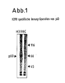
- Fig. 1
- the specific reaction of the antibody H398: it precipitates an iodine-labeled membrane protein with the
MW 60 kDa from HL-60 cells, - Fig. 2
- the neutralization of the cytotoxic or cytostatic effects caused by TNF by the antibody H398,
- Fig. 3
- neutralization of TNF-induced HLA gene expression by the antibody H398.
-
1.1 Präparation von Zellmembranen
- Puffer A:
- 125 mM Sucrose
7,5 mM Tris/HCl pH 7,4
2 mM EDTA - Puffer B:
- 100 mM Tris/HCl pH 7,4
10 mM MgCl₂
10 mM CaCl₂
2 % Fötales Kälberserum
(Seramed, Biochrom KG, Berlin)
Jeweils am Präparationstag wurden die Puffer A und B mit 1 mM Phenylmethylsulfonyfluorid (PMSF) versetzt. Die frisch geernteten Zellen wurden vor der Homogenisation in einem Glas/Glas-Homogenisator bei einer Zelldichte von 1· 10⁸/ml in Puffer A für 30 Minuten inkubiert. Diese Vorbehandlung in hypotonem Medium erleichterte die anschließende Homogenisation mit 30 Schüben bei 1000 Upm. Das Homogenat wurde mit dem gleichen Volumen Puffer A verdünnt und bei 50 x g für 10 Minuten zentrifugiert, um Kerne sowie noch intakte Zellen abzutrennen. Das Sediment wurde einmal mit dem gleichen Volumen Puffer A gewaschen und die vereinigten Überstände bei 12000 x g für 30 Minuten zentrifugiert. Die pelletierten Membranen wurden anschließend zweimal mit Puffer B gewaschen. Alle Präparationsschritte wurden bei 4°C durchgeführt.1.1 Preparation of cell membranes- Buffer A:
- 125 mM sucrose
7.5 mM Tris / HCl pH 7.4
2mM EDTA - Buffer B:
- 100 mM Tris / HCl pH 7.4
10 mM MgCl₂
10 mM CaCl₂
2% fetal calf serum
(Seramed, Biochrom KG, Berlin)
Buffer A and B were mixed with 1 mM phenylmethylsulfonyl fluoride (PMSF) on each preparation day. The freshly harvested cells were prior to homogenization in a glass / glass homogenizer at a cell density of 1 × 10⁸ / ml in buffer A for 30 Minutes incubated. This pretreatment in hypotonic medium facilitated the subsequent homogenization with 30 batches at 1000 rpm. The homogenate was diluted with the same volume of buffer A and centrifuged at 50 xg for 10 minutes in order to separate nuclei and cells that were still intact. The sediment was washed once with the same volume of buffer A and the combined supernatants were centrifuged at 12,000 xg for 30 minutes. The pelleted membranes were then washed twice with Buffer B. All preparation steps were carried out at 4 ° C. -
1.2 Solubilisierung des TNF-Rezeptors
- Solubilisierungspuffer:
- 20 mM Tris/HCl pH 7,4
2 mM MgCl₂
2 mM CaCl₂
1 % Triton X-100
10 µg/ml Leupeptin
1000 KIE/ml Aprotinin
0,5 µl/ml DFP
Der Proteasehemmstoff DFP (Diisopropyl-fluorophosphat (Sigma) wurde jeweils frisch zum Puffer gegeben. Das Membranpellet wurde in eiskaltem Solubilisierungspuffer mit 1 ml pro 5 x 10⁸ eingesetzter Zellen suspendiert und mit 10 Schüben in einem Glas/Glas-Homogenisator bei 1000 Upm homogenisiert. Nach einstündiger Inkubation auf Eis wurde der Ansatz für 30 Minuten bei 100.000 x g zentrifugiert und nur der klare Überstand weiter verwendet.1.2 Solubilization of the TNF receptor- Solubilization buffer:
- 20 mM Tris / HCl pH 7.4
2 mM MgCl₂
2 mM CaCl₂
1% Triton X-100
10 µg / ml leupeptin
1000 KIE / ml aprotinin
0.5 µl / ml DFP
The protease inhibitor DFP (diisopropyl fluorophosphate (Sigma) was added fresh to the buffer. The membrane pellet was suspended in ice-cold solubilization buffer with 1 ml per 5 × 10⁸ cells used and homogenized with 10 batches in a glass / glass homogenizer at 1000 rpm After incubation on ice for 1 hour, the mixture was centrifuged for 30 minutes at 100,000 × g and only the clear supernatant was used. -
1.3 Herstellung der TNF-Affinitätssäule
Zur Herstellung der TNF-Affinitätssäule wurden 5 mg gereinigtes, rekombinantes, humanes TNFα an 1 ml Affigel 15 gekoppelt, entsprechend der Vorschrift des Herstellers (Biorad). Zuvor wurde das TNF 24 Stunden in einem geeigneten Dialyseschlauch gegen den Kopplungspuffer (0,1 M MOPS/HCl pH 7,5) dialysiert. Das Affigel wurde dreimal mit bidestilliertem Wasser gewaschen und mit der dialysierten TNFα-Lösung 4 Stunden kräftig geschüttelt. Die nicht umgesetzten reaktiven Gruppen des Affigels wurden danach durch Zugabe von 1 M Tris/HCl pH 7,4 (Endkonzentration 50 mM) für eine Stunde abgesättigt. Anschließend wurde das TNF-Affigel zweimal mit Waschpuffer pH 7,4 (siehe 1.4) gewaschen und in eine FPLC-Säule (HR 5/5, Fa. Pharmacia, Freiburg) gefüllt. Vor der ersten Verwendung der Säule wurde das gesamte Wasch- und Elutionsprogramm der Affinitätsreinigung mit allen dort verwendeten Puffern einmal durchgeführt. Die gesamte Herstellung der Säule wurde bei 4°C durchgeführt; ebenfalls bei 4°C wurde sie in Waschpuffer pH 7,4, versetzt mit 20 mM Natriumazid, aufbewahrt.1.3 Preparation of the TNF affinity column
To prepare the TNF affinity column, 5 mg of purified, recombinant, human TNFα were coupled to 1 ml of Affigel 15, in accordance with the manufacturer's instructions (Biorad). Before this, the TNF was dialyzed against the coupling buffer (0.1 M MOPS / HCl pH 7.5) in a suitable dialysis tube. The Affigel was washed three times with bidistilled water and shaken vigorously with the dialyzed TNFα solution for 4 hours. The unreacted reactive groups of the Affigel were then saturated for one hour by adding 1 M Tris / HCl pH 7.4 (final concentration 50 mM). The TNF-Affigel was then washed twice with washing buffer pH 7.4 (see 1.4) and filled into an FPLC column (HR 5/5, from Pharmacia, Freiburg). Before the column was used for the first time, the entire affinity purification and elution program was carried out once with all the buffers used there. The entire preparation of the column was carried out at 4 ° C; likewise at 4 ° C., it was kept in washing buffer pH 7.4, mixed with 20 mM sodium azide. -
1.4 Affinitätschromatographie
- Waschpuffer pH 7,4:
- 10 mM Tris/HCl pH 7,4
150 mM NaCl
0,1 % Triton X-100 - Waschpuffer 3D:
- 10 mM Tris/HCl pH 7,4
150 mM NaCl
10 % Glycerin
1 % Natriumdesoxycholat
0,1 % SDS
0,1 % Triton X-100 - Waschpuffer pH 8,6:
- 10 mM Tris/HCl pH 8,6
500 mM KCl
0,5 % Triton X-100 - Waschpuffer pH 4,5:
- 50 mM Glycin/HCl pH 4,5
150 mM NaCl
0,1 % Triton X-100 - Elutionspuffer pH 3,0:
- 150 mM Glycin/HCl pH 3,0
0,1 % Triton X-100.
Die gesamte Affinitätsreinigung von solubilisiertem Material wurde auf einer FPLC-Anlage (Fa. Pharmacia, Freiburg) bei 4°C durchgeführt. Zur Proben- bzw. Pufferaufgabe wurde eine Schleife (Superloop) mit 50 ml Fassungsvermögen verwendet.
Der Membranextrakt von 7·10¹⁰ Zellen wurde mit einer Flußgeschwindigkeit von 0,2 ml/min auf die TNF-Affinitätssäule aufgegeben, und anschließend wurde die Säule mit einer Flußgeschwindigkeit von 0,5 ml/min mit den Puffern in folgender Reihenfolge (s.u.) gewaschen:- 1) 10 ml Waschpuffer pH 7,4
- 2) 10 ml Waschpuffer 3D
- 3) 2 ml Waschpuffer pH 7,4
- 4) 10 ml Waschpuffer pH 8,6
- 5) 2 ml Waschpuffer pH 7,4
- 6) 5 ml Waschpuffer pH 4,5
Der Rezeptor wurde durch den sauren Elutionspuffer (pH 3,0) mit einer Flußgeschwindigkeit von 0,1 ml/min von der Säule eluiert. Dabei wurde das Eluat in 10 Fraktionen zu je 1 ml gesammelt und durch 200 µl eines 1 M Tris/HCl Puffers pH 7,4, der in die Fraktionsröhrchen vorgelegt wurde, sofort neutralisiert. Die Fraktionen wurden bei -20°C aufbewahrt.1.4 Affinity chromatography- Wash buffer pH 7.4:
- 10 mM Tris / HCl pH 7.4
150 mM NaCl
0.1% Triton X-100 - Wash buffer 3D:
- 10 mM Tris / HCl pH 7.4
150 mM NaCl
10% glycerin
1% sodium deoxycholate
0.1% SDS
0.1% Triton X-100 - Wash buffer pH 8.6:
- 10 mM Tris / HCl pH 8.6
500 mM KCl
0.5% Triton X-100 - Wash buffer pH 4.5:
- 50 mM glycine / HCl pH 4.5
150 mM NaCl
0.1% Triton X-100 - Elution buffer pH 3.0:
- 150 mM glycine / HCl pH 3.0
0.1% Triton X-100.
The entire affinity purification of solubilized material was carried out on an FPLC system (from Pharmacia, Freiburg) at 4 ° C. A loop (Superloop) with a capacity of 50 ml was used for sample and buffer application.
The membrane extract of 7 × 10¹⁰ cells was applied to the TNF affinity column at a flow rate of 0.2 ml / min, and then the column was washed with the buffers at a flow rate of 0.5 ml / min in the following order (see below) :- 1) 10 ml washing buffer pH 7.4
- 2) 10 ml washing buffer 3D
- 3) 2 ml washing buffer pH 7.4
- 4) 10 ml wash buffer pH 8.6
- 5) 2 ml washing buffer pH 7.4
- 6) 5 ml washing buffer pH 4.5
The receptor was eluted from the column through the acidic elution buffer (pH 3.0) at a flow rate of 0.1 ml / min. The eluate was collected in 10 fractions of 1 ml each and immediately neutralized by 200 μl of a 1 M Tris / HCl buffer pH 7.4, which was placed in the fraction tubes. The fractions were stored at -20 ° C. -
1.5 Fällung von Proteinen mit Aceton
Das gesammelte Eluat wurde mit -80°C kaltem Aceton in einem Verhältnis von 1 Teil Probe/3 Teile Aceton versetzt und 14 Stunden bei -80°C aufbewahrt. Nach dieser Zeit wurde der entstandene Niederschlag 45 Minuten bei 12000 x g in einer kühlbaren Zentrifuge bei 0°C pelletiert. Der Überstand wurde anschließend sofort abgesaugt und das Pellet in 400 µl 20 mM Tris-Puffer, pH 7,4, versetzt mit 0,1 % Triton X-100, wieder aufgelöst. TNF-Bindungsstudien zeigten, daß diese Präparation insgesamt 4 µg aktives Rezeptorprotein enthielt (Gesamtproteingehalt: 80 µg).1.5 Precipitation of proteins with acetone
The eluate collected was mixed with -80 ° C cold acetone in a ratio of 1 part sample / 3 parts acetone and stored at -80 ° C for 14 hours. After this time, the resulting precipitate was pelleted for 45 minutes at 12,000 xg in a coolable centrifuge at 0 ° C. The supernatant was then immediately aspirated and the pellet redissolved in 400μl 20 mM Tris buffer, pH 7.4, mixed with 0.1% Triton X-100. TNF binding studies showed that this preparation contained a total of 4 µg active receptor protein (total protein content: 80 µg). -
2. Immunisierung
Eine 6 Wochen alte weibliche Maus vom Stamm BALB/c wurde mit dem Rezeptormaterial nach folgendem Schema immunisiert:- Tag 1:
- Intraperitoneale Injektion von 100 µl Rezeptorpräparation (1 µg Rezeptorprotein), versetzt mit komplettem Freund'schen Adjuvans (Behringwerke AG, OREC 20/21) (100 µl),
- Tag 40:
- Intraperitoneale Injektion von 100 µl (1 µg Rezeptorprotein), versetzt mit inkomplettem Freund'schem Adjuvans (Behringwerke AG, ORED 20/21),
- Tag 55:
- Intraperitoneale Injektion von 200 µl (2 µg Rezeptorprotein), ohne Adjuvans
- Tag 58:
- Entnahme der Milz unter sterilen Bedingungen, Herstellung einer Milzzellsuspension zur Zellfusion mit Myelomzellen.
A 6 week old female mouse from the BALB / c strain was immunized with the receptor material according to the following scheme:- Day 1:
- Intraperitoneal injection of 100 µl receptor preparation (1 µg receptor protein), mixed with complete Freund's adjuvant (Behringwerke AG,
OREC 20/21) (100 µl), - Day 40:
- Intraperitoneal injection of 100 µl (1 µg receptor protein), mixed with incomplete Freund's adjuvant (Behringwerke AG,
ORED 20/21), - Day 55:
- Intraperitoneal injection of 200 µl (2 µg receptor protein) without adjuvant
- Day 58:
- Removal of the spleen under sterile conditions, production of a spleen cell suspension for cell fusion with myeloma cells.
-
3. Herstellung von Hybridomen
Die Fusion der Milzzellen einer hyperimmunisierten BALB-C-Maus mit NSO-Zellen wurde genau nach Vorschrift von Peters et al. (Monoklonale Antikörper, Springer Verlag, Berlin, Heidelberg, 1985) im Verhältnis 5:1 durchgeführt und nach Standardselektionsbedingungen (HAT-Medium) kultiviert. Insgesamt wurden 413 Hybridklone isoliert und nach Transfer in Mikrotiterplatten auf Reaktion mit TNF-R-positiven Zellen (HL-60) untersucht.3. Production of hybridomas
The fusion of the spleen cells of a hyperimmunized BALB-C mouse with NSO cells was carried out exactly according to the instructions of Peters et al. (Monoclonal antibodies, Springer Verlag, Berlin, Heidelberg, 1985) in a ratio of 5: 1 and cultivated according to standard selection conditions (HAT medium). A total of 413 hybrid clones were isolated and examined for reaction with TNF-R positive cells (HL-60) after transfer in microtiter plates. -
4. Screening-Verfahren zur Identifikation von TNF-R-spezifischen, monoklonalen Antikörpern: Modulation der TNF-Bindungsfähigkeit
1 x 10⁶ HL-60 Zellen wurden mit Hybridkulturüberständen (20 % Endkonzentration) für 2 Stunden bei 37°C inkubiert, der Überstand nach Zentrifugation abgesaugt und die Zellen für weitere 60 Minuten bei 4°C mit radioaktivem TNF (20 ng/ml) inkubiert. Die spezifische TNF-Bindung solcher Zellen wurde mit der von unbehandelten (NSO-Überstände) Kontrollzellen verglichen. Von den 413 untersuchten Klonen erwies sich einer (H398) nach zweifacher Subklonierung als stabiles IgG produzierendes Hybridom, welches reproduzierbar die TNF-Bindung blockiert.4. Screening method for the identification of TNF-R-specific monoclonal antibodies: modulation of the TNF binding ability
1 x 10⁶ HL-60 cells were incubated with hybrid culture supernatants (20% final concentration) for 2 hours at 37 ° C, the supernatant was aspirated after centrifugation and the cells were incubated for a further 60 minutes at 4 ° C with radioactive TNF (20 ng / ml) . The specific TNF binding of such cells was compared with that of untreated (NSO supernatants) control cells. Of the 413 clones examined, one (H398), after double subcloning, proved to be a stable IgG-producing hybridoma which reproducibly blocks TNF binding. -
5. Immunpräzipitation des TNF-Rezeptors p60 mittels Antikörpern H398
2x10⁸ HL60 Zellen/Gruppe wurde nach der Lactoperoxidase-Methode oberflächenjodiert, gewaschen und mit Triton X-100 (1 %) solubilisiert. Aus dem Triton X-100-Extrakt wurden nach Vorinkubation (Preclearing) mit Prot-A-Sepharose (2 Stunden 0°) mit Prot-A-Sepharose gekoppeltem mAK H398 bzw. IgG-Kontrollantikörper (Spur C) immunpräzipitiert (16 Stunden, 0°C), das Präzipitat in Tris Puffer (20 mM) 0,1 % Triton X-100, 1 M NaCl (pH 7,8) dreimal gewaschen, in SDS-Probenpuffer 3' bei 100°C inkubiert und der Überstand auf einer 7,5 % PAGE unter nicht reduzierten Bedingungen aufgetrennt. Abb. 1 zeigt ein Autoradiogramm nach 64 Stunden Exposition des getrockneten Gels. Man erkennt nur auf der Spur des H398-Immunpräzipitates eine spezifische Bande bei 60 kD. Immunpräzipitiertes p60 besitzt auch nach SDS-Gelelektrophorese im Liganden-blot mit jodmarkiertem TNF spezifische Bindungseigenschaften für TNFα.5. Immunoprecipitation of the TNF receptor p60 using antibodies H398
2x10⁸ HL60 cells / group was surface iodinated according to the lactoperoxidase method, washed and solubilized with Triton X-100 (1%). After pre-incubation (preclearing) with Prot-A-Sepharose (2hours 0 °), mAK H398 coupled with Prot-A-Sepharose or IgG control antibody (lane C) were immunoprecipitated from the Triton X-100 extract (16 hours, 0 ° C), the precipitate in Tris buffer (20 mM) 0.1% Triton X-100, 1 M NaCl (pH 7.8) washed three times, incubated in SDS sample buffer 3 'at 100 ° C and the supernatant on a 7.5% PAGE separated under non-reduced conditions. Fig. 1 shows an autoradiogram after 64 hours of exposure to the dried gel. A specific band at 60 kD can only be seen on the trace of the H398 immunoprecipitate. Immunoprecipitated p60 also has specific binding properties for TNFα after SDS gel electrophoresis in ligand blot with iodine-labeled TNF.
-
1. H398 als TNF-Antagonist bei der zytotoxischen Wirkung von TNF auf HeLa- und MCF-7-Zellen.
Menschliche HeLa- bzw. MCF-7-Zellen werden in Mikrokulturen (3x10⁴ Zellen in 0,2 ml Kulturmedium Clicks/RPMI (Kat.Nr. T125); 5 % Fötales Kälberserum (beide Seramed, Biochrom, Berlin, FRG); für 18 Stunden vorkultiviert, dann für 7 Stunden in Gegenwart von 5 µg/ml Cycloheximid mit 1 ng/ml (50 Units) TNF und seriellen Verdünnungen von H398 behandelt. Danach wird die Anzahl überlebender Zellen durch Anfärben mit Kristallviolett quantifiziert. Abbildung 2 (graue Säule) zeigt, daß H398 HeLa-Zellen vor der zytotoxischen TNF-Wirkung konzentrationsabhängig schützt. Vergleichbare Daten wurden mit MCF-7 erhalten (Tabelle 1).1. H398 as a TNF antagonist in the cytotoxic effect of TNF on HeLa and MCF-7 cells.
Human HeLa or MCF-7 cells are grown in microcultures (3x10⁴ cells in 0.2 ml Clicks / RPMI culture medium (Cat.No. T125); 5% fetal calf serum (both Seramed, Biochrom, Berlin, FRG); for 18 Pre-cultured for hours, then treated with 1 ng / ml (50 units) TNF and serial dilutions of H398 for 7 hours in the presence of 5 µg / ml cycloheximide, after which the number of surviving cells is quantified by staining with crystal violet. Figure 2 (gray column) shows that H398 protects HeLa cells against the cytotoxic TNF effect depending on the concentration.Comparative data were obtained with MCF-7 (Table 1). -
2. Blockierung der wachstumshemmenden Wirkung von TNF auf U937-Zellen.
Tumor-Nekrose-Faktoren zeigen auf der menschlichen U937-Zellinie eine wachstumsinhibitorische Wirkung im Synergismus mit Interferon-gamma (Pfizenmaier et al., Blut 55 (1978), 1-10). Die Zellen (5x10³ in 0,2 ml Kulturmedium) wurden für 48 Stunden in Gegenwart von 10 ng/ml Interferon-gamma, 1 ng/ml TNF und seriellen Verdünnungen an H398 kultiviert. Die Proliferationskapazität der U937-Zellen wurden durch einen sechsstündigen ³H-Thymidinpuls für 6 Stunden bestimmt. In Anwesenheit von H398 (10 µg/ml) kommt es zur vollständigen Inhibition der durch TNF vermittelten Zytostase (Tabelle 1 und Abbildung 2, schwarze Säule). In ähnlicher Weise schützt H398 auch vor TNFβ (Lymphotoxin) und auch vor murinem TNF, welches auch auf menschlichen Zellen wirksam ist.2. Blocking the growth-inhibiting effect of TNF on U937 cells.
Tumor necrosis factors show a growth-inhibitory effect in synergism with interferon-gamma on the human U937 cell line (Pfizenmaier et al., Blut 55 (1978), 1-10). The cells (5x10³ in 0.2 ml culture medium) were cultured for 48 hours in the presence of 10 ng / ml interferon-gamma, 1 ng / ml TNF and serial dilutions of H398. The proliferation capacity of the U937 cells was determined by a 6 hour 3 H-thymidine pulse for 6 hours. In the presence of H398 (10 µg / ml) the TNF-mediated cytostasis is completely inhibited (Table 1 and Figure 2, black column). Similarly, H398 also protects against TNFβ (lymphotoxin) and also against murine TNF, which is also effective on human cells. -
3. H398 zeigt keine Wirkung auf Mauszellen.
H398 ist speziesspezifisch und erkennt TNF-Rezeptoren auf Humanzellen, nicht aber auf Mauszellen (vgl. auch Tabelle 1). Abb. 2 (weiße Säulen) zeigt, daß H398 auf Mauszellen keinerlei protektive Wirkung für humanen TNF besitzt. Der Experimentalansatz entspricht dem Standardtestsystem zur Bestimmung von TNF (24 Stunden Kultur von murinen L929-Zellen in der Gegenwart von 1 µg/ml Actinomycin D, 1 ng/ml TNF) in An- bzw. Abwesenheit von H398-Antikörper (10 µg/ml).3. H398 shows no effect on mouse cells.
H398 is species-specific and recognizes TNF receptors on human cells, but not on mouse cells (see also Table 1). Fig. 2 (white columns) shows that H398 has no protective effect on human TNF on mouse cells. The experimental approach corresponds to the standard test system for the determination of TNF (24 hour culture of murine L929 cells in the presence of 1 µg / ml actinomycin D, 1 ng / ml TNF) in the presence or absence of H398 antibody (10 µg / ml ).
Die TNF-induzierte Modulation von HLA-Genen wurde in einer Zellinie myeloischen Ursprungs (K562) und einer epithelialen Karzinomzellinie (Colo 205) untersucht. In beiden Systemen ist eine Kostimulation mit Interferongamma notwendig, um die jeweilige Reaktion auszulösen (Pfizenmaier et al., J. Immunol. 138 (1987), 975-980). In Anwesenheit von H398 kommt es in beiden Systemen zur vollständigen Inhibition der TNF-Antwort (vgl. auch Tabelle 1).
- 1. H398 als Antagonist der TNF-verstärkten HLA-A,B,C Antigenexpression auf K562.
K562-Zellen wurden für 24 Stunden in Gegenwart von 10 ng/ml Interferon-gamma, 3 ng/ml TNF und seriellen Verdünnungen von H398 kultiviert (5x10⁵ Zellen in 2 ml Kulturmedium). Danach wurden die Zellen geerntet und die HLA-Antigenexpression wurde mit Hilfe HLA-spezifischer monoklonaler Antikörper durch indirekte Immunfluoreszenzanalyse im Durchflußzytofluorographen quantifiziert. Die Ergebnisse sind in Abbildung 3 (graue Säule) dargestellt und zeigen, daß bei 10 µg/ml H398 (10⁻³ Verdünnung einer Ascites mit 10 mg/ml H398 monoklonalem Antikörper) die TNF-induzierte Verstärkung der HLA-A,B,C-Antigenexpression vollständig neutralisiert wird. Eine halbmaximale Inhibition wird mit etwa 1 µg/ml Antikörper erreicht. - 2. Antagonistische Wirkung von H398 der TNF-verstärkten HLA-DR Antigenexpression auf Colo 205-Zellen.
Der Experimentalansatz ist ähnlich dem oben beschrieben (jedoch 48 Stunden Kulturdauer, 0,02 ng/ml Interferon-gamma, 1 ng/ml TNF). Die Daten in Abbildung 3 (schwarze Säulen) zeigen ebenfalls eine vollständige Aufhebung der TNF-induzierten Verstärkung der HLA-DR-Genexpression bei 10 µg/ml H398; die halbmaximale Hemmung wird hier mit etwa 2 µg/ml H398 erreicht.
- 1. H398 as an antagonist of TNF-enhanced HLA-A, B, C antigen expression on K562.
K562 cells were cultured for 24 hours in the presence of 10 ng / ml interferon-gamma, 3 ng / ml TNF and serial dilutions of H398 (5x10⁵ cells in 2 ml culture medium). The cells were then harvested and the HLA antigen expression was quantified using HLA-specific monoclonal antibodies by indirect immunofluorescence analysis in a flow cytofluorograph. The results are shown in Figure 3 (gray column) and show that at 10 µg / ml H398 (10⁻³ dilution of an ascites with 10 mg / ml H398 monoclonal antibody) the TNF-induced enhancement of HLA-A, B, C -Antigen expression is completely neutralized. Half maximum inhibition is achieved with approximately 1 µg / ml antibody. - 2. Antagonistic effect of H398 of the TNF-enhanced HLA-DR antigen expression on Colo 205 cells.
The experimental approach is similar to that described above (however, 48 hours of culture, 0.02 ng / ml interferon-gamma, 1 ng / ml TNF). The data in Figure 3 (black columns) also show a complete abolition of the TNF-induced enhancement of HLA-DR gene expression at 10 µg / ml H398; half-maximal inhibition is achieved here with about 2 µg / ml H398.
Eine bedeutende immunregulatorische Funktion von TNF ist die Stimulation der Interleukin-2 Rezeptorexpression auf T-Lymphozyten und auf sogenannten Natürlichen Killerzellen (NK-Zellen) (Scheurich et al., J. Immunol. 138 (1987), 1786-1790). Auch in diesem System besitzt H398 antagonistische Wirkung. Es wurden 5x10⁵ Zellen der NK-Zellinie YT in 2 ml Kulturmedium für 24 Stunden in Gegenwart von 1 und 10 ng/ml TNF sowie seriellen Verdünnungen von H398 kultiviert. Die Auswertung erfolgte zytofluorographisch mit Hilfe von anti-Interleukin-2 Rezeptor Antikörpern (anti Tac). Die in Tabelle 1 dargestellten Ergebnisse belegen die komplette Inhibition auch dieser TNF-Antwort durch H398.An important immunoregulatory function of TNF is the stimulation of interleukin-2 receptor expression on T lymphocytes and on so-called natural killer cells (NK cells) (Scheurich et al., J. Immunol. 138 (1987), 1786-1790). H398 also has an antagonistic effect in this system. 5x10⁵ cells of the NK cell line YT were cultivated in 2 ml of culture medium for 24 hours in the presence of 1 and 10 ng / ml TNF and serial dilutions of H398. The evaluation was carried out cytofluorographically with the help of anti-interleukin-2 receptor antibodies (anti Tac). The results shown in Table 1 demonstrate the complete inhibition of this TNF response by H398 as well.
Claims (17)
Applications Claiming Priority (2)
| Application Number | Priority Date | Filing Date | Title |
|---|---|---|---|
| DE4006269 | 1990-02-28 | ||
| DE4006269A DE4006269A1 (en) | 1990-02-28 | 1990-02-28 | Antibody which binds to tumour necrosis factor receptors |
Publications (1)
| Publication Number | Publication Date |
|---|---|
| EP0444560A1 true EP0444560A1 (en) | 1991-09-04 |
Family
ID=6401127
Family Applications (1)
| Application Number | Title | Priority Date | Filing Date |
|---|---|---|---|
| EP91102711A Withdrawn EP0444560A1 (en) | 1990-02-28 | 1991-02-25 | Antibody specific for the tumor necrosis factor receptor |
Country Status (6)
| Country | Link |
|---|---|
| US (1) | US5736138A (en) |
| EP (1) | EP0444560A1 (en) |
| JP (1) | JPH04211392A (en) |
| DE (1) | DE4006269A1 (en) |
| FI (1) | FI910973L (en) |
| PT (1) | PT96910A (en) |
Cited By (6)
| Publication number | Priority date | Publication date | Assignee | Title |
|---|---|---|---|---|
| WO1992022666A1 (en) * | 1991-06-19 | 1992-12-23 | Boehringer Ingelheim International Gmbh | Monoclonal antibodies against human tnf-binding protein i (tnf-bp i) |
| WO1994026924A3 (en) * | 1993-05-14 | 1994-11-24 | Mini Ricerca Scient Tecnolog | MONOCLONAL ANTIBODIES AGAINST THE α-TNF TYPE I RECEPTOR, PROCESS FOR THE PRODUCTION AND USE THEREOF |
| EP0568925A3 (en) * | 1992-05-03 | 1995-03-15 | Yeda Res & Dev | Modulation of TNF Receptor Activity |
| EP0585939A3 (en) * | 1992-09-03 | 1995-04-05 | Yeda Res & Dev | TNF ligands. |
| EP1542722A4 (en) * | 2002-08-01 | 2006-02-01 | Wyeth Corp | Methods and reagents relating to inflammation and apoptosis |
| EP1972637A1 (en) * | 2007-03-19 | 2008-09-24 | Universität Stuttgart | huTNFR1 selective antagonists |
Families Citing this family (17)
| Publication number | Priority date | Publication date | Assignee | Title |
|---|---|---|---|---|
| US6270766B1 (en) | 1992-10-08 | 2001-08-07 | The Kennedy Institute Of Rheumatology | Anti-TNF antibodies and methotrexate in the treatment of arthritis and crohn's disease |
| US8197430B1 (en) * | 1998-05-22 | 2012-06-12 | Biopheresis Technologies, Inc. | Method and system to remove cytokine inhibitor in patients |
| US6620382B1 (en) | 1998-05-22 | 2003-09-16 | Biopheresis Technologies, Llc. | Method and compositions for treatment of cancers |
| EP1372720A4 (en) * | 2001-03-02 | 2006-07-26 | Medimmune Inc | Methods of preventing or treating inflammatory or autoimmune disorders by administering integrin alphav beta3 antagonists |
| US7371383B2 (en) * | 2002-04-12 | 2008-05-13 | Medimmune, Inc. | Recombinant anti-interleukin-9 antibodies |
| US9028822B2 (en) | 2002-06-28 | 2015-05-12 | Domantis Limited | Antagonists against TNFR1 and methods of use therefor |
| JP4764818B2 (en) | 2003-04-11 | 2011-09-07 | メディミューン,エルエルシー | Recombinant IL-9 antibody and use thereof |
| US7351739B2 (en) * | 2004-04-30 | 2008-04-01 | Wellgen, Inc. | Bioactive compounds and methods of uses thereof |
| WO2005107802A2 (en) * | 2004-04-30 | 2005-11-17 | Biopheresis Technologies, Llc | Method and system to remove soluble tnfr1, tnfr2, and il2 in patients |
| US20060121042A1 (en) | 2004-10-27 | 2006-06-08 | Medimmune, Inc. | Modulation of antibody specificity by tailoring the affinity to cognate antigens |
| GB0506708D0 (en) * | 2005-04-01 | 2005-05-11 | Queen Mary & Westfield College | Use of calcitonin as combined treatment therapy for the management of inflammatory disease conditions |
| AU2007272970C1 (en) | 2006-07-11 | 2013-01-10 | Roy C. Levitt | Rhinosinusitis prevention and therapy with proinflammatory cytokine inhibitors |
| EP3669878A1 (en) | 2006-08-10 | 2020-06-24 | Roy C. Levitt | Localized therapy of lower airways inflammatory disorders with proinflammatory cytokine inhibitors |
| EP2068925A4 (en) | 2007-05-07 | 2011-08-31 | Medimmune Llc | Anti-icos antibodies and their use in treatment of oncology, transplantation and autoimmune disease |
| JP5933975B2 (en) | 2008-11-12 | 2016-06-15 | メディミューン,エルエルシー | Antibody preparation |
| CN103249742B (en) * | 2010-09-16 | 2017-04-12 | 巴利奥医药股份公司 | Anti-hutnfr1 antibody |
| EA202091182A1 (en) | 2017-11-27 | 2020-07-29 | Балиофарм Аг | ANTI-huTNFR1 NON-ALCOHOLIC STEATHEPATITIS THERAPY |
Citations (3)
| Publication number | Priority date | Publication date | Assignee | Title |
|---|---|---|---|---|
| EP0230574A2 (en) * | 1986-01-31 | 1987-08-05 | Yale University | Pharmaceutical compositions against infections caused by LAV/HTLV III virus and the use thereof |
| US4770995A (en) * | 1985-08-29 | 1988-09-13 | New York Blood Center, Inc | Detection of the sensitivity of cells to the effects of tumor necrosis factor and lymphotoxin |
| EP0334165B1 (en) * | 1988-03-17 | 1995-12-27 | F. Hoffmann-La Roche Ag | Monoclonal antibodies |
-
1990
- 1990-02-28 DE DE4006269A patent/DE4006269A1/en not_active Withdrawn
-
1991
- 1991-02-12 US US07/654,257 patent/US5736138A/en not_active Expired - Lifetime
- 1991-02-25 EP EP91102711A patent/EP0444560A1/en not_active Withdrawn
- 1991-02-27 FI FI910973A patent/FI910973L/en not_active Application Discontinuation
- 1991-02-27 JP JP3031181A patent/JPH04211392A/en active Pending
- 1991-02-28 PT PT96910A patent/PT96910A/en not_active Application Discontinuation
Patent Citations (3)
| Publication number | Priority date | Publication date | Assignee | Title |
|---|---|---|---|---|
| US4770995A (en) * | 1985-08-29 | 1988-09-13 | New York Blood Center, Inc | Detection of the sensitivity of cells to the effects of tumor necrosis factor and lymphotoxin |
| EP0230574A2 (en) * | 1986-01-31 | 1987-08-05 | Yale University | Pharmaceutical compositions against infections caused by LAV/HTLV III virus and the use thereof |
| EP0334165B1 (en) * | 1988-03-17 | 1995-12-27 | F. Hoffmann-La Roche Ag | Monoclonal antibodies |
Non-Patent Citations (4)
| Title |
|---|
| J. Exp. Med., Band 171, Nr. 2, 1. Februar 1990, Seiten 415-426, The Rockefeller University Press, New York, US; T. ESPEVIK et al.: "Characterization of binding and biological effects of monoclonal antibodies against a human tumor necrosis factor receptor", Zusammenfassung. * |
| J. Exp. Med., Band 172, Nr. 4, 1. Oktober 1990, Seiten 1019-1023, The Rockefeller University Press, New York, US; B. THOMA et al.: "Identification of a 60-kD tumor necrosis factor (TNF) receptor as the major signal transducing component in TNF responses", der ganze Artikel. * |
| The Journal of Biological Chemistry, Band 264, Nr. 25, 5. September 1989, Seiten 14927-14934, The American Society for Biochemistry and Molecular Biology, Inc., Baltimore, US; H.-P. HOHMANN et al.: "Two different cell types have different major receptors for human tumor necrosis factor (TNFalpha)", Figur 4. * |
| The Journal of Biological Chemistry, Band 265, Nr. 3, 25. Januar 1990, Seiten 1531-1536, The American Society for Biochemistry and Molecular Biology, Inc., Baltimore US; H. ENGELMANN et al.: "Two tumor necrosis factor-binding proteins purified from human urine", Seite 1534, rechte Spalte, Zeilen 1-25; Seite 1535, rechte Spalte, Zeilen 4-21; Figur 6. * |
Cited By (14)
| Publication number | Priority date | Publication date | Assignee | Title |
|---|---|---|---|---|
| WO1992022666A1 (en) * | 1991-06-19 | 1992-12-23 | Boehringer Ingelheim International Gmbh | Monoclonal antibodies against human tnf-binding protein i (tnf-bp i) |
| US7056505B2 (en) | 1992-05-03 | 2006-06-06 | Yeda Research And Development Co., Ltd. | TNF receptor action modulation |
| US7728111B2 (en) | 1992-05-03 | 2010-06-01 | Yeda Research And Development Co., Ltd. | Anti-p55 TNF receptor antibody |
| EP0568925A3 (en) * | 1992-05-03 | 1995-03-15 | Yeda Res & Dev | Modulation of TNF Receptor Activity |
| US7429379B2 (en) | 1992-05-03 | 2008-09-30 | Yeda Research And Development Co., Ltd. | TNF receptor action modulation |
| EP1512697A1 (en) * | 1992-05-03 | 2005-03-09 | Yeda Research And Development Co. Ltd. | TNF Receptor action modulation |
| EP0585939A3 (en) * | 1992-09-03 | 1995-04-05 | Yeda Res & Dev | TNF ligands. |
| AU678369B2 (en) * | 1992-09-03 | 1997-05-29 | Yeda Research And Development Co. Ltd. | TNF ligands |
| WO1994026924A3 (en) * | 1993-05-14 | 1994-11-24 | Mini Ricerca Scient Tecnolog | MONOCLONAL ANTIBODIES AGAINST THE α-TNF TYPE I RECEPTOR, PROCESS FOR THE PRODUCTION AND USE THEREOF |
| EP1542722A4 (en) * | 2002-08-01 | 2006-02-01 | Wyeth Corp | Methods and reagents relating to inflammation and apoptosis |
| EP1972637A1 (en) * | 2007-03-19 | 2008-09-24 | Universität Stuttgart | huTNFR1 selective antagonists |
| WO2008113515A3 (en) * | 2007-03-19 | 2008-11-13 | Univ Stuttgart | huTNFR1 SELECTIVE ANTAGONISTS |
| US8404238B2 (en) | 2007-03-19 | 2013-03-26 | Universitaet Stuttgart | huTNFR1 selective antagonists |
| US9045535B2 (en) | 2007-03-19 | 2015-06-02 | Universitaet Stuttgart | huTNFR1 selective antagonists |
Also Published As
| Publication number | Publication date |
|---|---|
| JPH04211392A (en) | 1992-08-03 |
| FI910973A7 (en) | 1991-08-29 |
| FI910973L (en) | 1991-08-29 |
| DE4006269A1 (en) | 1991-08-29 |
| US5736138A (en) | 1998-04-07 |
| PT96910A (en) | 1991-10-31 |
| FI910973A0 (en) | 1991-02-27 |
Similar Documents
| Publication | Publication Date | Title |
|---|---|---|
| EP0444560A1 (en) | Antibody specific for the tumor necrosis factor receptor | |
| DE69017753T2 (en) | Tumor necrosis factor binding protein II, its purification and specific antibodies. | |
| DE69132629T2 (en) | SURFACE COMPLEX FORMATION OF LYMPHOTOXIN | |
| DE69332997T2 (en) | MONOCLONAL ANTIBODIES AGAINST THE INTERFERON RECEPTOR, WITH NEUTRALIZING ACTIVITY AGAINST TYPE I INTERFERON | |
| DE69028671T2 (en) | Soluble extracellular fragment of the human IFN-beta 2 / IL-6 receptor, its preparation and pharmaceutical mixture containing this fragment | |
| DE69014474T2 (en) | Antibodies against TNF binding protein I and its F (ab) fragments. | |
| DE69635480T2 (en) | APOPTOSIS INDUCTIVE CYTOKIN | |
| DE69223641T2 (en) | STABILIZED ANTIBODIES | |
| DE69100768T2 (en) | CD25 binding molecules. | |
| DE69427928T2 (en) | Human monoclonal anti-TNF alpha antibodies | |
| DE69433422T2 (en) | MONOCLONAL ANTI-HIV ANTIBODY | |
| DE68928641T2 (en) | Human IFN-beta2 / IL-6, its purification and uses | |
| DE3750342T2 (en) | Human gamma interferon-specific receptor protein, antibodies against this protein, methods for producing this protein and antibody and compositions containing this protein and antibody. | |
| DE68914244T2 (en) | Monoclonal antibody. | |
| DE69329558T2 (en) | Procedure for the treatment of multiple sclerosis | |
| DE3889062T2 (en) | Monoclonal antibody against human BCDF. | |
| EP0538754A2 (en) | Use of antibody-containing preparations for immunosuppression | |
| DE69528325T2 (en) | Human interleukin-6 inhibitor | |
| EP0350690A2 (en) | Neutralisation of the in vitro and in vivo toxic properties of TNF-alpha by monoclonal antibodies and by fragments derived therefrom | |
| DE69028095T2 (en) | M-CSF MONOCLONAL ANTIBODIES THAT DETECT A NEUTRALIZING CONFORMAL EPITOP | |
| EP0340604B1 (en) | Monoclonal antibody and its use | |
| DE68924464T2 (en) | Interferon-gamma binding proteins. | |
| EP0430193A1 (en) | Monoclonal antibodies and their use | |
| DE3306060A1 (en) | NEW IMMUNGLOBULIN-PRODUCING HYBRID CELL LINES, THEIR USE AND METHOD FOR THE PRODUCTION THEREOF | |
| DE69226448T2 (en) | GLYCOPROTEIN COMPLEX AND GLYCOPROTEINE |
Legal Events
| Date | Code | Title | Description |
|---|---|---|---|
| PUAI | Public reference made under article 153(3) epc to a published international application that has entered the european phase |
Free format text: ORIGINAL CODE: 0009012 |
|
| AK | Designated contracting states |
Kind code of ref document: A1 Designated state(s): AT BE CH DE DK ES FR GB GR IT LI LU NL SE |
|
| 17P | Request for examination filed |
Effective date: 19920131 |
|
| 17Q | First examination report despatched |
Effective date: 19931229 |
|
| STAA | Information on the status of an ep patent application or granted ep patent |
Free format text: STATUS: THE APPLICATION IS DEEMED TO BE WITHDRAWN |
|
| 18D | Application deemed to be withdrawn |
Effective date: 19960130 |