EP0449883B1 - Verfahren für laserinduzierte fluoreszenz von gewebe - Google Patents
Verfahren für laserinduzierte fluoreszenz von gewebe Download PDFInfo
- Publication number
- EP0449883B1 EP0449883B1 EP90900642A EP90900642A EP0449883B1 EP 0449883 B1 EP0449883 B1 EP 0449883B1 EP 90900642 A EP90900642 A EP 90900642A EP 90900642 A EP90900642 A EP 90900642A EP 0449883 B1 EP0449883 B1 EP 0449883B1
- Authority
- EP
- European Patent Office
- Prior art keywords
- tissue
- fluorescence
- chromophore
- chromophores
- irradiating
- Prior art date
- Legal status (The legal status is an assumption and is not a legal conclusion. Google has not performed a legal analysis and makes no representation as to the accuracy of the status listed.)
- Expired - Lifetime
Links
- 238000000034 method Methods 0.000 title claims abstract description 89
- 238000001499 laser induced fluorescence spectroscopy Methods 0.000 title abstract description 33
- 238000002189 fluorescence spectrum Methods 0.000 claims abstract description 26
- 230000005855 radiation Effects 0.000 claims abstract description 17
- 239000000203 mixture Substances 0.000 claims abstract description 14
- 230000001575 pathological effect Effects 0.000 claims abstract description 11
- 230000005284 excitation Effects 0.000 claims description 77
- 238000001228 spectrum Methods 0.000 claims description 42
- 102000008186 Collagen Human genes 0.000 claims description 38
- 108010035532 Collagen Proteins 0.000 claims description 38
- QIVBCDIJIAJPQS-VIFPVBQESA-N L-tryptophane Chemical compound C1=CC=C2C(C[C@H](N)C(O)=O)=CNC2=C1 QIVBCDIJIAJPQS-VIFPVBQESA-N 0.000 claims description 36
- 229920001436 collagen Polymers 0.000 claims description 36
- QIVBCDIJIAJPQS-UHFFFAOYSA-N Tryptophan Natural products C1=CC=C2C(CC(N)C(O)=O)=CNC2=C1 QIVBCDIJIAJPQS-UHFFFAOYSA-N 0.000 claims description 33
- 102000016942 Elastin Human genes 0.000 claims description 28
- 108010014258 Elastin Proteins 0.000 claims description 28
- 229920002549 elastin Polymers 0.000 claims description 26
- 239000000835 fiber Substances 0.000 claims description 26
- 230000003595 spectral effect Effects 0.000 claims description 22
- 238000000295 emission spectrum Methods 0.000 claims description 19
- 239000000523 sample Substances 0.000 claims description 18
- 230000001678 irradiating effect Effects 0.000 claims description 16
- 230000009103 reabsorption Effects 0.000 claims description 9
- 230000002596 correlated effect Effects 0.000 claims description 7
- 239000013074 reference sample Substances 0.000 claims 1
- 239000007787 solid Substances 0.000 claims 1
- 239000000126 substance Substances 0.000 abstract description 20
- 239000013307 optical fiber Substances 0.000 abstract description 8
- 210000001519 tissue Anatomy 0.000 description 185
- 229960004799 tryptophan Drugs 0.000 description 33
- 101710172711 Structural protein Proteins 0.000 description 29
- 230000009102 absorption Effects 0.000 description 25
- 238000010521 absorption reaction Methods 0.000 description 25
- 210000000709 aorta Anatomy 0.000 description 24
- 239000010410 layer Substances 0.000 description 21
- 108010054147 Hemoglobins Proteins 0.000 description 20
- 102000001554 Hemoglobins Human genes 0.000 description 20
- 238000009826 distribution Methods 0.000 description 18
- 230000000694 effects Effects 0.000 description 15
- 230000035945 sensitivity Effects 0.000 description 15
- 208000037260 Atherosclerotic Plaque Diseases 0.000 description 14
- 201000001320 Atherosclerosis Diseases 0.000 description 13
- 230000003143 atherosclerotic effect Effects 0.000 description 13
- 210000001367 artery Anatomy 0.000 description 11
- 238000003745 diagnosis Methods 0.000 description 9
- 238000006862 quantum yield reaction Methods 0.000 description 9
- VYPSYNLAJGMNEJ-UHFFFAOYSA-N Silicium dioxide Chemical compound O=[Si]=O VYPSYNLAJGMNEJ-UHFFFAOYSA-N 0.000 description 8
- 150000002632 lipids Chemical class 0.000 description 7
- 230000003287 optical effect Effects 0.000 description 7
- 238000002679 ablation Methods 0.000 description 6
- 201000010099 disease Diseases 0.000 description 6
- 208000037265 diseases, disorders, signs and symptoms Diseases 0.000 description 6
- 230000001338 necrotic effect Effects 0.000 description 6
- 230000007170 pathology Effects 0.000 description 6
- 108010064719 Oxyhemoglobins Proteins 0.000 description 5
- 238000004458 analytical method Methods 0.000 description 5
- 238000000338 in vitro Methods 0.000 description 5
- 230000035515 penetration Effects 0.000 description 5
- 230000008569 process Effects 0.000 description 5
- LCYXYLLJXMAEMT-SAXRGWBVSA-N Pyridinoline Chemical compound OC(=O)[C@@H](N)CCC1=C[N+](C[C@H](O)CC[C@H](N)C([O-])=O)=CC(O)=C1C[C@H](N)C(O)=O LCYXYLLJXMAEMT-SAXRGWBVSA-N 0.000 description 4
- 230000008859 change Effects 0.000 description 4
- 239000002131 composite material Substances 0.000 description 4
- 230000003176 fibrotic effect Effects 0.000 description 4
- 230000003902 lesion Effects 0.000 description 4
- 238000005259 measurement Methods 0.000 description 4
- 230000002784 sclerotic effect Effects 0.000 description 4
- 208000004434 Calcinosis Diseases 0.000 description 3
- HEMHJVSKTPXQMS-UHFFFAOYSA-M Sodium hydroxide Chemical compound [OH-].[Na+] HEMHJVSKTPXQMS-UHFFFAOYSA-M 0.000 description 3
- 238000013459 approach Methods 0.000 description 3
- 230000002308 calcification Effects 0.000 description 3
- 150000001875 compounds Chemical class 0.000 description 3
- 210000004351 coronary vessel Anatomy 0.000 description 3
- 238000002405 diagnostic procedure Methods 0.000 description 3
- 210000002950 fibroblast Anatomy 0.000 description 3
- 210000000497 foam cell Anatomy 0.000 description 3
- 238000007429 general method Methods 0.000 description 3
- 230000006872 improvement Effects 0.000 description 3
- 238000001727 in vivo Methods 0.000 description 3
- 102000039446 nucleic acids Human genes 0.000 description 3
- 108020004707 nucleic acids Proteins 0.000 description 3
- 150000007523 nucleic acids Chemical class 0.000 description 3
- 150000004032 porphyrins Chemical class 0.000 description 3
- 239000008057 potassium phosphate buffer Substances 0.000 description 3
- 230000035755 proliferation Effects 0.000 description 3
- 102000004169 proteins and genes Human genes 0.000 description 3
- 108090000623 proteins and genes Proteins 0.000 description 3
- 230000004044 response Effects 0.000 description 3
- 210000000329 smooth muscle myocyte Anatomy 0.000 description 3
- 239000000243 solution Substances 0.000 description 3
- 238000004611 spectroscopical analysis Methods 0.000 description 3
- IJGRMHOSHXDMSA-UHFFFAOYSA-N Atomic nitrogen Chemical compound N#N IJGRMHOSHXDMSA-UHFFFAOYSA-N 0.000 description 2
- 101100129500 Caenorhabditis elegans max-2 gene Proteins 0.000 description 2
- 230000004913 activation Effects 0.000 description 2
- 239000007864 aqueous solution Substances 0.000 description 2
- XKRFYHLGVUSROY-UHFFFAOYSA-N argon Substances [Ar] XKRFYHLGVUSROY-UHFFFAOYSA-N 0.000 description 2
- 229910052786 argon Inorganic materials 0.000 description 2
- -1 argon ion Chemical class 0.000 description 2
- 238000005311 autocorrelation function Methods 0.000 description 2
- 238000011888 autopsy Methods 0.000 description 2
- 230000008901 benefit Effects 0.000 description 2
- 230000015572 biosynthetic process Effects 0.000 description 2
- 239000008280 blood Substances 0.000 description 2
- 210000004369 blood Anatomy 0.000 description 2
- 210000002808 connective tissue Anatomy 0.000 description 2
- 239000000470 constituent Substances 0.000 description 2
- 230000000875 corresponding effect Effects 0.000 description 2
- 230000007423 decrease Effects 0.000 description 2
- 239000000975 dye Substances 0.000 description 2
- 238000004836 empirical method Methods 0.000 description 2
- 230000003511 endothelial effect Effects 0.000 description 2
- 238000000605 extraction Methods 0.000 description 2
- 238000001506 fluorescence spectroscopy Methods 0.000 description 2
- 230000002209 hydrophobic effect Effects 0.000 description 2
- 238000012623 in vivo measurement Methods 0.000 description 2
- QWTDNUCVQCZILF-UHFFFAOYSA-N isopentane Chemical compound CCC(C)C QWTDNUCVQCZILF-UHFFFAOYSA-N 0.000 description 2
- 230000004807 localization Effects 0.000 description 2
- 239000000843 powder Substances 0.000 description 2
- 230000009467 reduction Effects 0.000 description 2
- 238000000926 separation method Methods 0.000 description 2
- 239000000377 silicon dioxide Substances 0.000 description 2
- 239000002356 single layer Substances 0.000 description 2
- 239000002904 solvent Substances 0.000 description 2
- 238000012360 testing method Methods 0.000 description 2
- 238000012546 transfer Methods 0.000 description 2
- RYHBNJHYFVUHQT-UHFFFAOYSA-N 1,4-Dioxane Chemical compound C1COCCO1 RYHBNJHYFVUHQT-UHFFFAOYSA-N 0.000 description 1
- 102000007469 Actins Human genes 0.000 description 1
- 108010085238 Actins Proteins 0.000 description 1
- 241000283690 Bos taurus Species 0.000 description 1
- KCKDFIJGPQNRRZ-UHFFFAOYSA-N CC(CCC1)C1C1CC1 Chemical compound CC(CCC1)C1C1CC1 KCKDFIJGPQNRRZ-UHFFFAOYSA-N 0.000 description 1
- 239000004593 Epoxy Substances 0.000 description 1
- 241000588724 Escherichia coli Species 0.000 description 1
- 238000001069 Raman spectroscopy Methods 0.000 description 1
- 208000034189 Sclerosis Diseases 0.000 description 1
- FAPWRFPIFSIZLT-UHFFFAOYSA-M Sodium chloride Chemical compound [Na+].[Cl-] FAPWRFPIFSIZLT-UHFFFAOYSA-M 0.000 description 1
- 241000191963 Staphylococcus epidermidis Species 0.000 description 1
- 239000007983 Tris buffer Substances 0.000 description 1
- 239000006096 absorbing agent Substances 0.000 description 1
- 238000004847 absorption spectroscopy Methods 0.000 description 1
- 238000000862 absorption spectrum Methods 0.000 description 1
- 238000009825 accumulation Methods 0.000 description 1
- 230000004075 alteration Effects 0.000 description 1
- 150000001413 amino acids Chemical class 0.000 description 1
- 125000003118 aryl group Chemical group 0.000 description 1
- 230000002238 attenuated effect Effects 0.000 description 1
- 239000012620 biological material Substances 0.000 description 1
- 210000000481 breast Anatomy 0.000 description 1
- 239000007975 buffered saline Substances 0.000 description 1
- 238000004364 calculation method Methods 0.000 description 1
- 230000004663 cell proliferation Effects 0.000 description 1
- 230000001413 cellular effect Effects 0.000 description 1
- 238000006243 chemical reaction Methods 0.000 description 1
- 238000010276 construction Methods 0.000 description 1
- 230000008878 coupling Effects 0.000 description 1
- 238000010168 coupling process Methods 0.000 description 1
- 238000005859 coupling reaction Methods 0.000 description 1
- 238000004132 cross linking Methods 0.000 description 1
- 239000003431 cross linking reagent Substances 0.000 description 1
- 238000013480 data collection Methods 0.000 description 1
- 230000001419 dependent effect Effects 0.000 description 1
- AFABGHUZZDYHJO-UHFFFAOYSA-N dimethyl butane Natural products CCCC(C)C AFABGHUZZDYHJO-UHFFFAOYSA-N 0.000 description 1
- 238000000695 excitation spectrum Methods 0.000 description 1
- 230000005281 excited state Effects 0.000 description 1
- 238000002474 experimental method Methods 0.000 description 1
- 150000002211 flavins Chemical class 0.000 description 1
- 238000001857 fluorescence decay curve Methods 0.000 description 1
- 238000000799 fluorescence microscopy Methods 0.000 description 1
- 239000007850 fluorescent dye Substances 0.000 description 1
- 239000005350 fused silica glass Substances 0.000 description 1
- 150000003278 haem Chemical class 0.000 description 1
- 238000010438 heat treatment Methods 0.000 description 1
- 238000005286 illumination Methods 0.000 description 1
- 238000003780 insertion Methods 0.000 description 1
- 230000037431 insertion Effects 0.000 description 1
- 230000010354 integration Effects 0.000 description 1
- 230000008069 intimal proliferation Effects 0.000 description 1
- 230000003834 intracellular effect Effects 0.000 description 1
- 230000009545 invasion Effects 0.000 description 1
- 238000000608 laser ablation Methods 0.000 description 1
- 238000001307 laser spectroscopy Methods 0.000 description 1
- 230000031700 light absorption Effects 0.000 description 1
- 239000007788 liquid Substances 0.000 description 1
- 239000000463 material Substances 0.000 description 1
- 239000011159 matrix material Substances 0.000 description 1
- 238000000386 microscopy Methods 0.000 description 1
- 238000012544 monitoring process Methods 0.000 description 1
- 230000000877 morphologic effect Effects 0.000 description 1
- 229930027945 nicotinamide-adenine dinucleotide Natural products 0.000 description 1
- BOPGDPNILDQYTO-NNYOXOHSSA-N nicotinamide-adenine dinucleotide Chemical compound C1=CCC(C(=O)N)=CN1[C@H]1[C@H](O)[C@H](O)[C@@H](COP(O)(=O)OP(O)(=O)OC[C@@H]2[C@H]([C@@H](O)[C@@H](O2)N2C3=NC=NC(N)=C3N=C2)O)O1 BOPGDPNILDQYTO-NNYOXOHSSA-N 0.000 description 1
- 229910052757 nitrogen Inorganic materials 0.000 description 1
- 230000003647 oxidation Effects 0.000 description 1
- 238000007254 oxidation reaction Methods 0.000 description 1
- 239000010453 quartz Substances 0.000 description 1
- 210000004872 soft tissue Anatomy 0.000 description 1
- 238000012353 t test Methods 0.000 description 1
- 230000008685 targeting Effects 0.000 description 1
- 230000007704 transition Effects 0.000 description 1
- LENZDBCJOHFCAS-UHFFFAOYSA-N tris Chemical compound OCC(N)(CO)CO LENZDBCJOHFCAS-UHFFFAOYSA-N 0.000 description 1
- 238000011179 visual inspection Methods 0.000 description 1
Images





Classifications
-
- A—HUMAN NECESSITIES
- A61—MEDICAL OR VETERINARY SCIENCE; HYGIENE
- A61B—DIAGNOSIS; SURGERY; IDENTIFICATION
- A61B5/00—Measuring for diagnostic purposes; Identification of persons
- A61B5/0059—Measuring for diagnostic purposes; Identification of persons using light, e.g. diagnosis by transillumination, diascopy, fluorescence
- A61B5/0082—Measuring for diagnostic purposes; Identification of persons using light, e.g. diagnosis by transillumination, diascopy, fluorescence adapted for particular medical purposes
- A61B5/0084—Measuring for diagnostic purposes; Identification of persons using light, e.g. diagnosis by transillumination, diascopy, fluorescence adapted for particular medical purposes for introduction into the body, e.g. by catheters
-
- A—HUMAN NECESSITIES
- A61—MEDICAL OR VETERINARY SCIENCE; HYGIENE
- A61B—DIAGNOSIS; SURGERY; IDENTIFICATION
- A61B18/00—Surgical instruments, devices or methods for transferring non-mechanical forms of energy to or from the body
- A61B18/18—Surgical instruments, devices or methods for transferring non-mechanical forms of energy to or from the body by applying electromagnetic radiation, e.g. microwaves
- A61B18/20—Surgical instruments, devices or methods for transferring non-mechanical forms of energy to or from the body by applying electromagnetic radiation, e.g. microwaves using laser
- A61B18/22—Surgical instruments, devices or methods for transferring non-mechanical forms of energy to or from the body by applying electromagnetic radiation, e.g. microwaves using laser the beam being directed along or through a flexible conduit, e.g. an optical fibre; Couplings or hand-pieces therefor
- A61B18/24—Surgical instruments, devices or methods for transferring non-mechanical forms of energy to or from the body by applying electromagnetic radiation, e.g. microwaves using laser the beam being directed along or through a flexible conduit, e.g. an optical fibre; Couplings or hand-pieces therefor with a catheter
- A61B18/245—Surgical instruments, devices or methods for transferring non-mechanical forms of energy to or from the body by applying electromagnetic radiation, e.g. microwaves using laser the beam being directed along or through a flexible conduit, e.g. an optical fibre; Couplings or hand-pieces therefor with a catheter for removing obstructions in blood vessels or calculi
-
- A—HUMAN NECESSITIES
- A61—MEDICAL OR VETERINARY SCIENCE; HYGIENE
- A61B—DIAGNOSIS; SURGERY; IDENTIFICATION
- A61B5/00—Measuring for diagnostic purposes; Identification of persons
- A61B5/0059—Measuring for diagnostic purposes; Identification of persons using light, e.g. diagnosis by transillumination, diascopy, fluorescence
- A61B5/0071—Measuring for diagnostic purposes; Identification of persons using light, e.g. diagnosis by transillumination, diascopy, fluorescence by measuring fluorescence emission
-
- A—HUMAN NECESSITIES
- A61—MEDICAL OR VETERINARY SCIENCE; HYGIENE
- A61B—DIAGNOSIS; SURGERY; IDENTIFICATION
- A61B5/00—Measuring for diagnostic purposes; Identification of persons
- A61B5/0059—Measuring for diagnostic purposes; Identification of persons using light, e.g. diagnosis by transillumination, diascopy, fluorescence
- A61B5/0075—Measuring for diagnostic purposes; Identification of persons using light, e.g. diagnosis by transillumination, diascopy, fluorescence by spectroscopy, i.e. measuring spectra, e.g. Raman spectroscopy, infrared absorption spectroscopy
-
- A—HUMAN NECESSITIES
- A61—MEDICAL OR VETERINARY SCIENCE; HYGIENE
- A61B—DIAGNOSIS; SURGERY; IDENTIFICATION
- A61B5/00—Measuring for diagnostic purposes; Identification of persons
- A61B5/145—Measuring characteristics of blood in vivo, e.g. gas concentration or pH-value ; Measuring characteristics of body fluids or tissues, e.g. interstitial fluid or cerebral tissue
- A61B5/1455—Measuring characteristics of blood in vivo, e.g. gas concentration or pH-value ; Measuring characteristics of body fluids or tissues, e.g. interstitial fluid or cerebral tissue using optical sensors, e.g. spectral photometrical oximeters
- A61B5/1459—Measuring characteristics of blood in vivo, e.g. gas concentration or pH-value ; Measuring characteristics of body fluids or tissues, e.g. interstitial fluid or cerebral tissue using optical sensors, e.g. spectral photometrical oximeters invasive, e.g. introduced into the body by a catheter
-
- A—HUMAN NECESSITIES
- A61—MEDICAL OR VETERINARY SCIENCE; HYGIENE
- A61B—DIAGNOSIS; SURGERY; IDENTIFICATION
- A61B2562/00—Details of sensors; Constructional details of sensor housings or probes; Accessories for sensors
- A61B2562/02—Details of sensors specially adapted for in-vivo measurements
- A61B2562/0233—Special features of optical sensors or probes classified in A61B5/00
- A61B2562/0238—Optical sensor arrangements for performing transmission measurements on body tissue
Definitions
- the present invention relates to optical and laser spectroscopy and, in particular, laser induced fluorescence (LIF) spectroscopy which has recently been explored as a technique for medical diagnosis.
- LIF laser induced fluorescence
- fluorescence spectroscopy can be used to diagnose the presence of atherosclerosis in human artery wall, emphasizing the use of empirically defined algorithms to determine tissue type from fluorescence spectra. For example, using 476nm LIF spectroscopy, Kittrell et al., "Diagnosis of Fibrous Atherosclerosis Using Fluorescence", Applied Optics 24 , 2280 (1985), have demonstrated that such an empirical algorithm can be used to differentiate normal aorta and early atherosclerotic plaque in vitro . Fluorescence spectra of normal aorta and fibrous plaque were differentiated by the peak to valley ratio of the fluorescence intensity at 600nm to the intensity at 580nm.
- Catheters employing optical fibers for the illumination, viewing and treatment of tissue are now used with sources of laser radiation for a variety of medical applications.
- laser radiation of a given wavelength can be used to illuminate tissue within the body such that the tissue fluoresces. Radiation generated by tissue fluorescence is then conveyed by one or more of the optical fibers to the proximal end of the catheter where it can be analyzed to yield information about the tissue under examination.
- the method of the present invention relates to the diagnosis of tissue and particularly targeting of chromophores in soft tissue with optical radiation and analyzing returning radiation to determine the type and abundance of those chromophores.
- tissue By utilizing the laser induced fluorescence of chromophores contained in the tissue the concentration of those chromophores yields significant diagnostic information about the tissue.
- the method provides a technique for rapid identification and diagnosis of tissue without the highly invasive procedures currently in use.
- Chromophores that naturally occur in tissue respond to incident light by several processes. Absorption of light by the tissue will occur based on the characteristics of each of the chromophores that are present. Single or multiple wavelength excitation will provide a characteristic spectrum or profile of the tissue being illuminated. Some of the chromophores in the tissue will fluoresce depending upon the excitation wave length or wavelengths so that the fluorescence emission will define a characteristic profile for that tissue.
- the emission also has a fluorescence lifetime which may be measured and analyzed to reveal important diagnostic information.
- the chemical moieties in the tissue exhibit inelastic or Raman scattering, which also provide useful spectral information.
- the total spectrum is the sum of the individual contributions of the many chemical constituents of the tissue.
- additional excitation wavelengths are used, chromophore excitation is also altered, some emitting at stronger intensities and some weaker.
- the overall profile changes as each individual contribution changes.
- the use of many excitation wavelengths assists in the identification of the chromophores present in the tissue.
- tuning through the absorption edge of a chromophore at a particular wavelength can result in sharp changes in the emission spectrum. Tuning close to the edge allows the spectral profile to be adjusted like a dimmer switch relative to the other chromophore contributions to the spectrum.
- spectroscopy can be used to identify different chromophores present in the tissue being diagnosed. By measuring the presence and amount of the chromophores, the tissue type can be more fully characterized. For increased accuracy in diagnosis, or to help distinguish between similar but non-identical tissues, several wavelengths can be used to target several chromophores.
- In vivo applications utilize a catheter containing one or more optical fibers which is inserted into an artery, bodily cavity or tissue surface such that the distal end is positioned adjacent to the tissue to be diagnosed.
- a specific chromophore or group of chromophores is then selected for analysis by selecting an irradiating wavelength known to excite the targeted chromophore or group of chromophores.
- a source of optical or laser radiation is then coupled to the proximal end of selected fibers within the catheter and the tissue being analyzed is then illuminated with radiation, causing it to fluoresce.
- the scattered tissue and fluorescence light from the tissue is then transmitted along the fibers to the proximal end of the catheter, where it is analyzed to determine the chromophores and, hence, diagnose the tissue.
- the laser wave length can be tuned to match the peak absorption of the chromophore being detected.
- the wavelength or wavelengths can be selected to discriminate between adjacent peaks of known chromophores. It can also be tuned to the absorption edge of a particular chromophore to identify its contribution to the spectrum. Other characteristics of the tissue can be ascertained by exciting two chromophores and determining the ratio of the fluorescence peaks.
- This present method provides information regarding the chemical constituents of the tissue being examined.
- the presence of certain fluorophores can be detected whose presence is an indicator of specific tissue pathology.
- Tissue fluorescence spectra can be deconvolved to yield more information regarding the fluorescence spectra.
- the method shows that in an optically thick sample of tissue the observed fluorescence signal is not simply the sum of the fluorescent contributions from individual chromophores but that frequency dependent absorption must be taken into account to properly deconvolve the spectrum.
- a further embodiment of the present invention relates to a method of tissue analysis involving the use of time-decay laser induced fluorescence spectroscopy.
- the monitoring of fluorescence decay times also provides information regarding the chromophores present in human tissue.
- endogeneous tissue that is, tissue that has not been treated with dyes or stains commonly used to enhance the fluorescence characteristics of biological materials or tissue.
- the present method thus provides for the rapid diagnosis of "native" or endogeneous tissue, including tissue within human subjects accessed by catheters having optical fibers to couple precisely controlled amounts of radiation to the tissue.
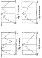
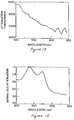
- Figures 1A, 1B, 1C and 1D are graphical illustrations of emission spectra resulting from a 310nm excitation wavelength of normal intima (1A), fibrous plaque (1B), fatty plaque (1C) and calcified plaque (1D) where the typical pulse energy is 150nJ.
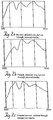
- Figures 2A, 2B and 2C are graphical illustrations of emission spectra of ablation damaged tissue excited at a 305nm wavelength.
- Figure 2A shows the emission spectrum of normal tissue after ablation into but not through the tissue.
- Figure 2B shows the emission spectrum after ablation into, but not through fatty plaque.
- Figure 2C shows the emission spectrum of tissue after ablation through plaque into normal tissue.
- Figures 3A, 3B, 3C and 3D illustrate a probe devices and the optical system used in performing the method of the present invention.
- Figure 4 graphically illustrates excitation and emission profiles of tryptophan (10 ⁇ 5M)(owing), S. epidermidis (-----), and E. coli (-----).
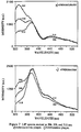
- Figures 5A, 5B and 5C are graphical illustrations of spectra showing the UV absorption of pyridinoline in 0.1N HCl (- ⁇ - ⁇ -), in 0.1M potassium phosphate buffer, pH 7.4 (-----), and 0.1N NaOH (-----) (Figure 5A); the activation spectrum in 0.02M potassium phosphate buffer, pH 7.4 with fluorescence at 400nm ( Figure 5B); and the fluorescence spectrum in 0.02M potassium phosphate buffer, pH 7.4, with activation at 325nm ( Figure 5C).
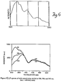
- Figure 6 is a graphical illustration of the emission spectrum of normal aortic tissue taken with a fiber-optic device with excitation at 305nm.
- Figures 7A-7E are graphical illustrations of the fluorescence emission intensity plotted versus wavelength for the these different excitation wavelengths 306nm, 308nm, and 310nm for A) bulk normal aorta, B) atherosclerotic plaque, C) atheromatous plaque, D) nonulcerated calcified plaque, and E) ulcerated calcified plaque.
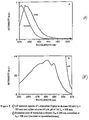
- Figures 8A and 8B are schematic graphical illustrations plotting intensity (arbitrary units) versus wavelength of an emission scan (8A) with excitation at 308nm, and an excitation scan (8B) of L-tryptophan in tris buffer with the emission peak at 350nm.
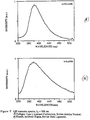
- Figures 9A and 9B are graphical illustrations of emission spectra at 308nm excitation for A) collagen with a peak at 383nm and B) elastin powder with a peak at 379nm.
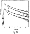
- Figure 10 is a graphical illustration plotting fluorescence decay curves of human aorta at (a) 305nm excitation and 440nm emission, (b) 305nm excitation and 350nm emission, (c) 310nm excitation and 440nm emission and (d) 310nm excitation and 350nm emission. The curves have been offset relative to one another.
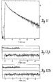
- Figure 11 is a graphical illustration of a fit of the fluorescence decay of human aorta at 305nm excitation and 350nm emission.
- the residuals for three and four exponentials are similar, however, the autocorrelation for three exponentials shows a long range deviation, indicating a poor fit, while the autocorrelation for four exponentials shows no such deviation, indicating a good fit.
- Figure 13 is a graphical illustration of the emission spectrum (a) of bulk normal aorta excited at 476 nm and the product ⁇ SP (476) F SP (476) dz(b).
- Figure 14 graphically illustrates the attenuation lineshape ⁇ SP (476) A SP (476, ⁇ m ).
- Figure 15 graphically illustrates an average plot of several curves showing the normalized hemoglobin attenuation lineshape.
- Figure 16 is a graphical illustration of the normalized ceroid fluorescence lineshape measured from the bulk necrotic core of several fatty plaque specimens.
- Figure 17 graphically illustrates the correlation of actual fluorescence lineshapes for normal (0), calcified atheromatous (5) and atheroschrotic (2) sample with fluorescence lineshapes estimated by the disclosed procedure.
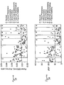
- Figure 18 is a scatter plot for an empirical procedure.
- Figure 19 is a scatter plot of ⁇ SP for the measured samples.
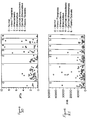
- Figure 20 is a scatter plot of ⁇ ' Ce for the measured samples.
- Figure 21 is a scatter plot of ⁇ Hb for the measured samples.
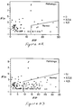
- Figure 22 is a graph of ⁇ ' Ce versus ⁇ SP for all classifications except intimal fibroplasia.
- Figure 23 is a graph of ⁇ ' Ce versus ⁇ SP for all classifications including intimal fibroplasia samples.
- Figure 24 is a graphical illustration of truncated normal distributions of ⁇ HSPY.
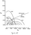
- Figure 25 is a representation of the truncated normal probability distributions of the binary classification scheme of ⁇ ' Ce versus ⁇ SP shown as a contour plot.
- the excitation source for Laser Induced Fluorescence (LIF)in a preferred embodiment of the invention was a dye laser pumped by a 10 Hertz Nd:YAG laser and frequency doubled. Gated integration minimized background noise by taking advantage of the ten nsec duration pulse. The results demonstrate that the tissue LIF has several spectral features which vary with tissue type and condition.
- the spectra taken in bulk tissue indicate that normal aorta can be distinguished from plaque, and that different types of plaque (fibrous, fatty and calcified) also exhibit different emission spectra (Fig. 1).
- the tissue tolerance to the low power diagnostic ultraviolet light is good. Even after substantial exposure, distinctive spectral features remain even though there is a significant loss of fluorescence energy.
- the remaining aortic plaque can be distinguished from damaged normal media (Fig. 2).
- the excitation wavelength is changed from 310nm to 305nm in this particular example.
- Spectral features are very sensitive to excitation wavelength; changing from 305nm to 310nm makes a substantial difference in the emission lineshape. Tryptophan fluoresces strongly with 285nm excitation and dominates the spectra, but the long wavelength weak absorption "tail” occurs in the 305-310nm range, so that a careful selection of the excitation wavelength will act as a chromophore "dimmer switch" and control the size of the tryptophan emission peak at about 340nm.
- Figure 7A displays three emission spectra, excited at 306, 308 and 310nm, from a typical sample of normal aorta.
- the emission lineshape is extremely sensitive to the excitation wavelength, as a very small change in wavelength results in a quite different spectrum. These spectra illustrate the sharpness of the transition between the short wavelength and long wavelength bands.
- the large emission peak at 340 nm dominates the spectrum.
- a minor shoulder at 380 nm and a valley at 418 nm are also present.
- the 340 nm peak disappears, leaving a peak at 380 nm, which was apparently hidden under the larger 340 nm emission band at 306nm excitation.
- the lineshape at wavelengths longer than 380nm remains relatively unchanged.
- the emission scan at 308nm has a lineshape intermediate to the 306nm and 310nm spectra, and can be regarded as the transitional point at which the short and long wavelength bands contribute in roughly equal amounts to the total emission.
- the fluorescence signal is a simple sum of the fluorescence contributions of the individual components.
- the effects of fluorescence reabsorption can produce profound alterations in the emission lineshapes of these components. These effects must be understood and separated from the intrinsic fluorescence of the components in order to accurately identify the active species.
- Figures 7B-7E display LIF spectra excited at 306, 308, and 310 nm of typical samples of an atherosclerotic plaque, an atheromatous plaque, a nonulcerated calcified plaque, and an ulcerated calcified plaque. Similar to normal aorta, the fluorescence lineshapes of each can be divided into a short and a long wavelength band. The short wavelength band is assigned to tryptophan, and the long wavelength band to collagen/elastin using equivalent arguments as those employed for normal aorta. The differences in lineshapes between the atherosclerotic lesions and normal are suggested below to be a result of a change in the relative concentrations of these components. Additional, distinct fluorophores in the atherosclerotic specimens are not observed at these excitation wavelengths.
- the LIF lineshapes of each type of atherosclerotic lesion are quite distinct, and certain spectral features of each are worth noting.
- the intensity of the short wavelength band relative to the long band is slightly higher in the atherosclerotic plaque (Figure 7B) than in normal aorta ( Figure 7A).
- this intensity ratio is largest in the atherosclerotic plaque, smaller in the atheromatous and nonulcerated calcified plaques, and smallest in the ulcerated calcified plaque.
- the atherosclerotic plaque spectrum also shows a reduced 440-540nm fluorescence tail compared with normal, although this reduction is not as great as in the case of atheromatous and nonulcerated calcified plaques.
- the 440-540nm tail in the spectra of the calcified plaque shows no such reduction as compared with normal.
- the fluorescence peak at 340nm is attributed to the aromatic amino acid tryptophan, a known UV fluorophore. Tryptophan in aqueous solution shows a 60nm FWHM fluorescence band peaking at 348nm when excited in the ultraviolet. The peak position is somewhat sensitive to environment; for instance, tryptophan powder peaks at 330nm when excited with 308nm. Tryptophan emission ( Figure 8A) peaks between 330 and 350 nm, depending on its local environment. In the buffered saline, it peaks at 350 nm, while in 1,4 dioxane (a hydrophobic solvent), it peaks at 329 nm.
- the short wavelength band in optically thin normal aorta spectra peaks around 332 nm, so it appears that tryptophan is located in a more hydrophobic environment in the tissue.
- tryptophan emission from the common intracellular protein actin in aqueous solution (pH7) has a high fluorescence quantum yield (0.24) and a peak emission wavelength of 332 nm.
- the assignment of the short wavelength band to tryptophan has been confirmed by more extensive fluorescence excitation-emission matrix studies of human aorta.
- the excitation spectrum of tryptophan is shown in Figure 8B.
- the appearance of a peak in an emission spectrum is thus associated with a distinct fluorophore having its maximum emission at that wavelength.
- the 450nm peak in the present spectra is the result of the singly-peaked broadband fluorescence at 387nm, distinct from the fluorescence at 340nm, modulated by reabsorption near 417nm. It has been demonstrated that the valleys in their visible LIF spectra of arterial wall above 500nm are due to reabsorption by oxy-hemoglobin or other porphyrin type absorbers.
- the Soret absorption band of oxy-hemoglobin occurs at 415nm, matching the wavelength of the valley in our spectra.
- the long wavelength band, peaking at 380nm, is a combination of collagen and elastin, each of which makes up 20-30% of the dry weight of normal intima.
- the emission lineshapes of collagen and elastin are similar, with the peak of collagen at 383nm and that of elastin at 379nm (See Figure 9). In addition, both are relatively insensitive to excitation wavelength. However, their emission lineshapes do have an important difference: the collagen bandshape is significantly narrower than the elastin bandshape. For example, the fluorescence intensity of elastin at 480 nm as compared to its peak is 0.3, while that of collagen is 0.1. This distinction allows collagen and elastin to be spectroscopically separated.
- the excitation wavelength is on the short wavelength absorption maximum of the collagen peak, so the collagen intensity can vary in the opposite direction of tryptophan, and more slowly. Therefore, excitation wavelength can be carefully selected so that tryptophan emission is relatively strong in tissue which contains it, such as normal media; and is relatively weak in tissue with low tryptophan content, such as fatty or calcified plaque.
- pyridinoline is a fluorescent compound found in elastin and collagen (See the absorption spectra of pyridmoline in Fig. 5A) and contributes to the 400nm signal which is strongest for tissue containing elastin.
- correlating observed LIF spectra to tissue composition is used when excitation wavelengths can be selected to enhance differences based on known differences in chemical make-up of different types of tissue.
- the argon ion laser tissue ablation substantially reduces the tryptophan contribution, and it appears that heating degrades this chromophore. This is observed as a substantial decrease in fluorescence yield with 280nm excitation. To compensate for this, the excitation wavelength is shifted to 305nm for studying ablation-damaged tissue (See Fig. 2). The emission signal from the small amount of tryptophan remaining is enhanced, by using a shorter wavelength, and ablation-damaged plaque and media can be distinguished.
- Spectra were recorded using a fiber-optic device (See the more detailed description of the collection system below).
- An epoxy plug containing a 200 micron core silica fiber was inserted into a 3mm silica test tube.
- a central fiber illuminated the tissue and the nine collector ring fibers had the proximal ends placed at the monochromator slit.
- One collection fiber can be separately connected to a photodiode to monitor laser pulse energy impinging on the tissue, which can be ratioed, or if coupling optics are used, the scattered laser light can be split off near the monochromator.
- Laser catheter devices used in connection with the present invention have been more fully described in issued U.S. Patent No. 4,913,142 having a priority date of March 22, 1985.
- FIG. 3B A preferred embodiment of a probe used for laser induced fluorescence of tissue is shown in Figure 3B in which samples were excited and collected through a fiber optic probe.
- the distal end of this probe consists of a center 200 ⁇ m fused silica core excitation fiber and nine surrounding 100 ⁇ m fused silica core collection fibers arranged in a close-packed circular array.
- a fused silica shield is bonded in front of the polished fiber ends, so that a constant excitation spot diameter (0.65mm) is maintained on the shield output surface and the collection geometry is reproducible and well-defined. With this arrangement the fractional area of overlap of the excitation and collection spots is calculated to be about 0.71.
- the excitation fiber 18 is isolated from the collection fibers 24 and is connected to a fiber-to-fiber excitation coupler 14.
- the nine collection fibers are regrouped into a linear close-packed array and are optically coupled to the monochromator 28.
- Time Decay ultraviolet-excited LIF has been performed with the psec laser system using Time Correlated Photon Counting (TCPC).
- TCPC Time Correlated Photon Counting
- the fluorescence time decay of human aorta excited in the same range referenced above was measured at 350 and 440nm, wavelengths at which the emissions of the two fluorophores are well separated. At 305nm excitation and 350nm emission, the shorter wavelength fluorophore dominates the fluorescence; at 310nm excitation and 440nm emission, the longer wavelength fluorophore prevails.
- Figure 10 shows time decays at 305 and 310nm excitations. Both decays are similar at the two excitations; however, the 350nm emission decays somewhat faster than the emission at 440nm.
- Deconvolution analysis was done by an iterative reconvolution method with parameter adjustment by the Marquardt procedure. The quality of the fit was judged by visual inspection of the autocorrelation function of residuals, the weighted residuals, and by ⁇ 2.
- the measured decays could not be fit with two or three exponentials, as indicated by the drift in the autocorrelation function (Fig. 12A). A minimum of four exponentials was needed to obtain a good fit (Fig. 12B); an attempt to a fifth exponential did not improve the fit for this particular analysis.
- the 350nm emission excited with 305nm decayed with time constants of 0.003(0.73), 0.65(0.14), 2.1(0.070), and 4.9(0.058). (All time constants ⁇ are reported in nanoseconds, and their corresponding normalized amplitudes A appear in parentheses.)
- the 440nm emission with 310nm excitation yielded time constants of 0.071(0.52), 0.61(0.29), 2.95(0.14), 8.6(0.052). With both excitations, the last two time constants are longer at 440nm emission compared with emission at 350nm.
- One possible explanation is energy transfer to the longer wavelength fluorophore.
- the fluorescence at 350nm excited by 305nm is due to tryptophan.
- Tryptophan fluorescence in solution has two exponentially decaying components with time constants of 0.6 and 2.5 ns. Longer decay constants for tryptophan fluorescence have been observed in other environments. The three long time decay constants of 0.65, 2.1 and 4.9 ns are in the correct range for the known values of tryptophan lifetimes in different environments.
- tryptophan is an efficient fluorescing moiety in tissue; in fact it will totally dominate if excited at its absorption peak at 280nm.
- the excitation wavelength about 310nm
- the tryptophan absorption cross-section is much smaller and the emission does not overwhelm much weaker fluorescing chromophores. Therefore, tissues containing relatively more tryptophan (normal vs. plaque) will have more of this efficient chromophore.
- efficient fluorescence means that other competing non-radiative depopulating processes are less effective than for weakly emitting chromophores.
- wavelengths of interest There are other wavelengths of interest: 365nm excites NAD/NADH; are 400nm excites blue-emitting chromophores, and 470nm excites the 500-620nm chromophores. Similar lifetimes for the various "peaks" confirms the hypothesis of a single fluorescing chromophore which is given structure by hemoglobin reabsorption in bulk tissue. Dissimilar lifetimes would indicate more than one overlapping chromophore emission spectra. Non-exponential decays tend to indicate this also, although one chromophore in two different environments can also be bi-exponential. Changing excitation wavelength helps here; as has already been demonstrated for ultraviolet tissue spectroscopy, the chromophore fluorescence is enhanced or inhibited relative to another.
- LIF measurements of human artery wall indicate that at least two distinct fluorophores are being excited in aorta in the 305 to 310nm region.
- the structural proteins collagen and elastin, via similar cross-linking agents, are probably responsible in large part for the longer wavelength fluorescence.
- the shorter wavelength fluorescence is attributed to tryptophan.
- the present procedure establishes the ability to spectroscopically observe two or more fluorophores and determine relative contributions to the spectrum.
- the spectral lineshape is very sensitive to excitation wavelengths near the absorption edge. For example, tryptophan shows such sensitivity between 305 and 310nm, the relative contributions of each fluorophore to the overall spectrum can be precisely controlled. Either fluorophore can be emphasized to aid in diagnosis by simply tuning the excitation wavelength a few nanometers; in addition, two or more fluorophores can be simultaneously observed by an appropriate choice of excitation wavelength and the ratio of emitted peaks can be adjusted for optimal comparison. This control is very important in identifying the chromophores and, hence, diagnosing the tissue type and condition for all kinds of bodily tissue. Differences in relative concentrations of the molecules, for instance in normal and diseased arteries, can be spectroscopically monitored; in addition, such relative measurements are both easier to perform and less sensitive to uncertainties in collection efficiency compared to absolute measurements.
- LIF spectral signatures of arterial tissues have been observed using ultraviolet excitation.
- Excitation wavelength is critical to the spectral signatures, and selection of the wavelength has a basis in knowledge of the composition of the tissue.
- Spectral emission can therefore be tailored by selecting the excitation wavelength to enhance differences.
- Individual tissue components and localized laser damage may be studied microscopically.
- Spectral "tours" of the thin sections can help determine the individual contributions to the bulk tissue spectra which will help generate a physical and chemical basis for understanding the LIF spectral fingerprint of bulk tissue.
- Photochemically induced changes in spectra reflect changes in the oxidation state of native compounds. Picosecond time resolved measurements will aid in this understanding. This is of general applicability and should be readily adapted for studying cancerous and other diseased and healthy tissue.
- the following relates to a general method of extracting clinical information about tissue type, degree of disease, and physicochemical composition from tissue fluorescence spectra, utilizing a general model of tissue fluorescence.
- Our approach which is based on a single-layer method analyzing of tissue fluorescence, includes contributions from intrinsic fluorescence, scattering and absorption from individual tissue components.
- this physicochemical information is intimately related to the pathologic state of the tissue, the method should provide more precise diagnostic criteria than empirical methods.
- the 476nm laser induced fluorescence (LIF) spectra of human artery wall of 148 samples from 30 autopsy sources has been analyzed.
- the utility of empirical methods and the present method based on a general diagnostic is compared.
- the extracted parameters are procedure correlated to the chemical composition of the tissue. The results demonstrate that the method characterizes the chemical composition of the tissue, and can be used to classify the pathology of the tissue.
- the one layer method of tissue fluorescence is presented and applied to human artery wall.
- the apparatus used to obtain tissue LIF spectra has been presented earlier.
- Application of the method to different arterial tissue types is demonstrated and the results are summarized.
- the extracted parameters are correlated to the chemical composition of artery and the use of the method for diagnosing different stages of atherosclerosis in terms of sensitivity, specificity and predictive value are presented.
- tissue is represented as a single layer, which is infinitely thick with respect to the penetration depth of the irradiating light. This proposition is most appropriate for excitation wavelengths for which 1/e of the penetration depth is less than the thickness of the top layer.
- light is attenuated exponentially in tissue, due to absorption and scattering. In describing this attenuation, scattering and absorption are not treated separately but are considered together as total attenuation.
- chromophores are distributed homogeneously in the tissue and the excitation beam is assumed to be uniform in profile and infinitely wide.
- P( ⁇ x ) denotes the incident excitation power
- ⁇ t ( ⁇ ) and ⁇ a ( ⁇ ), in units of length ⁇ 1 are the total attenuation and absorption coefficients of the tissue, respectively
- ⁇ ( ⁇ x , ⁇ m ) is the quantum yield of fluorescence, defined as the total fluorescence energy emitted at ⁇ m over the total absorbed energy at ⁇ x
- z is the distance from the irradiated surface.
- Equation (1) has a simple physical interpretation.
- Term A represents the attenuation of the incident radiation;
- term B represents the conversion of this light into backward directed fluorescence; and
- term C represents the attenuation of this net backward directed fluorescence.
- the net backward directed fluorescence is half that of the total net fluorescence, resulting in the factor of 2 in term B.
- the quantities ⁇ a ⁇ and ⁇ t contain contributions from all of the individual tissue chromophores. It is this information which requires separation. Thus, a more useful way to write Eq.
- the luminal layer, intima is comprised of an endothelial layer, a few collagen fibers and some fibroblasts.
- the second layer, media is made up mainly of elastin and some smooth muscle cells.
- the outer layer, adventitia is comprised of loose connective tissue.
- intimal fibroplasia is characterized initially by intimal proliferation of smooth muscle cells and fibroblasts.
- Table 1 Category Tissue Type Intimal Morphology 0 Normal Endothelial layer Some collagen Some fibroblasts 1 Intimal Fibroplasia Smooth muscle cell proliferation Collagen accumulation 2 Atherosclerotic Collagen proliferation Invasion of lipids and foam cells 3 Atheromatous Collagen proliferation Formation of necrotic core 4 Calcified Atherosclerotic Atherosclerotic + Calcifications 5 Calcified Atheromatous Atheromatous + Calcifications 6 Fibrotic/Sclerotic Dense collagen proliferation Loss of lipids intimal fibroplasia, collagen accumulates.
- Atherosclerotic plaque (2) is indicated when the intima is infiltrated by lipids and foam cells. When the center of the plaque becomes necrotic, the plaque is characterized as atheromatous (3). Both atherosclerotic and atheromatous plaques can develop significant calcifications, and are then classified as calcified atherosclerotic (4) and calcified atheromatous (5) plaques respectively. Fibrotic/sclerotic lesions (6) are end stage plaques, characterized by intimal sclerosis with loss of lipid and foam cells.
- ceroid In normal tissue, fluorophores in or associated with the structural proteins (SP), collagen and elastin, are important. Fluorescence contributions from collagen and elastin are not treated separately below, as these compounds have identical fluorescence lineshapes with 476 nm excitation. In atherosclerotic tissue, ceroid (Ce) contributes to the fluorescence in addition to the structural proteins, as detailed in Table 2. Ceroid is a morphologic term used to denote substances which (i) stain like a lipid, (ii) are not dissolved by usual lipid solvents, and (iii) fluoresce in the visible when excited by UV light. Ceroid is a complex of protein associated with oxidized lipids and its exact identity is unknown.
- hemoglobin (Hb) is an important attenuator in normal and atherosclerotic tissues.
- ceroid attenuation can be neglected for structural protein fluorescence, they are important for ceroid fluorescence. These effects in an approximate way.
- the ceroid attenuation terms in the denominator of the second term in eq. (5) are neglected, but the ceroid fluorescence term in the numerator is replaced with an effective ceroid fluorescence lineshape. This lineshape is approximated as the intrinsic fluorescence lineshape divided by a normalized ceroid attenuation lineshape.
- ceroid fluorescence lineshape As discussed above, the modulation of ceroid fluorescence by ceroid attenuation is included by dividing the ceroid fluorescence by a normalized ceroid attenuation lineshape. This represents a relaxation of the assumption that ceroid is distributed homogeneously, in order to better reflect the physical situation. This approximation allows the ceroid fluorescence lineshape to be extracted from bulk samples (samples thick with respect to the penetration depth of the excitation light), eliminating difficulties which would be associated with otaining an unattenuated ceroid fluorescence lineshape.
- ⁇ Max and ⁇ tMax are the maxiumum yield and attenuation coefficient over the emission wave length range, respectively.
- F represents a normalized intrinsic fluorescence lineshape and A represents a normalized round trip attenuation lineshape.
- ⁇ is proportional to the chromophore concentration
- ⁇ and x are proportional to the chromophore concentration
- ⁇ is proportional to the quantum yield at the peak emission wavelength.
- ⁇ if replaced by ⁇ '; ⁇ ' represents an effective quantum yield for the net ceroid fluorescence which has been modulated by ceroid absorption.
- ⁇ ' Ce is proportional to the ceroid concentration and its maximum effective quantum yield.
- F Ce is the normalized fluorescence lineshape from a sample thick compared to the penetration depth of 476 nm radiation.
- Eq. (7) is the key equation to understanding the fluorescence from artery wall. It shows that the fluorescence from both the structural proteins and ceroid are modulated by attenuation of the structural proteins and hemoglobin. Ceroid fluorescence is also modulated by ceroid attenuation. In the following description the lineshapes needed to utilize Eq. (7) are extracted using fluorescence spectra of optically thick tissues.
- FIGS 3A-3D the apparatus used to collect the tissue fluorescence spectra of Figures 13-17 is illustrated in Figures 3A-3D and consists of an argon ion laser 10, an optical fiber laser catheter or probe 20, and a scanning monochromator 28 with a photomultiplier tube 30.
- an optical fiber laser catheter used to collect data contains nine optical fibers 24 encased in a transparent quartz shield 45 the fibers 24 concentric around a central excitation fiber 18, coupled to the laser, which in this embodiment provides a 1 mm diameter spot at the surface of the shield.
- the collection fibers 24, surrounding the central fiber 18 are arranged to view only the tissue illuminated by the central fiber, and are used to collect tissue fluorescence.
- FIG. 3B shows the distal end 46 of the probe where the distal ends 50,48 of the excitation and collection fibers respectively are concentrically arranged.
- Figures 3C and 3D show an alternative embodiment using a larger number of fibers 52 concentric around a central lumen 58 and coupled to an optical shield 56 having a distal surface 60 on which overlapping spots of light 62 are formed.
- the data from the photomultiplier tube 30 can be averaged with the use of signals from the ratioing photodiode 16 and subsequently processed 34 and displayed 36.
- the well defined collection geometry of this system represents a substantial improvement over that of typical spectrofluorimeters, in which recorded signals are significantly distorted by reabsorption.
- an excitation intensity of 100 ⁇ W/mm2 was used in a 1.0 mm diameter spot.
- Data collection time was 40 seconds
- Spectral resolution of the detector was 6 nm FWHM, and a time constant of 250 ms was used to record all data. Data presented has been corrected for the non-uniform spectral response of this system.
- tissue studied was human cadaveric aorta obtained at autopsy within 24 hours of expiration. Samples were snap-frozen in liquid nitrogen and isopentane and then stored at -70°C until use. Spectra were obtained from samples kept moist using a buffered, isotonic saline solution, pH 7.4, at room temperature. Tissue type was determined histologically according to Table 1.
- the composite spectrum will arise from two (or conceivably three) layers; a one layer model will then describe the tissue in terms of some average of its optical properties.
- the extension to more than one layer is straightforward.
- Flavins which have broad absorption at 450nm and broad emission at 450nm can also contribute to the fluorescence.
- the chromophores also contribute to the composite tissue fluorescence spectrum through reabsorption.
- a heme containing compound, believed to be oxy-hemoglobin, is also present mainly in the intima of normal aorta, but is found in lesser concentrations in media and atherosclerotic intima as well.
- the 540 and 580 nm absorption peaks of oxy-hemoglobin produce the valleys at 540 and 580 nm in the composite tissue fluorescence spectra.
- the first system utilized a single optically thick section of media, blunt dissected from a sample of normal aorta.
- a fluorescence spectrum of the bulk tissue was recorded, yielding S(476 ⁇ m ), and is shown in Fig. 13.
- the 476 nm excited fluorescence lineshape of media is found to be identical to that of powdered bovine collagen and elastin.
- the fluorescence spectrum of this section of media did not exhibit valleys at 540 and 580 nm, which have been attributed to the presence of hemoglobin.
- the important chromophores in this system are limited to structural proteins, but the use of the present procedures is not limited to these proteins. Eq.
- the next step was to obtain the hemoglobin attenuation lineshape, x Hb (476)A Hb (476, ⁇ m ). It has been shown that the hemoglobin content of tissue can be increased by exposing the tissue to hemolysed blood. We exploited this fact to extract the hemoglobin attenuation lineshape. The same thick section of media used above was exposed to hemolysed blood and a fluorescence spectrum of the bulk tissue was recorded. As hemoglobin is an important chromophore, Eq.
- F Ce The only remaining lineshape in Eq. (7) is F Ce .
- F Ce was measured directly from bulk necrotic core which was blunt dissected from specimens of fatty plaque. The localization of ceroid in the necrotic core permits this approach. The average lineshape from several samples is shown in Fig. 16.
- the experimental LIF spectra from bulk tissue can now be modeled.
- the parameters in the model are ⁇ SP to vary while holding x SP constant, as ⁇ SP depends on the quantum yields, absorption coefficients and concentrations of collagen and elastin, while x SP depends on the attenuation coefficients and concentrations.
- ⁇ SP was significantly higher than that for pathologic tissues, (2,3), (4,5), with p ⁇ .01. ⁇ ' Ce for calcified tissues, (4,5), was significantly higher than that of other tissues: (0,1), with p ⁇ .01 (2,3), with p ⁇ .10. Interestingly, the values of ⁇ SP for (0) and (1) were not found to be significantly different, while the values of ⁇ ' Ce were (p ⁇ .10).
- the information obtained is a combination of intrinsic fluorescence and attenuation.
- a low value of absolute intensity can be due to either reduced intrinsic fluorescence or to increased attenuation.
- the changing attenuation is due to the presence or absence of hemoglobin, as represented by the parameter x Hb .
- x Hb is higher for normal tissues.
- the usefulness of extracting x Hb lies in the ability to separate the effects of fluorescence and attenuation.
- in vivo measurements of arterial fluorescence have demonstrated that in vitro tissue may contain different concentrations of hemoglobin.
- in vivo measurements indicate that hemoglobin is not an important attenuator in normal tissue but is important in calcified plaques.
- a non-zero ⁇ ' Ce indicates the presence of ceroid.
- Ceroid has been correlated with the presence of extensive atherosclerosis.
- Figure 20 and Table 3 show the data are consistant with this: the average value of ⁇ ' Ce is higher for pathologic specimens and appears to increase with the severity of the disease.
- Calcified plaques were associated with a particularly high value of ⁇ ' Ce . This is consistent with observations made with a fluorescence microscope indicating ceroid deposits are often localized adjacent to calcified areas of tissue. In fibrotic/sclerotic specimens, the amount of ceroid is negligible, reflecting the pathology of this tissue type. Detecting the presence of sub-surface ceroid fluorescence utilizing surface excitation is an important feature. The method provides a means of assessing ceroid content of tissues in vivo and in vitro .
- ⁇ SP is complicated by the fact that ⁇ SP contains contributions from both collagen and elastin. Results indicate that the quantum efficiency of elastin is higher than that of collagen with 476 nm excitation, although the band shapes are identical. Thus, a high value of ⁇ SP indicates a high ratio of elastin concentration to collagen concentration. Since the media consists mainly of elastin, and the intima mainly collagen, the effective ratio of these concentrations, reflected by our one-layer value of ⁇ SP , decreases as the intimal thickness increases. As shown in Table 3, ⁇ SP is highest for normal tissue, is slightly lower for intimal fibroplasia, and is significantly lower for pathologic specimens.
- the method parameters contain chemical information about tissue composition which is related to the presence of pathology in the tissue.
- the challenge in obtaining a clinical diagnostic is to find criteria which separate normal and pathologic tissue, despite the wide variance in the distributions of these parameters.
- intimal fibroplasia is considered 'non-pathologic'. In the context of atherosclerosis, this classification is justified, since in the aorta intimal fibroplasia is rarely hemodynamically significant.
- the ⁇ SP algorithm should be compared to a simple empirical diagnostic utilizing the intensity of 520 nm (Fig. 19). This empirical procedure is chosen because this wavelength corresponds to the fluorescence maximum observed for collagen and elastin, and thus should be related to ⁇ SP .
- sensitivity no. of true positives/total no. of diseased samples
- specificity no. of true negatives/total no. of normal samples
- predictive value no. of true positives/total no. of positives
- positives refer to those samples diagnosed as having disease. True positives denote positives actually displaying disease, whereas, true negatives denote negatives which are actually normal.
- the sensitivity measures the ability of the procedure to detect disease, while the specificity determines its ability to detect normal samples.
- the sensitivity, specificity and predictive value of the empirical procedure in Fig. 18 are 69%, 85% and 82%, respectively, which improved to 84%, 85% and 84% for the procedure shown for ⁇ SP in Fig. 19.
- the improvement in sensitivity reflects the ability of the ⁇ SP procedure to diagnose calcified samples.
- Fig. 22 shows a plot of ⁇ ' Ce vs ⁇ SP for all classifications except intimal fibroplasia.
- Fig. 23 shows a plot of ⁇ ' Ce vs ⁇ SP which includes the intimal fibroplasia samples.
- the divisions in this plot indicate a different decision surface for classification of tissue type than that in Fig. 22.
- This surface correctly diagnoses 88% of the samples.
- the sensitivity of the scheme is 91%, while its specificity is 85%.
- This decision surface has a predictive value of 85%.
- the poorer performance of the binary scheme when intimal fibroplasia is included reflects the fact that intimal fibroplasia is an initial step in the formation of atherosclerosis. Thus, this tissue type is more difficult to differentiate from samples displaying well developed atherosclerotic plaques.
- Figure 24 shows truncated normal distributions for ⁇ SP . These distributions were not greatly different from histograms of the data given in Fig. 18, and can therefore be considered reasonable approximations. Using the same decision vector as in Fig. 18, the sensitivity, specificity and predictive value were assessed using the area under the distribution. These quantities were found to be 79%, 82% and 90%, respectively, close to those values calculated from Fig. 18.
- FIG. 25 A representation of the truncated normal probability distributions for the binary classification scheme ⁇ ' Ce vs ⁇ SP is shown in Fig. 25 as a contour plot.
- the decision surface is that of Fig. 23. Similar to the method used for the single truncated normal distribution for ⁇ SP , the sensitivity, specificity and predictive value were found to be 82%, and 88%.
- Figs. 24 and 25 provide the values for sensitivity, specificity and predictive value.
- the above presents a method for analyzing fluorescence spectra of human tissues with the goal of diagnosing the presence of pathology.
- the method of tissue fluorescence was developed, and the steps necessary for applying this method to a specific type of tissue were described. It has been established that analysis of tissue fluorescence data within the framework of the method yields two related types of information, the chemical composition of the tissue and the tissue type.
- the method presented here allows for separation of normal and atherosclerotic tissues. However, the method can be used to separate tissues of each classification (Table 1). Another embodiment of the current method extends to two layers which allow extraction of an additional parameter, the thickness of the first layer, which improves the ability to separate tissue types.
- the model can also be extended to include the layered structure of tissue and additional chromophores.
- independent measures of tissue fluorescence efficiency and oxy-hemoglobin concentration can be obtained in order to correlate these values to those calculated in the model.
Landscapes
- Health & Medical Sciences (AREA)
- Life Sciences & Earth Sciences (AREA)
- Physics & Mathematics (AREA)
- Surgery (AREA)
- Engineering & Computer Science (AREA)
- Public Health (AREA)
- Biomedical Technology (AREA)
- Heart & Thoracic Surgery (AREA)
- Medical Informatics (AREA)
- Molecular Biology (AREA)
- Veterinary Medicine (AREA)
- Animal Behavior & Ethology (AREA)
- General Health & Medical Sciences (AREA)
- Pathology (AREA)
- Biophysics (AREA)
- Spectroscopy & Molecular Physics (AREA)
- Optics & Photonics (AREA)
- Vascular Medicine (AREA)
- Electromagnetism (AREA)
- Nuclear Medicine, Radiotherapy & Molecular Imaging (AREA)
- Otolaryngology (AREA)
- Investigating, Analyzing Materials By Fluorescence Or Luminescence (AREA)
- Laser Surgery Devices (AREA)
Claims (20)
- Verfahren zur Bestrahlung von Chromophoren in körpereigenem Gewebe, um die Zusammensetzung des Gewebes zu identifizieren, wobei
das körpereigene Gewebe mit Strahlung einer Wellenlänge gleich einer Erregungswellenlänge eines Chromophors bestrahlt wird, bei welcher der Chromophor eine intrinsische Fluoreszenz aufweist;
ein Fluoreszenzemissionsspektrum des Gewebes bestimmt wird, das sich durch die Bestrahlung ergibt und dadurch gekennzeichnet wird;
Zerlegen des Fluoreszenzemissionsspektrums in eine Vielzahl von einzelnen Spektralanteilen durch Vergleichen des Emissionsspektrums mit einer Vielzahl von Bezugskurven und Bestimmung, welche der Bezugskurven am Emissionsspektrum beteiligt sind, wobei die beteiligten Kurven jeweils einen der Spektralanteile bestimmt;
und Analyse der Spektralanteile, um das Vorhandensein des Chromophors in dem Gewebe zu bestimmen. - Verfahren zur Bestrahlung von Chromophoren gemäß Anspruch 1, bei welchem jede Bezugskurve jeweils ein erfaßtes, intrinsisches Fluoreszenzspektrum darstellt, welches von einem Abschwächungsspektrum moduliert wird.
- Verfahren zur Bestrahlung von Chromophoren gemäß Anspruch 1, welches weiterhin die folgenden Schritte umfaßt:
Auswahl einer zweiten bekannten Erregungswellenlänge eines zweiten Chromophors, bei welcher der zweite Chromophor Fluoreszenz aufweist;
gleichzeitige Bestrahlung des körpereigenen Gewebes mit einer Strahlung der zweiten Wellenlänge;
Beobachtung einer zweiten Fluoreszenzemission des Gewebes, welche von der Bestrahlung innerhalb der zweiten Wellenlänge herrührt; und
Analyse des beobachteten Spektrums, um das Vorhandensein des zweiten Chromophors in dem Gewebe zu bestimmen. - Verfahren gemäß Anspruch 3, bei welchem außerdem das Gewebe bei weiteren ausgewählten Wellenlängen beleuchtet wird, um Fluoreszenz von weiteren Chromophoren herbeizuführen und weitere pathologische Merkmale des Gewebes zu bestimmen.
- Verfahren zur Bestrahlung von Chromophoren gemäß Anspruch 3, bei welchem außerdem die Wellenlänge der Peakfluoreszenz des ersten und zweiten Chromophors in der ersten und zweiten Emission bestimmt wird.
- Verfahren gemäß Anspruch 5, bei welchem außerdem das Verhältnis der beiden Fluoreszenzpeaks bestimmt wird.
- Verfahren zur Bestrahlung von Chromophoren gemäß Anspruch 1, wobeia) außerdem die Wellenlänge der Peakfluoreszenz des Chromophors in dem Emissionsspektrum bestimmt wird und das Verhältnis des Peaks zu einem ausgewählten Tal in der Emission bestimmt wird; oderb) in dem Analysevorgang ein Peakabsorptionsspektrum von dem Fluoreszenzemissionspektrum abgetrennt wird; oderc) außerdem die Konzentration des Chromophors in dem Gewebe bestimmt wird.
- Verfahren zur Bestrahlung von Chromophoren gemäß Anspruch 1, bei welchem das Chromophor(a) Tryptophan in arteriellem Gewebe,(b) Kollagen oder(c) Elastinenthält oder daraus besteht.
- Verfahren zur Bestrahlung von Chromophoren gemäß Anspruch 1, bei welchem außerdem die Konzentration des Chromophors in dem Gewebe gegenüber der Konzentration des Chromophors in einer Bezugsprobe bestimmt wird.
- Verfahren zur Bestrahlung von Chromophoren gemäß Anspruch 1, bei welchem außerdem das Gewebe mit einem Laser bestrahlt wird.
- Verfahren zur Bestrahlung von Chromophoren gemäß Anspruch 1, bei welchem außerdem die Strahlung mittels einer Faseroptikvorrichtung auf das Gewebe gerichtet wird.
- Ein Verfahren zur Analyse des Gewebes gemäß Anspruch 1, das folgende Schritte umfaßt:
Beleuchten des Gewebes mit Licht einer ersten Wellenlänge, welche die Fluoreszenz eines ersten Chromophors stimuliert, um ein erstes pathologisches Merkmal des Gewebes zu bestimmen; und
Beleuchten des Gewebes mit Licht einer zweiten Wellenlänge, welche die Fluoreszenz eines zweiten Chromophors stimuliert, um ein zweites pathologisches Merkmal des Gewebes zu bestimmen. - Ein Verfahren gemäß einem der vorhergehenden Ansprüche, bei welchem sich das Gewebe im lebenden Menschen befindet.
- Vorrichtung zum Durchführen eines Verfahrens gemäß einem der Ansprüche 1 bis 10, welche folgendes aufweist:
Einrichtung zum Bestrahlen des körpereigenen Gewebes mit einer Strahlung, welche eine bekannte Erregungswellenlänge des Chromophors aufweist;
Einrichtung zum Bestimmen eines Fluoreszenzemissionsspektrums des Gewebes, welches durch die Bestrahlung erzeugt und gekennzeichnet wird;
Einrichtung zum Zerlegen des Fluoreszenzemissionsspektrums in eine Vielzahl von einzelnen Spektralanteilen durch Vergleichen des Emissionsspektrums mit einer Vielzahl von Bezugskurven und Bestimmung, welche der Bezugskurven zum Emissionsspektrum beiträgt, wobei die beteiligten Kurven jeweils einen der Spektralanteile bestimmt, der mit der Fluoreszenzemission des Chromophers korreliert wird; und
Einrichtung zum Analysieren des Spektralanteils, welcher mit der Fluoreszenzemission des Chromophors korreliert, um so das Vorhandensein des Chromophors in dem Gewebe zu bestimmen. - Vorrichtung gemäß Anspruch 14, bei welchem die Einrichtung zur Bestrahlung des Gewebes eine Festkörper-Laser-Erregungsquelle ist.
- Vorrichtung gemäß Anspruch 15, bei welchem der Laser ein Nd/YAG ist, welcher frequenzverdoppelt werden kann.
- Vorrichtung gemäß einem der vorhergehenden Ansprüche 14 bis 16, mit einer Sonde zur intraluminalen Verwendung mit einer zentral angeordneten Erregungsfaser und einer Vielzahl von Empfangsfasern, die die zentral angeordnete Faser umgeben.
- Vorrichtung gemäß Anspruch 17, bei welchem die Fasern der Sonde einander überlappen.
- Vorrichtung gemäß einem der Ansprüche 17 und 18, bei welchem die Sonde einen zentralen Hohlraum aufweist.
- Vorrichtung gemäß einem der vorhergehenden Ansprüche 14 bis 19, bei welchem die Einrichtung zum Analysieren des Spektralanteils einen Datenprozessor aufweist.
Applications Claiming Priority (3)
| Application Number | Priority Date | Filing Date | Title |
|---|---|---|---|
| US28877288A | 1988-12-21 | 1988-12-21 | |
| US288772 | 1988-12-21 | ||
| PCT/US1989/005174 WO1990006718A1 (en) | 1988-12-21 | 1989-11-17 | A method for laser induced fluorescence of tissue |
Publications (2)
| Publication Number | Publication Date |
|---|---|
| EP0449883A1 EP0449883A1 (de) | 1991-10-09 |
| EP0449883B1 true EP0449883B1 (de) | 1996-01-31 |
Family
ID=23108573
Family Applications (1)
| Application Number | Title | Priority Date | Filing Date |
|---|---|---|---|
| EP90900642A Expired - Lifetime EP0449883B1 (de) | 1988-12-21 | 1989-11-17 | Verfahren für laserinduzierte fluoreszenz von gewebe |
Country Status (5)
| Country | Link |
|---|---|
| US (2) | US5419323A (de) |
| EP (1) | EP0449883B1 (de) |
| AT (1) | ATE133545T1 (de) |
| DE (1) | DE68925586T2 (de) |
| WO (1) | WO1990006718A1 (de) |
Families Citing this family (353)
| Publication number | Priority date | Publication date | Assignee | Title |
|---|---|---|---|---|
| EP0573535B1 (de) * | 1991-02-26 | 2000-12-27 | Massachusetts Institute Of Technology | Molekularspektroskopieverfahren und -einrichtungen zur gewebediagnose |
| US6111645A (en) | 1991-04-29 | 2000-08-29 | Massachusetts Institute Of Technology | Grating based phase control optical delay line |
| US6485413B1 (en) | 1991-04-29 | 2002-11-26 | The General Hospital Corporation | Methods and apparatus for forward-directed optical scanning instruments |
| US6564087B1 (en) | 1991-04-29 | 2003-05-13 | Massachusetts Institute Of Technology | Fiber optic needle probes for optical coherence tomography imaging |
| DE4325529C2 (de) * | 1993-07-29 | 1996-11-28 | Pulsion Verwaltungs Gmbh & Co | Vorrichtung zum Bestimmen der Konzentration von Stoffen im Blut |
| US6304771B1 (en) * | 1993-10-29 | 2001-10-16 | The Trustees Of The University Of Pennsylvania | Systems and methods for imaging fluorophores |
| US5697373A (en) * | 1995-03-14 | 1997-12-16 | Board Of Regents, The University Of Texas System | Optical method and apparatus for the diagnosis of cervical precancers using raman and fluorescence spectroscopies |
| US6258576B1 (en) | 1996-06-19 | 2001-07-10 | Board Of Regents, The University Of Texas System | Diagnostic method and apparatus for cervical squamous intraepithelial lesions in vitro and in vivo using fluorescence spectroscopy |
| US5991653A (en) * | 1995-03-14 | 1999-11-23 | Board Of Regents, The University Of Texas System | Near-infrared raman spectroscopy for in vitro and in vivo detection of cervical precancers |
| US5735276A (en) * | 1995-03-21 | 1998-04-07 | Lemelson; Jerome | Method and apparatus for scanning and evaluating matter |
| US5612540A (en) * | 1995-03-31 | 1997-03-18 | Board Of Regents, The University Of Texas Systems | Optical method for the detection of cervical neoplasias using fluorescence spectroscopy |
| FR2737845B1 (fr) * | 1995-08-16 | 1998-01-02 | Centre Nat Rech Scient | Dispositif d'imagerie endoscopique pour la detection precoce de lesions superficielles cancereuses ou precancereuses |
| US6571119B2 (en) | 1996-03-06 | 2003-05-27 | Fuji Photo Film Co., Ltd. | Fluorescence detecting apparatus |
| JP3796635B2 (ja) * | 1996-03-06 | 2006-07-12 | 富士写真フイルム株式会社 | 蛍光検出装置 |
| US5773835A (en) * | 1996-06-07 | 1998-06-30 | Rare Earth Medical, Inc. | Fiber optic spectroscopy |
| US5842995A (en) * | 1996-06-28 | 1998-12-01 | Board Of Regents, The Univerisity Of Texas System | Spectroscopic probe for in vivo measurement of raman signals |
| US6296608B1 (en) * | 1996-07-08 | 2001-10-02 | Boston Scientific Corporation | Diagnosing and performing interventional procedures on tissue in vivo |
| US6119031A (en) | 1996-11-21 | 2000-09-12 | Boston Scientific Corporation | Miniature spectrometer |
| US6826422B1 (en) * | 1997-01-13 | 2004-11-30 | Medispectra, Inc. | Spectral volume microprobe arrays |
| US6847490B1 (en) * | 1997-01-13 | 2005-01-25 | Medispectra, Inc. | Optical probe accessory device for use in vivo diagnostic procedures |
| US6317624B1 (en) | 1997-05-05 | 2001-11-13 | The General Hospital Corporation | Apparatus and method for demarcating tumors |
| US6124597A (en) * | 1997-07-07 | 2000-09-26 | Cedars-Sinai Medical Center | Method and devices for laser induced fluorescence attenuation spectroscopy |
| US5984861A (en) | 1997-09-29 | 1999-11-16 | Boston Scientific Corporation | Endofluorescence imaging module for an endoscope |
| US6096065A (en) * | 1997-09-29 | 2000-08-01 | Boston Scientific Corporation | Sheath for tissue spectroscopy |
| US6324418B1 (en) | 1997-09-29 | 2001-11-27 | Boston Scientific Corporation | Portable tissue spectroscopy apparatus and method |
| US6185443B1 (en) | 1997-09-29 | 2001-02-06 | Boston Scientific Corporation | Visible display for an interventional device |
| US6238348B1 (en) | 1997-07-22 | 2001-05-29 | Scimed Life Systems, Inc. | Miniature spectrometer system and method |
| WO1999013764A1 (fr) * | 1997-09-12 | 1999-03-25 | Communaute Europeenne | Detection et cartographie de zones enflammees de tissus vivants |
| US5980468A (en) * | 1997-09-22 | 1999-11-09 | Zimmon Scientific Corporation | Apparatus and method for serial collection storage and processing of biopsy specimens |
| US6055451A (en) | 1997-12-12 | 2000-04-25 | Spectrx, Inc. | Apparatus and method for determining tissue characteristics |
| US20030135122A1 (en) * | 1997-12-12 | 2003-07-17 | Spectrx, Inc. | Multi-modal optical tissue diagnostic system |
| US6289229B1 (en) | 1998-01-20 | 2001-09-11 | Scimed Life Systems, Inc. | Readable probe array for in vivo use |
| US6174291B1 (en) | 1998-03-09 | 2001-01-16 | Spectrascience, Inc. | Optical biopsy system and methods for tissue diagnosis |
| US6066102A (en) * | 1998-03-09 | 2000-05-23 | Spectrascience, Inc. | Optical biopsy forceps system and method of diagnosing tissue |
| US6462770B1 (en) | 1998-04-20 | 2002-10-08 | Xillix Technologies Corp. | Imaging system with automatic gain control for reflectance and fluorescence endoscopy |
| US6590651B1 (en) | 1998-05-19 | 2003-07-08 | Spectrx, Inc. | Apparatus and method for determining tissue characteristics |
| US6064897A (en) * | 1998-06-01 | 2000-05-16 | Abbott Laboratories | Sensor utilizing Raman spectroscopy for non-invasive monitoring of analytes in biological fluid and method of use |
| US6405070B1 (en) * | 1998-06-16 | 2002-06-11 | Bhaskar Banerjee | Detection of cancer using cellular autofluorescence |
| GB9813179D0 (en) * | 1998-06-19 | 1998-08-19 | Aortech Europ Ltd | Spectrophotometry |
| AU6139199A (en) * | 1998-09-11 | 2000-04-03 | Spectrx, Inc. | Multi-modal optical tissue diagnostic system |
| US6377842B1 (en) | 1998-09-22 | 2002-04-23 | Aurora Optics, Inc. | Method for quantitative measurement of fluorescent and phosphorescent drugs within tissue utilizing a fiber optic probe |
| US6678541B1 (en) | 1998-10-28 | 2004-01-13 | The Governmemt Of The United States Of America | Optical fiber probe and methods for measuring optical properties |
| WO2000037917A2 (en) * | 1998-12-23 | 2000-06-29 | Medispectra, Inc. | Systems and methods for optical examination of samples |
| AU760402B2 (en) | 1998-12-23 | 2003-05-15 | Medispectra, Inc. | Optical methods and systems for cervical screening |
| US6272376B1 (en) | 1999-01-22 | 2001-08-07 | Cedars-Sinai Medical Center | Time-resolved, laser-induced fluorescence for the characterization of organic material |
| AU3855499A (en) * | 1999-02-12 | 2000-08-29 | Mikhail Leonidovich Stakhanov | Method for diagnosing proliferation regions and device for realising the same |
| JP2000262460A (ja) * | 1999-03-18 | 2000-09-26 | Inst Of Physical & Chemical Res | 生体試料の特定部位検出方法、生体試料の生理学的測定方法、生体試料の特定部位検出装置、光ファイバ保持装置 |
| US20040147843A1 (en) * | 1999-11-05 | 2004-07-29 | Shabbir Bambot | System and method for determining tissue characteristics |
| US6697666B1 (en) | 1999-06-22 | 2004-02-24 | Board Of Regents, The University Of Texas System | Apparatus for the characterization of tissue of epithelial lined viscus |
| US6219566B1 (en) | 1999-07-13 | 2001-04-17 | Photonics Research Ontario | Method of measuring concentration of luminescent materials in turbid media |
| US6445939B1 (en) | 1999-08-09 | 2002-09-03 | Lightlab Imaging, Llc | Ultra-small optical probes, imaging optics, and methods for using same |
| US7187810B2 (en) | 1999-12-15 | 2007-03-06 | Medispectra, Inc. | Methods and systems for correcting image misalignment |
| US7260248B2 (en) | 1999-12-15 | 2007-08-21 | Medispectra, Inc. | Image processing using measures of similarity |
| US6902935B2 (en) * | 1999-12-15 | 2005-06-07 | Medispectra, Inc. | Methods of monitoring effects of chemical agents on a sample |
| US6603552B1 (en) * | 1999-12-22 | 2003-08-05 | Xillix Technologies Corp. | Portable system for detecting skin abnormalities based on characteristic autofluorescence |
| EP1259163A4 (de) * | 2000-02-08 | 2006-05-03 | Cornell Res Foundation Inc | Multiphoton-anregung durch optische fasern für fluoreszenzspektroskopie |
| US6749623B1 (en) * | 2000-03-31 | 2004-06-15 | Richard A Hsi | Method and apparatus for catheter phototherapy with dose sensing |
| WO2002007587A2 (en) | 2000-07-14 | 2002-01-31 | Xillix Technologies Corporation | Compact fluorescent endoscopy video system |
| EP1307214A4 (de) * | 2000-08-11 | 2006-01-18 | Invitrogen Corp | Hochhomogene molekulare marker für ektrophorese |
| US20040014082A1 (en) * | 2000-08-11 | 2004-01-22 | Invitrogen Corporation | Highly homogeneous molecular markers for electrophoresis |
| CA2424894A1 (en) | 2000-10-06 | 2002-04-11 | Victor X. D. Yang | Multi-spectral fluorescence imaging and spectroscopy device |
| DE60141090D1 (de) | 2000-10-30 | 2010-03-04 | Gen Hospital Corp | Optische systeme zur gewebeanalyse |
| DE60128138T2 (de) | 2000-11-02 | 2008-01-03 | Cornell Research Foundation, Inc. | In vivo multiphoton diagnostische detektion und bilddarstellung einer neurodegenerativen erkrankung |
| US9295391B1 (en) | 2000-11-10 | 2016-03-29 | The General Hospital Corporation | Spectrally encoded miniature endoscopic imaging probe |
| EP1207387A1 (de) * | 2000-11-20 | 2002-05-22 | Institut Curie | Multiphotonabbildungsvorrichtung |
| US6839661B2 (en) * | 2000-12-15 | 2005-01-04 | Medispectra, Inc. | System for normalizing spectra |
| US6697652B2 (en) | 2001-01-19 | 2004-02-24 | Massachusetts Institute Of Technology | Fluorescence, reflectance and light scattering spectroscopy for measuring tissue |
| US20040038320A1 (en) * | 2001-03-09 | 2004-02-26 | Bhaskar Banerjee | Methods of detecting cancer using cellular autofluorescence |
| US20070276199A1 (en) * | 2002-04-04 | 2007-11-29 | Ediger Marwood N | Determination of a Measure of a Glycation End-Product or Disease State Using Tissue Fluorescence |
| US7139598B2 (en) * | 2002-04-04 | 2006-11-21 | Veralight, Inc. | Determination of a measure of a glycation end-product or disease state using tissue fluorescence |
| EP2333521B1 (de) * | 2001-04-30 | 2019-12-04 | The General Hospital Corporation | Verfahren und vorrichtung zur verbesserung der bildklarheit und empfindlichkeit bei der optischen kohärenz-tomographie unter verwendung von dynamischer rückkopplung zur kontrolle der fokussierungseigenschaften und der kohärenzsteuerung |
| US7865231B2 (en) | 2001-05-01 | 2011-01-04 | The General Hospital Corporation | Method and apparatus for determination of atherosclerotic plaque type by measurement of tissue optical properties |
| IL158532A0 (en) | 2001-05-02 | 2004-05-12 | Purdue Research Foundation | Treatment and diagnosis of macrophage mediated disease |
| JP2005502618A (ja) * | 2001-06-04 | 2005-01-27 | ザ・ジェネラル・ホスピタル・コーポレイション | 光力学的化合物を用いて脆弱なプラークを検出および治療する方法 |
| US6694159B2 (en) * | 2001-07-16 | 2004-02-17 | Art, Advanced Research Technologies Inc. | Choice of wavelengths for multiwavelength optical imaging |
| DE10239028B4 (de) * | 2001-08-21 | 2004-07-29 | Becker & Hickl Gmbh | Verfahren zur Identifizierung von natürlich vorkommenden oder synthetisch hergestellten Melaninsorten |
| US6980299B1 (en) | 2001-10-16 | 2005-12-27 | General Hospital Corporation | Systems and methods for imaging a sample |
| US6723308B2 (en) * | 2001-11-02 | 2004-04-20 | Kenra, Llc | Hair clarifying treatment |
| US20030109787A1 (en) * | 2001-12-12 | 2003-06-12 | Michael Black | Multiple laser diagnostics |
| US20050049582A1 (en) * | 2001-12-12 | 2005-03-03 | Debenedictis Leonard C. | Method and apparatus for fractional photo therapy of skin |
| US20040082940A1 (en) * | 2002-10-22 | 2004-04-29 | Michael Black | Dermatological apparatus and method |
| US7310150B2 (en) * | 2002-01-11 | 2007-12-18 | The General Hospital Corporation | Apparatus and method for low coherence ranging |
| US6899675B2 (en) * | 2002-01-15 | 2005-05-31 | Xillix Technologies Corp. | Fluorescence endoscopy video systems with no moving parts in the camera |
| US20060241496A1 (en) | 2002-01-15 | 2006-10-26 | Xillix Technologies Corp. | Filter for use with imaging endoscopes |
| US7333189B2 (en) * | 2002-01-18 | 2008-02-19 | Pentax Corporation | Spectroscopic diagnostic methods and system |
| US7355716B2 (en) | 2002-01-24 | 2008-04-08 | The General Hospital Corporation | Apparatus and method for ranging and noise reduction of low coherence interferometry LCI and optical coherence tomography OCT signals by parallel detection of spectral bands |
| US8043603B2 (en) | 2002-02-07 | 2011-10-25 | Endocyte, Inc. | Folate targeted enhanced tumor and folate receptor positive tissue optical imaging technology |
| US8043602B2 (en) | 2002-02-07 | 2011-10-25 | Endocyte, Inc. | Folate targeted enhanced tumor and folate receptor positive tissue optical imaging technology |
| DE60325564D1 (de) * | 2002-03-05 | 2009-02-12 | Univ Texas | Biospezifische kontrastmittel |
| AU2003260583A1 (en) * | 2002-04-03 | 2003-10-20 | The Regents Of The University Of California | System and method for quantitative or qualitative measurement of exogenous substances in tissue and other materials using laser-induced fluorescence spectroscopy |
| US8131332B2 (en) * | 2002-04-04 | 2012-03-06 | Veralight, Inc. | Determination of a measure of a glycation end-product or disease state using tissue fluorescence of various sites |
| US8140147B2 (en) * | 2002-04-04 | 2012-03-20 | Veralight, Inc. | Determination of a measure of a glycation end-product or disease state using a flexible probe to determine tissue fluorescence of various sites |
| US7309867B2 (en) | 2003-04-18 | 2007-12-18 | Medispectra, Inc. | Methods and apparatus for characterization of tissue samples |
| US7136518B2 (en) | 2003-04-18 | 2006-11-14 | Medispectra, Inc. | Methods and apparatus for displaying diagnostic data |
| US7469160B2 (en) | 2003-04-18 | 2008-12-23 | Banks Perry S | Methods and apparatus for evaluating image focus |
| US7282723B2 (en) * | 2002-07-09 | 2007-10-16 | Medispectra, Inc. | Methods and apparatus for processing spectral data for use in tissue characterization |
| US7459696B2 (en) | 2003-04-18 | 2008-12-02 | Schomacker Kevin T | Methods and apparatus for calibrating spectral data |
| US6933154B2 (en) * | 2002-07-09 | 2005-08-23 | Medispectra, Inc. | Optimal windows for obtaining optical data for characterization of tissue samples |
| US20040208385A1 (en) * | 2003-04-18 | 2004-10-21 | Medispectra, Inc. | Methods and apparatus for visually enhancing images |
| US6818903B2 (en) | 2002-07-09 | 2004-11-16 | Medispectra, Inc. | Method and apparatus for identifying spectral artifacts |
| US20040209237A1 (en) * | 2003-04-18 | 2004-10-21 | Medispectra, Inc. | Methods and apparatus for characterization of tissue samples |
| US6768918B2 (en) | 2002-07-10 | 2004-07-27 | Medispectra, Inc. | Fluorescent fiberoptic probe for tissue health discrimination and method of use thereof |
| US7103401B2 (en) | 2002-07-10 | 2006-09-05 | Medispectra, Inc. | Colonic polyp discrimination by tissue fluorescence and fiberoptic probe |
| US20040064053A1 (en) * | 2002-09-30 | 2004-04-01 | Chang Sung K. | Diagnostic fluorescence and reflectance |
| US7643153B2 (en) | 2003-01-24 | 2010-01-05 | The General Hospital Corporation | Apparatus and method for ranging and noise reduction of low coherence interferometry LCI and optical coherence tomography OCT signals by parallel detection of spectral bands |
| US7761139B2 (en) * | 2003-01-24 | 2010-07-20 | The General Hospital Corporation | System and method for identifying tissue using low-coherence interferometry |
| US20070265606A1 (en) * | 2003-02-14 | 2007-11-15 | Reliant Technologies, Inc. | Method and Apparatus for Fractional Light-based Treatment of Obstructive Sleep Apnea |
| CA2519937C (en) | 2003-03-31 | 2012-11-20 | Guillermo J. Tearney | Speckle reduction in optical coherence tomography by path length encoded angular compounding |
| JP4680185B2 (ja) | 2003-05-30 | 2011-05-11 | パーデュー・リサーチ・ファウンデーション | アテローム性動脈硬化症の診断法 |
| KR101204370B1 (ko) | 2003-06-06 | 2012-11-23 | 더 제너럴 하스피탈 코포레이션 | 파장 동조 소스용 방법 및 장치 |
| WO2005057244A2 (en) * | 2003-08-19 | 2005-06-23 | Cornell Research Foundation, Inc. | Optical fiber delivery and collection system for biological applications |
| DE202004021954U1 (de) | 2003-09-12 | 2013-06-19 | Vessix Vascular, Inc. | Auswählbare exzentrische Remodellierung und/oder Ablation von atherosklerotischem Material |
| WO2005047813A1 (en) | 2003-10-27 | 2005-05-26 | The General Hospital Corporation | Method and apparatus for performing optical imaging using frequency-domain interferometry |
| EP1687587B1 (de) * | 2003-11-28 | 2020-01-08 | The General Hospital Corporation | Verfahren und vorrichtung für dreidimensionale spektralcodierte bildgebung |
| WO2005117534A2 (en) | 2004-05-29 | 2005-12-15 | The General Hospital Corporation | Process, system and software arrangement for a chromatic dispersion compensation using reflective layers in optical coherence tomography (oct) imaging |
| US7413572B2 (en) * | 2004-06-14 | 2008-08-19 | Reliant Technologies, Inc. | Adaptive control of optical pulses for laser medicine |
| US20060035302A1 (en) * | 2004-06-21 | 2006-02-16 | Applera Corporation | Kinase substrates with multiple phosphorylation sites |
| JP4995720B2 (ja) | 2004-07-02 | 2012-08-08 | ザ ジェネラル ホスピタル コーポレイション | ダブルクラッドファイバを有する内視鏡撮像プローブ |
| JP5053845B2 (ja) | 2004-08-06 | 2012-10-24 | ザ ジェネラル ホスピタル コーポレイション | 光学コヒーレンス断層撮影法を使用して試料中の少なくとも1つの位置を決定するための方法、システムおよびソフトウェア装置 |
| JP5324095B2 (ja) | 2004-08-24 | 2013-10-23 | ザ ジェネラル ホスピタル コーポレイション | 血管セグメントを画像化する方法および装置 |
| ATE538714T1 (de) | 2004-08-24 | 2012-01-15 | Gen Hospital Corp | Verfahren, system und software-anordnung zur bestimmung des elastizitätsmoduls |
| KR101269455B1 (ko) * | 2004-09-10 | 2013-05-30 | 더 제너럴 하스피탈 코포레이션 | 광 간섭 영상화를 위한 시스템 및 방법 |
| US8920414B2 (en) | 2004-09-10 | 2014-12-30 | Vessix Vascular, Inc. | Tuned RF energy and electrical tissue characterization for selective treatment of target tissues |
| US8396548B2 (en) | 2008-11-14 | 2013-03-12 | Vessix Vascular, Inc. | Selective drug delivery in a lumen |
| US9713730B2 (en) | 2004-09-10 | 2017-07-25 | Boston Scientific Scimed, Inc. | Apparatus and method for treatment of in-stent restenosis |
| US20060094940A1 (en) * | 2004-09-21 | 2006-05-04 | Art, Advanced Research Technologies, Inc. | Method for selecting wavelengths for optical data acquisition |
| EP1804638B1 (de) | 2004-09-29 | 2012-12-19 | The General Hospital Corporation | System und verfahren zur abbildung optischer kohärenz |
| EP2272424A1 (de) * | 2004-10-29 | 2011-01-12 | The General Hospital Corporation | Polarisationssensitive optische Kohärenztomographie |
| JP5623692B2 (ja) * | 2004-11-02 | 2014-11-12 | ザ ジェネラル ホスピタル コーポレイション | 試料の画像形成のための光ファイバ回転装置、光学システム及び方法 |
| US20060229515A1 (en) * | 2004-11-17 | 2006-10-12 | The Regents Of The University Of California | Fiber optic evaluation of tissue modification |
| US10413188B2 (en) | 2004-11-17 | 2019-09-17 | Lawrence Livermore National Security, Llc | Assessment of tissue or lesion depth using temporally resolved light scattering spectroscopy |
| EP1825214A1 (de) | 2004-11-24 | 2007-08-29 | The General Hospital Corporation | Interferometer mit gemeinsamem pfad für endoskopische optische kohärenztomographie |
| EP1816949A1 (de) | 2004-11-29 | 2007-08-15 | The General Hospital Corporation | Anordnungen, vorrichtungen, endoskope, katheter und verfahren für die optische bilddarstellung durch gleichzeitige beleuchtung und nachweis von mehreren punkten auf einer probe |
| US20070016078A1 (en) * | 2004-12-06 | 2007-01-18 | Hoyt Clifford C | Systems and methods for in-vivo optical imaging and measurement |
| US7854705B2 (en) * | 2004-12-16 | 2010-12-21 | Olga Pawluczyk | Ex vivo verification of biopsy tissue samples |
| CA2592302C (en) * | 2004-12-23 | 2013-10-29 | Purdue Research Foundation | Positron emission tomography imaging method |
| US7307774B1 (en) | 2005-01-24 | 2007-12-11 | The Board Of Trustees Of The Leland Standford Junior University | Micro-optical analysis system and approach therefor |
| US8788021B1 (en) | 2005-01-24 | 2014-07-22 | The Board Of Trustees Of The Leland Stanford Junior Univerity | Live being optical analysis system and approach |
| US8346346B1 (en) | 2005-01-24 | 2013-01-01 | The Board Of Trustees Of The Leland Stanford Junior University | Optical analysis system and approach therefor |
| US7651851B2 (en) * | 2005-01-27 | 2010-01-26 | Prescient Medical, Inc. | Handheld Raman body fluid analyzer |
| US7688440B2 (en) | 2005-01-27 | 2010-03-30 | Prescient Medical, Inc. | Raman spectroscopic test strip systems |
| US7524671B2 (en) * | 2005-01-27 | 2009-04-28 | Prescient Medical, Inc. | Handheld raman blood analyzer |
| JP2008533167A (ja) * | 2005-03-15 | 2008-08-21 | アプレラ コーポレイション | 分析物の検出のための、抗体の代替抗原系の使用 |
| CN101511292B (zh) | 2005-03-28 | 2011-04-06 | 明诺医学有限公司 | 用于选择性地治疗动脉粥样硬化和其他目标组织的内腔电组织表征和调谐射频能量 |
| US8351665B2 (en) | 2005-04-28 | 2013-01-08 | The General Hospital Corporation | Systems, processes and software arrangements for evaluating information associated with an anatomical structure by an optical coherence ranging technique |
| EP1887926B1 (de) * | 2005-05-31 | 2014-07-30 | The General Hospital Corporation | System und verfahren die spektrale interferometrietechniken zur codierungsüberlagerung zur bildgebung benutzen |
| EP1889037A2 (de) | 2005-06-01 | 2008-02-20 | The General Hospital Corporation | Vorrichtung, verfahren und system zur abbildung phasenaufgelöster optischer frequenzdomänen |
| US20070038126A1 (en) * | 2005-06-23 | 2007-02-15 | Pyle Jason L | System and method for monitoring of end organ oxygenation by measurement of in vivo cellular energy status |
| US7499161B2 (en) * | 2005-07-05 | 2009-03-03 | The Board Of Regents Of The University Of Texas System | Depth-resolved spectroscopy method and apparatus |
| JP5175723B2 (ja) | 2005-07-05 | 2013-04-03 | パーデュー・リサーチ・ファウンデーション | 単球介在性疾患を治療するための組成物の調製 |
| EP1913332B1 (de) | 2005-08-09 | 2010-10-13 | The General Hospital Corporation | Gerät und verfahren zur durchführung von polarisationsbasierter quadraturdemodulation bei optischer kohärenztomographie |
| US7824395B2 (en) * | 2005-08-29 | 2010-11-02 | Reliant Technologies, Inc. | Method and apparatus for monitoring and controlling thermally induced tissue treatment |
| US20070049996A1 (en) * | 2005-08-29 | 2007-03-01 | Reliant Technologies, Inc. | Monitoring Method and Apparatus for Fractional Photo-Therapy Treatment |
| US20070122344A1 (en) | 2005-09-02 | 2007-05-31 | University Of Rochester Medical Center Office Of Technology Transfer | Intraoperative determination of nerve location |
| US8795633B2 (en) | 2005-09-23 | 2014-08-05 | Purdue Research Foundation | Multiphoton in vivo flow cytometry method and device |
| US20070121196A1 (en) | 2005-09-29 | 2007-05-31 | The General Hospital Corporation | Method and apparatus for method for viewing and analyzing of one or more biological samples with progressively increasing resolutions |
| US20070173736A1 (en) * | 2005-10-07 | 2007-07-26 | Femspec Llc | Apparatus and methods for endometrial biopsies |
| US7889348B2 (en) * | 2005-10-14 | 2011-02-15 | The General Hospital Corporation | Arrangements and methods for facilitating photoluminescence imaging |
| WO2007081811A2 (en) | 2006-01-04 | 2007-07-19 | The Trustees Of The University Of Pennsylvania | Oxygen sensor for internal monitoring of tissue oxygen in vivo |
| WO2007082228A1 (en) | 2006-01-10 | 2007-07-19 | The General Hospital Corporation | Systems and methods for generating data based on one or more spectrally-encoded endoscopy techniques |
| EP2289397A3 (de) | 2006-01-19 | 2011-04-06 | The General Hospital Corporation | Verfahren und Systeme zur optischen Bildgebung von epithelialen Luminalorganen durch Strahlenabtastung dieser |
| US20070223006A1 (en) * | 2006-01-19 | 2007-09-27 | The General Hospital Corporation | Systems and methods for performing rapid fluorescence lifetime, excitation and emission spectral measurements |
| WO2007084903A2 (en) | 2006-01-19 | 2007-07-26 | The General Hospital Corporation | Apparatus for obtaining information for a structure using spectrally-encoded endoscopy techniques and method for producing one or more optical arrangements |
| WO2007084959A1 (en) * | 2006-01-20 | 2007-07-26 | The General Hospital Corporation | Systems and methods for providing mirror tunnel microscopy |
| EP1986545A2 (de) | 2006-02-01 | 2008-11-05 | The General Hospital Corporation | Vorrichtung zur anwendung mehrerer elektromagnetischer strahlungen auf einer probe |
| JP2009537024A (ja) * | 2006-02-01 | 2009-10-22 | ザ ジェネラル ホスピタル コーポレイション | 少なくとも一つのファイバの少なくとも二つの部位の少なくとも一つを制御する装置 |
| WO2007149602A2 (en) | 2006-02-01 | 2007-12-27 | The General Hospital Corporation | Methods and systems for providing electromagnetic radiation to at least one portion of a sample using conformal laser therapy procedures |
| WO2007106624A2 (en) | 2006-02-07 | 2007-09-20 | Novadaq Technologies Inc. | Near infrared imaging |
| JP5519152B2 (ja) | 2006-02-08 | 2014-06-11 | ザ ジェネラル ホスピタル コーポレイション | 光学顕微鏡法を用いて解剖学的サンプルに関わる情報を取得するための装置 |
| WO2007101026A2 (en) | 2006-02-24 | 2007-09-07 | The General Hospital Corporation | Methods and systems for performing angle-resolved fourier-domain optical coherence tomography |
| US20070208400A1 (en) * | 2006-03-01 | 2007-09-06 | The General Hospital Corporation | System and method for providing cell specific laser therapy of atherosclerotic plaques by targeting light absorbers in macrophages |
| US20070212335A1 (en) * | 2006-03-07 | 2007-09-13 | Hantash Basil M | Treatment of alopecia by micropore delivery of stem cells |
| WO2007109540A2 (en) * | 2006-03-17 | 2007-09-27 | The General Hospital Corporation | Arrangement, method and computer-accessible medium for identifying characteristics of at least a portion of a blood vessel contained within a tissue using spectral domain low coherence interferometry |
| EP2564769B1 (de) * | 2006-04-05 | 2015-06-03 | The General Hospital Corporation | Vorrichtung zur polarisationsempfindlichen optischen Frequenzbereichsabbildung einer Probe |
| US8019435B2 (en) | 2006-05-02 | 2011-09-13 | Boston Scientific Scimed, Inc. | Control of arterial smooth muscle tone |
| EP2015669A2 (de) | 2006-05-10 | 2009-01-21 | The General Hospital Corporation | Verfahren, anordnungen und systeme für frequenzbereichsabbildung einer probe |
| WO2007133964A2 (en) * | 2006-05-12 | 2007-11-22 | The General Hospital Corporation | Processes, arrangements and systems for providing a fiber layer thickness map based on optical coherence tomography images |
| WO2008011722A1 (en) | 2006-07-28 | 2008-01-31 | Novadaq Technologies Inc. | System and method for deposition and removal of an optical element on an endoscope objective |
| US7920271B2 (en) | 2006-08-25 | 2011-04-05 | The General Hospital Corporation | Apparatus and methods for enhancing optical coherence tomography imaging using volumetric filtering techniques |
| US20080058782A1 (en) * | 2006-08-29 | 2008-03-06 | Reliant Technologies, Inc. | Method and apparatus for monitoring and controlling density of fractional tissue treatments |
| CA2662893A1 (en) * | 2006-09-06 | 2008-03-13 | University Health Network | Fluorescence quantification and image acquisition in highly turbid media |
| US20080161744A1 (en) | 2006-09-07 | 2008-07-03 | University Of Rochester Medical Center | Pre-And Intra-Operative Localization of Penile Sentinel Nodes |
| JP5559539B2 (ja) | 2006-10-18 | 2014-07-23 | べシックス・バスキュラー・インコーポレイテッド | 身体組織に望ましい温度作用を誘発するシステム |
| WO2008049082A2 (en) | 2006-10-18 | 2008-04-24 | Minnow Medical, Inc. | Inducing desirable temperature effects on body tissue |
| US8838213B2 (en) | 2006-10-19 | 2014-09-16 | The General Hospital Corporation | Apparatus and method for obtaining and providing imaging information associated with at least one portion of a sample, and effecting such portion(s) |
| US8155730B2 (en) * | 2006-10-24 | 2012-04-10 | The Research Foundation Of State University Of New York | Composition, method, system, and kit for optical electrophysiology |
| JP2010509570A (ja) | 2006-11-03 | 2010-03-25 | パーデュー・リサーチ・ファウンデーション | エクスビボフローサイトメトリーの方法および装置 |
| US7654716B1 (en) | 2006-11-10 | 2010-02-02 | Doheny Eye Institute | Enhanced visualization illumination system |
| US8417959B2 (en) | 2006-12-18 | 2013-04-09 | Southwest Research Institute | Biometric device based on luminescence |
| US8498695B2 (en) | 2006-12-22 | 2013-07-30 | Novadaq Technologies Inc. | Imaging system with a single color image sensor for simultaneous fluorescence and color video endoscopy |
| US7949019B2 (en) | 2007-01-19 | 2011-05-24 | The General Hospital | Wavelength tuning source based on a rotatable reflector |
| US20080206804A1 (en) * | 2007-01-19 | 2008-08-28 | The General Hospital Corporation | Arrangements and methods for multidimensional multiplexed luminescence imaging and diagnosis |
| JP5507258B2 (ja) | 2007-01-19 | 2014-05-28 | ザ ジェネラル ホスピタル コーポレイション | 光周波数領域イメージングにおける測定深度を制御するための装置及び方法 |
| US20080177140A1 (en) * | 2007-01-23 | 2008-07-24 | Xillix Technologies Corp. | Cameras for fluorescence and reflectance imaging |
| US8586595B2 (en) * | 2007-02-07 | 2013-11-19 | Purdue Research Foundation | Positron emission tomography imaging method |
| JP2010520589A (ja) * | 2007-02-28 | 2010-06-10 | ドヘニー アイ インスティテュート | 可搬手持ち照明装置 |
| WO2008116010A1 (en) * | 2007-03-19 | 2008-09-25 | The General Hospital Corporation | System and method for providing noninvasive diagnosis of compartment syndrome exemplary laser speckle imaging procedure |
| US9176319B2 (en) | 2007-03-23 | 2015-11-03 | The General Hospital Corporation | Methods, arrangements and apparatus for utilizing a wavelength-swept laser using angular scanning and dispersion procedures |
| US10534129B2 (en) | 2007-03-30 | 2020-01-14 | The General Hospital Corporation | System and method providing intracoronary laser speckle imaging for the detection of vulnerable plaque |
| GB0707199D0 (en) * | 2007-04-13 | 2007-05-23 | Univ St Andrews | Nash Matthews |
| US8045177B2 (en) | 2007-04-17 | 2011-10-25 | The General Hospital Corporation | Apparatus and methods for measuring vibrations using spectrally-encoded endoscopy |
| US8496653B2 (en) | 2007-04-23 | 2013-07-30 | Boston Scientific Scimed, Inc. | Thrombus removal |
| US8115919B2 (en) * | 2007-05-04 | 2012-02-14 | The General Hospital Corporation | Methods, arrangements and systems for obtaining information associated with a sample using optical microscopy |
| WO2008148001A2 (en) * | 2007-05-25 | 2008-12-04 | Purdue Research Foundation | Method of imaging localized infections |
| WO2009018456A2 (en) | 2007-07-31 | 2009-02-05 | The General Hospital Corporation | Systems and methods for providing beam scan patterns for high speed doppler optical frequency domain imaging |
| WO2009029843A1 (en) | 2007-08-31 | 2009-03-05 | The General Hospital Corporation | System and method for self-interference fluoresence microscopy, and computer-accessible medium associated therewith |
| WO2009059034A1 (en) * | 2007-10-30 | 2009-05-07 | The General Hospital Corporation | System and method for cladding mode detection |
| US8406860B2 (en) | 2008-01-25 | 2013-03-26 | Novadaq Technologies Inc. | Method for evaluating blush in myocardial tissue |
| US11123047B2 (en) | 2008-01-28 | 2021-09-21 | The General Hospital Corporation | Hybrid systems and methods for multi-modal acquisition of intravascular imaging data and counteracting the effects of signal absorption in blood |
| US9332942B2 (en) * | 2008-01-28 | 2016-05-10 | The General Hospital Corporation | Systems, processes and computer-accessible medium for providing hybrid flourescence and optical coherence tomography imaging |
| DE102008011013B4 (de) * | 2008-02-25 | 2014-11-13 | Mevitec Gmbh | Verfahren und Einrichtung zur komplexen Stoffwechselanalyse |
| EP2268194B1 (de) | 2008-03-18 | 2016-08-31 | Novadaq Technologies Inc. | Bildgebungssystem für kombinierte vollfarben-reflexions- und nah-infrarot-darstellung |
| US10219742B2 (en) | 2008-04-14 | 2019-03-05 | Novadaq Technologies ULC | Locating and analyzing perforator flaps for plastic and reconstructive surgery |
| WO2009135178A2 (en) | 2008-05-02 | 2009-11-05 | Flower Robert W | Methods for production and use of substance-loaded erythrocytes (s-les) for observation and treatment of microvascular hemodynamics |
| JP5607610B2 (ja) | 2008-05-07 | 2014-10-15 | ザ ジェネラル ホスピタル コーポレイション | 構造の特徴を決定する装置、装置の作動方法およびコンピュータアクセス可能な媒体 |
| CA2724973C (en) | 2008-05-20 | 2015-08-11 | University Health Network | Device and method for fluorescence-based imaging and monitoring |
| JP5795531B2 (ja) | 2008-06-20 | 2015-10-14 | ザ ジェネラル ホスピタル コーポレイション | フューズドファイバオプティックカプラ構造、及びその使用方法 |
| EP2309923B1 (de) | 2008-07-14 | 2020-11-25 | The General Hospital Corporation | Vorrichtung und verfahren für eine farbendoskopie |
| US20100249607A1 (en) * | 2008-09-26 | 2010-09-30 | Massachusetts Institute Of Technology | Quantitative spectroscopic imaging |
| KR20110104504A (ko) | 2008-11-17 | 2011-09-22 | 미노우 메디컬, 인코포레이티드 | 조직 토폴로지의 지식 여하에 따른 에너지의 선택적 축적 |
| JP5731394B2 (ja) | 2008-12-10 | 2015-06-10 | ザ ジェネラル ホスピタル コーポレイション | 光サブサンプリングを通じて、光コヒーレンストモグラヒィーのイメージング深度範囲を伸ばすためのシステム、装置及び方法 |
| DE102009005953A1 (de) * | 2009-01-19 | 2010-07-22 | Universität Tübingen | Verfahren und System zur Charakterisierung einer Probe mittels bildgebender Fluoreszenzmikroskopie |
| EP2382456A4 (de) | 2009-01-26 | 2012-07-25 | Gen Hospital Corp | System, verfahren und computermedium für mikroskopie mit weitem feld und sehr hoher auflösung |
| WO2010091190A2 (en) * | 2009-02-04 | 2010-08-12 | The General Hospital Corporation | Apparatus and method for utilization of a high-speed optical wavelength tuning source |
| WO2010105197A2 (en) | 2009-03-12 | 2010-09-16 | The General Hospital Corporation | Non-contact optical system, computer-accessible medium and method for measuring at least one mechanical property of tissue using coherent speckle techniques(s) |
| US10492671B2 (en) | 2009-05-08 | 2019-12-03 | Novadaq Technologies ULC | Near infra red fluorescence imaging for visualization of blood vessels during endoscopic harvest |
| US8551096B2 (en) | 2009-05-13 | 2013-10-08 | Boston Scientific Scimed, Inc. | Directional delivery of energy and bioactives |
| EP2251675A1 (de) | 2009-05-15 | 2010-11-17 | Fraunhofer-Gesellschaft zur Förderung der angewandten Forschung e.V. | Verfahren zur Erkennung von Tumorbehaftetem Zellgewebe |
| US8981317B2 (en) | 2009-05-15 | 2015-03-17 | Fraunhofer-Gesellschaft Zur Foerderung Der Angewandten Forschung E.V. | Method and device for detecting tumorous living cell tissue |
| CN102469943A (zh) | 2009-07-14 | 2012-05-23 | 通用医疗公司 | 用于测量脉管内流动和压力的设备、系统和方法 |
| FR2948007B1 (fr) | 2009-07-20 | 2012-06-08 | Chab Lama Al | Sonde a aiguille fibree tranchante pour le diagnostic optique en profondeur de tumeurs. |
| SMT202000624T1 (it) | 2010-03-05 | 2021-01-05 | Massachusetts Gen Hospital | Apparecchio per fornire radiazione elettromagnetica a un campione |
| AU2011238925B2 (en) | 2010-04-09 | 2016-06-16 | Vessix Vascular, Inc. | Power generating and control apparatus for the treatment of tissue |
| US9192790B2 (en) | 2010-04-14 | 2015-11-24 | Boston Scientific Scimed, Inc. | Focused ultrasonic renal denervation |
| US9069130B2 (en) | 2010-05-03 | 2015-06-30 | The General Hospital Corporation | Apparatus, method and system for generating optical radiation from biological gain media |
| EP3372206A1 (de) | 2010-05-13 | 2018-09-12 | Doheny Eye Institute | Autonome beleuchtete infusionskanülensysteme und vorrichtungen |
| US9557154B2 (en) | 2010-05-25 | 2017-01-31 | The General Hospital Corporation | Systems, devices, methods, apparatus and computer-accessible media for providing optical imaging of structures and compositions |
| EP2575598A2 (de) | 2010-05-25 | 2013-04-10 | The General Hospital Corporation | Vorrichtungen, systeme, verfahren und computerlesbares medium zur spektralanalyse von bildern aus einer optischen kohärenz-tomographie |
| US10285568B2 (en) | 2010-06-03 | 2019-05-14 | The General Hospital Corporation | Apparatus and method for devices for imaging structures in or at one or more luminal organs |
| US8473067B2 (en) | 2010-06-11 | 2013-06-25 | Boston Scientific Scimed, Inc. | Renal denervation and stimulation employing wireless vascular energy transfer arrangement |
| DE102010024732A1 (de) | 2010-06-23 | 2011-12-29 | Siemens Aktiengesellschaft | Verfahren und Vorrichtung zur Erkennung von tumorbehaftetem Gewebe im Gastrointestinaltrakt mit Hilfe einer Endokapsel |
| US9408661B2 (en) | 2010-07-30 | 2016-08-09 | Patrick A. Haverkost | RF electrodes on multiple flexible wires for renal nerve ablation |
| US9358365B2 (en) | 2010-07-30 | 2016-06-07 | Boston Scientific Scimed, Inc. | Precision electrode movement control for renal nerve ablation |
| US9155589B2 (en) | 2010-07-30 | 2015-10-13 | Boston Scientific Scimed, Inc. | Sequential activation RF electrode set for renal nerve ablation |
| US9463062B2 (en) | 2010-07-30 | 2016-10-11 | Boston Scientific Scimed, Inc. | Cooled conductive balloon RF catheter for renal nerve ablation |
| US9084609B2 (en) | 2010-07-30 | 2015-07-21 | Boston Scientific Scime, Inc. | Spiral balloon catheter for renal nerve ablation |
| US8974451B2 (en) | 2010-10-25 | 2015-03-10 | Boston Scientific Scimed, Inc. | Renal nerve ablation using conductive fluid jet and RF energy |
| US9220558B2 (en) | 2010-10-27 | 2015-12-29 | Boston Scientific Scimed, Inc. | RF renal denervation catheter with multiple independent electrodes |
| WO2012058381A2 (en) | 2010-10-27 | 2012-05-03 | The General Hospital Corporation | Apparatus, systems and methods for measuring blood pressure within at least one vessel |
| US9028485B2 (en) | 2010-11-15 | 2015-05-12 | Boston Scientific Scimed, Inc. | Self-expanding cooling electrode for renal nerve ablation |
| US9668811B2 (en) | 2010-11-16 | 2017-06-06 | Boston Scientific Scimed, Inc. | Minimally invasive access for renal nerve ablation |
| US9089350B2 (en) | 2010-11-16 | 2015-07-28 | Boston Scientific Scimed, Inc. | Renal denervation catheter with RF electrode and integral contrast dye injection arrangement |
| US9326751B2 (en) | 2010-11-17 | 2016-05-03 | Boston Scientific Scimed, Inc. | Catheter guidance of external energy for renal denervation |
| US9060761B2 (en) | 2010-11-18 | 2015-06-23 | Boston Scientific Scime, Inc. | Catheter-focused magnetic field induced renal nerve ablation |
| US9192435B2 (en) | 2010-11-22 | 2015-11-24 | Boston Scientific Scimed, Inc. | Renal denervation catheter with cooled RF electrode |
| US9023034B2 (en) | 2010-11-22 | 2015-05-05 | Boston Scientific Scimed, Inc. | Renal ablation electrode with force-activatable conduction apparatus |
| US20120157993A1 (en) | 2010-12-15 | 2012-06-21 | Jenson Mark L | Bipolar Off-Wall Electrode Device for Renal Nerve Ablation |
| WO2012100095A1 (en) | 2011-01-19 | 2012-07-26 | Boston Scientific Scimed, Inc. | Guide-compatible large-electrode catheter for renal nerve ablation with reduced arterial injury |
| KR101840177B1 (ko) | 2011-03-08 | 2018-03-19 | 노바다크 테크놀러지즈 인코포레이티드 | 풀 스펙트럼 led 조명기 |
| US8721077B2 (en) | 2011-04-29 | 2014-05-13 | The General Hospital Corporation | Systems, methods and computer-readable medium for determining depth-resolved physical and/or optical properties of scattering media by analyzing measured data over a range of depths |
| WO2013013049A1 (en) | 2011-07-19 | 2013-01-24 | The General Hospital Corporation | Systems, methods, apparatus and computer-accessible-medium for providing polarization-mode dispersion compensation in optical coherence tomography |
| AU2012283908B2 (en) | 2011-07-20 | 2017-02-16 | Boston Scientific Scimed, Inc. | Percutaneous devices and methods to visualize, target and ablate nerves |
| WO2013016203A1 (en) | 2011-07-22 | 2013-01-31 | Boston Scientific Scimed, Inc. | Nerve modulation system with a nerve modulation element positionable in a helical guide |
| WO2013029047A1 (en) | 2011-08-25 | 2013-02-28 | The General Hospital Corporation | Methods, systems, arrangements and computer-accessible medium for providing micro-optical coherence tomography procedures |
| JP5926806B2 (ja) * | 2011-09-22 | 2016-05-25 | ザ・ジョージ・ワシントン・ユニバーシティThe George Washingtonuniversity | アブレーションされた組織を視覚化するシステムと方法 |
| EP2757933B1 (de) * | 2011-09-22 | 2019-02-06 | The George Washington University | Systeme zur visualisierung von ablatiertem gewebe |
| EP2765942B1 (de) | 2011-10-10 | 2016-02-24 | Boston Scientific Scimed, Inc. | Medizinische vorrichtungen mit ablationselektroden |
| EP2765940B1 (de) | 2011-10-11 | 2015-08-26 | Boston Scientific Scimed, Inc. | Wandferne elektrodenvorrichtung zur nervenmodulation |
| US9420955B2 (en) | 2011-10-11 | 2016-08-23 | Boston Scientific Scimed, Inc. | Intravascular temperature monitoring system and method |
| US9364284B2 (en) | 2011-10-12 | 2016-06-14 | Boston Scientific Scimed, Inc. | Method of making an off-wall spacer cage |
| US9162046B2 (en) | 2011-10-18 | 2015-10-20 | Boston Scientific Scimed, Inc. | Deflectable medical devices |
| EP2769491A4 (de) | 2011-10-18 | 2015-07-22 | Gen Hospital Corp | Vorrichtung und verfahren zur herstellung und/oder bereitstellung rezirkulierender optischer verzögerung(en) |
| WO2013059202A1 (en) | 2011-10-18 | 2013-04-25 | Boston Scientific Scimed, Inc. | Integrated crossing balloon catheter |
| CN108095821B (zh) | 2011-11-08 | 2021-05-25 | 波士顿科学西美德公司 | 孔部肾神经消融 |
| US9119600B2 (en) | 2011-11-15 | 2015-09-01 | Boston Scientific Scimed, Inc. | Device and methods for renal nerve modulation monitoring |
| US9119632B2 (en) | 2011-11-21 | 2015-09-01 | Boston Scientific Scimed, Inc. | Deflectable renal nerve ablation catheter |
| US9265969B2 (en) | 2011-12-21 | 2016-02-23 | Cardiac Pacemakers, Inc. | Methods for modulating cell function |
| JP6158830B2 (ja) | 2011-12-23 | 2017-07-05 | べシックス・バスキュラー・インコーポレイテッド | 身体通路の組織又は身体通路に隣接する組織をリモデリングするためのシステム、方法及び装置 |
| US9433760B2 (en) | 2011-12-28 | 2016-09-06 | Boston Scientific Scimed, Inc. | Device and methods for nerve modulation using a novel ablation catheter with polymeric ablative elements |
| US9050106B2 (en) | 2011-12-29 | 2015-06-09 | Boston Scientific Scimed, Inc. | Off-wall electrode device and methods for nerve modulation |
| DE102012002086A1 (de) | 2012-02-06 | 2013-08-08 | Carl Zeiss Meditec Ag | Verfahren zum Untersuchen von biologischem Gewebe und Vorrichtungen zum Untersuchen und Behandeln des Gewebes |
| WO2013148306A1 (en) | 2012-03-30 | 2013-10-03 | The General Hospital Corporation | Imaging system, method and distal attachment for multidirectional field of view endoscopy |
| WO2013169927A1 (en) | 2012-05-08 | 2013-11-14 | Boston Scientific Scimed, Inc. | Renal nerve modulation devices |
| US11490797B2 (en) | 2012-05-21 | 2022-11-08 | The General Hospital Corporation | Apparatus, device and method for capsule microscopy |
| EP2863801B8 (de) | 2012-06-21 | 2024-06-12 | Stryker Corporation | Quantifizierung und analyse von angiografie und perfusionen |
| US9415550B2 (en) | 2012-08-22 | 2016-08-16 | The General Hospital Corporation | System, method, and computer-accessible medium for fabrication miniature endoscope using soft lithography |
| WO2014032016A1 (en) | 2012-08-24 | 2014-02-27 | Boston Scientific Scimed, Inc. | Intravascular catheter with a balloon comprising separate microporous regions |
| WO2014043687A2 (en) | 2012-09-17 | 2014-03-20 | Boston Scientific Scimed, Inc. | Self-positioning electrode system and method for renal nerve modulation |
| WO2014047454A2 (en) | 2012-09-21 | 2014-03-27 | Boston Scientific Scimed, Inc. | Self-cooling ultrasound ablation catheter |
| US10398464B2 (en) | 2012-09-21 | 2019-09-03 | Boston Scientific Scimed, Inc. | System for nerve modulation and innocuous thermal gradient nerve block |
| WO2014059165A2 (en) | 2012-10-10 | 2014-04-17 | Boston Scientific Scimed, Inc. | Renal nerve modulation devices and methods |
| WO2014120791A1 (en) | 2013-01-29 | 2014-08-07 | The General Hospital Corporation | Apparatus, systems and methods for providing information regarding the aortic valve |
| US11179028B2 (en) | 2013-02-01 | 2021-11-23 | The General Hospital Corporation | Objective lens arrangement for confocal endomicroscopy |
| WO2014143571A1 (en) | 2013-03-11 | 2014-09-18 | Boston Scientific Scimed, Inc. | Medical devices for modulating nerves |
| WO2014163987A1 (en) | 2013-03-11 | 2014-10-09 | Boston Scientific Scimed, Inc. | Medical devices for modulating nerves |
| US9808311B2 (en) | 2013-03-13 | 2017-11-07 | Boston Scientific Scimed, Inc. | Deflectable medical devices |
| WO2014144709A2 (en) | 2013-03-15 | 2014-09-18 | The General Hospital Corporation | Methods and systems for characterizing an object |
| WO2014168734A1 (en) * | 2013-03-15 | 2014-10-16 | Cedars-Sinai Medical Center | Time-resolved laser-induced fluorescence spectroscopy systems and uses thereof |
| EP2967725B1 (de) | 2013-03-15 | 2019-12-11 | Boston Scientific Scimed, Inc. | Steuervorrichtung zur leckstromdetektion zwischen elektrodenpads und anlage mit einer solchen steuervorrichtung |
| EP2967734B1 (de) | 2013-03-15 | 2019-05-15 | Boston Scientific Scimed, Inc. | Verfahren und vorrichtungen zur gewebeneumodellierung in oder neben einem körperdurchgang |
| US10265122B2 (en) | 2013-03-15 | 2019-04-23 | Boston Scientific Scimed, Inc. | Nerve ablation devices and related methods of use |
| US9784681B2 (en) | 2013-05-13 | 2017-10-10 | The General Hospital Corporation | System and method for efficient detection of the phase and amplitude of a periodic modulation associated with self-interfering fluorescence |
| EP3003177B1 (de) * | 2013-05-31 | 2021-03-10 | Covidien LP | Chirurgische vorrichtung mit einer endeffektorbaugruppe zur gewebeüberwachung bei chirurgischen eingriffen |
| US9943365B2 (en) | 2013-06-21 | 2018-04-17 | Boston Scientific Scimed, Inc. | Renal denervation balloon catheter with ride along electrode support |
| US10022182B2 (en) | 2013-06-21 | 2018-07-17 | Boston Scientific Scimed, Inc. | Medical devices for renal nerve ablation having rotatable shafts |
| US9707036B2 (en) | 2013-06-25 | 2017-07-18 | Boston Scientific Scimed, Inc. | Devices and methods for nerve modulation using localized indifferent electrodes |
| WO2015002787A1 (en) | 2013-07-01 | 2015-01-08 | Boston Scientific Scimed, Inc. | Medical devices for renal nerve ablation |
| US10413357B2 (en) | 2013-07-11 | 2019-09-17 | Boston Scientific Scimed, Inc. | Medical device with stretchable electrode assemblies |
| WO2015006480A1 (en) | 2013-07-11 | 2015-01-15 | Boston Scientific Scimed, Inc. | Devices and methods for nerve modulation |
| WO2015009932A1 (en) | 2013-07-19 | 2015-01-22 | The General Hospital Corporation | Imaging apparatus and method which utilizes multidirectional field of view endoscopy |
| US10117576B2 (en) | 2013-07-19 | 2018-11-06 | The General Hospital Corporation | System, method and computer accessible medium for determining eye motion by imaging retina and providing feedback for acquisition of signals from the retina |
| CN105682594B (zh) | 2013-07-19 | 2018-06-22 | 波士顿科学国际有限公司 | 螺旋双极电极肾脏去神经支配气囊 |
| JP6122217B2 (ja) | 2013-07-22 | 2017-04-26 | ボストン サイエンティフィック サイムド,インコーポレイテッドBoston Scientific Scimed,Inc. | 腎神経アブレーション用医療器具 |
| CN105555220B (zh) | 2013-07-22 | 2019-05-17 | 波士顿科学国际有限公司 | 用于肾神经消融的医疗器械 |
| WO2015013651A2 (en) | 2013-07-26 | 2015-01-29 | The General Hospital Corporation | System, apparatus and method utilizing optical dispersion for fourier-domain optical coherence tomography |
| EP4049605A1 (de) | 2013-08-22 | 2022-08-31 | Boston Scientific Scimed Inc. | Flexibler kreislauf mit verbesserter haftung an einem nierennervenmodulationsballon |
| CN105555218B (zh) | 2013-09-04 | 2019-01-15 | 波士顿科学国际有限公司 | 具有冲洗和冷却能力的射频(rf)球囊导管 |
| CN105530885B (zh) | 2013-09-13 | 2020-09-22 | 波士顿科学国际有限公司 | 具有气相沉积覆盖层的消融球囊 |
| US11246654B2 (en) | 2013-10-14 | 2022-02-15 | Boston Scientific Scimed, Inc. | Flexible renal nerve ablation devices and related methods of use and manufacture |
| US9687166B2 (en) | 2013-10-14 | 2017-06-27 | Boston Scientific Scimed, Inc. | High resolution cardiac mapping electrode array catheter |
| EP3057520A1 (de) | 2013-10-15 | 2016-08-24 | Boston Scientific Scimed, Inc. | Ballon für medizinische vorrichtung |
| US9770606B2 (en) | 2013-10-15 | 2017-09-26 | Boston Scientific Scimed, Inc. | Ultrasound ablation catheter with cooling infusion and centering basket |
| JP6259099B2 (ja) | 2013-10-18 | 2018-01-10 | ボストン サイエンティフィック サイムド,インコーポレイテッドBoston Scientific Scimed,Inc. | 可撓性を備える導電性ワイヤを備えるバルーン・カテーテル、並びに関連する使用および製造方法 |
| WO2015061457A1 (en) | 2013-10-25 | 2015-04-30 | Boston Scientific Scimed, Inc. | Embedded thermocouple in denervation flex circuit |
| JP6737705B2 (ja) | 2013-11-14 | 2020-08-12 | ザ・ジョージ・ワシントン・ユニバーシティThe George Washingtonuniversity | 損傷部位の深さを決定するシステムの動作方法及び心臓組織の画像を生成するシステム |
| JP2017500550A (ja) | 2013-11-20 | 2017-01-05 | ザ・ジョージ・ワシントン・ユニバーシティThe George Washingtonuniversity | 心臓組織のハイパースペクトル分析のためのシステム及び方法 |
| US11202671B2 (en) | 2014-01-06 | 2021-12-21 | Boston Scientific Scimed, Inc. | Tear resistant flex circuit assembly |
| WO2015105870A1 (en) | 2014-01-08 | 2015-07-16 | The General Hospital Corporation | Method and apparatus for microscopic imaging |
| US10736494B2 (en) | 2014-01-31 | 2020-08-11 | The General Hospital Corporation | System and method for facilitating manual and/or automatic volumetric imaging with real-time tension or force feedback using a tethered imaging device |
| US11000679B2 (en) | 2014-02-04 | 2021-05-11 | Boston Scientific Scimed, Inc. | Balloon protection and rewrapping devices and related methods of use |
| JP6325121B2 (ja) | 2014-02-04 | 2018-05-16 | ボストン サイエンティフィック サイムド,インコーポレイテッドBoston Scientific Scimed,Inc. | 双極電極上の温度センサの代替配置 |
| WO2015153982A1 (en) | 2014-04-04 | 2015-10-08 | The General Hospital Corporation | Apparatus and method for controlling propagation and/or transmission of electromagnetic radiation in flexible waveguide(s) |
| WO2016011534A1 (en) | 2014-07-24 | 2016-01-28 | University Health Network | Collection and analysis of data for diagnostic purposes |
| WO2016015052A1 (en) | 2014-07-25 | 2016-01-28 | The General Hospital Corporation | Apparatus, devices and methods for in vivo imaging and diagnosis |
| EP3201607B1 (de) | 2014-09-29 | 2020-12-30 | Novadaq Technologies ULC | Bildgebung eines zielfluorophors in einem biologischen material in anwesenheit von autofluoreszenz |
| EP3203902B1 (de) | 2014-10-09 | 2021-07-07 | Novadaq Technologies ULC | Quantifizierung des absoluten blutströmung in gewebe mittels fluoreszenzvermittelter photoplethysmographie |
| CN113208723B (zh) | 2014-11-03 | 2024-09-20 | 460医学股份有限公司 | 用于接触质量的评估的系统和方法 |
| CN113143440B (zh) | 2014-11-03 | 2024-07-30 | 乔治华盛顿大学 | 用于损伤评估的系统和方法 |
| US10779904B2 (en) | 2015-07-19 | 2020-09-22 | 460Medical, Inc. | Systems and methods for lesion formation and assessment |
| CA2998920A1 (en) | 2015-11-13 | 2017-05-18 | Novadaq Technologies ULC | Systems and methods for illumination and imaging of a target |
| WO2017127929A1 (en) | 2016-01-26 | 2017-08-03 | Novadaq Technologies Inc. | Configurable platform |
| US10293122B2 (en) | 2016-03-17 | 2019-05-21 | Novadaq Technologies ULC | Endoluminal introducer with contamination avoidance |
| CN115561211A (zh) | 2016-04-01 | 2023-01-03 | 黑光外科公司 | 用于时间分辨荧光光谱法的系统、装置和方法 |
| USD916294S1 (en) | 2016-04-28 | 2021-04-13 | Stryker European Operations Limited | Illumination and imaging device |
| DE102016210357A1 (de) * | 2016-06-10 | 2017-12-14 | Fraunhofer-Gesellschaft zur Förderung der angewandten Forschung e.V. | Verfahren und Vorrichtung zur Erfassung einer Belegung einer Oberfläche mittels induzierter Fluoreszenz |
| US10869645B2 (en) | 2016-06-14 | 2020-12-22 | Stryker European Operations Limited | Methods and systems for adaptive imaging for low light signal enhancement in medical visualization |
| CA3049922A1 (en) | 2017-02-10 | 2018-08-16 | Novadaq Technologies ULC | Open-field handheld fluorescence imaging systems and methods |
| US10952616B2 (en) | 2018-03-30 | 2021-03-23 | Canon U.S.A., Inc. | Fluorescence imaging apparatus |
| US11262309B2 (en) * | 2018-07-11 | 2022-03-01 | Advanced Cytometry Instrumentation Systems, Llc | Methods for lipid measurement in cells |
| US10743749B2 (en) | 2018-09-14 | 2020-08-18 | Canon U.S.A., Inc. | System and method for detecting optical probe connection |
| US12076081B2 (en) | 2020-01-08 | 2024-09-03 | 460Medical, Inc. | Systems and methods for optical interrogation of ablation lesions |
| US12539043B2 (en) | 2020-05-27 | 2026-02-03 | The George Washington University | Lesion visualization using dual wavelength approach |
| US12518875B2 (en) * | 2020-06-19 | 2026-01-06 | The General Hospital Corporation | Near-infrared autofluorescence imaging systems and methods |
Family Cites Families (24)
| Publication number | Priority date | Publication date | Assignee | Title |
|---|---|---|---|---|
| US4236526A (en) * | 1978-01-31 | 1980-12-02 | Richard Patricia A | Method of screening for sickle cell disease by detection of porphyrins and porphyrin metabolites in human dentition |
| SU929050A1 (ru) * | 1978-04-12 | 1982-05-23 | Больница N1 Четвертого Главного Управления При Министерстве Здравоохранения Рсфср | Эндоскоп |
| US4162405A (en) * | 1978-05-23 | 1979-07-24 | Britton Chance | Flying spot fluoro-meter for oxidized flavoprotein and reduced pyridine nucleotide |
| US4178917A (en) * | 1979-01-03 | 1979-12-18 | Shapiro Howard M | Method and system for non-invasive detection of zinc protoporphyrin in erythrocytes |
| USRE31815E (en) * | 1979-08-20 | 1985-01-29 | Philips Medical Systems, Inc. | Method and apparatus for detecting the presence of caries in teeth using visible luminescence |
| FR2521727A2 (fr) * | 1981-03-25 | 1983-08-19 | Cilas | Dispositif pour mesurer l'etat d'oxydo-reduction d'un organe vivant in situ |
| JPS5940830A (ja) * | 1982-08-31 | 1984-03-06 | 浜松ホトニクス株式会社 | レ−ザ光パルスを用いた癌の診断装置 |
| US4541438A (en) * | 1983-06-02 | 1985-09-17 | The Johns Hopkins University | Localization of cancerous tissue by monitoring infrared fluorescence emitted by intravenously injected porphyrin tumor-specific markers excited by long wavelength light |
| US4737628A (en) * | 1984-02-07 | 1988-04-12 | International Technical Associates | Method and system for controlled and selective removal of material |
| US4641650A (en) * | 1985-03-11 | 1987-02-10 | Mcm Laboratories, Inc. | Probe-and-fire lasers |
| US4718417A (en) * | 1985-03-22 | 1988-01-12 | Massachusetts Institute Of Technology | Visible fluorescence spectral diagnostic for laser angiosurgery |
| US5106387A (en) * | 1985-03-22 | 1992-04-21 | Massachusetts Institute Of Technology | Method for spectroscopic diagnosis of tissue |
| CN85100424B (zh) * | 1985-04-01 | 1986-10-29 | 上海医疗器械研究所 | 恶性肿瘤固有荧光诊断仪 |
| US5042494A (en) * | 1985-11-13 | 1991-08-27 | Alfano Robert R | Method and apparatus for detecting cancerous tissue using luminescence excitation spectra |
| GB2203831B (en) * | 1986-07-07 | 1991-02-06 | Academy Of Applied Sciences | Apparatus and method for the diagnosis of malignant tumours |
| DE3623862A1 (de) * | 1986-07-11 | 1988-01-21 | Schering Ag | Verwendung von dicarbonsaeuren mit 7 - 13 kohlenstoffatomen oder physiologisch vertraeglichen salzen davon zur topischen behandlung von rosazea |
| US4835871A (en) * | 1986-10-31 | 1989-06-06 | Brown & Sharpe Manufacturing Co. | Counterweight for coordinate measuring machine |
| DE3718202C1 (de) * | 1987-05-29 | 1988-11-17 | Wolfgang Prof Dr Lohmann | Anordnung zur Messung eines Zustandswertes fuer organische Gewebeflaechen |
| WO1989002718A1 (en) * | 1987-09-24 | 1989-04-06 | Massachusetts Institute Of Technology | Catheter system for imaging |
| WO1989005295A1 (en) * | 1987-12-02 | 1989-06-15 | Pfizer Inc. | Acyl derivatives of hydroxypyrimidines |
| US4981138A (en) * | 1988-06-30 | 1991-01-01 | Yale University | Endoscopic fiberoptic fluorescence spectrometer |
| US5111821A (en) * | 1988-11-08 | 1992-05-12 | Health Research, Inc. | Fluorometric method for detecting abnormal tissue using dual long-wavelength excitation |
| CA2003535A1 (en) * | 1988-11-22 | 1990-05-22 | Michael D. House | Method and apparatus for detecting and removing plaque from arteries by laser pulses |
| US5046501A (en) * | 1989-01-18 | 1991-09-10 | Wayne State University | Atherosclerotic identification |
-
1989
- 1989-11-17 EP EP90900642A patent/EP0449883B1/de not_active Expired - Lifetime
- 1989-11-17 US US07/720,465 patent/US5419323A/en not_active Expired - Lifetime
- 1989-11-17 DE DE68925586T patent/DE68925586T2/de not_active Expired - Lifetime
- 1989-11-17 WO PCT/US1989/005174 patent/WO1990006718A1/en not_active Ceased
- 1989-11-17 AT AT90900642T patent/ATE133545T1/de not_active IP Right Cessation
-
1994
- 1994-05-25 US US08/249,086 patent/US5562100A/en not_active Expired - Lifetime
Non-Patent Citations (1)
| Title |
|---|
| Spectroscopy of Biological Systems; edited by Clark et al. 1986; Mantsch et al: "Resolution enhancement of infrared spectra of biological systems" * |
Also Published As
| Publication number | Publication date |
|---|---|
| DE68925586D1 (de) | 1996-03-14 |
| ATE133545T1 (de) | 1996-02-15 |
| US5562100A (en) | 1996-10-08 |
| US5419323A (en) | 1995-05-30 |
| WO1990006718A1 (en) | 1990-06-28 |
| EP0449883A1 (de) | 1991-10-09 |
| DE68925586T2 (de) | 1996-10-24 |
Similar Documents
| Publication | Publication Date | Title |
|---|---|---|
| US5419323A (en) | Method for laser induced fluorescence of tissue | |
| Baraga et al. | Laser induced fluorescence spectroscopy of normal and atherosclerotic human aorta using 306–310 nm excitation | |
| US5042494A (en) | Method and apparatus for detecting cancerous tissue using luminescence excitation spectra | |
| US5201318A (en) | Contour mapping of spectral diagnostics | |
| Lieber et al. | In vivo nonmelanoma skin cancer diagnosis using Raman microspectroscopy | |
| Schomacker et al. | Ultraviolet laser-induced fluorescence of colonic polyps | |
| CN100533127C (zh) | 高波数拉曼光谱学用于测定组织的用途 | |
| US4930516A (en) | Method for detecting cancerous tissue using visible native luminescence | |
| US5421337A (en) | Spectral diagnosis of diseased tissue | |
| US6258576B1 (en) | Diagnostic method and apparatus for cervical squamous intraepithelial lesions in vitro and in vivo using fluorescence spectroscopy | |
| EP0573535B1 (de) | Molekularspektroskopieverfahren und -einrichtungen zur gewebediagnose | |
| Choo‐Smith et al. | Medical applications of Raman spectroscopy: from proof of principle to clinical implementation | |
| Richards‐Kortum et al. | Spectroscopic diagnosis of colonic dysplasia | |
| JP3752693B2 (ja) | レーザ誘導微分正規化蛍光式癌診断方法及び装置 | |
| Yong et al. | Distinction of brain tissue, low grade and high grade glioma with time-resolved fluorescence spectroscopy | |
| US5046501A (en) | Atherosclerotic identification | |
| US20040010187A1 (en) | Colonic polyp discrimination by tissue fluorescence and fiberoptic probe | |
| EP0466828A1 (de) | Spektraldiagnose von erkranktem gewebe | |
| van de Poll et al. | Raman spectroscopy of atherosclerosis | |
| Vo-Dinh et al. | Laser-induced differential fluorescence for cancer diagnosis without biopsy | |
| US20060084876A1 (en) | Method for reducing auto-fluorescence signals in confocal Raman microscopy | |
| Baraga et al. | Characterization of the fluorescent morphological structures in human arterial wall using ultraviolet-excited microspectrofluorimetry | |
| Crilly et al. | Fluorescence spectra of normal and atherosclerotic human aorta: Optimum discriminant analysis | |
| van de Poll et al. | Prospects of laser spectroscopy to detect vulnerable plaque | |
| Chen et al. | Laser-induced fluorescence spectroscopy of human normal and cancerous tissues |
Legal Events
| Date | Code | Title | Description |
|---|---|---|---|
| PUAI | Public reference made under article 153(3) epc to a published international application that has entered the european phase |
Free format text: ORIGINAL CODE: 0009012 |
|
| 17P | Request for examination filed |
Effective date: 19910722 |
|
| AK | Designated contracting states |
Kind code of ref document: A1 Designated state(s): AT BE CH DE ES FR GB IT LI |
|
| RIN1 | Information on inventor provided before grant (corrected) |
Inventor name: AN, KYUNGWON Inventor name: PARK, YOUNG, D. Inventor name: RAVA, RICHARD, P. Inventor name: RICHARDS-KORTUM, REBECCA Inventor name: FELD, MICHAEL, S. Inventor name: COTHREN, ROBERT, M. Inventor name: KITTRELL, CARTER Inventor name: BARAGA, JOSEPH, J. |
|
| 17Q | First examination report despatched |
Effective date: 19930924 |
|
| GRAA | (expected) grant |
Free format text: ORIGINAL CODE: 0009210 |
|
| AK | Designated contracting states |
Kind code of ref document: B1 Designated state(s): AT BE CH DE ES FR GB IT LI |
|
| PG25 | Lapsed in a contracting state [announced via postgrant information from national office to epo] |
Ref country code: BE Effective date: 19960131 Ref country code: IT Free format text: LAPSE BECAUSE OF FAILURE TO SUBMIT A TRANSLATION OF THE DESCRIPTION OR TO PAY THE FEE WITHIN THE PRE;WARNING: LAPSES OF ITALIAN PATENTS WITH EFFECTIVE DATE BEFORE 2007 MAY HAVE OCCURRED AT ANY TIME BEFORE 2007. THE CORRECT EFFECTIVE DATE MAY BE DIFFERENT FROM THE ONE RECORDED.SCRIBED TIME-LIMIT Effective date: 19960131 Ref country code: CH Effective date: 19960131 Ref country code: AT Effective date: 19960131 Ref country code: LI Effective date: 19960131 Ref country code: ES Free format text: THE PATENT HAS BEEN ANNULLED BY A DECISION OF A NATIONAL AUTHORITY Effective date: 19960131 |
|
| REF | Corresponds to: |
Ref document number: 133545 Country of ref document: AT Date of ref document: 19960215 Kind code of ref document: T |
|
| REF | Corresponds to: |
Ref document number: 68925586 Country of ref document: DE Date of ref document: 19960314 |
|
| ET | Fr: translation filed | ||
| REG | Reference to a national code |
Ref country code: CH Ref legal event code: PL |
|
| PLBE | No opposition filed within time limit |
Free format text: ORIGINAL CODE: 0009261 |
|
| STAA | Information on the status of an ep patent application or granted ep patent |
Free format text: STATUS: NO OPPOSITION FILED WITHIN TIME LIMIT |
|
| 26N | No opposition filed | ||
| REG | Reference to a national code |
Ref country code: GB Ref legal event code: IF02 |
|
| PGFP | Annual fee paid to national office [announced via postgrant information from national office to epo] |
Ref country code: GB Payment date: 20051109 Year of fee payment: 17 |
|
| PGFP | Annual fee paid to national office [announced via postgrant information from national office to epo] |
Ref country code: FR Payment date: 20051117 Year of fee payment: 17 |
|
| GBPC | Gb: european patent ceased through non-payment of renewal fee |
Effective date: 20061117 |
|
| REG | Reference to a national code |
Ref country code: FR Ref legal event code: ST Effective date: 20070731 |
|
| PG25 | Lapsed in a contracting state [announced via postgrant information from national office to epo] |
Ref country code: GB Free format text: LAPSE BECAUSE OF NON-PAYMENT OF DUE FEES Effective date: 20061117 |
|
| PG25 | Lapsed in a contracting state [announced via postgrant information from national office to epo] |
Ref country code: FR Free format text: LAPSE BECAUSE OF NON-PAYMENT OF DUE FEES Effective date: 20061130 |
|
| PGFP | Annual fee paid to national office [announced via postgrant information from national office to epo] |
Ref country code: DE Payment date: 20081223 Year of fee payment: 20 |