EP0455319B1 - Procédé d'essai cytotoxique - Google Patents
Procédé d'essai cytotoxique Download PDFInfo
- Publication number
- EP0455319B1 EP0455319B1 EP91250120A EP91250120A EP0455319B1 EP 0455319 B1 EP0455319 B1 EP 0455319B1 EP 91250120 A EP91250120 A EP 91250120A EP 91250120 A EP91250120 A EP 91250120A EP 0455319 B1 EP0455319 B1 EP 0455319B1
- Authority
- EP
- European Patent Office
- Prior art keywords
- cell
- cells
- culture
- temperature
- cell sheet
- Prior art date
- Legal status (The legal status is an assumption and is not a legal conclusion. Google has not performed a legal analysis and makes no representation as to the accuracy of the status listed.)
- Expired - Lifetime
Links
- 231100000263 cytotoxicity test Toxicity 0.000 title claims description 24
- 238000010998 test method Methods 0.000 title claims description 24
- 238000004113 cell culture Methods 0.000 claims description 49
- 239000000126 substance Substances 0.000 claims description 37
- 238000012360 testing method Methods 0.000 claims description 29
- 238000000034 method Methods 0.000 claims description 24
- 239000001963 growth medium Substances 0.000 claims description 21
- 108010035532 Collagen Proteins 0.000 claims description 19
- 102000008186 Collagen Human genes 0.000 claims description 19
- 229920001436 collagen Polymers 0.000 claims description 19
- 239000011248 coating agent Substances 0.000 claims description 17
- 238000000576 coating method Methods 0.000 claims description 17
- 229920000208 temperature-responsive polymer Polymers 0.000 claims description 15
- 230000000638 stimulation Effects 0.000 claims description 13
- 239000000203 mixture Substances 0.000 claims description 11
- 230000004660 morphological change Effects 0.000 claims description 10
- 239000000853 adhesive Substances 0.000 claims description 9
- 230000001070 adhesive effect Effects 0.000 claims description 9
- 238000012258 culturing Methods 0.000 claims description 8
- 229920001577 copolymer Chemical class 0.000 claims description 6
- 150000003926 acrylamides Chemical class 0.000 claims description 4
- 230000007704 transition Effects 0.000 claims description 4
- 239000011557 critical solution Substances 0.000 claims description 3
- FQPSGWSUVKBHSU-UHFFFAOYSA-N methacrylamide Chemical class CC(=C)C(N)=O FQPSGWSUVKBHSU-UHFFFAOYSA-N 0.000 claims description 3
- 239000004372 Polyvinyl alcohol Substances 0.000 claims description 2
- 229920002451 polyvinyl alcohol Polymers 0.000 claims description 2
- 229920002432 poly(vinyl methyl ether) polymer Polymers 0.000 claims 1
- 235000019422 polyvinyl alcohol Nutrition 0.000 claims 1
- 210000004027 cell Anatomy 0.000 description 155
- RJURFGZVJUQBHK-UHFFFAOYSA-N actinomycin D Natural products CC1OC(=O)C(C(C)C)N(C)C(=O)CN(C)C(=O)C2CCCN2C(=O)C(C(C)C)NC(=O)C1NC(=O)C1=C(N)C(=O)C(C)=C2OC(C(C)=CC=C3C(=O)NC4C(=O)NC(C(N5CCCC5C(=O)N(C)CC(=O)N(C)C(C(C)C)C(=O)OC4C)=O)C(C)C)=C3N=C21 RJURFGZVJUQBHK-UHFFFAOYSA-N 0.000 description 24
- YPHMISFOHDHNIV-FSZOTQKASA-N cycloheximide Chemical compound C1[C@@H](C)C[C@H](C)C(=O)[C@@H]1[C@H](O)CC1CC(=O)NC(=O)C1 YPHMISFOHDHNIV-FSZOTQKASA-N 0.000 description 24
- 238000004264 monolayer culture Methods 0.000 description 19
- 210000000056 organ Anatomy 0.000 description 19
- 239000000758 substrate Substances 0.000 description 16
- 239000000499 gel Substances 0.000 description 14
- 230000006870 function Effects 0.000 description 13
- 108010092160 Dactinomycin Proteins 0.000 description 12
- RJURFGZVJUQBHK-IIXSONLDSA-N actinomycin D Chemical compound C[C@H]1OC(=O)[C@H](C(C)C)N(C)C(=O)CN(C)C(=O)[C@@H]2CCCN2C(=O)[C@@H](C(C)C)NC(=O)[C@H]1NC(=O)C1=C(N)C(=O)C(C)=C2OC(C(C)=CC=C3C(=O)N[C@@H]4C(=O)N[C@@H](C(N5CCC[C@H]5C(=O)N(C)CC(=O)N(C)[C@@H](C(C)C)C(=O)O[C@@H]4C)=O)C(C)C)=C3N=C21 RJURFGZVJUQBHK-IIXSONLDSA-N 0.000 description 12
- 229960000640 dactinomycin Drugs 0.000 description 12
- 229920000642 polymer Polymers 0.000 description 12
- 230000000694 effects Effects 0.000 description 10
- 239000000178 monomer Substances 0.000 description 10
- 230000008569 process Effects 0.000 description 10
- 231100000135 cytotoxicity Toxicity 0.000 description 9
- 230000003013 cytotoxicity Effects 0.000 description 9
- 239000002356 single layer Substances 0.000 description 9
- 210000001519 tissue Anatomy 0.000 description 9
- 230000002209 hydrophobic effect Effects 0.000 description 8
- 229920003213 poly(N-isopropyl acrylamide) Polymers 0.000 description 8
- 230000035755 proliferation Effects 0.000 description 7
- 230000035945 sensitivity Effects 0.000 description 7
- 238000004114 suspension culture Methods 0.000 description 7
- UHOVQNZJYSORNB-UHFFFAOYSA-N Benzene Chemical compound C1=CC=CC=C1 UHOVQNZJYSORNB-UHFFFAOYSA-N 0.000 description 6
- 230000001419 dependent effect Effects 0.000 description 6
- 239000000243 solution Substances 0.000 description 6
- 230000015572 biosynthetic process Effects 0.000 description 5
- 230000003915 cell function Effects 0.000 description 5
- 230000008859 change Effects 0.000 description 5
- 239000002609 medium Substances 0.000 description 5
- QNILTEGFHQSKFF-UHFFFAOYSA-N n-propan-2-ylprop-2-enamide Chemical compound CC(C)NC(=O)C=C QNILTEGFHQSKFF-UHFFFAOYSA-N 0.000 description 5
- 235000015097 nutrients Nutrition 0.000 description 5
- 239000002699 waste material Substances 0.000 description 5
- 206010028851 Necrosis Diseases 0.000 description 4
- 206010028980 Neoplasm Diseases 0.000 description 4
- WYURNTSHIVDZCO-UHFFFAOYSA-N Tetrahydrofuran Chemical compound C1CCOC1 WYURNTSHIVDZCO-UHFFFAOYSA-N 0.000 description 4
- 230000006399 behavior Effects 0.000 description 4
- 201000011510 cancer Diseases 0.000 description 4
- 239000003795 chemical substances by application Substances 0.000 description 4
- 239000003814 drug Substances 0.000 description 4
- 210000002950 fibroblast Anatomy 0.000 description 4
- 230000017074 necrotic cell death Effects 0.000 description 4
- 230000035899 viability Effects 0.000 description 4
- OZAIFHULBGXAKX-UHFFFAOYSA-N 2-(2-cyanopropan-2-yldiazenyl)-2-methylpropanenitrile Chemical compound N#CC(C)(C)N=NC(C)(C)C#N OZAIFHULBGXAKX-UHFFFAOYSA-N 0.000 description 3
- HRPVXLWXLXDGHG-UHFFFAOYSA-N Acrylamide Chemical compound NC(=O)C=C HRPVXLWXLXDGHG-UHFFFAOYSA-N 0.000 description 3
- QVGXLLKOCUKJST-UHFFFAOYSA-N atomic oxygen Chemical compound [O] QVGXLLKOCUKJST-UHFFFAOYSA-N 0.000 description 3
- 230000008901 benefit Effects 0.000 description 3
- 150000001875 compounds Chemical class 0.000 description 3
- 238000007796 conventional method Methods 0.000 description 3
- 238000007334 copolymerization reaction Methods 0.000 description 3
- 210000004748 cultured cell Anatomy 0.000 description 3
- 230000018044 dehydration Effects 0.000 description 3
- 238000006297 dehydration reaction Methods 0.000 description 3
- 238000009792 diffusion process Methods 0.000 description 3
- 229940079593 drug Drugs 0.000 description 3
- 230000008611 intercellular interaction Effects 0.000 description 3
- 230000003834 intracellular effect Effects 0.000 description 3
- 239000001301 oxygen Substances 0.000 description 3
- 229910052760 oxygen Inorganic materials 0.000 description 3
- 239000000047 product Substances 0.000 description 3
- 230000002194 synthesizing effect Effects 0.000 description 3
- 230000001988 toxicity Effects 0.000 description 3
- 231100000419 toxicity Toxicity 0.000 description 3
- XLYOFNOQVPJJNP-UHFFFAOYSA-N water Substances O XLYOFNOQVPJJNP-UHFFFAOYSA-N 0.000 description 3
- RTZKZFJDLAIYFH-UHFFFAOYSA-N Diethyl ether Chemical compound CCOCC RTZKZFJDLAIYFH-UHFFFAOYSA-N 0.000 description 2
- 239000006144 Dulbecco’s modified Eagle's medium Substances 0.000 description 2
- KCXVZYZYPLLWCC-UHFFFAOYSA-N EDTA Chemical compound OC(=O)CN(CC(O)=O)CCN(CC(O)=O)CC(O)=O KCXVZYZYPLLWCC-UHFFFAOYSA-N 0.000 description 2
- PPBRXRYQALVLMV-UHFFFAOYSA-N Styrene Chemical compound C=CC1=CC=CC=C1 PPBRXRYQALVLMV-UHFFFAOYSA-N 0.000 description 2
- 102000004142 Trypsin Human genes 0.000 description 2
- 108090000631 Trypsin Proteins 0.000 description 2
- 230000024245 cell differentiation Effects 0.000 description 2
- 210000000170 cell membrane Anatomy 0.000 description 2
- 239000003153 chemical reaction reagent Substances 0.000 description 2
- 230000000052 comparative effect Effects 0.000 description 2
- 239000002537 cosmetic Substances 0.000 description 2
- 238000012136 culture method Methods 0.000 description 2
- 210000001840 diploid cell Anatomy 0.000 description 2
- 238000001035 drying Methods 0.000 description 2
- 239000000975 dye Substances 0.000 description 2
- 230000005684 electric field Effects 0.000 description 2
- 239000002778 food additive Substances 0.000 description 2
- 235000013373 food additive Nutrition 0.000 description 2
- 239000012737 fresh medium Substances 0.000 description 2
- 230000036571 hydration Effects 0.000 description 2
- 238000006703 hydration reaction Methods 0.000 description 2
- 239000003112 inhibitor Substances 0.000 description 2
- 239000002917 insecticide Substances 0.000 description 2
- 210000004692 intercellular junction Anatomy 0.000 description 2
- 230000005291 magnetic effect Effects 0.000 description 2
- 239000003550 marker Substances 0.000 description 2
- 108020004999 messenger RNA Proteins 0.000 description 2
- 239000011259 mixed solution Substances 0.000 description 2
- 238000002135 phase contrast microscopy Methods 0.000 description 2
- 230000001766 physiological effect Effects 0.000 description 2
- 229920003023 plastic Polymers 0.000 description 2
- 239000004033 plastic Substances 0.000 description 2
- -1 polymethylvinylether Polymers 0.000 description 2
- 239000002244 precipitate Substances 0.000 description 2
- 102000004169 proteins and genes Human genes 0.000 description 2
- 108090000623 proteins and genes Proteins 0.000 description 2
- 238000012113 quantitative test Methods 0.000 description 2
- 230000005855 radiation Effects 0.000 description 2
- 150000003839 salts Chemical class 0.000 description 2
- 239000007787 solid Substances 0.000 description 2
- 238000003786 synthesis reaction Methods 0.000 description 2
- YLQBMQCUIZJEEH-UHFFFAOYSA-N tetrahydrofuran Natural products C=1C=COC=1 YLQBMQCUIZJEEH-UHFFFAOYSA-N 0.000 description 2
- 230000009466 transformation Effects 0.000 description 2
- 239000012588 trypsin Substances 0.000 description 2
- 238000005406 washing Methods 0.000 description 2
- 229920002818 (Hydroxyethyl)methacrylate Polymers 0.000 description 1
- RESPXSHDJQUNTN-UHFFFAOYSA-N 1-piperidin-1-ylprop-2-en-1-one Chemical compound C=CC(=O)N1CCCCC1 RESPXSHDJQUNTN-UHFFFAOYSA-N 0.000 description 1
- WLPAQAXAZQUXBG-UHFFFAOYSA-N 1-pyrrolidin-1-ylprop-2-en-1-one Chemical compound C=CC(=O)N1CCCC1 WLPAQAXAZQUXBG-UHFFFAOYSA-N 0.000 description 1
- SMZOUWXMTYCWNB-UHFFFAOYSA-N 2-(2-methoxy-5-methylphenyl)ethanamine Chemical compound COC1=CC=C(C)C=C1CCN SMZOUWXMTYCWNB-UHFFFAOYSA-N 0.000 description 1
- SJIXRGNQPBQWMK-UHFFFAOYSA-N 2-(diethylamino)ethyl 2-methylprop-2-enoate Chemical compound CCN(CC)CCOC(=O)C(C)=C SJIXRGNQPBQWMK-UHFFFAOYSA-N 0.000 description 1
- JKNCOURZONDCGV-UHFFFAOYSA-N 2-(dimethylamino)ethyl 2-methylprop-2-enoate Chemical compound CN(C)CCOC(=O)C(C)=C JKNCOURZONDCGV-UHFFFAOYSA-N 0.000 description 1
- NIXOWILDQLNWCW-UHFFFAOYSA-N 2-Propenoic acid Natural products OC(=O)C=C NIXOWILDQLNWCW-UHFFFAOYSA-N 0.000 description 1
- OMIGHNLMNHATMP-UHFFFAOYSA-N 2-hydroxyethyl prop-2-enoate Chemical compound OCCOC(=O)C=C OMIGHNLMNHATMP-UHFFFAOYSA-N 0.000 description 1
- YQIGLEFUZMIVHU-UHFFFAOYSA-N 2-methyl-n-propan-2-ylprop-2-enamide Chemical compound CC(C)NC(=O)C(C)=C YQIGLEFUZMIVHU-UHFFFAOYSA-N 0.000 description 1
- CCIDRBFZPRURMU-UHFFFAOYSA-N 2-methyl-n-propylprop-2-enamide Chemical compound CCCNC(=O)C(C)=C CCIDRBFZPRURMU-UHFFFAOYSA-N 0.000 description 1
- KGIGUEBEKRSTEW-UHFFFAOYSA-N 2-vinylpyridine Chemical compound C=CC1=CC=CC=N1 KGIGUEBEKRSTEW-UHFFFAOYSA-N 0.000 description 1
- OZAIFHULBGXAKX-VAWYXSNFSA-N AIBN Substances N#CC(C)(C)\N=N\C(C)(C)C#N OZAIFHULBGXAKX-VAWYXSNFSA-N 0.000 description 1
- NLHHRLWOUZZQLW-UHFFFAOYSA-N Acrylonitrile Chemical compound C=CC#N NLHHRLWOUZZQLW-UHFFFAOYSA-N 0.000 description 1
- 229920001817 Agar Polymers 0.000 description 1
- IJGRMHOSHXDMSA-UHFFFAOYSA-N Atomic nitrogen Chemical compound N#N IJGRMHOSHXDMSA-UHFFFAOYSA-N 0.000 description 1
- 241000283690 Bos taurus Species 0.000 description 1
- 102000012422 Collagen Type I Human genes 0.000 description 1
- 108010022452 Collagen Type I Proteins 0.000 description 1
- 102000010970 Connexin Human genes 0.000 description 1
- 108050001175 Connexin Proteins 0.000 description 1
- JIGUQPWFLRLWPJ-UHFFFAOYSA-N Ethyl acrylate Chemical compound CCOC(=O)C=C JIGUQPWFLRLWPJ-UHFFFAOYSA-N 0.000 description 1
- 108010067306 Fibronectins Proteins 0.000 description 1
- 102000016359 Fibronectins Human genes 0.000 description 1
- 108010010803 Gelatin Proteins 0.000 description 1
- 229920002683 Glycosaminoglycan Polymers 0.000 description 1
- WOBHKFSMXKNTIM-UHFFFAOYSA-N Hydroxyethyl methacrylate Chemical compound CC(=C)C(=O)OCCO WOBHKFSMXKNTIM-UHFFFAOYSA-N 0.000 description 1
- 102000007547 Laminin Human genes 0.000 description 1
- 108010085895 Laminin Proteins 0.000 description 1
- 102000004856 Lectins Human genes 0.000 description 1
- 108090001090 Lectins Proteins 0.000 description 1
- CERQOIWHTDAKMF-UHFFFAOYSA-N Methacrylic acid Chemical compound CC(=C)C(O)=O CERQOIWHTDAKMF-UHFFFAOYSA-N 0.000 description 1
- VVQNEPGJFQJSBK-UHFFFAOYSA-N Methyl methacrylate Chemical compound COC(=O)C(C)=C VVQNEPGJFQJSBK-UHFFFAOYSA-N 0.000 description 1
- WHNWPMSKXPGLAX-UHFFFAOYSA-N N-Vinyl-2-pyrrolidone Chemical compound C=CN1CCCC1=O WHNWPMSKXPGLAX-UHFFFAOYSA-N 0.000 description 1
- 102000015636 Oligopeptides Human genes 0.000 description 1
- 108010038807 Oligopeptides Proteins 0.000 description 1
- 229920003171 Poly (ethylene oxide) Polymers 0.000 description 1
- 102000016611 Proteoglycans Human genes 0.000 description 1
- 108010067787 Proteoglycans Proteins 0.000 description 1
- 102000002938 Thrombospondin Human genes 0.000 description 1
- 108060008245 Thrombospondin Proteins 0.000 description 1
- IQFYYKKMVGJFEH-XLPZGREQSA-N Thymidine Chemical compound O=C1NC(=O)C(C)=CN1[C@@H]1O[C@H](CO)[C@@H](O)C1 IQFYYKKMVGJFEH-XLPZGREQSA-N 0.000 description 1
- 102000000591 Tight Junction Proteins Human genes 0.000 description 1
- 108010002321 Tight Junction Proteins Proteins 0.000 description 1
- GLNADSQYFUSGOU-GPTZEZBUSA-J Trypan blue Chemical compound [Na+].[Na+].[Na+].[Na+].C1=C(S([O-])(=O)=O)C=C2C=C(S([O-])(=O)=O)C(/N=N/C3=CC=C(C=C3C)C=3C=C(C(=CC=3)\N=N\C=3C(=CC4=CC(=CC(N)=C4C=3O)S([O-])(=O)=O)S([O-])(=O)=O)C)=C(O)C2=C1N GLNADSQYFUSGOU-GPTZEZBUSA-J 0.000 description 1
- XTXRWKRVRITETP-UHFFFAOYSA-N Vinyl acetate Chemical compound CC(=O)OC=C XTXRWKRVRITETP-UHFFFAOYSA-N 0.000 description 1
- BZHJMEDXRYGGRV-UHFFFAOYSA-N Vinyl chloride Chemical compound ClC=C BZHJMEDXRYGGRV-UHFFFAOYSA-N 0.000 description 1
- 102100035140 Vitronectin Human genes 0.000 description 1
- 108010031318 Vitronectin Proteins 0.000 description 1
- 238000009825 accumulation Methods 0.000 description 1
- 239000002253 acid Substances 0.000 description 1
- 150000001252 acrylic acid derivatives Chemical class 0.000 description 1
- 230000009471 action Effects 0.000 description 1
- 230000002776 aggregation Effects 0.000 description 1
- 238000004220 aggregation Methods 0.000 description 1
- 238000013019 agitation Methods 0.000 description 1
- 210000004102 animal cell Anatomy 0.000 description 1
- 239000007864 aqueous solution Substances 0.000 description 1
- IQFYYKKMVGJFEH-UHFFFAOYSA-N beta-L-thymidine Natural products O=C1NC(=O)C(C)=CN1C1OC(CO)C(O)C1 IQFYYKKMVGJFEH-UHFFFAOYSA-N 0.000 description 1
- 239000008280 blood Substances 0.000 description 1
- 210000004369 blood Anatomy 0.000 description 1
- 244000309466 calf Species 0.000 description 1
- 239000006285 cell suspension Substances 0.000 description 1
- 230000003833 cell viability Effects 0.000 description 1
- 230000019522 cellular metabolic process Effects 0.000 description 1
- 239000000512 collagen gel Substances 0.000 description 1
- 238000004891 communication Methods 0.000 description 1
- 238000010908 decantation Methods 0.000 description 1
- 230000007423 decrease Effects 0.000 description 1
- 230000003247 decreasing effect Effects 0.000 description 1
- 230000007812 deficiency Effects 0.000 description 1
- 210000001047 desmosome Anatomy 0.000 description 1
- 238000001514 detection method Methods 0.000 description 1
- 230000004069 differentiation Effects 0.000 description 1
- 229910001873 dinitrogen Inorganic materials 0.000 description 1
- 238000007598 dipping method Methods 0.000 description 1
- 230000000857 drug effect Effects 0.000 description 1
- 210000002889 endothelial cell Anatomy 0.000 description 1
- 210000002919 epithelial cell Anatomy 0.000 description 1
- 238000011156 evaluation Methods 0.000 description 1
- 230000007717 exclusion Effects 0.000 description 1
- 238000001914 filtration Methods 0.000 description 1
- 210000003976 gap junction Anatomy 0.000 description 1
- 239000007789 gas Substances 0.000 description 1
- 239000008273 gelatin Substances 0.000 description 1
- 229920000159 gelatin Polymers 0.000 description 1
- 235000019322 gelatine Nutrition 0.000 description 1
- 235000011852 gelatine desserts Nutrition 0.000 description 1
- VOZRXNHHFUQHIL-UHFFFAOYSA-N glycidyl methacrylate Chemical compound CC(=C)C(=O)OCC1CO1 VOZRXNHHFUQHIL-UHFFFAOYSA-N 0.000 description 1
- JEGUKCSWCFPDGT-UHFFFAOYSA-N h2o hydrate Chemical compound O.O JEGUKCSWCFPDGT-UHFFFAOYSA-N 0.000 description 1
- 210000003494 hepatocyte Anatomy 0.000 description 1
- 229920001519 homopolymer Polymers 0.000 description 1
- 239000005556 hormone Substances 0.000 description 1
- 229940088597 hormone Drugs 0.000 description 1
- 239000001257 hydrogen Substances 0.000 description 1
- 229910052739 hydrogen Inorganic materials 0.000 description 1
- UACSZOWTRIJIFU-UHFFFAOYSA-N hydroxymethyl 2-methylprop-2-enoate Chemical compound CC(=C)C(=O)OCO UACSZOWTRIJIFU-UHFFFAOYSA-N 0.000 description 1
- GJIDOLBZYSCZRX-UHFFFAOYSA-N hydroxymethyl prop-2-enoate Chemical compound OCOC(=O)C=C GJIDOLBZYSCZRX-UHFFFAOYSA-N 0.000 description 1
- 238000010348 incorporation Methods 0.000 description 1
- 230000006698 induction Effects 0.000 description 1
- 239000002523 lectin Substances 0.000 description 1
- 102000006240 membrane receptors Human genes 0.000 description 1
- 108020004084 membrane receptors Proteins 0.000 description 1
- 150000002734 metacrylic acid derivatives Chemical class 0.000 description 1
- 229920000609 methyl cellulose Polymers 0.000 description 1
- 239000001923 methylcellulose Substances 0.000 description 1
- 210000003470 mitochondria Anatomy 0.000 description 1
- 238000002156 mixing Methods 0.000 description 1
- DFENKTCEEGOWLB-UHFFFAOYSA-N n,n-bis(methylamino)-2-methylidenepentanamide Chemical compound CCCC(=C)C(=O)N(NC)NC DFENKTCEEGOWLB-UHFFFAOYSA-N 0.000 description 1
- OVHHHVAVHBHXAK-UHFFFAOYSA-N n,n-diethylprop-2-enamide Chemical compound CCN(CC)C(=O)C=C OVHHHVAVHBHXAK-UHFFFAOYSA-N 0.000 description 1
- FIBUWQFQYAAXHD-UHFFFAOYSA-N n-cyclopropyl-2-methylprop-2-enamide Chemical compound CC(=C)C(=O)NC1CC1 FIBUWQFQYAAXHD-UHFFFAOYSA-N 0.000 description 1
- LCXIFAOALNZGDO-UHFFFAOYSA-N n-cyclopropylprop-2-enamide Chemical compound C=CC(=O)NC1CC1 LCXIFAOALNZGDO-UHFFFAOYSA-N 0.000 description 1
- SWPMNMYLORDLJE-UHFFFAOYSA-N n-ethylprop-2-enamide Chemical compound CCNC(=O)C=C SWPMNMYLORDLJE-UHFFFAOYSA-N 0.000 description 1
- YPHQUSNPXDGUHL-UHFFFAOYSA-N n-methylprop-2-enamide Chemical compound CNC(=O)C=C YPHQUSNPXDGUHL-UHFFFAOYSA-N 0.000 description 1
- 235000016709 nutrition Nutrition 0.000 description 1
- 210000004789 organ system Anatomy 0.000 description 1
- PNJWIWWMYCMZRO-UHFFFAOYSA-N pent‐4‐en‐2‐one Natural products CC(=O)CC=C PNJWIWWMYCMZRO-UHFFFAOYSA-N 0.000 description 1
- 230000000144 pharmacologic effect Effects 0.000 description 1
- 230000037081 physical activity Effects 0.000 description 1
- 230000000704 physical effect Effects 0.000 description 1
- 239000003505 polymerization initiator Substances 0.000 description 1
- 238000006116 polymerization reaction Methods 0.000 description 1
- 230000003161 proteinsynthetic effect Effects 0.000 description 1
- 238000009790 rate-determining step (RDS) Methods 0.000 description 1
- 238000011160 research Methods 0.000 description 1
- 210000002966 serum Anatomy 0.000 description 1
- 235000015170 shellfish Nutrition 0.000 description 1
- 230000011664 signaling Effects 0.000 description 1
- 239000000725 suspension Substances 0.000 description 1
- 210000001578 tight junction Anatomy 0.000 description 1
- 238000013417 toxicology model Methods 0.000 description 1
- NLVXSWCKKBEXTG-UHFFFAOYSA-N vinylsulfonic acid Chemical compound OS(=O)(=O)C=C NLVXSWCKKBEXTG-UHFFFAOYSA-N 0.000 description 1
Images

Classifications
-
- G—PHYSICS
- G01—MEASURING; TESTING
- G01N—INVESTIGATING OR ANALYSING MATERIALS BY DETERMINING THEIR CHEMICAL OR PHYSICAL PROPERTIES
- G01N33/00—Investigating or analysing materials by specific methods not covered by groups G01N1/00 - G01N31/00
- G01N33/48—Biological material, e.g. blood, urine; Haemocytometers
- G01N33/50—Chemical analysis of biological material, e.g. blood, urine; Testing involving biospecific ligand binding methods; Immunological testing
- G01N33/5005—Chemical analysis of biological material, e.g. blood, urine; Testing involving biospecific ligand binding methods; Immunological testing involving human or animal cells
- G01N33/5008—Chemical analysis of biological material, e.g. blood, urine; Testing involving biospecific ligand binding methods; Immunological testing involving human or animal cells for testing or evaluating the effect of chemical or biological compounds, e.g. drugs, cosmetics
- G01N33/5014—Chemical analysis of biological material, e.g. blood, urine; Testing involving biospecific ligand binding methods; Immunological testing involving human or animal cells for testing or evaluating the effect of chemical or biological compounds, e.g. drugs, cosmetics for testing toxicity
-
- G—PHYSICS
- G01—MEASURING; TESTING
- G01N—INVESTIGATING OR ANALYSING MATERIALS BY DETERMINING THEIR CHEMICAL OR PHYSICAL PROPERTIES
- G01N33/00—Investigating or analysing materials by specific methods not covered by groups G01N1/00 - G01N31/00
- G01N33/48—Biological material, e.g. blood, urine; Haemocytometers
- G01N33/50—Chemical analysis of biological material, e.g. blood, urine; Testing involving biospecific ligand binding methods; Immunological testing
- G01N33/53—Immunoassay; Biospecific binding assay; Materials therefor
- G01N33/569—Immunoassay; Biospecific binding assay; Materials therefor for microorganisms, e.g. protozoa, bacteria, viruses
- G01N33/56966—Animal cells
- G01N33/56977—HLA or MHC typing
Definitions
- the present invention relates to a cytotoxicity test method which can simply and sensitively measure the effect of various test substances such as drugs, cosmetics, food additives, insecticides or industrial reagents or physical stimulation on cells or aggregated cells. More particularly, the present invention relates to a cytotoxicity test method which can sensitively measure cytotoxicity and the pharmacological activity of test substances and physical activity or stimulation on cell metabolism or intercellular interaction.
- Cell culture or organ culture systems have been used to evaluate the physiological toxicity or physiological activity of various chemical substances or biologically derived substances. Further, cell culture or organ culture systems have been considered effective for evaluating physiological action or damage of physical stimulations such as temperature, radiation, electrical field or magnetic field to the body.
- Organ culture systems have significant advantages over cell culture systems. These advantages include that the relationship between various tissue components and various cells is maintained in a manner similar to that of the body, and therefore organ culture systems are a more meaningful physiological test method.
- organ culture systems have many disadvantages, such as difficulty to supply problems associated with large amounts of living organs, difficulty in duplicating organ function, difficulty in quantifying the effect of the drug due to heterogeneity of the cells or variation of the size of the tissue, difficulty in defining the effect only on the target cells because the system is made of numerous cells, and difficulty associated with culturing the organ systems for a long period of time.
- organ culture systems are not at a stage which can be used practically as a general cytotoxicity test method.
- the suspension culture system is a culture system where individual cells are separately suspended in culture medium.
- Animal cells which are used in cell culture systems can be divided into two types, i.e., anchorage independent cells and anchorage dependent cells.
- Anchorage dependent cells are cells which cannot exhibit normal cells functions such as viability, proliferation and substance-producing ability without the presence of a substrate which serves as the foothold of cells. The majority of normal diploid cells such as the primary cells, and established cell lines which can indefinitely proliferate show anchorage dependency.
- anchorage independent cells are cells which can exhibit cellular functions such as viability or proliferation without a substrate. Cells of the blood system and cancer cells are anchorage independent.
- suspension culture systems can be applied only to anchorage independent cells, and the majority of the cells forming the tissues or organs are outside the application of the suspension culture system. Since the majority of the target cells used for test substances are anchorage dependent cells, this method has a fatal flaw as a cytotoxicity test method.
- the system most commonly used as the culture system of anchorage dependent cells is the monolayer culture system.
- the monolayer culture system can simply and conveniently test many test substances even if the number of cells is small. Also, the cells can be proliferated and subcultured if necessary.
- the major problem associated with the monolayer culture system is that the majority of organ cells do not exist as a monolayer in the body but rather form a three dimensional structure. Thus, the monolayer culture system is significantly different from the cell structure in the living body. Even with epithelial or endothelial cells, which exist as a monolayer in the living body, the condition under which they are anchored on the artificial substrate used in the monolayer culture system is drastically different from the environment existing in the living body.
- the monolayer culture system is the system which is most suitable for proliferation of cells, since the supply of nutrients and removal of obsolete waste materials can be carried out efficiently in this system.
- monolayer culture is considered unsuitable when it is desirable or necessary for cells to perform a specific function.
- monolayer culture is unsuitable as the system to induce differentiation of cells.
- the monolayer culture system which reduces or loses the specific function of target cells is not suitable as the cytotoxicity test method because it has drastically low sensitivity.
- the culture system in gel should be closer to the environment in the living body and more physiological than the monolayer culture system.
- Numerous experimental data have proven that the specific function of cells or the differentiation induction of cells is more active in gels than in the monolayer culture system [J. Enami: Soshiki Baiyo Kenkyu, 4 , 76, 1985].
- Today, collagen gel and agar gel, etc. are used in the culture system in gel. But, major flaws of this culture system are the reduced efficiency of the supply of nutrients such as oxygen and the removal of obsolete waste materials, which results in necrosis of cells.
- the diffusion process of the added test substance to the cells in the interior of the gel is the rate-limiting factor, and thus a quantitative test is difficult.
- Aggregated cell culture system is closer to the state existing in the living body and is a better physiological model than the monolayer culture system in the aspect of aggregation of cells that forms a three-dimensional structure.
- the aggregated cell culture system has also a significant problem with the efficiency of the supply of nutrients such as oxygen to the cells and the removal of obsolete waste materials from the cells in the interior of the aggregates.
- the diffusion process of the test substance to the cells in the interior of the aggregate becomes a rate-limiting step, and thus a quantitative test is difficult.
- cell functions such as viability, proliferation ability, and DNA synthesizing ability have been used as the criteria to determine cytotoxicity.
- the most common method to judge the viability of cells is the dye exclusion test method that employs dyes such as trypan blue, erythromine, or almoin blue and so on.
- a method by which cytotoxicity is judged by release of [51Cr or 3H]-leucine or [3H]-proline which has been incorporated in the cells ahead of time from the cell membrane has been developed also.
- a typical method is determination of viable cell number and cell morphology.
- the DNA synthesizing ability is also a good marker to measure the proliferation ability of cells.
- a method of measuring the incorporation of [3H]-thymidine into DNA has been practiced. And, a method of measuring the morphological change of the intracellular organs such as the nucleus or mitochondria as the marker of cytotoxicity has been practiced.
- the conventional methods described above can be applied.
- the object of the present invention is to solve various problems of the cytotoxicity test methods which employ the above described conventional cell culture systems.
- LCST is used herein to mean a lower critical solution temperature which is a transition temperature of a temperature-responsive polymeric compound between hydration and dehydration.
- a cytotoxicity test method in accordance with the invention comprises the steps of:
- a cytotoxicity test method comprises, prior to step (iii), the step of applying the same amount of said physical stimulation or adding the same amount of said test substance as in step (v) to the culture medium and further maintaining the culture in the above described cytotoxicity test method.
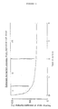
- Figure 1 is a graph to illustrate the change of % shrinkage with time in the process of transformation from a cell sheet to a spherical cell aggregate.
- Figure 2 are graphs to illustrate the effect of cycloheximide (CH) and actinomycin D (AD) on the change of % shrinkage with time in the process of transformation from a cell sheet to a spherical cell aggregate in comparison with the absence of these substances as shown by "cont.”
- CH cycloheximide
- AD actinomycin D
- the temperature-responsive polymeric compound having an LCST lower than the cell culture temperature which can be used as one component of the cell culture substrate in the first cell culture vessel of the present invention is in a solid state at the cell culture temperature, and if the temperature is lowered to a temperature below the LCST, the temperature-responsive polymeric compound becomes soluble in the culture medium and the cells on the first cell culture vessel will be detached therefrom.
- Examples of the temperature-responsive polymeric compounds having an LCST lower than the cell culture temperature which can be used as one component of the coating on the first cell culture vessel in the present invention are poly-N-substituted (meth)acrylamide derivatives and their copolymers, polymethylvinylether, polyethylene oxide, etherized methylcellulose, and partially acetylated polyvinyl alcohol. Of these preferred compounds more preferred are poly-N-substituted acrylamide derivatives, poly-N- substituted methacrylamide derivatives and their copolymers.
- thermo-responsive polymeric compounds in the present invention are listed below, but this invention is not limited to these examples.
- the LCSTs of these polymers rise with the sequence of polymers listed below.
- Poly-N-acryloyl piperidine poly-N-n-propyl methacrylamide, poly-N-isopropyl acrylamide, poly-N,N-diethyl acrylamide, poly-N-isopropyl methacrylamide, poly-N-cyclopropyl acrylamide, poly-N-acryloylpyrrolidine, poly-N,N-ethylmethyl acrylamide, poly-N-cyclopropyl methacrylamide, poly-N-ethyl acrylamide.
- the above described polymers may be homopolymers or copolymers with other monomers. Any hydrophilic monomers or hydrophobic monomers can be used as the monomers for copolymerization. Generally speaking, the copolymerization with a hydrophilic monomer will raise the LCST, and the copolymerization with a hydrophobic monomer will lower the LCST. With a proper selection of monomers, a copolymer with a desired LCST can be achieved.
- hydrophilic monomers are N-vinylpyrrolidone, vinylpyridine, acrylamide, methacrylamide, N-methyl acrylamide, hydroxyethyl methacrylate, hydroxyethyl acrylate, hydroxymethyl methacrylate, hydroxymethyl acrylate, acrylic acid, methacrylic acid, vinyl sulfonic acid, and styrylsulfonic acid and their salts, and N,N- dimethylaminoethyl methacrylate, N,N-diethyl-aminoethyl methacrylate, and N,N-dimethylaminopropyl acrylamide and their salts, but the present invention is not limited to these compounds.
- hydrophobic monomers examples include acrylate derivatives and methacrylate derivatives such as ethyl acrylate, methyl methacrylate and glycidyl methacrylate; N-substituted alkyl (meth)acrylamide derivatives such as N-n-butyl (meth)acrylamide; vinyl chloride, acrylonitrile, styrene and vinyl acetate but the present invention is not limited to these compounds.
- the molecular weight of the temperature-responsive polymeric compound which can be employed in the present invention is preferably at least about 1.0 x 10⁵ and more preferably higher than about 1.0 x 10⁶.
- the molecular weight herein means a number average molecular weight obtained from the viscosity.
- M n number average molecular weight obtained from the viscosity.
- M n number average molecular weight of poly-N-isopropyl acrylamide
- poly-N-isopropylacrylamide (hereinafter referred to as "PNIPAAm”) is a polymeric compound which shows a negative temperature coefficient of solubility in water (M. Heskins and J. E. Guillet, J. Macromol. Sci. Chem. , A2 (8), 1441, 1968).
- the hydrate (oxonium hydroxide) which depends on the hydrogen bonding formed at a lower temperature between a water molecule and the polymer molecule will decompose at a higher temperature, so that the polymer aggregates by dehydration to form a precipitate.
- the transition temperature of this hydration and dehydration is called “lower critical solution temperature” or "LCST”.
- LCST transition temperature of this hydration and dehydration
- the present invention takes advantage of such properties of the temperature-responsive polymeric compounds.
- any type of collagen can be used as the other component of the cell culture substrate of the first cell culture vessel in the present invention.
- the adhesive substances which can be used as a minor component together with collagen as a major component include fibronectin, laminin, vitronectin, proteoglycan, glycosaminoglycan, thrombospondin, gelatin, lectins, anchorage oligopeptides and adhesive proteins isolated from shellfish.
- the mixing weight ratio of the temperature-responsive polymeric compound to collagen or a mixture of collagen as a major component and the adhesive substance is typically from about 1:1 to about 1:3.
- the coating on the first cell culture vessel consists of the temperature-responsive polymeric compound having an LCST lower than the cell culture temperature and collagen or a mixture of collagen and the adhesive substance.
- a coating is formed on the first cell culture vessel by coating an aqueous solution of the mixture of the temperature-responsive polymeric compound and collagen or a mixture of the collagen and the adhesive substance on the first cell culture vessel at a temperature below the LCST and drying the coating thus obtained.
- the coating is also prepared by dipping the first cell culture vessel into the aqueous mixture solution of the temperature-responsive polymeric compound and collagen or a mixture of collagen and the adhesive substance at a temperature below the LCST and drying the coating thus obtained.
- the thickness of the coating after it is dried is at least about 0.2 »m, preferably at least 0.5 »m and more preferably at least about 1.0 »m.
- the thickness is below about 0.2 »m, the cell detachability remarkably worsens and it takes a very long time for the detachment of the cells and as a result, the cell functions are rendered unstable.
- cells are cultured on a coating of the cell culture substrate formed on the first cell culture vessel of a monolayer system suitable for the proliferation of anchorage dependent cells at a cell culture temperature higher than the LCST of the temperature-responsive polymeric compound.
- the cell culture temperature is lowered to a temperature below the LCST.
- the cultured cells can be detached from the cell culture substrate in the form of a cell sheet or monolayer which maintains the intercellular junctions without causing any damage to the cells.
- the detached cell sheet is transferred onto a non-adhesive substrate of the second cell culture vessel where a physical stimulation is applied or a test substance is added to the culture medium for the cell sheet, and the cell sheet is further cultured.
- the cell sheet by suspension culture, changes into a three dimensional cell aggregate. Since the morphological change of such a cell aggregate depends on the applied physical stimulation or the added test substance, the degree of cytotoxicity can be evaluated by measuring the morphological change of the cell aggregate.
- the physical stimulation or the test substance can be applied or added to the culture medium, and the culture can be maintained prior to the lowering of the cell culture temperature to a temperature below the LCST in the first cell culture vessel.
- morphological changes used herein includes not only the external change such as size and form of the cells but also any detectable changes such as change of intracellular organs and intercellular linkages and so on.
- the "physical stimulation” to be applied to the culture system broadly includes temperature, radiation, electrical field, magnetic field, light, microwave, and so on.
- the "test substance” to be added to the culture system broadly includes chemical substances such as medicinal drugs, cosmetics, food additives, insecticides and industrial reagents and so on. And, the setting or the change of the culture conditions such as oxygen concentration, pH and nutritional condition are also included in the meaning of the term "application of a physical stimulation or addition of a test substance.
- the first step in this invention plays a role of increasing the number of cells to a quantity necessary for the cytotoxicity test by culturing the cells in monolayer.
- the cells anchored and multiplied on the substrate are significantly different from the cell state in a actual living body, and thus they are unsuitable as the system for each individual cell to show its characteristic function and as a system to induce differential function of the cells.
- the characteristic function to serve as the target cells in the test of test substances or test of physical effect is decreased or lost, and thus the sensitivity of the cytotoxicity test method will decline drastically.
- the second step of this invention is a process to activate the characteristic function of the cells, which have declined in the monolayer culture system.
- the cells are detached from the substrate which has restricted the cells function in the monolayer state which is different from the living environment.
- the cells are transferred from the monolayer state to the three dimensional culture system which is closer to the environment existing within the living body, to induce the differential function of the cells and to improve the sensitivity of the cytotoxicity method.
- the problems with the conventional three-dimensional culture method such as the culture method in gel and aggregated cell culture method, include (1) test substances cannot reach the interior of the three-dimensional culture system and it is difficult to quantitatively evaluate the drug effect, and (2) cells may necrotize because supply of nutrients or removal of waste materials cannot be carried out efficiently in the interior of the culture system.
- problems can be solved by using the culture system of this invention.
- Problem (1) can be solved in the first step of this invention by addition of a test substance in the monolayer system which allows the test substance to reach homogeneously all cells.
- problem (2) is drastically reduced since in the second step of this invention the cell sheet is detached from the substrate and a three-dimensional structure is gradually formed.
- the risk of necrosis of internal cells will be drastically reduced, compared to the conventional culture system in gel or aggregated cell culture system which forms a tight structure from the start. Furthermore, since the test method of this invention is carried out mainly in the transition state until a three-dimensional structure is formed from the cell sheet and the test is completed by the time when a perfect three-dimensional structure is formed, the risk of necrosis is further reduced.
- Another characteristic feature of the culture system to be used in the cytotoxicity test of this invention is that it does not use trypsin/EDTA which is the cell detaching agent conventionally used to detach cultured cells from the surface of a substrate.
- Cell detaching agents such as trypsin/EDTA, not only destroy the bond between cells and the substrate, but also destroy the tight junction, desmosome, or gap junction which is the intercellular bonds, and thus it separates the cells into individual cells. Therefore, it is impossible to form a cell sheet or a cell aggregate.
- these cell detaching agents destroy various membrane receptors which exist on the surface of the cell membrane, and cause significant damage to the susceptibility of cells to signaling molecules such as hormone and local chemical mediators. Therefore, detachment of cells by conventional detaching agents will significantly reduce the sensitivity of the cytotoxicity test.
- cytotoxicity test method of the present invention As described before, conventional methods of measuring cellular functions such as cell viability, proliferation ability, and DNA synthesizing ability, can naturally be applied in the cytotoxicity test method of the present invention. However, these methods are indirect in the aspect of damage to the aggregates of cells because these cellular functions are markers of toxicity mainly against individual cells. In a living body, cells do not exist individually, but instead they form a society by the strong intercellular interaction and function as a member of that society.
- the cytotoxicity test method of the presnt invention is suitable for the determination of toxicity against cell aggregates (cell society) rather than against individual cells and therefore, the evaluation of the conditions closer to the cytotoxicity occurring actually in the body is possible.
- the method of the present invention can measure the effect of test substances on the morphological change of the entire cell aggregates as it shifts from the cell sheet to the cell aggregate, the effect on the morphological change of the intracellular organs and intercellular junctions within the cell aggregates, and the effect on the distribution of stained cells in the aggregates by the stain method and so on, and thus many toxicity models of tissues and organs can be provided.
- N-Isopropylacrylamide monomer here "NIPAAm", a product of Eastman Kodak Co. 50 g was dissolved in 500 ml benzene.
- AIBN 2,2'-Azobisisobutyronitrile
- Polymerization was carried out at 60°C for 12 hours in a stream of nitrogen gas with constant agitation. The polymer thus obtained was precipitated in benzene and after decantation the precipitates formed were dissolved in tetrahydrofuran and purified with ethyl ether.
- the poly-N-isopropyl acrylamide (here "PNIPAAm”) thus prepared had a number average molecular weight of 2.0 x 10⁶ and the LCSTs of the PNIPAAm in PBS and culture medium [polymer concentration: 1% (w/v)] were 28.9°C ⁇ 0.1°C and 28.6°C ⁇ 0.1°C.
- PNIPAAm poly-N-isopropyl acrylamide
- the aqueous polymer solution thus obtained was mixed with an equal amount of the aqueous 0.5% (w/v) solution of type I collagen which was solubilized from a cow's skin by pepsinization (sterilized, a product of Kohken K.K.), to prepare a mixed solution of the PNIPAAm and the collagen.
- This mixed solution was poured on the bottom of a commercially available plastic culture dish (Falcon, 35 mm), and it was air-dried aseptically in a clean bench at about 10°C. A dish where a 1:1 mixture of collagen and PNIPAAm was coated on the bottom of the dish to a thickness of about 2.0 »m was prepared by the above described method.
- Fibroblasts derived from human skin were suspended in Dulbecco's modified Eagle medium ("DMEM", a product of GIBCO, containing 10% (w/v) calf serum) (cell concentration in the suspension: about 2 x 10⁵ cells/ml). Two milliliters of this cell suspension was incubated at 37°C, and then it was poured in the coated dish which was pre-incubated at 37°C. It was cultured at 37°C for 3 days in an air/5 volume% CO2 gas incubator. After 3 days at which point the cultured cells covered the surface of the bottom of the dish completely, the dish was taken out from the 37°C incubator and left to stand at 4°C on ice.
- DMEM Dulbecco's modified Eagle medium
- the old medium was discarded and the sheet was washed twice with fresh medium at 4°C to remove the dissolved polymer and the collagen. Then, the cell sheet was transferred and incubated in a hydrophobic dish (Falcon, 35 mm containing 2 ml of fresh medium. In this culture system the cell sheet gradually shrank and finally became a spherical cell aggregate having an outer diameter of about 900 »m.
- phase I is a very significant shrinkage period for about 0.5 day from the time of the detachment of the cell sheet
- phase II is a weak shrinkage period for about 2 days after the 0.5th day
- phase III is a stabilized period without shrinkage after the second day.
- the fibroblasts derived from human skin were cultured for 3 days on a dish coated with the mixture of PNIPAAm and collagen as described in Control Example in the same manner as in Control Example.
- cycloheximide here "CH" was added to the culture medium for the cell sheet to bring the concentration to 0.3 and 3 »g/ml of medium, respectively.
- the shrinkage process of the detached cell sheet was examined by the phase contrast microscopy while the cell sheet was cultured in the culture medium containing CH in the hydrophobic dish.
- the shrinkage behavior of the cell aggregate is shown in Fig. 2. As shown in Fig.
- the fibroblasts derived from human skin were cultured for 3 days on the dish coated with the mixture of PNIPAAm and collagen as used in Control Example in the same manner as Control Example. Twelve hours before a cell sheet was detached from the cell culture substrate and transferred onto a hydrophobic dish in the same manner as in Control Example, actinomycin D (here "AD") was added to the culture medium to bring the concentration to 0.003 and 0.03 »g/ml of medium, respectively. Then the shrinkage process of the detached cell sheet was examined by phase contrast microscopy while the detached cell sheet was cultured in the culture medium containing 0.003 and 0.03 »g/ml (medium) of AD, respectively, in the hydrophobic dish. The shrinkage behavior of the cell aggregate is shown in Fig. 2.
- AD actinomycin D
- the shrinkage behavior of phase II ranging from the 0.5th to the second day after the detachment was significantly interrupted by the addition of more than 0.03 »g/ml of AD and no formation of any spherical cell aggregate was observed.
- the added AD was completely removed by washing the cell aggregate with fresh culture medium and then transferring it into the vessel containing fresh culture medium.
- the shrinkage started again.
- the concentration of AD which inhibits phase II was about 1/100 of that of CH. Since AD is an inhibitor of the mRNA synthesis, the shrinkage process of the phase II seems to be closely related to the mRNA synthesis of the cell.
- the fibroblasts derived from human skin as used in Example 1 were cultured for 3 days on a plastic tissue culture dish (Falcon, 35 mm), and then cycloheximide and actinomycin D were added, respectively, in such an amount that the concentration of the medium would be 10 »g/ml, and the morphological change of the cells was examined under a phase contrast microscope. No morphological change was observed at all.
- Example 1 illustrating the cytotoxicity test method of this invention, cytotoxicity was noted with cycloheximide at 3 »g/ml and with actinomycin D at 0.03 »g/ml.
- the concentration for the detection of cytotoxicity by the conventional monolayer culture such as shown in this Comparative Example was higher than 10 »g/ml.
- the sensitivity was significantly lower, compared to the culture system of this invention.
- the cytotoxicity test method using the cell culture system of the present invention is more convenient and has better sensitivity than the conventional method. Furthermore, the cell culture system of the present invention can measure sensitively the intercellular interaction or the effect on the cell aggregates, which has been difficult to evaluate by the methods of the prior art.
Landscapes
- Health & Medical Sciences (AREA)
- Life Sciences & Earth Sciences (AREA)
- Immunology (AREA)
- Engineering & Computer Science (AREA)
- Biomedical Technology (AREA)
- Chemical & Material Sciences (AREA)
- Hematology (AREA)
- Urology & Nephrology (AREA)
- Molecular Biology (AREA)
- Cell Biology (AREA)
- Medicinal Chemistry (AREA)
- Biochemistry (AREA)
- Biotechnology (AREA)
- Pathology (AREA)
- Toxicology (AREA)
- Food Science & Technology (AREA)
- Tropical Medicine & Parasitology (AREA)
- Physics & Mathematics (AREA)
- Analytical Chemistry (AREA)
- Microbiology (AREA)
- General Health & Medical Sciences (AREA)
- General Physics & Mathematics (AREA)
- Bioinformatics & Cheminformatics (AREA)
- Zoology (AREA)
- Virology (AREA)
- Apparatus Associated With Microorganisms And Enzymes (AREA)
- Micro-Organisms Or Cultivation Processes Thereof (AREA)
- Measuring Or Testing Involving Enzymes Or Micro-Organisms (AREA)
Claims (3)
- Méthode de test de cytotoxicité qui comprend les étapes de :(i) culture de cellules sur un revêtement formé sur un premier récipient de culture de cellules où ledit revêtement comprend(a) un composé polymérique répondant à la température ayant une température de solution critique inférieure (LCST) à la température de culture des cellules et(b) du collagène ou un mélange de collagène en tant que composant majoritaire et au moins une substance adhévise en tant que composant minoritaire,à une température de culture des cellules supérieure à ladite LCST,(ii) maintien de la culture pour former une feuille de cellules sur ledit revêtement,(iii) abaissement de la température de la culture de cellules à une température inférieure à ladite LCST pour détacher ladite feuille de cellules dudit revêtement,(iv) transfert de ladite feuille de cellules dans un second récipient de culture de cellules n'ayant pas d'adhésivité pour ladite feuille de cellules,(v) application d'une stimulation physique ou ajout d'une substance test au milieu de culture pour cultiver ladite feuille de cellules,(vi) culture de ladite feuille de cellules dans ledit milieu de culture et,(vii) mesure du changement morphologique de ladite feuille de cellules dans ledit milieu de culture dans la phase de transition de ladite feuille de cellules en un sphéroïde.
- Méthode selon la revendication 1 comprenant, avant l'étape (iii), l'étape d'appliquer la même quantité de ladite stimulation physique ou l'ajout de la même quantité de ladite substance test qu'à l'étape (v) au milieu de culture et de plus le maintien de la culture.
- Méthode selon la revendication 1 ou 2, où ledit composé polymérique répondant à la température est choisi dans le groupe consistant d'un dérivé de poly-N-substitué acrylamide ou d'un copolymère de celui-ci, d'un dérivé de poly-N-substitué méthacrylamide ou d'un copolymère de celui-ci, d'un polyvinylméthyléther, et d'un polyvinylalcool partiellement acétylé.
Applications Claiming Priority (2)
| Application Number | Priority Date | Filing Date | Title |
|---|---|---|---|
| JP116687/90 | 1990-05-02 | ||
| JP2116687A JPH0489000A (ja) | 1990-05-02 | 1990-05-02 | 細胞培養キットおよび細胞傷害試験法 |
Publications (3)
| Publication Number | Publication Date |
|---|---|
| EP0455319A2 EP0455319A2 (fr) | 1991-11-06 |
| EP0455319A3 EP0455319A3 (en) | 1993-05-05 |
| EP0455319B1 true EP0455319B1 (fr) | 1995-06-28 |
Family
ID=14693384
Family Applications (1)
| Application Number | Title | Priority Date | Filing Date |
|---|---|---|---|
| EP91250120A Expired - Lifetime EP0455319B1 (fr) | 1990-05-02 | 1991-04-27 | Procédé d'essai cytotoxique |
Country Status (5)
| Country | Link |
|---|---|
| US (1) | US5229288A (fr) |
| EP (1) | EP0455319B1 (fr) |
| JP (1) | JPH0489000A (fr) |
| CA (1) | CA2041367A1 (fr) |
| DE (1) | DE69110750D1 (fr) |
Families Citing this family (12)
| Publication number | Priority date | Publication date | Assignee | Title |
|---|---|---|---|---|
| EP0499932A1 (fr) * | 1991-02-16 | 1992-08-26 | W.R. Grace & Co.-Conn. | Amas de cellules cancéreuses comprenant des cellules cancéreuses et des cellules normales et leurs préparations |
| US5262055A (en) * | 1992-10-19 | 1993-11-16 | The University Of Utah | Implantable and refillable biohybrid artificial pancreas |
| BR9508092A (pt) * | 1994-06-21 | 1997-08-12 | Mukoyama Orchids Ltd | Suporte para ocultivo/regeneração de corpo de planta e processo para cultivar/regenerar um corpo de planta |
| US6035228A (en) * | 1997-11-28 | 2000-03-07 | Picker International, Inc. | Frameless stereotactic arm apparatus and method of using same |
| JP2005506839A (ja) * | 2001-10-02 | 2005-03-10 | ベクトン・ディキンソン・アンド・カンパニー | 細胞外マトリックスおよび他の分子を使用する幹細胞の増殖および分化 |
| WO2004096892A1 (fr) * | 2003-04-25 | 2004-11-11 | Japan Science And Technology Agency | Polymere fonctionnel presentant une fonction de piegeage de substance, trousse de reactif pour piegeage de substance comprenant ce polymere, et procede de recuperation d'une substance au moyen dudit polymere |
| EP2112196A1 (fr) * | 2008-04-25 | 2009-10-28 | Nederlandse Organisatie voor toegepast- natuurwetenschappelijk onderzoek TNO | Composition liquide comportant des chaînes de polymères et particules d'un matériau inorganique dans un liquide |
| JP5847733B2 (ja) | 2010-01-28 | 2016-01-27 | ザ リージェンツ オブ ザ ユニバーシティ オブ ミシガン | 懸滴用アレイプレート |
| JP5819056B2 (ja) * | 2010-11-18 | 2015-11-18 | 大日本印刷株式会社 | 細胞培養用基材 |
| JP2015149949A (ja) * | 2014-02-17 | 2015-08-24 | 学校法人明治大学 | 免疫抑制剤の評価方法、及び免疫抑制剤評価キット |
| KR20170055791A (ko) * | 2015-11-12 | 2017-05-22 | 한국과학기술연구원 | 인 비트로 3d 피부 진피 모델 및 그를 사용하여 약물을 스크리닝하는 방법 |
| US11378570B2 (en) | 2017-05-24 | 2022-07-05 | Alcon Inc. | Cytotoxicity test method for medical devices |
Family Cites Families (3)
| Publication number | Priority date | Publication date | Assignee | Title |
|---|---|---|---|---|
| US4963489A (en) * | 1987-04-14 | 1990-10-16 | Marrow-Tech, Inc. | Three-dimensional cell and tissue culture system |
| EP0355112A1 (fr) * | 1987-04-22 | 1990-02-28 | Michael Bay | Procedes de culture de cellules, et produits |
| JPH0383914A (ja) * | 1989-08-18 | 1991-04-09 | W R Grace & Co | ドラッグキャリアー |
-
1990
- 1990-05-02 JP JP2116687A patent/JPH0489000A/ja active Pending
-
1991
- 1991-04-23 US US07/690,132 patent/US5229288A/en not_active Expired - Fee Related
- 1991-04-26 CA CA002041367A patent/CA2041367A1/fr not_active Abandoned
- 1991-04-27 EP EP91250120A patent/EP0455319B1/fr not_active Expired - Lifetime
- 1991-04-27 DE DE69110750T patent/DE69110750D1/de not_active Expired - Lifetime
Also Published As
| Publication number | Publication date |
|---|---|
| US5229288A (en) | 1993-07-20 |
| EP0455319A3 (en) | 1993-05-05 |
| DE69110750D1 (de) | 1995-08-03 |
| CA2041367A1 (fr) | 1991-11-03 |
| JPH0489000A (ja) | 1992-03-23 |
| EP0455319A2 (fr) | 1991-11-06 |
Similar Documents
| Publication | Publication Date | Title |
|---|---|---|
| EP0455319B1 (fr) | Procédé d'essai cytotoxique | |
| Kirkpatrick et al. | Theoretical and practical aspects of testing potential biomaterials in vitro | |
| Brown | Neutrophil granulocytes: adhesion and locomotion on collagen substrata and in collagen matrices | |
| Hampel et al. | Effect of intermittent shear stress on corneal epithelial cells using an in vitro flow culture model | |
| Avner et al. | Polypeptide growth factors in metanephric growth and segmental nephron differentiation | |
| EP0387975A1 (fr) | Support pour culture cellulaire, couche de cellules, amas cellulaire et leurs préparations | |
| EP2854885B1 (fr) | Micro-conception d'hydrogels | |
| CA2942491A1 (fr) | Chambre d'ecoulement a plaques paralleles a orifices et leurs procedes d'utilisation | |
| Kato et al. | The design of polymer microcarrier surfaces for enhanced cell growth | |
| EP0470681A2 (fr) | Amas cellulaire ou couches de cellules comprenant une pluralité d'espèces cellulaires et leurs préparations | |
| EP1980613A1 (fr) | Procede de maintien de la fonction des cellules de tissus du foie sur une periode prolongee | |
| US5175092A (en) | Vitro toxicology kit and method therefor | |
| JPH04278083A (ja) | 細胞数を制御した細胞塊状体形成法およびそれに用いる細胞培養用基材 | |
| JPH05168470A (ja) | 複数の細胞種からなる細胞塊状体とシートおよびその製造法 | |
| JP7647047B2 (ja) | 低組織因子活性細胞、およびその製法 | |
| EP0463508A1 (fr) | Méthode d'injection de substances dans des cellules | |
| Briles et al. | Isolation of cloned variants of a rat hepatoma cell line with altered attachment to collagen, but normal attachment to fibronectin | |
| EP0499932A1 (fr) | Amas de cellules cancéreuses comprenant des cellules cancéreuses et des cellules normales et leurs préparations | |
| Singer et al. | Stability of polyadenylated RNA in differentiating myogenic cells | |
| JP5674255B2 (ja) | 細胞培養基材の評価方法 | |
| JPH05276923A (ja) | 細胞培養用基材およびその製造方法 | |
| JPH0466082A (ja) | 細胞数を制御した細胞塊状体形成法およびそれに用いる細胞培養基材 | |
| JP3105011B2 (ja) | 培養癌組織 | |
| EP3766958B1 (fr) | Substrat de culture, méthode de fabrication de substrat de culture, et méthode de culture et dispositif de culture pour cellule souche | |
| JP2000217570A (ja) | 培養癌組織およびその製造方法 |
Legal Events
| Date | Code | Title | Description |
|---|---|---|---|
| PUAI | Public reference made under article 153(3) epc to a published international application that has entered the european phase |
Free format text: ORIGINAL CODE: 0009012 |
|
| AK | Designated contracting states |
Kind code of ref document: A2 Designated state(s): DE FR GB IT |
|
| PUAL | Search report despatched |
Free format text: ORIGINAL CODE: 0009013 |
|
| AK | Designated contracting states |
Kind code of ref document: A3 Designated state(s): DE FR GB IT |
|
| 17P | Request for examination filed |
Effective date: 19930920 |
|
| 17Q | First examination report despatched |
Effective date: 19931130 |
|
| GRAA | (expected) grant |
Free format text: ORIGINAL CODE: 0009210 |
|
| AK | Designated contracting states |
Kind code of ref document: B1 Designated state(s): DE FR GB IT |
|
| PG25 | Lapsed in a contracting state [announced via postgrant information from national office to epo] |
Ref country code: IT Free format text: LAPSE BECAUSE OF FAILURE TO SUBMIT A TRANSLATION OF THE DESCRIPTION OR TO PAY THE FEE WITHIN THE PRE;WARNING: LAPSES OF ITALIAN PATENTS WITH EFFECTIVE DATE BEFORE 2007 MAY HAVE OCCURRED AT ANY TIME BEFORE 2007. THE CORRECT EFFECTIVE DATE MAY BE DIFFERENT FROM THE ONE RECORDED.SCRIBED TIME-LIMIT Effective date: 19950628 Ref country code: FR Effective date: 19950628 |
|
| REF | Corresponds to: |
Ref document number: 69110750 Country of ref document: DE Date of ref document: 19950803 |
|
| PG25 | Lapsed in a contracting state [announced via postgrant information from national office to epo] |
Ref country code: DE Effective date: 19950929 |
|
| EN | Fr: translation not filed | ||
| PG25 | Lapsed in a contracting state [announced via postgrant information from national office to epo] |
Ref country code: GB Effective date: 19960427 |
|
| PLBE | No opposition filed within time limit |
Free format text: ORIGINAL CODE: 0009261 |
|
| STAA | Information on the status of an ep patent application or granted ep patent |
Free format text: STATUS: NO OPPOSITION FILED WITHIN TIME LIMIT |
|
| 26N | No opposition filed | ||
| GBPC | Gb: european patent ceased through non-payment of renewal fee |
Effective date: 19960427 |