EP0463114B1 - Testverfahren für und behandlung von autoimmunkrankheiten - Google Patents
Testverfahren für und behandlung von autoimmunkrankheiten Download PDFInfo
- Publication number
- EP0463114B1 EP0463114B1 EP90906028A EP90906028A EP0463114B1 EP 0463114 B1 EP0463114 B1 EP 0463114B1 EP 90906028 A EP90906028 A EP 90906028A EP 90906028 A EP90906028 A EP 90906028A EP 0463114 B1 EP0463114 B1 EP 0463114B1
- Authority
- EP
- European Patent Office
- Prior art keywords
- cells
- channels
- type
- mice
- autoimmune disease
- Prior art date
- Legal status (The legal status is an assumption and is not a legal conclusion. Google has not performed a legal analysis and makes no representation as to the accuracy of the status listed.)
- Expired - Lifetime
Links
Images



Classifications
-
- G—PHYSICS
- G01—MEASURING; TESTING
- G01N—INVESTIGATING OR ANALYSING MATERIALS BY DETERMINING THEIR CHEMICAL OR PHYSICAL PROPERTIES
- G01N33/00—Investigating or analysing materials by specific methods not covered by groups G01N1/00 - G01N31/00
- G01N33/48—Biological material, e.g. blood, urine; Haemocytometers
- G01N33/50—Chemical analysis of biological material, e.g. blood, urine; Testing involving biospecific ligand binding methods; Immunological testing
- G01N33/5005—Chemical analysis of biological material, e.g. blood, urine; Testing involving biospecific ligand binding methods; Immunological testing involving human or animal cells
- G01N33/5008—Chemical analysis of biological material, e.g. blood, urine; Testing involving biospecific ligand binding methods; Immunological testing involving human or animal cells for testing or evaluating the effect of chemical or biological compounds, e.g. drugs, cosmetics
-
- A—HUMAN NECESSITIES
- A61—MEDICAL OR VETERINARY SCIENCE; HYGIENE
- A61K—PREPARATIONS FOR MEDICAL, DENTAL OR TOILETRY PURPOSES
- A61K39/00—Medicinal preparations containing antigens or antibodies
- A61K39/395—Antibodies; Immunoglobulins; Immune serum, e.g. antilymphocytic serum
- A61K39/39533—Antibodies; Immunoglobulins; Immune serum, e.g. antilymphocytic serum against materials from animals
-
- A—HUMAN NECESSITIES
- A61—MEDICAL OR VETERINARY SCIENCE; HYGIENE
- A61K—PREPARATIONS FOR MEDICAL, DENTAL OR TOILETRY PURPOSES
- A61K47/00—Medicinal preparations characterised by the non-active ingredients used, e.g. carriers or inert additives; Targeting or modifying agents chemically bound to the active ingredient
- A61K47/50—Medicinal preparations characterised by the non-active ingredients used, e.g. carriers or inert additives; Targeting or modifying agents chemically bound to the active ingredient the non-active ingredient being chemically bound to the active ingredient, e.g. polymer-drug conjugates
- A61K47/51—Medicinal preparations characterised by the non-active ingredients used, e.g. carriers or inert additives; Targeting or modifying agents chemically bound to the active ingredient the non-active ingredient being chemically bound to the active ingredient, e.g. polymer-drug conjugates the non-active ingredient being a modifying agent
- A61K47/68—Medicinal preparations characterised by the non-active ingredients used, e.g. carriers or inert additives; Targeting or modifying agents chemically bound to the active ingredient the non-active ingredient being chemically bound to the active ingredient, e.g. polymer-drug conjugates the non-active ingredient being a modifying agent the modifying agent being an antibody, an immunoglobulin or a fragment thereof, e.g. an Fc-fragment
- A61K47/6835—Medicinal preparations characterised by the non-active ingredients used, e.g. carriers or inert additives; Targeting or modifying agents chemically bound to the active ingredient the non-active ingredient being chemically bound to the active ingredient, e.g. polymer-drug conjugates the non-active ingredient being a modifying agent the modifying agent being an antibody, an immunoglobulin or a fragment thereof, e.g. an Fc-fragment the modifying agent being an antibody or an immunoglobulin bearing at least one antigen-binding site
- A61K47/6849—Medicinal preparations characterised by the non-active ingredients used, e.g. carriers or inert additives; Targeting or modifying agents chemically bound to the active ingredient the non-active ingredient being chemically bound to the active ingredient, e.g. polymer-drug conjugates the non-active ingredient being a modifying agent the modifying agent being an antibody, an immunoglobulin or a fragment thereof, e.g. an Fc-fragment the modifying agent being an antibody or an immunoglobulin bearing at least one antigen-binding site the antibody targeting a receptor, a cell surface antigen or a cell surface determinant
-
- G—PHYSICS
- G01—MEASURING; TESTING
- G01N—INVESTIGATING OR ANALYSING MATERIALS BY DETERMINING THEIR CHEMICAL OR PHYSICAL PROPERTIES
- G01N33/00—Investigating or analysing materials by specific methods not covered by groups G01N1/00 - G01N31/00
- G01N33/48—Biological material, e.g. blood, urine; Haemocytometers
- G01N33/50—Chemical analysis of biological material, e.g. blood, urine; Testing involving biospecific ligand binding methods; Immunological testing
- G01N33/53—Immunoassay; Biospecific binding assay; Materials therefor
- G01N33/564—Immunoassay; Biospecific binding assay; Materials therefor for pre-existing immune complex or autoimmune disease, i.e. systemic lupus erythematosus, rheumatoid arthritis, multiple sclerosis, rheumatoid factors or complement components C1-C9
-
- G—PHYSICS
- G01—MEASURING; TESTING
- G01N—INVESTIGATING OR ANALYSING MATERIALS BY DETERMINING THEIR CHEMICAL OR PHYSICAL PROPERTIES
- G01N2800/00—Detection or diagnosis of diseases
- G01N2800/24—Immunology or allergic disorders
Definitions
- the present invention relates generally to advances made in the field of autoimmune disease prognosis, diagnosis and treatment. More particularly, the present invention is directed to certain assays for, and consequent treatment of, autoimmune diseases based upon the identification and characterization herein of a unique ion channel aberration found to be associated with abnormal T cells linked to such diseases. Assays, prognostic markers and therapeutic modalities for such autoimmune disease states are defined and described.
- Autoimmune diseases have been the subject of widespread press attention because of the considerable morbidity worldwide that they cause. Autoimmune diseases include rheumatoid arthritis, type-1 diabetes mellitus (insulin dependent), multiple sclerosis, myasthenia gravis, systemic lupus erythematosus, Sjogren's syndrome, mixed connective tissue disease, experimental allergic encephalomyelitis (EAE), to name a few.
- EAE allergic encephalomyelitis
- autoimmune diseases result from abnormal cells of the immune system destroying 5 target tissues, either by direct killing or by producing autoantibodies.
- SLE systemic lupus erythematosus
- abnormal B-lymphocytes produce anti-DNA antibodies that are positively charged and aggregate on negatively charged kidney cells causing inflammation and nephritis, which is symptomatic of SLE.
- T cells In diabetes mellitus, abnormal T cells systematically destroy pancreatic islet cells such that they prove incapable of producing insulin, a necessary hormone for proper metabolic balance in an organism.
- abnormal T cells are believed to damage myelin basic protein, a major component of nerve cells, which systematically destroys certain nerve cells, causing a spectrum of neurological symptoms.
- myelin basic protein a major component of nerve cells, which systematically destroys certain nerve cells, causing a spectrum of neurological symptoms.
- autoimmune diseases studied to date, there seems to emerge a common pattern of abnormal immune system cells producing materials that either destroy or retard certain target tissues causing symptoms manifest for that disease state.
- the next step was to determine whether the same abnormal expression pattern could be observed for cells from mice that were not genetically predisposed to SLE, as were the murine models of Chandy, et al ., Supra .
- murine models that were not prone for SLE, developed similar abnormal expression patterns associated with the onset of SLE and lymphoproliferation.
- n , n' and l types Three types of ion channel types, classified pharmacologically and electrophysiologically, were identified, the so-called n , n' and l types.
- T cells in the peripheral lymphoid tissues for present purposes are characterized into relevant types: CD4+CD8 ⁇ , CD4 ⁇ CD8+, CD4 ⁇ CD8 ⁇ .
- the CD4+CD8 ⁇ cells are thought to express approximately 20 n channels per cell and are believed to have no n ' and l channels.
- CD4 ⁇ CD8+ T cells are thought to have about 20 n ' and l channels per cell and no n channels.
- CD4 ⁇ CD8 ⁇ T cells are believed to express about 20 channels per cell, all three types being represented.
- n channel types are increased upwards of ten fold in the cells that are activated.
- normal T cells when stimulated by mitogens, show as a normal immune response elevation in the number of n channels. Blocking these n channels, for example with tetraethyl ammonium (TEA), would serve to shut down and effectively block an immune response.
- TAA tetraethyl ammonium
- Abnormal T cells may also be subject to a similar blockage. However, such abnormal T cells are represented by double negative CD4 ⁇ CD8 ⁇ cells, manifesting a proliferation of type l channels that seem implicated in the induction of autoimmune disease states.
- Type l K+ channels are not use dependent, close more rapidly on repolarization than do n channels and are much more sensitive to blockage, for example by TEA. It was the goal of the present research to establish a link, if any, hence an etiology between abnormal ion channel expression in immune cells and systemic symptoms of autoimmune disease.
- the threshold experiment establishing the aforementioned link was performed with murine models that are representative of human SLE, namely murine models NZBxNZW, NZBxSWR, BXSB-Yaa and MRL +/+.
- murine models that are representative of human SLE, namely murine models NZBxNZW, NZBxSWR, BXSB-Yaa and MRL +/+.
- the present invention is predicated upon the identification and characterization of an etiology of autoimmune diseases, represented by SLE, type-1 diabetes mellitus, experimental allergic encephalomyelitis (EAE), and others, namely, that all such autoimmune diseases demonstrate the presence of abnormal CD4 ⁇ CD8 ⁇ T cells expressing large numbers of type l K+ ion channels.
- EAE experimental allergic encephalomyelitis
- the present invention is directed to several consequential aspects: 1) an assay for diagnosing autoimmune diseases by measuring the relative numbers of type l K+ channels in abnormal CD4 ⁇ CD8 ⁇ T cells as defined in independent Claim 4, 2) an assay for testing extrinsic materials for their ability to selectively modulate type l K+ channels in abnormal CD4 ⁇ CD8 ⁇ T cells as defined in independent Claim 1, 3) the use of a selected identified extrinsic material for the preparation of a drug useful for treating an autoimmune disease as defined in independent Claim 7, and 4) a process, as defined in independent Claim 16, for preparing a composition containing such selected identified extrinsic material and the use, as defined in independent Claim 17, of a composition obtained by the said process.
- the present invention is predicated upon the identification and characterization of a diagnostic marker for autoimmune diseases, that leads to a diagnostic and a therapeutic target for such diseases.
- This predicate makes possible the identification of the etiology of a given autoimmune disease state, an assay to detect presence of the marker, an assay to identify (selective) modulators of the marker, suitable modulators identified from such an assay and therapeutic modalities based upon such modulators (drugs) and/or specific antibodies raised against type 1 K+ channels of abnormal CD4 ⁇ CD8 ⁇ T cells.
- the quintessence of the present invention enables in turn the consequence that the identified elevated numbers of type l K+ channels in abnormal CD4 ⁇ CD8 ⁇ T cells can be exploited by a method of screening for extrinsic materials having a modulating effect on such channels comprising providing an in vitro system containing elevated type l K+ channels of abnormal CD4 ⁇ CD8 ⁇ T cells, challenging such cells with one or more of a battery of test materials having the putative potential to modulate said type l K+ channels, monitoring the effect of said test materials on said l K+ channels and selecting candidates capable of modulating type l K+ channels.
- the assay for determining presence of an autoimmune disease of the invention comprises providing T cells containing K+ channels from a test individual, identifying abnormal T cells from among the population of the provided T cells, measuring the relative numbers of type l K+ channels of the abnormal cells, and determining whether said type l K+ channels are elevated or normal.
- the drug prepared from an extrinsic material according to the invention can be used for treating autoimmune diseases that are symptomatized by elevated numbers of type l K+ channels in abnormal CD4 ⁇ CD8 ⁇ T cells of the immune system of the organism manifesting said autoimmune disease in contacting said organism and/or said immune cell system with the said drug having a modulating effect on said type l K+ channels, such materials optionally identified via the assay system described Supra .
- autoimmune diseases as described above selectively without disturbing type n and n ' ion channels expression characteristic of normal immune response can be treated as described above with, as an example, tetraethyl ammonium (TEA) or such other selective modulator defined and optionally selected via the assay described supra .
- TAA tetraethyl ammonium
- the present invention is thus directed to the identification, management and control of autoimmune diseases, including:
- a reactive antibody with a cell toxin such that the antibody would locate and bind to a type l K+ channel of an aberrant CD4 ⁇ CD8 ⁇ T cell (responsible for an autoimmune disease) and thereby place the cell toxin, e.g., ricin, in close proximity to the target abnormal cell with consequential cytotoxic activity.
- Psoralen to image the aberrant target cells followed by activation to a cytotoxin.
- the present invention is illustrated herein by use of murine models representative of human etiology.
- Evidence for this lies in the fact that (1) CD4 ⁇ CD8 ⁇ cell types induce, in all systems studied, abnormal autoimmune disease causing cells and (2) type l K+ channels are known to exist in humans. See Shapiro et al. , Biophysical Journal 53 , 550a, No. W-Pos203 (1988).
- the present invention uses such murine models, there has been found an established link to autoimmune diseases of elevated type l K+ channels in abnormal T cells; therefore, the present invention is translatable to immune systems having CD4 and CD8 T cells bearing type l K+ channels, for example, as found in humans.
- the present invention is illustrated using characteristic murine models that establish, with proper correlation and translation into the human system, that type l K+ channels are elevated in abnormal CD4 ⁇ CD8 ⁇ T cells on onset of a given autoimmune disease.
- Illustrative autoimmune diseases tested were SLE, type-1 diabetes, and EAE, all with characteristic murine models. Because the present research is so properly based, given the instant disclosure of how to assay for and diagnose and treat an autoimmune disease, one skilled in the art will well enough know how to carry out such endeavors using the human system, when it is possible to proceed in this manner unhampered by regulatory impediments.
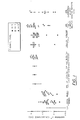
- Figure 1 K+ channel expression in splenic and lymph node derived CD4 ⁇ CD8 ⁇ Thy-1.2+ T cells in normal mice and mice with SLE, type 1 diabetes mellitus, or experimental allergic encephalomyelitis (EAE; a model for multiple sclerosis).
- Figure 2 K+ channel expression in splenic and lymph node CD4 ⁇ CD8 ⁇ Thy-1.2+ T cells in young and old SLE mice.
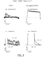
- FIG. 3 Whole cell K+ currents in CD4 ⁇ CD8 ⁇ Thy-1.2+ T cells in normal mice (left) and mice with collagen arthritis (right). Top, K+ currents were elicited with 200 ms depolarizing pulses to 40 mV from a holding potential of -80 mV. Pulse interval was 1 s to assay cumulative (use-dependent) inactivation. Note the different current scale. Bottom, tail currents were elicited by voltage steps to -60 and -30 mV after a 15 ms prepulse to 40 mV. Note the different current and time scale.
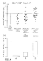
- FIG. 4 Maximal K+ conductance (g K ) of CD4 ⁇ CD8 ⁇ Thy-1.2+ T cells: a , , , represent n , l , n' K+ channel types. These data are shown as mean ⁇ SD. b , Percentage of cells with g K > 1000 pS of type l K+ conductance. Data for normal mice are pooled together from C3H, C57BL, BALB/c, and DBA/1-LACJ (uninjected).
- Figure 5 Maximal K+ conductance (g K ) of CD4+CD8 ⁇ and CD4 ⁇ CD8+ T cell subsets in normal mice and mice with collagen arthritis.
- ⁇ , ⁇ , • represent n , l , n' K+ channel types.
- Figure 6 Maximal K+ conductance (g K ) of CD4 ⁇ CD8 ⁇ Thy-1.2+ T cells in uninjected DBA/l-LACJ mice, BALC/c mice injected with heat-killed E . coli, and DBA/l-LACJ mice injected with CFA at day -26, -62 and -26, -26 and -2.
- Day 0 refers to the day the electrophysiological experiments were performed.
- ⁇ , ⁇ , • represent n , l , n' K+ channel types.
- extentrinsic material herein is meant any entity that is not ordinarily present or functional with respect to type l K+ channels and/or abnormal T cells and that affects same.
- the term has a functional definition and includes such identified entities as hormones; toxins; growth factors/cytokines; secondary messenger modulating substances; organic compounds and inorganic compounds; agents that indirectly impact on the l channel through effects on intracellular second messengers, or other channels, or membrane potential or receptors; cell types targeted to type l K+ channels or abnormal CD4 ⁇ CD8 ⁇ T cells; genetic manipulation products impacting on l channels or on CD4 ⁇ CD8 ⁇ T cells; or bacterial or viral materials that impact on CD4 ⁇ CD8 ⁇ T cells having many l channels.
- modulating effect or “properties”, or grammatical equivalents, herein is meant both active and passive impact on type l K+ channels and/or abnormal T cells. These include, but shall not be construed as limited to, blocking the channel or the function of the channel protein(s), reducing the number of ion channels per cell and use of secondary cell(s) or channel(s) to impact on a primary abnormal cell.
- monitoring in respect of effect of test materials on type l K+ channels and/or abnormal T cells herein is meant any method known or devised for measuring the impact of a test material on said channels/cells. These include, but shall not be construed as limited to, measuring current, measuring membrane potential, measuring K+ flux, such as with radioactive tracers, measuring K+ concentration and measurements of indirect consequences to other receptors, second messengers and/or channels.
- autoimmune disease herein has meaning beyond the classical definition, and therefore, shall include any disease state where CD4 ⁇ CD8 ⁇ T cells act as accessory cells, for example, to induce the immune system to result in destruction of organism cells.
- Figure 1 shows the maximum K+ conductance, g K , of CD4 ⁇ CD8 ⁇ Thy-1.2+ T cells from normal mice and from mice with genetically determined SLE or type 1 diabetes mellitus.
- the cells under investigation were bathed in Ringer solution (in mM): 160 NaCl, 4.5 KCl, 2 MgCl2, 1 CaCl2 , 2 MgCl2, and 10 K-HEPES (pH 7.4).
- the patch pipette contained 134 KF, 11 K2-EGTA, 1.1 CaCl2, 2 MgCl2, and 10 K-HEPES (pH 7.2).
- Each point represents g K in one single cell except the ones with the bars that reflect the mean ⁇ SD of previously reported data.
- g K of CD4 ⁇ CD8 ⁇ T cells from three young MRL-+/+ mice, before the onset of clinically symptomatic disease and two old MRL-+/+ mice with lupus nephritis (proteinuria: >2000 mg protein/dl) was determined as described above in connection with Fig. 1.
- Each point represents g K in one single cell except the ones with the bars that reflect the mean ⁇ SD of previously reported data (Grissmer et al. , J. Immunol. 141 , 1137 (1988) and Chandy et al. , Science 233 , 1197 (1986).) See Figure 2.
- CD4 ⁇ CD8 ⁇ Thy-1.2+ T cells from every murine model for SLE display an abnormally elevated number of type l K+ channels, demonstrating from the (as labeled) Lupus models that the alteration is linked to the disease.
- non-obese diabetes (NOD) mouse strain is another genetically determined murine model for autoimmune disease [Makino et al. , Exp. Anim. 29 , 1 (1980) and Hattori et al. , Science 231 , 733 (1986)]. These mice spontaneously develop type 1 diabetes mellitus. CD4+CD8 ⁇ and CD4 ⁇ CD8+ T cells have been implicated in the pathogenesis of this disease [Miller et al. , Nature 307 , 465 (1984), O'Neil et al. , J. Immunol. 140 , 52 (1988) and Koike et al. , Diabetes 36 , 539 (1987)].
- CD4 ⁇ CD8 ⁇ Thy-1.2+ T cells The role of CD4 ⁇ CD8 ⁇ Thy-1.2+ T cells has not been examined.
- This pattern of K+ channel expression is almost identical to that in CD4 ⁇ CD8 ⁇ Thy-1.2+ T cells from SLE mice, showing a common mechanism underlying type 1 diabetes mellitus and SLE.
- This difference in K+ channel expression in the spleen may reflect the organ-specific nature of the autoimmune manifestations in type 1 diabetes mellitus, which are primarily confined to the pancreas, and the systemic autoimmune manifestations in SLE.
- EAE Experimental allergic encephalomyelitis
- Mice inoculated with myelin basic protein develop paralysis within 1-2 weeks (acute EAE). Some of these mice recover and then develop a chronic relapsing disease (chronic EAE) which closely resembles human multiple sclerosis.
- CD4 ⁇ CD8 ⁇ Thy1.2+ splenic T cells were isolated from mice with chronic EAE and their K+ channel expression was studied. A greater frequency of cells exhibited large numbers of type l K+ channels compared to normal mice.
- T lymphocytes CD4+CD8 ⁇ or CD4 ⁇ CD8+
- C3H- gld mice exhibited small numbers of types l , n , or n ' K+ channels like phenotypically identical cells from normal mice.
- Normal mitogen activated T cells express ⁇ 200 type n K+ channels/cell [DeCoursey et al. , J. Gen. Physiol. 89 , 405 (1987)], in contrast to the numerous type l K+ channels displayed by CD4 ⁇ CD8 ⁇ Thy-1.2+ T cells in mice with SLE and type 1 diabetes mellitus.
- K+ channels were identified on the basis of their inactivation properties, channel closing kinetics, and sensitivity to block by tetraethylammonium (TEA+).
- type n K+ channels are use-dependent, i.e., show cumulative inactivation by repetitive depolarizing pulses. Use dependence was determined by measuring the decline in the size of the peak K+ current elicited with one per second repetitive depolarizing pulses of 200 ms duration.
- Type n ' K+ channels are not use dependent, close slowly like type n K+ channels upon repolarization, but are less sensitive to lock by TEA+ (K D ⁇ 100 mM).
- mice were housed in the Vivarium at U.C. Irvine, and care was taken in accordance with institutional guidelines. The mice were lightly anesthetized with metofane, and sacrificed by cervical dislocation. The spleen was removed and single cell suspensions prepared. Cells were washed three times with RPMI-1640 (Irvine Scientific, Santa Ana, CA) supplemented with 10% fetal calf serum (Hyclone, Logan, Utah). Cells were resuspended at 10 x 106 cells/ml and T cells separated from non-T cells by a nylon wool column. The cells were washed three times in medium, and resuspended at 1-2 x 10⁶ cells/ml.
- the cells were stained as follows: Cells were first incubated on ice for 30 minutes with rat-anti-mouse-CD4 and rat-anti-mouse-CD8 antibodies, washed three times in medium, incubated for 30 minutes on ice with phycoerythrin-conjugated goat-anti-rat-IgG (affinity purified and absorbed for mouse Ig), washed three times in medium, incubated on ice for 30 minutes with fluoresceinated rat-anti-mouse-Thy-1.2, washed three times in medium, and then resuspended at 2 x 10⁶ cells/ml in medium. The cells were then patch-clamped as described in the Fig. 1 legend.
- CD4 ⁇ CD8 ⁇ Thy-1.2+ T cells were identified as green cells. Other T cells (CD4+CD8 ⁇ Thy-1.2+, CD4 ⁇ CD8+Thy ⁇ 1.2+,) were yellow, and B cells and monocytes (CD4 ⁇ CD8 ⁇ Thy-1.2 ⁇ ) were unstained.
- mice used were: C57BL: 6 and 19 months; BALB/c: 1 and 7 months; BXSB-males: 2 mice aged 4 months; NZBxNZW: 2 mice aged 6 months, one aged 9 months; NZBxSWR: 3 mice aged 7-8 months; old MRL-+/+: 2 mice older than 20 months; NOD mice: 6 and 9 months; young MRL-+/+: 6, 10, and 12 months.
- DBA-J/Lac-J male mice were purchased from the Jackson Laboratory (Bar Harbor, ME). They were injected intradermally near the base of their tails with 100ug of highly purified chick type II collagen emulsified in complete Freunds Adjuvant (CFA) that contained additional Mycobactrium butyricum (Difco).
- CFA complete Freunds Adjuvant
- the collagen/CFA emulsion was prepared by adding 1 volume of modified CFA (30 ug of M. butyricum per 50 ul of 0.01 N acetic acid). Animals were immunized with 100 ul of this mixture using a 26 gauge needle. One group of control animals was not injected and another group was injected with modified CFA without collagen. Paw thickness was measured with a Schnelltaster constant-tension caliper.
- the type II collagen was prepared from chick sternal cartilage by established methods Miller, Biochemistry 10, 1652 (1971).
- E. coli ATCC 8739, wild type was homogenized in a Manton-Gaulin homogenizer at 4,19.108 Pa (6000 psi) for 5 min, and heatinactivated for 30 min at 60°C. The bacteria were then adjusted to a concentration of 0.5 mg/ml stock in phosphate buffered saline (PBS). BALB/c mice were then immunized intraperitoneally with 1 ml of solution containing 0.5 ml antigen (15 ug. E. Coli ) in PBS and 0.5 ml CFA. Mice received booster injections of antigen (15 ug E.Coli ) and incomplete Freund's adjuvant 4 weeks and 6 weeks later. Mice were sacrified 1 week after the second booster immunization.
- PBS phosphate buffered saline
- PE-conjugated-anti-CD4 L3T4
- FITC-conjugated-anti-CD8 FITC-conjugated-anti-Thy-1,2, anti-CD4 and anti-CD8 were purchased from Becton Dickinson (Mountain View, CA).
- PE-conjugated-goat-anti-rat IgG affinity purified and absorbed against mouse IgG
- Caltag Caltag
- T Cells Mice were killed, and single-cell suspensions were prepared from the spleen. T cells were enriched by passage through a nylon wool column. Cells were then suspended in RPMI-1640 medium supplemented with 10% heat-inactivated FCS (Hyclone, Logan, UT) and 2 mM l -glutamine (medium).
- FCS heat-inactivated FCS
- DN T cells To identify DN T cells, cells were incubated with anti-CD4 and anti-CD8 for 30 min on ice, washed three times with medium, incubated with PE-labeled-goat-anti-rat IgG for 30 min on ice, washed five times with medium incubated with FITC-labeled-anti-Thy-1.2 for 30 min on ice, washed three times with medium, and then resuspended in 1 ml of medium. The cells were visualized under the microscope and three populations were evident: CD4 ⁇ CD8 ⁇ Thy-1.2+ cells appeared green, CD4+CD8 ⁇ Thy-1.2+ and CD4 ⁇ CD8+Thy-1.2+ cells appeared yellow, and B cells and macrophages were unstained.
- Electrophysiology After phenotypic identification by epifluorescence -microscopy, single T cells were patch-clamped at room temperature (22° to 26° C). Details of the giga-ohm voltage-clamp technique used here are described elsewhere (Chandy, et al. Eur. J. Immunol. (in press); Lewis and Cahalan (1988), Science 239, 771); Grissmer, et al. (1988) J. Immunol. 141, 1137); Chandy, et al. (1986) Science 233, 1197; Cahalan, et al. (1985) J. Physiol. 358, 197.)
- K+ channels were identified on the basis of their inactivation properties, channel closing kinetics, and sensitivity to block by TEA+ (Chandy, et al. Eur. J. Immunol. (in press) ; Lewis and Cahalan (1988), Science 239, 771; Grissmer, et al. (1988), J. Immunol. 141, 1137; Chandy, et al. (1986), Science 233, 1197; DeCoursey, et al. (1987), J. Gen. Physiol. 89, 379; DeCoursey, et al. (1987), J. Gen. Physiol. 89, 405).
- Type n' K+ channels are not use-dependent, close slowly as do type n K+ channels upon repolarization, but are less sensitive to block by TEA+ (K D - 100 mM).
- g K maximal K+ conductance
- g K was calculated from the largest K+ current recorded in each cell.
- the number of K+ channels per cell was calculated by dividing g K by the single-channel conductances of the appropriate channel type; the single-channel conductances are 18, 27, and 17 pS for n , l and n' , respectively (Chandy, et al. Eur. J. Immunol. (in press); Lewis and Cahalan (1988), Science 239, 771); Grissmer, et al. (1988) J. Immunol. 141, 1137).
- DN T cells from mice with collagen arthritis possess abnormally large numbers of type 1 K+ channels .
- Fig. 3 shows K+ outward currents in DN T cells from normal DBA/1-LacJ mice and mice with collagen arthritis.
- the decline in the size of the K+ current elicited by a repetitive (1/s) depolarizing voltage-step from -80 mV to +40 mV for a duration of 200 ms was measured.
- This property of K+ channels which is termed use dependence is characteristic of type n K+ channels, but not of type l or n'.
- Another feature of type n K+ channels is their closing kinetics during membrane hyperpolarization, after an initial period of depolarization.
- deactivation or tail currents is shown in the bottom panel of Fig. 3.
- the membrane potential was held at -80 mV, then stepped to +40 mV for a 15 ms duration to open all the channels, and then stepped back to either -30 or -60 mV.
- Type n and n' channels close slowly (time constants for closure are 75 ms and 40 ms at -30 mV and -60 mV, respectively), whereas the l K+ channels close rapidly (time constants for closure are 6 ms and 2 ms at -30 mV and -60 mV, respectively.
- Normal DN T cells (left panel of Fig. 3) have small K+ currents that are use-dependent (top), display slow deactivation kinetics (bottom), are half-blocked by 10 mM TEA+ (not shown), indicating that they express small numbers of type n K+ channels.
- DN T cells from diseased mice have large K+ currents (right panel of Fig. 3) that are not use dependent (top), exhibit rapid deactivation kinetics (bottom), are half-blocked by 0.1 mM TEA+ (not shown), indicating that these cells express an abundance of type l K+ channels.
- Fig. 4a shows the maximum K+ conductance or g Kmax , of splenic DN T cells from normal and diseased mice
- Fig. 4b represents the fraction of cells with large numbers of type l K+ channels.
- DN T cells express roughly 10-20 K+ channels/cell of types n , l or n'.
- 14 out of 34 DN T cells from diseased DBA/1-LacJ mice immunized with type II collagen exhibited a g K greater than 1000 pS (Fig. 4a and 4b), and the channels in these cells were exclusively type l , averaging 180 channels/cell.
- DN T cells exhibiting large numbers of type l K+ channels were substantially fewer (3/25) in DBA/I-LacJ mice immunized with type II collagen with no obvious clinical signs of arthritis (Fig. 4a and 4b) compared with mice with obvious disease.
- FIG. 5 shows the g K of helper (CD4+CD8 ⁇ ) and cytotoxic (CD4 ⁇ CD8+) T cells from normal and diseased mice.
- CD4+CD8 ⁇ T cells from both normal and diseased mice exhibited small number of K+ channels, (averaging 10-20 channels per cell), which were predominantly type n .
- CD4 ⁇ CD8+ T cells from mice with collagen arthritis displayed small numbers of types n' or l K+ channels like their phenotypic counterparts from normal mice.
- the augmentation of type l K+ channel numbers associated with disease appears to be a feature confined to DN T cells.
- DN T cells Abundant type l K+ channel expression in DN T cells is not a feature of a generalized immune response in vivo.
- T cells activated by mitogens in vitro enlarge (have an average membrane capacitance > 2 pF corresponding to a cell diameter > 8 um), we compared g K of large (> 2 pF) and small ( ⁇ 2 pF) DN T cells in CFA immunized mice.
- DN T cells from mice immunized with heat-killed E. Coli also display small numbers of K+ channels of one of the three types. Taken together these data show that expression of a high number of type l K+ channels by DN T cells is associated with symptoms of autoimmune diseases.
- CD4+CD8 ⁇ (helper) T cells display about 20-100 type n K+ channels whereas CD4 ⁇ CD8+ (cytotoxic) T cells exhibit 20-200 type l K+ channels (Chandy, et al. Eur. J. Immunol. (in press); Lewis and Cahalan (1988), Science 239, 771; Grissmer, et al. (1988) J. Immunol. 141, 1137).
- Mitogen-activated T cells express about 20-times more K+ channels than unstimulated cells, which are exclusively type n (22).
- Quiescent DN T cells possess small numbers of one of these types of channels (Chandy, et al. Eur. J. Immunol.
- Abundant type l K+ channel expression is a marker for DN T cells associated with murine SLE, type-1 diabetes mellitus or chronic EAE; other T-cell subsets from mice with these autoimmune diseases retain their normal pattern of K+ channel expression.
- phenotypically similar cells from normal mice (Fig. 3 and 4), or from mice immunized with CFA or heat-killed E. Coli (Fig. 6), or mice inoculated with type II collagen without evidence of active disease exhibit small numbers of K+ channels which may be types n , n' or l .
- the pattern of K+ channel expression in helper (CD4+CD8 ⁇ ) and cytotoxic (CD4 ⁇ CD8+) T cells from arthritic mice closely resemble that of their phenotypic equivalents in normal strains of mice.
- altered K+ expression appears to be a marker for DN T cells associated with four disparate autoimmune disorders.
- gamma/delta DN T cells respond to mycobacterial antigens and accumulate in leprosy skin lesions, cutaneous leishmaniasis, and rheumatoid arthritic joints (Modlin, et al. (1989), Nature 339, 544); Holishitz, et al. (1989), Nature 339, 226). By inducing the aggregation of monocytes, these DN T cells may contribute to inflammatory processes (Modlin, et al. (1989), Nature 339, 544).
- Alpha/beta TCR+ DN T cells have been reported to act as helper cells, inducing autoreactive B cells to secrete pathogenic Anti-DNA antibodies (Datta, et al.
- DN T cells have also been reported to abrogate oral tolerance (Kitamura, et al. (1987), J. Immunol. 139, 3251). Collectively, these observations show that DN T cells apparently have a significant role in biologically relevant immune responses and apparently are involved in the mechanisms that result in tissue damage found in autoimmune diseases. Activation via a pathway distinct from that triggered by mitogens or antigens (e.g., E. coli or CFA), may induce abundant expression of type l K+ channels on DN T cells, regardless of the type of TCR they display on their cell surface.
- mitogens or antigens e.g., E. coli or CFA
Landscapes
- Health & Medical Sciences (AREA)
- Life Sciences & Earth Sciences (AREA)
- Engineering & Computer Science (AREA)
- Immunology (AREA)
- Chemical & Material Sciences (AREA)
- Biomedical Technology (AREA)
- Medicinal Chemistry (AREA)
- Hematology (AREA)
- Molecular Biology (AREA)
- Urology & Nephrology (AREA)
- General Health & Medical Sciences (AREA)
- Microbiology (AREA)
- Cell Biology (AREA)
- Bioinformatics & Cheminformatics (AREA)
- Biotechnology (AREA)
- Analytical Chemistry (AREA)
- Pathology (AREA)
- General Physics & Mathematics (AREA)
- Public Health (AREA)
- Animal Behavior & Ethology (AREA)
- Epidemiology (AREA)
- Pharmacology & Pharmacy (AREA)
- Biochemistry (AREA)
- Food Science & Technology (AREA)
- Physics & Mathematics (AREA)
- Veterinary Medicine (AREA)
- Mycology (AREA)
- Tropical Medicine & Parasitology (AREA)
- Toxicology (AREA)
- Rehabilitation Therapy (AREA)
- Rheumatology (AREA)
- Medicines That Contain Protein Lipid Enzymes And Other Medicines (AREA)
- Investigating Or Analysing Biological Materials (AREA)
- Measuring Or Testing Involving Enzymes Or Micro-Organisms (AREA)
- Medicines Containing Material From Animals Or Micro-Organisms (AREA)
- Medicines Containing Antibodies Or Antigens For Use As Internal Diagnostic Agents (AREA)
- Pharmaceuticals Containing Other Organic And Inorganic Compounds (AREA)
Claims (17)
- Assay zum Screenen und Identifizieren extrinsischer Materialien mit einer Modulationswirkung auf Typ-l-K⁺-Kanäle von abnormalen doppelt-negativen (CD- 4CD- 8) T-Zellen, die einen Überschuß an l-K⁺-Kanälen zeigen, verbunden mit einer Autoimmunkrankheit, umfassend die Stufen:a) Schaffung einer Kultur aus abnormalen doppelt-negativen T-Zellen, abnormal weil sie einzigartige K⁺-Kanaländerungseigenschaften zeigen von einer Autoimmunkrankheit und damit verbunden und charakterisiert durch eine erhöhte Zahl von Typ-l-K⁺-Kanälen,b) Behandlung der Zellkultur mit einem oder mehreren Testmaterialien einer Serie, die die Typ-l-K⁺-Kanäle davon potentiell modulieren können,c) Überwachen der Wirkung der Testmaterialien auf die Typ-l-K⁺-Kanäle undd) Auswahl von chemischen Stoffen von der Serie von Testmaterialien, die die Typ-l-K⁺-Kanäle modulieren können.
- Assay nach Anspruch 1, dadurch gekennzeichnet, daß die Überwachung der Stufe c) durch Messung des elektrischen Stroms durch die K⁺-Kanäle, die mit dem Testmaterial befeuchtet sind, erfolgt.
- Assay nach Anspruch 2, dadurch gekennzeichnet, daß die Auswahl der Stufe d) auf Testmaterial, das wenig oder weniger als normal elektrischen Strom durch die Typ-l-K⁺-Kanäle induziert, oder auf der Änderung der Intensität eines Membranpotential-Erkennungsfarbstoffs oder eines Calciumerkennungsfarbstoffs beruht.
- Assaysystem zur Bestimmung des Vorhandenseins eines Zustands einer Autoimmunkrankheit in einem Individuum, umfassend die Stufen:a) Bereitstellung von T-Zellen, die K⁺-Ionenkanäne enthalten, von einem Testindividuum,b) Identifizierung abnormaler doppelt-negativer T-Zellen, die einen Überschuß an Typ-l-K⁺-Kanälen zeigen, von der Population der T-Zellen der Stufe a),c) Messung der relativen Zahlen der Typ-l-K⁺-Kanäle der in Stufe b) identifizierten Zellen undd) Bestimmung, ob die Typ-l-K⁺-Xanäle, verglichen mit normal, erhöht sind.
- Assay nach Anspruch 4, dadurch gekennzeichnet, daß die Messung der Stufe c) durch Messung des elektrischen Stroms durch die Typ-l-K⁺-Kanäle erfolgt.
- Assay nach Anspruch 5, dadurch gekennzeichnet, daß die Bestimmung der Stufe d) auf der Messung des elektrischen Stroms durch Typ-l-K⁺-Kanäle oder auf der Intensität der Farbstoffärbung in einer normalen, phänotypisch ähnlichen T-Zelle erfolgt.
- Verwendung eines extrinsischen Materials für die Herstellung eines Arzneimittels, das für die Behandlung einer Autoimmunkrankheit nützlich ist, wobei das extrinsische Material eine Modulationswirkung auf Typ-l-K⁺-Kanäle in abnormalen doppelt-negativen T-Zellen zeigt, die einen Überschuß an Typ-l-K⁺-Kanälen zeigen und mit einer Autoimmunkrankheit verbunden sind.
- Verwendung nach Anspruch 7, dadurch gekennzeichnet, daß das Material mit einem Cytotoxin konjugiert ist und einen abnormalen doppelt-negativen, T-Zellen-Typ-l-spezifischen Antikörper für die Lokalisierung der abnormalen doppelt-negativen T-Zellen aufweist.
- Verwendung nach Anspruch 7, dadurch gekennzeichnet, daß das Material Tetraethylammonium ist.
- Verwendung nach Anspruch 7, dadurch gekennzeichnet, daß die Autoimmunkrankheit systemischer Lupus erythematodes ist.
- Verwendung nach Anspruch 7, dadurch gekennzeichnet, daß die Autoimmunkrankheit Typ-l-Diabetes-mellitus ist.
- Verwendung nach Anspruch 7, dadurch gekennzeichnet, daß die Autoimmunkrankheit kollagenrheumatoide Arthritis ist.
- Verwendung nach Anspruch 7, dadurch gekennzeichnet, daß die Autoimmunkrankheit Multiple sklerose (EAE-Enzephalomyelitis) ist.
- Verwendung nach Anspruch 7, dadurch gekennzeichnet, daß die Autoimmunkrankheit Myasthenia gravis ist.
- Verwendung nach Anspruch 7, dadurch gekennzeichnet, daß das Material ein chemischer Stoff ist, der entsprechend dem Assay nach Anspruch 1 identifiziert wurde.
- Verfahren zur Verarbeitung eines chemischen Stoffes, der entsprechend dem Assay nach Anspruch 1 ausgewählt wurde, bei der Herstellung einer Zusammensetzung, die den chemischen Stoff als wesentliche Komponente enthält, wobei die Zusammensetzung nützlich ist, wenn sie in vivo mit abnormalen doppelt-negativen T-Zellen, die einen Überschuß an Typ-l-K⁺-Kanälen enthalten, des Immunsystems in Kontakt kommt, ihre Modulationseigenschaften zu vermitteln.
- Verwendung einer Zusammensetzung erhalten nach dem Verfahren nach Anspruch 16 für die Herstellung eines Arzneimittels zur Behandlung einer Autoimmunkrankheit.
Applications Claiming Priority (3)
| Application Number | Priority Date | Filing Date | Title |
|---|---|---|---|
| US319499 | 1981-11-09 | ||
| US31949989A | 1989-03-06 | 1989-03-06 | |
| PCT/US1990/001197 WO1990010871A1 (en) | 1989-03-06 | 1990-03-05 | Assay for and treatment of autoimmune diseases |
Publications (3)
| Publication Number | Publication Date |
|---|---|
| EP0463114A1 EP0463114A1 (de) | 1992-01-02 |
| EP0463114A4 EP0463114A4 (en) | 1992-01-22 |
| EP0463114B1 true EP0463114B1 (de) | 1996-05-15 |
Family
ID=23242496
Family Applications (1)
| Application Number | Title | Priority Date | Filing Date |
|---|---|---|---|
| EP90906028A Expired - Lifetime EP0463114B1 (de) | 1989-03-06 | 1990-03-05 | Testverfahren für und behandlung von autoimmunkrankheiten |
Country Status (10)
| Country | Link |
|---|---|
| EP (1) | EP0463114B1 (de) |
| JP (1) | JPH04504173A (de) |
| AT (1) | ATE138196T1 (de) |
| CA (1) | CA2049007A1 (de) |
| DE (1) | DE69027025T2 (de) |
| FI (1) | FI914160A7 (de) |
| IE (1) | IE900786L (de) |
| NO (1) | NO913499L (de) |
| PT (1) | PT93337B (de) |
| WO (1) | WO1990010871A1 (de) |
Families Citing this family (4)
| Publication number | Priority date | Publication date | Assignee | Title |
|---|---|---|---|---|
| DE69232722T2 (de) * | 1991-08-23 | 2002-12-05 | The Board Of Regents Of The University Of Nebraska, Lincoln | Verfahren und zusammensetzungen für zelluläre reprogrammierung |
| WO1996010091A1 (en) * | 1994-09-29 | 1996-04-04 | Merck & Co., Inc. | A high capacity screen for immunoregulants |
| GB9520411D0 (en) * | 1995-10-06 | 1995-12-06 | Wales College Of Medicine | Method of testing |
| US6979547B2 (en) | 1998-03-25 | 2005-12-27 | Cornell Research Foundation, Inc. | Methods for designing specific ion channel blockers |
Family Cites Families (2)
| Publication number | Priority date | Publication date | Assignee | Title |
|---|---|---|---|---|
| JPH03503887A (ja) * | 1988-02-03 | 1991-08-29 | エクソウマ コーポレーション | 自己免疫疾患に対する抗t細胞イムノトキシンの治療的使用 |
| JPH03503164A (ja) * | 1988-02-03 | 1991-07-18 | エクソウマ コーポレーション | 抗‐パンt‐細胞イムノトキシン組成物による免疫抑制 |
-
1990
- 1990-03-05 AT AT90906028T patent/ATE138196T1/de active
- 1990-03-05 CA CA002049007A patent/CA2049007A1/en not_active Abandoned
- 1990-03-05 EP EP90906028A patent/EP0463114B1/de not_active Expired - Lifetime
- 1990-03-05 DE DE69027025T patent/DE69027025T2/de not_active Expired - Fee Related
- 1990-03-05 PT PT93337A patent/PT93337B/pt not_active IP Right Cessation
- 1990-03-05 FI FI914160A patent/FI914160A7/fi not_active Application Discontinuation
- 1990-03-05 WO PCT/US1990/001197 patent/WO1990010871A1/en not_active Ceased
- 1990-03-05 JP JP2505690A patent/JPH04504173A/ja active Pending
- 1990-03-06 IE IE900786A patent/IE900786L/xx unknown
-
1991
- 1991-09-05 NO NO91913499A patent/NO913499L/no unknown
Non-Patent Citations (4)
| Title |
|---|
| BIOLOGICAL ABSTRACTS, vol. 85, no. 7, 1988, Philadelphia, PA (US); F.E. KATZ et al., p. AB-651, no. 69872/ * |
| BIOLOGICAL ABSTRACTS, vol. 87, no. 4, 1989, Philadelphia, PA (US); M.A. TILL et al., p. AB-167, no. 34914/ * |
| Dorland's Illustrated Medical Dictionary, 26th ed., page 760 * |
| JOURNAL OF IMMUNOLOGY, vol. 134, no. 2, February 1985; D. WOFSY et al., abstract/ * |
Also Published As
| Publication number | Publication date |
|---|---|
| CA2049007A1 (en) | 1990-09-07 |
| NO913499D0 (no) | 1991-09-05 |
| PT93337A (pt) | 1990-11-07 |
| DE69027025D1 (de) | 1996-06-20 |
| FI914160A0 (fi) | 1991-09-03 |
| EP0463114A4 (en) | 1992-01-22 |
| PT93337B (pt) | 1997-01-31 |
| IE900786L (en) | 1990-09-06 |
| EP0463114A1 (de) | 1992-01-02 |
| ATE138196T1 (de) | 1996-06-15 |
| DE69027025T2 (de) | 1996-10-24 |
| FI914160A7 (fi) | 1991-09-03 |
| JPH04504173A (ja) | 1992-07-23 |
| WO1990010871A1 (en) | 1990-09-20 |
| NO913499L (no) | 1991-09-05 |
Similar Documents
| Publication | Publication Date | Title |
|---|---|---|
| DE69031919T3 (de) | T-zell-rezeptor-peptide als heilmittel für autoimmune und bösartige krankheiten | |
| JP2755458B2 (ja) | 自己免疫の改善に有用なmhcによって媒介される毒性抱合体 | |
| White et al. | The roles of Fas, Fas ligand and Bcl-2 in T cell apoptosis in the central nervous system in experimental autoimmune encephalomyelitis | |
| DE69532605T2 (de) | Peptidfragmente von mikrobiellen stressproteinen und daraus hergestellte pharmazeutische zusammensetzungen für die behandlung und vorbeugung von entzündungserkrankungen | |
| EP1771204A2 (de) | Zusammensetzungen und verfahren zur modulation von ror-gamma-t | |
| US20100203053A1 (en) | Method and composition for treating immune complex associated disorders | |
| JP5394916B2 (ja) | Nkg2dの調節方法 | |
| US5397702A (en) | Assay for and treatment of autoimmune diseases | |
| DE69230612T2 (de) | T-zell rezeptorpeptide als therapeutika für immun-verwandte krankheiten | |
| Velázquez et al. | Sialomucin CD 43 regulates T helper type 17 cell intercellular adhesion molecule 1 dependent adhesion, apical migration and transendothelial migration | |
| Chang et al. | Recovery from EAE is associated with decreased survival of encephalitogenic T cells in the CNS of B7‐1/B7‐2‐deficient mice | |
| KR20240007162A (ko) | Magec2 면역원성 펩티드, magec2 면역원성 펩티드를 인식하는 결합 단백질 및 이의 용도 | |
| EP0764273B1 (de) | Alpha b crystallin zur verwendung in diagnose und therapie von autoimmunkrankheiten, besonders multipler skerose | |
| Traunecker et al. | Blocking of LFA‐1 enhances expansion of Th17 cells induced by human CD14+ CD16++ nonclassical monocytes | |
| JP2002537357A (ja) | 受容体脱感作に関連する症状を治療するための生産物及び方法 | |
| Chen et al. | Roles of cytotoxic T‐lymphocyte‐associated antigen‐4 in the inductive phase of oral tolerance | |
| WO1990010871A1 (en) | Assay for and treatment of autoimmune diseases | |
| Gumanovskaya et al. | Intravenous tolerization with type II collagen induces interleukin‐4‐and interleukin‐10‐producing CD4+ T cells | |
| Lider et al. | Nonencephalitogenic CD4-CD8-V alpha 2V beta 8.2+ anti-myelin basic protein rat T lymphocytes inhibit disease induction | |
| DE69532714T2 (de) | Verwendung von wirkstoffen zur herstellung von arzeinmitteln zur behandlung der rheumatischen arthritis | |
| Jiang | Negative immunoregulatory role of CD8 T cells in peripheral tolerance | |
| Belles | Heat-shock protein 60 and gamma-delta T cells: Investigations of HSP60 as a potential antigen for gamma-delta T cells in an infectious disease model | |
| Yaseen | Characterisation of the chemical basis of T-cell mediated β-lactam hypersensitivity reactions | |
| Kieseier et al. | 19 Guillain–Barré syndrome and chronic inflammatory demyelinating polyradiculoneuropathy | |
| Zhang | Mechanisms of pathogenesis in experimental Chagas' disease |
Legal Events
| Date | Code | Title | Description |
|---|---|---|---|
| PUAI | Public reference made under article 153(3) epc to a published international application that has entered the european phase |
Free format text: ORIGINAL CODE: 0009012 |
|
| 17P | Request for examination filed |
Effective date: 19910829 |
|
| AK | Designated contracting states |
Kind code of ref document: A1 Designated state(s): AT BE CH DE DK ES FR GB IT LI LU NL SE |
|
| A4 | Supplementary search report drawn up and despatched |
Effective date: 19911202 |
|
| AK | Designated contracting states |
Kind code of ref document: A4 Designated state(s): AT BE CH DE DK ES FR GB IT LI LU NL SE |
|
| 17Q | First examination report despatched |
Effective date: 19940328 |
|
| GRAH | Despatch of communication of intention to grant a patent |
Free format text: ORIGINAL CODE: EPIDOS IGRA |
|
| GRAA | (expected) grant |
Free format text: ORIGINAL CODE: 0009210 |
|
| AK | Designated contracting states |
Kind code of ref document: B1 Designated state(s): AT BE CH DE DK ES FR GB IT LI LU NL SE |
|
| PG25 | Lapsed in a contracting state [announced via postgrant information from national office to epo] |
Ref country code: DK Effective date: 19960515 Ref country code: IT Free format text: LAPSE BECAUSE OF FAILURE TO SUBMIT A TRANSLATION OF THE DESCRIPTION OR TO PAY THE FEE WITHIN THE PRE;WARNING: LAPSES OF ITALIAN PATENTS WITH EFFECTIVE DATE BEFORE 2007 MAY HAVE OCCURRED AT ANY TIME BEFORE 2007. THE CORRECT EFFECTIVE DATE MAY BE DIFFERENT FROM THE ONE RECORDED.SCRIBED TIME-LIMIT Effective date: 19960515 Ref country code: FR Effective date: 19960515 Ref country code: AT Effective date: 19960515 Ref country code: ES Free format text: THE PATENT HAS BEEN ANNULLED BY A DECISION OF A NATIONAL AUTHORITY Effective date: 19960515 Ref country code: NL Free format text: LAPSE BECAUSE OF FAILURE TO SUBMIT A TRANSLATION OF THE DESCRIPTION OR TO PAY THE FEE WITHIN THE PRESCRIBED TIME-LIMIT Effective date: 19960515 Ref country code: BE Effective date: 19960515 |
|
| REF | Corresponds to: |
Ref document number: 138196 Country of ref document: AT Date of ref document: 19960615 Kind code of ref document: T |
|
| REF | Corresponds to: |
Ref document number: 69027025 Country of ref document: DE Date of ref document: 19960620 |
|
| REG | Reference to a national code |
Ref country code: CH Ref legal event code: NV Representative=s name: ISLER & PEDRAZZINI AG |
|
| PG25 | Lapsed in a contracting state [announced via postgrant information from national office to epo] |
Ref country code: SE Effective date: 19960815 |
|
| EN | Fr: translation not filed | ||
| NLV1 | Nl: lapsed or annulled due to failure to fulfill the requirements of art. 29p and 29m of the patents act | ||
| PG25 | Lapsed in a contracting state [announced via postgrant information from national office to epo] |
Ref country code: GB Effective date: 19970305 |
|
| PLBE | No opposition filed within time limit |
Free format text: ORIGINAL CODE: 0009261 |
|
| STAA | Information on the status of an ep patent application or granted ep patent |
Free format text: STATUS: NO OPPOSITION FILED WITHIN TIME LIMIT |
|
| PG25 | Lapsed in a contracting state [announced via postgrant information from national office to epo] |
Ref country code: LI Effective date: 19970331 Ref country code: CH Effective date: 19970331 Ref country code: LU Free format text: LAPSE BECAUSE OF NON-PAYMENT OF DUE FEES Effective date: 19970331 |
|
| 26N | No opposition filed | ||
| GBPC | Gb: european patent ceased through non-payment of renewal fee |
Effective date: 19970305 |
|
| REG | Reference to a national code |
Ref country code: CH Ref legal event code: PL |
|
| PG25 | Lapsed in a contracting state [announced via postgrant information from national office to epo] |
Ref country code: DE Effective date: 19971202 |