EP0468100B1 - Procédé pour la régulation automatique du point focal dans un cytomètre pour la représentation d'un écoulement - Google Patents
Procédé pour la régulation automatique du point focal dans un cytomètre pour la représentation d'un écoulement Download PDFInfo
- Publication number
- EP0468100B1 EP0468100B1 EP90125596A EP90125596A EP0468100B1 EP 0468100 B1 EP0468100 B1 EP 0468100B1 EP 90125596 A EP90125596 A EP 90125596A EP 90125596 A EP90125596 A EP 90125596A EP 0468100 B1 EP0468100 B1 EP 0468100B1
- Authority
- EP
- European Patent Office
- Prior art keywords
- evaluation parameter
- flow cell
- flow
- value
- adjustment
- Prior art date
- Legal status (The legal status is an assumption and is not a legal conclusion. Google has not performed a legal analysis and makes no representation as to the accuracy of the status listed.)
- Expired - Lifetime
Links
- 238000000034 method Methods 0.000 title claims description 26
- 238000003384 imaging method Methods 0.000 title claims description 11
- 238000011156 evaluation Methods 0.000 claims description 49
- 239000002245 particle Substances 0.000 claims description 19
- 238000012545 processing Methods 0.000 claims description 15
- 230000005856 abnormality Effects 0.000 claims description 4
- 238000004458 analytical method Methods 0.000 claims description 3
- 238000012544 monitoring process Methods 0.000 claims description 2
- 230000003287 optical effect Effects 0.000 claims 4
- 230000001678 irradiating effect Effects 0.000 claims 1
- 239000007788 liquid Substances 0.000 claims 1
- 238000005259 measurement Methods 0.000 claims 1
- 238000004163 cytometry Methods 0.000 description 6
- 230000001186 cumulative effect Effects 0.000 description 5
- 230000008569 process Effects 0.000 description 5
- 230000008859 change Effects 0.000 description 3
- 230000015654 memory Effects 0.000 description 3
- 238000010586 diagram Methods 0.000 description 2
- 238000009499 grossing Methods 0.000 description 2
- 230000003936 working memory Effects 0.000 description 2
- 239000008280 blood Substances 0.000 description 1
- 210000004369 blood Anatomy 0.000 description 1
- 238000004364 calculation method Methods 0.000 description 1
- 238000006243 chemical reaction Methods 0.000 description 1
- 239000000428 dust Substances 0.000 description 1
- 238000005516 engineering process Methods 0.000 description 1
- 239000012530 fluid Substances 0.000 description 1
- 230000006870 function Effects 0.000 description 1
- 230000008571 general function Effects 0.000 description 1
- 230000007246 mechanism Effects 0.000 description 1
- 230000035945 sensitivity Effects 0.000 description 1
- 210000002700 urine Anatomy 0.000 description 1
- 238000012795 verification Methods 0.000 description 1
Images


Classifications
-
- G—PHYSICS
- G01—MEASURING; TESTING
- G01N—INVESTIGATING OR ANALYSING MATERIALS BY DETERMINING THEIR CHEMICAL OR PHYSICAL PROPERTIES
- G01N15/00—Investigating characteristics of particles; Investigating permeability, pore-volume or surface-area of porous materials
- G01N15/10—Investigating individual particles
- G01N15/14—Optical investigation techniques, e.g. flow cytometry
- G01N15/1425—Optical investigation techniques, e.g. flow cytometry using an analyser being characterised by its control arrangement
-
- G—PHYSICS
- G01—MEASURING; TESTING
- G01N—INVESTIGATING OR ANALYSING MATERIALS BY DETERMINING THEIR CHEMICAL OR PHYSICAL PROPERTIES
- G01N15/00—Investigating characteristics of particles; Investigating permeability, pore-volume or surface-area of porous materials
- G01N15/10—Investigating individual particles
- G01N15/1012—Calibrating particle analysers; References therefor
-
- G—PHYSICS
- G01—MEASURING; TESTING
- G01N—INVESTIGATING OR ANALYSING MATERIALS BY DETERMINING THEIR CHEMICAL OR PHYSICAL PROPERTIES
- G01N15/00—Investigating characteristics of particles; Investigating permeability, pore-volume or surface-area of porous materials
- G01N15/10—Investigating individual particles
- G01N15/14—Optical investigation techniques, e.g. flow cytometry
- G01N15/1404—Handling flow, e.g. hydrodynamic focusing
- G01N15/1409—Handling samples, e.g. injecting samples
-
- G—PHYSICS
- G01—MEASURING; TESTING
- G01N—INVESTIGATING OR ANALYSING MATERIALS BY DETERMINING THEIR CHEMICAL OR PHYSICAL PROPERTIES
- G01N15/00—Investigating characteristics of particles; Investigating permeability, pore-volume or surface-area of porous materials
- G01N15/10—Investigating individual particles
- G01N15/14—Optical investigation techniques, e.g. flow cytometry
- G01N15/1434—Optical arrangements
- G01N2015/144—Imaging characterised by its optical setup
-
- G—PHYSICS
- G01—MEASURING; TESTING
- G01N—INVESTIGATING OR ANALYSING MATERIALS BY DETERMINING THEIR CHEMICAL OR PHYSICAL PROPERTIES
- G01N15/00—Investigating characteristics of particles; Investigating permeability, pore-volume or surface-area of porous materials
- G01N15/10—Investigating individual particles
- G01N15/14—Optical investigation techniques, e.g. flow cytometry
- G01N15/1434—Optical arrangements
- G01N2015/1452—Adjustment of focus; Alignment
Definitions
- This invention relates to flow imaging cytometry in which a specimen such a blood or urine suitably stained is introduced to a flow cell to form a flat, sheathed flow within the cell, the sheathed flow zone is irradiated with strobe light, and a cell image obtained by a video camera is analyzed by image processor. More particularly, the invention relates to an automatic focal-point adjustment method used in such flow imaging cytometry.
- a cell analyzing apparatus for imaging cells flowing in the form of a flat, sheathed stream and automatically classifying and counting the cells utilizing image processing technology has been disclosed in the specifications of Japanese Patent Application Laid-Open (KOKAI) No. 57-500995 (1982) and US Patent No. 4,338,024.
- KKAI Japanese Patent Application Laid-Open
- US Patent No. 4,338,024 In said apparatus the stream of particles in the imaging area is magnified and a series of still frame images of the particles are prepared and then algebraically combined to generate measures of the cell content of the original from stream. In this way more than one particle can be examined in a single field, and different particles can be optically distinguished.
- An automatic focusing method is also known in which the image of an object produced by a lens is formed in an automatically focused state at a prescribed position.
- Japanese Patent Publication (KOKOKU) No. 42-14096 (1967) describes a technique which uses an array of photoelectric elements having a very small surface area. Differences in output between mutually adjacent ones of the photoelectric elements are totaled and the total value is maximized when focusing is achieved. This technique detects the maximum point.
- various evaluation relations for evaluating the definition of an image For example, the specification of Japanese Patent Publication (KOKOKU) No. 58-41485 (1983) describes making use of a mean-square function.
- An object of the present invention is to provide a efficient automatic focal-point adjustment method used in flow imaging cytometry and adapted to solve the aforementioned problems encountered in cell analysis.
- a cumulative value obtained by totaling differential values at local areas of an image over the entire frame of the image is used as an evaluation parameter representing the degree of focusing of the image, such as the image of a cell.
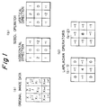
- Sobel operators shown in Fig. 1(b) are employed as an example of operators for obtaining a differential value in a local area of the image.
- Such new values are calculated with regard to all pixels in the entire image frame.
- new values are calculated in accordance with the following as differentiated values in the horizontal and vertical directions of the pixel of interest: In a background portion where particles do not appear the new value is approximately zero. However, when a particle enters the image frame, the calculated value at the contour thereof becomes larger in proportion to contrast as focusing is performed. Accordingly, a value obtained by accumulating such calculated values over the entire image frame can be utilized as an evaluation parameter indicating the degree of focusing.
- a solution containing control particles for precision management in which particles are uniform in dimensions and shape is introduced to a flow cell, and focusing is performed by processing the image in which the particles appear, thereby raising the precision of classification and analysis of a specimen containing a wide variety of particle components.
- the number of particles which appear in one frame will be only several tens at low magnification and several at high magnification.
- the cumulative differential value for one frame will exhibit a large statistical variance when compared with other frames.
- Laplacian operators Two types of Laplacian operators (c-1), (c-2) are illustrated in Fig. 1(c). When the Laplacian operator (c-1) is used, a 3 ⁇ 3 convolution takes on the following value:
- Equation (4) If the evaluation parameter P is obtained using Equation (4) or (5), P will be expressed by either of the following: or where h is a constant.
- the differential value Sbx ij which uses the horizontal operator and the differential value Sby ij which uses the vertical operator are obtained, and the evaluation parameter P is calculated using Equation (3).
- the Laplacian method it will be noted that one step of the calculations can be omitted.
- the above-mentioned evaluation parameter is obtained by real-time processing of the image data using an image processor with which the cytometer is furnished.
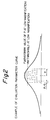
- a curve of the kind shown in Fig. 2 is obtained.
- the point at which the maximum value is obtained is the position at which focusing is achieved.
- An evaluation parameter according to Equation (3) is obtained using an image processor shown in Fig. 3.
- An example of the circuitry of an image processor board forming the nucleus of Fig. 3 is illustrated in Fig. 4.
- a morphology processor board is a board on which general functions necessary for processing a cell image are mounted; the board is not specially mounted in order to focus an image. Illustrated below is a method of obtaining evaluation parameters of a plurality of frames in which one field (1/60 of a second) is adopted as one step.
- an evaluation parameter for one frame will take one second assuming that one frame is composed of 256 ⁇ 256 pixels. Therefore, about 20 seconds will be required to obtain evaluation parameters for 20 frames. In actuality, however, a longer time is required since focus adjustment is performed while moving the lenses or flow cell.
- an evaluation parameter is obtained in real-time with respect to one frame which appears every 1/30 of a second, 20/30 of a second will be required to obtain evaluation parameters for 20 frames.
- an evaluation value for focusing should be obtained using a digital filter, namely a 3 ⁇ 3 convolution filter, described below, which incorporates a high-speed multiplier and a high-speed cumulative adder.
- step (i) Processing similar to that of step (ii) is executed with regard to the next frame in conjunction with step (iii).
- step (iv) Processing similar to that of step (ii) is executed with regard to the next frame.
- step (v) After processing similar to that of step (iii) is executed with regard to the next frame, the evaluation parameter of the initial frame and the evaluation parameter of the next frame are added. At the same time, processing similar to that of step (i) is executed with regard to the next frame. Thereafter, processing is applied to successive frames one after another in the same manner.
- the differential values Sbx, Sby of Equation (3) can be obtained in real-time by an FIR filter used as the 3 ⁇ 3 convoluter (Fig. 4) of process channel 2. Furthermore, processing for squaring both differential values and then multiplying them by 1/k can be performed in real-time by a look-up table LUT3.
- FIG. 6 showing a focusing flowchart. Focusing in the present invention is divided into two stages, namely, a coarse adjustment step and a fine adjustment step.
- a control solution is passed through a flow cell and imaged by a video camera, and an initial value P ol of the evaluation parameter of the image on the low-magnification mode is obtained.
- P ol of the evaluation parameter of the image on the low-magnification mode is obtained.
- Step 1 When it is determined that an image is too far out of focus, namely when the relation P ol ⁇ th l holds with regard to a threshold th l that gives the boundary between a coarse-adjustment region and a fine-adjustment region, it is required to bring adjustment to the fine-adjustment region much more speedily by following Step 1, which constitutes performing coarse adjustment at a high speed of flow cell or lens movement.
- Step 2 is executed in the next procedure in the above-described adjustment algorithm.
- the time required for adjustment at Step 2 is 6 - 7 seconds.
- Step 2 (Fine Adjustment): P o ⁇ th l
- Step 3 which is for a fine adjustment on the high-magnification mode, is executed through the following procedure:
- Step 3 Fine Adjustment on High-Magnification Mode
- the time required for the adjustment of Step 3 is 28 - 32 seconds.
- a still image of a specimen solution is captured every 1/30 of a second, and therefore identical frames do not occur.
- Evaluation parameters are obtained using these images that change from one instant to the next. Accordingly, though the evaluation parameters differ greatly from frame to frame, if a value obtained by adding up the evaluation parameters for a plurality of frames is adopted as the evaluation parameter, then the differences among a plurality of frames will be absorbed according to the rules of algebra, the characteristic features of the image are made to stand out and this is accurately reflected in the focused state of the cell image.
- the value of the evaluation parameter is obtained in real-time while continuously moving the flow cell or lens, and the position at which this value is maximized is rapidly determined.
- the time required for adjustment can be curtailed first by coarse adjustment of focus at low magnification and then by fine adjustment of focus at high magnification.
Landscapes
- Chemical & Material Sciences (AREA)
- Biochemistry (AREA)
- General Physics & Mathematics (AREA)
- Health & Medical Sciences (AREA)
- Life Sciences & Earth Sciences (AREA)
- Analytical Chemistry (AREA)
- Dispersion Chemistry (AREA)
- General Health & Medical Sciences (AREA)
- Physics & Mathematics (AREA)
- Immunology (AREA)
- Pathology (AREA)
- Investigating Or Analysing Biological Materials (AREA)
- Investigating Or Analysing Materials By Optical Means (AREA)
- Closed-Circuit Television Systems (AREA)
- Image Processing (AREA)
Claims (5)
- Procédé d'ajustement d'un point focal dans un cytomètre à formation d'images à circulation, dans lequel une solution échantillon contenant des particules constituantes s'écoule en étant enveloppée par un liquide d'enveloppe dans un passage plat de circulation d'une cellule à circulation, des images fixes de la solution échantillon sont saisies à l'aide d'un dispositif de projection de lumière et d'un dispositif capteur d'image placés de part et d'autre de la cellule à circulation et l'analyse des particules constituantes, contenues dans la solution échantillon, est réalisée par traitement d'image, le procédé d'ajustement du point focal comprenant les étapes suivantes :
l'introduction d'une solution témoin qui contient des particules de configurations et dimensions uniformes dans la cellule à circulation,
la saisie d'images fixes de la solution témoin avec déplacement d'un élément du circuit optique de mesure, par exemple de la cellule à circulation,
le calcul de paramètres d'évaluation qui représentent la définition des images fixes, et
l'exécution d'un ajustement du point focal par rapport au courant de l'échantillon par ajustement de la position de l'élément du circuit optique de manière que la valeur d'un paramètre d'évaluation soit rendue maximale. - Procédé selon la revendication 1, comprenant en outre les étapes suivantes :
la comparaison d'une valeur initiale Po du paramètre d'évaluation et d'une valeur prédéterminée de référence th lorsque la solution témoin est introduite dans la cellule à circulation,
l'obtention d'un paramètre d'évaluation Pn et l'établissement d'un état dans lequel la relation Pn > th est vérifiée par déplacement de la cellule à circulation lorsque la relation Po < th est vérifiée, et
l'établissement de la position par déplacement de la cellule à circulation vers une position dans laquelle le paramètre d'évaluation Pn est rendu maximal. - Procédé selon la revendication 2, dans lequel la position d'un élément optique, par exemple un ensemble à lentille du cytomètre à circulation, est déplacée à la place de la position de la cellule à circulation continue.
- Procédé selon l'une quelconque des revendications 1 à 3, comprenant en outre les étapes suivantes :
le calcul initial du paramètre d'évaluation en mode à faible grandissement du système optique, et
lorsque le paramètre d'évaluation a été rendu maximal, l'augmentation du paramètre d'évaluation à une valeur maximale en mode à grandissement élevé du système optique. - Procédé selon l'une quelconque des revendications 1 à 4, comprenant en outre les étapes suivantes :
la mémorisation d'une valeur maximale du paramètre d'évaluation, et
la détection d'une anomalie de l'ensemble de mesure par contrôle d'une fluctuation de la valeur maximale du paramètre d'évaluation qui est mémorisé.
Applications Claiming Priority (2)
| Application Number | Priority Date | Filing Date | Title |
|---|---|---|---|
| JP195934/90 | 1990-07-24 | ||
| JP2195934A JP2939647B2 (ja) | 1990-07-24 | 1990-07-24 | フローイメージングサイトメータにおける自動焦点調整方法 |
Publications (2)
| Publication Number | Publication Date |
|---|---|
| EP0468100A1 EP0468100A1 (fr) | 1992-01-29 |
| EP0468100B1 true EP0468100B1 (fr) | 1995-03-08 |
Family
ID=16349405
Family Applications (1)
| Application Number | Title | Priority Date | Filing Date |
|---|---|---|---|
| EP90125596A Expired - Lifetime EP0468100B1 (fr) | 1990-07-24 | 1990-12-27 | Procédé pour la régulation automatique du point focal dans un cytomètre pour la représentation d'un écoulement |
Country Status (5)
| Country | Link |
|---|---|
| US (1) | US5083014A (fr) |
| EP (1) | EP0468100B1 (fr) |
| JP (1) | JP2939647B2 (fr) |
| CA (1) | CA2039897C (fr) |
| DE (1) | DE69017675T2 (fr) |
Cited By (1)
| Publication number | Priority date | Publication date | Assignee | Title |
|---|---|---|---|---|
| US9617288B2 (en) | 2014-02-10 | 2017-04-11 | Enzytech, Ltd. | Method for preparing racemic or optically active α-glycerophosphorylcholine |
Families Citing this family (35)
| Publication number | Priority date | Publication date | Assignee | Title |
|---|---|---|---|---|
| JP3111706B2 (ja) * | 1992-02-18 | 2000-11-27 | 株式会社日立製作所 | 粒子分析装置及び粒子分析方法 |
| US5255089A (en) * | 1992-03-26 | 1993-10-19 | International Business Machines Corporation | Portable particle detector assembly |
| US6861265B1 (en) | 1994-10-14 | 2005-03-01 | University Of Washington | Flow cytometer droplet formation system |
| DE69532416T2 (de) * | 1994-10-14 | 2004-12-02 | University Of Washington, Seattle | Methode und vorrichtung zur tröpfchenerzeugung mit hoher rate bei der durchfluss-cytometrie |
| US5602349A (en) * | 1994-10-14 | 1997-02-11 | The University Of Washington | Sample introduction system for a flow cytometer |
| EP2264427B1 (fr) | 1997-01-31 | 2017-05-03 | Xy, Llc | Dispositif optique avec réflecteur focalisant pour faire converger la radiation sur un débit de particule, et procédé d'analyse associé |
| ATE514072T1 (de) | 1997-05-05 | 2011-07-15 | Chemometec As | Verfahren zur bestimmung von teilchen in einer flüssigen probe |
| US6071689A (en) | 1997-12-31 | 2000-06-06 | Xy, Inc. | System for improving yield of sexed embryos in mammals |
| US6149867A (en) | 1997-12-31 | 2000-11-21 | Xy, Inc. | Sheath fluids and collection systems for sex-specific cytometer sorting of sperm |
| US6248590B1 (en) | 1998-02-27 | 2001-06-19 | Cytomation, Inc. | Method and apparatus for flow cytometry |
| JPH11295208A (ja) * | 1998-04-13 | 1999-10-29 | Sysmex Corp | 粒子撮像装置 |
| PT1917974E (pt) | 1998-07-30 | 2011-02-22 | Xy Llc | Sistema de inseminação artificial não cirúrgica em equinos |
| US7024316B1 (en) | 1999-10-21 | 2006-04-04 | Dakocytomation Colorado, Inc. | Transiently dynamic flow cytometer analysis system |
| US7208265B1 (en) | 1999-11-24 | 2007-04-24 | Xy, Inc. | Method of cryopreserving selected sperm cells |
| IL152714A (en) | 2000-05-09 | 2014-03-31 | Xy Llc | High-purity spermatozoa populations carrying chromosome-x and chromosome-y |
| WO2002043486A1 (fr) | 2000-11-29 | 2002-06-06 | Xy, Inc. | Systeme permettant de realiser une fecondation in vitro avec des spermatozoides separes en population porteuse de chromosome x et en population porteuse de chromosome y |
| US7713687B2 (en) | 2000-11-29 | 2010-05-11 | Xy, Inc. | System to separate frozen-thawed spermatozoa into x-chromosome bearing and y-chromosome bearing populations |
| JP4595067B2 (ja) | 2002-08-01 | 2010-12-08 | エックスワイ,エルエルシー | 低圧精子細胞分離システム |
| US8486618B2 (en) | 2002-08-01 | 2013-07-16 | Xy, Llc | Heterogeneous inseminate system |
| CA2534394C (fr) | 2002-08-15 | 2013-01-08 | Xy, Inc. | Cytometre a flux haute resolution |
| US7169548B2 (en) | 2002-09-13 | 2007-01-30 | Xy, Inc. | Sperm cell processing and preservation systems |
| BRPI0408857B1 (pt) | 2003-03-28 | 2018-09-11 | Inguran Llc | aparelho, métodos e processos para separar partículas e para prover esperma de animal separado por sexo |
| ES2541121T3 (es) | 2003-05-15 | 2015-07-16 | Xy, Llc | Clasificación eficiente de células haploides por sistemas de citometría de flujo |
| DK2801363T3 (en) | 2004-03-29 | 2018-05-28 | Inguran Llc | PROCEDURE FOR STORING SORTED SPERMATOZOES |
| CA2574499C (fr) | 2004-07-22 | 2016-11-29 | Monsanto Technology Llc | Procede pour enrichir une population de spermatozoides |
| DK1771729T3 (en) | 2004-07-27 | 2015-11-23 | Beckman Coulter Inc | Improving flowcytometridiskrimination using geometric transformation |
| US7618770B2 (en) | 2005-07-29 | 2009-11-17 | Xy, Inc. | Methods and apparatus for reducing protein content in sperm cell extenders |
| US7804594B2 (en) | 2006-12-29 | 2010-09-28 | Abbott Laboratories, Inc. | Method and apparatus for rapidly counting and identifying biological particles in a flow stream |
| US8159670B2 (en) | 2007-11-05 | 2012-04-17 | Abbott Laboratories | Method and apparatus for rapidly counting and identifying biological particles in a flow stream |
| JP5254441B2 (ja) * | 2009-06-03 | 2013-08-07 | 株式会社日立ハイテクノロジーズ | フロー式粒子画像解析方法及び装置 |
| KR102095617B1 (ko) * | 2013-03-15 | 2020-03-31 | 아이리스 인터내셔널 인크. | 혈액 샘플에서의 입자 분석을 위한 오토포커스 시스템 및 방법 |
| US9857361B2 (en) | 2013-03-15 | 2018-01-02 | Iris International, Inc. | Flowcell, sheath fluid, and autofocus systems and methods for particle analysis in urine samples |
| US9316635B2 (en) | 2013-03-15 | 2016-04-19 | Iris International, Inc. | Sheath fluid systems and methods for particle analysis in blood samples |
| EP3202133B1 (fr) | 2014-09-29 | 2018-11-21 | Biosurfit, S.A. | Procédé de mise au point |
| JP7683484B2 (ja) * | 2019-11-06 | 2025-05-27 | ソニーグループ株式会社 | 位置調整方法、微小粒子分析装置、及びプログラム |
Family Cites Families (5)
| Publication number | Priority date | Publication date | Assignee | Title |
|---|---|---|---|---|
| US4338024A (en) * | 1980-05-02 | 1982-07-06 | International Remote Imaging Systems, Inc. | Flow analyzer and system for analysis of fluids with particles |
| US4519087A (en) * | 1983-06-20 | 1985-05-21 | International Remote Imaging Systems, Inc. | Method of determining the diagnostic significance of the content of a volume of biological sample containing particles |
| US4660971A (en) * | 1984-05-03 | 1987-04-28 | Becton, Dickinson And Company | Optical features of flow cytometry apparatus |
| JPS61280548A (ja) * | 1985-06-05 | 1986-12-11 | Canon Inc | 粒子解析装置 |
| US4804267A (en) * | 1986-07-10 | 1989-02-14 | Scientific Imaging Instruments, Inc. | System for microscopically analyzing fluids |
-
1990
- 1990-07-24 JP JP2195934A patent/JP2939647B2/ja not_active Expired - Fee Related
- 1990-12-26 US US07/633,783 patent/US5083014A/en not_active Expired - Lifetime
- 1990-12-27 EP EP90125596A patent/EP0468100B1/fr not_active Expired - Lifetime
- 1990-12-27 DE DE69017675T patent/DE69017675T2/de not_active Expired - Fee Related
-
1991
- 1991-04-05 CA CA002039897A patent/CA2039897C/fr not_active Expired - Fee Related
Cited By (1)
| Publication number | Priority date | Publication date | Assignee | Title |
|---|---|---|---|---|
| US9617288B2 (en) | 2014-02-10 | 2017-04-11 | Enzytech, Ltd. | Method for preparing racemic or optically active α-glycerophosphorylcholine |
Also Published As
| Publication number | Publication date |
|---|---|
| EP0468100A1 (fr) | 1992-01-29 |
| JP2939647B2 (ja) | 1999-08-25 |
| CA2039897C (fr) | 2001-10-16 |
| DE69017675T2 (de) | 1995-08-17 |
| DE69017675D1 (de) | 1995-04-13 |
| CA2039897A1 (fr) | 1992-01-25 |
| US5083014A (en) | 1992-01-21 |
| JPH0481640A (ja) | 1992-03-16 |
Similar Documents
| Publication | Publication Date | Title |
|---|---|---|
| EP0468100B1 (fr) | Procédé pour la régulation automatique du point focal dans un cytomètre pour la représentation d'un écoulement | |
| CA2202138C (fr) | Procede et appareil pour controler la fiabilite du fonctionnement repetitif d'un systeme optique automatise | |
| AU725820B2 (en) | Method and apparatus for assessing slide and specimen preparation quality | |
| EP0486747B1 (fr) | Mécanisme d'une cellule à écoulement dans un cytomètre d'écoulement à formation d'image | |
| AU709136B2 (en) | Automatic focusing of biomedical specimens apparatus | |
| DE69508248T2 (de) | Autofokussystem für abtastmikroskopie | |
| US7769219B2 (en) | Method for assessing image focus quality | |
| US5911002A (en) | Pattern recognition system | |
| EP3988985A2 (fr) | Focalisation automatique rapide dans imagerie microscopique | |
| US5715327A (en) | Method and apparatus for detection of unsuitable conditions for automated cytology scoring | |
| CN109361849A (zh) | 一种自动对焦的算法 | |
| US4667335A (en) | Method of determining the diagnostic significance of the content of a volume of biological sample containing particles | |
| US5995680A (en) | Cytological system illumination integrity checking apparatus and method | |
| US4519087A (en) | Method of determining the diagnostic significance of the content of a volume of biological sample containing particles | |
| CN120103597A (zh) | 一种自动聚焦方法及显微镜图像获取设备 | |
| Allen et al. | Automatic recognition of light microscope pollen images | |
| CN121541358B (zh) | 一种自动对焦控制方法、系统及介质 | |
| Li et al. | An self-focusing imaging method for leukocyte recognition | |
| AU687640C (en) | Method and apparatus for checking automated optical system performance repeatability | |
| CN121541358A (zh) | 一种自动对焦控制方法、系统及介质 | |
| CN120055608A (zh) | 一种运用ccd视觉传感器的封口高速寻焊缝工艺 | |
| HK1130345B (en) | Method for assessing image focus quality |
Legal Events
| Date | Code | Title | Description |
|---|---|---|---|
| PUAI | Public reference made under article 153(3) epc to a published international application that has entered the european phase |
Free format text: ORIGINAL CODE: 0009012 |
|
| AK | Designated contracting states |
Kind code of ref document: A1 Designated state(s): DE FR GB IT |
|
| 17P | Request for examination filed |
Effective date: 19920317 |
|
| 17Q | First examination report despatched |
Effective date: 19940309 |
|
| GRAA | (expected) grant |
Free format text: ORIGINAL CODE: 0009210 |
|
| AK | Designated contracting states |
Kind code of ref document: B1 Designated state(s): DE FR GB IT |
|
| PG25 | Lapsed in a contracting state [announced via postgrant information from national office to epo] |
Ref country code: IT Free format text: LAPSE BECAUSE OF FAILURE TO SUBMIT A TRANSLATION OF THE DESCRIPTION OR TO PAY THE FEE WITHIN THE PRE;WARNING: LAPSES OF ITALIAN PATENTS WITH EFFECTIVE DATE BEFORE 2007 MAY HAVE OCCURRED AT ANY TIME BEFORE 2007. THE CORRECT EFFECTIVE DATE MAY BE DIFFERENT FROM THE ONE RECORDED.SCRIBED TIME-LIMIT Effective date: 19950308 |
|
| REF | Corresponds to: |
Ref document number: 69017675 Country of ref document: DE Date of ref document: 19950413 |
|
| ET | Fr: translation filed | ||
| PLBE | No opposition filed within time limit |
Free format text: ORIGINAL CODE: 0009261 |
|
| STAA | Information on the status of an ep patent application or granted ep patent |
Free format text: STATUS: NO OPPOSITION FILED WITHIN TIME LIMIT |
|
| 26N | No opposition filed | ||
| REG | Reference to a national code |
Ref country code: GB Ref legal event code: IF02 |
|
| PGFP | Annual fee paid to national office [announced via postgrant information from national office to epo] |
Ref country code: FR Payment date: 20021210 Year of fee payment: 13 |
|
| PGFP | Annual fee paid to national office [announced via postgrant information from national office to epo] |
Ref country code: GB Payment date: 20021224 Year of fee payment: 13 |
|
| PGFP | Annual fee paid to national office [announced via postgrant information from national office to epo] |
Ref country code: DE Payment date: 20030109 Year of fee payment: 13 |
|
| PG25 | Lapsed in a contracting state [announced via postgrant information from national office to epo] |
Ref country code: GB Free format text: LAPSE BECAUSE OF NON-PAYMENT OF DUE FEES Effective date: 20031227 |
|
| PG25 | Lapsed in a contracting state [announced via postgrant information from national office to epo] |
Ref country code: DE Free format text: LAPSE BECAUSE OF NON-PAYMENT OF DUE FEES Effective date: 20040701 |
|
| GBPC | Gb: european patent ceased through non-payment of renewal fee |
Effective date: 20031227 |
|
| PG25 | Lapsed in a contracting state [announced via postgrant information from national office to epo] |
Ref country code: FR Free format text: LAPSE BECAUSE OF NON-PAYMENT OF DUE FEES Effective date: 20040831 |
|
| REG | Reference to a national code |
Ref country code: FR Ref legal event code: ST |