EP0486589B1 - Chirurgisches instrument und zellisolierung - Google Patents
Chirurgisches instrument und zellisolierung Download PDFInfo
- Publication number
- EP0486589B1 EP0486589B1 EP90912685A EP90912685A EP0486589B1 EP 0486589 B1 EP0486589 B1 EP 0486589B1 EP 90912685 A EP90912685 A EP 90912685A EP 90912685 A EP90912685 A EP 90912685A EP 0486589 B1 EP0486589 B1 EP 0486589B1
- Authority
- EP
- European Patent Office
- Prior art keywords
- retina
- tube
- cells
- graft
- instrument
- Prior art date
- Legal status (The legal status is an assumption and is not a legal conclusion. Google has not performed a legal analysis and makes no representation as to the accuracy of the status listed.)
- Expired - Lifetime
Links
- 238000002955 isolation Methods 0.000 title abstract description 10
- 210000001519 tissue Anatomy 0.000 claims abstract description 78
- 238000000034 method Methods 0.000 claims abstract description 61
- 238000002054 transplantation Methods 0.000 claims abstract description 45
- 230000002207 retinal effect Effects 0.000 claims abstract description 37
- 210000001525 retina Anatomy 0.000 claims description 191
- 210000004027 cell Anatomy 0.000 claims description 77
- 108010010803 Gelatin Proteins 0.000 claims description 36
- 239000008273 gelatin Substances 0.000 claims description 36
- 229920000159 gelatin Polymers 0.000 claims description 36
- 235000019322 gelatine Nutrition 0.000 claims description 36
- 235000011852 gelatine desserts Nutrition 0.000 claims description 36
- 210000000608 photoreceptor cell Anatomy 0.000 claims description 31
- 230000008520 organization Effects 0.000 claims description 22
- 210000003850 cellular structure Anatomy 0.000 claims description 19
- 239000012530 fluid Substances 0.000 claims description 17
- 239000000203 mixture Substances 0.000 claims description 11
- 238000003780 insertion Methods 0.000 claims description 10
- 230000037431 insertion Effects 0.000 claims description 10
- 210000000981 epithelium Anatomy 0.000 claims description 9
- 239000000835 fiber Substances 0.000 claims description 9
- 230000001413 cellular effect Effects 0.000 claims description 8
- 239000003795 chemical substances by application Substances 0.000 claims description 7
- 238000002360 preparation method Methods 0.000 claims description 7
- 230000036760 body temperature Effects 0.000 claims description 4
- 238000003306 harvesting Methods 0.000 claims description 4
- 238000002513 implantation Methods 0.000 claims description 4
- 231100000252 nontoxic Toxicity 0.000 claims description 3
- 230000003000 nontoxic effect Effects 0.000 claims description 3
- 239000002870 angiogenesis inducing agent Substances 0.000 claims description 2
- 230000001772 anti-angiogenic effect Effects 0.000 claims description 2
- 230000002927 anti-mitotic effect Effects 0.000 claims description 2
- 229960003444 immunosuppressant agent Drugs 0.000 claims description 2
- 239000003018 immunosuppressive agent Substances 0.000 claims description 2
- 230000001228 trophic effect Effects 0.000 claims description 2
- 230000001861 immunosuppressant effect Effects 0.000 claims 1
- 238000010030 laminating Methods 0.000 claims 1
- 108091008695 photoreceptors Proteins 0.000 abstract description 177
- 210000003583 retinal pigment epithelium Anatomy 0.000 abstract description 30
- 238000001356 surgical procedure Methods 0.000 abstract description 6
- 210000001508 eye Anatomy 0.000 description 98
- 239000000758 substrate Substances 0.000 description 28
- VRYALKFFQXWPIH-PBXRRBTRSA-N (3r,4s,5r)-3,4,5,6-tetrahydroxyhexanal Chemical compound OC[C@@H](O)[C@@H](O)[C@H](O)CC=O VRYALKFFQXWPIH-PBXRRBTRSA-N 0.000 description 25
- 241000700159 Rattus Species 0.000 description 25
- PMMURAAUARKVCB-UHFFFAOYSA-N alpha-D-ara-dHexp Natural products OCC1OC(O)CC(O)C1O PMMURAAUARKVCB-UHFFFAOYSA-N 0.000 description 24
- 230000001537 neural effect Effects 0.000 description 20
- 102000010175 Opsin Human genes 0.000 description 19
- 108050001704 Opsin Proteins 0.000 description 19
- 238000013459 approach Methods 0.000 description 18
- 241000699666 Mus <mouse, genus> Species 0.000 description 15
- 230000001179 pupillary effect Effects 0.000 description 10
- 230000004083 survival effect Effects 0.000 description 10
- 238000010276 construction Methods 0.000 description 9
- 210000005056 cell body Anatomy 0.000 description 8
- 210000003161 choroid Anatomy 0.000 description 8
- 230000011514 reflex Effects 0.000 description 8
- 241001465754 Metazoa Species 0.000 description 7
- 230000001419 dependent effect Effects 0.000 description 7
- 238000005286 illumination Methods 0.000 description 7
- 230000004913 activation Effects 0.000 description 6
- 230000000740 bleeding effect Effects 0.000 description 6
- 230000006378 damage Effects 0.000 description 6
- 208000014674 injury Diseases 0.000 description 6
- 238000001000 micrograph Methods 0.000 description 6
- 239000000049 pigment Substances 0.000 description 6
- 230000001953 sensory effect Effects 0.000 description 6
- 230000008733 trauma Effects 0.000 description 6
- 208000003098 Ganglion Cysts Diseases 0.000 description 5
- 241000283984 Rodentia Species 0.000 description 5
- 208000005400 Synovial Cyst Diseases 0.000 description 5
- 210000004556 brain Anatomy 0.000 description 5
- JVXZRNYCRFIEGV-UHFFFAOYSA-M dilC18(3) dye Chemical compound [O-]Cl(=O)(=O)=O.CC1(C)C2=CC=CC=C2N(CCCCCCCCCCCCCCCCCC)C1=CC=CC1=[N+](CCCCCCCCCCCCCCCCCC)C2=CC=CC=C2C1(C)C JVXZRNYCRFIEGV-UHFFFAOYSA-M 0.000 description 5
- 230000004438 eyesight Effects 0.000 description 5
- MHMNJMPURVTYEJ-UHFFFAOYSA-N fluorescein-5-isothiocyanate Chemical compound O1C(=O)C2=CC(N=C=S)=CC=C2C21C1=CC=C(O)C=C1OC1=CC(O)=CC=C21 MHMNJMPURVTYEJ-UHFFFAOYSA-N 0.000 description 5
- 239000007943 implant Substances 0.000 description 5
- 238000002372 labelling Methods 0.000 description 5
- 239000000463 material Substances 0.000 description 5
- 230000004044 response Effects 0.000 description 5
- 239000000243 solution Substances 0.000 description 5
- 210000000225 synapse Anatomy 0.000 description 5
- 241000288906 Primates Species 0.000 description 4
- 241000278713 Theora Species 0.000 description 4
- 210000003050 axon Anatomy 0.000 description 4
- 210000003169 central nervous system Anatomy 0.000 description 4
- 210000004087 cornea Anatomy 0.000 description 4
- 230000001404 mediated effect Effects 0.000 description 4
- 238000000386 microscopy Methods 0.000 description 4
- 238000010186 staining Methods 0.000 description 4
- 230000035899 viability Effects 0.000 description 4
- 230000000007 visual effect Effects 0.000 description 4
- 229930003347 Atropine Natural products 0.000 description 3
- 201000004569 Blindness Diseases 0.000 description 3
- 229930105110 Cyclosporin A Natural products 0.000 description 3
- PMATZTZNYRCHOR-CGLBZJNRSA-N Cyclosporin A Chemical compound CC[C@@H]1NC(=O)[C@H]([C@H](O)[C@H](C)C\C=C\C)N(C)C(=O)[C@H](C(C)C)N(C)C(=O)[C@H](CC(C)C)N(C)C(=O)[C@H](CC(C)C)N(C)C(=O)[C@@H](C)NC(=O)[C@H](C)NC(=O)[C@H](CC(C)C)N(C)C(=O)[C@H](C(C)C)NC(=O)[C@H](CC(C)C)N(C)C(=O)CN(C)C1=O PMATZTZNYRCHOR-CGLBZJNRSA-N 0.000 description 3
- 108010036949 Cyclosporine Proteins 0.000 description 3
- RKUNBYITZUJHSG-UHFFFAOYSA-N Hyosciamin-hydrochlorid Natural products CN1C(C2)CCC1CC2OC(=O)C(CO)C1=CC=CC=C1 RKUNBYITZUJHSG-UHFFFAOYSA-N 0.000 description 3
- 206010038848 Retinal detachment Diseases 0.000 description 3
- VREFGVBLTWBCJP-UHFFFAOYSA-N alprazolam Chemical compound C12=CC(Cl)=CC=C2N2C(C)=NN=C2CN=C1C1=CC=CC=C1 VREFGVBLTWBCJP-UHFFFAOYSA-N 0.000 description 3
- 238000010171 animal model Methods 0.000 description 3
- RKUNBYITZUJHSG-SPUOUPEWSA-N atropine Chemical compound O([C@H]1C[C@H]2CC[C@@H](C1)N2C)C(=O)C(CO)C1=CC=CC=C1 RKUNBYITZUJHSG-SPUOUPEWSA-N 0.000 description 3
- 229960000396 atropine Drugs 0.000 description 3
- 230000006399 behavior Effects 0.000 description 3
- 210000003986 cell retinal photoreceptor Anatomy 0.000 description 3
- 229960001265 ciclosporin Drugs 0.000 description 3
- 230000006735 deficit Effects 0.000 description 3
- UREBDLICKHMUKA-CXSFZGCWSA-N dexamethasone Chemical compound C1CC2=CC(=O)C=C[C@]2(C)[C@]2(F)[C@@H]1[C@@H]1C[C@@H](C)[C@@](C(=O)CO)(O)[C@@]1(C)C[C@@H]2O UREBDLICKHMUKA-CXSFZGCWSA-N 0.000 description 3
- 229960003957 dexamethasone Drugs 0.000 description 3
- 230000000694 effects Effects 0.000 description 3
- 230000012010 growth Effects 0.000 description 3
- 230000035876 healing Effects 0.000 description 3
- 238000000338 in vitro Methods 0.000 description 3
- 238000012423 maintenance Methods 0.000 description 3
- WEXRUCMBJFQVBZ-UHFFFAOYSA-N pentobarbital Chemical compound CCCC(C)C1(CC)C(=O)NC(=O)NC1=O WEXRUCMBJFQVBZ-UHFFFAOYSA-N 0.000 description 3
- 230000004264 retinal detachment Effects 0.000 description 3
- 230000000087 stabilizing effect Effects 0.000 description 3
- 239000000126 substance Substances 0.000 description 3
- 230000000699 topical effect Effects 0.000 description 3
- CSCPPACGZOOCGX-UHFFFAOYSA-N Acetone Chemical compound CC(C)=O CSCPPACGZOOCGX-UHFFFAOYSA-N 0.000 description 2
- CURLTUGMZLYLDI-UHFFFAOYSA-N Carbon dioxide Chemical compound O=C=O CURLTUGMZLYLDI-UHFFFAOYSA-N 0.000 description 2
- 229920002134 Carboxymethyl cellulose Polymers 0.000 description 2
- 206010061431 Glial scar Diseases 0.000 description 2
- 206010018341 Gliosis Diseases 0.000 description 2
- SXRSQZLOMIGNAQ-UHFFFAOYSA-N Glutaraldehyde Chemical compound O=CCCCC=O SXRSQZLOMIGNAQ-UHFFFAOYSA-N 0.000 description 2
- 108090001090 Lectins Proteins 0.000 description 2
- 102000004856 Lectins Human genes 0.000 description 2
- 241000282553 Macaca Species 0.000 description 2
- 241000699670 Mus sp. Species 0.000 description 2
- 208000007014 Retinitis pigmentosa Diseases 0.000 description 2
- 230000001594 aberrant effect Effects 0.000 description 2
- VLSMHEGGTFMBBZ-UHFFFAOYSA-N alpha-Kainic acid Natural products CC(=C)C1CNC(C(O)=O)C1CC(O)=O VLSMHEGGTFMBBZ-UHFFFAOYSA-N 0.000 description 2
- 210000002159 anterior chamber Anatomy 0.000 description 2
- 239000002260 anti-inflammatory agent Substances 0.000 description 2
- 239000003963 antioxidant agent Substances 0.000 description 2
- 235000006708 antioxidants Nutrition 0.000 description 2
- 230000003542 behavioural effect Effects 0.000 description 2
- 230000008901 benefit Effects 0.000 description 2
- 208000002352 blister Diseases 0.000 description 2
- 210000004204 blood vessel Anatomy 0.000 description 2
- 235000010948 carboxy methyl cellulose Nutrition 0.000 description 2
- 239000001768 carboxy methyl cellulose Substances 0.000 description 2
- 239000008112 carboxymethyl-cellulose Substances 0.000 description 2
- 230000007547 defect Effects 0.000 description 2
- 230000007850 degeneration Effects 0.000 description 2
- 230000018044 dehydration Effects 0.000 description 2
- 238000006297 dehydration reaction Methods 0.000 description 2
- 238000011161 development Methods 0.000 description 2
- 230000018109 developmental process Effects 0.000 description 2
- 230000000763 evoking effect Effects 0.000 description 2
- 238000002474 experimental method Methods 0.000 description 2
- 230000006870 function Effects 0.000 description 2
- 239000000499 gel Substances 0.000 description 2
- 238000002347 injection Methods 0.000 description 2
- 239000007924 injection Substances 0.000 description 2
- 230000010354 integration Effects 0.000 description 2
- 238000007917 intracranial administration Methods 0.000 description 2
- VLSMHEGGTFMBBZ-OOZYFLPDSA-N kainic acid Chemical compound CC(=C)[C@H]1CN[C@H](C(O)=O)[C@H]1CC(O)=O VLSMHEGGTFMBBZ-OOZYFLPDSA-N 0.000 description 2
- 229950006874 kainic acid Drugs 0.000 description 2
- 239000002523 lectin Substances 0.000 description 2
- 208000002780 macular degeneration Diseases 0.000 description 2
- 238000013507 mapping Methods 0.000 description 2
- 239000011159 matrix material Substances 0.000 description 2
- 239000012528 membrane Substances 0.000 description 2
- 230000028161 membrane depolarization Effects 0.000 description 2
- 210000000653 nervous system Anatomy 0.000 description 2
- 210000005157 neural retina Anatomy 0.000 description 2
- 239000002581 neurotoxin Substances 0.000 description 2
- 229960001412 pentobarbital Drugs 0.000 description 2
- 230000016732 phototransduction Effects 0.000 description 2
- -1 polyethylene Polymers 0.000 description 2
- 230000001242 postsynaptic effect Effects 0.000 description 2
- 210000001747 pupil Anatomy 0.000 description 2
- 238000011084 recovery Methods 0.000 description 2
- 238000011160 research Methods 0.000 description 2
- 210000003786 sclera Anatomy 0.000 description 2
- 238000012453 sprague-dawley rat model Methods 0.000 description 2
- 230000000638 stimulation Effects 0.000 description 2
- 238000003860 storage Methods 0.000 description 2
- 230000000946 synaptic effect Effects 0.000 description 2
- 238000012360 testing method Methods 0.000 description 2
- 229920000936 Agarose Polymers 0.000 description 1
- 229920003319 Araldite® Polymers 0.000 description 1
- 238000012935 Averaging Methods 0.000 description 1
- 239000011547 Bouin solution Substances 0.000 description 1
- 108091003079 Bovine Serum Albumin Proteins 0.000 description 1
- 108010062580 Concanavalin A Proteins 0.000 description 1
- LEVWYRKDKASIDU-QWWZWVQMSA-N D-cystine Chemical compound OC(=O)[C@H](N)CSSC[C@@H](N)C(O)=O LEVWYRKDKASIDU-QWWZWVQMSA-N 0.000 description 1
- 241001269524 Dura Species 0.000 description 1
- 102000018233 Fibroblast Growth Factor Human genes 0.000 description 1
- 108050007372 Fibroblast Growth Factor Proteins 0.000 description 1
- 241000282412 Homo Species 0.000 description 1
- 208000032578 Inherited retinal disease Diseases 0.000 description 1
- YQEZLKZALYSWHR-UHFFFAOYSA-N Ketamine Chemical compound C=1C=CC=C(Cl)C=1C1(NC)CCCCC1=O YQEZLKZALYSWHR-UHFFFAOYSA-N 0.000 description 1
- 206010025421 Macule Diseases 0.000 description 1
- CERQOIWHTDAKMF-UHFFFAOYSA-M Methacrylate Chemical compound CC(=C)C([O-])=O CERQOIWHTDAKMF-UHFFFAOYSA-M 0.000 description 1
- 108010025020 Nerve Growth Factor Proteins 0.000 description 1
- 102000007072 Nerve Growth Factors Human genes 0.000 description 1
- 229910019142 PO4 Inorganic materials 0.000 description 1
- 229930040373 Paraformaldehyde Natural products 0.000 description 1
- 229930182555 Penicillin Natural products 0.000 description 1
- JGSARLDLIJGVTE-MBNYWOFBSA-N Penicillin G Chemical compound N([C@H]1[C@H]2SC([C@@H](N2C1=O)C(O)=O)(C)C)C(=O)CC1=CC=CC=C1 JGSARLDLIJGVTE-MBNYWOFBSA-N 0.000 description 1
- 239000004698 Polyethylene Substances 0.000 description 1
- 201000007737 Retinal degeneration Diseases 0.000 description 1
- 208000032430 Retinal dystrophy Diseases 0.000 description 1
- FAPWRFPIFSIZLT-UHFFFAOYSA-M Sodium chloride Chemical compound [Na+].[Cl-] FAPWRFPIFSIZLT-UHFFFAOYSA-M 0.000 description 1
- 206010052779 Transplant rejections Diseases 0.000 description 1
- 241000209140 Triticum Species 0.000 description 1
- 235000021307 Triticum Nutrition 0.000 description 1
- GLNADSQYFUSGOU-GPTZEZBUSA-J Trypan blue Chemical compound [Na+].[Na+].[Na+].[Na+].C1=C(S([O-])(=O)=O)C=C2C=C(S([O-])(=O)=O)C(/N=N/C3=CC=C(C=C3C)C=3C=C(C(=CC=3)\N=N\C=3C(=CC4=CC(=CC(N)=C4C=3O)S([O-])(=O)=O)S([O-])(=O)=O)C)=C(O)C2=C1N GLNADSQYFUSGOU-GPTZEZBUSA-J 0.000 description 1
- 102000004142 Trypsin Human genes 0.000 description 1
- 108090000631 Trypsin Proteins 0.000 description 1
- COQLPRJCUIATTQ-UHFFFAOYSA-N Uranyl acetate Chemical compound O.O.O=[U]=O.CC(O)=O.CC(O)=O COQLPRJCUIATTQ-UHFFFAOYSA-N 0.000 description 1
- 241000251539 Vertebrata <Metazoa> Species 0.000 description 1
- 206010052428 Wound Diseases 0.000 description 1
- 208000027418 Wounds and injury Diseases 0.000 description 1
- 230000002159 abnormal effect Effects 0.000 description 1
- 239000002253 acid Substances 0.000 description 1
- NIXOWILDQLNWCW-UHFFFAOYSA-N acrylic acid group Chemical group C(C=C)(=O)O NIXOWILDQLNWCW-UHFFFAOYSA-N 0.000 description 1
- 230000009471 action Effects 0.000 description 1
- 230000001464 adherent effect Effects 0.000 description 1
- 239000000853 adhesive Substances 0.000 description 1
- 230000001070 adhesive effect Effects 0.000 description 1
- 210000000411 amacrine cell Anatomy 0.000 description 1
- 229940035674 anesthetics Drugs 0.000 description 1
- 229940121363 anti-inflammatory agent Drugs 0.000 description 1
- 230000003110 anti-inflammatory effect Effects 0.000 description 1
- 239000000427 antigen Substances 0.000 description 1
- 102000036639 antigens Human genes 0.000 description 1
- 108091007433 antigens Proteins 0.000 description 1
- 239000003080 antimitotic agent Substances 0.000 description 1
- 230000009118 appropriate response Effects 0.000 description 1
- QVGXLLKOCUKJST-UHFFFAOYSA-N atomic oxygen Chemical compound [O] QVGXLLKOCUKJST-UHFFFAOYSA-N 0.000 description 1
- 239000003855 balanced salt solution Substances 0.000 description 1
- 230000003115 biocidal effect Effects 0.000 description 1
- 239000004621 biodegradable polymer Substances 0.000 description 1
- 229920002988 biodegradable polymer Polymers 0.000 description 1
- 230000015572 biosynthetic process Effects 0.000 description 1
- HOQPTLCRWVZIQZ-UHFFFAOYSA-H bis[[2-(5-hydroxy-4,7-dioxo-1,3,2$l^{2}-dioxaplumbepan-5-yl)acetyl]oxy]lead Chemical compound [Pb+2].[Pb+2].[Pb+2].[O-]C(=O)CC(O)(CC([O-])=O)C([O-])=O.[O-]C(=O)CC(O)(CC([O-])=O)C([O-])=O HOQPTLCRWVZIQZ-UHFFFAOYSA-H 0.000 description 1
- 229910002092 carbon dioxide Inorganic materials 0.000 description 1
- 239000001569 carbon dioxide Substances 0.000 description 1
- 239000006285 cell suspension Substances 0.000 description 1
- 210000004081 cilia Anatomy 0.000 description 1
- 230000027288 circadian rhythm Effects 0.000 description 1
- 230000001427 coherent effect Effects 0.000 description 1
- 230000001054 cortical effect Effects 0.000 description 1
- 239000003431 cross linking reagent Substances 0.000 description 1
- 238000012258 culturing Methods 0.000 description 1
- 229960003067 cystine Drugs 0.000 description 1
- 230000007812 deficiency Effects 0.000 description 1
- 230000002950 deficient Effects 0.000 description 1
- 230000010339 dilation Effects 0.000 description 1
- 238000004090 dissolution Methods 0.000 description 1
- 230000008030 elimination Effects 0.000 description 1
- 238000003379 elimination reaction Methods 0.000 description 1
- 230000007613 environmental effect Effects 0.000 description 1
- 230000002255 enzymatic effect Effects 0.000 description 1
- 230000007717 exclusion Effects 0.000 description 1
- 239000012894 fetal calf serum Substances 0.000 description 1
- 229940126864 fibroblast growth factor Drugs 0.000 description 1
- 239000000834 fixative Substances 0.000 description 1
- 235000013305 food Nutrition 0.000 description 1
- 230000008014 freezing Effects 0.000 description 1
- 238000007710 freezing Methods 0.000 description 1
- 201000006321 fundus dystrophy Diseases 0.000 description 1
- 239000003193 general anesthetic agent Substances 0.000 description 1
- 239000011521 glass Substances 0.000 description 1
- 230000003370 grooming effect Effects 0.000 description 1
- 238000011327 histological measurement Methods 0.000 description 1
- 230000001900 immune effect Effects 0.000 description 1
- 230000008629 immune suppression Effects 0.000 description 1
- 238000012151 immunohistochemical method Methods 0.000 description 1
- 238000003364 immunohistochemistry Methods 0.000 description 1
- 230000001939 inductive effect Effects 0.000 description 1
- 238000001802 infusion Methods 0.000 description 1
- 208000017532 inherited retinal dystrophy Diseases 0.000 description 1
- 238000012966 insertion method Methods 0.000 description 1
- 230000003993 interaction Effects 0.000 description 1
- 238000011835 investigation Methods 0.000 description 1
- JDNTWHVOXJZDSN-UHFFFAOYSA-N iodoacetic acid Chemical compound OC(=O)CI JDNTWHVOXJZDSN-UHFFFAOYSA-N 0.000 description 1
- 229960003299 ketamine Drugs 0.000 description 1
- 230000003902 lesion Effects 0.000 description 1
- 239000003589 local anesthetic agent Substances 0.000 description 1
- 229960005015 local anesthetics Drugs 0.000 description 1
- 239000003550 marker Substances 0.000 description 1
- 230000008774 maternal effect Effects 0.000 description 1
- 230000035800 maturation Effects 0.000 description 1
- 238000005259 measurement Methods 0.000 description 1
- 230000002503 metabolic effect Effects 0.000 description 1
- 230000004048 modification Effects 0.000 description 1
- 238000012986 modification Methods 0.000 description 1
- 230000000877 morphologic effect Effects 0.000 description 1
- 210000003928 nasal cavity Anatomy 0.000 description 1
- 208000015122 neurodegenerative disease Diseases 0.000 description 1
- 210000002569 neuron Anatomy 0.000 description 1
- 230000003957 neurotransmitter release Effects 0.000 description 1
- 239000003900 neurotrophic factor Substances 0.000 description 1
- 230000007935 neutral effect Effects 0.000 description 1
- 239000002674 ointment Substances 0.000 description 1
- 210000001328 optic nerve Anatomy 0.000 description 1
- 230000003287 optical effect Effects 0.000 description 1
- 210000000056 organ Anatomy 0.000 description 1
- 229910052762 osmium Inorganic materials 0.000 description 1
- SYQBFIAQOQZEGI-UHFFFAOYSA-N osmium atom Chemical compound [Os] SYQBFIAQOQZEGI-UHFFFAOYSA-N 0.000 description 1
- 229910052760 oxygen Inorganic materials 0.000 description 1
- 239000001301 oxygen Substances 0.000 description 1
- 239000012188 paraffin wax Substances 0.000 description 1
- 229920002866 paraformaldehyde Polymers 0.000 description 1
- 230000035515 penetration Effects 0.000 description 1
- 229940049954 penicillin Drugs 0.000 description 1
- 230000002093 peripheral effect Effects 0.000 description 1
- 239000002831 pharmacologic agent Substances 0.000 description 1
- 230000000144 pharmacologic effect Effects 0.000 description 1
- NBIIXXVUZAFLBC-UHFFFAOYSA-K phosphate Chemical compound [O-]P([O-])([O-])=O NBIIXXVUZAFLBC-UHFFFAOYSA-K 0.000 description 1
- 239000010452 phosphate Substances 0.000 description 1
- 210000004694 pigment cell Anatomy 0.000 description 1
- 239000004033 plastic Substances 0.000 description 1
- 229920003023 plastic Polymers 0.000 description 1
- 229920003229 poly(methyl methacrylate) Polymers 0.000 description 1
- 229920000573 polyethylene Polymers 0.000 description 1
- 239000004926 polymethyl methacrylate Substances 0.000 description 1
- 210000000063 presynaptic terminal Anatomy 0.000 description 1
- 230000000750 progressive effect Effects 0.000 description 1
- 230000002035 prolonged effect Effects 0.000 description 1
- 230000000069 prophylactic effect Effects 0.000 description 1
- 102000004169 proteins and genes Human genes 0.000 description 1
- 108090000623 proteins and genes Proteins 0.000 description 1
- 230000004439 pupillary reactions Effects 0.000 description 1
- 230000002488 pyknotic effect Effects 0.000 description 1
- 239000012070 reactive reagent Substances 0.000 description 1
- 102000005962 receptors Human genes 0.000 description 1
- 108020003175 receptors Proteins 0.000 description 1
- 230000009467 reduction Effects 0.000 description 1
- 230000008929 regeneration Effects 0.000 description 1
- 238000011069 regeneration method Methods 0.000 description 1
- 230000004258 retinal degeneration Effects 0.000 description 1
- 210000003994 retinal ganglion cell Anatomy 0.000 description 1
- 239000000790 retinal pigment Substances 0.000 description 1
- 210000000844 retinal pigment epithelial cell Anatomy 0.000 description 1
- 210000000880 retinal rod photoreceptor cell Anatomy 0.000 description 1
- 231100000241 scar Toxicity 0.000 description 1
- 230000036573 scar formation Effects 0.000 description 1
- 230000002784 sclerotic effect Effects 0.000 description 1
- 229920000260 silastic Polymers 0.000 description 1
- 229920002379 silicone rubber Polymers 0.000 description 1
- 239000004945 silicone rubber Substances 0.000 description 1
- 210000003625 skull Anatomy 0.000 description 1
- 239000007787 solid Substances 0.000 description 1
- 229910001220 stainless steel Inorganic materials 0.000 description 1
- 239000010935 stainless steel Substances 0.000 description 1
- 230000002459 sustained effect Effects 0.000 description 1
- 230000001225 therapeutic effect Effects 0.000 description 1
- 230000008467 tissue growth Effects 0.000 description 1
- 229950003937 tolonium Drugs 0.000 description 1
- HNONEKILPDHFOL-UHFFFAOYSA-M tolonium chloride Chemical compound [Cl-].C1=C(C)C(N)=CC2=[S+]C3=CC(N(C)C)=CC=C3N=C21 HNONEKILPDHFOL-UHFFFAOYSA-M 0.000 description 1
- 231100000331 toxic Toxicity 0.000 description 1
- 230000002588 toxic effect Effects 0.000 description 1
- 231100000419 toxicity Toxicity 0.000 description 1
- 230000001988 toxicity Effects 0.000 description 1
- 239000012588 trypsin Substances 0.000 description 1
- 238000010246 ultrastructural analysis Methods 0.000 description 1
- 230000002792 vascular Effects 0.000 description 1
- 230000003966 vascular damage Effects 0.000 description 1
- 230000004304 visual acuity Effects 0.000 description 1
- 210000000857 visual cortex Anatomy 0.000 description 1
- 238000005406 washing Methods 0.000 description 1
- XLYOFNOQVPJJNP-UHFFFAOYSA-N water Substances O XLYOFNOQVPJJNP-UHFFFAOYSA-N 0.000 description 1
Images







Classifications
-
- A—HUMAN NECESSITIES
- A61—MEDICAL OR VETERINARY SCIENCE; HYGIENE
- A61B—DIAGNOSIS; SURGERY; IDENTIFICATION
- A61B90/00—Instruments, implements or accessories specially adapted for surgery or diagnosis and not covered by any of the groups A61B1/00 - A61B50/00, e.g. for luxation treatment or for protecting wound edges
-
- A—HUMAN NECESSITIES
- A61—MEDICAL OR VETERINARY SCIENCE; HYGIENE
- A61F—FILTERS IMPLANTABLE INTO BLOOD VESSELS; PROSTHESES; DEVICES PROVIDING PATENCY TO, OR PREVENTING COLLAPSING OF, TUBULAR STRUCTURES OF THE BODY, e.g. STENTS; ORTHOPAEDIC, NURSING OR CONTRACEPTIVE DEVICES; FOMENTATION; TREATMENT OR PROTECTION OF EYES OR EARS; BANDAGES, DRESSINGS OR ABSORBENT PADS; FIRST-AID KITS
- A61F2/00—Filters implantable into blood vessels; Prostheses, i.e. artificial substitutes or replacements for parts of the body; Appliances for connecting them with the body; Devices providing patency to, or preventing collapsing of, tubular structures of the body, e.g. stents
- A61F2/02—Prostheses implantable into the body
- A61F2/14—Eye parts, e.g. lenses or corneal implants; Artificial eyes
- A61F2/148—Implantation instruments specially adapted therefor
-
- A—HUMAN NECESSITIES
- A61—MEDICAL OR VETERINARY SCIENCE; HYGIENE
- A61F—FILTERS IMPLANTABLE INTO BLOOD VESSELS; PROSTHESES; DEVICES PROVIDING PATENCY TO, OR PREVENTING COLLAPSING OF, TUBULAR STRUCTURES OF THE BODY, e.g. STENTS; ORTHOPAEDIC, NURSING OR CONTRACEPTIVE DEVICES; FOMENTATION; TREATMENT OR PROTECTION OF EYES OR EARS; BANDAGES, DRESSINGS OR ABSORBENT PADS; FIRST-AID KITS
- A61F2/00—Filters implantable into blood vessels; Prostheses, i.e. artificial substitutes or replacements for parts of the body; Appliances for connecting them with the body; Devices providing patency to, or preventing collapsing of, tubular structures of the body, e.g. stents
- A61F2/02—Prostheses implantable into the body
- A61F2/14—Eye parts, e.g. lenses or corneal implants; Artificial eyes
-
- A—HUMAN NECESSITIES
- A61—MEDICAL OR VETERINARY SCIENCE; HYGIENE
- A61F—FILTERS IMPLANTABLE INTO BLOOD VESSELS; PROSTHESES; DEVICES PROVIDING PATENCY TO, OR PREVENTING COLLAPSING OF, TUBULAR STRUCTURES OF THE BODY, e.g. STENTS; ORTHOPAEDIC, NURSING OR CONTRACEPTIVE DEVICES; FOMENTATION; TREATMENT OR PROTECTION OF EYES OR EARS; BANDAGES, DRESSINGS OR ABSORBENT PADS; FIRST-AID KITS
- A61F9/00—Methods or devices for treatment of the eyes; Devices for putting in contact-lenses; Devices to correct squinting; Apparatus to guide the blind; Protective devices for the eyes, carried on the body or in the hand
- A61F9/007—Methods or devices for eye surgery
-
- A—HUMAN NECESSITIES
- A61—MEDICAL OR VETERINARY SCIENCE; HYGIENE
- A61L—METHODS OR APPARATUS FOR STERILISING MATERIALS OR OBJECTS IN GENERAL; DISINFECTION, STERILISATION OR DEODORISATION OF AIR; CHEMICAL ASPECTS OF BANDAGES, DRESSINGS, ABSORBENT PADS OR SURGICAL ARTICLES; MATERIALS FOR BANDAGES, DRESSINGS, ABSORBENT PADS OR SURGICAL ARTICLES
- A61L27/00—Materials for grafts or prostheses or for coating grafts or prostheses
- A61L27/14—Macromolecular materials
- A61L27/20—Polysaccharides
-
- A—HUMAN NECESSITIES
- A61—MEDICAL OR VETERINARY SCIENCE; HYGIENE
- A61L—METHODS OR APPARATUS FOR STERILISING MATERIALS OR OBJECTS IN GENERAL; DISINFECTION, STERILISATION OR DEODORISATION OF AIR; CHEMICAL ASPECTS OF BANDAGES, DRESSINGS, ABSORBENT PADS OR SURGICAL ARTICLES; MATERIALS FOR BANDAGES, DRESSINGS, ABSORBENT PADS OR SURGICAL ARTICLES
- A61L27/00—Materials for grafts or prostheses or for coating grafts or prostheses
- A61L27/14—Macromolecular materials
- A61L27/22—Polypeptides or derivatives thereof, e.g. degradation products
- A61L27/222—Gelatin
-
- A—HUMAN NECESSITIES
- A61—MEDICAL OR VETERINARY SCIENCE; HYGIENE
- A61L—METHODS OR APPARATUS FOR STERILISING MATERIALS OR OBJECTS IN GENERAL; DISINFECTION, STERILISATION OR DEODORISATION OF AIR; CHEMICAL ASPECTS OF BANDAGES, DRESSINGS, ABSORBENT PADS OR SURGICAL ARTICLES; MATERIALS FOR BANDAGES, DRESSINGS, ABSORBENT PADS OR SURGICAL ARTICLES
- A61L27/00—Materials for grafts or prostheses or for coating grafts or prostheses
- A61L27/36—Materials for grafts or prostheses or for coating grafts or prostheses containing ingredients of undetermined constitution or reaction products thereof, e.g. transplant tissue, natural bone, extracellular matrix
- A61L27/38—Materials for grafts or prostheses or for coating grafts or prostheses containing ingredients of undetermined constitution or reaction products thereof, e.g. transplant tissue, natural bone, extracellular matrix containing added animal cells
-
- A—HUMAN NECESSITIES
- A61—MEDICAL OR VETERINARY SCIENCE; HYGIENE
- A61L—METHODS OR APPARATUS FOR STERILISING MATERIALS OR OBJECTS IN GENERAL; DISINFECTION, STERILISATION OR DEODORISATION OF AIR; CHEMICAL ASPECTS OF BANDAGES, DRESSINGS, ABSORBENT PADS OR SURGICAL ARTICLES; MATERIALS FOR BANDAGES, DRESSINGS, ABSORBENT PADS OR SURGICAL ARTICLES
- A61L27/00—Materials for grafts or prostheses or for coating grafts or prostheses
- A61L27/36—Materials for grafts or prostheses or for coating grafts or prostheses containing ingredients of undetermined constitution or reaction products thereof, e.g. transplant tissue, natural bone, extracellular matrix
- A61L27/38—Materials for grafts or prostheses or for coating grafts or prostheses containing ingredients of undetermined constitution or reaction products thereof, e.g. transplant tissue, natural bone, extracellular matrix containing added animal cells
- A61L27/3839—Materials for grafts or prostheses or for coating grafts or prostheses containing ingredients of undetermined constitution or reaction products thereof, e.g. transplant tissue, natural bone, extracellular matrix containing added animal cells characterised by the site of application in the body
-
- A—HUMAN NECESSITIES
- A61—MEDICAL OR VETERINARY SCIENCE; HYGIENE
- A61B—DIAGNOSIS; SURGERY; IDENTIFICATION
- A61B17/00—Surgical instruments, devices or methods
- A61B2017/00969—Surgical instruments, devices or methods used for transplantation
-
- A—HUMAN NECESSITIES
- A61—MEDICAL OR VETERINARY SCIENCE; HYGIENE
- A61L—METHODS OR APPARATUS FOR STERILISING MATERIALS OR OBJECTS IN GENERAL; DISINFECTION, STERILISATION OR DEODORISATION OF AIR; CHEMICAL ASPECTS OF BANDAGES, DRESSINGS, ABSORBENT PADS OR SURGICAL ARTICLES; MATERIALS FOR BANDAGES, DRESSINGS, ABSORBENT PADS OR SURGICAL ARTICLES
- A61L2300/00—Biologically active materials used in bandages, wound dressings, absorbent pads or medical devices
- A61L2300/40—Biologically active materials used in bandages, wound dressings, absorbent pads or medical devices characterised by a specific therapeutic activity or mode of action
- A61L2300/412—Tissue-regenerating or healing or proliferative agents
- A61L2300/414—Growth factors
-
- A—HUMAN NECESSITIES
- A61—MEDICAL OR VETERINARY SCIENCE; HYGIENE
- A61L—METHODS OR APPARATUS FOR STERILISING MATERIALS OR OBJECTS IN GENERAL; DISINFECTION, STERILISATION OR DEODORISATION OF AIR; CHEMICAL ASPECTS OF BANDAGES, DRESSINGS, ABSORBENT PADS OR SURGICAL ARTICLES; MATERIALS FOR BANDAGES, DRESSINGS, ABSORBENT PADS OR SURGICAL ARTICLES
- A61L2300/00—Biologically active materials used in bandages, wound dressings, absorbent pads or medical devices
- A61L2300/60—Biologically active materials used in bandages, wound dressings, absorbent pads or medical devices characterised by a special physical form
- A61L2300/64—Animal cells
-
- A—HUMAN NECESSITIES
- A61—MEDICAL OR VETERINARY SCIENCE; HYGIENE
- A61L—METHODS OR APPARATUS FOR STERILISING MATERIALS OR OBJECTS IN GENERAL; DISINFECTION, STERILISATION OR DEODORISATION OF AIR; CHEMICAL ASPECTS OF BANDAGES, DRESSINGS, ABSORBENT PADS OR SURGICAL ARTICLES; MATERIALS FOR BANDAGES, DRESSINGS, ABSORBENT PADS OR SURGICAL ARTICLES
- A61L2430/00—Materials or treatment for tissue regeneration
- A61L2430/16—Materials or treatment for tissue regeneration for reconstruction of eye parts, e.g. intraocular lens, cornea
-
- A—HUMAN NECESSITIES
- A61—MEDICAL OR VETERINARY SCIENCE; HYGIENE
- A61M—DEVICES FOR INTRODUCING MEDIA INTO, OR ONTO, THE BODY; DEVICES FOR TRANSDUCING BODY MEDIA OR FOR TAKING MEDIA FROM THE BODY; DEVICES FOR PRODUCING OR ENDING SLEEP OR STUPOR
- A61M25/00—Catheters; Hollow probes
- A61M25/0021—Catheters; Hollow probes characterised by the form of the tubing
- A61M2025/0042—Microcatheters, cannula or the like having outside diameters around 1 mm or less
Definitions
- the present invention relates in general to surgical instruments, and cell and tissue isolation techniques. More particularly, the present invention is directed to a surgical tool for transplanting retinal cells, epithelium and choroidea within their normal planar configuration, a graft for transplantation to the subretinal region of the eye, a method for preparing such grafts for transplantation.
- the retina is the sensory epithelial surface that lines the posterior aspect of the eye, receives the image formed by the lens, transduces this image into neural impulses and conveys this information to the brain by the optic nerve.
- the retina comprises a number of layers, namely, the ganglion cell layer, inner plexiform layer, inner nuclear layer, outer plexiform layer, outer nuclear layer, photoreceptor inner segments and outer segments.
- the outer nuclear layer comprises the cell bodies of the photoreceptor cells with the inner and outer segments being extensions of the cell bodies.
- the choroid is a vascular membrane containing large branched pigment-cells that lies between the retina and the sclerotic coat of the vertebrate eye. Immediately between the choroid and the retina is the retinal pigment epihtelium which forms an intimate structural and functional relationship with the photoreceptor cells.
- Several forms of blindness are primarily related to the loss of photoreceptor cells caused by defects in the retina, retinal pigment epithelium, choroid or possibly other factors (e.g. intense light, retinal detachment, intraocular bleeding).
- select populations of cells are lost.
- retinitis pigmentosa retinal photoreceptors degenerate while other cells in the retina as well as the retina's central connections are maintained.
- researchers have suggested various forms of grafts and transplantation techniques, none of which constitute an effective manner for reconstructing a dystrophic retina.
- retinal cells to the eye can be traced to a report by Royo et al., Growth 23 : 313-336 (1959) in which embryonic retina was transplanted to the anterior chamber of the maternal eye. A variety of cells were reported to survive, including photoreceptors. Subsequently del Cerro was able to repeat and extend these experiments (del Cerro et al., Invest. Ophthalmol. Vis. Sci . 26 : 1182-1185, 1985). Soon afterward Turner, et al. Dev. Brain Res . 26 :91-104 (1986) showed that neonatal retinal tissue could be transplanted into retinal wounds.
- RPE retinal pigment epithelium
- a 1 ⁇ l injection of RPE (40,000 - 60,000 cells) was made at the incision site into the subretinal space by means of a 10 ⁇ l syringe to which was attached a 0,30 mm (30 gauge) needle.
- this method destroys the cellular polarity and native organization of the donor retinal pigment epithelium which is desirable for transplants.
- del Cerro (del Cerro et al., Invest. Ophthalmol. Vis. Sci. 26 : 1182-1185, 1985) reported a method for the transplantation of tissue strips into the anterior chamber or into the host retina.
- the strips were prepared by excising the neural retina from the donor eye.
- the retina was then cut into suitable tissue strips which were then injected into the appropriate location by means of a 0,30 mm (30 gauge) needle or micropipette with the width of the strip limited to the inner diameter of the needle (250 micrometers) and the length of the strip being less than 1 millimeter.
- del Cerro reports that the intraocular transplantation of retinal strips can survive, he notes that the procedure has some definite limitations.
- his techniques do not allow for the replacement of just the missing cells (e.g. photoreceptors) but always include a mixture of retinal cells.
- the dystrophic retina that lacks a specific population of cells (e.g., photoreceptors) is not possible.
- a method for preparation of a graft for use in the reconstruction of a dystrophic retina the provision of such a method which conserves relatively large expanses of the tissue harvested from a donor eye from a dead donor; the provision of such a method in which the polarity and organization of the cells at the time of harvest are maintained in the graft; the provision of a graft for use in the reconstruction of a dystrophic retina; the provision of such a graft which facilitates regrowth of photoreceptor axons by maintaining the polar organization of the photoreceptor and the close proximity of their postsynaptic targets with the adjacent outer plexiform layer upon transplantation; the provision of a surgical tool for use in the transplantation method which allows appropriate retinotopic positioning and which protects photoreceptors or other grafted tissue from damage prior to and as the surgical device is positioned in the eye.
- the present invention is directed to a method for the preparation of a graft for transplantation to the subretinal area of a host eye.
- the method comprises providing donor tissue from a dead donor and harvesting from that tissue a population of cells selected from retinal cells, epithelial tissue or choroidal tissue, the population of cells having the same organization and cellular polarity as is present in normal tissue of that type.
- the population of cells is laminated to a non-toxic and flexible composition which substantially dissolves at body temperature.
- the present invention is further directed to a method for the preparation of a graft comprising photoreceptor cell bodies for transplantation to the subretinal area of a host eye.
- the method comprises providing a donor retina from a dead donor containing a plurality of layers of cells including a layer of photoreceptor cells bodies.
- the layer of photoreceptor cell bodies is isolated from at least one of the plurality of layers in a manner that maintains the layer of photoreceptor cell bodies in the same organization and cellular polarity as is present in normal tissue of that type.
- the present invention is further directed to a graft for transplantation to the subretinal area of a host eye.
- the graft comprises a population of photoreceptor cells bodies harvested from a donor retina, the population of photoreceptor cell bodies having the same organization and cellular polarity as is present in normal tissue of that type, wherein at least one layer of cells present in the donor retina has been substantially removed.
- the present invention is further directed to an instrument for the implantation of a graft, as described above, between the retina and supporting tissues in an eye.
- the instrument comprises an elongate tube, having a flat, wide cross-section, with a top, a bottom for supporting the planar cellular structure, and opposing sides.
- the tube has a beveled distal edge facilitating the insertion of the tube into the eye and the advancement of the tube between the retina and the supporting tissues.
- the instrument also comprises plunger means for ejecting the planar cellular structure from the distal end of the tube.
- the term "donor” shall mean the same or different organism relative to the host and the term “donor tissue” shall mean tissue harvested from the same or different organism relative to the host. Although examples are disclosed in which the donor is alive, the invention as claimed is only directed to dead donors.
- retinitis pigmentosa retinitis pigmentosa
- retinal detachment retinitis pigmentosa
- macular degeneration retinitis pigmentosa
- light exposure-related blindness primarily related to the loss of the photoreceptors in the eye.
- destruction of the photoreceptors does not necessarily lead to the loss of the remaining retina or axons that connect the retina to the brain.
- some degree of vision can be restored by replacing damaged photoreceptors with photoreceptors harvested from a donor and which are maintained in their original organization and cellular polarity.
- Fig. 1 is a photograph of a cryostat section of normal rat retina.
- Fig. 2 is a photograph of a cryostat section of a rat retina following constant illumination which destroys the photoreceptor (outer nuclear) layer while leaving other retinal layers and cells largely intact.
- the retina or layers thereof e.g., the ganglion cell layer ("G"), inner plexiform layer ("IPL”), inner nuclear layer (“INL”), outer plexiform layer (“OPL”), outer nuclear layer (“ONL”), inner segments (“IS”), outer segments (“OS”), and retinal pigment epithelium (“RPE”), are shown, respectively, from top to bottom.
- a graft comprising photoreceptor cells is prepared in accordance with a method of the present invention by removing a donor retina 50 comprising inner retina layers 52 and photoreceptor layer 54 from a donor eye.
- the donor retina 50 is flattened (Fig. 4) by making a plurality of cuts through the retina from locations near the center of the retina to the outer edges thereof (see Fig. 8). Cuts can be made in other directions if necessary.
- the flattened retina 56 is placed with the photoreceptor side 54 down on a gelatin slab 58 which has been surfaced so as to provide a flat surface 60 that is parallel to the blade of a vibratome apparatus.
- the gelatin slab 58 is secured to a conventional vibratome chuck of the vibratome apparatus. Molten four to five per cent gelatin solution is deposited adjacent the flattened retina/gelatin surface interface 61 and is drawn by capillary action under the flattened retina causing the flattened retina to float upon the gelatin slab 58.
- the inner retina portion 52 is sectioned from the top down at approximately 20 to 50 millimicrons until the photoreceptor layer 54 is reached, thereby isolating the photoreceptor layer from the inner layers of the retina, i.e., the ganglion cell layer, inner plexiform layer, inner nuclear layer, and outer plexiform layer.
- the vibratome stage is advanced and a section from approximately 200 to 300 millimicrons thick obtained as shown in Fig. 7. The thickness of this section is sufficient to undercut the photoreceptor and form a laminate 62 consisting of a layer of photoreceptor cells and the gelatin adhered thereto.
- the laminate 62 is cut vertically along the dashed lines to create a graft 63 having a size appropriate for transplantation.
- the surface of the graft should have a surface area greater than about 1 square millimeter, preferably greater than 2 square millimeters, and most preferably greater than 4 square millimeters or as large as may be practically handled. Thus constructed, the graft may subtend a considerable extent of the retinal surface.
- the gelatin substrate adds mechanical strength and stability to the easily damaged photoreceptor layer. As a result, the flattened retinal tissue is less likely to be damaged and is more easily manipulated during the transplantation procedure.
- Gelatin is presently preferred as a substrate because of its flexibility, apparent lack of toxicity to neural tissue and ability to dissolve at body temperature.
- other compositions such as auger or agarose which also have the desirable characteristics of gelatin may be substituted.
- gelatin has not been found to interfere with tissue growth or post-transplant interaction between the graft and the underlying retinal pigment epithelium.
- Gelatin is presently preferred as an adhesive to laminate the retinal tissue to the substrate.
- other compositions including lectins such as concanavalin A, wheat germ agglutin, or photo reactive reagents which gel or solidify upon exposure to light and which also have the desirable characteristics of gelatin may be substituted.
- the gelatin or other substrate may additionally serve as a carrier for any of a number of trophic factors such as fibroblast growth factor, pharmacologic agents including immunosuppressants such as cyclosporin A, anti-inflamation agents such as dexamethasone, anti-angiogenic factors, anti-glial agents, and anti-mitotic factors.
- trophic factors such as fibroblast growth factor
- pharmacologic agents including immunosuppressants such as cyclosporin A, anti-inflamation agents such as dexamethasone, anti-angiogenic factors, anti-glial agents, and anti-mitotic factors.
- the factor or agent Upon dissolution of the substrate, the factor or agent becomes available to impart the desired effect upon the surrounding tissue.
- the dosage can be determined by established experimental techniques.
- the substrate may contain biodegradable polymers to act as slow release agents for pharmacologic substances that may be included in the substrate.
- the thickness of the graft comprising the sectioned flattened retinal tissue and the substrate as discussed above is only approximate and will vary as donor material varies.
- sectioning may be facilitated and vibratome thickness further calibrated from histological measurements of the thickness of the retina, thereby providing further guides to sectioning depth. Appropriate sectioning thicknesses or depth may be further determined by microscopic examination and observation of the sections.
- the donor retina may be chemically sectioned.
- neurotoxic agents such as kainic acid is toxic to cells in all retinal layer except to photoreceptor layer (i.e., kainic acid does not damage photoreceptor cells). Therefore if the donor retina is treated with an appropriate neurotoxic agent the photoreceptor layer can be isolated.
- This technique has the advantage of maintaining the retinal Müller cells (which are not killed by kainic treatment) with the photoreceptor cells. Since it is known that Müller cells help maintain photoreceptor cells (both biochemically and structurally) the isolation of Müller cells along with the photoreceptor cells could be advantageous.
- the graft may additionally contain the retinal pigment epithelial cells. Because the RPE is tenuously adherent to the retina, mechanical detachment of the retina from a donor eye ordinarily will cause the RPE to separate from the retina and remain attached to the choroid. However, through the use of enzymatic techniques such as those described in Mayerson et al., Invest. Opthalmol. Vis. Sci . 25 : 1599-1609, 1985, the retina can be separated from the donor eye with the RPE attached. .
- grafts comprising the choroid may additionally be prepared.
- the choroid is stripped off of the scleral lining of the eye (with or without the RPE attached), and flattened by making radial cuts.
- the donor choroid may then be adhered to a substrate as previously described for the photoreceptor cells and/or combined with a photoreceptor layer which has been prepared as described above to form a laminate comprising a photoreceptor layer adhered to a substrate, a RPE layer and a choroidal layer.
- the surgical instruments are described in connection with a photoreceptor isolation and transplantation method, which method is merely described in order to explain the functioning of the instrument.
- the surgical instruments and methods of this invention are particularly adapted for isolation and transplantation of an intact sheet of cells from a donor retina to a recipient retina and are characterized by the maintenance of cell organization of the transplanted tissue layer.
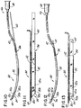
- a first embodiment of an instrument for implanting a graft comprising an intact planar cellular structure as described above between the retina and supporting tissues in an eye is indicated generally as 10 in Figure 9.
- the instrument 10 may be made from acrylic, or some other suitable material that is flexible and sterilizable.
- the instrument 10 comprises an elongate platform 11 for holding the planar cellular structure.
- the platform 11 has a distal end 16 for insertion into the eye of the recipient, and a proximal end 18.
- the platform 11 is approximately 2 to 10 centimeters long, which is an appropriate length for making implants in rodents and lower primates.
- the platform 11 must be sufficiently long to extend into the eye, between the retina and the supporting tissue, and thus different platform lengths may be used, depending upon the procedure being employed and upon the recipient.
- the platform is approximately 2.5 millimeters wide, which is sufficiently wide for making implants in rodents and lower primates.
- the platform 11 must be sufficiently wide to carry and intact cellular structure for implanting, and thus different platform widths may be used, depending upon the recipient.
- the edge 11a of the platform 11 at the distal end 16 is preferably convexly curved to facilitate both the insertion of the instrument 10 into the eye, and the advancement of the instrument between the retina and the supporting tissue to temporarily detach the retina, with a minimum of trauma.
- the platform 11 is preferably concavely curved (with respect to the top surface of the platform 11) along its longitudinal axis from the distal end 16 to the proximal end 18.
- the curvature of the platform 11 facilitates the manipulation of the instrument 10 within the eye, particularly the manipulation of the instrument between the retina and the supporting tissue on the curved walls of the eye.
- the radius of the curvature of the platform 11 will depend upon the procedure and the recipient.
- the platform 11 has side rails 12 and 14 on opposite sides for retaining the planar cellular structure on the platform.
- the distal portions 12a and 14a of the side rails taper from a point intermediate the distal and proximal ends of the side rails toward their distal ends.
- the distal ends of the side rails terminate in smoothly curved ends 12b and 14b, which are proximal of the distal end 16 of the platform.
- the offset of the distal ends of the rails, together with their rounded configuration facilitates the insertion of the instrument into the eye and the advancement of the instrument between the retina and the supporting tissue.
- the proximal portions of the side rails 12 and 14 are approximately 1 millimeter high, while the distal portions 12a and 14a taper to about 0.5 millimeters.
- the height of the side rails is made as small as possible, but they must be slightly greater than the thickness of the planar cell structure and the supporting substrate, and thus may vary depending on the donor and the type of implantation being made ( i.e. , how many cell layers are being implanted and thickness of the substrate).
- a second embodiment of an instrument for implanting a graft comprising an intact planar cellular structure as described above between the retina and supporting tissues in an eye is indicated generally as 30 in Figures 10-14, and 18.
- the instrument 30 may be made from polyethylene, or some other suitable material that is flexible and sterilizable.
- the instrument might be made of silicone rubber or silastic.
- the instrument 30 comprises an elongate tube 32 having a flat, wide cross-section, with a top 32a, a bottom 32b for supporting the planar cellular structure, and opposing sides 32c and 32d.
- the tube 32 has a distal end 34 for insertion into the eye, and a proximal end 36.
- the distal end 34 of the tube 32 is open for the discharge of the planar cellular structure.
- the instrument 30 of the second embodiment is preferable to the instrument 10 of the first embodiment in at least one respect because the tube 32 has a top 32a which provides better protection for the planar cellular structure to be implanted than the open platform 11.
- the tube 32 is approximately 3.5 centimeters long, which is an appropriate length for making implants in rodents and lower primates.
- the tube 32 must be sufficiently long to extend into the eye, between the retina and the supporting tissue, and thus the different tube lengths may be used, depending upon the procedure being employed and upon the recipient.
- the tube is approximately 2.5 centimeters wide, which is sufficiently wide for making implants in rodents and lower primates.
- the tube must be sufficiently wide to carry an intact cellular structure for implanting, and thus different tube widths may be used, depending upon the recipient.
- the sides 32c and 32d are approximately 0.75 millimeters high.
- the height of the sides is made as small as possible, but they must be slightly greater than the thickness of the planar cell structure and substrate, and thus may vary depending on the donor and the type of implantation being made (i.e. how many cell layers are being implanted and thickness of the substrate).
- the distal end 34 of the tube can be beveled to facilitate both the insertion of the tube into the eye, and the advancement of the tube between the retina and the supporting tissues, with a minimum of trauma.
- the end is preferably beveled at about 45°, from the top 32a to the bottom 32b.
- the distal end 34 of the tube 32 is also preferably raked transversely across the tube (i.e. from side 32c to 32d) toward the proximal end.
- the rake angle is preferably about 45°.
- the raked distal end also facilitates the insertion of the tube into the eye, and the advancement of the tube between the retina and the supporting tissue. Moreover, raking the distal end eliminates a sharp corner that could damage tissue.
- the tube 32 is preferably concavely curved along its longitudinal axis from the distal end 34 to the proximal end 36, so that the top 32a is on the inside of the curve, and the bottom 32b is on the outside of the curve.
- the curvature of the tube facilitates the manipulation of the instrument 30 within the eye, particularly the manipulation of the instrument between the retina and the supporting tissue on the curved walls of the eye.
- the radius of the curvature of the tube will depend on the procedure and on the recipient.
- the instrument 30 also comprises plunger means.
- the plunger means is preferably a flat plunger 40 slidably received in the tube so that relative sliding motion between the tube 32 and the plunger 40 urges a planar cellular structure that is in the tube out the distal end of the tube.
- the plunger 40 may be made of polymethylmethacrylate.
- the proximal end of the plunger 40 projects a sufficient amount from the proximal end of the tube 32 that the end of the plunger can be manipulated even when the distal portion of the tube is in an eye.
- the preferred method of operating the instrument 30 is that once the distal end of the tube is properly located within the subretinal area, the plunger 40 is held in place as the tube 32 is gradually withdrawn to eject the cellular structure.
- the plunger means may comprise means for applying hydraulic pressure on the contents of the tube.
- the proximal end 36 of the tube 32 is connected to a line 41 connected to a source of fluid under pressure. Fluid can be selectively supplied via the line 41 to the proximal end of the tube, to displace the contents of the tube.
- the fluid may be viscous, for example a 2% carboxymethylcellulose, or non-viscous.
- Gelatin is satisfactory because it is a simi-solid, and because it will dissolve harmlessly if it is ejected from the tube.
- the instrument 30 preferably also includes a lumen 42, extending generally parallel with the tube 32.
- lumen refers to any tube-like vessel, whether separately provided or formed as a passageway in another structure.
- the lumen 42 is attached to one of the sides of the tube 32, and preferably side 32c so that the distal end of the tube rakes away from the lumen.
- the lumen 42 has a distal end 44 generally adjacent the distal end of the tube, and preferably slightly advanced relative to the distal end of the tube.
- the proximal end 46 is remote from the distal end, and may be provided with a connector 48 for connection with a source of fluid under pressure.
- the lumen 42 can eject a stream of fluid from its distal end 44 which creates a fluid space ahead of the instrument, which helps separate or detach the retina from the supporting tissue as the instrument is advanced.
- the fluid may be a saline solution, or some other fluid that will not harm the delicate eye tissues.
- Various substances, such as anti-oxidants, anti-inflammatories, anti-mitotic agents and local anesthetics can be provided in the fluid for treatment of the eye or implanted tissue.
- the raked distal end of the tube 32 follows generally in the path opened by the fluid, thus minimizing direct contact of the instrument and the eye tissue.
- the distal end of the lumen may be beveled to facilitate the advancement of the instrument, particularly at times when fluid is not being ejected from the lumen.
- the end is preferably beveled at about 45°.
- the lumen could be formed integrally in the walls of the tube 32.
- a first alternate construction of instrument 30 is indicated as 30A in Figure 15.
- the instrument 30A is very similar in construction to instrument 30, and corresponding parts are identified with corresponding reference numerals.
- the instrument 30A includes a fiber optic filament 64 extending generally parallel with lumen 42, and positioned between the lumen 42 and the tube 32.
- the fiber optic filament 64 facilitates the manipulation of the instrument and the proper placement of the implant in two ways: a light source can be provided at the proximal end of the fiber optic filament so that the filament provides light at the distal end of the instrument, to facilitate the visual observation procedure through the pupil.
- a lens could be provided at the proximal end of the fiber optic filament so that the filament can also be used for direct observation at the distal end of the instrument.
- the fiber optic filament could allow for laser-light cautery to control subretinal bleeding.
- fiber optic filaments could be incorporated into the walls of the tube 32 or the lumen 42.
- a second alternative construction of instrument 30 is indicated as 30B in Figure 16.
- the instrument 30B is very similar in construction to instrument 30, and corresponding parts are identified with corresponding reference numerals.
- the instrument 30B includes a lumen 66 extending generally parallel with lumen 42, and positioned between the lumen 42 and the tube 32.
- the lumen 66 allows for the aspiration of material from the distal end of the instrument.
- the proximal end of the lumen 66 can be connected to a source of suction, to remove excess fluid and debris. It is possible to incorporate the lumen 66 into the wall of the tube 32.
- a third alternative construction of the instrument 30 is indicated as 30C in Figure 17.
- the instrument 30C is very similar in construction to instrument 30, and corresponding parts are identified with corresponding reference numerals.
- the instrument 30C includes a pair of lead wires 65, terminating in an electrode 67 at their distal ends.
- the electrode 67 allows for cauterization of blood vessels.
- the proximal ends of the leads 65 can be connected to a source of electrical power to seal broken blood vessels. It is possible to incorporate the leads 65 into the wall of the tube 32.
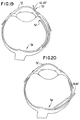
- the preferred surgical approach in the rodent, Fig. 19, includes making a transverse incision 70 in a cornea 72 of sufficient size so as to allow insertion of a surgical instrument illustrated schematically by reference characters 10 or 30.
- the instrument 10 is advanced under the iris, through the cornea 72 and to the ora serrata 74 as illustrated in Fig. 19.
- the iris should be dilated for example, with topical atropine.
- the instrument 10 When the instrument 10 is used, it detaches the retina as it is advanced under the retina and into the sub-retinal space to the posterior pole 76 of the eye.
- the channel defined by the side rails 12, 14 and the intermediate cell supporting platform provides for the graft comprising a photoreceptor layer 54 attached to the gelatin substrate to be placed on the instrument 10 and guided into the sub-retinal space, preferably with forceps or other suitable instruments.
- the gelatin is held in position with the forceps while the carrier is removed.
- the edges of the corneal incision are abutted after removal of the forceps to allow rapid, sutureless healing.
- the eye should be patched during recovery.
- the surgical instrument 30 ( Figures 10-14, and 18) is used instead of the instrument 10, the graft comprising intact generally planar sheet 54 of donor photoreceptors attached to the gelatin substrate 62 is drawn into the elongate tube 32.
- the instrument 30 is then inserted through an appropriate sized incision in the cornea and advanced under the iris. The iris will have been dilated, for example, with topical atropine.
- the instrument 30 is advanced to the ora serrata 74 of the host eye.
- the instrument 30 includes a lumen 42, the retina is detached by the gentle force of a perfusate such as a saline-like fluid, carboxymethylcellulose, or 1-2% hyluronic acid ejected from the lumen 42.
- the fluid may additionally contain anti-oxidants, anti-inflammation agents, anesthetics or agents that slow the metabolic demand of the host retina.
- the instrument 30 does not include a lumen 42
- the retina is detached by the walls of the surgical instrument as it is advanced under the retina and into the subretinal space to the posterior pole 76 of the eye.
- the graft comprising a photoreceptor layer attached to the gelatin substrate is then transplanted by moving the tube 32 in a direction away from the eye while keeping the plunger 40 stationary.
- the plunger 40 is carefully withdrawn out of the eye and the edges of the corneal incision are abutted after removal to allow rapid, sutureless healing.
- Retinal reattachment occurs rapidly and the photoreceptor sheet is held in place in a sandwich-like arrangement between the retina and the underlying eye tissues.
- the incision may require suturing.
- Fig. 20 depicts a trans-choroidal and scleral surgical approach as an alternative to the transcorneal approached described above. Except for the point of entry, the surgical technique is essentially the same as outlined above. Nevertheless, the transcorneal approach is preferred because it has been found to reduce bleeding and surgical trauma.
- a further surgical approach is to diathermize in the pars planna region to eliminate bleeding.
- the sclera is then incised and the choroidal and epithelial tissue is diathermized.
- the surgical tool is then inserted through the incision, the retina is intercepted at the ora serrata and the graft is deposited in the subretinal area otherwise as outlined elsewhere herein.
- the regrowth of photoreceptor axons may be facilitated by the proximity of the post-synaptic targets of the photoreceptor within the adjacent outer plexiform layer.
- growth across substantial intervening neural or glial scar tissue is not necessary in order for transplanted photoreceptors to make appropriate connections with the recipient retina including neural connections.
- the retina from the anesthetized 8-day-old rat was removed, flattened with radial cuts and placed with the receptor side down on a gelatin slab secured to the vibratome chuck.
- Molten gelatin (4-5% solution) was deposited adjacent the retina at the retina/gelatin interface and then cooled to 4°C with ice-cold Ringer's solution.
- the retina was sectioned at 20 to 50 ⁇ m until the photoreceptor layer was reached. When the photoreceptor layer was reached, the stage was advanced and a thick (200 to 300 ⁇ m) section was taken, undercutting the photoreceptor layer secured to the gelatin base.
- the isolated outer nuclear layer was cultured overnight with 40 ⁇ g/ml of diI (1,1'-dioctadecyl-3,3,3',3'-tetramethylindocarbocyanine perchlorate in Earle's MEM containing 10% fetal calf serum, incubated under 95%/5% oxygen/carbon dioxide mixture at room temperature. Labeling techniques and fluorescent microscopy were otherwise as outlined by Honig et al., J. Cell. Biol. 103 :17, 1986. DiI was removed from sections that were to be counterstained with FITC-labeled RET-P1 opsin antibody by prior washing in acetone.
- a transverse incision was made in the cornea sufficient to allow insertion of a surgical instrument 10 that is 2.5 mm wide with side rails 0.5 mm high or surgical instrument 30.
- the instrument was advanced under the iris (dilated with topical atropine) to the ora serrata, detaching the retina.
- the carrier was then advanced under the retina into the subretinal space to the posterior pole of the eye.
- the instrument allowed a graft comprising a piece of the photoreceptor layer attached to the gelatin substrate (up to 2.5 X 4 mm) to be guided into the retinal space by fine forceps.
- the instrument was then removed while the gelatin was held in position by the forceps.
- the edges of the corneal incision were abutted to allow rapid, sutureless healing.
- the eye was patched during recovery and a prophylactic dose of penicillin was administered.
- a verterinary ophthalmic antibiotic ointment was applied.
- Transplant recipients were maintained on a 12 hr/12 hr light/dark cycle with an average light intensity of 50 lux. Following appropriate survival times, the animal was overdosed with pentobarbital and perfused transcardially with phosphate buffered 3% paraformaldehyde-2% gluteraldehyde solution. Cryostat sections of both the light-blinded eye (control) and the eye receiving the photoreceptor transplant were then cut (20 ⁇ m).
- Antibody labeling for opsin was performed on retinas fixed with 3% paraformaldehyde and cryosectioned at 20 ⁇ m. Immunohistochemical methods were otherwise as described in Hicks, et al., J. Histochem Cytochem 35:1317 (1987). Elimination of the primary antibody eliminated specific labeling for opsin.
- Figs. 22-24 Cryostat sections made at 4 weeks post-transplantation are shown in Figs. 22-24.
- Figs 25A-C Cryostat sections made at 3 weeks post-transplantation are shown in Figs 25A-C.
- Fig. 25A is a H & E-stained photomicrograph of a transplant and host retina. Note the cell-sparse layer that resembles the outer plexiform layer interposed between the host retina and the transplant.
- Fig. 25B is a diI fluorescent photomicrograph of adjacent section. Transplanted photoreceptors show diI fluorescence, identifying them as donor tissue.
- cryostat sections (20 ⁇ m) were made from both the blinded eye (control) and the eye receiving the photoreceptor transplant at 2, 4, or 6 weeks after transplantation. It was found that the photoreceptors survived transplantation at all times tested (36 out of 54 transplants). In most instances, the surviving transplant approximated its size at the time of transplantation. More importantly, there was no apparent reduction in transplant size with longer survival times, suggesting that the transplants were stable.
- the contralateral eyes that did not receive a photoreceptor transplant were examined.
- the retinas possessed very few residual photoreceptors located adjacent to the outer plexiform layer and the RPE.
- these residual cells were abnormal in their appearance, having flattened, pyknotic cell bodies instead of the rounded cell bodies of normal photoreceptors.
- the residual photoreceptors did not form an outer nuclear layer composed of columnarly stacked cell bodies, but instead were found in isolation, or at most appear as a single or double layer of cells (see Fig. 23) located mainly in the peripheral retina.
- the transplanted cells were easily distinguished from the residual photoreceptors by a number of parameters. First, they were found in discrete patches and have the characteristic columnar stacking arrangement of up to about 12 cell bodies that is characteristic of photoreceptor cells in the outer nuclear layer of the normal retina. They did not have the flattened appearance of the residual native photoreceptors, but instead have the round, nonpyknotic cell body typical of normal transplanted cells. Furthermore, the transplanted photoreceptors can form rosette configurations, a characteristic of transplanted and cultured retina while residual photoreceptors were not found in these configurations.
- the donor outer nuclear layer was labeled with the fluorescent marker diI prior to the transplantation.
- the photoreceptor patches were fluorescently labeled while the host retina did not show diI fluorescence.
- RET-P1 a monoclonal antibody specific for opsin
- any cell showing labeling for opsin was, therefore, identified as a photoreceptor.
- the transplanted cells stain intensely for opsin whereas other retinal cells are unstained. Positive staining for opsin not only identifies these cells as photoreceptors but indicates that these cells are still capable of producing the protein moiety of visual pigment.
- Retina adjacent to the region of the transplant shows only a few isolated photoreceptor cell bodies (Fig. 23) that do not stain for opsin (Fig. 24) suggesting that they are cones. Their lack of opsin staining, as well as their location and appearance in H & E-stained material, confirms that these cells are the host's residual photoreceptors (Fig. 23).
- Example 1 The procedures of Example 1 were repeated except as noted. Substituted for the Sprague-Dawley rats were the rd mouse and the RCS rat which are afflicted with inherited retinal degeneration. In the rd mouse it is thought that the deficit resides in the photoreceptor whereas in the RCS it is thought that the deficit resides in the pigment epithelium. In these animals, almost all photoreceptors are eliminated while the remaining retina is preserved; neither the rd mouse or the RCS rat were blinded by constant illumination as set forth in Example 1. The rd mouse and the RCS respectively received transplants of immature (7-8 day old mouse or rat) and mature rat photoreceptors.
- the transplantation technique was adapted to the smaller size of the mouse eye. This modification allowed sheets of intact outernuclear layer to be transplanted to the subretinal space of the mouse eye.
- Neonatal (8 days old) photoreceptors were transplanted from rd control mice to the subretinal space of adult rd mice. Survival times were for 2 weeks to 3 months. At all survival times, it was found that the transplanted photoreceptors survived, becoming physically attached to the outer portion of the host retina and stained positive for opsin. In addition, the host retina became reattached to the pigment epithelium.
- Photoreceptors from 7 to 8 day old RCS controls were transplanted to the subretinal space in the eye of adult (3 month old) RCS rats. A two month survival period was allowed because in this time period almost all host photoreceptors degenerate in the RCS rat. It was found that the grafted photoreceptors survive transplantation to the subretinal space of the RCS rat and show histotypic as well as immunological characteristics of normal photoreceptors. In addition, it was found that transplanted photoreceptors survive within their homotopic location in the RCS rat whereas the RCS's own photoreceptors do not.
- Example 1 The procedures of Example 1 were repeated except as noted.
- Donor photoreceptors were originally harvested at the earliest ontogenetic time in which the photoreceptors could be isolated from other portions of the retina (7-8 days old) since it is generally believed that more embryonic and undifferentiated neural tissue survives transplantation far better than more mature and differentiated tissue. To determine the effect of developmental age on photoreceptor survival and ability to integrate with the host retina, photoreceptors were subsequently transplanted from 8, 9, 12, 15 and 30 day old rats into light damaged adults. These show progressive development and maturation of the photoreceptors including mature outer segments (at 15 and 30 days).
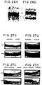
- FIG 26 is a photograph of a transplant of mature photoreceptors (30 day old donor) to adult light damaged host. (T, Transplant). 120X. These observations suggest that photoreceptors have characteristics that differ from other neural tissue that permits them to be transplanted when they are essentially mature while other neural tissue must be at a very immature stage for successful transplantation to occur.
- Example 1 The procedures of Example 1 were repeated except as noted. Photoreceptors were taken from the retina of donated human eyes (obtained from the MO Lions and St. Louis Eye Banks) following corneal removal. A portion of the retinas were tested for viability by dye exclusion with trypan blue and didansyl cystine staining. The photoreceptors excluded dye and appeared to be in good condition. Hosts were adult albino rats (immune-suppressed with cyclosporin A or immune-competent) exposed to constant illumination.
- Fig. 26B shows a transplant of human photoreceptors from adult donor to adult light damaged rat host. (T, transplant). 120X.
- transplants stained positive for antiopsin antibody RET-P1, identifying the transplanted cells as photoreceptors and further indicating that they are still capable of producing visual pigment.
- transplants to immune-competent hosts showed signs of rejection within one week of transplantation. Sham operated animals showed no repopulation of the host retina with photoreceptors.
- Example 1 The procedures of Example 1 were repeated except as noted.
- the 2DG functional mapping technique developed by Sokoloff et al., J. Neurochem . 28:897-916 (1977) allows the measurement of the relative levels of neural activity for a given stimulus condition. For this reason, the 2DG technique appeared to be an appropriate method of assessing the functional characteristics of the transplant and its ability to activate the light damaged retina.
- the outer retina might be expected to show high 2DG uptake in the dark since photoreceptors, horizontal and some bipolar cells are maximally depolarized in this situation.
- strobe flicker is a strong stimulus for the retina including the amacrine and retinal ganglion cells
- 2DG uptake across the entire retina is also to be expected. It therefore appears that the 2DG uptake pattern in normal retina reflects relative degrees of neural activity or neural depolarization, and therefore is a useful indicator of neural activity in the retina as it is in other areas of the nervous system.
- both the normal retina and the light-damaged retina receiving the photoreceptor transplant show relatively high uptake of 2DG in the photoreceptor and inner nuclear layers.
- the similarity in the relative uptake patterns between these cases suggests that the transplanted photoreceptors may have similar functional characteristics as normal photoreceptors (i.e., they depolarize in the dark and are capable of inducing a sustained depolarization of some cells in the host's inner nuclear layer).
- Example 1 The procedures of Example 1 were repeated except as noted.
- Example 1 The procedures of Example 1 were repeated except as noted.
- VEP visually evoked cortical potentials
- VEP VEP-induced strobe flash test stimuli generated by a Grass PS-2 photostimulator directed toward one eye with the other eye covered by a patch.
- Responses were differentially amplified (Grass P-15D preamp), displayed on a Tectronix #564 oscilliscope and then averaged by a Macintosch IIx computer using LabView.
- the reconstructed retina can produce a light-evoked electrical response in the visual cortex whereas the unreconstructed fellow eye showed little or no response to the same light stimulus.
- Example 1 The procedures of Example 1 were repeated except as noted.
- neural transplants to the brain can restore appropriate behavioral activity (Bjorklund et al., Neural Grafting in the Mammalian CNS . Elsevier, Amsterdam, 1985).
- Klassen and Lund Proc. Natl. Acad. Sci. USA 84 : 6958-6960, 1987; and Exp. Neurol. 102 : 102-108, 1988 have shown that neural transplants can restore the pupillary reflex mediated by intracranial transplantation of embryonic retinas thus showing that neural transplants consisting of sensory tissue are capable of mediating a behaviorally appropriate response to sensory stimulation.
- animals were anesthesized and held in a stereotaxic device.
- An infrared video camera was focused on the eye through an operating microscope and the eye illuminated with infrared light.
- a light beam controlled by a camera shutter within the operating microscope was used. This light was focused on the eye.
- the pupillary response to the light at graded intensities (intensity of the light was controlled by neutral density filters) was recorded by video camera connected to a frame grabber system.
- the pupillary reflex was then analysed using automated imageprocessing software (Ultimage, GTFS, Inc.)
- Panels a and b are of the reconstructed retina.
- Panel a shows the iris at light onset whereas panel b shows the same eye at 5 seconds after light onset.
- Comparison of panel a to panel b shows a normal pupillary constriction mediated by light.
- Panels c and d show the fellow blinded eye that received sham surgery with panel c showing the iris at light onset and panel d showing the iris 5 seconds later.
- Comparison of panels c and d show an increase in pupil size with light. This response is aberrant in form and is characteristic of individuals suffering from severe retinal dystrophy of a photoreceptor type.
- Example 1 The procedures of Example 1 were repeated except as noted.
- Photoreceptors were taken from mature macaque retina (animals sacrificed for other research) or the retina of donated human eyes (obtained from the St. Louis Eye Bank) using vibratome section of the flat-mounted retina to isolate the intact outer nuclear layer.
- Hosts were mature macaque monkeys treated with iodoacetic acid (30 mg/kg given on 3 successive days) which selectively eliminates host photoreceptors in non-macular areas of the retina while leaving the remaining retina intact. This treatment did not compromise central vision and therefore maintained sight required for behavioral and physiologically important functions (e.g., locating of food and water, visually guided locomoter activities, grooming, maintenance of circadian rhythms).
- the isolated outer nuclear layer was transplanted following a pars plana vitrectomy (a standard surgical technique) using a trans-scleral approach to the subretinal space.
- the photoreceptors were inserted under a focal retinal detachment induced by the formation of a subretinal bleb.
- the bleb was created by the infusion of ophthalmic balanced salt solution.
- the reconstructed retina was reattached to the back of the eye by pneumatic tamponade with the transplanted photoreceptors interposed between the retina and the underlying pigment epithelium.
- Daily injections of cyclosporin A and dexamethasone were made to suppress any possible transplant rejection.
- the present invention provides an improved surgical instrument that is adapted to provide cell organization during transplantation of the photoreceptors. With the surgical instrument of this invention cell organization is maintained during photoreceptor, RPE, and choroidal transplantation while minimizing trauma to the transplanted tissues, the host eye and retina. It is believed that retina reattachment and subsequent substantially normal function of the reconstructed retina, in view of the transplant, is thereby facilitated.
- the present invention provides an improved surgical instrument that is constructed to allow relatively large expanses of the RPE, choroidea, and photoreceptor cell matrix or column to be transplanted to a sub-retinal space.
- the present invention provides an improved surgical instrument that allows appropriate retinotopic positioning.
- the present invention provides an improved surgical instrument that protects photoreceptors from damage as the surgical device is positioned in the eye.
- the present invention provides a method of photoreceptor or retinal pigment epithelium isolation that, maintains to the extent possible the normal organization of the outer nuclear layer and these other tissues.
- the present invention provides a method of cell and tissue isolation by which cells can be isolated without disruption of their intercellular organization. With the method of this invention retinal cells, such as retinal photoreceptors can be isolated without the disruption of the intercellular organization of the outer nuclear layer or other layer of the retina, RPE, and choroidea.
- transplanted cells A number of properties of the transplanted cells are that they are and remain alive; they produce opsin, important for phototransduction; they are functional (i.e., activated by light); and the transplanted photoreceptors activate a previously blinded retina in a light dependent fashion.
- Attachment of retinal tissue to the gelatin substrate allows extended periods of in vitro culture of retinal tissues by: maintaining organization of tissue in culture; and allowing for a better viability of cultured tissue.
- Photoreceptors may also be used for transplantation to retina in which the host's or recipient's photoreceptors are lost by environmental (constant light) or inherited defects.
- environmental constant light
- inherited defects See: S. E. Hughes and M. S. Silverman (1988) in "Transplantation of retinal photoreceptors to dystrophic retina", Soc. Neuorosci. Abstr. , 18 : 1278.
- transplanted photoreceptor cells maintain basic characteristics of normal photoreceptor cells by producing opsin and maintaining an intercellular organization and apposition to the host retina that is similar to that seen in the normal outer nuclear layer.
- the surgical instruments may be larger for use in humans.
- trans-scleral, choroidal Other approaches to the subretinal space may be used, e.g., trans-scleral, choroidal.
- substrates besides gelatin can be used, e.g. augar, augarose, in fact improved substrates could include factors that can be integrated into gelatin, for example, neurotrophic factors). It is believed that attachment to gelatin or equivalent substrates will allow prolonged in vitro culture, or cryogenic freezing, and similar storage, while allowing for the maintenance of tissue organization and viability. Finally, it is believed that other methods of attaching retina to substrate can be used, such as lectins, or photo-activated cross-linking agents.
- this invention provides a method to isolate the intact photoreceptor layer. This is significant because it will be necessary to maintain tight matrix organization if coherent vision is to be restored to the retina comprised by the loss of photoreceptors.
- the surgical approach allowed by the instrument has been described merely as an an example which minimizes trauma to the eye and allows controlled positioning of sheets of transplanted photoreceptors to their homotopic location within the eye.
- thee methods for preparation transplantation and isolation of photoreceptors could be utilized to prepare other retinal layers so that selected populations of retinal cells can be used in other neurobiological investigations and clinical procedures.
- retinal layers once they are flattened, appropriately sectioned, and appropriately affixed to a stabilizing substrate or base, could be prepared for transplantation, storage (e.g., in vitro, cryogenic), or culturing similar to the methods described herein for photoreceptor layers.
- the necessity for prompt re-vascularization typically limits the ability to transplant most neural tissue, but not photoreceptors.
- the photoreceptor layer of a retina and the ("RPE") is non-vascularized.
- Non-vascularized tissue shows the least amount of tissue rejection. Consequently, it is believed that genetically dissimilar photoreceptor cells may be transplanted in accordance with the method described above as example. Matching of host and donor histocompatibility antigens will probably be necessary for transplantation of the retinal pigment epithelium and choroidea.
- Photoreceptors can be transplanted when developing or when mature. Not only can mature rat photoreceptors be transplanted, but mature photoreceptors from human donors can be transplanted as well. This is significantly different from neurons which must be immature in order to be transplanted. At present the reason for this difference is not known but has obvious importance for retinal and neural transplantation research in general.
- transplanted photoreceptors activate the host's or recipient's dystrophic retina in a light dependent manner that closely resembles the activation pattern seen in normal retina.
Landscapes
- Health & Medical Sciences (AREA)
- Life Sciences & Earth Sciences (AREA)
- Biomedical Technology (AREA)
- Engineering & Computer Science (AREA)
- Veterinary Medicine (AREA)
- Animal Behavior & Ethology (AREA)
- General Health & Medical Sciences (AREA)
- Public Health (AREA)
- Oral & Maxillofacial Surgery (AREA)
- Chemical & Material Sciences (AREA)
- Transplantation (AREA)
- Ophthalmology & Optometry (AREA)
- Heart & Thoracic Surgery (AREA)
- Surgery (AREA)
- Medicinal Chemistry (AREA)
- Dermatology (AREA)
- Epidemiology (AREA)
- Vascular Medicine (AREA)
- Nuclear Medicine, Radiotherapy & Molecular Imaging (AREA)
- Cell Biology (AREA)
- Chemical Kinetics & Catalysis (AREA)
- Botany (AREA)
- Zoology (AREA)
- Medical Informatics (AREA)
- Cardiology (AREA)
- Pathology (AREA)
- Molecular Biology (AREA)
- Materials For Medical Uses (AREA)
- Prostheses (AREA)
- Surgical Instruments (AREA)
Claims (16)
- Verfahren zur Vorbereitung bzw. Herstellung eines Implantats zur Transplantation in den subretinalen Bereich eines Wirtsauges, die Verfahrensschritte umfassend:Bereitstellen von Spendergewebe von einem toten Spender,Sammeln einer Zellpopulation von diesem Gewebe, wobei diese Zellpopulation auf eine solche Weise aus Retinazellen, Epithelgewebe und Choroideagewebe ausgewählt ist, daß die Zellpopulation in derselben interzellulären Organisation und zellulären Polarität verbleibt, wie sie in normalem Gewebe von diesem Typus vorliegt undLaminieren der Zellpopulation auf eine nicht toxische und flexible Zusammensetzung, die sich im wesentlichen bei Körpertemperatur auflöst.
- Verfahren gemäß Anspruch 1, bei dem die Zusammensetzung Gelatine ist und das gemäß den genannten Schritten vorbereitete bzw. hergestellte Implantat eine Oberfläche besitzt, deren Fläche größer als 1 mm2 ist.
- Verfahren zur Vorbereitung bzw. Herstellung eines Implantats, das Photorezeptorzellkörper zur Transplantation in den subretinalen Bereich eines Wirtsauges umfasst, die folgenden Verfahrensschritte aufweisend:Bereitstellen einer Spenderretina von einem toten Spender, umfassend eine Mehrzahl von Zellschichten, einschließlich einer Schicht von Photorezeptorzellkörpern undIsolieren der Schicht der Photorezeptorzellkörper der Donorretina von wenigstens einer der Mehrzahl von Schichten auf eine Weise, daß die Photorezeptorzellkörper in derselben interzellulären Organisation und zellulären Polarität gehalten werden, wie sie in normalem Gewebe dieses Typus vorliegt.
- Implantat zur Transplantation in den subretinalen Bereich eines Wirtsauges, umfassend eine Population von Photorezeptorzellkörpern, die von einer Spenderretina gesammelt wurden, wobei die Population von Photorezeptorzellkörpern dieselbe interzelluläre Organisation und zelluläre Polarität besitzt, wie sie in normalem Gewebe dieses Typus vorliegt, wobei wenigstens eine bei einer normalen Retina vorliegende Zellschicht im wesentlichen entfernt worden ist.
- Implantat gemäß Anspruch 4, weiterhin wenigstens eine andere Schicht von Retinazellen umfassend.
- Implantat gemäß Anspruch 4 oder 5, wobei das Implantat ein Laminat umfasst, das eine Population von Photorezeptorzellkörpern und eine nicht toxische und flexible Zusammensetzung aufweist, die sich im wesentlichen bei Körpertemperatur auflöst.
- Implantat gemäß einem der Ansprüche 4 bis 6, wobei die Zusammensetzung einen trophischen Faktor, ein Immunsuppressivum, ein entzündungshemmendes Mittel, einen anti-angiogenen Faktor, ein Antigliamittel oder einen Antimitosefaktor aufweist.
- Implantat gemäß einem der Ansprüche 4 bis 6, bei dem die Zusammensetzung Gelatine ist.
- Implantat gemäß einem der Ansprüche 4 bis 6, bei dem das Implantat eine Oberfläche mit einer Fläche größer als 4 mm2 besitzt.
- Implantat gemäß einem der Ansprüche 4 bis 9, bei dem die Zellpopulation Retinazellen, Epithelgewebe und Choroideagewebe sind.
- Instrument zur Implantation eines Implantats gemäß einem der Ansprüche 4 bis 10 in ein Auge, wobei das Instrument aufweist:eine längliche Röhre (32) mit einem flachen, breiten Querschnitt, mit einer Oberseite (32a) einer Unterseite (32b) zur Stütze der ebenen, zellulären Struktur und einander gegenüberliegende Seiten (32c, 32d), wobei die Röhre eine abgeschrägte distale Kante (34) besitzt, die das Einführen der Röhre in das Auge und das Vordringen der Röhre zwischen der Retina und den Stützgeweben erleichtert; undeine Kolbeneinrichtung (40) zum Austragen der ebenen, zellulären Strutur aus dem distalen Ende der Röhre.
- Instrument gemäß Anspruch 11, bei dem das distale Ende der Röhre (32) quer über die Röhre in Richtung auf das proximale Ende abgeschrägt ist und beidem die Kolbeneinrichtung einen flachen Kolben (40) umfasst, der aus dem proximalen Ende der Röhre hervorsteht, wobei der Kolben gleitend verschiebbar in der Röhre (32) angebracht ist, so daß eine relative Gleitbewegung zwischen der Röhre (32) und dem Kolben (40) die ebene, zelluläre Struktur aus dem distalen Ende hinausdrängt.
- Instrument gemäß Anspruch 12, weiter ein Lumen (42) aufweisend, das sich im allgemeinen parallel zu der Röhre erstreckt, wobei es zum Ausstoß einer Flüssigkeit an dem distalen Ende des Instruments zum Trennen der Retina vom Trägergewebe oder zum Ansaugen von Flüssigkeit und Trümmer an dem distalen Ende des Instruments dient.
- Instrument gemäß Anspruch 12, ferner ein fieberoptisches Filament (64) aufweisend, das sich im allgemeinen parallel zu dem Lumen (42) erstreckt, wobei dieses zwischen dem Lumen und der Röhre angeordnet ist, und eine Lichtquelle an dem proximalen Ende des fiberoptischen Filaments.
- Instrument gemäß Anspruch 12, wobei die Röhre entlang seiner longitudinalen Achse vom distalen Ende zum proximalen Ende eine solche Kurve beschreibt, daß die Oberseite der Röhre sich auf der Innenseite der Kurve befindet und die Aussenseite der Röhre auf der Aussenseite der Kurve.
- Instrument gemäß Anspruch 12, ferner eine Kauterisierungselektrode (67) aufweisend, allgemein angrenzend an das distale Ende der Röhre , ein Paar elektrischer Leitungen (65), die sich zu der Elektrode erstrecken, und Mittel zur Verbindung der Leitungen mit einer elektrischen Stromquelle, um dem Instrument das Kauterisieren beschädigter Gefäße zu ermöglichen.
Applications Claiming Priority (3)
| Application Number | Priority Date | Filing Date | Title |
|---|---|---|---|
| US39437789A | 1989-08-14 | 1989-08-14 | |
| US394377 | 1989-08-14 | ||
| PCT/US1990/004550 WO1991002499A1 (en) | 1989-08-14 | 1990-08-13 | Surgical instrument and cell isolation and transplantation |
Publications (3)
| Publication Number | Publication Date |
|---|---|
| EP0486589A1 EP0486589A1 (de) | 1992-05-27 |
| EP0486589A4 EP0486589A4 (en) | 1993-03-03 |
| EP0486589B1 true EP0486589B1 (de) | 1998-03-04 |
Family
ID=23558712
Family Applications (1)
| Application Number | Title | Priority Date | Filing Date |
|---|---|---|---|
| EP90912685A Expired - Lifetime EP0486589B1 (de) | 1989-08-14 | 1990-08-13 | Chirurgisches instrument und zellisolierung |
Country Status (9)
| Country | Link |
|---|---|
| EP (1) | EP0486589B1 (de) |
| JP (1) | JP2533693B2 (de) |
| AT (1) | ATE163531T1 (de) |
| AU (1) | AU653499B2 (de) |
| CA (1) | CA2065230C (de) |
| DE (1) | DE69032100T2 (de) |
| DK (1) | DK0486589T3 (de) |
| ES (1) | ES2115594T3 (de) |
| WO (1) | WO1991002499A1 (de) |
Cited By (1)
| Publication number | Priority date | Publication date | Assignee | Title |
|---|---|---|---|---|
| US5868728A (en) * | 1995-02-28 | 1999-02-09 | Photogenesis, Inc. | Medical linear actuator for surgical delivery, manipulation, and extraction |
Families Citing this family (15)
| Publication number | Priority date | Publication date | Assignee | Title |
|---|---|---|---|---|
| SG49267A1 (en) * | 1989-08-14 | 1998-05-18 | Photogenesis Inc | Surgical instrument and cell isolation and transplantation |
| US5817075A (en) * | 1989-08-14 | 1998-10-06 | Photogenesis, Inc. | Method for preparation and transplantation of planar implants and surgical instrument therefor |
| US5273530A (en) * | 1990-11-14 | 1993-12-28 | The University Of Rochester | Intraretinal delivery and withdrawal instruments |
| US6045791A (en) * | 1992-03-06 | 2000-04-04 | Photogenesis, Inc. | Retinal pigment epithelium transplantation |
| SG49754A1 (en) * | 1993-03-16 | 1998-06-15 | Photogenesis Inc | Method for preparation and transplantation of volute grafts and surgical instrument therefor |
| CA2158442A1 (en) * | 1993-03-16 | 1994-09-29 | Stephen E. Hughes | Method for rescuing endogenous cone photoreceptor population |
| JP3490447B2 (ja) * | 1993-04-30 | 2004-01-26 | フォトジェネシス インコーポレイテッド | 網膜色素上皮移植 |
| US5941250A (en) * | 1996-11-21 | 1999-08-24 | University Of Louisville Research Foundation Inc. | Retinal tissue implantation method |
| US6156042A (en) * | 1997-11-17 | 2000-12-05 | Aramant; Robert B. | Retinal tissue implantation instrument |
| FR2787993B1 (fr) * | 1999-01-06 | 2001-07-13 | Fci France Chirurgie Instrumen | Instrument pour la pose d'une sangle pour la chirurgie d'un decollement de retine |
| US6159218A (en) * | 1999-05-19 | 2000-12-12 | Aramant; Robert B. | Retinal tissue implantation tool |
| US6416777B1 (en) * | 1999-10-21 | 2002-07-09 | Alcon Universal Ltd. | Ophthalmic drug delivery device |
| GB0328021D0 (en) | 2003-12-03 | 2004-01-07 | Inst Of Ophthalmology | Method |
| JP4569971B2 (ja) * | 2007-01-19 | 2010-10-27 | Hoya株式会社 | 治療用物質の運搬投与器具 |
| EP3166550B1 (de) * | 2014-07-11 | 2023-12-13 | The United States of America, as represented by the Secretary, Department of Health and Human Services | Chirurgisches werkzeug zur augengewebetransplantation |
Family Cites Families (7)
| Publication number | Priority date | Publication date | Assignee | Title |
|---|---|---|---|---|
| US3934591A (en) * | 1974-03-20 | 1976-01-27 | Gleason Robert W | Dermatome |
| US4014342A (en) * | 1975-04-11 | 1977-03-29 | Concept, Inc. | Vitreous cutter |
| US4418691A (en) * | 1981-10-26 | 1983-12-06 | Massachusetts Institute Of Technology | Method of promoting the regeneration of tissue at a wound |
| US4662869A (en) * | 1984-11-19 | 1987-05-05 | Wright Kenneth W | Precision intraocular apparatus |
| DE3681787D1 (de) * | 1985-07-05 | 1991-11-07 | Whitehead Biomedical Inst | Expression von fremdem genetischem material in epithelzellen. |
| US4900300A (en) * | 1987-07-06 | 1990-02-13 | Lee David A | Surgical instrument |
| US4940468A (en) * | 1988-01-13 | 1990-07-10 | Petillo Phillip J | Apparatus for microsurgery |
-
1990
- 1990-08-13 ES ES90912685T patent/ES2115594T3/es not_active Expired - Lifetime
- 1990-08-13 CA CA002065230A patent/CA2065230C/en not_active Expired - Fee Related
- 1990-08-13 WO PCT/US1990/004550 patent/WO1991002499A1/en not_active Ceased
- 1990-08-13 JP JP2511904A patent/JP2533693B2/ja not_active Expired - Fee Related
- 1990-08-13 DK DK90912685T patent/DK0486589T3/da active
- 1990-08-13 AU AU62715/90A patent/AU653499B2/en not_active Ceased
- 1990-08-13 AT AT90912685T patent/ATE163531T1/de not_active IP Right Cessation
- 1990-08-13 DE DE69032100T patent/DE69032100T2/de not_active Expired - Fee Related
- 1990-08-13 EP EP90912685A patent/EP0486589B1/de not_active Expired - Lifetime
Cited By (1)
| Publication number | Priority date | Publication date | Assignee | Title |
|---|---|---|---|---|
| US5868728A (en) * | 1995-02-28 | 1999-02-09 | Photogenesis, Inc. | Medical linear actuator for surgical delivery, manipulation, and extraction |
Also Published As
| Publication number | Publication date |
|---|---|
| DK0486589T3 (da) | 1999-02-22 |
| JP2533693B2 (ja) | 1996-09-11 |
| EP0486589A4 (en) | 1993-03-03 |
| DE69032100T2 (de) | 1998-10-29 |
| CA2065230C (en) | 1999-07-20 |
| ATE163531T1 (de) | 1998-03-15 |
| WO1991002499A1 (en) | 1991-03-07 |
| AU6271590A (en) | 1991-04-03 |
| JPH05501969A (ja) | 1993-04-15 |
| AU653499B2 (en) | 1994-10-06 |
| DE69032100D1 (de) | 1998-04-09 |
| ES2115594T3 (es) | 1998-07-01 |
| CA2065230A1 (en) | 1991-02-15 |
| EP0486589A1 (de) | 1992-05-27 |
Similar Documents
| Publication | Publication Date | Title |
|---|---|---|
| US5962027A (en) | Retinal cell transplant | |
| US6514238B1 (en) | Method for preparation and transplantation of volute grafts and surgical instrument therefor | |
| CA2158443C (en) | Method for preparation and transplantation of volute grafts and surgical instrument therefor | |
| US5817075A (en) | Method for preparation and transplantation of planar implants and surgical instrument therefor | |
| AU653499B2 (en) | Surgical instrument and cell isolation and transplantation | |
| Wongpichedchai et al. | Comparison of external and internal approaches for transplantation of autologous retinal pigment epithelium. | |
| WO1996026759A9 (en) | Method for preparation and transplantation of planar implants and surgical instruments therefor | |
| JP3490447B2 (ja) | 網膜色素上皮移植 | |
| Matsuo et al. | Visual evoked potential in rabbits’ eyes with subretinal implantation by vitrectomy of Okayama University-type retinal prosthesis (OURePTM) | |
| del Cerro et al. | Retinal transplants into adult eyes affected by phototoxic retinopathy | |
| AU716820B2 (en) | Method for preparation and transplantation of volute grafts and surgical instrument therefor | |
| TW524681B (en) | Volute grafts and method for preparing the same | |
| D’Agostino et al. | 3.5. Decellularized descemet’s membrane: a new scaffold on sight for full-thickness macular hole | |
| Palanker | Transplantation of Photoreceptors for Restoration of Sight | |
| CN120789103A (zh) | M2型小胶质细胞外泌体的应用 | |
| AU672207B2 (en) | Retinal pigment epithelium transplantation |
Legal Events
| Date | Code | Title | Description |
|---|---|---|---|
| PUAI | Public reference made under article 153(3) epc to a published international application that has entered the european phase |
Free format text: ORIGINAL CODE: 0009012 |
|
| 17P | Request for examination filed |
Effective date: 19920217 |
|
| AK | Designated contracting states |
Kind code of ref document: A1 Designated state(s): AT BE CH DE DK ES FR GB IT LI LU NL SE |
|
| A4 | Supplementary search report drawn up and despatched |
Effective date: 19930109 |
|
| AK | Designated contracting states |
Kind code of ref document: A4 Designated state(s): AT BE CH DE DK ES FR GB IT LI LU NL SE |
|
| RAP1 | Party data changed (applicant data changed or rights of an application transferred) |
Owner name: PHOTOGENESIS INCORPORATED |
|
| 17Q | First examination report despatched |
Effective date: 19950410 |
|
| RIN1 | Information on inventor provided before grant (corrected) |
Inventor name: HUGHES, DR. STEPHAN E. Inventor name: SILVERMAN, MARTIN, S. |
|
| GRAG | Despatch of communication of intention to grant |
Free format text: ORIGINAL CODE: EPIDOS AGRA |
|
| GRAG | Despatch of communication of intention to grant |
Free format text: ORIGINAL CODE: EPIDOS AGRA |
|
| GRAH | Despatch of communication of intention to grant a patent |
Free format text: ORIGINAL CODE: EPIDOS IGRA |
|
| GRAH | Despatch of communication of intention to grant a patent |
Free format text: ORIGINAL CODE: EPIDOS IGRA |
|
| GRAA | (expected) grant |
Free format text: ORIGINAL CODE: 0009210 |
|
| AK | Designated contracting states |
Kind code of ref document: B1 Designated state(s): AT BE CH DE DK ES FR GB IT LI LU NL SE |
|
| REF | Corresponds to: |
Ref document number: 163531 Country of ref document: AT Date of ref document: 19980315 Kind code of ref document: T |
|
| REG | Reference to a national code |
Ref country code: CH Ref legal event code: EP |
|
| REF | Corresponds to: |
Ref document number: 69032100 Country of ref document: DE Date of ref document: 19980409 |
|
| ITF | It: translation for a ep patent filed | ||
| REG | Reference to a national code |
Ref country code: ES Ref legal event code: FG2A Ref document number: 2115594 Country of ref document: ES Kind code of ref document: T3 |
|
| ET | Fr: translation filed | ||
| PLBE | No opposition filed within time limit |
Free format text: ORIGINAL CODE: 0009261 |
|
| STAA | Information on the status of an ep patent application or granted ep patent |
Free format text: STATUS: NO OPPOSITION FILED WITHIN TIME LIMIT |
|
| REG | Reference to a national code |
Ref country code: DK Ref legal event code: T3 |
|
| 26N | No opposition filed | ||
| PGFP | Annual fee paid to national office [announced via postgrant information from national office to epo] |
Ref country code: DK Payment date: 20010820 Year of fee payment: 12 |
|
| PGFP | Annual fee paid to national office [announced via postgrant information from national office to epo] |
Ref country code: AT Payment date: 20010821 Year of fee payment: 12 |
|
| PGFP | Annual fee paid to national office [announced via postgrant information from national office to epo] |
Ref country code: BE Payment date: 20010903 Year of fee payment: 12 Ref country code: CH Payment date: 20010903 Year of fee payment: 12 |
|
| PGFP | Annual fee paid to national office [announced via postgrant information from national office to epo] |
Ref country code: LU Payment date: 20010914 Year of fee payment: 12 |
|
| REG | Reference to a national code |
Ref country code: GB Ref legal event code: IF02 |
|
| PG25 | Lapsed in a contracting state [announced via postgrant information from national office to epo] |
Ref country code: LU Free format text: LAPSE BECAUSE OF NON-PAYMENT OF DUE FEES Effective date: 20020813 Ref country code: AT Free format text: LAPSE BECAUSE OF NON-PAYMENT OF DUE FEES Effective date: 20020813 |
|
| PG25 | Lapsed in a contracting state [announced via postgrant information from national office to epo] |
Ref country code: CH Free format text: LAPSE BECAUSE OF NON-PAYMENT OF DUE FEES Effective date: 20020831 Ref country code: BE Free format text: LAPSE BECAUSE OF NON-PAYMENT OF DUE FEES Effective date: 20020831 Ref country code: LI Free format text: LAPSE BECAUSE OF NON-PAYMENT OF DUE FEES Effective date: 20020831 |
|
| PG25 | Lapsed in a contracting state [announced via postgrant information from national office to epo] |
Ref country code: DK Free format text: LAPSE BECAUSE OF NON-PAYMENT OF DUE FEES Effective date: 20020930 |
|
| BERE | Be: lapsed |
Owner name: *PHOTOGENESIS INC. Effective date: 20020831 |
|
| REG | Reference to a national code |
Ref country code: DK Ref legal event code: EBP |
|
| REG | Reference to a national code |
Ref country code: CH Ref legal event code: PL |
|
| PGFP | Annual fee paid to national office [announced via postgrant information from national office to epo] |
Ref country code: ES Payment date: 20050920 Year of fee payment: 16 |
|
| PGFP | Annual fee paid to national office [announced via postgrant information from national office to epo] |
Ref country code: GB Payment date: 20060717 Year of fee payment: 17 |
|
| PGFP | Annual fee paid to national office [announced via postgrant information from national office to epo] |
Ref country code: DE Payment date: 20060725 Year of fee payment: 17 |
|
| PGFP | Annual fee paid to national office [announced via postgrant information from national office to epo] |
Ref country code: FR Payment date: 20060726 Year of fee payment: 17 |
|
| PGFP | Annual fee paid to national office [announced via postgrant information from national office to epo] |
Ref country code: NL Payment date: 20060731 Year of fee payment: 17 |
|
| PGFP | Annual fee paid to national office [announced via postgrant information from national office to epo] |
Ref country code: IT Payment date: 20060831 Year of fee payment: 17 |
|
| PGFP | Annual fee paid to national office [announced via postgrant information from national office to epo] |
Ref country code: SE Payment date: 20060830 Year of fee payment: 17 |
|
| EUG | Se: european patent has lapsed | ||
| GBPC | Gb: european patent ceased through non-payment of renewal fee |
Effective date: 20070813 |
|
| PG25 | Lapsed in a contracting state [announced via postgrant information from national office to epo] |
Ref country code: SE Free format text: LAPSE BECAUSE OF NON-PAYMENT OF DUE FEES Effective date: 20070814 Ref country code: NL Free format text: LAPSE BECAUSE OF NON-PAYMENT OF DUE FEES Effective date: 20080301 |
|
| NLV4 | Nl: lapsed or anulled due to non-payment of the annual fee |
Effective date: 20080301 |
|
| REG | Reference to a national code |
Ref country code: FR Ref legal event code: ST Effective date: 20080430 |
|
| PG25 | Lapsed in a contracting state [announced via postgrant information from national office to epo] |
Ref country code: DE Free format text: LAPSE BECAUSE OF NON-PAYMENT OF DUE FEES Effective date: 20080301 |
|
| PG25 | Lapsed in a contracting state [announced via postgrant information from national office to epo] |
Ref country code: FR Free format text: LAPSE BECAUSE OF NON-PAYMENT OF DUE FEES Effective date: 20070831 |
|
| REG | Reference to a national code |
Ref country code: ES Ref legal event code: FD2A Effective date: 20070814 |
|
| PG25 | Lapsed in a contracting state [announced via postgrant information from national office to epo] |
Ref country code: GB Free format text: LAPSE BECAUSE OF NON-PAYMENT OF DUE FEES Effective date: 20070813 |
|
| PG25 | Lapsed in a contracting state [announced via postgrant information from national office to epo] |
Ref country code: ES Free format text: LAPSE BECAUSE OF NON-PAYMENT OF DUE FEES Effective date: 20070814 |
|
| PG25 | Lapsed in a contracting state [announced via postgrant information from national office to epo] |
Ref country code: IT Free format text: LAPSE BECAUSE OF NON-PAYMENT OF DUE FEES Effective date: 20070813 |