EP0487429A1 - Einrichtung zur Verhinderung von Herzfunktionstörungen - Google Patents
Einrichtung zur Verhinderung von Herzfunktionstörungen Download PDFInfo
- Publication number
- EP0487429A1 EP0487429A1 EP91403161A EP91403161A EP0487429A1 EP 0487429 A1 EP0487429 A1 EP 0487429A1 EP 91403161 A EP91403161 A EP 91403161A EP 91403161 A EP91403161 A EP 91403161A EP 0487429 A1 EP0487429 A1 EP 0487429A1
- Authority
- EP
- European Patent Office
- Prior art keywords
- parameters
- cardiac
- sequence
- cycles
- intervention
- Prior art date
- Legal status (The legal status is an assumption and is not a legal conclusion. Google has not performed a legal analysis and makes no representation as to the accuracy of the status listed.)
- Granted
Links
Images

Classifications
-
- A—HUMAN NECESSITIES
- A61—MEDICAL OR VETERINARY SCIENCE; HYGIENE
- A61N—ELECTROTHERAPY; MAGNETOTHERAPY; RADIATION THERAPY; ULTRASOUND THERAPY
- A61N1/00—Electrotherapy; Circuits therefor
- A61N1/18—Applying electric currents by contact electrodes
- A61N1/32—Applying electric currents by contact electrodes alternating or intermittent currents
- A61N1/38—Applying electric currents by contact electrodes alternating or intermittent currents for producing shock effects
- A61N1/39—Heart defibrillators
- A61N1/3956—Implantable devices for applying electric shocks to the heart, e.g. for cardioversion
-
- A—HUMAN NECESSITIES
- A61—MEDICAL OR VETERINARY SCIENCE; HYGIENE
- A61N—ELECTROTHERAPY; MAGNETOTHERAPY; RADIATION THERAPY; ULTRASOUND THERAPY
- A61N1/00—Electrotherapy; Circuits therefor
- A61N1/18—Applying electric currents by contact electrodes
- A61N1/32—Applying electric currents by contact electrodes alternating or intermittent currents
- A61N1/36—Applying electric currents by contact electrodes alternating or intermittent currents for stimulation
- A61N1/362—Heart stimulators
- A61N1/365—Heart stimulators controlled by a physiological parameter, e.g. heart potential
- A61N1/36507—Heart stimulators controlled by a physiological parameter, e.g. heart potential controlled by gradient or slope of the heart potential
-
- A—HUMAN NECESSITIES
- A61—MEDICAL OR VETERINARY SCIENCE; HYGIENE
- A61N—ELECTROTHERAPY; MAGNETOTHERAPY; RADIATION THERAPY; ULTRASOUND THERAPY
- A61N1/00—Electrotherapy; Circuits therefor
- A61N1/18—Applying electric currents by contact electrodes
- A61N1/32—Applying electric currents by contact electrodes alternating or intermittent currents
- A61N1/36—Applying electric currents by contact electrodes alternating or intermittent currents for stimulation
- A61N1/362—Heart stimulators
- A61N1/365—Heart stimulators controlled by a physiological parameter, e.g. heart potential
- A61N1/36585—Heart stimulators controlled by a physiological parameter, e.g. heart potential controlled by two or more physical parameters
-
- A—HUMAN NECESSITIES
- A61—MEDICAL OR VETERINARY SCIENCE; HYGIENE
- A61N—ELECTROTHERAPY; MAGNETOTHERAPY; RADIATION THERAPY; ULTRASOUND THERAPY
- A61N1/00—Electrotherapy; Circuits therefor
- A61N1/18—Applying electric currents by contact electrodes
- A61N1/32—Applying electric currents by contact electrodes alternating or intermittent currents
- A61N1/36—Applying electric currents by contact electrodes alternating or intermittent currents for stimulation
- A61N1/362—Heart stimulators
- A61N1/37—Monitoring; Protecting
- A61N1/3702—Physiological parameters
Definitions
- the invention relates to a device for preventing, on the one hand, heart rhythm disturbances, in particular tachycardias or ventricular tachyarrhythmias, and ventricular fibrillations, cause of sudden death, and on the other hand mechanical cardiovascular insufficiencies, thrombosis and hemorrhages.
- the automatic treatment of cardiac disorders using implanted devices essentially presents a momentary curative or palliative aspect, without preventing relapses or treating the cause.
- the first pacemakers were designed to compensate for the shortcomings of cardiac electrogenesis, especially in the case of bradycardia by atrioventricular block.
- the automatic treatment of tachycardias or tachyarrhythmias, in particular ventricular proved difficult and it was not until the invention of the orthorythmic stimulator (patent US-A-3,857,399 and US-A-4,052,991 Paris 1970) by the depositing, to achieve automatically reduce, with great chances of success, tachycardias or tachyarrhythmias.
- Another device designed by the applicant in patent application EP-A-0.384 271 makes it possible to detect the risks of immediate thrombosis or ischemia, for example of the coronary arteries which are the cause of myocardial infarction and postischemic lesions, to trigger a drug action.
- the present invention proposes to remedy these drawbacks and to provide a device for preventing sudden cardiac failures and mechanical heart failure and which makes it possible to prevent, in a high number of cases, the occurrence of ventricular extrasystoles, tachyarrhythmias or ventricular tachycardias, and ventricular fibrillations as well as failure by heart failure or thromboses or hemorrhages.
- Another object of the invention is to provide such a device which remains effective in a very large diversity of clinical situations including hemopathies and blood clotting disorders.
- Another object of the invention is to provide such a device which can be produced from usual electronic components used for a long time in implanted devices.
- Yet another objective of the invention is to provide such a device which improves the already known orthorhythmic stimulator.
- Another objective, again, of the invention is to provide such a device that can easily be associated, in combination, with already known devices and in particular pacemakers, defibrillators, implanted devices for hemodynamic assistance, anti-ischemia devices, etc.
- comparison means can be materially the same, just as the memories retaining the active sequences as well as the information previously memorized, can be materially the same as those mentioned above, the invention then being implemented by the logical implementation of these means.
- the means described above are wholly or partly self-programmable so as to allow the device to be adapted to the development of the natural cardiovascular state or to changes on the one hand.
- active sequence of cardiac cycles in the sense of the invention, is meant a sequence of cardiac cycles, preferably directly consecutive, which has just ended, or which just precedes the instant of said comparison.
- the active sequence of cardiac cycles is the sequence constituted by the last n cardiac cycles which have just occurred at the time considered, the number n preferably being between 6 and 8 and a few hundred or thousands.
- the active sequence is the sequence of the last 72 cycles that have just occurred. In such an embodiment, each time a new cardiac cycle is detected, this new cycle is part of the active sequence and the oldest cycle is eliminated from it so that n remains constant.
- the active sequence could also consist of a non-constant number of cycles, for example all the cycles which have occurred during a determined duration, for example. example between 30 seconds to 120 minutes, before the time of comparison.
- the active sequence may consist of a sequence of cycles which does not end immediately with the current cycle but which ends prior to the current cycle at the determined instant.
- said storage means keep in memory the set of ordered values corresponding to the (x) parameter (s) measured during the active sequence.
- cardiac parameter in the sense of the invention, is meant any detectable, measurable or calculable parameter giving information on the cardiovascular and / or blood state.
- the memorized values of these parameters, at each cycle, can be the values actually measured or detected of a parameter. However, it is understood that it is also possible, from the measured values, to create values calculated as a function of the several parameters or of the analysis of a complex parameter. In particular, when one of the parameters relating to the T wave is used, it is possible to use the ratio of the interval QT of the cycle in progress to the value of the cycle RR, for example immediately preceding, in progress or at come.
- Said so-called electrical parameters can, in an extremely simple embodiment, be detected between two electrical points, for example between a straight intracavitary electrode and an electrode carried on the surface of the housing of a device implanted according to the invention.
- the parameters for example the electrovectocardiogram
- the detection can be carried out in an original way which does not trace the simple result of the electric vectors of the left and right ventricles but on the contrary respects separately the vector paths of or in each ventricle at each cycle in order to be able to detect the propagations and critical intervals of each ventricle.
- the small isolated endocardial electrodes connected in monopolar with respect to epicardial, juxtacardiac or subcutaneous electrodes allow the recording of endocardial potentials.
- this device makes it possible to switch simultaneously or successively, the endocardial electrodes in position of electrical continuity between them and with the blood medium (by electrical union with the metallic box of the pacemaker and an ear electrode for example) and simultaneously switch the isolated epi-or juxtacardiac or skin electrodes in the monopolar position.
- the epicardial potentials we obtain the epicardial potentials.
- the simultaneous or successive alternating acquisition of epicardial and endocardial potentials on the same axes or vectors allows a very precise measurement of the AQRS and AT transparietal gradients; this opens up the e.c.g. very early ischemia or other myocardial lesions, even slight and silent with their location, necessary for the automatic prevention of cardiac failures by internal polygraphs analyzed and instant selective intervention.
- the device can also carry out electrophysiological tests used until now to cause ventricular tachycardias after programmed extrastimulis, and this by extrastimulis with orthorythmic coupling intervals (in percentage or in milliseconds) to analyze the cardiovascular polygraphic reactions. , hemostasis and others (including ecg, durations, gradients, and dispersions QT and AT, QRS, cardiovascular mechanograms and other parameters).
- the device may include, in memory, programmed information or, or as a variant acquired during operation, on the normal modifications of the parameters to the effort, to the emotion and to the return to rest, these modifications should not be confused with pathological changes.
- This identification is carried out by means of multiple detection of the effort already known in pacemakers of the WIR type and storage in a special register.
- the electrical axis of the heart which can vary pathologically in the event of cardiac involvement, also varies temporarily depending on the respiratory cycle or the postural situation of the person connected to the device according to the invention.
- the instantaneous acquisition of the direction of the electrical axis of the heart can be obtained, for example, by comparison of the signals obtained on different leads, for example D1, D2, D3 of the heart.
- cardiac, blood or cardiovascular parameters can be detected and used in the invention, for example chemical parameters such as oxygen or CO2 saturation, pH, lactic acid, myosin alpha or beta, catecholamines, endothelin, ion concentration such as K +, Ca++, Na+, content of certain coagulating, anticoagulant, fibrinogenic or fibrinolytic factors, content of myocardium in glutathione or other antioxidants in toxic free radicals or mechanical parameters such as duration or isovolumetric concentration reports (PEP) ), pressure-diagram or pressure-volume, volume of the core, value of the ejection pressure, derivative dP / dt of the pressure, integral of the pressure (pressure.time) per cycle, etc.
- chemical parameters such as oxygen or CO2 saturation, pH, lactic acid, myosin alpha or beta, catecholamines, endothelin, ion concentration such as K +, Ca++, Na+, content of certain coagulating, anticoagulant, fibrinogenic or fibrinolytic factors, content of my
- the device can prevent the installation of a vascular thrombosis or hemorrhage by using means capable of detecting in situ the functional state of blood coagulation, for example by using a device described by the inventor in EP-AO 348 271. and to inject substances using known implantable pumps immediately, for example small molecule trombin inhibitors such as for example (D / N-methyl-Phenylglycyl-Pro-Arginal 0.5 mg / kg and / or hirudin 0.25 mg / Kg, and / or Aspirin 0.02 g or a factor XIII inhibitor, such as an iodacetamide derivative (eg fluoracetamide, 1 mg).
- small molecule trombin inhibitors such as for example (D / N-methyl-Phenylglycyl-Pro-Arginal 0.5 mg / kg and / or hirudin 0.25 mg / Kg, and / or Aspirin 0.02 g or a factor XIII inhibitor, such as an iod
- Such inhibition of thrombin may be sufficient to potentiate natural intrinsic tPA enough to dissolve the clot being formed; if this is not the case, the inventor's blood clotting detector registers the new wave of spontaneous fibrinolysis and the apparatuscan then automatically inject a very low dose of a paper clip.
- the intravascular detection device allows to know the beginning of fibrin formation in another vessel using the bio-chemical equivalent (EP-AO 348 271) of a small permanent wound around which the balance fibrinolysis, fibrinogenesis switches to an intense and rapid fibrinolysis observable and characteristic of a trombosis of a certain importance. Depending on whether the trombosis is dissolved or not the fibrinolysis stops or continues, allowing to distinger the stop or not of the trombosis, to signal it to a monitor and to control the continuation or not of the automatic administration of a drug.
- bio-chemical equivalent EP-AO 348 271
- the device may include an implanted optical sensor, such as an illuminating, orientable and cleanable micro-camera, currently available, in the intracardiac position and capable of visualizing a part of the heart on a cellular, intercellular and infra-cellular scale. cellular.
- an implanted optical sensor such as an illuminating, orientable and cleanable micro-camera, currently available, in the intracardiac position and capable of visualizing a part of the heart on a cellular, intercellular and infra-cellular scale. cellular.
- the device can also include a miniature intracardiac probe which can be directed to ultrasonic emission and reception, the signals of which are processed by the implanted microcontroller.
- Another interesting parameter can be the value of the arterial pressure, which can be measured, for example by amplification of spontaneous sound or infrasonic vibrations at the resonance frequencies of the arterial tree. If necessary, these vibrations can be caused by endocavitary sound emission at low frequency or sound pulse whose deformations and damping are proportional to the pressure in the arterial shaft.
- the analysis of the resonance frequency, in particular of the systolic noise detected on or near a ventricular wall, allows the monitoring of its contractile function.
- the detection of the resonant frequencies of valvular, myocardial, arterial or aortic cardiac prostheses allows monitoring of these prostheses.
- the parameter values can be acquired at each cycle of the active sequence, or only at certain cycles, for example 1 cycle out of 10 or according to another selection, for example 1 cycle out of 500, these selections being able to be different from one parameter to another over time, according to the detection state of each parameter and from one parameter to another within the same sequence.
- the device can be arranged to carry out exploration or diagnostic tests by causing variations in the parameters, for example by electrical stimulation at a programmed interval (for example search for inhibition of ventricular extrasystoles by slight acceleration of the rhythm stimulation), or by the action of pharmacological, chemical or biological products.
- results of these tests can then advantageously be used within the framework of a self-learning program, either for modifications of the comparison logic according to the evolutionary tendencies of the pathological or physiological parameters, or for modifications of the treatment, for example. variation in intensity of electrical stimulation or drug doses.
- cardiac intervention means in the sense of the invention means any means making it possible to prevent the appearance of extrasystoles, tachycardias or ventricular tachyarrhythmias, or sudden failures or mechanical insufficiency.
- These means can include implanted means such as micropumps, making it possible to deliver a drug, for example in the blood or in the myocardium or a partial or total hemodynamic assistance, for example, provided for in patent application EP-AO.348.271.
- these means can be electrical stimulation means of a conventional type allowing the sending of one or more stimulation pulses with a potentially high potential, sufficient to resynchronize the myocardial cells. .
- the means of intervention may include peripheral electrodes, for example subcutaneous, at two opposite points of the thorax to generate electrical stimulation pulses, which are sub-linear for the heart muscle, but sufficient to stimulate the neurovegetative system, in particular in the event of excessive stability of the RR period in the active sequence possibly associated with a reduction in the mechanical reactions to the effort or after injection of hypertensive substances.
- peripheral electrodes for example subcutaneous
- electrical stimulation pulses which are sub-linear for the heart muscle, but sufficient to stimulate the neurovegetative system, in particular in the event of excessive stability of the RR period in the active sequence possibly associated with a reduction in the mechanical reactions to the effort or after injection of hypertensive substances.
- Comparison means in the sense of the invention are logic type comparison means and preferably implantable, articulated for example around a microprocessor.
- the device according to the invention stores in memory the values of the parameters of at least one old sequence which it is known to have been followed by a cardiac incident, for example bradycardia, ventricular tachycardia or ventricular tachyarrhythmia, ventricular fibrillation, cardiac arrest, or where the doctor thinks it is associated with a risk of mechanical failure.
- a cardiac incident for example bradycardia, ventricular tachycardia or ventricular tachyarrhythmia, ventricular fibrillation, cardiac arrest, or where the doctor thinks it is associated with a risk of mechanical failure.
- the device automatically by the device according to the invention which is then arranged to keep permanently in memory the sequence of the values of the parameters of the sequence of n (for example 72) cycles which have been followed, immediately, or preferably, remotely by l incident, for example tachycardia or fibrillation or purely mechanical failure of the myocardium or an abrupt fibrinolysis or fibrinogenesis.
- the device is designed not only to memorize the active sequence but also to keep, indefinitely or not, the old active sequence which may or may not have preceded the incident directly.
- Means are provided for canceling old critical sequences, for example after pharmacological, genetic or surgical treatment.
- said comparison means compare the sequence of values of parameters of the active sequence which they have just acquired, to the sequence or sequences of values of the old dangerous sequences memorized automatically or programmed.
- the device triggers the activation of the intervention means, for example electrical stimulation, and / or an alarm received on an extracorporeal receiver.
- similarity in the sense of the present invention, is meant a certain degree of resemblance between the parameters of the two sequences and we can refer here to techniques close to known techniques of image analysis making it possible to decide that an image looks like another from a certain degree of similarity without a perfect coincidence being essential.
- the parameter consists simply of measuring the heart rate
- the device will be arranged so that it considers that there is similarity not only in the event of similarity at a predetermined percentage near, for example 4%, values absolute of the different cycles but also in case of analogical or homothetic similarity of the two sequences, or even of significant parts of these sequences.
- the device in particular in the case where the device is arranged so as to detect and memorize itself the dangerous sequences which actually preceded an incident, it will be preferable not to trigger the intervention at the very last moment but at otherwise before. This can be done, for example, in the case where n equals 36, by keeping in memory for example a sequence of 72 cycles immediately prior to the occurrence of the incident and by comparing the active sequence with the first 36 cycles of the dangerous sequence memorized.
- the sequence of values of the parameter of the active sequence is compared, not to a series of values from a sequence which preceded a dangerous event, but on the contrary to information relating to values or developments considered normal in the patient and it is decided that the comparison is positive, i.e. - saying triggers the alert or the intervention of the means, for example of stimulation, when the parameter values of the active sequence differ significantly from said information.
- the series of parameter values is constituted by the ratio QT / RR (RR can be the duration of a cycle of the QT interval in question, or alternatively, the duration of a previous cycle )
- the function, normal in the patient, of the variations in said QT / RR ratio is determined, either before the installation of the device or directly by the device itself, and stimulation is carried out if, during of the active sequence, the measured curve of this function differs by at least a certain predetermined quantity or percentage, from the curve usual in the patient.
- the device may also include means of comparison instantly sensitive to an abrupt change, that is to say practically instantaneous, of a parameter to trigger an immediate intervention.
- the device can incorporate all or part of the means of the orthorhythmic device previously described in the aforementioned US patents.
- the device is arranged to discriminate on separate memories the parameter values according to whether they come from a spontaneous, electrostimulated or fused rhythm.
- a large and instantaneous decrease in the length of the RR cycle will be considered an extrasystole and a pacing intervention will occur immediately after a scheduled interval.
- a rapid modification of the QT offset in one of the electrical leads detecting the electrocardiogram compared to another lead could cause an immediate response of the device during the same cycle or not.
- the device according to the invention is preferably of the implantable type and, in this case, it is preferred that it comprises, in addition to the means of detection and acquisition of the parameter values, which may possibly be of a conventional type, and the stimulation intervention means, which may also optionally be of the conventional type, memorization means of any type and a central unit performing the calculations and comparisons and managing the operation of the assembly, for example adaptation to physiological changes of the rest-effort type, such as by example, a microprocessor.
- the device if installed, also includes an energy source, for example an electric battery.
- the device according to the invention when it is implanted, can advantageously include remote transmission means from and to the implanted device.
- These means are already currently used in implanted pacemakers and need not be detailed. They are arranged to allow remote control or remote programming, remote alarm and / or remote monitoring, preferably with remote display on a monitor, in particular for monitoring the proper functioning of the parameter sensors and the microprocessor, to carry out automatic tests of diagnostic and therapeutic efficacy, reprogram new definitions of active sequence, comparison criteria, and evolutionary trends. They can also be used to transmit reactions to tests that monitor the effect of chemical or other medications.
- the remote control means can be arranged to be able to control from the outside the automatic and immediate intravascular administration of preset doses of drug (for example trinitin), including by the patient himself and, in the event of persistence of pain, automatic administration of thrombin inhibitors or low doses of fibrinolytics or others.
- the device can comprise, in association or entirely independently of the rest of the invention, implantable means for dispensing very low doses of at least one medicament into the blood circulation, for example a pump with automatic control.
- this device allows the delivery of trombin inhibitors, in particular small peptides such as (D) N-methyl-phenylglycyl-pro-argynal or other similar products, or even hirudin.
- the dose of such a short peptide inhibitor is preferably of the order of 0.1 to 1 mg per kg body.
- the device can also comprise a means of administration of a coronary vasodilator such as trinitrine or its derivatives for administration of a very low dose, for example of the order of 10 mg.
- the device may comprise, in addition to the means for administering a trombin inhibitor, a source for delivering a dose of aspirin and / or a source for delivering a fibrinolytic at a very low dose such as TPA, by example in a single dose of 10 to 20 mg.
- the remote control unit in particular when it is intended to be used by the wearer of the device itself, can advantageously comprise several commands, such as for example buttons, the possible case associated with an unlocking system, preferably a command or pusher by type of medication.
- the manual action device can advantageously be provided to impose the following order: voluntary administration of trinitrine followed by a waiting period of a few minutes, voluntary administration of a dose of inhibitor of the trombine, followed by a duration of several minutes, then, in the event that this source is present, voluntary administration of the very small dose of fibrinolytic. Provision may be made for the device to authorize the voluntary delivery, at a minimum interval of several timers, of trinitrine or of thrombin inhibitors.
- the patient will be forced to start, in the event of pain, by the administration of at least one dose of trinitrine and then, if the pain does not subside, the administration of a dose of inhibitor, then , if the pain persists and if the device includes it, the administration of a dose of fibrinolytic.
- the trinitrine distribution device will not be present or supplied.
- this device is associated with means for acquiring and interpreting the electrocardiogram which may be of a completely conventional type, using a microprocessor to analyze the electrocardiogram and detect the existence of a variation. may reflect coronary thrombosis or pulmonary embolism so that the administration of the inhibitor or fibronolytic will only be authorized after positive electrocardiographic detection of this thrombosis.
- the device is associated with the other means described in the invention allowing the prevention of cardiac and / or blood disorders, including the permanent monitoring of coagulation factors.
- the doctor will be able to carry out the conventional measurement of the arterial pressure by cuff and obtain the values, at this instant, of multiple parameters corresponding to this tension, in particular dp / dt, PEP, integral QRS, integral of the segment ST , systolic volume, diastolic volume, transparietal impedance of the myocardium, etc.
- the doctor will be able to carry out the conventional measurement of the arterial pressure by cuff and obtain the values, at this instant, of multiple parameters corresponding to this tension, in particular dp / dt, PEP, integral QRS, integral of the segment ST , systolic volume, diastolic volume, transparietal impedance of the myocardium, etc.
- the device can also be external and, in this case, it can include a computer of any type with possibly peripherals such as keyboard, screen and printer, as well as the means of detection, and of stimulation with the interfaces.
- analog / digital required, opto-electronic coupling means can be interposed, for the electrical safety of the subject between the parts connected to the body such as sensor, electrodes and stimulation means and the rest of the device.
- the subject of the invention is also the method of processing the heart consisting in detecting cardiac or blood parameters, in memorizing, at least temporarily, the series of parameter values corresponding to a sequence of parameter values, in a previously memorized information of parameter values, and, in the event of a positive comparison, to trigger the implementation of means of alert or cardiac or blood intervention.
- the effective prevention of cardiovascular failure can provide for an adaptation to the evolution of new therapeutic possibilities. These include A- partial or total cardiac tissue grafts and B- partial or total artificial hearts and C- treatment of pathological substrates.
- genetic repair can be carried out by simple injection of exogenous genes capable of obtaining their expression by gene transfer, for example gene of the cardiac myosin.
- the invention in a variant, applies to these hearts subjected to reconstitution by repaired cells which may have serious rhythm disturbances or failures due to mechanical insufficiencies or excess of their hemodynamics.
- the invention will use the multiple cardiovascular detection analyzed, the software of which will be programmed and will program itself taking into account the functioning and normal evolution of the repaired heart.
- monitoring and preventive regulation of an artificial heart becomes a new application of the invention.
- the parameters measured will preferably be the central and arterial venous pressures and, in the event of detection of a drop in venous pressure, the device sends a command instruction to the artificial heart, for example to reduce the flow and avoid collapse or administer a drug such as the adrenaline.
- a variant of the invention applies to hearts, the general state of which is still satisfactory but only a part of which constitutes a very pathological substrate, for example ischemia, necrosis or localized fibroblastic degeneration producing slowing and deviation of activation and repolarization.
- the device of the invention will detect and locate the diseased substrate in the healthy myocardium.
- This detection can advantageously be carried out by detection of the electrocardiographic modifications, by modification of the regional appearance of the T waves and of the duration QT and of the gradient AT, as well as of a mechanical anomaly (increase in the PEP and decrease in dp / dt, systolic volume and FE ejection fraction (the device can trigger either an alarm on the telemonitor with indication of the detected parameters of the diseased substrate by displaying, for example anterior left local ischemia, need for intervention, or proceed itself using a mini-pump implanted for blood perfusion, for example certain fibroblastic growth factors (FGB (capable of triggering revascularization and reimplantation of normal myocardial cells in the ischemic or necrotic area).
- FGB fibroblastic growth factors
- the invention also advantageously applies to hearts carrying valve, vascular or ventricular prostheses as well as hearts transplanted or receiving tissue grafts.
- the device will incorporate a logical program of artificial intelligence so as to adapt its operation to physiological or pathophysiological evolution.
- a logical program of artificial intelligence so as to adapt its operation to physiological or pathophysiological evolution.
- this could be done by known detection and evaluation of the systolic and diastolic function which would, for example, gradually modify the means of comparison.
- FIG. 1 represents a view in the form of a functional block diagram of an implantable device according to the invention.
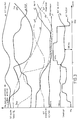
- Figures 2 and 3 show curves of parameter values used in the device.
- the implantable device comprises a central unit 1 such as a microprocessor, to which are added an electric current source 2 and memories such as a memory 3, preferably of RAM type, intended for memorizing the active sequence in progress, a memory 4, preferably of the RAM type, intended for memorizing the sequences of parameters of several sequences, for example a sequence entered by programming, and one or more sequences which can be produce and which are followed by a cardiac incident detected by the device, as well as a memory 5, preferably of the ROM type comprising instructions for the general operation of the microprocessor 1.
- a central unit 1 such as a microprocessor, to which are added an electric current source 2 and memories such as a memory 3, preferably of RAM type, intended for memorizing the active sequence in progress, a memory 4, preferably of the RAM type, intended for memorizing the sequences of parameters of several sequences, for example a sequence entered by programming, and one or more sequences which can be produce and which are followed by a cardiac incident detected by the device, as well as a memory 5, preferably of the
- Additional memories may possibly include other information such as for example information relating to the patient, his disease and his evolution, the physiological modifications as well as information relating to the type of intervention, for example power intensity, and localization and orientation of electrical stimulations, etc.
- the microprocessor 1 is on the other hand connected to an analog / digital conversion unit 6 to which lead, by suitable implanted conductors, the information of different sensors 7, 8, 9 such as detection electrodes, pressure sensors, etc.
- the central unit 1 is connected to a stimulation logic 10, for example of a known type, capable of processing the heart, by sending stimulation pulses, actuating effectors such as 11, 12 , for example stimulation electrodes. All of the components 1-6, 10 are contained in a sealed housing of the type used in implanted cardiac pacemakers which can itself carry, for example, an electrode leading to the converter 6.
- FIG. 2 shows, for a determined patient, the curve of parameter values of a sequence which the doctor has deemed dangerous for the patient following a study on a HOLTER recording.
- the parameter is the RR period of the cardiac cycle.
- the curve a in FIG. 2 represents the evolution of the period of the cardiac cycles during 72 consecutive cycles. As we can see, this evolution is characterized on the one hand by a great constancy of the RR ratio in which the RR interval of two consecutive cycles differ by less than 5%, as well as by a trend general to a very gradual decrease in the duration of the RR interval.
- the curve thus formed is stored in memory 4.
- the microprocessor constantly maintains in memory 3, the analogous curve b constituted by the values of the RR intervals of the last 72 detected cycles, that is to say of the active sequence.
- the microprocessor 1 compares the curve b of the active sequence which has just been updated in memory 3 with the curve a recorded in memory 4.
- the active curve b represented in FIG. 2 differs from the curve a by variations in rhythm sometimes greater than 5%, as well as by fluctuations in increase and decrease. This sequence is not considered dangerous.
- the comparison with the sequence a is positive and the microprocessor 1 then commands the sending of a series of stimulation pulses by the device 10, at a rhythm slightly higher than the rhythm spontaneous and with a power of four times the threshold.
- the active sequence this time includes 1152 cycles.
- the device also comprises a right intra-ventricular pressure sensor making it possible to obtain the value of the PEP (a6) and also, by the microprocessor, to calculate the variation of the pressure dp / dt (a7).
- It also includes logic means calculating the area of the pressure mechanogram (a8), a partial oxygen pressure detector (a9), and an endocavitary arterial pressure sensor (a10).
- the device measures and calculates these different parameters for each cycle which occurs and stores the values of the last 1152 cycles, that is to say of the active sequence, in memory 3.
- the device keeps in memory 3 the sequence of parameter values of the last 9216 cycles, which of course includes the last 1152 cycles.
- the microprocessor is programmed to be able to see the occurrence of a heart failure. For example, the device considers that a rate of at least 15% of identified ventricular extrasystoles indicates a failure, as does the detection of ventricular fibrillation.
- the combination of electronic detection means and heart pressure is already described in the literature, for example French patent FR-A-1,237,702.
- the device according to the invention which is preferably combined with an automatic defibrillator and with an anti-tachycardia device as described in patent US-A-4,052,991, detects the occurrence of heart failure, it extracts from memory 3 the values of the parameters corresponding to the 9216 cycles which preceded the occurrence of the fault and enters them in memory 4.
- FIG. 3 shows the curves 1 to 10 of the different parameters during a sequence of 1152 consecutive cycles constituted by the cycles 1252 to 100 which preceded the occurrence of the failure.
- the device according to the invention constantly compares the values of the various parameters mentioned above during the active sequence in progress with the values stored and represented on the curves (a1 ... a10) of FIG. 3.
- the device For the curve a2 (percentage of extrasystoles), the device considers that there is similarity in the comparison as soon as a neighboring number of extrasystoles has been counted during the sequence.
- the active sequence is compared with the sequence shown in FIG. 3 which has been stored and which corresponds to cycles 1252 to 100 before the failure which has been recorded.
- the device can be arranged by programming the microprocessor so as to retain as stored comparison sequence, one of the sequences of 1152 consecutive cycles occurring before the recorded failure and having a particularly characteristic appearance, for example a high number ventricular extrasystoles, QT prolongation and a significant shift of the QT intervals on two different leads giving an abnormal QT dispersion.
- the device is, as has been said, preferably combined with the tachycardia reduction means described in the aforementioned US patent application.
- This method detects the occurrence of a number of extrasystoles or incipient tachycardia and automatically causes an isolated stimulation, or preferably a sequence of electrical stimulations, at a frequency higher than that of the extrasystoles which have just been detected.
- the device according to the invention detects whether the extrasystoles persist or not. If the extrasystoles do not persist, the device can be arranged so as not to involve the implementation of the intervention means.
Landscapes
- Health & Medical Sciences (AREA)
- Cardiology (AREA)
- Life Sciences & Earth Sciences (AREA)
- Heart & Thoracic Surgery (AREA)
- Nuclear Medicine, Radiotherapy & Molecular Imaging (AREA)
- Public Health (AREA)
- Engineering & Computer Science (AREA)
- Biomedical Technology (AREA)
- Veterinary Medicine (AREA)
- Radiology & Medical Imaging (AREA)
- Animal Behavior & Ethology (AREA)
- General Health & Medical Sciences (AREA)
- Biophysics (AREA)
- Physiology (AREA)
- Electrotherapy Devices (AREA)
- Prostheses (AREA)
- Paper (AREA)
- External Artificial Organs (AREA)
- Soil Working Implements (AREA)
Applications Claiming Priority (2)
| Application Number | Priority Date | Filing Date | Title |
|---|---|---|---|
| FR9014643 | 1990-11-23 | ||
| FR9014643A FR2669523B1 (fr) | 1990-11-23 | 1990-11-23 | Dispositif de prevention des defaillances cardiaques |
Publications (2)
| Publication Number | Publication Date |
|---|---|
| EP0487429A1 true EP0487429A1 (de) | 1992-05-27 |
| EP0487429B1 EP0487429B1 (de) | 1996-03-27 |
Family
ID=9402523
Family Applications (1)
| Application Number | Title | Priority Date | Filing Date |
|---|---|---|---|
| EP91403161A Revoked EP0487429B1 (de) | 1990-11-23 | 1991-11-22 | Einrichtung zur Verhinderung von Herzfunktionstörungen |
Country Status (6)
| Country | Link |
|---|---|
| US (1) | US5306293A (de) |
| EP (1) | EP0487429B1 (de) |
| AT (1) | ATE135927T1 (de) |
| CA (1) | CA2056003A1 (de) |
| DE (1) | DE69118325D1 (de) |
| FR (1) | FR2669523B1 (de) |
Cited By (7)
| Publication number | Priority date | Publication date | Assignee | Title |
|---|---|---|---|---|
| EP0653226A1 (de) * | 1993-11-17 | 1995-05-17 | ELA MEDICAL (Société anonyme) | Medizinisches Gerät, insbesondere implantierbarer Defibrillator mit integrierten Aufzeichnungsfunktionen von Holter |
| WO1999021475A1 (de) * | 1997-10-24 | 1999-05-06 | Pulsion Medical Systems Gmbh | Vorrichtung zur feststellung eines perikardergusses |
| EP0920885A1 (de) * | 1997-12-04 | 1999-06-09 | Vitatron Medical B.V. | Herzbehandlungssystem zur Feststellung und Behandlung von Herzversagen |
| ES2157848A1 (es) * | 1999-12-23 | 2001-08-16 | Gullon Miguel Santiago Gullon | Sistema inteligente y autonomo de recuperacion cardiaca. |
| DE10008792A1 (de) * | 2000-02-18 | 2001-08-23 | Biotronik Mess & Therapieg | Vorrichtung zur Verarbeitung von Körpersignalen |
| WO2002020088A1 (en) * | 2000-09-01 | 2002-03-14 | Medtronic, Inc. | Method and system for myocardial infarction repair |
| US7031775B2 (en) | 1997-11-07 | 2006-04-18 | Medtronic, Inc. | Method and system for myocardial infarction repair |
Families Citing this family (60)
| Publication number | Priority date | Publication date | Assignee | Title |
|---|---|---|---|---|
| SE9304029D0 (sv) * | 1993-12-03 | 1993-12-03 | Siemens Elema Ab | Analysanordning för att analysera elektriska signaler från ett hjärta |
| US5568126A (en) * | 1995-07-10 | 1996-10-22 | Andersen; Stig L. | Providing an alarm in response to a determination that a person may have suddenly experienced fear |
| GB9518094D0 (en) † | 1995-09-05 | 1995-11-08 | Cardionics Ltd | Heart monitoring apparatus |
| US5753182A (en) * | 1996-02-14 | 1998-05-19 | Biomet, Inc. | Method for reducing the number of free radicals present in ultrahigh molecular weight polyethylene orthopedic components |
| US7269457B2 (en) | 1996-04-30 | 2007-09-11 | Medtronic, Inc. | Method and system for vagal nerve stimulation with multi-site cardiac pacing |
| US6449507B1 (en) * | 1996-04-30 | 2002-09-10 | Medtronic, Inc. | Method and system for nerve stimulation prior to and during a medical procedure |
| US5690682A (en) * | 1996-06-17 | 1997-11-25 | Pharmatarget, Inc. | Device and method for treatment of cardiac arrhythmia |
| US6151525A (en) * | 1997-11-07 | 2000-11-21 | Medtronic, Inc. | Method and system for myocardial identifier repair |
| US6038476A (en) * | 1998-06-12 | 2000-03-14 | Pacesetter, Inc. | System and method for analyzing the efficacy of cardiac stimulation therapy |
| US6370431B1 (en) * | 1998-10-26 | 2002-04-09 | Medtronic, Inc. | Pacemaker system for preventing ventricular tachycardia |
| NL1012223C2 (nl) * | 1999-06-03 | 2000-12-06 | Martil Instr B V | Hartgangmaker alsmede gangmakereenheid en elektrische draad daarvoor. |
| FR2796292B1 (fr) * | 1999-07-15 | 2001-09-21 | Ela Medical Sa | Dispositif medical actif implantable, notamment stimulateur cardiaque, defibrillateur ou cardioverteur, asservi et protege contre les effets des extrasystolies brady et/ou tachy-dependantes |
| US6810286B2 (en) * | 2000-03-06 | 2004-10-26 | Medtronic, Inc | Stimulation for delivery of molecular therapy |
| US6456880B1 (en) | 2000-05-01 | 2002-09-24 | Pacesetter, Inc. | Implantable cardiac stimulation device for and method of monitoring progression or regression of a patient's heart condition by monitoring ventricular repolarization interval dispersion |
| US6847840B2 (en) * | 2000-09-25 | 2005-01-25 | Pfizer, Inc. | System and method for statistical analysis of QT interval as a function of changes in RR interval |
| US6571125B2 (en) | 2001-02-12 | 2003-05-27 | Medtronic, Inc. | Drug delivery device |
| US20020188327A1 (en) * | 2001-04-26 | 2002-12-12 | Chester Struble | Drug delivery for treatment of cardiac arrhythmia |
| US7463921B2 (en) * | 2001-08-13 | 2008-12-09 | University Of Rochester | Method and system for analyzing an electrocardiographic signal |
| FR2829029B1 (fr) * | 2001-09-04 | 2005-04-01 | Ela Medical Sa | Dispositif medical implantable actif pour le traitement des troubles du rythme cardiaque, comprenant des moyens de detection, d'analyse et de traitement des pauses ventriculaires |
| EP1451306A1 (de) * | 2001-11-06 | 2004-09-01 | Medtronic, Inc. | Verfahren und system zur operativen versorgung nach myokardinfarkt |
| US8391989B2 (en) * | 2002-12-18 | 2013-03-05 | Cardiac Pacemakers, Inc. | Advanced patient management for defining, identifying and using predetermined health-related events |
| US20040122294A1 (en) | 2002-12-18 | 2004-06-24 | John Hatlestad | Advanced patient management with environmental data |
| US7378955B2 (en) | 2003-01-03 | 2008-05-27 | Cardiac Pacemakers, Inc. | System and method for correlating biometric trends with a related temporal event |
| US7392081B2 (en) * | 2003-02-28 | 2008-06-24 | Cardiac Pacemakers, Inc. | Subcutaneous cardiac stimulator employing post-shock transthoracic asystole prevention pacing |
| JP4270382B2 (ja) * | 2003-03-13 | 2009-05-27 | 大日精化工業株式会社 | 感熱記録材料 |
| US7979122B2 (en) * | 2003-04-11 | 2011-07-12 | Cardiac Pacemakers, Inc. | Implantable sudden cardiac death prevention device with reduced programmable feature set |
| US7302294B2 (en) | 2003-04-11 | 2007-11-27 | Cardiac Pacemakers, Inc. | Subcutaneous cardiac sensing and stimulation system employing blood sensor |
| US7570997B2 (en) * | 2003-04-11 | 2009-08-04 | Cardiac Pacemakers, Inc. | Subcutaneous cardiac rhythm management with asystole prevention therapy |
| US20050004615A1 (en) * | 2003-04-11 | 2005-01-06 | Sanders Richard S. | Reconfigurable implantable cardiac monitoring and therapy delivery device |
| US20040215240A1 (en) * | 2003-04-11 | 2004-10-28 | Lovett Eric G. | Reconfigurable subcutaneous cardiac device |
| US20040230229A1 (en) * | 2003-04-11 | 2004-11-18 | Lovett Eric G. | Hybrid transthoracic/intrathoracic cardiac stimulation devices and methods |
| EP3064242A1 (de) | 2003-04-28 | 2016-09-07 | Advanced Circulatory Systems Inc. | Lüfter und verfahren zur behandlung von kopftrauma und geringer blutzirkulation |
| FR2854971B1 (fr) * | 2003-05-14 | 2005-07-29 | Ela Medical Sa | Dispositif medical actif pourvu d'une memoire pour le stockage de donnees holter et d'instructions de pilotage d'un microprocesseur |
| US7074194B2 (en) * | 2003-05-19 | 2006-07-11 | Ischemia Technologies, Inc. | Apparatus and method for risk stratification of patients with chest pain of suspected cardiac origin |
| US7887493B2 (en) | 2003-09-18 | 2011-02-15 | Cardiac Pacemakers, Inc. | Implantable device employing movement sensing for detecting sleep-related disorders |
| US8002553B2 (en) * | 2003-08-18 | 2011-08-23 | Cardiac Pacemakers, Inc. | Sleep quality data collection and evaluation |
| EP2008581B1 (de) | 2003-08-18 | 2011-08-17 | Cardiac Pacemakers, Inc. | Patientenüberwachungs-, Diagnose- und/oder Therapiesysteme und -verfahren |
| DE10340268A1 (de) * | 2003-08-29 | 2005-04-14 | Corscience Gmbh & Co.Kg | Verfahren zum Detektieren der pulslosen elektrischen Aktivität |
| FR2861997A1 (fr) * | 2003-11-06 | 2005-05-13 | Fred Zacouto | Stimulateur cardiaque orthorythmique inotrope |
| US7577478B1 (en) * | 2006-02-01 | 2009-08-18 | Pacesetter, Inc. | Ischemia detection for anti-arrhythmia therapy |
| US7933650B2 (en) * | 2006-02-16 | 2011-04-26 | Cardiac Pacemakers, Inc. | Method and apparatus for selecting and timing anti-tachyarrhythmia pacing using cardiac signal morphology |
| US8151790B2 (en) | 2007-04-19 | 2012-04-10 | Advanced Circulatory Systems, Inc. | Volume exchanger valve system and method to increase circulation during CPR |
| US9352111B2 (en) | 2007-04-19 | 2016-05-31 | Advanced Circulatory Systems, Inc. | Systems and methods to increase survival with favorable neurological function after cardiac arrest |
| US9724266B2 (en) | 2010-02-12 | 2017-08-08 | Zoll Medical Corporation | Enhanced guided active compression decompression cardiopulmonary resuscitation systems and methods |
| US20120203147A1 (en) * | 2010-07-02 | 2012-08-09 | ResQSystems, Inc. | Vasodilator-enhanced cardiopulmonary resuscitation |
| US12016820B2 (en) | 2010-02-12 | 2024-06-25 | Zoll Medical Corporation | Enhanced guided active compression decompression cardiopulmonary resuscitation systems and methods |
| US8406868B2 (en) | 2010-04-29 | 2013-03-26 | Medtronic, Inc. | Therapy using perturbation and effect of physiological systems |
| US8620425B2 (en) | 2010-04-29 | 2013-12-31 | Medtronic, Inc. | Nerve signal differentiation in cardiac therapy |
| US8639327B2 (en) | 2010-04-29 | 2014-01-28 | Medtronic, Inc. | Nerve signal differentiation in cardiac therapy |
| US8706223B2 (en) | 2011-01-19 | 2014-04-22 | Medtronic, Inc. | Preventative vagal stimulation |
| US8718763B2 (en) | 2011-01-19 | 2014-05-06 | Medtronic, Inc. | Vagal stimulation |
| US8781583B2 (en) | 2011-01-19 | 2014-07-15 | Medtronic, Inc. | Vagal stimulation |
| US8781582B2 (en) | 2011-01-19 | 2014-07-15 | Medtronic, Inc. | Vagal stimulation |
| US8725259B2 (en) | 2011-01-19 | 2014-05-13 | Medtronic, Inc. | Vagal stimulation |
| AU2012358977A1 (en) | 2011-12-19 | 2014-07-24 | ResQSystems, Inc. | Systems and methods for therapeutic intrathoracic pressure regulation |
| US8874197B2 (en) | 2012-10-30 | 2014-10-28 | Medtronic, Inc. | Risk determination for ventricular arrhythmia |
| US9811634B2 (en) | 2013-04-25 | 2017-11-07 | Zoll Medical Corporation | Systems and methods to predict the chances of neurologically intact survival while performing CPR |
| US20140358047A1 (en) | 2013-05-30 | 2014-12-04 | ResQSystems, Inc. | End-tidal carbon dioxide and amplitude spectral area as non-invasive markers of coronary perfusion pressure and arterial pressure |
| US10265495B2 (en) | 2013-11-22 | 2019-04-23 | Zoll Medical Corporation | Pressure actuated valve systems and methods |
| US10143839B1 (en) * | 2017-07-05 | 2018-12-04 | Medtronic, Inc. | Lead navigation guidance |
Citations (4)
| Publication number | Priority date | Publication date | Assignee | Title |
|---|---|---|---|---|
| US4473078A (en) * | 1982-05-24 | 1984-09-25 | Cardiac Resuscitator Corporation | Cardiac arrhythmia analysis system |
| EP0348271A1 (de) * | 1988-06-13 | 1989-12-27 | Fred Zacouto | Schutzeinrichtung gegen gesundheitliche Schädigung des Blutes, besonders Thrombosen, Embolien, Gefässpasmen, Hämorrhagien, Hämopathien und das Vorhandensein von anormalen Elementen im Blut |
| EP0387363A1 (de) * | 1989-03-13 | 1990-09-19 | Pacesetter AB | Implantierbares medizinisches Gerät zur Stimulation eines physiologischen Vorganges eines Lebewesens mit an die körperliche Aktivität des Lebewesens anpassbarer Stimulationsintensität |
| EP0398548A2 (de) * | 1989-05-15 | 1990-11-22 | Cardiac Pacemakers, Inc. | Demand-Herzschrittmacher mit aus lebendem Gewebe bestehenden physiologischen Sensoren |
Family Cites Families (5)
| Publication number | Priority date | Publication date | Assignee | Title |
|---|---|---|---|---|
| FR1237702A (fr) * | 1953-07-11 | 1960-08-05 | Perfectionnements aux appareils électro-médicaux | |
| US4052991A (en) * | 1970-03-24 | 1977-10-11 | Fred Zacouto | Method of stimulating the heart |
| US3857399A (en) * | 1970-03-24 | 1974-12-31 | F Zacouto | Heart pacer |
| US4552150A (en) * | 1983-06-14 | 1985-11-12 | Fred Zacouto | Method and apparatus to assist cardiac muscle functioning |
| US5053008A (en) * | 1990-11-21 | 1991-10-01 | Sandeep Bajaj | Intracardiac catheter |
-
1990
- 1990-11-23 FR FR9014643A patent/FR2669523B1/fr not_active Expired - Fee Related
-
1991
- 1991-11-21 US US07/796,250 patent/US5306293A/en not_active Expired - Lifetime
- 1991-11-22 CA CA002056003A patent/CA2056003A1/fr not_active Abandoned
- 1991-11-22 EP EP91403161A patent/EP0487429B1/de not_active Revoked
- 1991-11-22 DE DE69118325T patent/DE69118325D1/de not_active Expired - Lifetime
- 1991-11-22 AT AT91403161T patent/ATE135927T1/de not_active IP Right Cessation
Patent Citations (4)
| Publication number | Priority date | Publication date | Assignee | Title |
|---|---|---|---|---|
| US4473078A (en) * | 1982-05-24 | 1984-09-25 | Cardiac Resuscitator Corporation | Cardiac arrhythmia analysis system |
| EP0348271A1 (de) * | 1988-06-13 | 1989-12-27 | Fred Zacouto | Schutzeinrichtung gegen gesundheitliche Schädigung des Blutes, besonders Thrombosen, Embolien, Gefässpasmen, Hämorrhagien, Hämopathien und das Vorhandensein von anormalen Elementen im Blut |
| EP0387363A1 (de) * | 1989-03-13 | 1990-09-19 | Pacesetter AB | Implantierbares medizinisches Gerät zur Stimulation eines physiologischen Vorganges eines Lebewesens mit an die körperliche Aktivität des Lebewesens anpassbarer Stimulationsintensität |
| EP0398548A2 (de) * | 1989-05-15 | 1990-11-22 | Cardiac Pacemakers, Inc. | Demand-Herzschrittmacher mit aus lebendem Gewebe bestehenden physiologischen Sensoren |
Cited By (15)
| Publication number | Priority date | Publication date | Assignee | Title |
|---|---|---|---|---|
| FR2712501A1 (fr) * | 1993-11-17 | 1995-05-24 | Ela Medical Sa | Appareil médical, notamment défibrillateur implantable, à fonctions d'enregistrement Holter intégrées . |
| EP0653226A1 (de) * | 1993-11-17 | 1995-05-17 | ELA MEDICAL (Société anonyme) | Medizinisches Gerät, insbesondere implantierbarer Defibrillator mit integrierten Aufzeichnungsfunktionen von Holter |
| US6351667B1 (en) | 1997-10-24 | 2002-02-26 | Pulsion Medical Systems Ag | Device for detecting pericardial effusion |
| WO1999021475A1 (de) * | 1997-10-24 | 1999-05-06 | Pulsion Medical Systems Gmbh | Vorrichtung zur feststellung eines perikardergusses |
| US7155288B2 (en) | 1997-11-07 | 2006-12-26 | Medtronic, Inc | Method and system for myocardial infarction repair |
| US7031775B2 (en) | 1997-11-07 | 2006-04-18 | Medtronic, Inc. | Method and system for myocardial infarction repair |
| EP0920885A1 (de) * | 1997-12-04 | 1999-06-09 | Vitatron Medical B.V. | Herzbehandlungssystem zur Feststellung und Behandlung von Herzversagen |
| EP1101509A1 (de) | 1997-12-04 | 2001-05-23 | Vitatron Medical B.V. | Implantierbares Herzschrittmachersystem |
| ES2157848A1 (es) * | 1999-12-23 | 2001-08-16 | Gullon Miguel Santiago Gullon | Sistema inteligente y autonomo de recuperacion cardiaca. |
| EP1129661A2 (de) | 2000-02-18 | 2001-09-05 | BIOTRONIK Mess- und Therapiegeräte GmbH & Co Ingenieurbüro Berlin | Vorrichtung zur Verarbeitung von Körpersignalen |
| EP1129661A3 (de) * | 2000-02-18 | 2001-11-14 | BIOTRONIK Mess- und Therapiegeräte GmbH & Co Ingenieurbüro Berlin | Vorrichtung zur Verarbeitung von Körpersignalen |
| DE10008792A1 (de) * | 2000-02-18 | 2001-08-23 | Biotronik Mess & Therapieg | Vorrichtung zur Verarbeitung von Körpersignalen |
| US6577892B2 (en) | 2000-02-18 | 2003-06-10 | Biotonik Mess -Und Therapiegeraete Gmbh & Co. Ingenieurbuero Berlin | Apparatus for processing body signals |
| WO2002020088A1 (en) * | 2000-09-01 | 2002-03-14 | Medtronic, Inc. | Method and system for myocardial infarction repair |
| EP1728532A1 (de) | 2000-09-01 | 2006-12-06 | Medtronic, Inc. | Verfahren und Vorrichtung zur Myokardwiederherstellung bei Myokard-Infarkt |
Also Published As
| Publication number | Publication date |
|---|---|
| US5306293A (en) | 1994-04-26 |
| CA2056003A1 (fr) | 1992-05-24 |
| FR2669523A1 (fr) | 1992-05-29 |
| EP0487429B1 (de) | 1996-03-27 |
| ATE135927T1 (de) | 1996-04-15 |
| FR2669523B1 (fr) | 1997-06-06 |
| DE69118325D1 (de) | 1996-05-02 |
Similar Documents
| Publication | Publication Date | Title |
|---|---|---|
| EP0487429B1 (de) | Einrichtung zur Verhinderung von Herzfunktionstörungen | |
| EP0348271A1 (de) | Schutzeinrichtung gegen gesundheitliche Schädigung des Blutes, besonders Thrombosen, Embolien, Gefässpasmen, Hämorrhagien, Hämopathien und das Vorhandensein von anormalen Elementen im Blut | |
| FR2808213A1 (fr) | Dispositif medical implantable et son procede utilisant un analyseur integre d'alternance d'onde t | |
| JP5048057B2 (ja) | 心臓保護ペーシング方法及び装置 | |
| US8328728B2 (en) | Implantable hemodynamic monitor and methods for use therewith | |
| EP0467695A2 (de) | Erkennung und Behandlung von Herzinfarkten in einem Antitachy-Arrythmien- und Bradycardien-Herzschrittmacher | |
| FR2808214A1 (fr) | Methode et systeme pour stimuler un coeur de mammifere | |
| WO2005046790A2 (fr) | Stimulateur cardiaque orthorythmique inotrope | |
| EP0862927B1 (de) | Multi-Ort Herzschrittmacher zur Behandlung von Herzinsuffizienzen mit Reizimpulsen | |
| US8433397B2 (en) | Methods and systems to correlate arrhythmic and ischemic events | |
| van der Burg et al. | Long‐term follow‐up after radiofrequency catheter ablation of ventricular tachycardia: A successful approach? | |
| FR2618339A1 (fr) | Methode et appareil medical pour le traitement de tachyarythmies | |
| EP0566588A1 (de) | Gerät zur überwachung der herzfrequenz | |
| US8670820B2 (en) | Near field-based systems and methods for assessing impedance and admittance for use with an implantable medical device | |
| EP3111991B1 (de) | Stimulationsbehandlungssystem, insbesondere für die stimulation des vagusnervs durch anwendung eines zustandsübergangsmodells, das sich automatisch an die physischen und physiologischen niveaus anpasst | |
| US20050234517A1 (en) | Apparatus and method for hemodynamic-based optimization of cardiac pacing | |
| EP2508227B1 (de) | Implantierbare Herzprothese zur Resynchronisation des Herzens durch biventrikuläre Stimulation, die Mittel zur inversen Herzrekonfiguration umfasst | |
| EP2495013B1 (de) | Anordnung für die Suche nach einer optimalen Konfiguration eines Implantats zur bi-, tri- oder multiventrikulären Resynchronisation des Herzens | |
| Schaefer et al. | Clinical and hemodynamic characteristics of patients with inducible pulsus alternans | |
| US8521277B2 (en) | Implantable systems and method for use therewith for tracking changes in hemodynamics and cardiac disease | |
| US20140257070A1 (en) | Processing of lap signals | |
| RU2164077C1 (ru) | Способ определения коронарной и миокардиальной недостаточности | |
| EP4558215B1 (de) | Akute nichtexzitatorische elektrische herzinsuffizienztherapie | |
| Lévy et al. | Refractory supraventricular tachycardias: Successful therapy with double demand sequential pacing | |
| FR3157209A1 (fr) | Pompe cardiaque dotée de fonctions de resynchronisation cardiaque. |
Legal Events
| Date | Code | Title | Description |
|---|---|---|---|
| PUAI | Public reference made under article 153(3) epc to a published international application that has entered the european phase |
Free format text: ORIGINAL CODE: 0009012 |
|
| AK | Designated contracting states |
Kind code of ref document: A1 Designated state(s): AT BE CH DE DK ES FR GB GR IT LI LU NL SE |
|
| 17P | Request for examination filed |
Effective date: 19921002 |
|
| 17Q | First examination report despatched |
Effective date: 19941004 |
|
| GRAA | (expected) grant |
Free format text: ORIGINAL CODE: 0009210 |
|
| AK | Designated contracting states |
Kind code of ref document: B1 Designated state(s): AT BE CH DE DK ES FR GB GR IT LI LU NL SE |
|
| PG25 | Lapsed in a contracting state [announced via postgrant information from national office to epo] |
Ref country code: IT Free format text: LAPSE BECAUSE OF FAILURE TO SUBMIT A TRANSLATION OF THE DESCRIPTION OR TO PAY THE FEE WITHIN THE PRE;WARNING: LAPSES OF ITALIAN PATENTS WITH EFFECTIVE DATE BEFORE 2007 MAY HAVE OCCURRED AT ANY TIME BEFORE 2007. THE CORRECT EFFECTIVE DATE MAY BE DIFFERENT FROM THE ONE RECORDED.SCRIBED TIME-LIMIT Effective date: 19960327 Ref country code: DK Effective date: 19960327 Ref country code: AT Effective date: 19960327 Ref country code: ES Free format text: THE PATENT HAS BEEN ANNULLED BY A DECISION OF A NATIONAL AUTHORITY Effective date: 19960327 Ref country code: NL Free format text: LAPSE BECAUSE OF FAILURE TO SUBMIT A TRANSLATION OF THE DESCRIPTION OR TO PAY THE FEE WITHIN THE PRESCRIBED TIME-LIMIT Effective date: 19960327 Ref country code: GR Free format text: LAPSE BECAUSE OF FAILURE TO SUBMIT A TRANSLATION OF THE DESCRIPTION OR TO PAY THE FEE WITHIN THE PRESCRIBED TIME-LIMIT Effective date: 19960327 |
|
| REF | Corresponds to: |
Ref document number: 135927 Country of ref document: AT Date of ref document: 19960415 Kind code of ref document: T |
|
| REF | Corresponds to: |
Ref document number: 69118325 Country of ref document: DE Date of ref document: 19960502 |
|
| PG25 | Lapsed in a contracting state [announced via postgrant information from national office to epo] |
Ref country code: SE Effective date: 19960627 |
|
| PG25 | Lapsed in a contracting state [announced via postgrant information from national office to epo] |
Ref country code: DE Effective date: 19960628 |
|
| GBT | Gb: translation of ep patent filed (gb section 77(6)(a)/1977) |
Effective date: 19960704 |
|
| GRAH | Despatch of communication of intention to grant a patent |
Free format text: ORIGINAL CODE: EPIDOS IGRA |
|
| NLV1 | Nl: lapsed or annulled due to failure to fulfill the requirements of art. 29p and 29m of the patents act | ||
| PG25 | Lapsed in a contracting state [announced via postgrant information from national office to epo] |
Ref country code: LU Free format text: LAPSE BECAUSE OF NON-PAYMENT OF DUE FEES Effective date: 19961130 |
|
| PLBQ | Unpublished change to opponent data |
Free format text: ORIGINAL CODE: EPIDOS OPPO |
|
| PLBI | Opposition filed |
Free format text: ORIGINAL CODE: 0009260 |
|
| PLBF | Reply of patent proprietor to notice(s) of opposition |
Free format text: ORIGINAL CODE: EPIDOS OBSO |
|
| 26 | Opposition filed |
Opponent name: BIOTRONIK MESS- UND THERAPIEGERAETE GMBH & CO ING Effective date: 19961223 |
|
| PLBF | Reply of patent proprietor to notice(s) of opposition |
Free format text: ORIGINAL CODE: EPIDOS OBSO |
|
| PLBF | Reply of patent proprietor to notice(s) of opposition |
Free format text: ORIGINAL CODE: EPIDOS OBSO |
|
| PLBF | Reply of patent proprietor to notice(s) of opposition |
Free format text: ORIGINAL CODE: EPIDOS OBSO |
|
| PGFP | Annual fee paid to national office [announced via postgrant information from national office to epo] |
Ref country code: GB Payment date: 19981117 Year of fee payment: 8 |
|
| PGFP | Annual fee paid to national office [announced via postgrant information from national office to epo] |
Ref country code: BE Payment date: 19981210 Year of fee payment: 8 |
|
| PGFP | Annual fee paid to national office [announced via postgrant information from national office to epo] |
Ref country code: CH Payment date: 19991015 Year of fee payment: 9 |
|
| PG25 | Lapsed in a contracting state [announced via postgrant information from national office to epo] |
Ref country code: GB Free format text: LAPSE BECAUSE OF NON-PAYMENT OF DUE FEES Effective date: 19991122 |
|
| PG25 | Lapsed in a contracting state [announced via postgrant information from national office to epo] |
Ref country code: BE Free format text: LAPSE BECAUSE OF NON-PAYMENT OF DUE FEES Effective date: 19991130 |
|
| PLBQ | Unpublished change to opponent data |
Free format text: ORIGINAL CODE: EPIDOS OPPO |
|
| PLAB | Opposition data, opponent's data or that of the opponent's representative modified |
Free format text: ORIGINAL CODE: 0009299OPPO |
|
| R26 | Opposition filed (corrected) |
Opponent name: BIOTRONIK MESS- UND THERAPIEGERAETE GMBH & CO INGE Effective date: 19961223 |
|
| PLBO | Opposition rejected |
Free format text: ORIGINAL CODE: EPIDOS REJO |
|
| APAC | Appeal dossier modified |
Free format text: ORIGINAL CODE: EPIDOS NOAPO |
|
| BERE | Be: lapsed |
Owner name: ZACOUTO FRED Effective date: 19991130 |
|
| APAE | Appeal reference modified |
Free format text: ORIGINAL CODE: EPIDOS REFNO |
|
| GBPC | Gb: european patent ceased through non-payment of renewal fee |
Effective date: 19991122 |
|
| APAC | Appeal dossier modified |
Free format text: ORIGINAL CODE: EPIDOS NOAPO |
|
| PG25 | Lapsed in a contracting state [announced via postgrant information from national office to epo] |
Ref country code: LI Free format text: LAPSE BECAUSE OF NON-PAYMENT OF DUE FEES Effective date: 20001130 Ref country code: CH Free format text: LAPSE BECAUSE OF NON-PAYMENT OF DUE FEES Effective date: 20001130 |
|
| PLBQ | Unpublished change to opponent data |
Free format text: ORIGINAL CODE: EPIDOS OPPO |
|
| PLAB | Opposition data, opponent's data or that of the opponent's representative modified |
Free format text: ORIGINAL CODE: 0009299OPPO |
|
| R26 | Opposition filed (corrected) |
Opponent name: BIOTRONIK MESS- UND THERAPIEGERAETE GMBH & CO INGE Effective date: 19961223 |
|
| REG | Reference to a national code |
Ref country code: CH Ref legal event code: PL |
|
| PLAB | Opposition data, opponent's data or that of the opponent's representative modified |
Free format text: ORIGINAL CODE: 0009299OPPO |
|
| R26 | Opposition filed (corrected) |
Opponent name: BIOTRONIK GMBH & CO. KG Effective date: 19961223 |
|
| PGFP | Annual fee paid to national office [announced via postgrant information from national office to epo] |
Ref country code: FR Payment date: 20040129 Year of fee payment: 13 |
|
| APBU | Appeal procedure closed |
Free format text: ORIGINAL CODE: EPIDOSNNOA9O |
|
| RDAF | Communication despatched that patent is revoked |
Free format text: ORIGINAL CODE: EPIDOSNREV1 |
|
| RDAG | Patent revoked |
Free format text: ORIGINAL CODE: 0009271 |
|
| STAA | Information on the status of an ep patent application or granted ep patent |
Free format text: STATUS: PATENT REVOKED |
|
| 27W | Patent revoked |
Effective date: 20040317 |
|
| APAH | Appeal reference modified |
Free format text: ORIGINAL CODE: EPIDOSCREFNO |