EP0489732B1 - Uv-induzierter faktor für immununterdrückung - Google Patents
Uv-induzierter faktor für immununterdrückung Download PDFInfo
- Publication number
- EP0489732B1 EP0489732B1 EP90905146A EP90905146A EP0489732B1 EP 0489732 B1 EP0489732 B1 EP 0489732B1 EP 90905146 A EP90905146 A EP 90905146A EP 90905146 A EP90905146 A EP 90905146A EP 0489732 B1 EP0489732 B1 EP 0489732B1
- Authority
- EP
- European Patent Office
- Prior art keywords
- cells
- radiation
- irradiated
- uvb
- mice
- Prior art date
- Legal status (The legal status is an assumption and is not a legal conclusion. Google has not performed a legal analysis and makes no representation as to the accuracy of the status listed.)
- Expired - Lifetime
Links
Images


Classifications
-
- C—CHEMISTRY; METALLURGY
- C07—ORGANIC CHEMISTRY
- C07K—PEPTIDES
- C07K14/00—Peptides having more than 20 amino acids; Gastrins; Somatostatins; Melanotropins; Derivatives thereof
- C07K14/435—Peptides having more than 20 amino acids; Gastrins; Somatostatins; Melanotropins; Derivatives thereof from animals; from humans
- C07K14/52—Cytokines; Lymphokines; Interferons
- C07K14/54—Interleukins [IL]
- C07K14/5428—IL-10
-
- A—HUMAN NECESSITIES
- A61—MEDICAL OR VETERINARY SCIENCE; HYGIENE
- A61K—PREPARATIONS FOR MEDICAL, DENTAL OR TOILETRY PURPOSES
- A61K41/00—Medicinal preparations obtained by treating materials with wave energy or particle radiation ; Therapies using these preparations
- A61K41/10—Inactivation or decontamination of a medicinal preparation prior to administration to an animal or a person
- A61K41/17—Inactivation or decontamination of a medicinal preparation prior to administration to an animal or a person by ultraviolet [UV] or infrared [IR] light, X-rays or gamma rays
-
- A—HUMAN NECESSITIES
- A61—MEDICAL OR VETERINARY SCIENCE; HYGIENE
- A61P—SPECIFIC THERAPEUTIC ACTIVITY OF CHEMICAL COMPOUNDS OR MEDICINAL PREPARATIONS
- A61P37/00—Drugs for immunological or allergic disorders
- A61P37/02—Immunomodulators
- A61P37/06—Immunosuppressants, e.g. drugs for graft rejection
-
- A—HUMAN NECESSITIES
- A61—MEDICAL OR VETERINARY SCIENCE; HYGIENE
- A61K—PREPARATIONS FOR MEDICAL, DENTAL OR TOILETRY PURPOSES
- A61K38/00—Medicinal preparations containing peptides
-
- A—HUMAN NECESSITIES
- A61—MEDICAL OR VETERINARY SCIENCE; HYGIENE
- A61N—ELECTROTHERAPY; MAGNETOTHERAPY; RADIATION THERAPY; ULTRASOUND THERAPY
- A61N5/00—Radiation therapy
- A61N5/06—Radiation therapy using light
- A61N2005/0658—Radiation therapy using light characterised by the wavelength of light used
- A61N2005/0661—Radiation therapy using light characterised by the wavelength of light used ultraviolet
Definitions
- UV radiation suppresses the immune response [1].
- CHS contact hypersensitivity
- Mice exposed to a single dose of UV radiation are unable to generate a contact hypersensitivity (CHS) reaction to contact allergens applied to a distant unirradiated site [3].
- CHS contact hypersensitivity
- a single exposure to UV radiation also inhibits the generation of a delayed hypersensitivity (DTH) response to hapten modified cells [4], foreign erythrocytes and protein antigens [5] and allogeneic spleen cells [6.7].
- DTH delayed hypersensitivity
- UV-induced CHS and DTH suppression Although it is yet to be determined what the mechanism of UV-induced CHS and DTH suppression is, it has generally been considered by researchers in this area that a single mechanism is responsible for both. However, the present inventor has demonstrated that at least two factors are involved, each being released by cells after irradiation with different wavelengths of UV-radiation. The present inventor has determined that supernatant from epidermal cells exposed to long-wave UV radiation, UVA, (320-400 nm) would suppress CHS but not DTH. On the other hand, supernatants from short-wave UV-radiation, UVB, (280-320 nm) would suppress DTH but not CHS. This result shows that two different immunosuppressive factors are released by UV-irradiated cells.
- the first immunosuppressive is released on exposure to UVB and suppresses DTH and the second is released on exposure to UVA and suppresses CHS. Therefore, by using a predetermined wavelength of ultraviolet radiation (UVR), e.g., UVA or UVB, the immune response of a mammal can be selectively suppressed.
- UVR ultraviolet radiation
- immunosuppressive drugs are used to overcome the immunological rejection of transplanted tissue (allografts).
- pan-immunosuppression is produced.
- all other immune responses such as those involved in the protection of the host from viral and bacterial pathogens, are also suppressed.
- the immunosuppressed patient is susceptible to a variety of opportunistic infections. Accordingly, a method of suppressing only the immune response to the allografted tissue while leaving other immunological functions intact would be highly advantageous.
- UVR direct UV-irradiation
- specific immunosuppressive factors are produced in vitro by a mammalian cell when it is irradiated with a sufficient amount of UVB-radiation.
- These immunosuppressive factors combined with subsequent antigenic sensitization of an animal, induce an immunosuppression which is specific for the antigenic determinants used to sensitize the animal.
- pan-immunosuppression is avoided.
- the use of UVB-radiation to induce selective immunosuppression may have a marked advantage over the use of immunosuppressive drugs such as azathioprine or corticosteriods. For example, it would be advantageous to selectively suppress DTH and allograft rejection while leaving the immune system of a patient otherwise uncompromised.
- An aspect of the present invention is an immunosuppressive factor for use in a method for selectively suppressing an immune response in a mammal to a particular alloantigen.
- the method for producing said immunosuppressive factor may include multiple steps. One step is irradiating a plurality of mammalian cells with a sufficient amount of UVB-radiation to create immunosuppressive factors.
- One step is irradiating a plurality of mammalian cells with a sufficient amount of UVB-radiation to create immunosuppressive factors.
- mammalian epidermal cells irradiated with UVB radiation pre-determined wavelength of UVB-radiation being 280 to 320 nm
- Another step in the inventive method involves extracting the immunosuppressive factors from the UV-irradiated cells.
- a further step involves administering an effective amount of the immunosuppressive factors to the mammal. Thereafter, the mammal is sensitized to the particular alloantigen for which immunotolerance is preferred.
- Yet another aspect of the present invention is the process for producing immunosuppressive factors and the immunological suppressive factors themselves.
- This aspect of the invention preferably produces immunosuppressive factors which may be subsequently administered to a subject animal to result in a selective suppression of a specific immune response to a particular alloantigen.
- the inventive process includes the steps of radiating a plurality of mammalian cells in vitro with a sufficient amount of UVB-radiation to produce UV-irradiated cells producing immunosuppressive factors. It has been determined that mammalian cells irradiated in vitro with a sufficient amount of UVB (pre-determined wavelength being 280 to 320 nm) will produce immunosuppressive factors which selectively suppress the DTH response in mammals when administered in a sufficient amount.
- a single exposure to ultraviolet radiation induces a systemic suppression of the immune response to allogeneic histocompatibility antigens.
- the suppression is associated with the appearance of splenic alloantigen-specific suppressor T cells.
- How exposing the skin to UV radiation results in the induction of splenic suppressor T cells is not entirely clear.
- the data described herein suggest the involvement of a UV-induced keratinocyte-derived suppressive factor.
- the keratinocyte line, Pam 212 was exposed to 200 J/m2 of UVB radiation from a single FS-40 sunlamp and cultured overnight in serum-free medium. Injecting mice with culture supernatants from UV-irradiated keratinocytes suppressed the induction of delayed type hypersensitivity to alloantigen.
- Figure 1 shows the effect of the supernatants from UV-irradiated primary epidermal cell cultures on the induction of CHS(A) or DTH(B).
- Mice were injected with supernatants from the UVB-irradiated (UVB-SN) or control (NRSN) epidermal cell cultures or exposed to 40 kJ/m2 of UVB radiation (UVB).
- UVB-SN UVB-irradiated
- NRSN control
- FIG. 2 shows the effect of the supernatants from UV-irradiated Pam 212 cells on the induction of CHS(A) or DTH(B).
- Mice were injected with supernatants from the UV-irradiated (UVSN) or control (NRSN) non-irradiated keratinocyte cell cultures or exposed to 40 kJ/m2 of UVB radiation (UVB).
- UVSN UV-irradiated
- NRSN control non-irradiated keratinocyte cell cultures or exposed to 40 kJ/m2 of UVB radiation
- panel A Balb/c mice were sensitized with TNCB
- panel B Balb c mice were sensitized with C3H spleen cells.
- the asterisk indicates a significant difference (P ⁇ 0.001) from the response observed in the positive control (NR).
- FIG. 3 shows the effect of the supernatants from UVB-irradiated Pam 212 cells on the induction of DTH to TNP-conjugated syngeneic spleen cells.
- Mice were injected with supernatants from the UVB-irradiated (UVB-SN) or control (NR SN) keratinocyte cell cultures or exposed to 40 kJ/m2 of UV radiation (UV).
- UVB-SN UVB-irradiated
- NR SN control keratinocyte cell cultures or exposed to 40 kJ/m2 of UV radiation (UV).
- the asterisk indicates a significant difference (P ⁇ 0.001) from the response observed in the positive control (NR).
- Figure 4 shows the phenotype of the suppressor cells induced by injecting supernatants from UV-irradiated keratinocytes into mice.
- Spleen cells from mice injected with supernatants from the UV-irradiated keratinocytes where added to one way MLR cultures containing normal C3H responder cells and gamma-irradiated B6 stimulator cells.
- Spleen cells from the mice injected with the suppressive cytokines were treated with various monoclonal antibodies and complement.
- One group of cells was exposed to 2000 rad of gamma radiation.
- Control cells (UV and NR) were treated with complement. * indicates a significant difference from the proliferation of the control; P ⁇ 0.001.
- Figure 5 shows physical properties of the suppressive cytokine released from UV-irradiated keratinocytes.
- Pam 212 cells were exposed to 200 J/m2 of UV radiation and then treated with 10 micrograms/ml of indomethacin or 10 microgram/ml of cycloheximide.
- Supernatants from the treated cells and the control cultures (UV and NR) were dialyzed and then injected into mice.
- panel B the supernatants were harvested and then treated with heat or trypsin (10 microgram/ml). The treated supernatants were then injected into mice and the resulting MLR was measured.
- the background response of responder cells cultured alone was 4290 ⁇ 960 CPM. * indicates a significant difference from the control; P ⁇ 0.001.
- Figure 6 shows dose-response curve for suppressing the MLR.
- Various concentrations of culture supernatants from the UV-irradiated keratinocytes were injected into mice and the ability of their spleen cells to proliferate in response to alloantigen was measured. The data are expressed as a percentage of the control response (mice injected with media; 34,456 ⁇ 2215 cpm is equal to 100%; the background response was 3,072 ⁇ 495 cpm).
- the cross-hatched region represents the proliferation of spleen cells from mice injected with supernatants from non-irradiated keratinocytes.
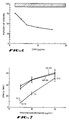
- Figure 7 shows production of IL-1 by the UV-irradiated and control keratinocytes.
- Supernatants were obtained from the UV-irradiated or control nonirradiated Pam 212 cells. Protein concentration was measured and various concentrations were added to the IL-1 dependent D10.G4.1 helper cell line. A standard curve was generated by adding dilutions of murine rIL-1 to the cells.
- Figure 8 shows sodium dodecyl sulfate-polyacrylamide gel electrophoresis (SDS-PAGE) analysis of the suppressive material eluted from conconavalin-A (Con A)-agarose columns. Equivalent amounts (200 ng) of the material eluted from the Con A-agarose columns were analyzed on 12.5% SDS-PAGE gels under reducing conditions. Lane 1 contained the UV mannoside eluate, lane 2 the UV glucoside eluate, lane 3 the control mannoside eluate from non-irradiated cells, lane 4 the control glucoside eluate from non-irradiated cells. A unique band, present only in the immunosuppressive fraction (lane 1), and not in any of the non-suppressive fractions (lane 2-4), appears to migrate with a molecular weight of 68kDa.
- SDS-PAGE sodium dodecyl sulfate-polyacrylamide gel electrophoresis
- the selective suppression of a specific immune response to a particular alloantigen by a pre-determined wavelength of ultraviolet radiation is illustrated by experiments in which mice are irradiated with a sufficient dose of UV-radiation and subsequently sensitized to particular alloantigen.
- the CHS response to the particular alloantigen is depressed by whole body UVB-irradiation (pre-determined wavelength 280 nm - 320 nm) and subsequent sensitization with the particular alloantigen.
- Whole body irradiation is defined as the process of irradiating the epidermis of the subject animal.
- the DTH response to a particular alloantigen is suppressed by whole body UVB-irradiation (pre-determined wavelength 280 nm - 320 nm) and subsequent sensitization with the particular alloantigen.
- the mechanism of whole body UV-induced immunosuppression is the release of immunosuppressive factors by UV-irradiated cells.
- these immunosuppressive factors are contained in the supernatants from epidermal cell cultures exposed to predetermined wavelengths of UV-radiation.
- the immunosuppressive factors produced in vitro are potent, suppressing the induction of CHS or DTH, depending on the wavelength of UV-radiation used.
- the suppression induced by these immunosuppressive factors remains selective in nature.
- cells in vitro irradiated with selected or pre-determined wavelengths of UV-radiation e.g., UVA (320 nm to 400 nm), or UVB (280 nm to 320 nm)
- produce immunosuppressive factors which, when administered to a subject individual, will selectively suppress an immune response in the subject individual. Accordingly, the immune response of the subject individual is not totally incapacitated, leaving much of the immune response uncompromised to protect against opportunist pathogens.
- the suppression of a specific immune response to a particular alloantigen in a mammal can be performed by administering immunosuppressive factors obtained from UV-irradiated cells to the subject mammal, thereafter sensitized to the particular alloantigen.
- the suppression of a specific immune response to a particular alloantigen can be further performed by whole body UV-irradiation and subsequent sensitization to the particular alloantigen.
- immunosuppressive factors released from either UVB-irradiated Pam 212 cells or primary epidermal cell cultures can mimic the effects of whole-body UVB-irradiation and suppress DTH. On the other hand, these same immunosuppressive factors were unable to suppress CHS.
- An aspect of the present invention is directed to methods of making immunosuppressive factors which induce a specific immunotolerance to a particular alloantigen in a subject mammal.
- the inventive method includes the steps of: (a) irradiating a plurality of mammalian cells in vitro , with an effective amount of UVB-radiation to create UV-irradiated cells producing immunosuppressive factors; (b) extracting the immunosuppressive factors from the UV-irradiated cells. These immunosuppressive factors are administered in an effective amount to a mammal; and the mammal is sensitized thereafter to the particular alloantigen.
- the first step of the inventive method is irradiating a plurality of mammalian cells in vitro with an effective amount UVB-radiation to create UV-irradiated cells producing immunosuppressive factors.
- Preferred cells include epidermal cells and may be those of the subject to be treated.
- mammalian cells irradiated in vitro with an effective amount of UVA-radiation produce immunosuppressive factors which, when administered in an effective amount to a subject mammal which is subsequently sensitized to a particular alloantigen, selectively suppress the CHS response in that mammal to that particular alloantigen.
- mammalian cells irradiated in vitro with an effective amount of UVB-radiation produce immunosuppressive factors which, when administered in an effective amount to a subject mammal which is subsequently sensitized to a particular alloantigen, selectively suppress the DTH response in that mammal to that particular alloantigen.
- the mammalian cells preferably epidermal cells
- the cells are placed in suspension in a non-toxic, nutritive medium. While in suspension, the cells are irradiated with UVB-radiation.
- the source of the UV-radiation may be, for example, any commercially available "sunlamp," generating UV-radiation in pre-determined wavelengths.
- FS-40 sunlamps, Westinghouse provided UVB-radiation.
- Dermalight 2001, (Dermalight Systems, Studio City, CA) provided UVA-radiation.
- the UV-radiation administered to the cells must be sufficient to result in UV-irradiated cells which produce immunosuppressive factors.
- the amount of radiation administered to the cells is from about 10 to about 100 J/m2, and most preferably from about 10 to about 40 J/m2.
- the immunosuppressive factors are preferably secreted by the cells into the nutritive media.
- the immunosuppressive factors are thereafter extracted from the UV-irradiated cells. This extraction step may simply be the process of separating the UV-irradiated cells from the nutritive media. However, any known separation technique can be employed in the practice of the inventive method.
- the immunosuppressive factors may also, prior to administration, be concentrated by techniques well known in the art. Thereafter, a therapeutically effective amount of the immunosuppressive factors are administered to a mammal.
- the therapeutically effective amount of the immunosuppressive factors is determined by the subject's physician (or veterinarian if the subject is an animal) as that amount of immunosuppressive factor required to suppress the particular immune response to the particular alloantigen. Most preferably, however, the therapeutically effective amount is that amount which prevents the occurrence of a particular pathology related to a specific alloantigen.
- the administration of the immunosuppressive factors may be administered as a single or divided dose.
- the immunosuppressive factors are preferably administered by injection, for example, intraperitoneally, subcutaneously, intramuscularly or intravascularly. However, the immunosuppressive factors are most preferably administered by intravenous injection or infusion.
- the subject mammal is thereafter sensitized to the particular alloantigen for which immunotolerance is sought.
- Epidermal cells derived from a skin graft comprise the alloantigen and are used to sensitize the subject mammal to produce immunotolerance to the later transplanted skin graft, generally referred to as an allograft.
- Spleen cells are used to sensitize the subject mammal.
- the epidermal cells or the spleen cells (alloantigens) may have substantially identical antigenic profiles as the later transplanted graft (allograft).
- the immunosuppressive factors can be used to treat or to prevent an occurrence of an immunological-related pathology.
- the immunological-related pathology is the DTH response to a particular alloantigen.
- the immunological-related pathology is a pathological condition for which the suppression of a specific immune response to a particular alloantigen would be beneficial.
- the immune response to allogeneic histocompatibility antigens can be suppressed by injecting allogeneic spleen cells into mice that have been previously exposed to UV radiation.
- the suppression is mediated by antigen-specific suppressor T cells found in the spleens of the UV-irradiated mice.
- a previously unanswered question is, how does the irradiation of the animal's dorsal skin lead to the induction of splenic antigen specific suppressor cells? Findings suggest that soluble factors released by UV-irradiated keratinocytes are involved in the induction of antigen-specific suppressor cells.
- the dorsal fur of the mice was removed by shaving with electric clippers.
- the mice were then exposed for 3 hrs. to UVB (280 nm - 320 nm) radiation provided by a bank of 6 FS-40 sunlamps (Westinghouse, Bloomfield, NJ). Approximately 70% of the radiation emitted by these lamps is within the UVB range.
- the irradiance of the source averaged 10 W/m2, as measured by an IL-700 radiometer, using a PT171C UVB detector equipped with a UVB 320 filter and A127 quartz diffuser (International Light, Inc., Newburyport, MA). Due to shielding by the cage lids the incident dose received by the animals was approximately 4.5 W/m2. The total dose of UV received was approximately 40 kJ/m2.
- the ears of the mice were covered with tape to prevent damage from the UV radiation.
- Epidermal cell suspensions were prepared from the ears and trunk skin of mice. The fur was removed by shaving and the skin was removed and cut into 1 mm2 pieces. These were floated at 37°C in 0.75% trypsin/EDTA. After 60 minutes the dermis was separated from the epidermis by teasing apart with forceps. The epidermis was cut into small pieces and stirred for 30 minutes in 0.25% trypsin/EDTA.
- the resulting cell suspension was filtered through nylon mesh, counted and resuspended to 1 x 10⁶ cell/ml in minimal essential media (MEM) supplemented with; 5% fetal calf serum, 2 mM glutamine and 1% non-essential amino acids (Gibco Laboratories, Grand Island, NY).
- MEM minimal essential media
- Five ml of the cell suspension was added to 100 mm tissue culture dishes. Twenty-four hours later the non-adherent cells were removed and the monolayers resuspended in PBS and irradiated with 200 J/m2 of either UVA or UVB radiation.
- the source of the UVB radiation was a single FS-40 sunlamp with an output of 1.43 W/m2, at a tube to target distance of 20 cm.
- This lamp emits a continuous spectrum from 270 nm to 390 nm with peaks at 313 nm and 365 nm. Approximately 70% of the energy emitted by this lamp was within the UVB region.
- the source of the UVA radiation was a Dermalight 2001 equipped with an optical filter (H-1) to remove contaminating UVB (Dermalight Systems, Studio City, CA). Essentially all the radiation (99.5%) emitted by this lamp was within the UVA range as determined with an Optronics 742 Spectroradiometer (Optronic Laboratories Inc., Orlando, FL.). The output of this lamp was 56 W/m2 at a tube to target distance of 20 cm.
- the cells were washed 3 times with PBS and resuspended in serum free MEM. Eighteen to 24 hours later the supernatant (UVB-SN or UVA-SN) from the cultures was removed and passed through 0.22 micron filters. In some of the experiments the keratinocyte line, PAM 212 [19] was used. The cells were adjusted to 1 X 10⁶ cells/ml and 5 ml of the cell suspension was plated in 100 mm tissue culture dishes. Twenty-four hours later these cells were irradiated as described above. The cells were resuspended in serum-free MEM and the supernatants obtained 18 to 24 hours later. Control supernatants (NR-SN) were obtained from cells treated in an identical manner but not exposed to UV radiation.
- mice were injected via the tail vein with 0.5 ml of UVA-SN, UVB-SN or NR-SN.
- TNCB trinitrochlorobenzene
- DNFB dinitrofluorobenzene
- mice were challenged by applying 5 ul of a 1% solution of TNCB or a 0.2% solution of DNFB onto each ear surface.
- the thickness of the pinna of each ear was measured with a spring loaded micrometer (Swiss Precision Instruments, Los Angeles, CA.) immediately prior to challenge and 24 hours later.
- the background response was determined by measuring the swelling found in animals that were not sensitized but were challenged. The specific swelling was calculated by subtracting the background swelling from that seen in the experimental groups. There were 5 mice per group.
- mice were injected via the tail vein with 0.5 ml of UVB-SN, UVA-SN or the control, NR-SN. Five days later these mice were sensitized with allogeneic C3H spleen cells by injecting 2.5 X 10⁷ cells into each flank. Six days later the mice were challenged with C3H spleen cells by injecting 107 cells into each hind footpad. The resulting footpad swelling was read 24 hours later. As before, the background swelling was determined by challenging non-sensitized mice with C3H cells, and the specific swelling was calculated by subtracting the background swelling from the footpad swelling of the experimental groups. There were 5 mice per group.
- mice were sensitized with 5 X 10⁷ syngeneic spleen cells modified with the trinitrophenol (TNP) hapten as described by Shearer [20]. Six days later these mice were challenged by injecting 107 TNP-conjugated spleen cells into each hind footpad. Twenty-four hours later the footpad swelling was determined.
- TNP trinitrophenol
- the slide modification [21] of the Jerne and Nordin [22] plaque assay was used.
- Mice were injected with 0.5 ml of UVB-SN, UVA-SN or NR-SN and 5 days later were immunized by the intravenous (iv) injection of a 1% solution of sheep erythrocytes (SRBC).
- SRBC sheep erythrocytes
- HRBC horse erythrocytes
- mice Primary epidermal cell cultures were prepared from the back skin and ears of C3H mice. The cultures were exposed to 200 J/m2 of UVB radiation and 24 hours later the supernatants from these cultures were injected into normal mice. Five days later half of the mice were sensitized with TNCB and the other half injected with Balb/c spleen cells. Six days later the mice were challenged with the respective antigen and the CHS and DTH response measured 24 hours later. As controls for the experiment, two groups of mice were also irradiated with 40 kJ/m2 of UVB-radiation (whole body irradiation).
- a keratinocyte line, Pam 212 was also used as a source of potentially immunosuppressive supernatants.
- the effect that the injection of these supernatants had on the generation of CHS and DTH is shown in Figure 2 (A and B).
- the control for this experiment consisted of exposing mice to 40 mJ/m2 of UVB radiation. There was a significant suppression (P ⁇ 0.001) of both CHS and DTH after whole-body UVB radiation (UVB) when compared to the immune response generated in unirradiated animals (NR).
- UVB-SN was injected into BALB/c mice that were subsequently sensitized with TNCB, it had no suppressive effect (Figure 2A).
- the source of the UVB-radiation used was an Osram Vitalux bulb emitting a continuous spectrum between 300 nm and 600 nm, with peaks at 310 nm and 390 nm.
- the FS-40 lamp used in the present invention has a continuous spectrum of from about 270 nm to 390 nm, with peaks at 312 nm and 365 nm.
- mice were injected with the UVB-SN produced as described in Example 2 or exposed to 40 kJ/m2 of UVB-radiation. Five days later they were injected with 0.1 ml of a 1% solution of SRBC via the tail vein. Five days after this immunization, their spleens were removed and the numbers of antibody-forming cells were determined. This experiment is summarized in Table 2.
- mice were then injected with sheep erythrocytes.
- the number of plaque-forming cells (PFC) was determined by using SRBC as the indicator cells.
- the response of normal mice immunized with sheep erythrocytes (+SRBC) was compared to the response found in mice exposed to UVB or injected with supernatants from the epidermal cell cultures.
- the background response was determined by injecting mice with horse erythrocytes (+HRBC) and measuring the number of anti-SRBC plaques.
- HRBC horse erythrocytes
- mice to UV radiation followed by injection of allogeneic cells results in suppression of the immune response to alloantigen.
- DTH delayed hypersensitivity
- MLR mixed lymphocyte reaction
- Antigen-specific Thy 1.2+, Lyt 1+, 2- suppressor cells are found in the spleens of these mice. Two signals are required to induce the suppressor cells, the mice must be exposed to UV radiation and sensitized with alloantigen. Exposure to UV radiation alone or simple antigenic sensitization is not sufficient to induce suppression. Allograft rejection is also suppressed in mice exposed to UV and sensitized with alloantigen (32). The ability to reject allogeneic heart fragments is significantly suppressed by treating the recipient mice with UV radiation. Here, also the suppression is specific, exposure of BALB/c mice to UV followed by injection of C3H spleen cells results in a prolonged survival of C3H heart fragments but not B6 heart fragments.
- mice Specific-pathogen-free female C3H/HeN, BALB/c, and C57B1/6 mice were obtained from the Animal Production Area, Frederick Cancer Research Facility, Frederick, MD. The animals were housed and cared for according to the guide for the care and use of laboratory animals (DHHS publication # (NIH) 78-23), and their use was approved by the institutional animal care and use committee.
- DHHS publication # (NIH) 78-23 DHHS publication # (NIH) 78-23
- the method used has been described in detail elsewhere (6).
- the dorsal skin of the mice was shaved and the animals were exposed to UVB (280 nm - 320 nm) radiation provided by a bank of six FS-40 sunlamps (Westinghouse, Bloomfield, NJ.).
- UVB 280 nm - 320 nm
- the total dose received by the mice during a 3 hr exposure was 40 kJ/m2.
- the procedure of Schwarz et al . (35) was used to irradiate epidermal cell cultures.
- Five million Pam 212 cells (kindly provided to us by Dr. Stuart Yuspa, National Cancer Institute) were added to 100 mm tissue culture dishes in minimum essential medium (MEM) supplemented with 10% fetal calf serum and cultured overnight. The medium was removed and the cells were resuspended with phosphate-buffered saline (PBS). The monolayers were then exposed to 200 J/m2 of UVB radiation.
- the source of the radiation was a single FS-40 sunlight bulb (Westinghouse, Bloomfield, N.J.), with an output of 1.43 W/m2, at a tube to target distance of 20 cm.
- mice C3H or BALB/c mice were injected i.v. with 0.5 ml of the supernatants from UV-irradiated Pam 212 cells or with 0.5 ml of control supernatants.
- Five days later the mice were immunized by a subcutaneous injection of 5 x 10⁷ allogeneic spleen cells.
- Six days later the mice were challenged by injecting 107 allogeneic spleen cells into each hind footpad.
- the footpad swelling was measured 24-h later with an engineer's micrometer (Swiss Precision Instruments, Los Angeles, CA).
- the background response was calculated from the footpad swelling found in non-immunized mice.
- the specific footpad swelling was determined by subtracting the background response from the response found in the immunized mice.
- mice Spleens were removed from mice that had a suppressed DTH response.
- Single-cell suspensions were prepared and 108 cells were injected into the tail veins of syngeneic recipient mice. Immediately after the cell transfer these mice were immunized with 5 x 10⁷ allogeneic spleen cells. Six days later the mice were challenged as described above. The immune response to the allogeneic spleen cells was determined by measuring the animal's footpad swelling 24 hours later.
- mice were injected i.v. with 0.5 ml (5 to 10 micrograms of protein) of supernatants from the UV-irradiated Pam 212 cells. Five days later the mice were immunized by a subcutaneous injection of 5 x 10⁷ B6 spleen cells and, seven days later, their spleens were removed and single-cell suspensions prepared.
- the responder cells were resuspended in RPMI medium (1), and 2 x 10⁵ responder cells were mixed with an equal number of gamma-irradiated (5000 rads) B6 stimulator cells and cultured for 5 days in a 96-well round-bottomed microtiter plate.
- T lymphocytes and T cell subsets were depleted by the use of monoclonal antibodies and complement as described previously (32).
- Pam 212 cells were treated with UV radiation as described above. Immediately after exposure, 10 micrograms/ml of indomethacin or 10 micrograms/ml of cycloheximide was added to the cultures (Sigma Chemical Co., St. Louis, MO). 24 hr later the supernatants were collected and the low molecular weight inhibitors were removed by dialysis against PBS (Spectrophore dialysis tubing, 6-8000 molecular weight cut off, Fisher Scientific, Houston, TX). The supernatants were injected i.v. into C3H mice that were sensitized with BALB/c spleen cells as described above. A one way MLR against allogeneic spleen cells was set up 7 days later as described previously.
- Il-1 activity was measured by the proliferation of the IL-1 dependent murine helper cell line D10.G4.1.1 as described (25).
- the cells (104 per well) were added to a 96-well microtiter dish in medium containing 2.5 »g/ml of Con-A (Sigma Chemical Co. St. Louis, MO) together with various dilutions of the supernatants from the UV-irradiated or control keratinocytes.
- various amounts of murine rIL-1 (Genzyme Corp. Boston, MA) was used to generate a standard curve. After a 48 hour culture period, 1»Ci/well of tritiated thymidine was added and 24 hours later the cells were harvested on glass fiber filters and the radioactivity incorporated was measured as described above.
- mice per group there were 5 mice per group.
- MLR response there were generally 2-3 mice per group. The response of each individual animal was measured and the data pooled. Each experiment was repeated at least twice.
- mice previously exposed to UV were significantly less than that observed in the non-irradiated control mice (NR).
- the response seen in mice injected with supernatants from the UV-irradiated Pam 212 cells was significantly suppressed, whereas, the injection of supernatants from the non-irradiated Pam 212 cells did not cause a significant suppression of DTH.
- the Pam 212 cell line is of BALB/c origin. Because the intravenous introduction of foreign histocompatibility antigens can suppress DTH (37) it is possible that the suppression observed in Experiment 1 was an artifact resulting from the release of H-2 antigens into the supernatant by the UV-irradiated Pam 212 cells.
- mice injected with control supernatants did not significantly suppress the recipient animals' immune response.
- the specificity of the suppression was also examined.
- Spleen cells from C3H mice, injected with the supernatants from the UV-irradiated keratinocytes and sensitized with BALB/c cells were transferred into normal C3H mice. The recipients were then sensitized and challenged with B6 spleen cells. While the transfer of suppressor cells from mice injected with the suppressive supernatants and sensitized with BALB/c cells did suppress DTH when the recipients were sensitized with BALB/c cells, these cells had no effect on the magnitude of the DTH response against B6, indicating the specificity of the suppressor cells.
- mice C3H mice were injected with supernatant from the UV-irradiated keratinocytes (10 micrograms of protein) or exposed to UV radiation. Five days later all the mice were injected with B6 spleen cells. This was done because a previous study had demonstrated that in order to suppress the MLR, mice must be first exposed to UV radiation and then sensitized with the alloantigen. Exposure to UV radiation alone will not induce suppression [6]. Seven days later, spleen cells from these mice were used as responder cells in the MLR.
- Spleen cells were obtained from the non-irradiated controls (NR), mice exposed to 46 kJ/m2 of UV radiation (UV), mice injected with supernatants from the non-irradiated control cultures (Pam SN), or mice injected with supernatants (10»g protein) from the UV-irradiated keratinocytes (UV Pam SN). All mice were sensitized with alloantigen 5 days after UV exposure or injection of the keratinocyte-derived supernatants. Cells were cultured with gamma-irradiated allogeneic stimulator cells for 3 or 5 days. Data is expressed as ⁇ CPM; the background response of the responder cells cultured alone was subtracted. * P ⁇ 0.001, two-tailed Student's t-test vs. the NR control.
- mice immunized with alloantigen, NR Compared to the response seen in the control (normal mice immunized with alloantigen, NR), exposure of mice to UV radiation prior to sensitization, or injecting the supernatant from the UV-irradiated keratinocytes, caused a significant depression of the proliferative response.
- the injection of the supernatant from the non-irradiated keratinocytes had no suppressive effect (P>0.05). Note that the cells were harvested after 3 or 5 days of culture. Regardless of duration of the culture period, exposure of mice to UV radiation or injecting the supernatant from the UV-irradiated keratinocytes, resulted in a significant suppression (P ⁇ 0.001) of the proliferative response.
- mice injected with the suppressive supernatants and sensitized with B6 cells were obtained from mice injected with the suppressive supernatants and sensitized with B6 cells. As before, when these cells were cultured with gamma-irradiated B6 stimulator cells there was little to no proliferation (Table 6).
- T-cells were responsible for suppressing the MLR.
- spleen cells from C3H mice injected with the suppressive supernatants and sensitized with B6 were treated with anti-Thy 1.2 monoclonal antibody and complement. The remaining cells were added to cultures of normal C3H spleen cells and gamma-irradiated BALB/c stimulator cells.
- the data from this experiment shown in Figure 4 demonstrate that Ts are generated in the spleens of mice injected with supernatants from the UV-irradiated Pam 212 cells.
- complement treated cells C3H + B6 + UV
- C3H + B6 + UV complement treated cells
- the depletion of T lymphocytes totally abrogated the suppressive effect.
- depletion of the Lyt 1+ subset of T cells also also caused a total abrogation of suppression.
- Depletion of the Lyt 2+ cells had no effect on the suppression of the MLR. Irradiation of the suppressor cells with 20 Gy of gamma radiation reduced the suppressive effect to a degree, however there was still a significant difference from the control (P ⁇ 0.001).
- mice initially injected with the suppressive supernatants and sensitized with B6 cells had no suppressive effect when BALB/c spleen cells were used as stimulators (34528 ⁇ 4868 CPM, C3H +B6) compared with (31983 ⁇ 4524 (PM, C3H + B6 UVB) when supernatants from the UV-irradiated Pam 212 cells were injected).
- a Thy 1+Lyt 1+, 2-, radiation resistant, antigen-specific suppressor cell is induced after injecting the supernatants from the UV-irradiated keratinocytes into mice.
- FIG. 5 Certain characteristics of the suppressive material are shown in Figure 5.
- panel A the Pam 212 cells were exposed to UV and then treated with either the prostaglandin synthetase inhibitor, indomethacin or with cycloheximide, which interferes with protein synthesis. Twenty-four hours later all the supernatants were collected, dialyzed to remove the low molecular weight inhibitors and injected into mice. Note that the inhibition of protein synthesis interferes with the ability of the UV-irradiated cells to generate the suppressive cytokine, while the inhibition of prostaglandin synthesis did not. Dialysis had no effect on the ability of the supernatant from the UV-irradiated keratinocytes to suppress the induction of the MLR.
- the present invention relates to using specific UV radiation to suppress, in an antigen-specific manner, the immune response to alloantigen.
- the ability of cytokines from UV-irradiated keratinocytes to induce alloantigen specific suppressor T cells was tested as described herein.
- DTH to alloantigens can be suppressed by the factor released from UVB-irradiated keratinocytes; (2) the suppressive activity of the factor is not H-2 restricted; (3) suppressor cells are induced; (4) the suppressor cells are specific for the antigen used to sensitize the mice injected with the suppressive cytokine; and (5) the suppressor cells are T cells. Since the immunosuppression induced by the injection of this factor is very similar to that seen after exposure of mice to UV radiation [6, 32], these findings support the hypothesis that the systemic suppression of DTH in vivo following UVB-irradiation is the result of the release of suppressive cytokines by UVB-irradiated keratinocytes.
- the identity of the suppressive substance is not totally defined at the present time.
- HPLC analysis was unable to demonstrate the presence of cis-urocanic acid in the suppressive supernatant.
- the removal of all suppressive activity by cycloheximide treatment of the cells and/or trypsin treatment of the supernatant strongly suggest the protein nature of the suppressive material.
- the active material released by the UV-irradiated keratinocytes was bound to Con A-agarose beads, suggesting that it is a glycoprotein.
- the UVA-induced factor would suppress CHS but not DTH and the UVB-induced factor would suppress DTH but not CHS.
- two different suppressive cytokines are released from keratinocytes, depending on the wavelength of light used to irradiate the cells.
- the dose-response curve for the suppression of the MLR is shown in Figure 6.
- C3H mice were injected with various concentrations of supernatants from the UV-irradiated and non-irradiated keratinocytes and 5 days later were sensitized with B6 cells. One week later their spleens were removed, and the proliferation in response to alloantigen was measured.
- injecting supernatants from the non-irradiated cells had no suppressive effect, injecting increasing amounts of supernatants from the UV-irradiated keratinocytes increased the degree of suppression. From these data, we determined that the amount of suppressive material needed to cause a 50% suppression of the response was between 7 and 10»g of protein. Therefore, in all subsequent experiments at least 10»g of protein was injected.
- Pam 212 constitutively produce IL-1 because data published by Robertson et al . (20) indicated that the iv injection of IL-1 can suppress the induction of a contact hypersensitivity reaction and since UV exposure has been shown to modulate the expression of IL-1 mRNA, and the release of IL-1 by keratinocytes (27, 28) it is possible that the overproduction of IL-1 by our UV-irradiated Pam 212 cells may be responsible for the suppression we see. To address this question we measured the amount of IL-1 released into the medium after exposure of the keratinocytes to UV radiation.
- the keratinocytes were exposed to UV radiation as described and 18 hours later the supernatants collected and added to the IL-1 dependent, D10.G4.1, T helper cell line. Control supernatants were obtained from keratinocytes treated in an identical manner but not exposed to UV radiation. As can be seen from the data presented in Figure 7, exposing the Pam 212 cells to 200 J/m2 of UV radiation did not cause a significant increase in the release of IL-1. The proliferation of the D10.G4.1 cells cultured with the supernatant from the UV-irradiated Pam 212 cells was identical to that seen when supernatants from the non-irradiated control keratinocytes were used.
- the eluted fractions were dialyzed against PBS, concentrated by ultrafiltration, and 10»g of protein was injected into C3H mice. The mice were sensitized with alloantigen. The MLR was measured as described in Materials and Methods. b.* P ⁇ 0.001, Student's two-tailed t-test vs. medium control.
- D10.G4.1 Because the ability of the supernatants from both the UV-irradiated cells and the control non-irradiated cells to support the proliferation of the IL-1-dependent cell line, D10.G4.1 is equivalent it does not appear that the overproduction of IL-1 by the UV-irradiated keratinocytes is responsible for the suppression we describe.
- the suppressive factor described here binds to concanavalin-A agarose columns, indicating that it is a glycoprotein, whereas IL-1 is not glycosylated, further indicating that the suppression of DTH observed after injecting the supernatants from the UV-irradiated keratinocytes was not due to the injection of IL-1.
- immunosuppressive glycoprotein such as keratinocytes or other epidermal clls, it is believed, may also be used as a source of the 68 kDa glycoprotein.
- this immunosuppressive glycoprotein should prove useable to preclude undesired immune responses as described elsewhere herein for crude cellular UV-induced cell products.
- a major goal of transplantation biology is to suppress, in an antigen-specific manner, the response of the host against a foreign graft.
- Perhaps the most significant aspect about the data presented herein is the ability to use supernatants from UV-irradiated keratinocytes or analogous preparations with the 68 kDa glycoprotein to suppress, in an antigen-specific manner, the immune response to alloantigen. It may be possible therefore to use this factor to induce antigen-specific suppressor cells and suppress the rejection of foreign tissue grafts.
- the injection of suppressive cytokines from UV-irradiated keratinocytes should provide a novel method of inducing a specific suppression of allograft rejection.
Landscapes
- Health & Medical Sciences (AREA)
- Chemical & Material Sciences (AREA)
- Life Sciences & Earth Sciences (AREA)
- Medicinal Chemistry (AREA)
- General Health & Medical Sciences (AREA)
- Immunology (AREA)
- Organic Chemistry (AREA)
- Veterinary Medicine (AREA)
- Animal Behavior & Ethology (AREA)
- Engineering & Computer Science (AREA)
- Public Health (AREA)
- Pharmacology & Pharmacy (AREA)
- Bioinformatics & Cheminformatics (AREA)
- Gastroenterology & Hepatology (AREA)
- Biophysics (AREA)
- Chemical Kinetics & Catalysis (AREA)
- Nuclear Medicine, Radiotherapy & Molecular Imaging (AREA)
- Epidemiology (AREA)
- Toxicology (AREA)
- Zoology (AREA)
- General Chemical & Material Sciences (AREA)
- Biochemistry (AREA)
- Transplantation (AREA)
- Genetics & Genomics (AREA)
- Molecular Biology (AREA)
- Proteomics, Peptides & Aminoacids (AREA)
- Medicines Containing Material From Animals Or Micro-Organisms (AREA)
- Medicines Containing Antibodies Or Antigens For Use As Internal Diagnostic Agents (AREA)
- Peptides Or Proteins (AREA)
- Medicines That Contain Protein Lipid Enzymes And Other Medicines (AREA)
- Cosmetics (AREA)
- Pharmaceuticals Containing Other Organic And Inorganic Compounds (AREA)
- Nitrogen And Oxygen Or Sulfur-Condensed Heterocyclic Ring Systems (AREA)
Claims (19)
- Immunsupprimierender Faktor, erhältlich durch
Bestrahlung einer Säugerzellkultur von Zellen, die zur Bildung eines immunsupprimierenden Faktors imstande sind, mit einer Menge an UVB-Strahlung mit einer vorbestimmten Wellenlänge von 280 bis 320 nm, wobei die Menge ausreichend ist, die Zellen zur Bildung des immunsupprimierenden Faktors zu induzieren; und
Extraktion des immunsupprimierenden Faktors, der durch die bestrahlten Zellen gebildet wird,
für die Verwendung bei einem Verfahren zur selektiven Suppression der DTH-Antwort in einem Säuger gegenüber einem besonderen Alloantigen. - Immunsupprimierender Faktor nach Anspruch 1, wobei es sich bei den Zellen um epidermale Säugerzellen oder somatische Säugerzellen eines einzigen Zelltyps handelt.
- Immunsupprimierender Faktor nach Anspruch 1, wobei die Bestrahlungsstufe eine Menge an UVB-Strahlung von 10 J/m² bis 200 J/m² beinhaltet.
- Immunsupprimierender Faktor nach Anspruch 1, wobei unmittelbar nach der Bestrahlungsstufe bestrahlte Zellen in einem nicht-toxischen Nährmedium suspendiert werden.
- Immunsupprimierender Faktor nach Anspruch 1, wobei die Extraktion die immunsupprimierenden Faktors die Abtrennung bestrahlter Zellen vom nicht-toxischen Nährmedium umfaßt, wobei das nicht-toxische Nährmedium nun den immunsupprimierenden Faktor umfaßt.
- Verfahren zur Herstellung eines immunsupprimierenden Faktors, der selektiv die DTH bei einem Säuger supprimiert, umfassend:
Bestrahlung einer Mehrzahl von Säugerzellen, die zur Bildung eines immunsupprimierenden Faktors imstande sind, in vitro mit einer Menge an UVB-Strahlung, um die Bildung eines immunsupprimierenden Faktors durch die Zellen zu induzieren; und
Extraktion des immunsupprimierenden Faktors. - Verfahren nach Anspruch 6, wobei es sich bei den Säugerzellen um epidermale Zellen handelt.
- Verfahren nach Anspruch 6 oder 7, worin die Menge an Strahlung 10 J/m² bis 200 J/m² beträgt.
- Glycoprotein zur Verwendung bei der Suppression der DTH-Antwort bei einem Säuger, wobei das Glycoprotein aus Säugerzellen erhalten wird, die UVB-Strahlung unterworfen werden.
- Glycoprotein nach Anspruch 9, ferner dadurch definiert, daß es von einer Concanavalin A-Agarose-Affinitätsmatrix gebunden wird und von dieser Matrix durch α-D-Mannopyranosid eluiert wird.
- Glycoprotein nach Anspruch 9, worin es sich bei den Säugerzellen um Keratinocyten oder epidermale Zellen oder Pam 212-Zellen handelt.
- Immunsupprimierendes Glycoprotein, erhältlich durch Bestrahlung einer Kultur von Säugerzellen mit einer Menge an UVB-Strahlung zur Verwendung bei einem Verfahren zur selektiven Suppressiven der DTH-Antwort bei einem Säuger gegenüber einem besonderen Alloantigen.
- Immunsupprimierendes Glycoprotein nach Anspruch 12, wobei es sich bei den Säugerzellen um epidermale Zellen oder somatische Zellen von einem einzigen Typ oder Keratinocyten oder Pam 212-Zellen handelt.
- Immunsupprimierendes Glycoprotein nach Anspruch 12, wobei die Bestrahlung eine Menge an UVB-Strahlung von 10 J/m² bis 200 J/m² umfaßt.
- Verfahren zur Herstellung eines immunsupprimierenden Glycoproteins, das selektiv die DTH-Antwort bei einem Säuger supprimiert, umfassend:
Bestrahlung von Säugerzellen, die zur Bildung eines immunsupprimierenden Glycoproteins imstande sind, in vitro mit einer Menge an UVB-Strahlung, wobei die Menge ausreicht, um die Bildung eines immunsupprimierenden Glycoproteins zu induzieren; und
Extraktion des immunsupprimierenden Glycoproteins. - Verfahren nach Anspruch 15, wobei es sich bei den Säugerzellen um epidermale Zellen oder Keratinocyten oder Pam 212-Zellen handelt.
- Verfahren nach Anspruch 15, wobei die Menge an UVB-Strahlung 10 J/m² bis 200 J/m² beträgt.
- Immunsupprimierender Faktor, erhältlich aus UVB-bestrahlten Säugerzellen, der selektiv die DTH bei einem Säuger supprimiert und die CHS unbeeinträchtigt läßt, wobei der Faktor durch Wärme oder proteolytischen Verdau inaktivierbar ist und nicht dialysierbar ist.
- Zubereitung, verwendbar, um eine Wirtsabstoßung eines Allo-Transplantats bei einem Säuger zu verhindern, wobei die Zubereitung aus Säugerzellen erhältlich ist, die mit 10 J/m² bis 100 J/m² bestrahlt wurden.
Applications Claiming Priority (3)
| Application Number | Priority Date | Filing Date | Title |
|---|---|---|---|
| US32361589A | 1989-03-14 | 1989-03-14 | |
| US323615 | 1989-03-14 | ||
| PCT/US1990/001402 WO1990010461A1 (en) | 1989-03-14 | 1990-03-14 | A uv-induced factor for immunosuppression |
Publications (2)
| Publication Number | Publication Date |
|---|---|
| EP0489732A1 EP0489732A1 (de) | 1992-06-17 |
| EP0489732B1 true EP0489732B1 (de) | 1994-09-28 |
Family
ID=23259963
Family Applications (1)
| Application Number | Title | Priority Date | Filing Date |
|---|---|---|---|
| EP90905146A Expired - Lifetime EP0489732B1 (de) | 1989-03-14 | 1990-03-14 | Uv-induzierter faktor für immununterdrückung |
Country Status (6)
| Country | Link |
|---|---|
| EP (1) | EP0489732B1 (de) |
| JP (1) | JPH04504574A (de) |
| AT (1) | ATE112171T1 (de) |
| AU (1) | AU630184B2 (de) |
| DE (1) | DE69013002T2 (de) |
| WO (1) | WO1990010461A1 (de) |
Families Citing this family (3)
| Publication number | Priority date | Publication date | Assignee | Title |
|---|---|---|---|---|
| US5910309A (en) * | 1989-03-14 | 1999-06-08 | Board Of Regents, The University Of Texas System | UV-induced factor for immunosuppression |
| ES2102670T3 (es) * | 1992-08-26 | 1997-08-01 | Beiersdorf Ag | Uso de captadores de radicales como agentes inmunomoduladores en composiciones cosmeticas y dermatologicas. |
| US11247067B2 (en) | 2020-03-11 | 2022-02-15 | Chine, Llc | Apparatus and methods for phototherapy |
-
1990
- 1990-03-14 EP EP90905146A patent/EP0489732B1/de not_active Expired - Lifetime
- 1990-03-14 AT AT90905146T patent/ATE112171T1/de not_active IP Right Cessation
- 1990-03-14 JP JP2505104A patent/JPH04504574A/ja active Pending
- 1990-03-14 DE DE69013002T patent/DE69013002T2/de not_active Expired - Fee Related
- 1990-03-14 AU AU53302/90A patent/AU630184B2/en not_active Ceased
- 1990-03-14 WO PCT/US1990/001402 patent/WO1990010461A1/en not_active Ceased
Also Published As
| Publication number | Publication date |
|---|---|
| WO1990010461A1 (en) | 1990-09-20 |
| AU5330290A (en) | 1990-10-09 |
| AU630184B2 (en) | 1992-10-22 |
| ATE112171T1 (de) | 1994-10-15 |
| JPH04504574A (ja) | 1992-08-13 |
| DE69013002T2 (de) | 1995-02-02 |
| EP0489732A1 (de) | 1992-06-17 |
| DE69013002D1 (de) | 1994-11-03 |
Similar Documents
| Publication | Publication Date | Title |
|---|---|---|
| Rivas et al. | Systemic suppression of delayed-type hypersensitivity by supernatants from UV-irradiated keratinocytes. An essential role for keratinocyte-derived IL-10 | |
| Ullrich et al. | Suppression of the immune response to alloantigen by factors released from ultraviolet-irradiated keratinocytes. | |
| Donawho et al. | Evidence that the local effect of ultraviolet radiation on the growth of murine melanomas is immunologically mediated | |
| Costa et al. | The cells of the allergic response: mast cells, basophils, and eosinophils | |
| Noonan et al. | Suppression of contact hypersensitivity by UV radiation and its relationship to UV‐induced suppression of tumor immunity | |
| JP2958372B2 (ja) | T細胞の調整方法 | |
| Sauder et al. | Ultraviolet radiation inhibits alloantigen presentation by epidermal cells: partial reversal by the soluble epidermal cell product, epidermal cell-derived thymocyte-activating factor (ETAF) | |
| Reeve et al. | Interferon-γ is involved in photoimmunoprotection by UVA (320–400 nm) radiation in mice | |
| Lee Choi et al. | The role of Langerhans cells and keratinocytes in epidermal immunity | |
| Teunissen et al. | In contrast to their murine counterparts, normal human keratinocytes and human epidermoid cell lines A431 and HaCaT fail to express IL‐10 mRNA and protein | |
| Gruner et al. | Inhibition of skin allograft rejection and acute graft-versus-host disease by cis-urocanic acid | |
| US5910309A (en) | UV-induced factor for immunosuppression | |
| Jr et al. | Ultraviolet‐A light prolongs survival and improves immune function in (New Zealand black× New Zealand white) F1 hybrid mice | |
| Keahey et al. | Studies on the mechanism of clinical tolerance in solar urticaria | |
| EP0489732B1 (de) | Uv-induzierter faktor für immununterdrückung | |
| Polla et al. | Enhancement of the elicitation phase of the murine contact hypersensitivity response by prior exposure to local ultraviolet radiation | |
| EP0313901A2 (de) | System zur In-Vivo-Behandlung von Tumorgeweben auf Körperoberflächen | |
| US5696081A (en) | UVB-induced factor for immunosupression | |
| Ullrich et al. | Specific suppression of allograft rejection after treatment of recipient mice with ultraviolet radiation and allogeneic spleen cells | |
| Lönnroth et al. | Tumor necrosis factor‐α and interleukin‐1α production in cachectic, tumor‐bearing mice | |
| Chu et al. | 5 The Keratinocyte | |
| Claas et al. | Influence of ultraviolet radiation treatment on the survival of heterotopic skin grafts in the mouse | |
| CA2050319A1 (en) | Uv-induced factor for immunosuppression | |
| Ullrich | The effect of ultraviolet radiation-induced suppressor cells on T-cell activity | |
| Krutmann et al. | Modulation of the expression of intercellular adhesion molecule-1 (ICAM-1) in human keratinocytes by ultraviolet (UV) radiation |
Legal Events
| Date | Code | Title | Description |
|---|---|---|---|
| PUAI | Public reference made under article 153(3) epc to a published international application that has entered the european phase |
Free format text: ORIGINAL CODE: 0009012 |
|
| 17P | Request for examination filed |
Effective date: 19911014 |
|
| AK | Designated contracting states |
Kind code of ref document: A1 Designated state(s): AT BE CH DE DK ES FR GB IT LI LU NL SE |
|
| 17Q | First examination report despatched |
Effective date: 19921218 |
|
| GRAA | (expected) grant |
Free format text: ORIGINAL CODE: 0009210 |
|
| AK | Designated contracting states |
Kind code of ref document: B1 Designated state(s): AT BE CH DE DK ES FR GB IT LI LU NL SE |
|
| PG25 | Lapsed in a contracting state [announced via postgrant information from national office to epo] |
Ref country code: AT Effective date: 19940928 Ref country code: DK Effective date: 19940928 Ref country code: LI Effective date: 19940928 Ref country code: BE Effective date: 19940928 Ref country code: CH Effective date: 19940928 Ref country code: NL Effective date: 19940928 Ref country code: IT Free format text: LAPSE BECAUSE OF FAILURE TO SUBMIT A TRANSLATION OF THE DESCRIPTION OR TO PAY THE FEE WITHIN THE PRE;WARNING: LAPSES OF ITALIAN PATENTS WITH EFFECTIVE DATE BEFORE 2007 MAY HAVE OCCURRED AT ANY TIME BEFORE 2007. THE CORRECT EFFECTIVE DATE MAY BE DIFFERENT FROM THE ONE RECORDED.SCRIBED TIME-LIMIT Effective date: 19940928 |
|
| REF | Corresponds to: |
Ref document number: 112171 Country of ref document: AT Date of ref document: 19941015 Kind code of ref document: T |
|
| REF | Corresponds to: |
Ref document number: 69013002 Country of ref document: DE Date of ref document: 19941103 |
|
| PG25 | Lapsed in a contracting state [announced via postgrant information from national office to epo] |
Ref country code: SE Effective date: 19941228 |
|
| PG25 | Lapsed in a contracting state [announced via postgrant information from national office to epo] |
Ref country code: ES Free format text: LAPSE BECAUSE OF FAILURE TO SUBMIT A TRANSLATION OF THE DESCRIPTION OR TO PAY THE FEE WITHIN THE PRESCRIBED TIME-LIMIT Effective date: 19950108 |
|
| REG | Reference to a national code |
Ref country code: CH Ref legal event code: PL |
|
| ET | Fr: translation filed | ||
| NLV1 | Nl: lapsed or annulled due to failure to fulfill the requirements of art. 29p and 29m of the patents act | ||
| PGFP | Annual fee paid to national office [announced via postgrant information from national office to epo] |
Ref country code: ES Payment date: 19950331 Year of fee payment: 6 |
|
| PGFP | Annual fee paid to national office [announced via postgrant information from national office to epo] |
Ref country code: LU Payment date: 19950401 Year of fee payment: 6 |
|
| PLBE | No opposition filed within time limit |
Free format text: ORIGINAL CODE: 0009261 |
|
| STAA | Information on the status of an ep patent application or granted ep patent |
Free format text: STATUS: NO OPPOSITION FILED WITHIN TIME LIMIT |
|
| 26N | No opposition filed | ||
| PG25 | Lapsed in a contracting state [announced via postgrant information from national office to epo] |
Ref country code: LU Free format text: LAPSE BECAUSE OF NON-PAYMENT OF DUE FEES Effective date: 19960314 |
|
| PGFP | Annual fee paid to national office [announced via postgrant information from national office to epo] |
Ref country code: FR Payment date: 19990309 Year of fee payment: 10 |
|
| PGFP | Annual fee paid to national office [announced via postgrant information from national office to epo] |
Ref country code: GB Payment date: 19990318 Year of fee payment: 10 |
|
| PGFP | Annual fee paid to national office [announced via postgrant information from national office to epo] |
Ref country code: DE Payment date: 19990319 Year of fee payment: 10 |
|
| PG25 | Lapsed in a contracting state [announced via postgrant information from national office to epo] |
Ref country code: GB Free format text: LAPSE BECAUSE OF NON-PAYMENT OF DUE FEES Effective date: 20000314 |
|
| GBPC | Gb: european patent ceased through non-payment of renewal fee |
Effective date: 20000314 |
|
| PG25 | Lapsed in a contracting state [announced via postgrant information from national office to epo] |
Ref country code: FR Free format text: LAPSE BECAUSE OF NON-PAYMENT OF DUE FEES Effective date: 20001130 |
|
| REG | Reference to a national code |
Ref country code: FR Ref legal event code: ST |
|
| PG25 | Lapsed in a contracting state [announced via postgrant information from national office to epo] |
Ref country code: DE Free format text: LAPSE BECAUSE OF NON-PAYMENT OF DUE FEES Effective date: 20010103 |