EP0502036B1 - Latenz assoziierte peptide und deren verwendung - Google Patents
Latenz assoziierte peptide und deren verwendung Download PDFInfo
- Publication number
- EP0502036B1 EP0502036B1 EP90917236A EP90917236A EP0502036B1 EP 0502036 B1 EP0502036 B1 EP 0502036B1 EP 90917236 A EP90917236 A EP 90917236A EP 90917236 A EP90917236 A EP 90917236A EP 0502036 B1 EP0502036 B1 EP 0502036B1
- Authority
- EP
- European Patent Office
- Prior art keywords
- tgf
- peptide
- lap
- sequence
- dna
- Prior art date
- Legal status (The legal status is an assumption and is not a legal conclusion. Google has not performed a legal analysis and makes no representation as to the accuracy of the status listed.)
- Expired - Lifetime
Links
- AFOIWNAPAYBQDT-UHFFFAOYSA-N CC(CC1)C11C(C)CC2C1C2 Chemical compound CC(CC1)C11C(C)CC2C1C2 AFOIWNAPAYBQDT-UHFFFAOYSA-N 0.000 description 1
Images







Classifications
-
- C—CHEMISTRY; METALLURGY
- C07—ORGANIC CHEMISTRY
- C07K—PEPTIDES
- C07K14/00—Peptides having more than 20 amino acids; Gastrins; Somatostatins; Melanotropins; Derivatives thereof
- C07K14/435—Peptides having more than 20 amino acids; Gastrins; Somatostatins; Melanotropins; Derivatives thereof from animals; from humans
- C07K14/475—Growth factors; Growth regulators
- C07K14/495—Transforming growth factor [TGF]
-
- A—HUMAN NECESSITIES
- A61—MEDICAL OR VETERINARY SCIENCE; HYGIENE
- A61K—PREPARATIONS FOR MEDICAL, DENTAL OR TOILETRY PURPOSES
- A61K38/00—Medicinal preparations containing peptides
- A61K38/16—Peptides having more than 20 amino acids; Gastrins; Somatostatins; Melanotropins; Derivatives thereof
- A61K38/17—Peptides having more than 20 amino acids; Gastrins; Somatostatins; Melanotropins; Derivatives thereof from animals; from humans
- A61K38/18—Growth factors; Growth regulators
- A61K38/1841—Transforming growth factor [TGF]
-
- A—HUMAN NECESSITIES
- A61—MEDICAL OR VETERINARY SCIENCE; HYGIENE
- A61K—PREPARATIONS FOR MEDICAL, DENTAL OR TOILETRY PURPOSES
- A61K38/00—Medicinal preparations containing peptides
- A61K38/16—Peptides having more than 20 amino acids; Gastrins; Somatostatins; Melanotropins; Derivatives thereof
- A61K38/17—Peptides having more than 20 amino acids; Gastrins; Somatostatins; Melanotropins; Derivatives thereof from animals; from humans
- A61K38/19—Cytokines; Lymphokines; Interferons
- A61K38/20—Interleukins [IL]
- A61K38/2006—IL-1
-
- A—HUMAN NECESSITIES
- A61—MEDICAL OR VETERINARY SCIENCE; HYGIENE
- A61K—PREPARATIONS FOR MEDICAL, DENTAL OR TOILETRY PURPOSES
- A61K38/00—Medicinal preparations containing peptides
- A61K38/16—Peptides having more than 20 amino acids; Gastrins; Somatostatins; Melanotropins; Derivatives thereof
- A61K38/17—Peptides having more than 20 amino acids; Gastrins; Somatostatins; Melanotropins; Derivatives thereof from animals; from humans
- A61K38/19—Cytokines; Lymphokines; Interferons
- A61K38/20—Interleukins [IL]
- A61K38/2013—IL-2
-
- A—HUMAN NECESSITIES
- A61—MEDICAL OR VETERINARY SCIENCE; HYGIENE
- A61K—PREPARATIONS FOR MEDICAL, DENTAL OR TOILETRY PURPOSES
- A61K38/00—Medicinal preparations containing peptides
- A61K38/16—Peptides having more than 20 amino acids; Gastrins; Somatostatins; Melanotropins; Derivatives thereof
- A61K38/17—Peptides having more than 20 amino acids; Gastrins; Somatostatins; Melanotropins; Derivatives thereof from animals; from humans
- A61K38/19—Cytokines; Lymphokines; Interferons
- A61K38/21—Interferons [IFN]
- A61K38/217—IFN-gamma
-
- A—HUMAN NECESSITIES
- A61—MEDICAL OR VETERINARY SCIENCE; HYGIENE
- A61P—SPECIFIC THERAPEUTIC ACTIVITY OF CHEMICAL COMPOUNDS OR MEDICINAL PREPARATIONS
- A61P37/00—Drugs for immunological or allergic disorders
- A61P37/02—Immunomodulators
- A61P37/06—Immunosuppressants, e.g. drugs for graft rejection
Definitions
- This invention relates to a latency associated peptide and to the means and methods for its production in therapeutically significant quantities, as well as to uses for the peptide.
- TGF- ⁇ s The beta transforming growth factors
- TGF- ⁇ s are multifunctional cytokines, produced by many types of cells, including hematopoietic, neural, heart, fibroblast, and tumor cells, that can regulate the growth and differentiation of cells from a variety of tissue origins (Sporn et al ., Science , 233 : 532 [1986]) and stimulate the formation and elaboration of various stromal elements.
- tissue origins Sporn et al ., Science 233 : 532 [1986]
- TGF- ⁇ include lymphokine activated killer (LAK) and cytotoxic T lymphocyte (CTL) inhibition (Ranges et al ., J. Exp. Med.
- TGF- ⁇ there are at least five forms of TGF- ⁇ currently identified, TGF- ⁇ 1, TGF- ⁇ 2, TGF- ⁇ 3, TGF- ⁇ 4, and TGF- ⁇ 5.
- Suitable methods are known for purifying this family of TGF- ⁇ s from various species such as human, mouse, green monkey, pig, bovine, chick, and frog, and from various body sources such as bone, platelets, or placenta, for producing it in recombinant cell culture, and for determining its activity. See, for example, R. Derynck et al ., Nature , 316 :701-705 [1985]; European Pat. Pub. Nos.
- TGF- ⁇ 1 is a homodimer formed by dimerization of the carboxy-terminal 112 amino acids of a 390 amino acid precursor (Derynck et al ., Nature , supra ).
- TGF- ⁇ 2 has a precursor form of 414 amino acids and is also processed to a homodimer from the carboxy-terminal 112 amino acids that shares approximately 70% homology with the active form of TGF- ⁇ 1 (Marquardt et al ., J. Biol. Chem. , 262 : 12127 [1987]).
- TGF- ⁇ 2 has been purified from porcine platelets (Seyedin et al ., J. Biol. Chem.
- TGF- ⁇ 3, TGF- ⁇ 4, and TGF- ⁇ 5 which are the most recently discovered forms of TGF- ⁇ , were identified by screening cDNA libraries. None of these three putative proteins has been isolated from natural sources, although Northern blots demonstrate expression of the corresponding mRNAs. Human and porcine TGF- ⁇ 3 have been cloned and described previously (Derynck et al ., EMBO J. , 7 : 3737-3743 (1988), ten Dijke et al ., Proc. Natl. Acad. Sci. USA , 85 : 4715 [1988]).
- TGF- ⁇ 4 and TGF- ⁇ 5 were cloned from a chicken chondrocyte cDNA library (Jakowlew et al ., Molec. Endocrinol. , 2 : 1186-1195 [1988]) and from a frog oocyte cDNA library, respectively.
- the frog oocyte cDNA library can be screened using a probe derived from one or more sequences of another type of TGF- ⁇ .
- TGF- ⁇ 4 mRNA is detectable in chick embryo chondrocytes, but is far less abundant than TGF- ⁇ 3 mRNA in developing embryos or in chick embryo fibroblasts.
- TGF- ⁇ 5 mRNA is expressed in frog embryos beyond the neurula state and in Xenopus tadpole (XTC) cells.
- TGF- ⁇ has been shown to have numerous regulatory actions on a wide variety of both normal and neoplastic cells. TGF- ⁇ is multifunctional, as it can either stimulate or inhibit cell proliferation, differentiation, and other critical processes in cell function (M. Sporn, Science , 233 :532 [1986]). For a general review of TGF- ⁇ and its actions, see Sporn et al. , J. Cell Biol. , 105 : 1039-1045 (1987), Sporn and Roberts, Nature , 332 : 217-219 (1988), and Roberts et al. , Recent Progress in Hormone Research , 44 : 157-197 (1988).
- Natural TGF- ⁇ 1 is made predominantly, if not exclusively, in a biologically latent form, which can be activated in vitro by denaturants such as urea, heat, plasmin, high salt, endoglycosidase F, cathepsin D, type IV collagenase, cocultured endothelial cells and pericytes, plasminogen activators such as urokinase, stimulated osteoclasts, or extremes of pH. See, e.g., Pircher et al., Canc. Res.
- TGF- ⁇ 1 secreted by human platelets.
- Pircher et al ., 1986, supra stated that it has an apparent molecular weight of 400 Kd. More recently, it has been characterized as a three-component complex of about 235 Kd, wherein the active TGF- ⁇ 1 (25 Kd dimer) is non-covalently associated with the remainder of the processed precursor (75 Kd dimer), which in turn is disulfide-bonded to an unrelated protein of 125-160 Kd (Wakefield et al ., J. Biol. Chem. , 263 , supra ; Miyazono et al ., supra ; Miyazono et al ., J.
- Latent TGF- ⁇ secreted by many cells in culture has a similar structure (Wakefield et al ., J. Biol. Chem. , supra ), and this is the form in which TGF- ⁇ 1 is probably perceived initially by target cells in vivo . It has been suggested that the precursor remainder of TGF- ⁇ may have an important independent biological function based on conservation of sequences in the precursor region (Roberts et al ., Recent Progress in Hormone Research , supra ). Additionally, a mutation at position 33 of precursor TGF- ⁇ 1 is reported to increase the yield of mature TGF- ⁇ 1, and dimerization of the precursor "pro" region is suggested as necessary to confer latency (Brunner et al ., J. Biol. Chem. , 264 : 13660-13664 [1989]).
- TGF- ⁇ purified from porcine platelets as a high molecular weight complex was analyzed.
- the mature TGF- ⁇ dimer is non-covalently linked to a component of 210 Kd on non-reducing SDS-PAGE. Upon reduction with dithiothreitol, this large component was resolved into bands of 40 Kd and 115-130 Kd.
- Kamerud et al ., J. Cell Biol. , 107 (6 Part 3), Jan. 29-Feb. 2, 1989, 49a.
- Another platelet-derived molecule able to form latent TGF- ⁇ is that described by Jap. Kokai No. 63-196600 published August 15, 1988. This is a glycoprotein subunit of about 46 Kd that in association with four or more molecules binds reversibly to TGF- ⁇ on platelets, controlling TGF- ⁇ activity, is stable to heat and acid, and loses its TGF- ⁇ -inhibition activity when treated with dithiothreitol or trypsin.
- An additional platelet-derived latent TGF- ⁇ is disclosed by lap. Kokai No. 63-150300 published June 22, 1988.
- This glycoprotein has a molecular weight of about 44 Kd, reversibly links to TGF- ⁇ to inhibit TGF- ⁇ activity, is stable to heat and acid, and loses TGF- ⁇ inhibiting activity by treating with dithiothreitol or trypsin.
- the N-terminal sequence of the 39 Kd subunit was found to be identical to the N-terminal part of the TGF- ⁇ precursor starting 30 residues from the initiation site of the transcribed precursor.
- the 180-210 Kd component was found to have a masking activity, but the 39 Kd and 105-120 Kd components had no activity, leading the authors to conclude on p. 243 that the 180-210 Kd component is the minimum active unit for masking TGF- ⁇ .
- the mechanism of activation in vivo is not known, but may involve proteolysis or dissociation (Lyons et al ., J. Cell. Biol. , 106 : 1659-1665 (1988); Keski-Oja et al ., J. Cell Biochem. Suppl. , 11A : 0, 60 [1987]). Since the TGF- ⁇ 1 receptor is almost ubiquitously expressed, target restriction for TGF- ⁇ 1 action may be defined by the presence or absence of the activating mechanism rather than the receptor (Wakefield et al ., J. Cell. Biol. , 105 : 965-975 [1987]), and it is anticipated that activation of the latent form will be a major regulatory step in TGF- ⁇ 1 action.
- TGF- ⁇ in serum consists of TGF- ⁇ bound to ⁇ 2-macroglobulin, which is a major serum protein involved in clearing serum endoproteases, including thrombin, nucleophiles, and trypsin, from the circulation.
- ⁇ 2-macroglobulin is a major serum protein involved in clearing serum endoproteases, including thrombin, nucleophiles, and trypsin.
- TGF- ⁇ 1 and TGF- ⁇ 2 activity by ⁇ 2-macroglobulin were observed by Danielpour and Sporn, J. Cell Biochem. , 13B : 83 (1989). It has been postulated that TGF- ⁇ 1 is stored in platelets in a latent complex containing the NH2-terminal part of its precursor and TGF- ⁇ 1 binding protein, and that other proteins, including ⁇ 2-macroglobulin, can act as scavengers and inactivate released TGF- ⁇ 1. Most recent findings indicate that the interaction of TGF- ⁇ with ⁇ 2-macroglobulin does not led to the clearance of the TGF- ⁇ - ⁇ 2-macroglobulin complex, as was previously postulated.
- TGF- ⁇ NY Acad. Sci. meeting abstract, May 18-20, 1989.
- the authors suggest that the complex may represent a circulating latent form of TGF- ⁇ that plays an important role in the endocrine actions of TGF- ⁇ .
- TGF- ⁇ 1 precursor coding region has been successfully expressed in Chinese hamster ovary cells (Gentry et al ., supra , and Lyons et al ., J. Cell Biol. , 107 (6 part 3) 1988, 51a for simian TGF- ⁇ 1) and in a human renal carcinoma cell line (Wakefield et al ., J. Cell Biochem. Supp. 0 (13 Part B) Jan. 21-27, 1989, p. 100 and Wakefield et al. , Growth Factors , 1 : (1989) for human TGF- ⁇ 1).
- the recombinant TGF- ⁇ 1 is secreted in a biologically latent form.
- the recombinant latent form requires transient acidification for activation.
- Immunoblot analysis and gel filtration indicate that the recombinant latent TGF- ⁇ 1 is a 100 Kd complex in which active 25 Kd TGF- ⁇ is non-covalently associated with the remaining 75 Kd of the processed precursor from which it has been biosynthetically cleaved.
- the recombinant latent complex contains no 135 Kd component.
- Gentry et al detected high levels of precursor sequences lacking mature TGF- ⁇ in the conditioned medium of the transfected CHO cells that appeared to be quite stable.
- TGF- ⁇ 2 The latent form of TGF- ⁇ 2 has also recently been produced in COS cells transfected with the appropriate vector. Madisen et al ., DNA , 8 : 205-212 (1989). The authors found additionally that DNA encoding the latency associated peptide of TGF- ⁇ 1 fused to DNA encoding mature TGF- ⁇ 2 and expressed in CHO cells resulted in secreted mature TGF- ⁇ 2 upon activation. Thus, the processing signals contained within the TGF- ⁇ 1 amino-terminal precursor portion can function in producing mature TGF- ⁇ 2.
- this invention provides an isolated nucleic acid sequence comprising a DNA sequence encoding the peptide shown in figure 8, or analogues or variants thereof, having the biological activity of a latency associated peptide, provided that the sequence does not also encode a mature TGF- ⁇ .
- This invention also provides an expression vector comprising the above nucleic acid sequence operably linked to control sequences recognized by a host transformed by the vector and host cells transformed with this expression vector.
- this invention provides a method of producing a latency associated peptide, which method comprises culturing the transformed host cells to express nucleic acid encoding the peptide in the host cell culture.
- the invention provides latency associated peptide that is completely free of source proteins.
- the invention provides a latency associated peptide for use in a method of medical treatment or diagnosis.
- the invention furnishes a pharmaceutical composition useful for antagonizing a TGF- ⁇ activity comprising a therapeutically effective amount of the above peptide in a pharmaceutically acceptable carrier.
- the invention provides the use of a latency associated peptide in the preparation of a medicament for antagonizing a TGF- ⁇ activity in a mammal.
- the invention provides a pharmaceutical composition useful as having a TGF- ⁇ activity comprising a therapeutically effective amount of the above peptide and a therapeutically effective amount of a mature TGF- ⁇ in a pharmaceutically acceptable carrier.
- the invention provides the use of a latency associated peptide and a TGF- ⁇ in the preparation of a medicament for treating a mammal in need of TGF- ⁇ treatment.
- a method for diagnosing the presence of a disease or disorder detectable by the pretence of mature TGF- ⁇ in serum sample of a patient comprising adding to the serum sample a labeled form of the latency associated peptide herein and assaying for the presence of labeled complex of the peptide and mature TGF- ⁇ in the serum sample.
- the pretent invention renders possible the production of a heat-labile latency associated peptide and/or derivatives thereof by recombinant techniques, as well as products and method related to such production. It is believed that the latency associated peptide prepared by the method described herein is useful for controlling TGF- ⁇ activity, as by avenging and inactivating excess active TGF- ⁇ in the circulation, tissue, or elsewhere, whether from a exogenous or endogenous source. It can be used, for example, in situations where TGF- ⁇ activation has been implicated in the pathogenesis of certain diseases, such a that characterized by excessive fibrosis.
- the latency associated peptide is useful in treating other deleterious conditions caused by TGF- ⁇ or resulting from the production of TGF- ⁇ , such as in the suppression of tumors that grow autonomously by a autocrine mechanism involving TGF- ⁇ or in the reversal of immunosuppression.
- the peptide can also be used a a diagnostic agent.
- Latent TGF- ⁇ can be made recombinantly by individually expressing and purifying the latency associated peptide and the active (mature) portion of TGF- ⁇ . When added together, these proteins reassociate to form an inactive (latent) complex.
- This complex may be useful in delivering TGF- ⁇ to sites capable of activating it, so that a different spectrum of activities will be seen with the latent form than that seen with mature TGF- ⁇ alone.
- the complex is administered systemically to target TGF- ⁇ to its receptor sites where activation usually occurs, thus precluding unwanted system effusing and obtaining desirable TGF- ⁇ activities such as immunosuppression and bone formation.
- Figure 1 discloses latent forms of TGF- ⁇ and their standardized nomenclature.
- the full length preproTGF- ⁇ , whether in monomeric or dimeric form, is designated as the "precursor.”
- the term "latency associated peptide” is assigned to that portion of the recombinantly produced latent TGF- ⁇ complex lacking the mature TGF- ⁇ (where the recombinant complexes include a "small latent TGF- ⁇ complex” and a "TGF- ⁇ disulfide-linked complex”).
- the 125-160 Kd binding protein that is disulfide linked to the latency associated peptide in the "large latent TGF- ⁇ complex" produced in vivo is designated as the "binding protein.”
- Figure 2 shows the construction of the vector pSVI-tPA used as a starting vector to construct the ultimate expression vector for latency associated peptide.
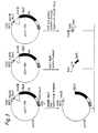
- Figure 3 shows the construction of the vector pSVI2-tPA.
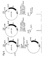
- Figure 4 shows the construction of the vector pSVI3-tPA.
- Figure 5 shows the construction of the vector pSVI5-tPA.
- Figure 6 shows the construction of the vector pSVI6B-tPA.
- Figure 7 shows the construction of the general expression vector pSVI6B5 into which the gene encoding latency associated peptide is conveniently inserted.
- Figure 8 depicts the complete nucleotide and predicted amino acid sequence of the latency associated peptide. Predicted amino acids of the peptide are shown below the DNA sequence and are numbered from the first residue of the N-terminus of the peptide sequence. The mature TGF- ⁇ sequence is not included. Negative numbers indicate the signal sequence, and positive numbers designate the sequence of the secreted peptide.
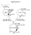
- Figure 9 depicts the construction of the expression vectors pSVI6B-LAP-ARG and pSVI6B-LAP-SER from pSVI6B5 used to transform mammalian host cells to produce the latency associated peptide.
- Figure 10 illustrates the amount of bioactive TGF- ⁇ in ng/ml as determined by a mink lung fibroblast cell assay as a function of the degree of diluted heat-activated TGF- ⁇ supernatant (circles), and when heat-inactivated latency associated peptide (squares with dashed lines), or untreated latency associated peptide (squares with solid lines) is added to the heat-activated TGF- ⁇ .
- Figure 11 depicts the sequences of human TGF- ⁇ 1, human TGF- ⁇ 2, human TGF- ⁇ 3, chick TGF- ⁇ 4, and frog TGF- ⁇ 5.
- LAP latency associated peptide
- TGF- ⁇ i.e., any TGF- ⁇ in the family of TGF- ⁇ s
- molecular weight of about 75 Kd (i.e., 75 up to about 78 Kd) when measured by SDS-PAGE on a non-reducing gel and having the amino acid sequence of Fig. 8, together with analogues and variants thereof having the biological activity of the corresponding recombinant LAP.
- the biological activity of recombinant LAP is shared by any analogue or variant thereof that is capable of antagonizing a biological activity of mature TGF- ⁇ .
- LAP that does not have the indicated biological activity, e.g., that is degraded by heat treatment, are excluded.
- the LAP is a dimeric molecule that forms a monomer on reduction having a molecular weight of about 37-39 Kd on reducing SDS-PAGE; the variants need not share this dimeric property.
- Analogues or variants are defined as molecules in which the amino acid sequence, glycosylation, or other feature of recombinant LAP has been modified covalently or noncovalently.
- variants may or may not have a molecular weight of approximately 70 kD (as determined by SDS-PAGE carried out in the absence of a reducing agent such as, e.g., ⁇ -mercaptoethanol or dithiothreitol).
- a reducing agent such as, e.g., ⁇ -mercaptoethanol or dithiothreitol
- unglycosylated LAP having the recombinant LAP sequence will have a lower molecular weight on non-reducing SDS-PAGE.
- Amino acid sequence variants include not only alleles of the Fig. 8 sequence, but also predetermined mutations thereof.
- amino acid sequence variables have an amino acid sequence with at least about 80% sequence identity, and more typically at least about 90% sequence identity, to that of the recombinant LAP of Fig. 8.
- LAP shall mean either the recombinantly produced sequence, a variant form, or fragments representing biologically active site(s) within the molecule, unless otherwise appropriate.
- LAP having the human recombinant LAP amino acid sequence as set forth in Figure 8, analogous LAP proteins from other species such as bovine, equine, porcine, ovine, canine, murine, feline LAP, and the like, LAP sequences from other types of TGF- ⁇ such as TGF- ⁇ 2 (see, e.g., Madisen et al., DNA , supra) , and biologically active amino acid sequence variants of these LAP molecules, including alleles and in vitro -generated covalent derivatives of LAP that demonstrate its biological activity.
- analogous LAP proteins from other species such as bovine, equine, porcine, ovine, canine, murine, feline LAP, and the like
- LAP sequences from other types of TGF- ⁇ such as TGF- ⁇ 2 (see, e.g., Madisen et al., DNA , supra)
- biologically active amino acid sequence variants of these LAP molecules including alleles and in vitro -
- TGF- ⁇ refers to the family of molecules described hereinabove that have the full-length, native amino acid sequence of any of the TGF- ⁇ s from any species. Reference to such TGF- ⁇ herein will be understood to be a reference to any one of the currently identified forms, including TGF- ⁇ 1, TGF- ⁇ 2, TGF- ⁇ 3, TGF- ⁇ 4, and TGF- ⁇ 5 (whose sequences are shown in Figure 11), as well as to TGF- ⁇ species identified in the future, including polypeptides derived from the sequence of any known TGF- ⁇ and identical at 75% or more of the residues.
- TGF- ⁇ 1, TGF- ⁇ 2
- TGF- ⁇ 3 refer to the TGF- ⁇ s defined in the literature, e.g., Derynck et al., Nature , supra , Seyedin et al ., J. Biol. Chem. , 262 , supra , and deMartin et al ., supra .
- TGF- ⁇ family are defined as those which have nine cysteine residues in the mature portion of the molecule, share at least 65% sequence identity with other known TGF- ⁇ sequences in the mature region, and may compete for the same receptor. In addition, they all appear to be encoded as a larger precursor that shares a region of high homology near the N-terminus and shows conservation of three cysteine residues in the portion of the precursor that will later be removed by processing. Moreover, the TGF- ⁇ s appear to have a processing site with four or five basic amino acids.
- neutralization of a TGF- ⁇ activity means neutralization of a TGF- ⁇ as measured by 3 H-thymidine uptake inhibition of a mink lung fibroblast cell line, e.g., Mv-3D9 and CCL64.
- the expression "antagonizing a biological activity of TGF- ⁇ ” refers to blocking an activity of a TGF- ⁇ , e.g., substantially preventing at least one of the undesirable growth inhibitory, immunosuppressive, stroma forming (the stromal elements including inflammatory cells, endothelial cells, and fibroblasts), or anchorage-independent growth promoting activities of a mature TGF- ⁇ , as it is defined in the literature.
- the LAP is thus able to block the activity of all endogenously released TGF- ⁇ produced by tumors and suppressor lymphoid cells (T cells). Identification of LAP molecules having this effect is by means of the neutralization test detailed above.
- conditions to be treated by therapy or prophylaxis using the processes and compositions of this invention include conditions caused by suppression of the immune system due to endogenous TGF- ⁇ production, including acquired immune deficiency syndrome, acute immune deficiencies resulting from severe injuries, burns, and illnesses such as viral or bacterial infections, cystic fibrosis, multi-organ systemic illnesses due to TGF- ⁇ production or overproduction, and TGF- ⁇ -producing tumors.
- Such conditions would not include those resulting from proliferation of cells of the lymphoid and myeloid lineages, which proliferation is inhibited by TGF- ⁇ (e.g., Burkitt's lymphoma, T-cell lymphomas, B-cell lymphomas, and various myelocytic leukemias).
- Such conditions also include diseases caused by circulating TGF- ⁇ or TGF- ⁇ activated at a local site, such as those diseases associated with chronic granulation or hyperproliferation, such as fibrotic diseases, including fibrosis of any organ, e.g., scleroderma, and fibrosis of the lung, kidney, peritoneal cavity, and liver, and diseases caused by an excess of active TGF- ⁇ , such as vitreoretinopathy caused by an excess of TGF- ⁇ 2. Also included is the prevention of illnesses and side effects caused by TGF- ⁇ and another regulator of immune function in a mammal as defined below.
- regulators of immune function in a mammal refers to cytokines and growth factors that regulate a mammal's immune function, including interleukins, tumor necrosis factor, lymphotoxin, epidermal growth factor, platelet-derived growth factor, TGF- ⁇ , macrophage migration inhibitory factor, macrophage activation factor, fibroblast growth factor, macrophage activating factors, interferons, and colony stimulating factors.
- These regulators may be derived from natural sources, formed by leukocytes, synthesized by chemical methods if appropriate, or prepared by recombinant techniques. Preferred of these are IL-1, IL-2, IL-6, and IFN-gamma.
- LAP Derivatives and amino acid sequence variants of LAP are useful for their biological activity as it relates to therapeutic utility, as is set forth elsewhere herein.
- LAP molecule Covalent modifications of a LAP molecule are included within the scope of this invention. Variant LAP fragments having up to about 100 residues may be conveniently prepared by in vitro synthesis. Such modifications may be introduced into the molecule by reacting targeted amino acid residues of the purified or crude protein with an organic derivatizing agent that is capable of reacting with selected side chains or terminal residues. The resulting covalent derivatives are useful in programs directed at identifying residues important for biological activity.
- Cysteinyl residues most commonly are reacted with ⁇ -haloacetates (and corresponding amines), such as chloroacetic acid or chloroacetamide, to give carboxymethyl or carboxyamidomethyl derivatives. Cysteinyl residues also are derivatized by reaction with bromotrifluoroacetone, ⁇ -bromo- ⁇ -(5-imidozoyl)propionicacid,chloroacetylphosphate,N-alkylmaleimides, 3-nitro-2-pyridyl disulfide, methyl 2-pyridyl disulfide, p-chloromercuribenzoate,2-chloromercuri-4-nitrophenol,orchloro-7-nitrobenzo-2-oxa-1,3-diazole.
- Histidyl residues are derivatized by reaction with diethylpyrocarbonate at pH 5.5-7.0 because this agent is relatively specific for the histidyl side chain.
- Para-bromophenacyl bromide also is useful; the reaction is preferably performed in 0.1M sodium cacodylate at pH 6.0.
- Lysinyl and amino terminal residues are reacted with succinic or other carboxylic acid anhydrides. Derivatization with these agents has the effect of reversing the charge of the lysinyl residues.
- Other suitable reagents for derivatizing ⁇ -amino-containing residues include imidoesters such as methyl picolinimidate; pyridoxal phosphate; pyridoxal; chloroborohydride; trinitrobenzenesulfonic acid; O-methylisourea; 2,4-pentanedione; and transaminase-catalyzed reaction with glyoxylate.
- Arginyl residues are modified by reaction with one or several conventional reagents, among them phenylglyoxal, 2,3-butanedione, 1,2-cyclohexanedione, and ninhydrin. Derivatization of arginine residues requires that the reaction be performed in alkaline conditions because of the high pK a of the guanidine functional group. Furthermore, these reagents may react with the groups of lysine as well as the arginine epsilon-amino group.
- tyrosyl residues per se has been studied extensively, with particular interest in introducing spectral labels into tyrosyl residues by reaction with aromatic diazonium compounds or tetranitromethane. Most commonly, N-acetylimidizol and tetranitromethane are used to form O-acetyl tyrosyl species and 3-nitro derivatives, respectively.
- Tyrosyl residues are iodinated using 125 I or 131 I to prepare labeled proteins for use in radioimmunoassay, the chloramine T method described above being suitable.
- Carboxyl side groups (aspartyl or glutamyl) are selectively modified by reaction with carbodiimides (R'-N-C-N-R') such as 1-cyclohexyl-3-(2-morpholinyl-(4-ethyl) carbodiimide or 1-ethyl-3-(4-azonia-4,4-dimethylpentyl) carbodiimide. Furthermore, aspartyl and glutamyl residues are converted to asparaginyl and glutaminyl residues by reaction with ammonium ions.
- Glutaminyl and asparaginyl residues are frequently deamidated to the corresponding glutamyl and aspartyl residues. Alternatively, these residues are deamidated under mildly acidic conditions. Either form of these residues falls within the scope of this invention.
- Amino acid sequence variants of LAP can also be prepared by mutations in the DNA. Such variants include, for example, deletions from, or insertions or substitutions of, residues within the amino acid sequence shown in Figure 8. Any combination of deletion, insertion, and substitution may also be made to arrive at the final construct, provided that the final construct possesses the desired activity. Obviously, the mutations that will be made in the DNA encoding the variant must not place the sequence out of reading frame and preferably will not create complementary regions that could produce secondary mRNA structure (see EP 75,444A).
- these variants ordinarily are prepared by site-directed mutagenesis of nucleotides in the DNA encoding the LAP, thereby producing DNA encoding the variant, and thereafter expressing the DNA in recombinant cell culture.
- the variants typically exhibit the same qualitative biological activity as the naturally occurring analog.
- Preparation of LAP variants in accordance herewith is preferably achieved by site-specific mutagenesis of DNA that encodes an earlier prepared variant or a nonvariant version of the protein.
- Site-specific mutagenesis allows the production of LAP variants through the use of specific oligonucleotide sequences that encode the DNA sequence of the desired mutation, as well as a sufficient number of adjacent nucleotides, to provide a primer sequence of sufficient size and sequence complexity to form a stable duplex on both sides of the deletion junction being traversed.
- a primer of about 20 to 25 nucleotides in length is preferred, with about 5 to 10 residues on both sides of the junction of the sequence being altered.
- the technique of site-specific mutagenesis is well known in the art, as exemplified by publications such as Adelman et al ., DNA , 2 : 183 (1983).
- the site-specific mutagenesis technique typically employs a phage vector that exists in both a single-stranded and double-stranded form.
- Typical vectors useful in site-directed mutagenesis include vectors such as the M13 phage, for example, as disclosed by Messing et al ., Third Cleveland Symposium on Macromolecules and Recombinant DNA , Editor A. Walton, Elsevier, Amsterdam (1981). These phage are readily commercially available and their use is generally well known to those skilled in the art.
- plasmid vectors that contain a single-stranded phage origin of replication may be employed to obtain single-stranded DNA.
- heteroduplex is formed wherein one strand encodes the original non-mutated sequence and the second strand bears the desired mutation.
- This heteroduplex vector is then used to transform appropriate cells such as JM101 cells and clones are selected that include recombinant vectors bearing the mutated sequence arrangement.
- Amino acid sequence deletions generally range from about 1 to 30 residues, more preferably 1 to 10 residues, and typically are contiguous.
- the third group of variants are those in which at least one amino acid residue in the LAP molecule, and preferably only one, has been removed and a different residue inserted in its place. Such substitutions preferably are made in accordance with the following Table 1 when it is desired to modulate finely the characteristics of a LAP molecule.
- substitutions that in general are expected to produce the greatest changes in LAP properties will be those in which (a) glycine and/or proline (P) is substituted by another amino acid or is deleted or inserted; (b) a hydrophilic residue, e.g., seryl or threonyl, is substituted for (or by) a hydrophobic residue, e.g., leucyl, isoleucyl, phenylalanyl, valyl, or alanyl; (c) a cysteine residue is substituted for (or by) any other residue; (d) a residue having an electropositive side chain, e.g., lysyl, arginyl, or histidyl, is substituted for (or by) a residue having an electronegative charge, e.g., glutamyl or aspartyl; or (e) a residue having a bulky side chain, e.g., phenylalanine, is substituted for (or
- a variant typically is made by site-specific mutagenesis of the native LAP-encoding nucleic acid, expression of the variant nucleic acid in recombinant cell culture, and, optionally, purification from the cell culture, for example, by immunoaffinity adsorption on a rabbit polyclonal anti-LAP column (to absorb the variant by binding it to at least one remaining immune epitope).
- LAP may aggregate into dimers, it is within the scope hereof to provide hetero- and homodimers, wherein one or both subunits are variants. Where both subunits are variants, the changes in amino acid sequence can be the same or different for each subunit chain.
- Heterodimers are readily produced by cotransforming host cells with DNA encoding both subunits and, if necessary, purifying the desired heterodimer, or by separately synthesizing the subunits, dissociating the subunits (e.g., by treatment with a chaotropic agent such as urea, guanidine hdyrochloride, or the like), mixing the dissociated subunits, and then reassociating the subunits by dialyzing away the chaotropic agent.
- a chaotropic agent such as urea, guanidine hdyrochloride, or the like
- the activity of the cell lysate or purified LAP variant is then screened in a suitable screening assay for the desired characteristic. For example, changes in the level of TGF- ⁇ activity by the candidate mutants are measured by the appropriate assay. Modifications of such protein properties as redox or thermal stability, hydrophobicity, susceptibility to proteolytic degradation, or the tendency to aggregate with carriers or into multimers are assayed by methods well known to the ordinarily skilled artisan.
- the LAP molecule desired may be prepared by any technique, including recombinant methods.
- an isolated DNA is understood herein to mean chemically synthesized DNA, cDNA, chromosomal, or extrachromosomal DNA with or without any 5'-flanking region.
- the desired LAP herein is made by synthesis in recombinant cell culture.
- DNA encoding a LAP molecule may be obtained from a source of cells for human TGF- ⁇ by (a) preparing a cDNA library from these cells, (b) conducting hybridization analysis with labeled DNA encoding TGF- ⁇ or fragments thereof (up to or more than 100 base pairs in length) to detect clones in the library containing homologous sequences, and (c) analyzing the clones by restriction enzyme analysis and nucleic acid sequencing to identify full-length clones. DNA that is capable of hybridizing to a TGF- ⁇ -encoding DNA under low stringency conditions is useful for identifying DNA encoding TGF- ⁇ .
- a LAP-encoding gene is expressed in mammalian cells by transformation with an expression vector comprising DNA encoding the LAP. It is preferable to transform host cells capable of accomplishing such processing so as to obtain the LAP in the culture medium or periplasm of the host cell, i.e., obtain a secreted molecule.
- vectors and methods disclosed herein are suitable for use in host cells over a wide range of prokaryotic and eukaryotic organisms.
- prokaryotes are preferred for initial cloning, amplifying, or storing the vectors of interest.
- Vector DNA is easily obtainable from certain prokaryotes.
- E. coli K12 strain MM 294 (ATCC No. 31,446) is particularly useful for this purpose.
- Other microbial strains that may be used include E. coli strains such as E. coli B and E. coli X1776 (ATCC No. 31,537). These examples are, of course, intended to be illustrative rather than limiting.
- Prokaryotes may also be used for expression.
- the aforementioned strains, as well as E. coli strains W3110 (F, lambda-, prototrophic, ATCC No. 27,325), K5772 (ATCC No. 53,635), and SR101, bacilli such as Bacillus subtilis , and other enterobacteriaceae such as Salmonella typhimurium or Serratia marcesans , and various pseudomonas species, may be used.
- plasmid vectors containing replicon and control sequences that are derived from species compatible with the host cell are used in connection with these prokaryotic hosts.
- the vector ordinarily carries a replication site, as well as marking sequences that are capable of providing phenotypic selection in transformed cells.
- E. coli is typically transformed using pBR322, a plasmid derived from an E. coli species (see, e.g., Bolivar et al ., Gene , 2: 95 [1977]).
- pBR322 contains genes for ampicillin and tetracycline resistance and thus provides easy means for identifying transformed cells.
- the pBR322 plasmid, or other microbial plasmid or phage must also contain, or be modified to contain, promoters that can be used by the microbial organism for expression of the selectable marker genes.
- promoters most commonly used in recombinant DNA construction include the ⁇ -lactamase (penicillinase) and lactose promoter systems (Chang et al ., Nature , 375 : 615 [1978]; Itakura et al ., Science , 198 : 1056 [1977]; Goeddel et al , Nature , 281 : 544 [1979]) and a tryptophan (trp) promoter system (Goeddel et al ., Nucleic Acids Res. , 8 4057 [1980]; EPO Appl. Publ. No. 0036,776).
- eukaryotic microbes such as yeast cultures
- Saccharomyces cerevisia e or common baker's yeast
- Saccharomyces cerevisia e or common baker's yeast
- the plasmid YRp7 for example (Stinchcomb et al , Nature , 282 : 39 [1979]; Kingsman et al ., Gene , 7 : 141 [1979]; Tschemper et al ., Gene , 10 : 157 [1980]), is commonly used.
- This plasmid already contains the trp 1 gene that provides a selection marker for a mutant strain of yeast lacking the ability to grow in tryptophan, for example, ATCC No. 44,076 or PEP4-1 (Jones, Genetics , 85 : 12 [1977]).
- the presence of the trp 1 lesion as a characteristic of the yeast host cell genome then provides an effective environment for detecting transformation by growth in the absence of tryptophan.
- Suitable promoting sequences in yeast vectors include the promoters for 3-phosphoglycerate kinase (Hitzeman et al ., J. Biol. Chem. , 255 : 2073 [1980]) or other glycolytic enzymes (Hess et al. , J. Adv. Enzyme Reg. , 7 : 149 [1968]; Holland et al.
- enolase such as enolase, glyceraldehyde-3-phosphate dehydrogenase, hexokinase, pyruvate decarboxylase, phosphofructokinase, glucose-6-phosphate isomerase, 3-phosphoglycerate mutase, pyruvate kinase, triosephosphate isomerase, phosphoglucose isomerase, and glucokinase.
- the termination sequences associated with these genes are also ligated into the expression vector 3' of the sequence desired to be expressed to provide polyadenylation of the mRNA and termination.
- promoters which have the additional advantage of transcription controlled by growth conditions, are the promoter region for alcohol dehydrogenase 2, isocytochrome C, acid phosphatase, degradative enzymes associated with nitrogen metabolism, and the aforementioned glyceraldehyde-3-phosphate dehydrogenase, and enzymes responsible for maltose and galactose utilization.
- Any plasmid vector containing yeast-compatible promoter, origin of replication and termination sequences is suitable.
- cultures of cells derived from multicellular organisms may also be used as hosts.
- any such cell culture is workable, whether from vertebrate or invertebrate culture.
- interest has been greatest in vertebrate cells, and propagation of vertebrate cells in culture (tissue culture) has become a routine procedure in recent years [ Tissue Culture , Academic Press, Kruse and Patterson, editors (1973)].
- Such useful host cell lines include monkey kidney CVI line transformed by SV40 sequences (COS-7, ATCC CRL 1651); human embryonic kidney line (293, Graham et al ., J. Gen. Virol. , 36 : 59 [1977]); baby hamster kidney cells (BHK, ATCC CCL 10); Chinese hamster ovary cells (Urlaub and Chasin, Proc. Natl. Acad. Sci. USA , 77 : 4216 [1980]); mouse sertoli cells (TM4, Mather, Biol. Reprod.
- monkey kidney cells CVI, ATCC CCL 70); African green monkey kidney cells (VERO-76, ATCC CRL-1587); human cervical carcinoma cells (HELA, ATCC CCL 2); canine kidney cells (MDCK, ATCC CCL 34); buffalo rat liver cells (BRL 3A, ATCC CRL 1442); human lung cells (W138, ATCC CCL 75); human liver cells (Hep G2, HB 8065); mouse mammary tumor cells (MMT 060562, ATCC CCL51); rat hepatoma cells (HTC, M1.54, Baumann et al ., J. Cell. Biol.
- the most preferred eukaryotic host herein for stable expression is a Chinese hamster ovary cell line.
- control functions on the expression vectors are often provided by viral material.
- promoters are derived from the genomes of polyoma, Adenovirus2, retroviruses, cytomegalovirus, and most frequently Simian Virus 40 (SV40).
- Other promoters are those from heterologous sources, e.g., the beta actin promoter.
- the early and late promoters of SV40 virus are particularly useful because both are obtained easily from the virus as a fragment that also contains the SV40 viral origin of replication [Fiers et al. , Nature , 273 : 113 (1978)].
- Smaller or larger SV40 fragments may also be used, provided there is included the approximately 250-bp sequence extending from the Hin dIII site toward the Bgl I site located in the viral origin of replication.
- the immediate early promoter of the human cytomegalovirus is conveniently obtained as a Hin dIII restriction fragment. Greenawa y et al ., Gene , 18 : 355-360 (1982).
- promoter or control sequences normally associated with the desired gene sequence provided such control sequences are compatible with the host cell systems.
- Enhancers are relatively orientation and position independent, having been found 5' (Laimins et al ., Proc. Natl. Acad. Sci. USA , 78 : 993 [1981]) and 3' (Lusky et al ., Mol. Cell Bio. , 3 : 1108 [1983]) to the transcription unit, within an intron (Banerji et al.
- the enhancer element is located upstream of the promoter sequence for this invention.
- Many enhancer sequences are now known from mammalian genes (globin, elastase, albumin, ⁇ -fetoprotein, and insulin). Typically, however, one will use an enhancer from a eukaryotic cell virus.
- Examples include the SV40 enhancer on the late side of the replication origin (bp 100-270), the cytomegalovirus early promoter enhancer, the polyoma enhancer on the late side of the replication origin, and adenovirus enhancers. Most preferred herein is the SV40 enhancer region.
- Expression vectors used in mammalian host cells will also contain polyadenylation sites.
- polyadenylation regions are those derived from viruses such as, e.g., the SV40 (early and late) or HBV.
- An origin of replication may be provided either by construction of the vector to include an exogenous origin, such as may be derived from SV40 or other viral (e.g., Polyoma, Adeno, VSV, BPV) source, or may be provided by the host cell. If the vector is integrated into the host cell chromosome, the latter is often sufficient.
- an exogenous origin such as may be derived from SV40 or other viral (e.g., Polyoma, Adeno, VSV, BPV) source, or may be provided by the host cell. If the vector is integrated into the host cell chromosome, the latter is often sufficient.
- the expression vectors may suitably contain a selection gene, also termed a selectable marker.
- a selection gene encodes a protein necessary for the survival or growth of a host cell transformed with the vector.
- suitable selectable markers for mammalian cells include dihydrofolate reductase (DHFR), thymidine kinase (TK), or neomycin. When such selectable markers are successfully transferred into a mammalian host cell, the transformed mammalian host cell can survive if placed under selective pressure.
- the first category is based on the metabolism of a cell and the use of a mutant cell line that lacks the ability to grow independent of a supplemented medium.
- Two examples are CHO DHFR - cells and mouse LTK - cells. These cells lack the ability to grow without the addition of such nutrients as thymidine or hypoxanthine. Because these cells lack certain genes necessary for a complete nucleotide synthesis pathway, they cannot survive unless the missing nucleotides are provided in a supplemented medium.
- An alternative to supplementing the medium is to introduce an intact DHFR or TK gene into cells lacking the respective genes, thus altering their growth requirements. Individual cells that were not transformed with the DHFR or TK gene will not be capable of survival in non-supplemented medium. Therefore, direct selection of those cells requires cell growth in the absence of supplemental nutrients.
- the second category is dominant selection, which refers to a selection scheme that does not require the use of a mutant cell line.
- This method typically employs a drug to arrest growth of a host cell. Those cells that have a novel gene would express a protein conveying drug resistance and would survive the selection.
- drugs used in dominant selection include neomycin (Southern and Berg, J. Molec. Appl. Genet. , 1 : 327 [1982]), mycophenolic acid (Mulligan and Berg, Science , 209 : 1422 [1980]), or hygromycin (Sugden et al. , Mol. Cell. Biol. , 5 : 410-413 [1985]).
- neomycin G418 or geneticin
- xgpt mycophenolic acid
- One secondary coding sequence comprises dihydrofolate reductase (DHFR) that is affected by an externally controlled parameter, such as methotrexate (MTX), thus permitting control of expression by control of the methotrexate concentration.
- DHFR dihydrofolate reductase
- MTX methotrexate
- a preferred host cell for transfection by the vectors of the invention that comprise DNA sequences encoding both LAP and DHFR protein it is appropriate to select the host according to the type of DHFR protein employed. If wild-type DHFR protein is employed, it is preferable to select a host cell that is deficient in DHFR, thus permitting the use of the DHFR coding sequence as a marker for successful transfection in selective medium that lacks hypoxanthine, glycine, and thymidine.
- An appropriate host cell in this case is the Chinese hamster ovary (CHO) cell line deficient in DHFR activity, prepared and propagated as described by Urlaub and Chasin, Proc. Natl. Acad. Sci. (USA) 77 : 4216 1980).
- DHFR protein with low binding affinity for MTX is used as the controlling sequence, it is not necessary to use DHFR-deficient cells. Because the mutant DHFR is resistant to methotrexate, MTX-containing media can be used as a means of selection provided that the host cells are themselves methotrexate sensitive. Most eukaryotic cells that are capable of absorbing MTX appear to be methotrexate sensitive.
- One such useful cell line is a CHO line, CHO-K1 (ATCC No. CCL 61).
- Extremely good amounts of polypeptide are produced by cell cultures using the method of this invention; however, refinements, using cotransfection with a separate vector encoding a secondary coding sequence.
- One secondary coding sequence comprises dihydrofolate reductase (DHFR) that is affected by an externally controlled parameter, such as methotrexate (MTX), thus permitting control of expression by control of the methotrexate concentration.
- DHFR dihydrofolate reductase
- Plasmid DNA fragments are cleaved, tailored, and religated to form the desired plasmid.
- the cleaved DNA preparation may be treated for 30 minutes at 37°C with DNA Polymerase I (Klenow fragment) or T4 DNA polymerase, phenol-chloroform extracted, and ethanol precipitated.
- DNA Polymerase I Klenow fragment
- T4 DNA polymerase phenol-chloroform extracted
- ethanol precipitated 3' protruding ends are removed by the 3' to 5' exonucleolytic activity of either enzyme, and the 5' protruding ends are made flush by the 5' to 3' polymerase activity incorporating complementary nt until the end of the fragment is reached.
- Size separation of the cleaved fragments may be performed using 6 percent polyacrylamide gel described by Goeddel et al ., Nucleic Acids Res. , 8 : 4057 (1980).
- the ligation mixtures are typically used to transform E. coli K12 strain 294 (ATCC 31,446) or other suitable E. coli strains, and successful transformants selected by ampicillin or tetracycline resistance where appropriate. Plasmids from the transformants are prepared and analyzed by restriction mapping and/or DNA sequencing by the method of Messing et al ., Nucleic Acids Res. , 9 : 309 (1981) or by the method of Maxam et al ., Meth. Enzym , 65 : 499 (1980).
- amplification of DHFR-protein-coding sequences is effected by growing host cell cultures in the presence of approximately 200-500 nM concentrations of methotrexate, a competitive inhibitor of DHFR activity.
- concentrations of methotrexate a competitive inhibitor of DHFR activity.
- concentrations of other folic acid analogs or other compounds that inhibit DHFR could also be used.
- MTX itself is, however, convenient, readily available, and effective.
- the LAP molecules herein have a number of therapeutic uses associated with the antagonism or neutralization of TGF- ⁇ activity. Such uses include the treatment of fibrotic diseases and patients who are immunosuppressed as measured by the ability to contract and sustain a secondary infection for a prolonged period of time. Particularly of interest are patients suffering from recurrent or chronic viral, bacterial, mycological, or parasitic infections. Other physiological conditions that could be improved based on the ability of LAP to bind reversibly to mature TGF- ⁇ are also included herein.
- LAP can be used as a means of preventing side effects of other immune regulators (e.g., recombinant cytokines) when they are used as pharmaceuticals or used in combination with such regulators, depending on the particular clinical scenario.
- immune regulators e.g., recombinant cytokines
- the LAP molecule will be formulated and dosed in a fashion consistent with good medical practice taking into account the specific disorder to be treated, the condition of the individual patient, the site of delivery of the LAP, the method of administration, and other factors known to practitioners.
- the "therapeutically effective amount" of the LAP is an amount that is effective either to prevent, lessen the worsening of, alleviate, or cure the treated condition, in particular that amount which is sufficient to antagonize the activity of a TGF- ⁇ in vivo .
- the LAP is prepared for storage or administration by mixing LAP having the desired degree of purity with physiologically acceptable carriers, excipients, or stabilizers. Such materials are non-toxic to recipients at the dosages and concentrations employed. If the LAP is water soluble, it may be formulated in a buffer such as phosphate or other organic acid salt preferably at a pH of about 7 to 8. If a LAP variant is only partially soluble in water, it may be prepared as a microemulsion by formulating it with a nonionic surfactant such as Tween, Pluronics, or PEG, e.g., Tween 80, in an amount of 0.04-0.05% (w/v), to increase its solubility.
- a nonionic surfactant such as Tween, Pluronics, or PEG, e.g., Tween 80, in an amount of 0.04-0.05% (w/v), to increase its solubility.
- antioxidants e.g., ascorbic acid
- low molecular weight polypeptides e.g., polyarginine or tripeptides
- proteins such as serum albumin, gelatin, or immunoglobulins
- hydrophilic polymers such as polyvinylpyrrolidone
- amino acids such as glycine, glutamic acid, aspartic acid, or arginine
- monosaccharides, disaccharides, and other carbohydrates including cellulose or its derivatives, glucose, mannose, or dextrins
- chelating agents such as EDTA
- sugar alcohols such as mannitol or sorbitol.
- the LAP to be used for therapeutic administration must be sterile. Sterility is readily accomplished by filtration through sterile filtration membranes (e.g., 0.2 micron membranes).
- the LAP ordinarily will be stored in lyophilized form or as an aqueous solution if it is highly stable to thermal and oxidative denaturation.
- the pH of the LAP preparations typically will be about from 6 to 8, although higher or lower pH values may also be appropriate in certain instances. It will be understood that use of certain of the foregoing excipients, carriers, or stabilizers will result in the formation of salts of the LAP.
- therapeutic compositions containing the LAP generally are placed into a container having a sterile access port, for example, an intravenous solution bag or vial having a stopper pierceable by a hypodermic injection needle.
- the disorder permits, one should formulate and dose the LAP for site-specific delivery. This is convenient in the case of fibrotic diseases and tumors.
- Sustained release formulations may also be prepared, and include the formation of microcapsular particles and implantable articles.
- the LAP is preferably incorporated into a biodegradable matrix or microcapsule.
- a suitable material for this purpose is a polylactide, although other polymers of poly-( ⁇ -hydroxycarboxylic acids), such as poly-D-(-)-3-hydroxybutyric acid (EP 133,988A), can be used.
- Other biodegradable polymers include poly(lactones), poly(acetals), poly(orthoesters), or poly(orthocarbonates).
- the initial consideration here must be that the carrier itself, or its degradation products, is nontoxic in the target tissue and will not further aggravate the condition. This can be determined by routine screening in animal models of the target disorder or, if such models are unavailable, in normal animals.
- the LAP When applied topically, the LAP is suitably combined with other ingredients, such as carriers and/or adjuvants.
- suitable vehicles include ointments, creams, gels, or suspensions, with or without purified collagen.
- the compositions also may be impregnated into transdermal patches, plasters, and bandages, preferably in liquid or semi-liquid form.
- the LAP formulated in a liquid composition may be mixed with an effective amount of a water-soluble polysaccharide or synthetic polymer such as polyethylene glycol to form a gel of the proper viscosity to be applied topically.
- the polysaccharide that may be used includes, for example, cellulose derivatives such as etherified cellulose derivatives, including alkyl celluloses, hydroxyalkyl celluloses, and alkylhydroxyalkyl celluloses, for example, methylcellulose, hydroxyethyl cellulose, carboxymethyl cellulose, hydroxypropyl methylcellulose, and hydroxypropyl cellulose; starch and fractionated starch; agar; alginic acid and alginates; gum arabic; pullullan; agarose; carrageenan; dextrans; dextrins; fructans; inulin; mannans; xylans; arabinans; chitosans; glycogens; glucans; and synthetic biopolymers; as well as gums such as xanthan gum; guar gum; locust bean gum; gum arabic; tragacanth gum; and karaya gum; and derivatives and mixtures thereof.
- the preferred gelling agent herein is one that is in
- the polysaccharide is an etherified cellulose derivative, more preferably one that is well defined, purified, and listed in USP, e.g., methylcellulose and the hydroxyalkyl cellulose derivatives, such as hydroxypropyl cellulose, hydroxyethyl cellulose, and hydroxypropyl methylcellulose. Most preferred herein is methylcellulose.
- the polyethylene glycol useful for gelling is typically a mixture of low and high molecular weight polyethylene glycols to obtain the proper viscosity.
- a mixture of a polyethylene glycol of molecular weight 400-600 with one of molecular weight 1500 would be effective for this purpose when mixed in the proper ratio to obtain a paste.
- water soluble as applied to the polysaccharides and polyethylene glycols is meant to include colloidal solutions and dispersions.
- solubility of the cellulose derivatives is determined by the degree of substitution of ether groups, and the stabilizing derivatives useful herein should have a sufficient quantity of such ether groups per anhydroglucose unit in the cellulose chain to render the derivatives water soluble.
- a degree of ether substitution of at least 0.35 ether groups per anhydroglucose unit is generally sufficient.
- the cellulose derivatives may be in the form of alkali metal salts, for example, the Li, Na, K, or Cs salts.
- methylcellulose is employed in the gel, preferably it comprises about 2-5%, more preferably about 3%, of the gel and the LAP is present in an amount of about 300-1000 »g per ml of gel.
- LAP LAP
- a dose of about 0.015 to 15 mg/kg of LAP may be administered to the patient, whether via, e.g., one or more single administrations, continuous infusion, or bolus injection.
- an initial dose of the LAP is administered to the patient by injection or infusion.
- the treatment is repeated until a desired suppression of disease symptoms occurs.
- other dosage regimens may be useful.
- the effectiveness of the LAP may be improved by administering it serially or in combination with another agent that is effective for this purpose, such as one or more anti-TGF- ⁇ antibodies, a regulator of immune function, as defined above, or one or more conventional therapeutic agents such as, for example, alkylating agents, folic acid antagonists, anti-metabolites of nucleic acid metabolism, spindle poisons, antibiotics, pyrimidine analogs, 5-FU, purine nucleosides, amines, amino acids, triazol nucleosides, corticosteroids, calcium, retinoids, lipoxygenase and cyclooxygenase inhibitors, fumaric acid and its salts, analgesics, psychopharmaceuticals, local anesthetics, spasmolytics, and beta-blockers.
- another agent that is effective for this purpose such as one or more anti-TGF- ⁇ antibodies, a regulator of immune function, as defined above, or one or more conventional therapeutic agents such as, for example, alkyl
- the LAP molecule is suitably administered serially or in combination with radiological treatments--irradiation or introduction of radioactive substances--such as those referred to in UICC (ed.) in Klinische Onkologie , Springer-Verlag (1982).
- the LAP is used in combination with a mature TGF- ⁇ , whether heterogeneous or homogeneous with the LAP.
- a LAP from TGF- ⁇ 1 can be employed with mature TGF- ⁇ 1, TGF- ⁇ 2, or TGF- ⁇ 3.
- the relative amounts of mature TGF- ⁇ to LAP are generally about equimolar.
- the mature TGF- ⁇ is administered simultaneously with the LAP, whether in the same formulation or in separate administrations to the same locale such as a vein.
- the TGF- ⁇ and LAP are mixed together before administration. If the complex is unstable, the ingredients are mixed together in approximately equimolar amounts just before administration. If the complex is stable, the TGF- ⁇ and LAP are mixed together and the inactive complex is purified away from the reaction mixture and the purified material is administered.
- the administration is systemic, because the advantage resides in the reduction of side effects associated with certain doses of systemically administered TGF- ⁇ .
- the combination of mature TGF- ⁇ with LAP can be used by any process enhanced by the pool of latent TGF- ⁇ , including soft and hard tissue generation, such as wound healing and bone formation, and immunosuppression.
- the therapy applies to surgical as well as non-surgical intervention; for example, the combination can be given to a patient a day before surgery so that tissue strength at the wound site will be increased, or given to a patient with a simple bone fracture to increase the bone healing rate.
- the LAP is suitably used in a diagnostic assay to capture active TGF- ⁇ in the serum of a patient suffering from a condition, including a disease, that can be diagnosed by detecting mature TGF- ⁇ in serum.
- This technique is particularly suitable for sensitive detection of minute amounts of mature TGF- ⁇ in the serum.
- the method comprises adding to the serum, whether in vitro or in vivo , LAP that has been labeled by any suitable detectable moiety, such as 32 P or alkaline phosphatase, and assaying for the presence of labeled complexes of LAP and mature TGF- ⁇ in the serum.
- Transfection refers to the taking up of an expression vector by a host cell whether or not any coding sequences are in fact expressed. Numerous methods of transfection are known to the ordinarily skilled artisan, for example, CaPO 4 and electroporation. Successful transfection is generally recognized when any indication of the operation of this vector occurs within the host cell.
- Transformation means introducing DNA into an organism so that the DNA is replicable, either as an extrachromosomal element or by chromosomal integrant. Depending on the host cell used, transformation is done using standard techniques appropriate to such cells.
- the calcium treatment employing calcium chloride as described by Cohen, S.N. Proc. Natl. Acad. Sci. (USA), 69 : 2110 (1972); Mandel et al ., J. Mol. Biol. 53 :154 (1970); and more recently Liljestrom et al ., Gene , 40 : 241-246 (1985), is generally used for prokaryotes or other cells that contain substantial cell-wall barriers. For mammalian cells without such cell walls, the calcium phosphate precipitation method of Graham, F.
- coding sequence “operably linked” to control sequences refers to a configuration wherein the coding sequence can be expressed under the control of these sequences and wherein the DNA sequences being linked are contiguous and, in the case of a secretory leader, contiguous and in reading phase.
- DNA for a presequence or secretory leader is operably linked to DNA for a polypeptide if it is expressed as a preprotein that participates in the secretion of the polypeptide; a promoter or enhancer is operably linked to a coding sequence if it effects the transcription of the sequence; or a ribosome binding site is operably linked to a coding sequence if it is positioned so as to facilitate translation.
- Linking is accomplished by ligation at convenient restriction sites. If such sites do not exist, then synthetic oligonucleotide adaptors or linkers are used in accord with conventional practice.
- Control sequences refers to DNA sequences necessary for the expression of an operably linked coding sequence in a particular host organism.
- the control sequences that are suitable for prokaryotes include a promoter, optionally an operator sequence, a ribosome binding site, and possibly, other as yet poorly understood sequences.
- Eukaryotic cells are known to utilize promoters, polyadenylation signals, and enhancers.
- “Expression system” refers to DNA sequences containing a desired coding sequence and control sequences in operable linkage, so that hosts transformed with these sequences are capable of producing the encoded proteins.
- the expression system may be included on a vector; however, the relevant DNA may then also be integrated into the host chromosome.
- cell As used herein, "cell,” “cell line,” and “cell culture” are used interchangeably and all such designations include progeny.
- “transformants” or “transformed cells” includes the initial transformant and cultures derived therefrom without regard for the number of transfers. It is also understood that all progeny may not be precisely identical in DNA content, due to deliberate or inadvertent mutations. Mutant progeny that have the same functionality as screened for in the originally transformed cell are included. Where distinct designations are intended, it will be clear from the context.
- Plasmids are designated by a lower case p preceded and/or followed by capital letters and/or numbers.
- the starting plasmids herein are commercially available, are publicly available on an unrestricted basis, or can be constructed from such available plasmids in accord with published procedures.
- other equivalent plasmids are known in the art and will be apparent to the ordinary artisan.
- PCR polymerase chain reaction
- sequence information from the ends of the stretch of interest or beyond needs to be available, such that oligonucleotide primers can be designed; these primers will point towards one another, and will be identical or similar in sequence to opposite strands of the template to be amplified.
- the 5' terminal nucleotides of the two primers will coincide with the ends of the amplified material.
- PCR can be used to amplify specific DNA sequences from total genomic DNA, cDNA transcribed from total cellular RNA, bacteriophage or plasmid sequences, etc. See generally H. Erlich, ed., PCR Technology , Stockton Press, NY, 1989.
- PCR mutagenesis refers to the following (see Erlich, supra , the chapter by R. Higuchi, p. 61-70):
- primers that differ slightly in sequence from the corresponding region in a template DNA can be used to generate relatively large quantities of a specific DNA fragment that differs from the template sequence only at the positions where the primers differ from the template.
- one of the primers is designed to overlap the position of the mutation and to contain the mutation; the sequence of the other primer must be identical to a stretch of sequence of the opposite strand of the plasmid, but this sequence can be located anywhere along the plasmid DNA.
- the sequence of the second primer is located within 200 nt from that of the first, such that in the end the entire amplified region of DNA bounded by the primers can be easily sequenced.
- PCR amplification using a primer pair like the one just described results in a population of DNA fragments that differ at the position of the mutation specified by the primer, and possibly at other positions, as template copying is somewhat error-prone.
- the ratio of template to product material is extremely low, and as a result, the vast majority of product DNA fragments incorporate the desired mutation(s).
- This product material is used to replace the corresponding region in the plasmid that served as PCR template using standard DNA technology. Mutations at separate positions can be introduced simultaneously by either using a mutant second primer, or performing a second PCR with different mutant primers and ligating the two resulting PCR fragments simultaneously to the vector fragment in a three (or more)-part ligation.
- PCR mutagenesis procedure employed in the examples below was as follows: Template plasmid DNA (1 »g) was linearized by digestion with a restriction endonuclease that has a unique recognition site in the plasmid DNA outside of the region to be amplified.
- reaction mixture was overlayed with 35 »l mineral oil and inserted into a DNA Thermal Cycler (purchased from Perkin-Elmer Cetus) programmed as follows: time-delay file 12 min. 94°C thermo-cycle file 1 min. 50°C 2-3 min. 68-72°C 1 min. 94°C 20 cycles time-delay file 4 min. 50°C time-delay file 12 min. 68°C soak file 4°C
- “Digestion” of DNA refers to catalytic cleavage of the DNA with an enzyme that acts only at specific nt sequences in the DNA. Such enzymes are called restriction enzymes, and the sequence for which each is specific is called a restriction site.
- restriction enzymes are commercially available and their reaction conditions, cofactors, and other requirements as established by the enzyme suppliers are used. Restriction enzymes commonly are designated by abbreviations composed of a capital letter followed by other letters representing the microorganism from which each restriction enzyme originally was obtained and then a number designating the particular enzyme. In general, about 1 »g of plasmid or DNA fragment is used with about 1-2 units of enzyme in about 20 »l of buffer solution.
- Appropriate buffers and substrate amounts for particular restriction enzymes are specified by the manufacturer. Incubation of about 1 hour at 37°C is ordinarily used, but may vary in accordance with the supplier's instructions. After incubation, protein is removed by extraction with phenol and chloroform, and the digested nucleic acid is recovered from the aqueous fraction by precipitation with ethanol. When appropriate, digestion with a restriction enzyme is followed by bacterial alkaline phosphatase-mediated hydrolysis of the terminal 5' phosphates to prevent the two ends of a DNA fragment from "circularizing" or forming a closed loop that would impede insertion of another DNA fragment at the restriction site. Unless otherwise stated, digestion of plasmids is not followed by 5' terminal dephosphorylation. Procedures and reagents for dephosphorylation are conventional (T. Maniatis et al ., 1982, Molecular Clonine : A Laboratory Manual (New York: Cold Spring Harbor Laboratory, 1982) pp. 133-134).
- Recovery or “isolation” of a given fragment of DNA from a restriction digest means separation of the digest on polyacrylamide or agarose gel by electrophoresis, identification of the fragment of interest by comparison of its mobility versus that of marker DNA fragments of known molecular weight, removal of the gel section containing the desired fragment, and separation of the gel from DNA.
- This procedure is known generally. For example, see R. Lawn et al ., Nucleic Acids Res . 9 :6103-6114 (1981), and D. Goeddel et al ., Nucleic Acids Res. 8 :4057 (1980).
- Ligase refers to the process of forming phosphodiester bonds between two double stranded nucleic acid fragments (T. Maniatis et al ., 1982, supra , p. 146). Unless otherwise provided, ligation may be accomplished using known buffers and conditions with 10 units of T4 DNA ligase ("ligase”) per 0.5 »g of approximately equimolar amounts of the DNA fragments to be ligated.
- ligase T4 DNA ligase
- Preparation of DNA from transformants means isolating plasmid DNA from microbial culture. Unless otherwise provided, the alkaline/SDS method of Maniatis et al ., 1982, supra , p. 90, may be used.
- Oligonucleotides are short-length, single- or double-stranded polydeoxynucleotides that are chemically synthesized by known methods (such as phosphotriester, phosphite, or phosphoramidite chemistry, using solid phase techniques such as described in EP Pat. Pub. No. 266,032 published May 4, 1988, or via deoxynucleoside H-phosphonate intermediates as described by Froehler et al ., Nucl. Acids Res. , 14 : 5399-5407 [1986]). They are then purified on polyacrylamide gels.
- t-PA vectors were derived from parent vector pSVI-tPA by altering a number of features of the splice donor-intron-splice acceptor unit.
- Mammalian expression vector pRK-tPA was prepared from pRK5 (described in EP 307,247, supra , where the pCIS2.8c28D starting plasmid is described in EP 278,776 published Aug. 17, 1988) and from t-PA cDNA (Pennica et al ., Nature , 301 : 214 [1983]).
- the cDNA was prepared for insertion into pRK5 by cutting with restriction endonuclease Hin dIII (which cuts 49 base pairs 5' of the ATG start codon) and restriction endonuclease Bal I (which cuts 276 base pairs downstream of the TGA stop codon).
- This cDNA was ligated into pRK5 previously cut with Hin dIII and Sma I using standard ligation methodology (Maniatis et al ., 1982, supra ). This construct was named pRK-tPA, and is shown in Figure 2.
- pRK-tPA drives the efficient synthesis of t-PA upon transient transfection into human 293 fibroblasts.
- the vector contains the cytomegalovirus immediate early gene enhancer and promoter, the CMV-IE splice donor site and a portion of the associated intron, the bacteriophage SP6 promoter, a portion of an IgV H intron and the associated splice acceptor, the cDNA encoding t-PA, the SV40 early polyadenylation ("polyA") region, and the SV40 origin of replication ("ori”) in plasmid pUC118.
- polyA SV40 early polyadenylation
- ori SV40 origin of replication
- the vector pE348DHFRUC (Vannice and Levinson, J. Virology , 62 : 1305-1313 (1988), where it is designated pE, Figure 1) contains the SV40 enhancer and early promoter region upstream of the Hin dIII site (position 5171 in the virus), preceding cDNA encoding murine dihydrofolate reductase (DHFR), which is followed by the 584-bp Hepatitis B virus (HBV) polyA signal in plasmid pML1 from the Bam HI to the Bgl II sites of HBV. This plasmid contains a polylinker immediately upstream of the SV40 sequences.
- the isolated pRK-tPA and pE348DHFRUC fragments were ligated to generate vector pSVI-tPA.
- the vector pSVI2-tPA was prepared as shown in Figure 3, wherein the splice donor site of vector pSVI-tPA was mutated by ligation of three fragments, prepared as described below. Nucleotides -3 (G) and -1 (C) relative to the 5' exon/intron boundary of pSVI-tPA were changed to C and G, respectively, to render the splice donor sequence identical to the consensus sequence CAG/GUAAGU. This was accomplished as follows:
- Plasmid DNA was isolated from several ampicillin-resistant colonies, and the nucleotide sequence of one isolate (pSVI2-tPA), that had the three fragments ligated in the desired order, was determined to verify the presence of the nucleotide changes specified by the two primers and the integrity of the region derived from the amplified fragment.
- the vector pSVI3-tPA was prepared as shown in Figure 4. Two adjacent regions of pSVI2-tPA were amplified by PCR using in each case one oligonucleotide identical to the pSVI2-tPA sequence and one mutant oligonucleotide specifying the desired changes. These fragments were used to replace the corresponding regions in pSVI2-tPA. The fragments isolated were as follows:
- the three fragments (Fig. 4) were ligated and as such were introduced into E. coli MM294. Plasmid DNA was isolated from a number of ampicillin-resistant colonies and analyzed by digestion with appropriate restriction enzymes and gel electrophoresis for the presence of all three fragments in one copy each. The relative orientation of the fragments was also determined by this analysis.
- the nucleotide sequence of one recombinant plasmid (pSVI3-tPA), which had the three constituent fragments arranged in the same order as in pSVI2-tPA, was determined in the region spanning the three sites of recombination (the two Hin dIII sites and the Bam HI site), including all sequences between these sites.
- Vector pSVI5-tPA was constructed from pSVI3-tPA by replacing a portion of the intron and the splice acceptor with a PCR-generated mutant fragment (as shown in Figure 5).
- Nucleotide -16 (G) relative to the 3' splice junction of pSVI3-tPA was modified to the nucleotide T to create an uninterrupted 16-nt-long polypyrimidine tract as part of the splice acceptor.
- the vector also was modified to contain a potential branch acceptor nt at position -23 relative to the 3' splice junction, embedded within the sequence GACTT A TT, which is complementary to the splice donor sequence (G/GTAAGT) in this vector.
- the first oligonucleotide, SVI4, had its 3' terminal thirteen nucleotides in common with pSVI3-tPA (positions 545-557). Preceding these were 22 nucleotides of sequence similar, but not identical, to the pSVI3-tPA sequence between positions 523 and 544. The differences were as follows (underlined below): a G to T change at position 528; an A to T at 535, a C to A at 537, a C to T at 538, and an A to T at 539; and a G to T change at position 544.
- the last change would cause an extension of the polypyrimidine tract of the splice acceptor; the first would alter the pSVI3-tPA sequence GACTGAC to conform to the BPS consensus sequence that matches U2.
- the remaining four single nucleotide changes were designed to create a sequence complementary to the splice donor.
- the sequence of SVI4 was: 5'-CTATA T ACTGAC T T ATT CTTT T CCTTTCTCTCCAC-3'.
- the second oligonucleotide overlapped the central portion of SVI4. It differed in the five 5' terminal nt of SVI4 and extended in the 5' direction to incorporate the yeast BPS (TACTAAC). Its sequence was (underlined are the differences from the corresponding pSVI3-tPA sequence): 5'- CTA CTA ACT ACTGAC T T ATT CTTT-3'.
- the BPS/splice acceptor region of pSVI3-tPA was amplified and mutated by PCR mutagenesis using the oligonucleotide pair SVI4/SVI625.
- the PCR products were diluted and reamplified with oligonucleotide pair SVI5/SVI625.
- the products were treated with T4 DNA polymerase and T4 polynucleotide kinase and cleaved with restriction enzyme Bst BI and the 71-nt fragment was gel isolated.
- Fig. 5 The two isolated fragments (Fig. 5) were ligated, and plasmid DNA was obtained from transformed (ampicillin-resistant) bacterial colonies and analyzed as described above for pSVI3-tPA. One isolate that carried all the desired intron changes was designated pSVI5-tPA.
- Sequence determination identified an additional unforeseen change outside of the splice donor-intron-splice acceptor unit.
- Two additional nucleotides were present in pSVI5-tPA within the Bst BI site of pSVI3-tPA (TTCGAA changed to TTCGCGAA), which site was therefore absent from pSVI5-tPA.
- the two additional nucleotides created a recognition site for the enzyme Nru I, which is unique in the vector.
- the additional nucleotides most likely resulted from an unanticipated filling-in of the Bst BI protruding ends of the two constituent fragments used for ligation.
- pSVI6B-tPA was created by an approach similar to the one described above for pSVI5-tPA, using two successive amplifications with partially overlapping mutant oligonucleotides to generate the desired changes in the intron-splice acceptor region of pSVI3-tPA (as shown in Fig. 6).
- the first oligonucleotide (SVI6A) had the following sequence (differences from pSVI3-tPA, positions 523-550, underlined): 5'-C CTG A C ACTGACATCCACTTT T CCTTTC-3'; and the second (SVI6B; compare to positions 523-545 of pSVI3-tPA): 5'-CTA C T G A C ACTGACATCCACTTT T C-3'.
- Nucleotide replacements in SVI6A were: T to C at 524, T to G at 526, G to C at 528, and G to T at 544.
- the changes specified by SVI6B were: insertion of a C between nucleotides 525 and 526 of pSVI3-tPA, insertion of a G between nucleotides 526 and 527, replacement of the G at position 528 by a C, and the G to T change at position 544.
- These changes were designed to optimize the pSVI3-tPA BPS and introduce a second BPS just flanking it to the 5' side; these two branchpoint sequences overlap at the central C and are shown in italics in the SVI6B sequence.
- Successive amplifications with oligonucleotides SVI6A and SVI6B in the presence of SVI625 were performed to modify the pSVI3-tPA intron-splice acceptor unit as described for pSVI5-tPA.
- Enzymatic treatment of the PCR products, gel isolation, and ligation to the pSVI3-tPA large Bam HI- Bst BI fragment were also as described for pSVI5-tPA.
- Nucleotide sequence analysis of plasmid DNA from one ampicillin-resistant bacterial colony (this plasmid isolate was designated pSVI6B-tPA) showed several unanticipated changes in addition to the modifications specified by the SVI6B oligonucleotide; position 545 was a T rather than the C of pSVI3-tPA; position 550 was also a T rather than a C; and the additional dinucleotide creating the Nru I restriction site in pSVI5-tPA was also present in the pSVI6B-tPA sequence. The first two changes would not be expected to affect adversely t-PA expression and were not corrected.
- a broadly applicable parental vector for expression of different polypeptides was derived from pSVI6B-tPA, as shown in Figure 7.
- This vector called pSVI6B5 (transformed E. coli strain ATCC No. 68,151) carries polylinker regions in place of the t-PA cDNA in pSVI6B-tPA.
- the polylinker for another useful parental vector, pSVI6B7 is the same as that for pSVI6B5 (from pRK5, EP Pub. No. 307,247 published March 15, 1989), except that the order of the endonuclease restriction sites in the polylinker region between Cla I and Hin dIII is reversed.
- polylinker regions provide convenient, unique restriction endonuclease recognition sites that can be used to introduce any sequence that encodes a polypeptide of interest, and expression of this sequence will be influenced by the modifications present in the pSVI6B-tPA splice unit.
- Vector pSVI6B5 was generated in four steps, as described below and illustrated in Figure 7.
- the first three steps involved the removal of the Bam HI, Hin dIII and Sal I restriction sites, respectively, from pSVI6B-tPA; as a consequence, upon replacement of the t-PA cDNA by the polylinker in the last step, the polylinker sites for these enzymes were unique in the resulting parental expression plasmid.
- Vector pSVI6B-tPA was digested with Bam HI, which cleaves only within the intron, the resulting 5' protruding ends were filled in with T4 DNA polymerase in the presence of dNTPs, and the linearized vector was gel isolated and treated with T4 DNA ligase to recircularize. This caused the loss of the Bam HI recognition sequence; instead, the recognition sequence for the enzyme Cla I was created. However, due to methylation, Cla I is unable to cleave this sequence when plasmid DNA is obtained from dam + E. coli .
- Plasmid pSVI6B-tPAd(b) was digested with Hin dIII, which cleaves to both sides of the splice unit, the 5' protruding ends were filled in with T4 DNA polymerase in the presence of all four dNTPs, and part of the reaction mixture was treated with BAP.
- the two fragments present in both the treated and untreated material were separated by gel electrophoresis; the larger fragment was isolated from the treated material, and the smaller fragment from the untreated material.
- the two fragments were ligated to create plasmid pSVI6B-tPAd(bh). Filling in of the Hin dIII ends of both constituent fragments caused the loss of the two Hin dIII sites in the resulting plasmid and concurrently created two new recognition sites for the enzyme Nhe I.
- the single Sal I recognition site was removed from plasmid pSVI6B-tPAd(bh) by digestion with this enzyme, T4 DNA polymerase treatment in the presence of dNTPs, gel isolation of the linear plasmid DNA, and recircularization using T4 DNA ligase. This resulted in the creation of a new Pvu I recognition site.
- Plasmid pSVI6B7 was created in a similar manner, using the following two oligonucleotides (7A and 7B; note that the order of the restriction sites in these oligonucleotides is reversed relative to the protruding ends when compared with oligonucleotides 5A and 5B):
- the polypeptide produced in this example is the latency associated peptide (LAP) that remains when mature TGF- ⁇ is cleaved from its precursor remainder. This peptide can be used to scavenge excess TGF- ⁇ or keep TGF- ⁇ latent until it reaches the site for activation.
- LAP latency associated peptide
- Figure 8 depicts the complete nucleotide and predicted amino acid sequence of LAP. Predicted amino acids of the peptide are shown below the DNA sequence and are numbered from the first residue of the N-terminus of the peptide sequence. The mature TGF- ⁇ sequence is not included. Negative numbers indicate the signal sequence, and positive numbers designate the sequence of the secreted peptide.
- Plasmid pSVI6B-LAP-SER contains the sequences for the pre-pro region of TGF- ⁇ 1 terminating at serine residue #275, which normally precedes the basic cleavage sites in the TGF- ⁇ 1 precursor.
- the plasmid pSVI6B-LAP-ARG is the same except that it terminates at arginine residue #279, which is encoded by a codon just 5' of a stop codon.
- the construction of pSVI6B-LAP-SER and pSVI6B-LAP-ARG is shown in Figure 9.
- the plasmid pSVI6B-LAP-ARG was transformed into an E. coli strain, and the resulting host was deposited in the American Type Culture Collection, Rockville, MD on October 25, 1989, as ATCC No. 68,150.
- these vectors were prepared as follows: Open reading frame DNA encoding preproTGF- ⁇ terminated with Eco RI and Bgl II (see EP Pub. 200,341 published Dec. 10, 1986) was isolated. The vector pRK5 was cleaved with Eco RI and Bam HI and the large vector fragment was isolated. The small Eco RI- Bgl II fragment and the large pRK5 vector fragment were ligated to result in plasmid pSB ⁇ IMF, which has a 21-bp deletion of the polylinker sequence of pRK5.
- pSB ⁇ IMF has the precursor TGF- ⁇ -encoding DNA driven by the CMV promoter/enhancer followed by a splice-donor-intron-splice-acceptor region 5' to the TGF- ⁇ -encoding DNA.
- SV40 early polyA At the 3' end of the TGF- ⁇ -encoding DNA is SV40 early polyA and an SV40 origin of replication.
- the vector pSB ⁇ IMF was digested with Apa I and Hin dIII and the large vector fragment was isolated. This fragment was ligated with one of the two oligonucleotides below: The resulting plasmids, pTGF- ⁇ LAP1+2 and pTGF- ⁇ LAP3+4, were cleaved with Eco RI and Hin dIII and the small fragments were isolated. These fragments were then ligated into the isolated vector fragment from pSVI6B5 that had been cut with Eco RI and Hin dIII so that the LAP fragments were inserted into the polylinker region of the vector. The resulting plasmids were named pSVI6B-LAP-SER and pSVI6B-LAP-ARG.
- Human 293S cells (Graham and van der Eb, Virology , 52: 456-467 [1973]) seeded at about 30% confluency in 60-mm dishes were transfected (Gorman et al ., Science , 221 : 551-553 [1983]) the following day with 2.5 »g of pSVI6B-LAP-SER or pSVI6B-LAP-ARG and 2 »g of plasmid RSV T-antigen. The next day cells were labeled in 1.0 ml serum-free DMEM with 250 »Ci/ml each of 35 S-cysteine and 35 S-methionine for 1-2 hours.
- Labeling medium was removed and secreted proteins were collected for 4-5 hours in 1 ml of serum-free medium. The supernatant was cleared by centrifugation and rinsed cells were lysed in 0.5 ml lysis buffer (1% Nonidet P40; 0.5% deoxycholate; 0.1% SDS; 5 mM EDTA in phosphate-buffered saline) and spun for ten minutes and the lysate was frozen until use.
- lysis buffer 1% Nonidet P40; 0.5% deoxycholate; 0.1% SDS; 5 mM EDTA in phosphate-buffered saline
- Secreted protein (100 »l) were immunoprecipitated with a murine antibody raised against recombinant human TGF- ⁇ 1. Supernatants to be precipitated with the TGF- ⁇ antibody were heated at 75°C for 5 minutes in the presence of 0.1% BSA and chilled on ice before addition of antibody. Immune complexes of secreted proteins were precipitated with Pansorbin (Calbiochem) and cell lysates with Protein A-Sepharose (Pharmacia). Denatured immunoprecipitates or total secreted proteins (15 »l) were electrophoresed on 13% SDS-PAGE, gels were permeated with Enhance (DuPont) and exposed three days at -70°C.
- An autoradiograph of an SDS-PAGE of 35 S-labeled total secreted proteins from the transfected cells indicates that the pSVI6B-LAP-SER yields a protein at about 75 Kd, representing the LAP.
- An autoradiograph of SDS-PAGE analysis of TGF- ⁇ 1 immuno-precipitation indicated that LAP did not immunoprecipitate with the antibody directed against mature TGF- ⁇ 1.
- CHO-dhfr - cells (such as those described by Urlaub and Chasin, supra ) were cotransfected with either pSVI6B-LAP-SER or pSVI6B-LAP-ARG and with a dhfr selection vector pFD11 (Simonsen and Levinson, Proc. Natl. Acad. Sci. USA , 80 : 2495-2499 [1983]) using the general procedure of Graham and van der Eb , supra .
- the latter plasmid encodes DHFR, thereby conferring methotrexate resistance on the transfected cells and allowing for selection of LAP-expressing transformants.
- the transformed dhfr- cells were selected by growth in glycine, hypoxanthine, and thymidine deficient medium. Colonies that arose on this selection medium were isolated using cotton swabs and propagated in the same medium to several generations. After cell growth the cells were amplified and selected with increasing amounts of methotrexate using standard techniques.
- Serum-free conditioned medium was collected from 293S cells transfected with either pSVI6B-LAP-SER or pRKTGF- ⁇ 1, which is pRK5 containing full-length TGF- ⁇ (precursor TGF- ⁇ ) as the insert.
- This conditioned media was used to examine the effects of the LAP on TGF- ⁇ in the mink lung fibroblast assay used as a standard bioassay for TGF- ⁇ .
- the TGF- ⁇ supernatant was heat activated by treatment at 75°C for 5 minutes, and a series of dilutions from 1:100, 1:200, 1:400, 1:800, and 1:1600 were prepared. A 1:200 dilution of the LAP supernatant, which was either untreated or heat treated, was added to each of these dilutions before submission to the assay.
- Serum-free conditioned medium (2.5 ml) was collected from the cells selected using methotrexate as described above in Example 1c. The medium was evaluated for bioactivity as described above for the transfected S293 cells, and the results are shown in the table below.
- the difference in activities between the LAPs generated by the two plasmids relates to the amount of each cell line required to get the protein out of the cell and is not due to the specific activity of the form of LAP generated.
- the results indicate that as the concentration of untreated LAP supernatant increased, the TGF- ⁇ activity decreased in a linear fashion.
- the L-929 assay system is a convenient in vitro assay that permits rapid measurement of the activity of the LAP herein optionally in conjunction with an appropriate regulator of immune function. Its degree of correlation with the in vivo tumor necrosis assay of Carswell et al. , Proc. Natl. Acad. Sci. USA , 72 :3666 (1975) is at present unknown; however, as it utilizes murine tumor cells specifically, the correlation is expected to be high.
- the proteins tumor necrosis factor (TNF- ⁇ ) and lymphotoxin (TNF- ⁇ ) give activity in this assay.
- the assay is similar in concept to that disclosed in U.S. Pat. No. 4,457,916, which used murine L-M cells and methylene blue staining. However, the L-929 assay has been shown to correlate (for TNF- ⁇ derived from HL-60 cells) with human tumor cell line cytotoxicity.
- L-929 cells are prepared overnight as monolayers in microtiter plates. The best samples are diluted twofold across the plate, UV irradiated, and then added onto the prepared cell monolayers. The culture media in the wells are then brought to 1 »g/ml actinomycin D. The plates are allowed to incubate 18 hours at 37°C and the plates are scored visually under the microscope. Each well is given a 25, 50, 75, or 100% mark signifying the extent of cell death in the well.
- One unit of TNF activity is defined as the reciprocal of the dilution at which 50% killing occurs.
- Preparations may also be tested for activity using the ability of the drug to kill or repress the growth of tumors and to protect the animal bearing the tumor from mortality.
- Balb/c mice are injected subcutaneously with various types of tumor cells to create a localized tumor.
- Tumor cell lines include MethA mouse fibrosarcoma, obtained as a cell suspension from ascites fluid, and MCF-7, a human breast carcinoma that is administered as a 1 mm 3 clump of cells.
- mice Female Balb/c mice (19-22 g) are injected subcutaneously by 26-gauge needle with either suspension containing 5 x 10 5 fibrosarcoma cells in 0.1 ml medium or with the MCF-7 clumps.
- the fibrosarcoma suspension is prepared from 8-day-old ascites fluid by cell counting and dilution with serum-free medium.
- 1 »g per mouse of TNF- ⁇ is injected intravenously, and administration of the TNF- ⁇ is repeated, if desired, on subsequent days. Results are assessed by measuring tumor volume and by survival rate. The test is repeated using separate sequential injections of 1 »g per mouse of TNF- ⁇ and 10 mg/kg per mouse of LAP prepared as described in Example 1 and purified by standard methods.
- the LAP is found to enhance the tumor-reduction activity of the TNF- ⁇ in both the in vivo and in vitro tests described above.
- normal 2-hour adherent C3H/HeN mouse peritoneal macrophages are incubated for one day in vitro with and without murine rM-CSF as murine L-cell-conditioned medium containing 1200 U/ml M-CSF (prepared as in U.S. Pat. No. 4,847,201) and with a formulation containing the above murine M-CSF medium and 1 mg/ml of the LAP used in Example 3.
- the incubates are mixed at a 20:1 ratio with 3 H-thymidine-labeled mouse sarcoma TU5 cells along with 10% v/v conA-induced (10 »g/ml) spleen lymphokine (LK), which contains gamma-interferon.
- LK spleen lymphokine
- the LAP is found to increase the ability of the M-CSF to stimulate tumor cell killing by murine macrophages.
- Example 3 when added to purified native IL-2 isolated from the induced Jurkat cell line, is found to increase the effectiveness of the IL-2, as measured by its specific bioactivity in the HT-2 cell proliferation assay (Watson, J.Exp. Med , 150 : 1510-1519 (1979) and Gillis et al ., J. Immunol. , 1230 : 2027-2032 [1979]).
- a 55-year-old male patient weighing 70 kg suffering from a Streptococcal infection is immunosuppressed and therefore contracts a secondary infection. He is continuously infused with a composition having a dose of 0.1 mg/kg of the LAP used in Example 3 for five weeks. As a result of treatment, the secondary infection is resolved.
- a 50-year-old male patient weighing 85 kg having a deep incision on his arm as a result of surgery and suffering from soft tissue infection resulting from his immunosuppressed condition is injected intravenously with a formulation containing a dose of 25 »g/m 2 recombinant human IFN-gamma (U.S. Pat. No. 4,762,791), 1 mg/kg of the LAP used in Example 3, 5 mg human serum albumin, 8 mg sodium chloride, 0.2 mg potassium hydrogen phosphate, and 1.4 mg disodium hydrogen phosphate.
- This treatment is carried out five times a week for four weeks. At the end of four weeks the infection is resolved.
Landscapes
- Health & Medical Sciences (AREA)
- Life Sciences & Earth Sciences (AREA)
- Chemical & Material Sciences (AREA)
- Immunology (AREA)
- Medicinal Chemistry (AREA)
- General Health & Medical Sciences (AREA)
- Engineering & Computer Science (AREA)
- Bioinformatics & Cheminformatics (AREA)
- Public Health (AREA)
- Pharmacology & Pharmacy (AREA)
- Proteomics, Peptides & Aminoacids (AREA)
- Animal Behavior & Ethology (AREA)
- Gastroenterology & Hepatology (AREA)
- Zoology (AREA)
- Veterinary Medicine (AREA)
- Epidemiology (AREA)
- Organic Chemistry (AREA)
- Genetics & Genomics (AREA)
- Biophysics (AREA)
- Biochemistry (AREA)
- Molecular Biology (AREA)
- Toxicology (AREA)
- Transplantation (AREA)
- Chemical Kinetics & Catalysis (AREA)
- General Chemical & Material Sciences (AREA)
- Nuclear Medicine, Radiotherapy & Molecular Imaging (AREA)
- Peptides Or Proteins (AREA)
- Medicines That Contain Protein Lipid Enzymes And Other Medicines (AREA)
- Preparation Of Compounds By Using Micro-Organisms (AREA)
- Micro-Organisms Or Cultivation Processes Thereof (AREA)
Claims (15)
- Isolierte Nukleinsäure-Sequenz, umfassend eine für das in Fig. 8 dargestellte Peptid kodierende DNA-Sequenz oder Analoge oder Varianten davon, die die biologische Aktivität eines latenzassoziierten Peptids besitzt, vorausgesetzt, daß die Sequenz nicht auch für einen reifen TGF-β kodiert.
- Nukleinsäure-Sequenz nach Anspruch 1, worin das von der Nukleinsäure-Sequenz kodierte Peptid eine zumindest 80%-ige Aminosäuresequenz-Identität mit dem rekombinanten, latenzassoziierten Peptid aus Fig. 8 aufweist.
- Expressionsvektor, umfassend die Nukleinsäure-Sequenz nach Anspruch 1 oder 2, die mit Steuersequenzen operabel verbunden ist, die von einem mit dem Vektor transformierten Wirt erkannt werden.
- Vektor nach Anspruch 3, der der in Fig. 9 (Forts. a) dargestellte Vektor pSV16B-LAP-SER ist.
- Vektor nach Anspruch 3, der der unter ATCC 68.150 hinterlegte Vektor pSV16B-LAP-ARG ist.
- Mit dem Expressionsvektor nach irgendeinem der Ansprüche 3 - 5 transformierte Wirtszelle.
- Verfahren zur Herstellung eines latenzassoziierten Peptids, umfassend das Kultivieren der Zellen nach Anspruch 6 zum Exprimieren von Nukleinsäure, die für das Peptid in der Wirtszellenkultur kodiert.
- Latenzassoziiertes Peptid, das nach dem Verfahren nach Anspruch 7 erhalten werden kann.
- Latenzassoziiertes Peptid mit einem Molekulargewicht von etwa 75.000, gemessen durch nicht-reduzierende SDS-PAGE, das in der Lage ist, einer biologischen Aktivität von reifem TGF-β entgegenzuwirken, undvollständig frei von Ursprungsproteinen ist.
- Verfahren zur Herstellung einer pharmazeutischen Zusammensetzung, die zum Entgegenwirken gegen eine TGF-β-Aktivität dient, umfassend das Vermischen einer therapeutisch wirksamen Menge des Peptids nach Anspruch 8 oder 9 mit einem pharmazeutisch annehmbaren Träger.
- Verfahren zur Herstellung einer pharmazeutischen Zusammensetzung, die aufgrund einer TGF-β-Aktivität nützlich ist, umfassend das Vermischen einer therapeutisch wirksamen Menge des Peptids nach Anspruch 8 oder 9 mit einem pharmazeutisch annehmbaren Träger.
- Verfahren zum Diagnostizieren des Vorliegens einer Krankheit oder Störung, die durch die Gegenwart von reifem TGF-β in einer Serumprobe eines Patienten nachweisbar ist, umfassend die Zugabe einer markierten Form des Peptids nach Anspruch 8 oder 9 zur Serumprobe und das Testen auf die Gegenwart von markierten Komplexen aus Peptid und reifem TGF-β in der Serumprobe.
- Verwendung des Peptids nach Anspruch 8 oder 9 zur Herstellung eines Medikaments zum Entgegenwirken gegen eine TGF-β-Aktivität in einem Säugetier.
- Verwendung des Peptids nach Anspruch 8 oder 9 und TGF-β zur Herstellung eines Medikaments zur Behandlung eines Säugetiers, das einer TGF-β-Behandlung bedarf.
Applications Claiming Priority (3)
| Application Number | Priority Date | Filing Date | Title |
|---|---|---|---|
| US44168089A | 1989-11-22 | 1989-11-22 | |
| PCT/US1990/006292 WO1991008291A2 (en) | 1989-11-22 | 1990-10-30 | Latency associated peptide and uses therefor |
| US441680 | 1995-05-15 |
Publications (2)
| Publication Number | Publication Date |
|---|---|
| EP0502036A1 EP0502036A1 (de) | 1992-09-09 |
| EP0502036B1 true EP0502036B1 (de) | 1995-12-20 |
Family
ID=23753857
Family Applications (1)
| Application Number | Title | Priority Date | Filing Date |
|---|---|---|---|
| EP90917236A Expired - Lifetime EP0502036B1 (de) | 1989-11-22 | 1990-10-30 | Latenz assoziierte peptide und deren verwendung |
Country Status (10)
| Country | Link |
|---|---|
| EP (1) | EP0502036B1 (de) |
| JP (1) | JPH05503003A (de) |
| AT (1) | ATE131872T1 (de) |
| AU (1) | AU639047B2 (de) |
| CA (1) | CA2068204C (de) |
| DE (1) | DE69024369T2 (de) |
| DK (1) | DK0502036T3 (de) |
| ES (1) | ES2083469T3 (de) |
| GR (1) | GR3019343T3 (de) |
| WO (1) | WO1991008291A2 (de) |
Cited By (1)
| Publication number | Priority date | Publication date | Assignee | Title |
|---|---|---|---|---|
| US9758786B2 (en) | 2016-02-09 | 2017-09-12 | Autotelic, Llc | Compositions and methods for treating pancreatic cancer |
Families Citing this family (68)
| Publication number | Priority date | Publication date | Assignee | Title |
|---|---|---|---|---|
| AU659412B2 (en) * | 1990-06-25 | 1995-05-18 | Osi Pharmaceuticals, Inc. | Tissue-derived tumor growth inhibitors |
| US6395494B1 (en) | 1993-05-13 | 2002-05-28 | Neorx Corporation | Method to determine TGF-β |
| DK0690873T3 (da) | 1993-03-19 | 2003-09-29 | Univ Johns Hopkins Med | Vækstdifferentieringsfaktor-8 |
| US6607884B1 (en) | 1993-03-19 | 2003-08-19 | The Johns Hopkins University School Of Medicine | Methods of detecting growth differentiation factor-8 |
| US5994618A (en) | 1997-02-05 | 1999-11-30 | Johns Hopkins University School Of Medicine | Growth differentiation factor-8 transgenic mice |
| US7393682B1 (en) * | 1993-03-19 | 2008-07-01 | The Johns Hopkins University School Of Medicine | Polynucleotides encoding promyostatin polypeptides |
| US7566768B1 (en) | 1995-10-26 | 2009-07-28 | The Johns Hopkins University School Of Medicine | Promyostatin peptides and methods of using same |
| US7332575B2 (en) | 1994-03-18 | 2008-02-19 | The Johns Hopkins University School Of Medicine | Growth differentiation factor-8 nucleic acid and polypeptide from aquatic species, and transgenic aquatic species |
| US5708142A (en) | 1994-05-27 | 1998-01-13 | Genentech, Inc. | Tumor necrosis factor receptor-associated factors |
| ES2202469T5 (es) | 1995-09-08 | 2011-06-06 | Genentech, Inc. | Proteína relacionada con el vegf. |
| US6030945A (en) | 1996-01-09 | 2000-02-29 | Genentech, Inc. | Apo-2 ligand |
| US6998116B1 (en) | 1996-01-09 | 2006-02-14 | Genentech, Inc. | Apo-2 ligand |
| CA2249206A1 (en) | 1996-04-01 | 1997-10-09 | Genentech, Inc. | Apo-2li and apo-3 apoptosis polypeptides |
| US5851984A (en) * | 1996-08-16 | 1998-12-22 | Genentech, Inc. | Method of enhancing proliferation or differentiation of hematopoietic stem cells using Wnt polypeptides |
| US6159462A (en) * | 1996-08-16 | 2000-12-12 | Genentech, Inc. | Uses of Wnt polypeptides |
| US6462176B1 (en) | 1996-09-23 | 2002-10-08 | Genentech, Inc. | Apo-3 polypeptide |
| DE69737457T2 (de) | 1997-01-31 | 2007-11-29 | Genentech, Inc., South San Francisco | O-fukosyltransferase |
| US6100076A (en) | 1997-01-31 | 2000-08-08 | Genentech, Inc. | O-fucosyltransferase |
| GB9702943D0 (en) * | 1997-02-13 | 1997-04-02 | Univ Manchester | Wound healing |
| US6117911A (en) | 1997-04-11 | 2000-09-12 | Neorx Corporation | Compounds and therapies for the prevention of vascular and non-vascular pathologies |
| US6342369B1 (en) | 1997-05-15 | 2002-01-29 | Genentech, Inc. | Apo-2-receptor |
| US6342220B1 (en) | 1997-08-25 | 2002-01-29 | Genentech, Inc. | Agonist antibodies |
| JP2002508962A (ja) | 1998-01-15 | 2002-03-26 | ジェネンテク・インコーポレイテッド | Apo−2リガンド |
| US7141392B2 (en) | 2001-01-09 | 2006-11-28 | Queen Mary And Westfield College | Latent fusion protein |
| US6942853B2 (en) * | 2001-01-09 | 2005-09-13 | Queen Mary And Westfield College | Latent fusion protein |
| KR100844062B1 (ko) | 2001-02-21 | 2008-07-07 | 미쓰이 가가쿠 가부시키가이샤 | 올레핀 중합용 촉매 및 이 촉매를 사용하는 올레핀중합체의 제조방법 |
| US6613083B2 (en) | 2001-05-02 | 2003-09-02 | Eckhard Alt | Stent device and method |
| EP2311960A3 (de) | 2001-08-29 | 2011-06-01 | Genentech, Inc. | Bv8-Nukleinsäuren und Polypeptide mit mitogener Aktivität |
| JP4912144B2 (ja) | 2003-03-12 | 2012-04-11 | ジェネンテック, インコーポレイテッド | 造血促進のためのbv8及び/又はeg−vegfの使用 |
| EP1740615B1 (de) | 2004-03-31 | 2014-11-05 | Genentech, Inc. | Humanisierte anti-tgf-beta-antikörper |
| ES2564187T3 (es) | 2005-04-05 | 2016-03-18 | Yale University | Agentes de modulación de glutamato en el tratamiento de trastornos mentales |
| EP2862867A3 (de) | 2005-10-25 | 2015-08-05 | The Johns Hopkins University | Verfahren und Zusammensetzungen zur Behandlung des Marfan-Syndroms und assoziierter Erkrankungen |
| US8454952B2 (en) | 2006-03-13 | 2013-06-04 | The Johns Hopkins University | Augmentation of endothelial thromboresistance |
| MX2009003518A (es) | 2006-10-03 | 2009-08-25 | Genzyme Corp | Uso de antagonistas de tgf-beta para tratar a los infantes en riesgo de desarrollar displasia brocopulmonar. |
| US9089564B2 (en) | 2007-03-22 | 2015-07-28 | The Catholic University of Korea Industry-Academic Cooperatior | Use of EC-SOD for treating angiogenesis-mediated eye diseases |
| WO2008115039A1 (en) | 2007-03-22 | 2008-09-25 | Industry Academic Cooperation Foundation, The Catholic University Of Korea | Novel use of ec-sod and method for preparing thereof |
| AU2008360729A1 (en) | 2008-08-18 | 2010-02-25 | Seoul National University Industry Foundation | Method for controlling cancer metastasis or cancer cell migration by modulating the cellular level of lysyl tRNA synthetase |
| AU2009290563A1 (en) | 2008-09-15 | 2010-03-18 | Genentech, Inc. | Compositions and methods for regulating cell osmolarity |
| EP2351575B1 (de) | 2008-10-10 | 2016-12-14 | SNU R & DB Foundation | Grs in der verwendung zur behandlung von krebs |
| SI2835053T1 (sl) | 2010-03-12 | 2016-09-30 | Genzyme Corporation | Kombinirana terapija za zdravljenje raka dojke |
| KR20130102534A (ko) | 2010-07-12 | 2013-09-17 | 에이티와이알 파마, 인코포레이티드 | 히스티딜trna 합성효소의 단백질 단편에 관련된 치료적, 진단적, 및 항체 조성물의 혁신적 발견 |
| CN103429732B (zh) | 2010-12-27 | 2015-09-16 | Lsip基金运营联合公司 | iPS细胞及其制造方法 |
| US20140308275A1 (en) | 2011-07-27 | 2014-10-16 | Inserm (Institut National De La Sante Et De La Recherche Medicale | Methods for diagnosing and treating myhre syndrome |
| AU2011379972B2 (en) | 2011-10-26 | 2016-05-12 | Seattle Children's Research Institute | Cysteamine in the treatment of fibrotic disease |
| WO2013077534A1 (en) | 2011-11-23 | 2013-05-30 | Cell Biotech Co., Ltd. | Protein p14 with anti-cancer and anti-allergy activity and pharmaceutical composition comprising the same |
| ES2897740T3 (es) | 2011-12-28 | 2022-03-02 | Kyoto Prefectural Public Univ Corp | Normalización del cultivo de células endoteliales de la córnea |
| EP2814514B1 (de) | 2012-02-16 | 2017-09-13 | Atyr Pharma, Inc. | Histidyl-trna-synthetasen zur behandlung von autoimmun- und entzündungserkrankungen |
| US9493744B2 (en) | 2012-06-20 | 2016-11-15 | Genentech, Inc. | Methods for viral inactivation and other adventitious agents |
| US20130281355A1 (en) | 2012-04-24 | 2013-10-24 | Genentech, Inc. | Cell culture compositions and methods for polypeptide production |
| ES2708565T3 (es) | 2013-03-15 | 2019-04-10 | Atyr Pharma Inc | Conjugados de Fc-histidil-ARNt sintetasa |
| CA2927898C (en) | 2013-10-31 | 2021-11-16 | Kyoto Prefectural Public University Corporation | Therapeutic drug for diseases related to endoplasmic reticulum cell death in corneal endothelium |
| WO2015073633A1 (en) | 2013-11-15 | 2015-05-21 | Genentech, Inc. | Methods for viral inactivation using eco-friendly detergents |
| KR101619863B1 (ko) | 2014-05-23 | 2016-05-24 | 주식회사 제노포커스 | 신규 베타-갈락토시다아제 |
| KR101730035B1 (ko) | 2015-03-20 | 2017-04-26 | 한국과학기술원 | L-아스파르트산 유도체를 생산하는 변이미생물 및 이를 이용한 l-아스파르트산 유도체의 제조방법 |
| KR101839595B1 (ko) | 2015-04-13 | 2018-04-26 | 한국과학기술원 | 락탐의 제조방법 |
| WO2016187312A1 (en) | 2015-05-18 | 2016-11-24 | The Arizona Board Of Regents On Behalf Of The University Of Arizona | Methods and compositions for treating an alphavirus infection |
| KR20160141123A (ko) | 2015-05-28 | 2016-12-08 | 주식회사 제노포커스 | 고당전이 활성의 베타-갈락토시다제 변이체 및 그 용도 |
| KR101702906B1 (ko) | 2015-07-15 | 2017-02-22 | 주식회사 제노포커스 | 신규 알파-1,3-글루카나제 |
| KR101756338B1 (ko) | 2016-01-15 | 2017-07-10 | 고려대학교 산학협력단 | L-시스테인 생산용 변이미생물 및 이를 이용한 l-시스테인의 제조방법 |
| BR112019001944A2 (pt) | 2016-08-02 | 2019-05-07 | Vaccinex, Inc. | métodos aperfeiçoados para produção de bibiliotecas de polinucleotídeo em células eucarióticas/vírus de vaccínia |
| CA3060514A1 (en) | 2017-04-20 | 2018-10-25 | Atyr Pharma, Inc. | Compositions and methods for treating lung inflammation |
| KR102658830B1 (ko) | 2018-08-02 | 2024-04-19 | 에스케이이노베이션 주식회사 | 내열성 탄산무수화효소 변이체 및 이를 포함하는 이산화탄소 포집용 조성물 |
| KR102346050B1 (ko) | 2019-01-25 | 2022-01-03 | 주식회사 네오믹스 | 미백 활성을 갖는 신규 폴리펩타이드 및 이의 용도 |
| EP4212544A1 (de) | 2020-09-07 | 2023-07-19 | GI Cell, Inc. | Von coronavirus abgeleitete rezeptorbindungsdomänenvariante mit reduzierter ace2-bindungskapazität und impfstoffzusammensetzung damit |
| KR20220059399A (ko) | 2020-11-02 | 2022-05-10 | 한국과학기술원 | 소수성 물질의 생산능이 향상된 재조합 미생물 및 이의 제조를 위한 세포막 엔지니어링 방법 |
| KR102661294B1 (ko) | 2020-12-24 | 2024-04-30 | 한국과학기술원 | 글루타르산 수송체 유전자가 도입된 재조합 미생물 및 이를 이용한 글루타르산의 제조방법 |
| JP7838772B2 (ja) | 2022-11-25 | 2026-04-01 | ギル メディカル センター | Fgf5の転写体変異体-2及びそのタンパク質の肝線維症の予防、抑制、及び治療用途 |
| WO2025048616A1 (ko) | 2023-08-31 | 2025-03-06 | 주식회사 지아이셀 | Il-2 단백질 및 il-15 단백질의 키메라 단백질 또는 변이체, 이를 포함하는 융합단백질, 및 이를 발현하는 유전자 조작된 세포 |
Family Cites Families (4)
| Publication number | Priority date | Publication date | Assignee | Title |
|---|---|---|---|---|
| EP0269408A3 (de) * | 1986-11-26 | 1989-08-30 | Genentech, Inc. | TGF-Beta für die Behandlung von entzündlichen Erkrankungen |
| EP0293785A3 (de) * | 1987-05-29 | 1990-05-02 | Oncogen Limited Partnership | Klonierung und Expression von transformierendem Affenwachstumsfaktor-SS 1 |
| US5221620A (en) * | 1987-10-06 | 1993-06-22 | Oncogen | Cloning and expression of transforming growth factor β2 |
| US5236905A (en) * | 1988-08-25 | 1993-08-17 | Oncogen | In vitro method of inhibition of HIV using transforming growth factor-.beta. |
-
1990
- 1990-10-30 EP EP90917236A patent/EP0502036B1/de not_active Expired - Lifetime
- 1990-10-30 DK DK90917236.3T patent/DK0502036T3/da active
- 1990-10-30 ES ES90917236T patent/ES2083469T3/es not_active Expired - Lifetime
- 1990-10-30 JP JP3500456A patent/JPH05503003A/ja active Pending
- 1990-10-30 AT AT90917236T patent/ATE131872T1/de not_active IP Right Cessation
- 1990-10-30 CA CA002068204A patent/CA2068204C/en not_active Expired - Fee Related
- 1990-10-30 AU AU67440/90A patent/AU639047B2/en not_active Ceased
- 1990-10-30 DE DE69024369T patent/DE69024369T2/de not_active Expired - Fee Related
- 1990-10-30 WO PCT/US1990/006292 patent/WO1991008291A2/en not_active Ceased
-
1996
- 1996-03-19 GR GR960400738T patent/GR3019343T3/el unknown
Non-Patent Citations (1)
| Title |
|---|
| JOURNAL OF BIOLOGICAL CHEMISTRY, vol. 263, no. 16, 05 June 1988, US;L.M. WAKEFIELD et al., pp. 7646-7654 * |
Cited By (2)
| Publication number | Priority date | Publication date | Assignee | Title |
|---|---|---|---|---|
| US9758786B2 (en) | 2016-02-09 | 2017-09-12 | Autotelic, Llc | Compositions and methods for treating pancreatic cancer |
| US9963703B2 (en) | 2016-02-09 | 2018-05-08 | Autotelic Llc | Compositions and methods for treating pancreatic cancer |
Also Published As
| Publication number | Publication date |
|---|---|
| CA2068204C (en) | 2002-02-12 |
| DK0502036T3 (da) | 1996-05-06 |
| ES2083469T3 (es) | 1996-04-16 |
| AU639047B2 (en) | 1993-07-15 |
| GR3019343T3 (en) | 1996-06-30 |
| WO1991008291A2 (en) | 1991-06-13 |
| AU6744090A (en) | 1991-06-26 |
| WO1991008291A3 (en) | 1991-12-12 |
| JPH05503003A (ja) | 1993-05-27 |
| DE69024369D1 (de) | 1996-02-01 |
| CA2068204A1 (en) | 1991-05-23 |
| DE69024369T2 (de) | 1996-06-13 |
| EP0502036A1 (de) | 1992-09-09 |
| ATE131872T1 (de) | 1996-01-15 |
Similar Documents
| Publication | Publication Date | Title |
|---|---|---|
| EP0502036B1 (de) | Latenz assoziierte peptide und deren verwendung | |
| EP0471754B1 (de) | Kultur des wachstumsfaktors der vaskularen endothelialzellen und dns, die dafür kodiert | |
| US6750044B1 (en) | Variants of vascular endothelial cell growth factor having antagonistic properties, nucleic acids encoding the same and host cells comprising those nucleic acids | |
| US7361738B2 (en) | Megakaryocyte stimulating factors | |
| US6485942B1 (en) | Variants of vascular endothelial cell growth factor having altered pharmacological properties, and recombinant methods of production | |
| NZ244155A (en) | Hybrid cytokine with parts from at least 2 of leukaemia inhibitory factor (lif), granulocyte-colony stimulating factor (g-csf), interleukin-6 and oncostatin-m | |
| SK281484B6 (sk) | Dna sekvencia, polypeptid majúci hematopoetickú biologickú vlastnosť, jeho použitie, farmaceutická kompozícia, spôsob transfekcie a kultivácie buniek, kit a protilátka | |
| IE63487B1 (en) | Lymphotoxin, nucleic acid encoding it, vectors incorporating the nucleic acid and cells transformed therewith, methods of obtaining lymphotoxin, and lymphotoxin neutralizing antibody | |
| EP0412149A1 (de) | Verwendung von il-7 bei arzneimitteln zur steigerung der plättchenherstellung. | |
| US6395707B1 (en) | Methods of treatment utilizing variants of vascular endothelial cell growth factor | |
| WO1998036075A9 (en) | Variants of vascular endothelial cell growth factor having altered pharmacological properties, and related aspects thereof | |
| US20070238642A1 (en) | Production of Vascular Endothelial Cell Growth Factor and DNA Encoding Same | |
| US7300791B2 (en) | Production of vascular endothelial cell growth factor and DNA encoding same | |
| US20030092617A1 (en) | Production of vascular endothelial cell growth factor and DNA enconding same | |
| Krasney et al. | Further aspects of IL-1β secretion revealed by transfected monkey kidney cells |
Legal Events
| Date | Code | Title | Description |
|---|---|---|---|
| PUAI | Public reference made under article 153(3) epc to a published international application that has entered the european phase |
Free format text: ORIGINAL CODE: 0009012 |
|
| 17P | Request for examination filed |
Effective date: 19920611 |
|
| AK | Designated contracting states |
Kind code of ref document: A1 Designated state(s): AT BE CH DE DK ES FR GB GR IT LI LU NL SE |
|
| 17Q | First examination report despatched |
Effective date: 19930419 |
|
| GRAA | (expected) grant |
Free format text: ORIGINAL CODE: 0009210 |
|
| AK | Designated contracting states |
Kind code of ref document: B1 Designated state(s): AT BE CH DE DK ES FR GB GR IT LI LU NL SE |
|
| REF | Corresponds to: |
Ref document number: 131872 Country of ref document: AT Date of ref document: 19960115 Kind code of ref document: T |
|
| REF | Corresponds to: |
Ref document number: 69024369 Country of ref document: DE Date of ref document: 19960201 |
|
| ET | Fr: translation filed | ||
| ITF | It: translation for a ep patent filed | ||
| REG | Reference to a national code |
Ref country code: ES Ref legal event code: FG2A Ref document number: 2083469 Country of ref document: ES Kind code of ref document: T3 |
|
| NLR4 | Nl: receipt of corrected translation in the netherlands language at the initiative of the proprietor of the patent | ||
| REG | Reference to a national code |
Ref country code: DK Ref legal event code: T3 |
|
| REG | Reference to a national code |
Ref country code: GR Ref legal event code: FG4A Free format text: 3019343 |
|
| PLBE | No opposition filed within time limit |
Free format text: ORIGINAL CODE: 0009261 |
|
| STAA | Information on the status of an ep patent application or granted ep patent |
Free format text: STATUS: NO OPPOSITION FILED WITHIN TIME LIMIT |
|
| K2C3 | Correction of patent specification (complete document) published |
Effective date: 19951220 |
|
| 26N | No opposition filed | ||
| ET1 | Fr: translation filed ** revision of the translation of the patent or the claims | ||
| REG | Reference to a national code |
Ref country code: GB Ref legal event code: IF02 |
|
| PGFP | Annual fee paid to national office [announced via postgrant information from national office to epo] |
Ref country code: DE Payment date: 20071025 Year of fee payment: 18 Ref country code: NL Payment date: 20071003 Year of fee payment: 18 Ref country code: LU Payment date: 20071025 Year of fee payment: 18 Ref country code: DK Payment date: 20071015 Year of fee payment: 18 Ref country code: ES Payment date: 20071026 Year of fee payment: 18 |
|
| PGFP | Annual fee paid to national office [announced via postgrant information from national office to epo] |
Ref country code: CH Payment date: 20071029 Year of fee payment: 18 Ref country code: AT Payment date: 20071011 Year of fee payment: 18 Ref country code: IT Payment date: 20071027 Year of fee payment: 18 |
|
| PGFP | Annual fee paid to national office [announced via postgrant information from national office to epo] |
Ref country code: BE Payment date: 20071220 Year of fee payment: 18 Ref country code: SE Payment date: 20071004 Year of fee payment: 18 |
|
| PGFP | Annual fee paid to national office [announced via postgrant information from national office to epo] |
Ref country code: FR Payment date: 20071009 Year of fee payment: 18 Ref country code: GB Payment date: 20071024 Year of fee payment: 18 |
|
| PGFP | Annual fee paid to national office [announced via postgrant information from national office to epo] |
Ref country code: GR Payment date: 20070913 Year of fee payment: 18 |
|
| BERE | Be: lapsed |
Owner name: *GENENTECH INC. Effective date: 20081031 |
|
| REG | Reference to a national code |
Ref country code: CH Ref legal event code: PL |
|
| REG | Reference to a national code |
Ref country code: DK Ref legal event code: EBP |
|
| EUG | Se: european patent has lapsed | ||
| GBPC | Gb: european patent ceased through non-payment of renewal fee |
Effective date: 20081030 |
|
| NLV4 | Nl: lapsed or anulled due to non-payment of the annual fee |
Effective date: 20090501 |
|
| REG | Reference to a national code |
Ref country code: FR Ref legal event code: ST Effective date: 20090630 |
|
| PG25 | Lapsed in a contracting state [announced via postgrant information from national office to epo] |
Ref country code: NL Free format text: LAPSE BECAUSE OF NON-PAYMENT OF DUE FEES Effective date: 20090501 |
|
| PG25 | Lapsed in a contracting state [announced via postgrant information from national office to epo] |
Ref country code: DE Free format text: LAPSE BECAUSE OF NON-PAYMENT OF DUE FEES Effective date: 20090501 Ref country code: AT Free format text: LAPSE BECAUSE OF NON-PAYMENT OF DUE FEES Effective date: 20081030 Ref country code: IT Free format text: LAPSE BECAUSE OF NON-PAYMENT OF DUE FEES Effective date: 20081030 |
|
| PG25 | Lapsed in a contracting state [announced via postgrant information from national office to epo] |
Ref country code: BE Free format text: LAPSE BECAUSE OF NON-PAYMENT OF DUE FEES Effective date: 20081031 |
|
| PG25 | Lapsed in a contracting state [announced via postgrant information from national office to epo] |
Ref country code: CH Free format text: LAPSE BECAUSE OF NON-PAYMENT OF DUE FEES Effective date: 20081031 Ref country code: LI Free format text: LAPSE BECAUSE OF NON-PAYMENT OF DUE FEES Effective date: 20081031 Ref country code: FR Free format text: LAPSE BECAUSE OF NON-PAYMENT OF DUE FEES Effective date: 20081031 Ref country code: DK Free format text: LAPSE BECAUSE OF NON-PAYMENT OF DUE FEES Effective date: 20081031 |
|
| PG25 | Lapsed in a contracting state [announced via postgrant information from national office to epo] |
Ref country code: GB Free format text: LAPSE BECAUSE OF NON-PAYMENT OF DUE FEES Effective date: 20081030 Ref country code: GR Free format text: LAPSE BECAUSE OF NON-PAYMENT OF DUE FEES Effective date: 20090505 |
|
| REG | Reference to a national code |
Ref country code: ES Ref legal event code: FD2A Effective date: 20081031 |
|
| PG25 | Lapsed in a contracting state [announced via postgrant information from national office to epo] |
Ref country code: ES Free format text: LAPSE BECAUSE OF NON-PAYMENT OF DUE FEES Effective date: 20081031 |
|
| PG25 | Lapsed in a contracting state [announced via postgrant information from national office to epo] |
Ref country code: LU Free format text: LAPSE BECAUSE OF NON-PAYMENT OF DUE FEES Effective date: 20081030 |
|
| PG25 | Lapsed in a contracting state [announced via postgrant information from national office to epo] |
Ref country code: SE Free format text: LAPSE BECAUSE OF NON-PAYMENT OF DUE FEES Effective date: 20081031 |