EP0502146B1 - DNA SEGMENT DES HERPESVIRUS DER MAREK-KRANKHEIT, WELCHES FÜR DAS GLYKOPROTEIN gE KODIERT - Google Patents
DNA SEGMENT DES HERPESVIRUS DER MAREK-KRANKHEIT, WELCHES FÜR DAS GLYKOPROTEIN gE KODIERT Download PDFInfo
- Publication number
- EP0502146B1 EP0502146B1 EP91915865A EP91915865A EP0502146B1 EP 0502146 B1 EP0502146 B1 EP 0502146B1 EP 91915865 A EP91915865 A EP 91915865A EP 91915865 A EP91915865 A EP 91915865A EP 0502146 B1 EP0502146 B1 EP 0502146B1
- Authority
- EP
- European Patent Office
- Prior art keywords
- dna
- mdv
- glycoprotein
- sequence
- marek
- Prior art date
- Legal status (The legal status is an assumption and is not a legal conclusion. Google has not performed a legal analysis and makes no representation as to the accuracy of the status listed.)
- Expired - Lifetime
Links
- 241000701047 Gallid alphaherpesvirus 2 Species 0.000 title claims abstract description 152
- 108090000288 Glycoproteins Proteins 0.000 title claims abstract description 40
- 102000003886 Glycoproteins Human genes 0.000 title claims abstract description 38
- 108090000623 proteins and genes Proteins 0.000 claims abstract description 89
- 241001529453 unidentified herpesvirus Species 0.000 claims abstract description 14
- 239000000523 sample Substances 0.000 claims abstract description 10
- 229960005486 vaccine Drugs 0.000 claims abstract description 6
- 108020004414 DNA Proteins 0.000 claims description 46
- 239000002773 nucleotide Substances 0.000 claims description 26
- 125000003729 nucleotide group Chemical group 0.000 claims description 25
- 238000000034 method Methods 0.000 claims description 18
- 150000001413 amino acids Chemical class 0.000 claims description 16
- 102000004169 proteins and genes Human genes 0.000 claims description 10
- 241000700605 Viruses Species 0.000 claims description 9
- 108091028043 Nucleic acid sequence Proteins 0.000 claims description 8
- 208000006758 Marek Disease Diseases 0.000 claims description 7
- 230000001105 regulatory effect Effects 0.000 claims description 7
- 239000002243 precursor Substances 0.000 claims description 6
- 238000000338 in vitro Methods 0.000 claims description 4
- 230000007918 pathogenicity Effects 0.000 claims description 3
- 239000013598 vector Substances 0.000 claims description 3
- 230000001018 virulence Effects 0.000 claims description 2
- 108010046807 Marek's disease virus glycoprotein E Proteins 0.000 claims 2
- 238000004519 manufacturing process Methods 0.000 claims 2
- 101000807236 Human cytomegalovirus (strain AD169) Membrane glycoprotein US3 Proteins 0.000 claims 1
- 108020004511 Recombinant DNA Proteins 0.000 claims 1
- 239000013604 expression vector Substances 0.000 claims 1
- 238000001727 in vivo Methods 0.000 claims 1
- 229940126580 vector vaccine Drugs 0.000 claims 1
- 239000000427 antigen Substances 0.000 abstract description 13
- 108091007433 antigens Proteins 0.000 abstract description 13
- 102000036639 antigens Human genes 0.000 abstract description 13
- 238000003780 insertion Methods 0.000 abstract description 7
- 230000037431 insertion Effects 0.000 abstract description 7
- 230000014509 gene expression Effects 0.000 abstract description 6
- 230000002779 inactivation Effects 0.000 abstract description 3
- 108700026244 Open Reading Frames Proteins 0.000 description 26
- 241000700588 Human alphaherpesvirus 1 Species 0.000 description 23
- 241000701085 Human alphaherpesvirus 3 Species 0.000 description 20
- 108091026890 Coding region Proteins 0.000 description 19
- 239000012634 fragment Substances 0.000 description 19
- 241000700584 Simplexvirus Species 0.000 description 17
- 235000001014 amino acid Nutrition 0.000 description 15
- 229940024606 amino acid Drugs 0.000 description 15
- 238000004458 analytical method Methods 0.000 description 15
- 230000008520 organization Effects 0.000 description 14
- 210000004027 cell Anatomy 0.000 description 13
- 101150053425 US1 gene Proteins 0.000 description 12
- 101150030521 gI gene Proteins 0.000 description 12
- 241000701044 Human gammaherpesvirus 4 Species 0.000 description 9
- 241000701093 Suid alphaherpesvirus 1 Species 0.000 description 9
- 125000003275 alpha amino acid group Chemical group 0.000 description 9
- 238000013519 translation Methods 0.000 description 9
- 108020004705 Codon Proteins 0.000 description 8
- 239000013612 plasmid Substances 0.000 description 8
- 241000701089 Equid alphaherpesvirus 4 Species 0.000 description 7
- 241001502481 Meleagrid alphaherpesvirus 1 Species 0.000 description 7
- 108010076504 Protein Sorting Signals Proteins 0.000 description 7
- 125000000151 cysteine group Chemical group N[C@@H](CS)C(=O)* 0.000 description 7
- 230000008488 polyadenylation Effects 0.000 description 7
- 101100533882 Caenorhabditis elegans sorf-2 gene Proteins 0.000 description 6
- 230000029087 digestion Effects 0.000 description 6
- 239000000499 gel Substances 0.000 description 6
- 235000018102 proteins Nutrition 0.000 description 6
- 238000001712 DNA sequencing Methods 0.000 description 5
- 241000701062 Saimiriine gammaherpesvirus 2 Species 0.000 description 5
- 238000004113 cell culture Methods 0.000 description 5
- 238000010369 molecular cloning Methods 0.000 description 5
- 108090000765 processed proteins & peptides Proteins 0.000 description 5
- 101100533881 Caenorhabditis elegans sorf-1 gene Proteins 0.000 description 4
- 108091035707 Consensus sequence Proteins 0.000 description 4
- 241000701027 Human herpesvirus 6 Species 0.000 description 4
- 108091081024 Start codon Proteins 0.000 description 4
- 239000002253 acid Substances 0.000 description 4
- 150000007513 acids Chemical class 0.000 description 4
- 238000010367 cloning Methods 0.000 description 4
- 230000001086 cytosolic effect Effects 0.000 description 4
- 208000037265 diseases, disorders, signs and symptoms Diseases 0.000 description 4
- 230000002068 genetic effect Effects 0.000 description 4
- UYTPUPDQBNUYGX-UHFFFAOYSA-N guanine Chemical group O=C1NC(N)=NC2=C1N=CN2 UYTPUPDQBNUYGX-UHFFFAOYSA-N 0.000 description 4
- 208000015181 infectious disease Diseases 0.000 description 4
- 108020004999 messenger RNA Proteins 0.000 description 4
- 239000000203 mixture Substances 0.000 description 4
- XUYJLQHKOGNDPB-UHFFFAOYSA-N phosphonoacetic acid Chemical compound OC(=O)CP(O)(O)=O XUYJLQHKOGNDPB-UHFFFAOYSA-N 0.000 description 4
- 229920001184 polypeptide Polymers 0.000 description 4
- 102000004196 processed proteins & peptides Human genes 0.000 description 4
- 238000011451 sequencing strategy Methods 0.000 description 4
- 238000012163 sequencing technique Methods 0.000 description 4
- 241000271566 Aves Species 0.000 description 3
- 241000287828 Gallus gallus Species 0.000 description 3
- DHMQDGOQFOQNFH-UHFFFAOYSA-N Glycine Chemical compound NCC(O)=O DHMQDGOQFOQNFH-UHFFFAOYSA-N 0.000 description 3
- 241000701074 Human alphaherpesvirus 2 Species 0.000 description 3
- 230000004988 N-glycosylation Effects 0.000 description 3
- 238000000636 Northern blotting Methods 0.000 description 3
- 101710163270 Nuclease Proteins 0.000 description 3
- KDCGOANMDULRCW-UHFFFAOYSA-N Purine Natural products N1=CNC2=NC=NC2=C1 KDCGOANMDULRCW-UHFFFAOYSA-N 0.000 description 3
- 238000012300 Sequence Analysis Methods 0.000 description 3
- 101000874347 Streptococcus agalactiae IgA FC receptor Proteins 0.000 description 3
- 230000015572 biosynthetic process Effects 0.000 description 3
- 238000006243 chemical reaction Methods 0.000 description 3
- 235000013330 chicken meat Nutrition 0.000 description 3
- 235000018417 cysteine Nutrition 0.000 description 3
- 238000012217 deletion Methods 0.000 description 3
- 230000037430 deletion Effects 0.000 description 3
- 201000010099 disease Diseases 0.000 description 3
- 210000002950 fibroblast Anatomy 0.000 description 3
- 239000011159 matrix material Substances 0.000 description 3
- 230000002276 neurotropic effect Effects 0.000 description 3
- 108020004707 nucleic acids Proteins 0.000 description 3
- 102000039446 nucleic acids Human genes 0.000 description 3
- 150000007523 nucleic acids Chemical class 0.000 description 3
- 230000007030 peptide scission Effects 0.000 description 3
- 239000000047 product Substances 0.000 description 3
- 230000010076 replication Effects 0.000 description 3
- 238000011160 research Methods 0.000 description 3
- 108020005065 3' Flanking Region Proteins 0.000 description 2
- 108020005029 5' Flanking Region Proteins 0.000 description 2
- 229920001817 Agar Polymers 0.000 description 2
- 0 C(C1)C2*=C1CC2 Chemical compound C(C1)C2*=C1CC2 0.000 description 2
- 108700010070 Codon Usage Proteins 0.000 description 2
- 102000004594 DNA Polymerase I Human genes 0.000 description 2
- 108010017826 DNA Polymerase I Proteins 0.000 description 2
- 102000016928 DNA-directed DNA polymerase Human genes 0.000 description 2
- 108010014303 DNA-directed DNA polymerase Proteins 0.000 description 2
- 241000701081 Equid alphaherpesvirus 1 Species 0.000 description 2
- 241000230501 Equine herpesvirus sp. Species 0.000 description 2
- WHUUTDBJXJRKMK-UHFFFAOYSA-N Glutamic acid Natural products OC(=O)C(N)CCC(O)=O WHUUTDBJXJRKMK-UHFFFAOYSA-N 0.000 description 2
- 241000701024 Human betaherpesvirus 5 Species 0.000 description 2
- DCXYFEDJOCDNAF-REOHCLBHSA-N L-asparagine Chemical compound OC(=O)[C@@H](N)CC(N)=O DCXYFEDJOCDNAF-REOHCLBHSA-N 0.000 description 2
- KDXKERNSBIXSRK-UHFFFAOYSA-N Lysine Natural products NCCCCC(N)C(O)=O KDXKERNSBIXSRK-UHFFFAOYSA-N 0.000 description 2
- 241000699670 Mus sp. Species 0.000 description 2
- 108010051791 Nuclear Antigens Proteins 0.000 description 2
- 102000019040 Nuclear Antigens Human genes 0.000 description 2
- 108091034117 Oligonucleotide Proteins 0.000 description 2
- 102000009516 Protein Serine-Threonine Kinases Human genes 0.000 description 2
- 108010009341 Protein Serine-Threonine Kinases Proteins 0.000 description 2
- 108020005038 Terminator Codon Proteins 0.000 description 2
- XSQUKJJJFZCRTK-UHFFFAOYSA-N Urea Chemical compound NC(N)=O XSQUKJJJFZCRTK-UHFFFAOYSA-N 0.000 description 2
- 108020005202 Viral DNA Proteins 0.000 description 2
- 239000008272 agar Substances 0.000 description 2
- 230000000692 anti-sense effect Effects 0.000 description 2
- 230000002238 attenuated effect Effects 0.000 description 2
- 238000003776 cleavage reaction Methods 0.000 description 2
- 238000004590 computer program Methods 0.000 description 2
- 238000010276 construction Methods 0.000 description 2
- 230000000120 cytopathologic effect Effects 0.000 description 2
- 210000002919 epithelial cell Anatomy 0.000 description 2
- 210000003746 feather Anatomy 0.000 description 2
- 230000012010 growth Effects 0.000 description 2
- 238000009396 hybridization Methods 0.000 description 2
- 230000002209 hydrophobic effect Effects 0.000 description 2
- 238000011534 incubation Methods 0.000 description 2
- 230000005764 inhibitory process Effects 0.000 description 2
- 230000000977 initiatory effect Effects 0.000 description 2
- 238000012423 maintenance Methods 0.000 description 2
- 238000013507 mapping Methods 0.000 description 2
- 239000000463 material Substances 0.000 description 2
- 229930182817 methionine Natural products 0.000 description 2
- 210000004897 n-terminal region Anatomy 0.000 description 2
- 230000001717 pathogenic effect Effects 0.000 description 2
- 238000012545 processing Methods 0.000 description 2
- 125000000561 purinyl group Chemical group N1=C(N=C2N=CNC2=C1)* 0.000 description 2
- 230000007017 scission Effects 0.000 description 2
- 239000006228 supernatant Substances 0.000 description 2
- 238000003786 synthesis reaction Methods 0.000 description 2
- 238000011144 upstream manufacturing Methods 0.000 description 2
- 210000002845 virion Anatomy 0.000 description 2
- 230000003612 virological effect Effects 0.000 description 2
- OIXLLKLZKCBCPS-RZVRUWJTSA-N (2s)-2-azanyl-5-[bis(azanyl)methylideneamino]pentanoic acid Chemical compound OC(=O)[C@@H](N)CCCNC(N)=N.OC(=O)[C@@H](N)CCCNC(N)=N OIXLLKLZKCBCPS-RZVRUWJTSA-N 0.000 description 1
- 108091032973 (ribonucleotides)n+m Proteins 0.000 description 1
- OAKPWEUQDVLTCN-NKWVEPMBSA-N 2',3'-Dideoxyadenosine-5-triphosphate Chemical compound C1=NC=2C(N)=NC=NC=2N1[C@H]1CC[C@@H](CO[P@@](O)(=O)O[P@](O)(=O)OP(O)(O)=O)O1 OAKPWEUQDVLTCN-NKWVEPMBSA-N 0.000 description 1
- KDELTXNPUXUBMU-UHFFFAOYSA-N 2-[2-[bis(carboxymethyl)amino]ethyl-(carboxymethyl)amino]acetic acid boric acid Chemical compound OB(O)O.OB(O)O.OB(O)O.OC(=O)CN(CC(O)=O)CCN(CC(O)=O)CC(O)=O KDELTXNPUXUBMU-UHFFFAOYSA-N 0.000 description 1
- YVOOPGWEIRIUOX-UHFFFAOYSA-N 2-azanyl-3-sulfanyl-propanoic acid Chemical compound SCC(N)C(O)=O.SCC(N)C(O)=O YVOOPGWEIRIUOX-UHFFFAOYSA-N 0.000 description 1
- FWMNVWWHGCHHJJ-SKKKGAJSSA-N 4-amino-1-[(2r)-6-amino-2-[[(2r)-2-[[(2r)-2-[[(2r)-2-amino-3-phenylpropanoyl]amino]-3-phenylpropanoyl]amino]-4-methylpentanoyl]amino]hexanoyl]piperidine-4-carboxylic acid Chemical compound C([C@H](C(=O)N[C@H](CC(C)C)C(=O)N[C@H](CCCCN)C(=O)N1CCC(N)(CC1)C(O)=O)NC(=O)[C@H](N)CC=1C=CC=CC=1)C1=CC=CC=C1 FWMNVWWHGCHHJJ-SKKKGAJSSA-N 0.000 description 1
- OPIFSICVWOWJMJ-AEOCFKNESA-N 5-bromo-4-chloro-3-indolyl beta-D-galactoside Chemical compound O[C@@H]1[C@@H](O)[C@@H](O)[C@@H](CO)O[C@H]1OC1=CNC2=CC=C(Br)C(Cl)=C12 OPIFSICVWOWJMJ-AEOCFKNESA-N 0.000 description 1
- 229920000936 Agarose Polymers 0.000 description 1
- DCXYFEDJOCDNAF-UHFFFAOYSA-N Asparagine Natural products OC(=O)C(N)CC(N)=O DCXYFEDJOCDNAF-UHFFFAOYSA-N 0.000 description 1
- 241000283690 Bos taurus Species 0.000 description 1
- UMRZSTCPUPJPOJ-UHFFFAOYSA-N C(C1)C2CC1CC2 Chemical compound C(C1)C2CC1CC2 UMRZSTCPUPJPOJ-UHFFFAOYSA-N 0.000 description 1
- OXCHTPAFSKXKKP-UHFFFAOYSA-N CC(C1)C2C1CCC2 Chemical compound CC(C1)C2C1CCC2 OXCHTPAFSKXKKP-UHFFFAOYSA-N 0.000 description 1
- 101710098119 Chaperonin GroEL 2 Proteins 0.000 description 1
- 239000003298 DNA probe Substances 0.000 description 1
- 208000016192 Demyelinating disease Diseases 0.000 description 1
- 206010012305 Demyelination Diseases 0.000 description 1
- 108010031111 EBV-encoded nuclear antigen 1 Proteins 0.000 description 1
- 101000877444 Enterobacteria phage T4 Recombination endonuclease VII Proteins 0.000 description 1
- 101710088235 Envelope glycoprotein C homolog Proteins 0.000 description 1
- 102000004190 Enzymes Human genes 0.000 description 1
- 108090000790 Enzymes Proteins 0.000 description 1
- 101000653753 Escherichia phage Mu Tail fiber protein S Proteins 0.000 description 1
- 241001524679 Escherichia virus M13 Species 0.000 description 1
- 241000206602 Eukaryota Species 0.000 description 1
- 102000009109 Fc receptors Human genes 0.000 description 1
- 108010087819 Fc receptors Proteins 0.000 description 1
- 239000004471 Glycine Substances 0.000 description 1
- 241000282412 Homo Species 0.000 description 1
- 101000773038 Human herpesvirus 7 (strain RK) U21 glycoprotein Proteins 0.000 description 1
- ONIBWKKTOPOVIA-BYPYZUCNSA-N L-Proline Chemical compound OC(=O)[C@@H]1CCCN1 ONIBWKKTOPOVIA-BYPYZUCNSA-N 0.000 description 1
- QNAYBMKLOCPYGJ-REOHCLBHSA-N L-alanine Chemical compound C[C@H](N)C(O)=O QNAYBMKLOCPYGJ-REOHCLBHSA-N 0.000 description 1
- CKLJMWTZIZZHCS-REOHCLBHSA-N L-aspartic acid Chemical compound OC(=O)[C@@H](N)CC(O)=O CKLJMWTZIZZHCS-REOHCLBHSA-N 0.000 description 1
- AGPKZVBTJJNPAG-WHFBIAKZSA-N L-isoleucine Chemical compound CC[C@H](C)[C@H](N)C(O)=O AGPKZVBTJJNPAG-WHFBIAKZSA-N 0.000 description 1
- ROHFNLRQFUQHCH-YFKPBYRVSA-N L-leucine Chemical compound CC(C)C[C@H](N)C(O)=O ROHFNLRQFUQHCH-YFKPBYRVSA-N 0.000 description 1
- FFEARJCKVFRZRR-BYPYZUCNSA-N L-methionine Chemical compound CSCC[C@H](N)C(O)=O FFEARJCKVFRZRR-BYPYZUCNSA-N 0.000 description 1
- COLNVLDHVKWLRT-QMMMGPOBSA-N L-phenylalanine Chemical compound OC(=O)[C@@H](N)CC1=CC=CC=C1 COLNVLDHVKWLRT-QMMMGPOBSA-N 0.000 description 1
- QIVBCDIJIAJPQS-VIFPVBQESA-N L-tryptophane Chemical compound C1=CC=C2C(C[C@H](N)C(O)=O)=CNC2=C1 QIVBCDIJIAJPQS-VIFPVBQESA-N 0.000 description 1
- OUYCCCASQSFEME-QMMMGPOBSA-N L-tyrosine Chemical compound OC(=O)[C@@H](N)CC1=CC=C(O)C=C1 OUYCCCASQSFEME-QMMMGPOBSA-N 0.000 description 1
- ROHFNLRQFUQHCH-UHFFFAOYSA-N Leucine Natural products CC(C)CC(N)C(O)=O ROHFNLRQFUQHCH-UHFFFAOYSA-N 0.000 description 1
- 206010025323 Lymphomas Diseases 0.000 description 1
- 239000004472 Lysine Substances 0.000 description 1
- 241001465754 Metazoa Species 0.000 description 1
- 241000288935 Platyrrhini Species 0.000 description 1
- 241000288906 Primates Species 0.000 description 1
- ONIBWKKTOPOVIA-UHFFFAOYSA-N Proline Natural products OC(=O)C1CCCN1 ONIBWKKTOPOVIA-UHFFFAOYSA-N 0.000 description 1
- 108091081062 Repeated sequence (DNA) Proteins 0.000 description 1
- 101000999689 Saimiriine herpesvirus 2 (strain 11) Transcriptional regulator ICP22 homolog Proteins 0.000 description 1
- MTCFGRXMJLQNBG-UHFFFAOYSA-N Serine Natural products OCC(N)C(O)=O MTCFGRXMJLQNBG-UHFFFAOYSA-N 0.000 description 1
- 241000272534 Struthio camelus Species 0.000 description 1
- 241000282887 Suidae Species 0.000 description 1
- 101800001271 Surface protein Proteins 0.000 description 1
- 241000282898 Sus scrofa Species 0.000 description 1
- 108010008038 Synthetic Vaccines Proteins 0.000 description 1
- 206010042971 T-cell lymphoma Diseases 0.000 description 1
- 101000588258 Taenia solium Paramyosin Proteins 0.000 description 1
- AYFVYJQAPQTCCC-UHFFFAOYSA-N Threonine Natural products CC(O)C(N)C(O)=O AYFVYJQAPQTCCC-UHFFFAOYSA-N 0.000 description 1
- 239000004473 Threonine Substances 0.000 description 1
- 108091061763 Triple-stranded DNA Proteins 0.000 description 1
- QIVBCDIJIAJPQS-UHFFFAOYSA-N Tryptophan Natural products C1=CC=C2C(CC(N)C(O)=O)=CNC2=C1 QIVBCDIJIAJPQS-UHFFFAOYSA-N 0.000 description 1
- 241000251539 Vertebrata <Metazoa> Species 0.000 description 1
- JLCPHMBAVCMARE-UHFFFAOYSA-N [3-[[3-[[3-[[3-[[3-[[3-[[3-[[3-[[3-[[3-[[3-[[5-(2-amino-6-oxo-1H-purin-9-yl)-3-[[3-[[3-[[3-[[3-[[3-[[5-(2-amino-6-oxo-1H-purin-9-yl)-3-[[5-(2-amino-6-oxo-1H-purin-9-yl)-3-hydroxyoxolan-2-yl]methoxy-hydroxyphosphoryl]oxyoxolan-2-yl]methoxy-hydroxyphosphoryl]oxy-5-(5-methyl-2,4-dioxopyrimidin-1-yl)oxolan-2-yl]methoxy-hydroxyphosphoryl]oxy-5-(6-aminopurin-9-yl)oxolan-2-yl]methoxy-hydroxyphosphoryl]oxy-5-(6-aminopurin-9-yl)oxolan-2-yl]methoxy-hydroxyphosphoryl]oxy-5-(6-aminopurin-9-yl)oxolan-2-yl]methoxy-hydroxyphosphoryl]oxy-5-(6-aminopurin-9-yl)oxolan-2-yl]methoxy-hydroxyphosphoryl]oxyoxolan-2-yl]methoxy-hydroxyphosphoryl]oxy-5-(5-methyl-2,4-dioxopyrimidin-1-yl)oxolan-2-yl]methoxy-hydroxyphosphoryl]oxy-5-(4-amino-2-oxopyrimidin-1-yl)oxolan-2-yl]methoxy-hydroxyphosphoryl]oxy-5-(5-methyl-2,4-dioxopyrimidin-1-yl)oxolan-2-yl]methoxy-hydroxyphosphoryl]oxy-5-(5-methyl-2,4-dioxopyrimidin-1-yl)oxolan-2-yl]methoxy-hydroxyphosphoryl]oxy-5-(6-aminopurin-9-yl)oxolan-2-yl]methoxy-hydroxyphosphoryl]oxy-5-(6-aminopurin-9-yl)oxolan-2-yl]methoxy-hydroxyphosphoryl]oxy-5-(4-amino-2-oxopyrimidin-1-yl)oxolan-2-yl]methoxy-hydroxyphosphoryl]oxy-5-(4-amino-2-oxopyrimidin-1-yl)oxolan-2-yl]methoxy-hydroxyphosphoryl]oxy-5-(4-amino-2-oxopyrimidin-1-yl)oxolan-2-yl]methoxy-hydroxyphosphoryl]oxy-5-(6-aminopurin-9-yl)oxolan-2-yl]methoxy-hydroxyphosphoryl]oxy-5-(4-amino-2-oxopyrimidin-1-yl)oxolan-2-yl]methyl [5-(6-aminopurin-9-yl)-2-(hydroxymethyl)oxolan-3-yl] hydrogen phosphate Polymers Cc1cn(C2CC(OP(O)(=O)OCC3OC(CC3OP(O)(=O)OCC3OC(CC3O)n3cnc4c3nc(N)[nH]c4=O)n3cnc4c3nc(N)[nH]c4=O)C(COP(O)(=O)OC3CC(OC3COP(O)(=O)OC3CC(OC3COP(O)(=O)OC3CC(OC3COP(O)(=O)OC3CC(OC3COP(O)(=O)OC3CC(OC3COP(O)(=O)OC3CC(OC3COP(O)(=O)OC3CC(OC3COP(O)(=O)OC3CC(OC3COP(O)(=O)OC3CC(OC3COP(O)(=O)OC3CC(OC3COP(O)(=O)OC3CC(OC3COP(O)(=O)OC3CC(OC3COP(O)(=O)OC3CC(OC3COP(O)(=O)OC3CC(OC3COP(O)(=O)OC3CC(OC3COP(O)(=O)OC3CC(OC3COP(O)(=O)OC3CC(OC3CO)n3cnc4c(N)ncnc34)n3ccc(N)nc3=O)n3cnc4c(N)ncnc34)n3ccc(N)nc3=O)n3ccc(N)nc3=O)n3ccc(N)nc3=O)n3cnc4c(N)ncnc34)n3cnc4c(N)ncnc34)n3cc(C)c(=O)[nH]c3=O)n3cc(C)c(=O)[nH]c3=O)n3ccc(N)nc3=O)n3cc(C)c(=O)[nH]c3=O)n3cnc4c3nc(N)[nH]c4=O)n3cnc4c(N)ncnc34)n3cnc4c(N)ncnc34)n3cnc4c(N)ncnc34)n3cnc4c(N)ncnc34)O2)c(=O)[nH]c1=O JLCPHMBAVCMARE-UHFFFAOYSA-N 0.000 description 1
- CCPIKNHZOWQALM-DLQJRSQOSA-N [[(2r,3s,5r)-5-(6-aminopurin-9-yl)-3-hydroxyoxolan-2-yl]methoxy-hydroxyphosphinothioyl] phosphono hydrogen phosphate Chemical compound C1=NC=2C(N)=NC=NC=2N1[C@H]1C[C@H](O)[C@@H](COP(O)(=S)OP(O)(=O)OP(O)(O)=O)O1 CCPIKNHZOWQALM-DLQJRSQOSA-N 0.000 description 1
- HDRRAMINWIWTNU-NTSWFWBYSA-N [[(2s,5r)-5-(2-amino-6-oxo-3h-purin-9-yl)oxolan-2-yl]methoxy-hydroxyphosphoryl] phosphono hydrogen phosphate Chemical compound C1=2NC(N)=NC(=O)C=2N=CN1[C@H]1CC[C@@H](COP(O)(=O)OP(O)(=O)OP(O)(O)=O)O1 HDRRAMINWIWTNU-NTSWFWBYSA-N 0.000 description 1
- ARLKCWCREKRROD-POYBYMJQSA-N [[(2s,5r)-5-(4-amino-2-oxopyrimidin-1-yl)oxolan-2-yl]methoxy-hydroxyphosphoryl] phosphono hydrogen phosphate Chemical compound O=C1N=C(N)C=CN1[C@@H]1O[C@H](COP(O)(=O)OP(O)(=O)OP(O)(O)=O)CC1 ARLKCWCREKRROD-POYBYMJQSA-N 0.000 description 1
- 239000011543 agarose gel Substances 0.000 description 1
- 235000004279 alanine Nutrition 0.000 description 1
- 125000000539 amino acid group Chemical group 0.000 description 1
- 238000010171 animal model Methods 0.000 description 1
- 230000000890 antigenic effect Effects 0.000 description 1
- 238000013459 approach Methods 0.000 description 1
- 235000009582 asparagine Nutrition 0.000 description 1
- 229960001230 asparagine Drugs 0.000 description 1
- 235000003704 aspartic acid Nutrition 0.000 description 1
- 229940031567 attenuated vaccine Drugs 0.000 description 1
- 238000000376 autoradiography Methods 0.000 description 1
- 210000003719 b-lymphocyte Anatomy 0.000 description 1
- OQFSQFPPLPISGP-UHFFFAOYSA-N beta-carboxyaspartic acid Natural products OC(=O)C(N)C(C(O)=O)C(O)=O OQFSQFPPLPISGP-UHFFFAOYSA-N 0.000 description 1
- 230000004071 biological effect Effects 0.000 description 1
- 210000004899 c-terminal region Anatomy 0.000 description 1
- 239000004202 carbamide Substances 0.000 description 1
- 230000010307 cell transformation Effects 0.000 description 1
- 238000005119 centrifugation Methods 0.000 description 1
- 239000007795 chemical reaction product Substances 0.000 description 1
- 239000003795 chemical substances by application Substances 0.000 description 1
- 239000013599 cloning vector Substances 0.000 description 1
- 230000000052 comparative effect Effects 0.000 description 1
- 230000000295 complement effect Effects 0.000 description 1
- 230000008602 contraction Effects 0.000 description 1
- XUJNEKJLAYXESH-UHFFFAOYSA-N cysteine Natural products SCC(N)C(O)=O XUJNEKJLAYXESH-UHFFFAOYSA-N 0.000 description 1
- 230000009089 cytolysis Effects 0.000 description 1
- URGJWIFLBWJRMF-JGVFFNPUSA-N ddTTP Chemical compound O=C1NC(=O)C(C)=CN1[C@@H]1O[C@H](COP(O)(=O)OP(O)(=O)OP(O)(O)=O)CC1 URGJWIFLBWJRMF-JGVFFNPUSA-N 0.000 description 1
- 230000002950 deficient Effects 0.000 description 1
- 238000011161 development Methods 0.000 description 1
- 239000005546 dideoxynucleotide Substances 0.000 description 1
- 208000035475 disorder Diseases 0.000 description 1
- 230000000694 effects Effects 0.000 description 1
- 230000002255 enzymatic effect Effects 0.000 description 1
- 210000004265 eukaryotic small ribosome subunit Anatomy 0.000 description 1
- 238000002474 experimental method Methods 0.000 description 1
- 230000002349 favourable effect Effects 0.000 description 1
- 210000003754 fetus Anatomy 0.000 description 1
- 238000001502 gel electrophoresis Methods 0.000 description 1
- 108091008053 gene clusters Proteins 0.000 description 1
- 238000012252 genetic analysis Methods 0.000 description 1
- 235000013922 glutamic acid Nutrition 0.000 description 1
- 239000004220 glutamic acid Substances 0.000 description 1
- ZDXPYRJPNDTMRX-UHFFFAOYSA-N glutamine Natural products OC(=O)C(N)CCC(N)=O ZDXPYRJPNDTMRX-UHFFFAOYSA-N 0.000 description 1
- HNDVDQJCIGZPNO-UHFFFAOYSA-N histidine Natural products OC(=O)C(N)CC1=CN=CN1 HNDVDQJCIGZPNO-UHFFFAOYSA-N 0.000 description 1
- 230000006801 homologous recombination Effects 0.000 description 1
- 238000002744 homologous recombination Methods 0.000 description 1
- 230000001900 immune effect Effects 0.000 description 1
- 230000036039 immunity Effects 0.000 description 1
- 230000002163 immunogen Effects 0.000 description 1
- 238000010348 incorporation Methods 0.000 description 1
- 230000006698 induction Effects 0.000 description 1
- 230000002458 infectious effect Effects 0.000 description 1
- 238000007689 inspection Methods 0.000 description 1
- AGPKZVBTJJNPAG-UHFFFAOYSA-N isoleucine Natural products CCC(C)C(N)C(O)=O AGPKZVBTJJNPAG-UHFFFAOYSA-N 0.000 description 1
- 229960000310 isoleucine Drugs 0.000 description 1
- BPHPUYQFMNQIOC-NXRLNHOXSA-N isopropyl beta-D-thiogalactopyranoside Chemical compound CC(C)S[C@@H]1O[C@H](CO)[C@H](O)[C@H](O)[C@H]1O BPHPUYQFMNQIOC-NXRLNHOXSA-N 0.000 description 1
- 238000013493 large scale plasmid preparation Methods 0.000 description 1
- 231100000518 lethal Toxicity 0.000 description 1
- 230000001665 lethal effect Effects 0.000 description 1
- 230000004807 localization Effects 0.000 description 1
- 210000004072 lung Anatomy 0.000 description 1
- 239000012528 membrane Substances 0.000 description 1
- 125000001360 methionine group Chemical group N[C@@H](CCSC)C(=O)* 0.000 description 1
- 230000004048 modification Effects 0.000 description 1
- 238000012986 modification Methods 0.000 description 1
- 238000002703 mutagenesis Methods 0.000 description 1
- 231100000350 mutagenesis Toxicity 0.000 description 1
- 229920001542 oligosaccharide Polymers 0.000 description 1
- 150000002482 oligosaccharides Chemical class 0.000 description 1
- 231100000590 oncogenic Toxicity 0.000 description 1
- 230000002246 oncogenic effect Effects 0.000 description 1
- 230000008506 pathogenesis Effects 0.000 description 1
- 210000000578 peripheral nerve Anatomy 0.000 description 1
- COLNVLDHVKWLRT-UHFFFAOYSA-N phenylalanine Natural products OC(=O)C(N)CC1=CC=CC=C1 COLNVLDHVKWLRT-UHFFFAOYSA-N 0.000 description 1
- 229920002401 polyacrylamide Polymers 0.000 description 1
- 230000004481 post-translational protein modification Effects 0.000 description 1
- 125000002924 primary amino group Chemical group [H]N([H])* 0.000 description 1
- 230000001681 protective effect Effects 0.000 description 1
- 108010058147 pseudorabies virus glycoprotein D Proteins 0.000 description 1
- 238000000746 purification Methods 0.000 description 1
- 230000008707 rearrangement Effects 0.000 description 1
- 229940124551 recombinant vaccine Drugs 0.000 description 1
- 230000006798 recombination Effects 0.000 description 1
- 238000005215 recombination Methods 0.000 description 1
- 108091008146 restriction endonucleases Proteins 0.000 description 1
- 238000012216 screening Methods 0.000 description 1
- 238000010583 slow cooling Methods 0.000 description 1
- 239000007787 solid Substances 0.000 description 1
- 238000010561 standard procedure Methods 0.000 description 1
- 230000001629 suppression Effects 0.000 description 1
- 230000004083 survival effect Effects 0.000 description 1
- 210000001519 tissue Anatomy 0.000 description 1
- 230000005100 tissue tropism Effects 0.000 description 1
- 238000013518 transcription Methods 0.000 description 1
- 230000035897 transcription Effects 0.000 description 1
- 230000002103 transcriptional effect Effects 0.000 description 1
- 230000014621 translational initiation Effects 0.000 description 1
- 239000001226 triphosphate Substances 0.000 description 1
- 235000011178 triphosphate Nutrition 0.000 description 1
- 125000002264 triphosphate group Chemical class [H]OP(=O)(O[H])OP(=O)(O[H])OP(=O)(O[H])O* 0.000 description 1
- OUYCCCASQSFEME-UHFFFAOYSA-N tyrosine Natural products OC(=O)C(N)CC1=CC=C(O)C=C1 OUYCCCASQSFEME-UHFFFAOYSA-N 0.000 description 1
- 238000002255 vaccination Methods 0.000 description 1
Images







Classifications
-
- C—CHEMISTRY; METALLURGY
- C07—ORGANIC CHEMISTRY
- C07K—PEPTIDES
- C07K14/00—Peptides having more than 20 amino acids; Gastrins; Somatostatins; Melanotropins; Derivatives thereof
- C07K14/005—Peptides having more than 20 amino acids; Gastrins; Somatostatins; Melanotropins; Derivatives thereof from viruses
-
- A—HUMAN NECESSITIES
- A61—MEDICAL OR VETERINARY SCIENCE; HYGIENE
- A61K—PREPARATIONS FOR MEDICAL, DENTAL OR TOILETRY PURPOSES
- A61K38/00—Medicinal preparations containing peptides
-
- C—CHEMISTRY; METALLURGY
- C12—BIOCHEMISTRY; BEER; SPIRITS; WINE; VINEGAR; MICROBIOLOGY; ENZYMOLOGY; MUTATION OR GENETIC ENGINEERING
- C12N—MICROORGANISMS OR ENZYMES; COMPOSITIONS THEREOF; PROPAGATING, PRESERVING, OR MAINTAINING MICROORGANISMS; MUTATION OR GENETIC ENGINEERING; CULTURE MEDIA
- C12N2710/00—MICROORGANISMS OR ENZYMES; COMPOSITIONS THEREOF; PROPAGATING, PRESERVING, OR MAINTAINING MICROORGANISMS; MUTATION OR GENETIC ENGINEERING; CULTURE MEDIA dsDNA viruses
- C12N2710/00011—Details
- C12N2710/16011—Herpesviridae
- C12N2710/16311—Mardivirus, e.g. Gallid herpesvirus 2, Marek-like viruses, turkey HV
- C12N2710/16322—New viral proteins or individual genes, new structural or functional aspects of known viral proteins or genes
Definitions
- the present invention relates to segments of the Marek's Disease Herpesvirus genome, from its unique short (U S ) region encoding glycoprotein gE, and to novel glycoproteins produced therefrom.
- the present invention relates to DNA segments containing genes encoding these glycoprotein antigens and containing potential promoter sequences up to 400 nucleotides 5' of each gene, segments which are useful for probing for Marek's disease herpesvirus, as a possible source for Marek's disease virus (MDV) promoters, for gene expression to produce the glycoproteins that in turn can be used for producing antibodies which recognize the three glycoprotein antigens, and in the case of the latter two genes, for potential insertion sites for foreign genes and as possible sites for gene inactivation to attenuate MDV field isolates for vaccine purposes.
- MDV Marek's disease virus
- MDV is an oncogenic herpesvirus of chickens, which is known to cause T cell lymphomas and peripheral nerve demyelination.
- the resulting disease, Marek's disease (MD) was the first naturally occurring lymphomatous disorder to be effectively controlled via vaccination, using either the antigenically related, yet apathogenic, herpesvirus of turkeys (HVT) or attenuated field isolates of MDV.
- HVT herpesvirus of turkeys
- MDV has been classified as a member of the gammaherpesvirus subfamily (Roizman, B., et al., Intervirology 16:201-217 (1981)). Of the three herpesvirus subfamilies, gammaherpesviruses exhibit particularly marked differences with regard to genome composition and organization.
- the B-lymphotropic Epstein-Barr virus (EBV) of humans has a 172.3 kbp genome with 60% G+C-content, is bounded by terminal 0.5 kbp direct repeats and contains a characteristic set of internal 3.07 kbp tandem repeats (Baer, R., et al., Nature (London) 310:207-211 (1984)).
- Herpesvirus saimiri (HVS) a T-lymphotropic herpesvirus of new-world monkeys and lower vertebrates, has an A+T rich coding sequence (112 kbp; 36% G+C; i.e.
- L-DNA without any large-scale internal redundancy, but contains instead greater than 30 reiterations of a 1.44 kbp sequence of 71% G+C at the termini of the genome (H-DNA)
- H-DNA herpes simplex virus
- VZV varicella-zoster virus
- MDV has a genome structure closely resembling that of the alphaherpesviruses (Cebrian, J., et al., Proc. Natl. Acad. Sci. USA 79:555-558 (1982); and Fukuchi, K., et al., J. Virol. 51:102-109 (1984)).
- Members of the latter subfamily have similar genome structures consisting of covalently joined long (L) and short (S) segments. Each segment comprises a unique (U) segment (U L , U S ) flanked by a pair (terminal and internals) of inverted repeat regions (TR L , IR L ; TR S ; respectively).
- Alphaherpesviruses include human HSV and VZV, porcine pseudorabies virus (PRV), bovine herpesvirus (BHV) and equine herpesvirus (EHV). Because MDV contains extensive repeat sequences flanking its U L region, its genome structure most resembles that of HSV (Cebrian, J., et al., Proc. Natl. Acad. Sci. USA 79:555-558 (1982); and Fukuchi, K., et al., J. Virol. 51:102-109 (1984)).
- PRV porcine pseudorabies virus
- BHV bovine herpesvirus
- EHV equine herpesvirus
- U L genes extends to other alphaherpesviruses such as HSV-1, HSV-2, EHV-1 and PRV as evidenced by both sequence analysis (McGeoch, D. J., et al., J. Gen. Virol. 69:1531-1574 (1988)) and DNA hybridization experiments (Davison, A. J., et al., J. Gen. Virol. 64:1927-1942 (1983)). Many of these U L genes are shared by other herpesviruses, including the beta- and gammaherpesviruses (Davison, A. J., et al., J. Gen. Virol. 68:1067-1079 (1987)).
- glycoproteins are encoded by Marek's disease herpesvirus genome.
- application Serial No. 07/572,711, filed August 24, 1990 by Leland F. Velicer, et al. the MDV DNA containing the genes encoding the MDV, gD, gI and part of gE glycoproteins is described, with MDV nucleotide sequences for the complete gD and gI genes and part of gE (MDV homologs of HSV genes US6, US7, US8, respectively).
- This MDV DNA segment is useful as probes, as a possible source for promoters in the gene's 5' regulatory region, and for producing antibodies by the sequence of events described above.
- the present invention is particularly directed to the complete gene (US8) encoding glycoprotein gE.
- FIG. 1A to C show map location, sequencing strategy and organization of MDV open reading frames (ORFs):
- Figure 1A includes MDV genomic structure and restriction maps outlining area sequenced.
- Figure 1B includes map location and sequencing strategy. Boxes define plasmid clones with BamHI, EcoRI or SalI-bound inserts that were used to generate M13mp18 and -19 templates for DNA sequencing. Rightward and leftward arrows define sequences derived from the top and bottom strands, respectively.
- Sequences derived from random libraries (Sau3A, TaqI, RsaI), specific cloned restriction fragments, Bal 31-digested libraries or using synthetically-derived oligonucleotides are denoted by a, b, c, and d, respectively.
- Figure 1C includes organization of the MDV U S ORFs. Numbers refer to homologs based on relation to HSV-1 U S ORF nomenclature (McGeoch, D. J., et al., J. Mol. Biol. 181:1-13 (1985)). Boxes represent location of MDV ORFs. Arrows define direction of transcription/translation. Names of ORFs are displayed above boxes. Potential polyadenylation signals on the top and bottom strands are highlighted by AATAAA and AAATAA, respectively. SORF1 and SORF2 are MDV-specific S component ORFs given arbitrary names.
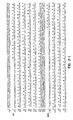
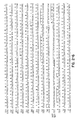
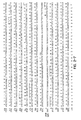
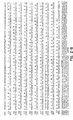
- Figure 2 shows nucleotide and predicted amino acid sequences.
- the nucleotide sequence is given as the rightward 5' to 3' strand only (numbered 1 to 10350).
- Rightward- and leftward- directed predicted amino acid sequences are shown above and below the corresponding nucleotide sequences in single-letter code, respectively.
- the name of each ORF is given to the left of the first line of the amino acid sequence.
- Amino acid sequences are numbered from the first M (three letter code) (ATG in the DNA) at the N-terminus to the last amino acid at the C-terminus, which precedes the termination codon (identified by an *).
- TATA consensus sites located within 400 nucleotides of the ATG are underlined and defined as sites containing at least six of seven matches to the TATA(AT)A(AT) consensus sequences defined by Corden et al. (Corden, B., et al., Science 209:1406-1414 (1980)). Underlines longer than seven nucleotides refer to areas containing overlapping TATA consensus sites.
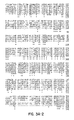
- Figure 3A shows alignment of S component homologs showing selected regions displaying maximum amino acid conservation. Gaps have been introduced to maximize alignment of identical amino acids as described in Methods.
- the consensus sequence (cons) indicates residues that are shared by at least all but one of the viruses and are indicated by capital letters. In alignments between more than two sequences, asterisks (*) indicate residues conserved by all of the viruses. Amino acid numbers (with respect to 5'-ATG) of corresponding regions aligned are listed before and after each sequence.
- Figure 3B shows the dot matrix analyses depicting overall homologies between selected MDV-alphaherpesvirus S segment homolog comparisons. Points were generated where at least 15 amino acids over a sliding window length of 30 were found identical or similar. The resulting diagonals illustrate regions showing greatest conservation. Amino acid numbers (with respect to 5'-ATG) of corresponding sequences are denoted above and to the right of each plot.
- Figure 4 shows a comparison of overall genome organization of available S component ORFs (Audonnet, J.-C., et al., J. Gen. Virol. 71:2969-2978 (1990); McGeoch, D. J., et al., J. Gen. Virol. 68:19-38 (1987); Tikoo, S. K., et al., J. Virol. 64:5132-5142 (1990); Van Zijl, M., et al., J. Gen. Virol. 71:1747-1755 (1990); Zhang, G., et al., J. Gen. Virol. 71:2433-2441 (1990); Cullinane, A. A., et al., J. Gen.
- ORF Numbers above each ORF refer to homologs based on relation to HSV-1 U S ORF nomenclature (McGeoch, D. J., et al., J. Mol. Biol. 181:1-13 (1985)). Alternative polypeptide designations common to each system are listed below those ORFs where applicable. Upper and lower case solid bars refer to rightward and leftward-directed ORFs, respectively. Arrows refer to identified IR S -U S and/or U S -TR S junction sites.
- PRV gene 4 is homologous to HSV-2 US4, rather than HSV-1 US4.
- Figure 5 shows the sequence of steps necessary to produce a complete segment of Marek's disease herpesvirus DNA encoding glycoprotein gI and the part of gE included in the application filed August 24, 1990.
- the present invention relates to a segment of DNA encoding glycoprotein gE precursor between a 8488 and 9978 nucleotide sequence of Marek's disease herpesvirus DNA, and containing potential promoter sequences up to 400 nucleotides 5' of each gene, as shown in Figure 2 (and identified as part of SEQ ID No:1) and subfragments of the DNA that recognize the DNA.
- the present invention relates to the novel glycoprotein precursors which are produced by expressions of the genes in the segments of DNA.
- the present invention relates to the potential MDV gene promoters, which are located in the 400 nucleotides 5' of each coding sequence.
- the present invention shows a sequence analysis of a 10.35 kbp DNA stretch encompassing a majority of the MDV U S region. Altogether seven MDV U S homologs, including three glycoprotein genes and two additional MDV-specific open reading frames, were identified.
- MDV EcoRl-0 and EcoRl-I of the pathogenic GA strain were previously cloned into pBR328 (Gibbs, C. P., et al., Proc. Natl. Acad. Sci. USA 81:3365-3369 (1984)), (Silva, R. F., et al., J. Virol. 54:690-696 (1985)) and made available by R. F. Silva, USDA Avian Disease and Oncology Lab, East Lansing, MI, where these clones are maintained.
- GA strain BamHI-A and BamHI-Pl were previously cloned into pACYC184 and pBR322, respectively (Fukuchi, K., et al., J.
- This phage clone was used to generate pUC18 subclones with smaller Sal I-bound inserts (psPl8-A, pSP18-B, and pSP18-C) containing the 3' BamHI-Pl-flanking region. These clones ( Figure 1B) were used to generate M13mp18 and -19 subclones for use as templates for nucleotide sequencing. Small- and large-scale plasmid preparations were made using the alkaline lysis procedure (Maniatis, T., et al., Molecular cloning: a laboratory manual. Cold Spring Harbor Laboratory, Cold Spring Harbor, New York (1982)).
- M13mp18 and M13mp19 phage subclones to be used as templates for sequencing were generated using specific restriction subfragments determined by restriction mapping or the use of Sau3A, Taq I or RsaI-digested viral DNA pools ligated into the unique BamHI, AccI or SmaI sites of M13 RF DNA, respectively.
- overlapping M13 deletion clones were obtained by processive Ba131 digestions from AccI, NaeI or NsiI restriction sites in EcoRl-0 by the method of Poncz et al (Poncz, M., et al., Proc. Natl. Acad. Sci. USA 79:4298-4302 (1982)).
- Ligated M13 products were transformed into CaCl 2 -competent JM107 host cells and added to melted B top agar containing 10 1 of 100 mM IPTG, 50 1 of 2% X-gal and 200 1 of a fresh overnight JM101 culture. These contents were then plated onto B agar plates and incubated at 37°C overnight. Recombinant (clear) plaques were then used to infect 5 ml of YT media diluted 1:50 with an overnight JM101 culture and rotated at 37°C for 6 hours. The resulting cells were pelleted by centrifugation for 5 minutes at room temperature and the supernatants were removed and stored at 4°C to retain viral stocks of each recombinant clone.
- single-stranded M13 phage DNA to be used as templates for DNA sequencing by the dideoxy-chain termination method was isolated according to instructions in the M13 Cloning/Dideoxy Sequencing Instruction Manual provided by Bethesda Research Laboratories. Recombinant M13mp phages were further screened by electrophoresing purified single-stranded viral DNA on 1% agarose mini-gels and selecting those templates showing reduced mobility in comparison to single-stranded M13mp 18 control DNA.
- DNA sequencing with single-stranded M13 templates was performed by the dideoxy-chain termination method (Sanger, F. S., et al., Proc. Natl. Acad. Sci. USA 74:5463-5467 (1977)) employing the modified T7 DNA polymerase, SequenaseTM (United States Biochemical Corp., Cleveland, Ohio).
- SequenaseTM United States Biochemical Corp., Cleveland, Ohio
- Figure 1B A summary of the sequencing strategy is included in Figure 1B.
- the specific step by step instructions provided with the SequenaseTM sequencing kit were employed. Briefly, single-stranded M13 templates were first annealed with the universal M13 synthetic oligonucleotide primer by incubation at 65°C for 2 minutes followed by slow cooling until the incubation temperature was below 30°C.
- dNTPs and ddNTPs radioactively labeled deoxyadenosine 5'-(alpha-thio) triphosphate
- 35 S-dATP 1000-1500 Ci/mmol
- NEN-DuPont radioactively labeled deoxyadenosine 5'-(alpha-thio) triphosphate
- SequenaseTM enzyme synthesis of radioactively labeled complementary strands was initiated from the annealed primer.
- Four separate synthesis reactions were each terminated by the incorporation of the specific ddNTP (ddATP, ddGTP, ddTTP or ddCTP) used in each tube.
- Reaction products were electrophoresed through 7% polyacrylamide/50% urea/Tris-Borate-EDTA gels and the labeled chains were visualized by autoradiography. Both strands were sequenced at least once. This was facilitated by the use of 16 synthetic 17-mer olignonucleotides generated based on previously determined sequences and substituted for the universal primer under similar reaction conditions above (0.5 pmoles/reaction) according to the general approach described by Strauss (Strauss, E. C., et al., Anal. Biochem. 154:353-360 (1986)).
- Dot matrix homology plots were generated by using the GCG program DOTPLOT with the output file from GCG's COMPARE. The latter creates a file of the points of similarity between two predicted amino acid sequences for which a window length of 30 and a stringency of 15 (in which conservative amino acid replacements are scored positive) were chosen.
- GAP GAP
- specific amino acid sequences were aligned using the algorithm of Needleman and Wunsch (Needleman, S. B., et al., J. Mol. Biol.
- the 10,350 nucleotide DNA sequence presented appears to encompass a majority of the MDV (GA) genome's unique short (U S ) region.
- a summary of the sequencing strategy is included in Materials and Methods and is depicted in Figure 1B. This sequence spans the U S fragments, EcoRl-0, EcoRl-I and extends to a SalI site 1.55 kbp downstream of the 3' end of BamHI-P 1 ( Figures 1A and 1B). Fukuchi et al. (Fukuchi, K., et al., J. Virol.
- this border is located about 100 bp upstream, or 1.1 and 2.7 kb downstream, respectively, of the termination codon of their respective US8 homologs (Cullinane, A. A., et al., J. Gen. Virol. 69:1575-1590 (1988); Davison, A. J., et al., J. Gen. Virol. 76:1759-1816 (1986); and McGeoch, D. J., et al., J. Gen. Virol. 69:1531-1574 (1988)).
- the region sequenced contains 9 complete ORFs likely to code for proteins (Fig. 1C, basis for names is given below). This prediction was based on: (1) homology and positional organization comparisons to other alphaherpesvirus genes and (2) presence of potential TATA and polyadenylation consensus sequences (Birnstiel, M. L., et al., Cell 41:349-359 (1985); and Corden, B., et al., Science 209:1406-1414 (1980)), and (3) possession of favorable contexts for translational initiation (Kozak, M., J. Cell Biool. 108:229-241 (1989)).
- alphaherpesviruses such as HSV and VZV tend to contain relatively tightly packed, unspliced and generally nonoverlapping coding regions (Davison, A. J., et al., J. Gen. Virol. 76:1759-1816 (1986); Davison, A. J., et al., J. Gen. Virol. 76:1759-1816 (1986); McGeoch, D. J., et al., J. Gen. Virol. 69:1531-1574 (1988); McGeoch, D. J., et al., J. Mol. Biol. 181:1-13 (1985); and McGeoch, D. J., et al., J. Gen. Virol.
- the predicted amino acid sequences of the predicted ORFs (beginning from the first ATG codon) are shown relative to the nucleotide sequence in Figure 2. Potential TATA sites within 400 nucleotides of the initiation codon are underlined. Proposed ORF and potential polyadenylation signal locations, identification of the -3, +4 ATG context nucleotides (Kozak, M., J. Cell Biol. 108:229-241 (1989)), as well as the lengths, relative molecular masses and predicted isoelectric points of the predicted translational products are shown in Table 1.
- the 40S ribosomal subunit binds initially at the 5'-end of mRNA and then migrates, stopping at the first AUG (ATG) codon in a favdrable context for initiating translation (Kozak, M., J. Cell Biol. 108:229-241 (1989)).
- AUG AUG
- definitive start sites for translation cannot be accurately predicted. Nevertheless, likely start sites are listed in Table 1; these refer to the location of the first inframe ATG codon found in the major open reading frame. According to Kozak (Kozak, M., J. Cell Biol.
- MDV US1 two transcriptional cap sites have been tentatively identified by 5' S1 nuclease protection analysis (data not shown). These sites appear to be located 18 and 25 nucleotides downstream of a TATATAA sequence at position 200 and 207, respectively ( Figure 2) Based on 3' S1 data, this transcript utilizes a polyadenylation signal located just downstream of the US10 coding region (Table 1, data not shown). Comparative Northern blot analyses of the U S region indicate that the MDV US1 transcript appears to be the most prominent transcript expressed at late times (72h) post-infection when extensive cytopathic effects are observed (data not shown). Phosphonoacetic acid inhibition studies have indicated that MDV US1, in contrast to its immediate-early HSV1 US1 counterpart, is regulated as a late class gene (data not shown).
- Dot matrix homology plots depicting overall homologies between selected MDV-alphaherpesvirus S segment homolog comparisons are included in Figure 3B. (Using a sliding window length of 30 amino acids, in which points are generated where at least 15 amino acids are found identical or similar). The resulting diagonals illustrate the regions showing greatest conservation. Such regions include and in some cases extend upon those regions depicted in Figure 3A.
- Herpesvirus glycoprotein homologs have generally been found to contain similar patterns of conserved cysteine residues. In comparing the gB homologs of seven different herpesviruses included in the alpha-, beta- and gammaherpesvirus subclasses, there is complete conservation of 10 cysteine residues (Ross, L. J. N., et al., J. Gen. Virol. 70:1789-1804 (1989)). HSV-1 US6 (gD) contains 7 cysteine residues: six appear critical for correct folding, antigenic structure and extent of oligosaccharide processing (Wilcox, W. C., et al., J. Virol. 62:1941-1947 (1988)).
- HSV-1 gE has Fc receptor activity (Johnson, D. C., et al., J. Virol. 62:1347-1354 (1988)), while its PRV and VZV counterparts do not (Edson, C. M., et al., Virology, 161:599-602 (1987); and Zuckerman, F. A., et al., J. Virol. 62:4622-4626 (1988)).
- N-terminal regions of the MDV gD, gI and gE homologs have revealed that they contain the three basic building blocks of signal peptide sequences: a basic, positively charged N-terminal region (n-region), a central hydrophobic region (h-region), and a more polar terminal region (c-region) that seems to define the cleavage site (von Heijne, G. J. Mol. Biol. 184:99-105 (1985)).
- Table 3 shows the likely position of these sites, the location of the hydrophobic transmembrane and charged cytoplasmic domains near the C-terminal end and the location of potential N-glycosylation sites.
- Table 3 shows MDV U S glycoprotein data on predicted signal peptide cleavage sites and locations of transmembrane and cytoplasmic domains and potential N-glycosylation sites (with respect to the ATG initiation codon).
- TABLE 3 Name Predicted Signal Peptide Cleavage Site Trans-membrane Domain Cytoplasmic Domain N-glycosylation Sites US6 G 30 -D 31 358-374 375-403 87,138,230,306 US7 S 18 -I 19 269-288 289-355 147,167,210,245, 253 US8 T 18 -A 19 394-419 420-497 60,133,148,203, 229,277,366,388
- MDV's counterpart contains a relatively long cytoplasmic domain.
- MDV gD's signal peptide contains a relatively long n-region (18 residues), that is unusually highly charged (+4; Figure 2) considering an overall mean value of +1.7 among eukaryotes, which generally does not vary with length (von Heijne, G, J. Mol. Biol. 184:99-105 (1985)).
- n-region 18 residues
- a more distal methionine codon exists directly before the initiation codon (as in the PRV gD homolog, Petrovskis, E. A., et al., J. Virol.
- MDV nuclear antigen MDV nuclear antigen expressed only in "latently" infected MDV-transformed lymphoblastoid cells.
- MDNA MDV nuclear antigen
- Alphaherpesvirus S segment genes have previously been found to be unique to members of this taxonomic subfamily (Davison, A. J., et al., J. Gen. Virol. 68:1067-1079 (1987); and Davison, A. J., et al., J. Gen. Virol. 67:597-611 (1986)).
- the identification of seven MDV homologs of alphaherpesvirus S segment genes in this study is consistent with the idea that MDV shares a closer evolutionary relationship with alphaherpesviruses than gammaherpesviruses.
- the MDV, VZV and EHV-4 US1 homologs lack approximately 120 aa of sequence comparable to the 5'-proximal portion of HSV-1 US1 (alpha 22)
- the MDV US1 gene appears to be regulated as an abundantly expressed late class gene (data not shown).
- MDV contains two apparently MDV-specific ORFs.
- the MDV U S region appears to contain approximately 2.6 to 4.0 kb of additional 5'-proximal sequences. Based on a comparison of Figure 4 and consideration of the expansion-contraction recombination scheme, it appears likely that there are additional MDV-specific U S genes.
- MDV has long been regarded as a gammaherpesvirus
- much of the previous work interpreting their properties has proceeded by analogy with the association between EBV and B cells (Nonoyama, M. p. 333-341. In B. Roizman (ed.), The herpesviruses, vol. 1. Plenum Press (1982); and Wilbur, W. J., et al., Proc. Natl. Acad. Sci. USA 80:726-730 (1983)).
- HHV-6 Because of a closer genetic relationship to the alphaherpesviruses, and keeping in mind the analysis of HHV-6 above, we agree with Lawrence et al. (Lawrence, G. L., et al., J. Virol.
- each HSV-1 L component gene possesses an equivalent in VZV (McGeoch, D. J., et al., J. Gen. Virol. 69:1531-1574 (1988)); a considerable number of these are related to beta- and gammaherpesvirus genes as well (29 of 67 EBV counterparts to VZV U L genes; Davison, A. J., et al., J. Gen. Virol. 68:1067-1079 (1987)).
- the S segments of HSV-1 and VZV differ significantly in size and appear to be among the least related parts of the two genomes (Davison, A.
- the 11 HSV-1 U S region genes dispensible for growth in tissue culture described above include HS 1 US7 (gI) and US8 (gE) (Longnecker, R., et al., Proc. Natl. Acad. Sci. USA 84:4303-4307 (1987); and Weber, P. C., et al., Science 236:576-579 (1987)). Assuming the MDV homologs have the same properties, these genes may be useful as sites for insertion of foreign genes.
- HSV's gE seem to play a role in HSV-1's ability to establish lethal infections and latency in mice (Meignier, B., et al., Virology 162:251-254 (1988)).
- gI and gE homologs of PRV of swine play a clear role in PRV virulence for 1-day-old chickens and young pigs (Mettenleiter, Thomas C., et al., Journal of Virology, p. 4030-4032 (Dec. 1987)).
- MDV US7 (gI) and US8 (gE) homologs it may be possible to inactivate one or both of these genes from very virulent MDV isolates which cause outbreaks not prevented by current vaccines, and thereby creating an attenuated vaccine viruses more closely related to field virus causing disease outbreaks.
- VZV lacks a gD homolog and is strongly cell-associated
- block in MDV virion formation in primary avian fibroblast cells is found to correlate with lack of expression (in these cells) of a glycoprotein, such as gD, and/or some other S component gene(s).
- Example 2 shows the molecular cloning of a construct containing the DNA encoding the complete MDV US7 (gI) gene and part of the MDV US8 (gE) gene. As can be seen, this is accomplished using segments of DNA spanning the gI and part of the gE coding region.
- pKS-MDgIl.59 a recombinant clone containing the complete MDV US7 (gI) coding sequence and a portion of the MDV US8 (gE) coding sequence requires two preexisting MDV clones, pKS-MDgD1.75 and p19P1 (Fig. 5).
- pKS-MDgDl.75 is a recombinant plasmid containing the 1.75 kbp NcoI-SstII subfragment of MDV EcoRl-I ligated into the SmaI-Sst II site of the cloning vector, pBluescript KS-.
- pl9Pl is a recombinant plasmid containing the 1.5 kbp BamHI-P 1 subfragment of MDV cloned into the unique BamHI site of pUC19.
- This clone contains the entire MDV gI coding sequence, except for the first 9 aa of its signal sequence.
- pl9Pl contains the first 104 aa of the MDV US8 (gE) coding region.
- pKS-MgD1.75 is first cut with HincII, which cuts once in the multiple cloning site of the pBluescript vector and once about 180 bp upstream of the insert's SstII terminus. This results in two fragments: one fragment (1.6 kbp) consists primarily of insert sequences encoding MDV US6 (gD); the larger fragment (3.1 kbp) consists of pBluescript vector sequences, in addition to about 180 bp which encode the N-terminus of MDV gI. The 3.1 kb fragment is gel purified and self-ligated by way of the two HincII ends.
- the resulting recombinant plasmid, pKS-MDgI0.18, is then cut with SstI (in the multiple cloning site, just downstream of the SstII site). Prior to subsequent digestion with SstII, the cohesive SstI ends is made blunt-ended with T4 DNA polymerase.
- the resulting 3.1 kbp SstII-SstI (blunt) fragment of pMDgI0.18 is gel purified and used in the final ligation step to create pKS-MDgI1.59.
- pl9Pl is cut with HindIII, which cuts just downstream of the partial MDV US8 (gE) coding sequence in the multiple cloning site of pUC19.
- HindIII cuts just downstream of the partial MDV US8 (gE) coding sequence in the multiple cloning site of pUC19.
- SstIId Prior to digestion with SstIId, the cohesive HindIII ends is made blunt-ended using Klenow fragment.
- the smaller SstII-HindIII (blunt) fragment (1.4 kbp) contains a majority of the MDV US7 (gI) coding sequence, in addition to 312 nucleotides at the 3' end which code for the 5' end of MDV gE.
- This 1.4 kbp SstII-HindIII(blunt) fragment is gel purified and ligated to the 3.1 kbp SstII-SstI(blunt) fragment of pKS-MDgD0.18.
- the resulting recombinant, pKS-MDgI1.59 contains the complete coding sequence for MDV gI and a portion of the N-terminal gE coding sequence. Digestion of pKS-MDgI1.59 with KpnI yields two fragments; the smaller 1.15 kbp fragment contains the complete coding sequence for MDV gI.
- Example 3 shows molecular subcloning of a construct containing the complete MDV US8 (gE) gene.
- the pathogenicity of the MDV may be reduced.
- the segments of DNA encoding the non-essential gI and gE can be used as insertion sites for segments of foreign DNA which encode proteins that are antigenically active for the purpose of producing a recombinant vaccine.
- MDV US6 The gene for MDV US6 (MDV gD) has been deposited in a plasmid (phagemid) pKS-MDgDl.75, as ATCC 40855, with The American Type Culture Collection, Rockville, MD, 20852, USA.
- MDV US7 The gene for MDV US7 (MDV gI) has been deposited in a plasmid (phagemid) pKS-MDgIl.59, as ATCC 75040, with The American Type Culture Collection, Rockville, MD, 20852, USA.
- MDV US8 The gene for MDV US8 (MDV gE) has been deposited in a plasmid pl8-MDgE 2.53, as ATCC 75039, with The American Type Culture Collection, Rockville, MD, 20852, USA.
Landscapes
- Chemical & Material Sciences (AREA)
- Organic Chemistry (AREA)
- Health & Medical Sciences (AREA)
- Life Sciences & Earth Sciences (AREA)
- Molecular Biology (AREA)
- Biophysics (AREA)
- General Health & Medical Sciences (AREA)
- Genetics & Genomics (AREA)
- Medicinal Chemistry (AREA)
- Gastroenterology & Hepatology (AREA)
- Proteomics, Peptides & Aminoacids (AREA)
- Virology (AREA)
- Biochemistry (AREA)
- Peptides Or Proteins (AREA)
- Micro-Organisms Or Cultivation Processes Thereof (AREA)
- Medicines Containing Antibodies Or Antigens For Use As Internal Diagnostic Agents (AREA)
- Saccharide Compounds (AREA)
- Pharmaceuticals Containing Other Organic And Inorganic Compounds (AREA)
- Medicines Containing Material From Animals Or Micro-Organisms (AREA)
- Medicines That Contain Protein Lipid Enzymes And Other Medicines (AREA)
- Agricultural Chemicals And Associated Chemicals (AREA)
Claims (10)
- DNA-Segment mit einem Gen, das den MDV-Glycoprotein E (gE) Vorläufer kodiert, lokalisiert zwischen den Nukleotiden 8488 und 9978 der DNA-Sequenz des Herpes-Virus der Marek-Krankheit und identifiziert als Teil von SEQ ID Nr. 1 und enthaltend potentielle Promotorsequenzen bis zu 400 Nukleotiden 5' vom Gen und Subfragmente der DNA, die die DNA selektiv erkennen, wenn sie in Form einer Sonde vorliegen.
- Das Segment des Anspruchs 1, worin das kodierte Glycoprotein 497 Aminosäuren enthält.
- Glycoprotein gE-Vorläufer, der folgendes umfaßt:


- Verfahren zur Reduktion der Pathogenität oder Virulenz eines Herpesvirus der Marek-Krankheit, wodurch ein Gen, welches für das Glycoprotein E kodiert, verändert wird.
- Verfahren zur Erzeugung eines Virusvektorimpfstoffes, zur Verwendung in vivo, oder eines in vitro-Expressionsvektors für ein Protein, das Antikörper gegen die Marek-Krankheit erzeugt, umfassend den Schritt der Insertion eines DNA-Segments in den Impfstoff oder den Vektor, das das gesamte oder ein Teil des Gens enthält, das für das Glycoprotein gE des Herpesvirus der Marek-Krankheit kodiert.
- Verfahren, um fremde Gene in einem Herpesvirus der Marek-Krankheit bereitzustellen, das die Insertion des fremden Gens in eine Region der DNA des Herpes-Virus umfaßt, die für das nicht essentielle Protein gE kodiert.
- DNA-Segment mit einem Gen, das für einen Teil des MDV-Glycoprotein E (gE)-Vorläufers kodiert, der zwischen den Nukleotiden 8488 und 8799 der DNA-Sequenz des Herpesvirus der Marek-Krankheit lokalisiert ist und als Teil der SEQ ID Nr. 1 identifiziert ist und der gegebenenfalls einen 5'-Regulatorbereich enthält, wobei das Gen bis zu 400 bp Länge aufweist, wie dargestellt in Fig. 2 und Subfragmente der DNA, die die DNA selektiv erkennen, wenn sie in Form einer Sonde vorliegen.
- Glycoprotein gE -Vorläufer, der folgendes umfaßt:

- 5'-Regulatorbereich für das Glycoprotein gE, das zwischen den Nukleotiden 8088 und 8487 der Sequenz, wie dargestellt in Fig. 2, lokalisiert ist.
- Sonde, die das DNA-Segment gemäß Anspruch 1 selektiv erkennt.
Applications Claiming Priority (5)
| Application Number | Priority Date | Filing Date | Title |
|---|---|---|---|
| US572711 | 1984-01-20 | ||
| US07/572,711 US5138033A (en) | 1990-08-24 | 1990-08-24 | Marek's disease herpesvirus glycoproteins GE |
| US07/736,335 US6087127A (en) | 1990-08-24 | 1991-07-25 | Marek's disease herpesvirus DNA segment encoding glycoproteins, gD, gI and gE |
| US736335 | 1991-07-25 | ||
| PCT/US1991/005870 WO1992003547A1 (en) | 1990-08-24 | 1991-08-19 | MAREK'S DISEASE HERPESVIRUS DNA SEGMENT ENCODING GLYCOPROTEINS, gD, gI AND gE |
Publications (3)
| Publication Number | Publication Date |
|---|---|
| EP0502146A1 EP0502146A1 (de) | 1992-09-09 |
| EP0502146A4 EP0502146A4 (de) | 1994-08-03 |
| EP0502146B1 true EP0502146B1 (de) | 1997-11-19 |
Family
ID=27075921
Family Applications (1)
| Application Number | Title | Priority Date | Filing Date |
|---|---|---|---|
| EP91915865A Expired - Lifetime EP0502146B1 (de) | 1990-08-24 | 1991-08-19 | DNA SEGMENT DES HERPESVIRUS DER MAREK-KRANKHEIT, WELCHES FÜR DAS GLYKOPROTEIN gE KODIERT |
Country Status (11)
| Country | Link |
|---|---|
| US (2) | US6087127A (de) |
| EP (1) | EP0502146B1 (de) |
| JP (1) | JP2527872B2 (de) |
| AT (1) | ATE160376T1 (de) |
| AU (1) | AU636247B2 (de) |
| CA (1) | CA2066160A1 (de) |
| DE (1) | DE69128239T2 (de) |
| DK (1) | DK0502146T3 (de) |
| ES (1) | ES2110999T3 (de) |
| GR (1) | GR3025821T3 (de) |
| WO (1) | WO1992003547A1 (de) |
Families Citing this family (9)
| Publication number | Priority date | Publication date | Assignee | Title |
|---|---|---|---|---|
| ES2056565T3 (es) * | 1990-07-30 | 1994-10-01 | Akzo Nobel Nv | Virus recombinante de la enfermedad de marek. |
| AU657144B2 (en) * | 1991-07-09 | 1995-03-02 | Juridical Foundation The Chemo-Sero-Therapeutic Research Institute | Recombinant Marek's disease virus, process for preparing the same and vaccine containing the same |
| US6913751B2 (en) * | 1992-06-12 | 2005-07-05 | Schering-Plough Veterinary Corporation | Recombinant avian herpesvirus useful in vaccine production |
| US6875856B2 (en) | 1993-09-24 | 2005-04-05 | Syntro Corporation | Recombinant infectious laryngotracheitis virus and uses thereof |
| FR2719056B1 (fr) * | 1994-04-20 | 1996-09-06 | Rhone Merieux | Herpesvirus transformés pour exprimer gD in vitro. |
| CA2216139C (en) * | 1995-03-23 | 2013-01-08 | Syntro Corporation | Recombinant infectious laryngotracheitis virus and uses thereof |
| US7314715B2 (en) | 2001-06-14 | 2008-01-01 | Schering-Plough Animal Health Corporation | Recombinant avian herpesvirus useful in vaccine production |
| GB0313132D0 (en) * | 2003-06-06 | 2003-07-09 | Ich Productions Ltd | Peptide ligands |
| CN116042709B (zh) * | 2022-09-23 | 2025-01-14 | 广西大学 | 一种共表达马立克病病毒糖蛋白gC和gD的DNA疫苗构建方法 |
Family Cites Families (4)
| Publication number | Priority date | Publication date | Assignee | Title |
|---|---|---|---|---|
| EP0434721B1 (de) * | 1988-09-13 | 2000-03-15 | Merial | Virale nucleotidsequenzen |
| ES2056565T3 (es) * | 1990-07-30 | 1994-10-01 | Akzo Nobel Nv | Virus recombinante de la enfermedad de marek. |
| FR2666589B1 (fr) * | 1990-09-07 | 1994-08-05 | Rhone Merieux | Nouveaux virus herpes recombinants, vaccin a base de ces recombinants, leur procede de preparation, genes, vecteurs et plasmides utilises dans ce procede. |
| CA2127700C (en) * | 1992-01-13 | 2006-06-20 | Enzo Paoletti | Marek's disease virus recombinant poxvirus vaccine |
-
1991
- 1991-07-25 US US07/736,335 patent/US6087127A/en not_active Expired - Fee Related
- 1991-08-19 AT AT91915865T patent/ATE160376T1/de active
- 1991-08-19 DK DK91915865T patent/DK0502146T3/da active
- 1991-08-19 EP EP91915865A patent/EP0502146B1/de not_active Expired - Lifetime
- 1991-08-19 CA CA002066160A patent/CA2066160A1/en not_active Abandoned
- 1991-08-19 JP JP3514685A patent/JP2527872B2/ja not_active Expired - Lifetime
- 1991-08-19 DE DE69128239T patent/DE69128239T2/de not_active Expired - Fee Related
- 1991-08-19 AU AU84965/91A patent/AU636247B2/en not_active Ceased
- 1991-08-19 ES ES91915865T patent/ES2110999T3/es not_active Expired - Lifetime
- 1991-08-19 WO PCT/US1991/005870 patent/WO1992003547A1/en not_active Ceased
-
1997
- 1997-12-30 GR GR970403471T patent/GR3025821T3/el unknown
-
1998
- 1998-03-24 US US09/047,125 patent/US5976787A/en not_active Expired - Fee Related
Also Published As
| Publication number | Publication date |
|---|---|
| JP2527872B2 (ja) | 1996-08-28 |
| WO1992003547A1 (en) | 1992-03-05 |
| ATE160376T1 (de) | 1997-12-15 |
| JPH04505106A (ja) | 1992-09-10 |
| DK0502146T3 (da) | 1998-07-27 |
| US6087127A (en) | 2000-07-11 |
| ES2110999T3 (es) | 1998-03-01 |
| US5976787A (en) | 1999-11-02 |
| AU8496591A (en) | 1992-03-17 |
| CA2066160A1 (en) | 1992-02-25 |
| GR3025821T3 (en) | 1998-03-31 |
| AU636247B2 (en) | 1993-04-22 |
| DE69128239D1 (de) | 1998-01-02 |
| DE69128239T2 (de) | 1998-03-12 |
| EP0502146A4 (de) | 1994-08-03 |
| EP0502146A1 (de) | 1992-09-09 |
Similar Documents
| Publication | Publication Date | Title |
|---|---|---|
| JP3339854B2 (ja) | ウイルスワクチン | |
| Swain et al. | Herpes simplex virus specifies two subunits of ribonucleotide reductase encoded by 3'-coterminal transcripts | |
| Gompels et al. | Conservation of gene organization in the lymphotropic herpesviruses herpesvirus saimiri and Epstein-Barr virus | |
| Barker et al. | The unique sequence of the herpes simplex virus 1 L component contains an additional translated open reading frame designated UL49. 5 | |
| Baumeister et al. | Pseudorabies virus and equine herpesvirus 1 share a nonessential gene which is absent in other herpesviruses and located adjacent to a highly conserved gene cluster | |
| Bowles et al. | The ICP0 protein of equine herpesvirus 1 is an early protein that independently transactivates expression of all classes of viral promoters | |
| Flowers et al. | The equine herpesvirus type 1 (EHV-1) homolog of herpes simplex virus type 1 US9 and the nature of a major deletion within the unique short segment of the EHV-1 KyA strain genome | |
| Klupp et al. | The virulence-determining genomic BamHI fragment 4 of pseudorabies virus contains genes corresponding to the UL15 (partial), UL18, UL19, UL20, and UL21 genes of herpes simplex virus and a putative origin of replication | |
| Ross et al. | Alterations in DNA sequence and RNA transcription of the Bam HI-H fragment accompany attenuation of oncogenic Marek's disease herpesvirus | |
| EP0502146B1 (de) | DNA SEGMENT DES HERPESVIRUS DER MAREK-KRANKHEIT, WELCHES FÜR DAS GLYKOPROTEIN gE KODIERT | |
| Robertson et al. | Sequence characteristics of a gene in equine herpesvirus 1 homologous to glycoprotein H of herpes simplex virus | |
| US5252717A (en) | Marek's disease herpesvirus DNA segments encoding glycoproteins, gD, gI and gE | |
| US5138033A (en) | Marek's disease herpesvirus glycoproteins GE | |
| US5252716A (en) | Marek's disease herpesvirus DNA segments encoding glycoproteins, gD, gI and gE | |
| EP0908517B1 (de) | Virale Nukleotidsequenzen | |
| US5558860A (en) | Viral vaccines | |
| Kaliman et al. | Large and small subunits of the Aujeszky's disease virus ribonucleotide reductase: nucleotide sequence and putative structure | |
| EP0668356A2 (de) | Rekombinante, infektöse Rinder-Rhinotracheitis-Mutante, Methode zu deren Herstellung, und Vakzine dieselbige enthaltend | |
| DAMIANI et al. | Nucleotide sequences of glycoprotein I and E genes of equine herpesvirus type 4 | |
| Tan | Transcriptional and translational analysis of Marek's disease virus | |
| IE84982B1 (en) | Viral Vaccines | |
| IE19990699A1 (en) | Viral Vaccines |
Legal Events
| Date | Code | Title | Description |
|---|---|---|---|
| PUAI | Public reference made under article 153(3) epc to a published international application that has entered the european phase |
Free format text: ORIGINAL CODE: 0009012 |
|
| 17P | Request for examination filed |
Effective date: 19920416 |
|
| AK | Designated contracting states |
Kind code of ref document: A1 Designated state(s): AT BE CH DE DK ES FR GB GR IT LI LU NL SE |
|
| A4 | Supplementary search report drawn up and despatched | ||
| AK | Designated contracting states |
Kind code of ref document: A4 Designated state(s): AT BE CH DE DK ES FR GB GR IT LI LU NL SE |
|
| 17Q | First examination report despatched |
Effective date: 19950711 |
|
| GRAG | Despatch of communication of intention to grant |
Free format text: ORIGINAL CODE: EPIDOS AGRA |
|
| GRAH | Despatch of communication of intention to grant a patent |
Free format text: ORIGINAL CODE: EPIDOS IGRA |
|
| GRAH | Despatch of communication of intention to grant a patent |
Free format text: ORIGINAL CODE: EPIDOS IGRA |
|
| GRAA | (expected) grant |
Free format text: ORIGINAL CODE: 0009210 |
|
| AK | Designated contracting states |
Kind code of ref document: B1 Designated state(s): AT BE CH DE DK ES FR GB GR IT LI LU NL SE |
|
| REF | Corresponds to: |
Ref document number: 160376 Country of ref document: AT Date of ref document: 19971215 Kind code of ref document: T |
|
| REG | Reference to a national code |
Ref country code: CH Ref legal event code: NV Representative=s name: PATENTANWALTSBUERO JEAN HUNZIKER Ref country code: CH Ref legal event code: EP |
|
| REF | Corresponds to: |
Ref document number: 69128239 Country of ref document: DE Date of ref document: 19980102 |
|
| ITF | It: translation for a ep patent filed | ||
| ET | Fr: translation filed | ||
| REG | Reference to a national code |
Ref country code: ES Ref legal event code: FG2A Ref document number: 2110999 Country of ref document: ES Kind code of ref document: T3 |
|
| REG | Reference to a national code |
Ref country code: DK Ref legal event code: T3 |
|
| PLBE | No opposition filed within time limit |
Free format text: ORIGINAL CODE: 0009261 |
|
| STAA | Information on the status of an ep patent application or granted ep patent |
Free format text: STATUS: NO OPPOSITION FILED WITHIN TIME LIMIT |
|
| 26N | No opposition filed | ||
| PGFP | Annual fee paid to national office [announced via postgrant information from national office to epo] |
Ref country code: FR Payment date: 20000801 Year of fee payment: 10 |
|
| PGFP | Annual fee paid to national office [announced via postgrant information from national office to epo] |
Ref country code: SE Payment date: 20000802 Year of fee payment: 10 Ref country code: DE Payment date: 20000802 Year of fee payment: 10 |
|
| PGFP | Annual fee paid to national office [announced via postgrant information from national office to epo] |
Ref country code: GB Payment date: 20000803 Year of fee payment: 10 Ref country code: DK Payment date: 20000803 Year of fee payment: 10 Ref country code: CH Payment date: 20000803 Year of fee payment: 10 Ref country code: AT Payment date: 20000803 Year of fee payment: 10 |
|
| PGFP | Annual fee paid to national office [announced via postgrant information from national office to epo] |
Ref country code: NL Payment date: 20000808 Year of fee payment: 10 |
|
| PGFP | Annual fee paid to national office [announced via postgrant information from national office to epo] |
Ref country code: LU Payment date: 20000809 Year of fee payment: 10 |
|
| PGFP | Annual fee paid to national office [announced via postgrant information from national office to epo] |
Ref country code: GR Payment date: 20000823 Year of fee payment: 10 |
|
| PGFP | Annual fee paid to national office [announced via postgrant information from national office to epo] |
Ref country code: BE Payment date: 20000830 Year of fee payment: 10 |
|
| PGFP | Annual fee paid to national office [announced via postgrant information from national office to epo] |
Ref country code: ES Payment date: 20000908 Year of fee payment: 10 |
|
| PG25 | Lapsed in a contracting state [announced via postgrant information from national office to epo] |
Ref country code: LU Free format text: LAPSE BECAUSE OF NON-PAYMENT OF DUE FEES Effective date: 20010819 Ref country code: GB Free format text: LAPSE BECAUSE OF NON-PAYMENT OF DUE FEES Effective date: 20010819 Ref country code: DK Free format text: LAPSE BECAUSE OF NON-PAYMENT OF DUE FEES Effective date: 20010819 Ref country code: AT Free format text: LAPSE BECAUSE OF NON-PAYMENT OF DUE FEES Effective date: 20010819 |
|
| PG25 | Lapsed in a contracting state [announced via postgrant information from national office to epo] |
Ref country code: SE Free format text: LAPSE BECAUSE OF NON-PAYMENT OF DUE FEES Effective date: 20010820 Ref country code: ES Free format text: LAPSE BECAUSE OF NON-PAYMENT OF DUE FEES Effective date: 20010820 |
|
| PG25 | Lapsed in a contracting state [announced via postgrant information from national office to epo] |
Ref country code: LI Free format text: LAPSE BECAUSE OF NON-PAYMENT OF DUE FEES Effective date: 20010831 Ref country code: CH Free format text: LAPSE BECAUSE OF NON-PAYMENT OF DUE FEES Effective date: 20010831 Ref country code: BE Free format text: LAPSE BECAUSE OF NON-PAYMENT OF DUE FEES Effective date: 20010831 |
|
| PG25 | Lapsed in a contracting state [announced via postgrant information from national office to epo] |
Ref country code: GR Free format text: LAPSE BECAUSE OF NON-PAYMENT OF DUE FEES Effective date: 20020209 |
|
| BERE | Be: lapsed |
Owner name: MICHIGAN STATE UNIVERSITY Effective date: 20010831 |
|
| PG25 | Lapsed in a contracting state [announced via postgrant information from national office to epo] |
Ref country code: NL Free format text: LAPSE BECAUSE OF NON-PAYMENT OF DUE FEES Effective date: 20020301 |
|
| EUG | Se: european patent has lapsed |
Ref document number: 91915865.9 |
|
| GBPC | Gb: european patent ceased through non-payment of renewal fee |
Effective date: 20010819 |
|
| REG | Reference to a national code |
Ref country code: CH Ref legal event code: PL |
|
| REG | Reference to a national code |
Ref country code: DK Ref legal event code: EBP |
|
| PG25 | Lapsed in a contracting state [announced via postgrant information from national office to epo] |
Ref country code: FR Free format text: LAPSE BECAUSE OF NON-PAYMENT OF DUE FEES Effective date: 20020430 |
|
| NLV4 | Nl: lapsed or anulled due to non-payment of the annual fee |
Effective date: 20020301 |
|
| PG25 | Lapsed in a contracting state [announced via postgrant information from national office to epo] |
Ref country code: DE Free format text: LAPSE BECAUSE OF NON-PAYMENT OF DUE FEES Effective date: 20020501 |
|
| REG | Reference to a national code |
Ref country code: FR Ref legal event code: ST |
|
| REG | Reference to a national code |
Ref country code: ES Ref legal event code: FD2A Effective date: 20020911 |
|
| PG25 | Lapsed in a contracting state [announced via postgrant information from national office to epo] |
Ref country code: IT Free format text: LAPSE BECAUSE OF NON-PAYMENT OF DUE FEES;WARNING: LAPSES OF ITALIAN PATENTS WITH EFFECTIVE DATE BEFORE 2007 MAY HAVE OCCURRED AT ANY TIME BEFORE 2007. THE CORRECT EFFECTIVE DATE MAY BE DIFFERENT FROM THE ONE RECORDED. Effective date: 20050819 |