EP0508417A2 - Monoklonaler Antikörper, Polypeptide und deren Herstellung - Google Patents
Monoklonaler Antikörper, Polypeptide und deren Herstellung Download PDFInfo
- Publication number
- EP0508417A2 EP0508417A2 EP92106093A EP92106093A EP0508417A2 EP 0508417 A2 EP0508417 A2 EP 0508417A2 EP 92106093 A EP92106093 A EP 92106093A EP 92106093 A EP92106093 A EP 92106093A EP 0508417 A2 EP0508417 A2 EP 0508417A2
- Authority
- EP
- European Patent Office
- Prior art keywords
- cells
- antibody
- cancer
- medium
- polypeptide
- Prior art date
- Legal status (The legal status is an assumption and is not a legal conclusion. Google has not performed a legal analysis and makes no representation as to the accuracy of the status listed.)
- Granted
Links
Images





Classifications
-
- C—CHEMISTRY; METALLURGY
- C07—ORGANIC CHEMISTRY
- C07K—PEPTIDES
- C07K16/00—Immunoglobulins [IG], e.g. monoclonal or polyclonal antibodies
- C07K16/18—Immunoglobulins [IG], e.g. monoclonal or polyclonal antibodies against material from animals or humans
- C07K16/28—Immunoglobulins [IG], e.g. monoclonal or polyclonal antibodies against material from animals or humans against receptors, cell surface antigens or cell surface determinants
- C07K16/30—Immunoglobulins [IG], e.g. monoclonal or polyclonal antibodies against material from animals or humans against receptors, cell surface antigens or cell surface determinants from tumour cells
-
- C—CHEMISTRY; METALLURGY
- C07—ORGANIC CHEMISTRY
- C07K—PEPTIDES
- C07K14/00—Peptides having more than 20 amino acids; Gastrins; Somatostatins; Melanotropins; Derivatives thereof
- C07K14/435—Peptides having more than 20 amino acids; Gastrins; Somatostatins; Melanotropins; Derivatives thereof from animals; from humans
- C07K14/46—Peptides having more than 20 amino acids; Gastrins; Somatostatins; Melanotropins; Derivatives thereof from animals; from humans from vertebrates
- C07K14/47—Peptides having more than 20 amino acids; Gastrins; Somatostatins; Melanotropins; Derivatives thereof from animals; from humans from vertebrates from mammals
-
- C—CHEMISTRY; METALLURGY
- C07—ORGANIC CHEMISTRY
- C07K—PEPTIDES
- C07K14/00—Peptides having more than 20 amino acids; Gastrins; Somatostatins; Melanotropins; Derivatives thereof
- C07K14/435—Peptides having more than 20 amino acids; Gastrins; Somatostatins; Melanotropins; Derivatives thereof from animals; from humans
- C07K14/705—Receptors; Cell surface antigens; Cell surface determinants
-
- A—HUMAN NECESSITIES
- A61—MEDICAL OR VETERINARY SCIENCE; HYGIENE
- A61K—PREPARATIONS FOR MEDICAL, DENTAL OR TOILETRY PURPOSES
- A61K38/00—Medicinal preparations containing peptides
Definitions
- the present invention relates to a monoclonal antibody which suppresses the motility of cancer cells.
- the present antibody show suppressing activity to the motility of human cancer cells such as lung carcinoma cells and grioblastoma cells, and can be used in prophylaxis or treatment of cancer.
- the present invention also relates to polypeptides and a method of producing the same. More particularly, the invention relates to polypeptides influencing the motility of cancer cells. Said polypeptides can be used in preventing the metastatis of cancer cells, in particular lung carcinoma, glioblastoma and other metastatic cancers.
- anti cancer antibodies have been prepared and used as a therapeutic or diagnostic drug, since the publication of Köhler and Milstein about production of monoclonal antibodies [Köhler, G. and Milstein, C., Nature, 256 , 495 (1975)]. These antibodies are used for antibody missile therapy in which antibody specifically binds to cancer cells and exclude or injured them thereby without injured normal tissues, and show some degree of success in the therapy of blood cancer such as leukemia or lymploma.
- AMF autocrine motility factor
- chemotactic factor Igaku no Ayumi (Advances in Medicine). 150 , 805-806 (1989)].
- the present inventors completed the present invention by determining the amino acid sequence of the peptide recognized by the above-mentioned monoclonal antibody, isolating a cDNA coding for the antigen, further constructed a plasmid comprising said cDNA, preparing a transformant by transformation with said plasmid and recovering the desired peptide from a culture broth of said transformant.
- the present invention relates to (1) an anti cancer monoclonal antibody (hereinafter sometimes abbreviated as MoAb) which prevents the motility of human cancer cells. More paticularly, the present antibody is a monoclonal IgG, preferably IgG, antibody specific to a human cancer cell such as lung carcinoma cell or glioblastoid cell and recognizes a peptide which participates in the motility of cancer cell as an antigen.
- a polypeptide containing the amino acid sequence of Seq. ID No. 1 [hereinafter such polypeptide shall be referred to as M]; wherein M can be recognizable by the antibody (1) above, (3) a polypeptide having a partial amino acid sequence taken from the amino acid sequence of Seq. ID No.
- the polypeptide (3) above may be any one provided that it has a partial amino acid sequence taken from the amino acid sequence of Seq. ID No. 1 and shows a cancer cell motility suppressing activity, for example a polypeptide having the sequence covering the amino acid residues of 35-60, 113-142, 131-166, or 163-191, said polypeptide being selected either from a site performing a receptor function or from a site interacting another protein on the cell membrane and extracellularly; (4) a recombinant DNA comprising a nucleotide sequence coding for M; (5) a vector comprising the DNA mentioned above (4); (6) a transformant as transformed with said vector; and (7) a method of producing M which comprises cultivating a transformant as transformed with a vector comprising the DNA mentioned above (4), producing and accumulating of M in the culture broth and harvesting the same.
- Fig. 1 shows the results of Western blotting analysis (see Example 4).
- Fig. 2 shows the result concerning the suppression of the motility of MAC10 cells by the purified antibody M31-15 (see Experimental Example 2).
- Fig. 3 shows the results of Western blotting analysis (see Example 5).
- Fig. 4 shows the nucleotide sequence of cDNA of the positive clone obtained in Example 7 and the amino acid sequence deducible from the nucleotide sequence.
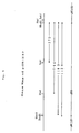
- Fig. 5 shows a gene map of the plasmid pTB1352 constructed in Example 7.
- Fig. 6 shows a restriction enzyme map of the M expression plasmid pTB1442 constructed in Example 10.
- Fig. 7 shows the FACStar analysis patterns obtained in Example 11 for CHO(dhfr ⁇ ) cells and M-expressing clones derived therefrom.
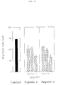
- Fig. 8 graphically shows the results of Experimental Example 4 concerning the suppression of the motility of MAC10 cells by some M fragment peptides.
- Fig. 9 shows the motilities of CHO(dhfr ⁇ ) cells and some M-expressing clones derived therefrom as measured in Experimental Example 5.
- Any DNA comprising a nucleotide sequence coding for the amino acid sequence of Seq. ID No. 1 may be used as the recombinant DNA mentioned above.
- a DNA comprising the nucleotide sequence of Seq. ID No. 2 is preferable, for instance.
- the antibodies of this invention can be used in the treatment of cancer, paticularly in the prevention of cancer metastasis, either alone or combination with substances inhibiting the adhesion of cancer cells and other metastasis inhibiting substances showing different mechanisms of action.
- the antibodies of this invention can be used as agents for missile therapy for the treatment of cancer.
- the antibodies of this invention are more advantageous in cancer therapy than the conventional missile therapy agents in that when an antitumor substance which will not inhibit the action of the antibodies of this invention is combinedly used, said antibodies can specifically kill cancer cells while preventing cancer metastasis.
- the antibodies of this invention can be obtained from among anti-human cancer antibodies, which are produced by anti-human cancer antibody-producing hybridomas produced by a known method, by screening using the cancer cell motility suppressing activity as an index.
- anti-human cancer antibody-producing hybridomas may be of any kind provided that they can produce antibodies specifically binding to human cancer cells. Mention may be made of the anti-human transferrin receptor (hereinafter sometimes abbreviated as hTfR) monoclonal antibody-producing mouse hybridoma 22C6 [IFO 50172, FERM BP-2054] [cf. Japanese Unexamined Publication No. 79970/1990] or the anti-human kidney cancer monoclonal antibody-producing mouse hybridoma RCS-1 [IFO 50184, FERM BP-2333] [cf. WO 91/09134], for instance.
- hTfR anti-human transferrin receptor
- the conventional method of hybridoma production can be used [G. Köhler et al.: Nature, 256 , 495 (1975)].
- animals are immunized with cancer cells in the conventional manner and antibody-producing cells obtained are fused with myeloma cells or the like.
- kidney cancer cells AM-RC-3, AM-RC-6, AM-RC-7, SK-RC-1, SK-RC-9, SK-RC-18
- bladder cancer cells T-24, KK-47, MGH-U-1
- prostate cancer cells DU-145
- stomach cancer cells NUGC-2, NUGC-3, NUGC-4, MKN-28, KATO-III, MRK-1
- intestinal cancer cells SW-403, SW-620, SW-1116, SW-1222, CaOV-4, HT-29
- cervical cancer cells ME-180), melanoma cells (SK-MEL-33, SK-MEL-37), breast carcinoma cells (MCF-7), glioma cells (MG-178), lung carcinoma cells (Luci-10, Calu-6, PC-10, ADLC-DA, SBC-3, SCLC-SA, Luci-6, CADO-LC3, OKADA, QG-56), T-cell leukemia cells (CCRF-CEM, HPB-ALL,
- Cancer cell lines established from cancer tissues of actual cancer patients, such as lung cancer patients, for example MAC8, can also be used.
- the yield of the antibodies of this invention can be increased by using, as an immunogen, a cell surface antigen recognizable by the antibodies of this invention.
- a cell surface antigen recognizable by the antibodies of this invention.
- FACS fluorescence-activated cell surface antigen
- a cell suspension is generally prepared using phosphate-bufferod saline (PBS) containing 5 mM EDTA. Trypsin, collagenase or the like may also be used as the case may be.
- PBS phosphate-bufferod saline
- Trypsin, collagenase or the like may also be used as the case may be.
- the animals to be immunized include, among others, mammals (e.g. human, mouse, rat, guinea pig, cat, dog, monkey, rabbit, sheep, goat, hamster, etc.) and birds (e.g. chicken, goose, duck, etc.).
- mammals e.g. human, mouse, rat, guinea pig, cat, dog, monkey, rabbit, sheep, goat, hamster, etc.
- birds e.g. chicken, goose, duck, etc.
- the mouse, rat and rabbit are preferred.
- the mouse is particularly preferred.
- the antibody-producing cells derived from immunized animals may be of the spleen, lymphatic node or peripheral blood lymphocyte origin, for instance, and splenocytes are particularly preferred.
- these antibody-producing cells are prepared by immunizing mice (4 to 10 weeks of age) with cancer cells.
- the method of immunization generally comprises suspending viable cells washed with phosphate-buffered saline (hereinafter sometimes abbreviated as PBS) in a small amount of PBS and administering the suspension to the mice subcutaneously at the back or abdomen or intraperitoneally in a dose of 106 to 108 cells/0.1 ml, preferably about 107 cells/0.1 ml. Then, cells are administered in the same manner 2 to 5 times at 2-week intervals.
- the last immunization is performed by intravenous administration. Three to four days after the last immunization, splenocytes can be prepared from the mice in the conventional manner.
- Myeloma cells e.g. NS-1, P3-X63-AgUI, X45, SP2, X68-Ag8, etc. are particularly preferred as counterpart cells for fusion.
- the MOPC21 (BALB/c mouse)-derived myeloma cell line X68-Ag8 and modifications thereof are preferred.
- a cell line selected from among these modifications and other myeloma-derived cell lines since cell lines of this kind are resistant to 8-azaguanine and are deficient in hypoxanthine guanine phosphoribosyltransferase and therefore cannot grow in HAT medium (containing hypoxanthine, aminopterine and thymidine), facilitating the hybridoma selection after fusion.
- the cell fusion is carried out by a known method and polyethylene glycol (hereinafter sometimes abbreviated as PEG), Sendai virus or the like, preferably PEG, is used as fusion agent.
- PEG polyethylene glycol
- Sendai virus or the like preferably PEG
- PEG should preferably have an average molecular weight of 1,000 to 6,000, and PEG 4000 is particularly preferred. PEG is used at a concentration of 10 to 80%, preferably 40 to 50%.
- antibody-producing cells and myeloma cells are washed with RPMI1640 medium and mixed generally in a ratio of 2:1 to 10:1, and the mixture is centrifuged at room temperature and 700 x g to give a cell pellet.
- the cell pellet is loosened with warming in a constant-temperature vessel maintained at 37°C and a PEG solution warmed in advance is gradually added with sufficient stirring.
- PEG is used in an amount of 1 ml per 108 cells but this amount may varies on the case-by-case basis.
- a medium warmed in advance is gradually added dropwise with stirring to thereby lower the PEG concentration.
- 1 to 30 ml of medium is added over about 10 minutes.
- Cells are collected by centrifugation at room temperature and suspended in a medium containing 10% fetal calf serum (hereinafter sometimes abbreviated as FCS) to a cell concentration of 1 to 2 x 10⁶ cells/ml, and the suspension is distributed in 100 ⁇ l portions into wells of 96 well plates. After overnight standing, a medium containing HAT and 10% FCS (HAT medium) is added (100 ⁇ l per well). For omitting this operation, a suspension of cells after cell fusion in HAT medium may be sowed.
- FCS fetal calf serum
- the method of medium exchange generally comprises removing 100 to 200 ⁇ l of medium from each well and adding 100 to 200 ⁇ l of fresh HAT medium to each well.
- the well or wells are submitted to an appropriate screening system to detectin which the desired antibody-producing hybridoma.
- the hybridomas obtained are immediately subjected to cloning by an appropriate method.
- MHA mixed hemagglutination method
- IF immunofluorescence method
- A549SA Antibody activity-positive hybridomas are immediately subjected to cloning, which is generally carries out with ease by the limiting dilution method, for instance.
- the culture supernatant of each clones hybridoma is assayed for its antibody titer by the method mentioned above.
- Hybridomas stably producing a high-titer antibody are selected and further subjected to screening for detecting antibodies capable of suppressing the cancer cell motility. Said screening is carried out, for example, by the modified Boyden chamber assay using the Nucleopore membrane filter mentioned above [J. Immunol. Methods, 33 , 239-247 (1980)] or by using commercially available Transwell plates (Costar).
- filters with a pore size of 5 ⁇ m to 8 ⁇ m may be selected depending on experiment conditions.
- Melanoma cells, glioma cells and the like are generally used as the cancer cells but appropriate cells screened out form among other cancer cells may be used as well.
- MAC10 cells screened out from among lung carcinoma cells may be used.
- Desired monoclonal hybridomas can be obtained by selecting, in this manner, hybridomas producing an antibody showing potent inhibitory effect on cancer cells.
- the production and accumulation of said anti human cancer cell monoclonal antibodies is effected by cultivating the hybridomas of this invention generally in a liquid medium, preferably a serum-free medium, or in the peritoneal cavity of a warm-blooded animal other than human (generally, mouse).
- a serum-free medium should desirably be used. Examples of such cultivation are given below.
- liquid medium there may be mentioned, for example, a basal medium for animal cell culture [e.g. an equivolume mixture (I ⁇ H medium) of Iscove medium and Ham's F-12 medium, or RPMI1640 medium] supplemented with fetal calf serum, or GIT medium (Wako Pure Chemical Industries) (cf. Japanese Unexamined Publication No. 145088/1985).
- the cultivation is generally carried out at about 30°C to about 38°C, preferably about 37°C, for about 3 to about 60 days, preferably about 5 to about 10 days.
- antibody-producing hybridoma cells are introduced into the peritoneal cavity of each mouse, whereby 3 to 10 ml of an ascitic fluid containing a quantity of an antibody is obtained after about 10 to about 35 days of feeding.
- the antibody in the liquid medium or ascitic fluid can be purified using a combination of known biochemical techniques.
- the antibody-containing liquid is centrifuged, and the supernatant is subjected to salting out (generally, with ammonium sulfate or sodium sulfate).
- the protein precipitate obtained is dissolved in an appropriate buffer solution, dialyzed and subjected to column chromatography (using a DEAE ion exchange column, hydroxyapatite column, gel filtration column, protein A column, protein G column, or the like) or immunoaffinity chromatography, for instance, for separating and purifying the desired antibody.
- Such a separation and purification procedure can give about 5 to about 30 mg of an anti-cancer cell MoAb with a purity of not less than 90% (on the protein weight basis) from, for example, 500 ml of liquid medium. Similarly, 50 ml of ascitic fluid gives 10 to 200 mg of such antibody.
- an antibody with a higher purity can be obtained by combining column chromatography using protein A or protein G with chromatography using a gel filtration column carrier, DEAE ion exchange column carrier, hydroxyapatite or the like.
- These purified antibody preparations have a heterologous protein content of not more than about 0.1% and ace suited for administration to humans as drugs.
- the monoclonal antibodies obtained in the above manner can give Fab, Fab' or F(ab')2 fragments while the ability to bind to cancer cells is retained. Such fragments can be used for the same purposes as the MoAb of this invention.
- a proteolytic enzyme e.g. papain, pepsin
- reductive treatment e.g. papain, pepsin
- Such fragments can be used for the same purposes as the MoAb of this invention.
- said hybridomas produce mouse IgG MoAb
- it is possible to produce a mouse-human chimera antibody by isolating a DNA coding for the antigen recognition site-containing variable region of said anti-cancer cell MoAb and joining thereto a gene coding for the constant region of human IgG using the gene manipulation technique [Steplewski, Z. et al.: Proc. Natl. Acad.
- Such a chimera antibody is advantageous in administering to humans because of its low antigenicity.
- the anti-cancer cell monoclonal antibodies of the present invention can be used either alone or admixed with pharmacologically acceptable appropriate carriers, excipients, diluents or the like to give pharmaceutical compositions such as injectable solutions.
- Such MoAb preparations, as anticancer agents can produce the cancer metastasis-inhibiting effect and, when bound to an appropriate anti-cancer agent, can expectedly be used as the therapeutic drug what is called missile therapy agents.
- the present antibody is useful for detecting human cancer cells, especially human lung carcinoma cells or human grioblastona cells, as well as other anti cancer antibodies.
- present antibody is also useful for screening or purification of the present M.
- the present M can be produced by a combination of known genetic engineering techniques.
- an expression vector comprising a DNA having the nucleotide sequence coding for M can be produced, for example, by (i) isolating a phage containing the desired DNA by means of a proto blot immunoscreening system using an appropriate cDNA library, (ii) excising the desired cloned DNA from said phage and (iii) joining said cloned DNA to a vehicle at a site downstream from the promoter in said vehicle.
- Escherichi a coli -derived plasmids e.g. pBR322, pBR325, pUC12, pUC13
- Bacillus subtilis -derived plasmids e.g. pUB110, pTB5, pC194
- Saccharomyces cerevisiae -derived plasmids e.g. pSH19, pSH15
- bacteriophages such as phage
- animal viruses such as retroviruses and vaccinia virus, among others.
- Said gene may has ATG as the translation initiation codon at its 5' end and it may also have TAA, TGA or TAG as the translation termination codon at its 3' end.
- a promoter is joined to said gene upstream therefrom. Any promoter that is appropriate to the host employed for gene expression may be used as the promoter in the practice of the invention.
- the T7 promoter, trp promoter, lac promoter, rec promoter, ⁇ P L promoter, 1pp promoter and the like are for instance, preferred.
- the SPO1 promoter, SPO2 promoter, penP promoter and the like are, for instance, preferred; and when the host is a yeast, the PHO5 promoter, PGK promoter, GAP promoter, ADH promoter and the like are, for instance, preferred. It is particularly preferred that the host is Escherichia coli and the promoter is the trp promoter or ⁇ P L promoter.
- SV40-derived promoters When animal cells are used as the host, mention may be made of SV40-derived promoters and retrovirus promoters, among others. In particular, SV-40-derived promoters are preferred.
- a transformant is produced using the thus-prepared DNA-comprising vector.
- prokaryotes such as Escherichia coli , Bacillus subtilis and actinomycetes as well as eukaryotes such as yeasts, fungi and animal cells.
- Escherichia coli K12 DH1 Proc. Natl. Acad. Sci. USA, 60 , 160 (1968)]
- JM103 Nucl. Acids Res., 9 , 309 (1981)]
- JA221 J. Mol. Biol., 120 , 517 (1978)]
- HB101 J. Mol. Biol., 41 , 459 (1969)]
- C600 C600 [Genetics, 39 , 440 1954]
- Bacillus subtilis MI114 Bacillus subtilis MI114 [Gene, 2 4, 255 (1983)] and 207-21 [J. Biochem., 95 , 87 (1984)].
- yeasts include, among others, Saccharomyces cerevisiae AH22, AH22R ⁇ , NA87-11A and DKD-5D.
- animal cells mentioned above include the simian COS-7 and Vero cells, chinese hamster CHO cells, and murine L cells, among others.
- Bacillu s subtilis The transformation of Bacillu s subtilis is carried out by the method described in Molec. Gen. Genet., 168 , 111 (1979), for instance.
- a liquid medium is suitable for the cultivation and those carbon sources, nitrogen sources, inorganic substances and other substances that are required for the growth of said transformant are contained in said medium.
- the carbon sources include, among others, glucose, dextrin, soluble starch, and sucrose
- the nitrogen sources include, among others, such inorganic or organic substances as ammonium salts, nitrate salts, corn steep liquor, peptone, casein, meat extract, soybean cake and potato extract
- the inorganic substances include calcium chloride, sodium dihydrogen phosphate, and magnesium chloride, among others.
- the medium preferably has a pH of about 5 to about 8.
- M9 medium containing glucose and casamino acids is preferred as the medium.
- the cultivation is carried out at about 14°C to about 43°C for about 3 to about 24 hours, with aeration and/or agitation may be performed when necessary.
- the cultivation is generally conducted at about 30°C to about 40°C for about 6 to about 24 hours, and aeration and/or agitation may be conducted when necessary may be conducted when necessary.
- Burkholder's minimum medium In cultivating a transformant derived from a yeast host, Burkholder's minimum medium [Bostian, K. L. et al: Proc. Natl. Acad. Sci. USA, 77 , 4505 (1980)], for instance, may be used as the medium.
- the pH of the medium is preferably adjusted to about 5 to about 8.
- the cultivation is generally carried out at about 20°C to about 35°C for about 24 to about 72 hours, with aeration and/or agitation when necessary.
- the medium to be used is, for example, MEM containing about 5-20% FBS [Science, 122 , 501 (1952)], DMEM [Virology, 8 , 396 (1959)], RPMI1640 medium [J. Am. Med. Assoc., 199 , 519 (1967)], or 199 medium [Proc. Soc. Exp. Biol. Med., 73 , 1 (1950)], for instance, may be used as the medium.
- the pH is preferably about 6 to about 8.
- the cultivation is generally carried out at about 30°C to about 40°C for about 15 to 60 hours, with aeration and/or agitation when necessary.
- the production method of this invention causes production and accumulation of the M protein in the culture broth, either intracellularly or extracellularly. While the whole molecule type, M, is produced mostly within cells, it may be caused to secrete as a fused protein by attaching an appropriate leader sequence thereto in the conventional manner.
- cultured cells are collected by a known method and suspended in a buffer containing a protein denaturing agent as guanidine hydrochloride or urea or in a buffer containing a surfactant such as Triton X-100 and the suspension is then subjected to centrifugation to give an M-containing supernatant, or cells are disrupted by ultrasonication, treatment with an enzyme such as lysozyme, or freez-thawing method and then the resultant mixture is centrifuged to give an M-containing supernatant. Any other appropriate extraction method may also be used.
- a protein denaturing agent as guanidine hydrochloride or urea
- a surfactant such as Triton X-100
- Such known separation and purification methods include those which utilize differences in solubility, such as salting out and solvent-caused precipitation, those which essentially utilize differences in molecular weight, such as dialysis, ultrafiltration, gel filtration and SDS-polyacrylamide gel electrophoresis, those which utilize the specific affinity, such as affinity chromatography, those which utilize differences in hydrophobicity, such as reversed-phase high-performance liquid chromatography, those which utilize differences in isoelectric point, such as electrofocusing, and so forth.
- solubility such as salting out and solvent-caused precipitation
- molecular weight such as dialysis, ultrafiltration, gel filtration and SDS-polyacrylamide gel electrophoresis
- affinity chromatography those which utilize differences in hydrophobicity, such as reversed-phase high-performance liquid chromatography
- isoelectric point such as electrofocusing, and so forth.
- M of the present invention and partial peptides thereof can also be chemically synthesized, for example using an automated peptide synthesizer.
- the basic synthetic process and other conditions are essentially as described by R. B. Merrifield [Advances in Enzymology, 32 , 221-296(1969)].
- the principle of this method consists in covalently binding the carboxyl-terminal amino acid to a carrier resin and extending the peptide chain toward the amino terminal by successively repeating elimination of the ⁇ -amino-protecting group and condensation with a protected amino acid to give a peptide-resin having the desired amino acid sequence.
- the condensation of each amino acid, the elimination of the ⁇ -amino-protecting group and other treatment are carried under substantially the same conditions without intermediate purification, so that, generally, high-level skill is not required for the synthesis. Moreover, this method can be performed speedily and is very convenient for the synthesis of various peptides.
- the thus-obtained peptide-resin is treated, for example, with anhydrous hydrogen fluoride, trifluoromethanesulfonic acid or trifluoroacetic acid, in the presence of a variety of additives, whereby the splitting off of the peptide from the resin and the removal of all the remaining protective groups can be realized simultaneously in one step.
- the thus-obtained crude peptide can be purified by known means for purifying peptides or proteins, for example by gel filtration, ion exchange chromatography using a cation or anion exchange resin or, further, any of column chromatography or high-performance liquid chromatography techniques based on various principles, such as hydrophobic chromatography, partitive adsorption chromatography, etc.
- the thus-obtained polypeptide of the present invention can be used in the form of various salts.
- the salts there may be mentioned, for example, salts with inorganic acids or with organic acids such as formic acid, acetic acid, tartaric acid citric acid, and salts with inorganic bases such as sodium and ammonia or with organic bases such as triethylamine, ethylamine and methylamine.
- polypeptide of the invention can be used as a therapeutic agent.
- the polypeptide of the invention may be safely administered either alon as such in the form of a powder or together with an other pharmacologically acceptable carrier, excipient or diluent, namely in the form of a pharmaceutical composition (e.g. injection, tablet, capsule, liquid, ointment), non-orally or orally to warm-blooded animals (e.g. human, mouse, rat, hamster, rabbit, dog, cat).
- a pharmaceutical composition e.g. injection, tablet, capsule, liquid, ointment
- warm-blooded animals e.g. human, mouse, rat, hamster, rabbit, dog, cat.
- Injectable solutions can be prepared in the conventional manner using, for example, physiological saline or an aqueous solution containing glucose and/or other auxiliary agents.
- Pharmaceutical compositions such as tablets and capsules can also be prepared in the conventional manner.
- the polypeptide of the invention is used as the above-mentioned drug, it is administered, for example to the warm-blooded animals mentioned above at an appropriate daily dose within the range of about 1 ⁇ g to about 1 mg/kg as selected in consideration of the route of administration, symptom and other factors.
- nucleotide amino acids and others are those employed by the IUPAC-IUB Commission on Biochemical Nomenclature or in conventional use in the relevant field of art. A partial listing thereof is given below. In case optical isomerism is possible for an amino acid, it is to be understood that said amino acid is in the L form unless otherwise specified.
- the direction of from left to right in each sequence is identical with the direction of from the N terminus to the C terminus as for amino acid sequence and with the direction of from the 5' end to the 3' end as for a nucleotide sequence.
- the amino acid sequence of M may be partially (e.g. up to about 5%) modified (e.g. by addition, deletion, or substitution by another or other amino acids).
- hybridomas or transformant cells obtained in the following Reference Examples or Working Examples have been deposited at the Institute for Fermentation, Osaka (IFO; 17-85 Juso-honmachi 2-chome, Yodogawaku, Osaka) and Fermentation Research Institute, Ministry of International Trade and Industry (FRI; 1-3 Higashi 1-chome, Tsukuba, Ibaraki Prefecture) under the Budapest treaty respectively as shown in the following table.
- a cancer tissue slice excised from a human lung carcinoma patient was rinsed with phosphate buffered saline (PBS) (137 mM NaCl, 2.7 mM KCl, 8.1 mM Na2HPO4, 1.5mM KH2PO4; pH 7.4) and then cut to pieces 1 to 2 mm square in size by means of scissors.
- PBS phosphate buffered saline
- the pieces obtained were subcutaneously transplanted into nude mice and, after 1 to 2 weeks, tumor tissues were excised from those nude mice that showed tumor growth.
- the tumor tissues were rinsed with PBS, then cut to pieces 1 to 2 mm square in size using scissors and subjected to digestion with PBS containing 0.25% trypsin for 10 minutes.
- the first digestion liquid was discarded and the second and third digestion liquids were pooled and centrifuged (700 x g, 5 minutes).
- the thus-obtained cells were rinsed with RPMI1640 medium containing 10% FBS and then cultured in the same medium, whereby a human lung carcinoma cell line, MAC8, was established.
- the thus-established cell line was then subjected to cloning by the limiting dilution method.
- the lung cancer cell line MAC8 established in Reference Example 1 was suspended in RPMI1640-10% FBS medium in a concentration of 4 x 10⁵ cells/ml, and the suspension was distributed in 100 ⁇ l portions into wells of a 96 well microplate and cultured overnight in a CO2 incubator maintained at 37°C. On the next day, the plate was rinsed with three portions of 50 mM Tris buffer (pH 7.4), then test culture supernatants were respectively distributed in 100 ⁇ l portions in wells of the plate, and the plate was incubated at 37°C for 1 hour. The plate was rinsed four times and then subjected to reaction with biotinylated goat anti mouse immunoglobulin for 1 hour.
- the plate was rinsed four times, avidin-biotinylated peroxidase (Vectastatin) was added to each well and the reaction was allowed to proceed for 1 hour.
- the plate was rinsed four times, 50 ⁇ l of an orthophenylenediamine solution (1 mg/ml, 0.5 ⁇ l aqueous hydrogen peroxide, 5.1 mg citric acid, 18.4 mg Na2HPO4 ⁇ 12H2O/ml) was added to each well, and the plate was allowed to stand for 10 minutes for color development. Then, 100 ⁇ l of 2 N H2SO4 was added to each well, and the absorbance of each well was measured at 450 nm using an optical densitometer for 96 well microplates.
- the MAC8 cell suspension (1 x 10⁷ cells/0.2 ml/mouse) in PBS as obtained in Reference Example 1 was intraperitoneally administered to Balb/c mice (8 week-old females) and booster was performed after 2, 3 and 4 days and after 7 weeks.
- the immunization at 7 weeks was carried out in the manner of intravenous administration.
- the mouse splenocyte suspension (2 x 10⁸ cells) obtained as described under (1) was mixed with a suspension of the mouse myeloma cell line SP2 (5 x 10⁷ cells), and the mixture was centrifuged at 700 x g for 5 minutes to give a cell pellet.
- To the cell pellet was added dropwise and gradually 1 ml of a 50% solution of polyethylene glycol 4000 (Sigma Chemical Island, NY) over 2 minutes with stirring.
- the polyethylene glycol concentration was gradually lowered by successive addition of 1, 1.5, 2, 3, 4 and 5 ml of RPMI1640 medium at 2 minute intervals.
- the resultant mixture was centrifuged at 700 x g for 5 minutes.
- the cells thus collected were washed with RPMI1640 medium and then suspended in RPMI1640 medium supplemented with 10% FBS/HAT (107 cells/ml), and the suspension was distributed in 100 ⁇ l portions into wells of 96 well plates. After 2, 5 and 10 days, the half of the medium was replaced with fresh HAT medium and, on day 21, 100 ⁇ l of the culture supernatant was taken from each well and subjected to antibody screening by ELISA as described in Reference Example 2.
- Each hybridoma screened out as described above under (2) was cultured in HAT medium, the culture was diluted to a cell count of 5 cells/ml, and the dilution was distributed in 100 ⁇ l portions into wells of a 96 well tissue culture plate and cultured. The culture fluids in those wells where single colony growth was observed were examined for antibody-producing ability by ELISA as described in Reference Example 2. Unique clone cells were thus obtained.
- a 0.6 ml portion of the culture supernatant of the hybridoma obtained in above (3) was added to the lower chamber portion of a Transwell plate, 0.1 ml of a suspension of the lung carcinoma cell line MAC10 (5 x 10⁵/ml; RPMI1640-10% FBS) was placed on a Nucleopore membrane filter (pore size 5 ⁇ m), and cultivation was carried out in a CO2 incubator at 37°C for 16 hours. Those cells that had passed through the Nucelopore membrane filter and settled on the bottom of the plate were counted using a phase contrast microscope. In this way, 135 hybridomas obtained in above (3) were screened and a mouse hybridoma M31-15 showing a potent inhibitory activity was selected.
- mice Five BALB/c mice (10 week-old females) were intraperitoneally administered with 0.1 ml of pristane (mineral oil; Wako Pure Chemical Industries Ltd.) and, after 2 weeks, intraperitoneally given 0.5 ml of a suspension of the hybridoma M31-15 (2 x 10⁷ cells/ml).
- pristane mineral oil; Wako Pure Chemical Industries Ltd.
- the ascitic fluid was recovered from the mice, centrifuged at 1,500 rpm for 10 minutes to thereby remove cells, further centrifuged at 18,000 rpm for 60 minutes, and then passed through a glass filter (Whatman GF/C) and a Milex GV filter (0.22 ⁇ m, Millipore) for sterilization by filtration, whereby the ascitic fluid was obtained in a yield of 28.5 ml.
- the MAPSII kit protein A column; Bio-Rad
- 0.5 ml of binding buffer was added to 0.5 ml of the ascitic fluid
- 0.5 ml of the mixture was applied to the protein A column (Affiprep; 30 x 4.6 mm), and elution was carried out according to the elution program given below and using a high-performance liquid chromatograph manufactured by Varian.
- Elution solvent A binding buffer
- Elusion solvent B PBS [8.1 mM Na2HPO4, 1.5 mM KH2PO4, 137 mM NaCl, 2.7 mM KCl (pH 7.4)]
- Elution solvent C elution buffer; Elution program; minute 0, 100% A; minute 3, 100% A; minute 3.01, 100% B; minute 15, 100% B; minute 15.01, 100% C; minute 20, 100% C; minute 20.01, 100% B; Elution speed: 0.8 ml/min.; Detection wavelength: 280 nm; Loop: 1 ml.
- the IgG antibody eluted from the column was immediately dialyzed against PBS and then passed through a Milex GV filter (Millipore) for sterilization by filtration, to give 0.4 mg of a purified sample of the mouse anti-human cancer antibody M31-15 (IgG1).
- the binding ability of the antibody M31-15 to cancer cells was estimated using FACStar (Becton Dickinson).
- the adherent cell line was detached with a PBS/0.02% EDTA solution containing 0.125% trypsin and washed with RPMI1640-10% FBS medium, and the cells, in a concentration of 1 x 10⁶ cells/ml, were maintained at 0°C for 1 hour.
- the purified antibody M31-15 was added (final concentration of 1 ⁇ g/ml) and the reaction was allowed to proceed at 0°C for 1 hour.
- the cells were washed three times with PBS-0.1% NaN3 and reacted with 100 ⁇ l of FITC-labeled anti mouse IgG (H+L) antibody (20 fold dilution) at 0°C for 30 minutes. After washing with PBS-0.1% NaN3, the cells were suspended in 2 ml of PBS-0.1% NaN3 and analyzed using FACStar.
- the antibody M31-15 bound to most of cancer cells except Hepatocellular carcinoma and some lymphoid cancer cells (Table 1).
- Example 1-(6) A 0.48 mg portion of the purified monoclonal antibody obtained in Example 1-(6) was dialyzed overnight against 1 liter of 0.1M NaHCO3 (pH 8.4). AffiGel 10 (Bio-Rad) (2 ml) was washed as directed in the relevant brochure and reacted with the antibody at 4°C for 24 hours for immobilizing the antibody M31-15. Then, the gel was washed with 10 ml of 0.1 M Tris-HCl (pH 8.1), allowed to stand in the same buffer at 4°C for 3 days for blocking remaining active groups, and then washed with PBS. A control gel was prepared by the same treatment procedure except that the buffer used was antibody-free.
- MIA PaCa-2 cells were cultured in a roller bottle (Corning 25240) using RPMI 1640-10% FCS medium. Cells were released with a scraper, washed twice with 0.1 M Tris-HCl (pH 8.1) and then solubilized by 10 minutes of treatment on ice with 2 ml of 1% Triton X114/0.1 M Tris-HCl/10 mM EDTA/0.2 mM APMSF/(2 U/ml) Trasylol (pH 8.1). The centrifugation supernatant (15,000 rpm, 10 minutes) was incubated at 37°C for 10 minutes and then centrifuged at room temperature and 3,000 rpm for 10 minutes.
- Example 3 In an Eppendorf tube was placed 5 ⁇ l of the supernatant, followed by addition of 15 ⁇ l of PBS and about 10 ⁇ l of the antibody M31-15 bound AffiGel 10 obtained in Example 3. The mixture was incubated at room temperature for 1 hour and centrifuged at 3,000 rpm for 10 minutes, and the supernatant was recovered using a microsyringe and subjected to SDS-PAGE followed by Western blotting. An attempt was made to elute the antigen bound to the gel by adding 5 ⁇ l of 10% SDS, 5 ⁇ l of 1% DTT and 10 ⁇ l of electrophoretic buffer and heating in boiling water for 5 minutes.
- the antigen was not bound to the gel but recovered in the supernatant whereas, in the case of the antibody M31-15 bound gel., the antigen did not remain in the supernatant but was wholly absorbed on the gel (Fig. 1).
- MAC8 cells cultured in a flask were detached using trypsin, rinsed once with 10% FBS/RPMI1640 medium and then twice with RPMI1640, and intravenously administered to BALB/c nu/nu mice through the caudal vein at a dose of 2 or 6 x 10⁵ cells.
- 50 ⁇ g of M31-15 was intravenously administered through the caudal vein 3 hours after cancer cell administration. After 3 months, the colonies of cancer cells that had metastasized to the lung were counted. The results are shown in Table 2. The administration of the antibody M31-15 resulted in a significant decrease in the number of metastatic colonies.
- Table 2 Metastatis positive cases/number of cases Average number of colonies Control group 6/7 54.4 Antibody group 3/4 1.8
- MAC10 cells were released from the culture flask wall using PBS containing 0.01% EDTA, washed with RPMI 1640-10% FCS medium, and suspended to a concentration of 5 x 10⁵ cells/ml.
- RPMI1640-10% FCS medium and the purified antibody M31-15 (0 to 10 ⁇ g) (0.6 ml) were added to the lower chamber of the Transwell plate (Costar) and 0.1 ml of the MAC10 cell suspension to the upper chamber. After 16 hours of incubation at 37°C, cells that had migrated into the medium in the lower chamber through the membrane were collected and, after concentration by centrifugation, transferred to a 96 well microplate and counted under a phase contrast microscope. As shown in Fig. 2, 50% motility suppression was observed at an antibody concentration of 0.1 ⁇ g/ml.
- T98G cells were grown in a 75 cm flask (Corning 25110) using MEM-10% FCS medium (supplemented with 1% nonessential amino acids and 1% sodium pyruvate. After formation of a dense monolayer of cells, the medium was discarded, cells were washed twice with the same medium as mentioned above except that it was serum-free, 3 ml of the medium was added, and incubation was conducted at 37°C for 24 hours. The culture supernatant was collected and deprived of cells by 5 minutes of centrifugation at 1,500 rpm to give a chemotactic factor.
- a mixed solution (30 ⁇ l) of the crude chemotactic factor and the purified antibody (0 to 1 ⁇ g/ml) was charged into the lower chamber of a microchemotaxis chamber (Academica), a Nucleopore membrane (PVPF) with a pore size of 8 ⁇ m was placed on said chamber, and a T98G cells suspension (2.5 x 10⁴ cells/50 ⁇ l) was added to the upper chamber. Incubation was conducted at 37°C for 4 hours. Cells on the upper surface of the membrane were removed, and cells that had passed through the membrane and were adhering to the lower surface of the membrane were stained with Diff-Quick (Green Cross Corp.).
- PVPF Nucleopore membrane
- T98G cells suspension 2.5 x 10⁴ cells/50 ⁇ l
- Example 3 About 20 ⁇ l of the gel obtained in Example 3 was placed in an Eppendorf tube (3810) using a spatula, 0.4 ml of the crude chemotactic factor obtained in Experimental Example 2-(2) was added, and the mixture was incubated at room temperature for 1 hour and then centrifuged at 3,000 rpm for 10 minutes. The supernatant was assayed for inhibitory activity by the method described in Example 2-(2). The chemotactic factor was used in a doubling dilution series. The chemotactic factor was not absorbed (Table 4).
- MIA PaCa-2 cells Pancreatic carcinoma cells
- HEL cells erythroleukemia cells
- ZR-75-30 cells breast carcinoma cells
- MAC10 cells lung carcinoma cells
- AZ521 Gastric carcinoma cells
- cells were collected using a scraper (Costar, 3010), washed by centrifugation and then solubilized by 2 hours of treatment at 0°C with 100 ⁇ l of 1% CHAPS/0.1 M Tris-HCl/10 mM EDTA/0.2 mM APMSF/Trasylol (2 U/ml).
- Escherichia coli Y1090 attached to the ⁇ gtll cDNA library kit (human breast carcinoma cDNA library; CLONTECH Lab. Inc.) was infected with said library and sowed onto an L broth soft agar plate.
- a nitrocellulose membrane containing IPTG isopropyl thiogalactoside was placed on the plate, and incubation was performed for 3.5 hours. Then the nitrocellulose membrane was separated, rinsed with TBST buffer (10 mM Tris-HCl, 150 mM NaCl, 0.05% Tween 20, pH 8.0) and immersed in a 20% FBS solution for 30 minutes.
- nitrocellulose membrane was immersed in TBST containing the antibody M31-15(2 ⁇ g/ml) and an Escherichia coli extract (1 mg/ml) and the reaction was allowed to proceed at room temperature for 50 minutes.
- the membrane was washed three times with TBST buffer, then reacted with alkaline phosphatase-labeled anti mouse IgG antibody (5,000-fold diluted) for 30 minutes, and washed three times with TBST, followed by color development using substrates (Nitro-blue tetrazolium and 5-bromo-4-chloro-3-indoyl phosphate). From among 1 x 10⁶ pfu of phage, 14 positive clones were selected. These 14 clones were rescreened by the method mentioned above. One clone was again found positive, hence said clone was selected as a positive one.
- Escherichia coli Y1090 was infected with the positive clone selected, then sowed onto an L broth soft agar medium and incubated at 37°C for 16 hours. About 109 pfu of phage were then extracted from the soft agar. DNA was purified from the thus-obtained phage using the QIAGEN>lambda ⁇ kit (DIAGEN GmbH). The purified DNA was digested with the restriction enzyme Eco RI, and the DNA fragment obtained was inserted into the plasmid pUC118 (Takara Shuzo) at the Eco RI site using T4 DNA ligase, whereby a plasmid, pTB1352, was constructed. The thus-obtained plasmid pTB1352 was used to transfect Escherichia coli DH5 ⁇ F'IQTM to give a transformant, Escherichia coli DH5 ⁇ F'IQTM/pTB1352.
- the transformant obtained in Example 6 was infected with the helper phage KO ⁇ 7 and then cultured in 2 x YT medium (1.6% Bacto-tryptone, 1% yeast extract, 0.5% NaCl) at 37°C for 16 hours, and a single-stranded DNA was purified from the culture supernatant by precipitation with PEG #6000, extraction with phenol and precipitation with ethanol.
- the nucleotide sequence of the cDNA portion of the purified DNA was determined using ⁇ -32p-dCTP ( ⁇ 800 Ci/mmole, Amersham) and the Sequenase Ver. 2.5 kit (U.B.C.). The thus-determined nucleotide sequence and the amino acid sequence deducible therefrom are shown in Fig. 4 and under Seq. ID No. 3.
- the transformant obtained in Example 7 was cultured in 2 x YT medium at 37°C for 16 hours, and a double-stranded DNA was purified therefrom using the QIAGEN>plasmid ⁇ kit.
- the DNA obtained was digested with the restriction enzymes Sac I, Kpn I, Sma I, Bam HI, Xba I, Sal I, Pst I, Sph I and Hind III, and the digests were analyzed by 1% agarose gel electrophoresis. It was found that the DNA had two Xba I cleavage sites and one Sph I cleavage site. Then double digestion was carried out with Xba I- Eco RI and with Sph I- Eco RI to complete the gene map shown in Fig. 5.
- the known plasmid pTB399 Japanese Unexamined Publication No. 63282/1985, Example 1 (v)] was cleaved with the restriction enzyme Eco RI, then rendered blunt-ended using DNA polymerase (Klenow fragment), and joined with a 5'-terminally phosphorylated Bgl II linker (CAGATCTG) by a known method. After cleavage of the product with Bgl II, a 3.9 Kb DNA fragment was separated and purified by agarose gel electrophoresis. This DNA fragment was circularized by the T4 DNA ligase reaction to give a plasmid, pTB1308, containing the A-MuLV LTR region, splicing region and poly-A addition region.
- the plasmid pTB1308 was then cleaved with the restriction enzyme Eco RI.
- the cleavage product was mixed with an gene fragment encoding M (1.1 Kb) separately prepared by cleavage of the plasmid pTB1352 obtained in Example 7 with the restriction enzyme Eco RI and isolation by agarose gel electrophoresis, and a plasmid, pTB1308/M, was constructed by the T4 DNA ligase reaction.
- the known plasmid pTB348 [Japanese Publication No. 63282/1985, Example 1 (v)] was cleaved with the restriction enzymes Sal I and Hind III, the cleavage product was mixed with a DNA fragment (2.7 Kb) separately prepared by cleavage of the plasmid pTB1308/M obtained as described above with the restriction enzymes Sal I and Hind III followed by isolation by agarose gel electrophoresis and containing the gene encoding M, A-MuLV LTR region, splicing region and poly-A addition region, and an expression plasmid for M, pTB1442 (Fig. 6) was constructed by the T4 DNA ligase reaction.
- the thus-obtained plasmid pTB1442 was used to transfect Escherichia coli DH1 to give a transformant, Escherichia coli DH1/pTB1442.
- CHO cells Hamster DHFR ⁇ CHO cells (hereinafter referred to briefly as CHO cells) [Proc. Natl. Acad. Sci. USA, 77 , 4216-4220 (1980)] were cultured in Falcon dishes (6 cm in diameter; Becton Dickinson, CA) using 5% FBS/Ham's F-12 medium. According to the method of Graham [Virology, 52 , 456-467 (1973)], CHO cells were transfected with the pTB1442 (10 ⁇ g per dish). After 2 days, the medium was replaced with 10% dialyzed FBS/proline (35 ⁇ g/ml)/Dulbecco's modified MEM and, 2 weeks later, colonies that had become DHFR+ and had grown were obtained. The detection of expression of M was done according to the procedure described in Example 2. The results are shown in Fig. 7.
- MAC10 cells For the transfection of MAC10 cells, the electroporation method [EMBO J., 1 , 841 (1982)] was used. Thus, 5 x 10⁶ MAC10 cells were transfected with a mixture of 20 ⁇ g of the plasmid pTB1442 and 2 ⁇ g of pRc/CMV (Invitrogen, CA), diluted to a concentration of 2.5 x 10⁵ cells/ml with 10% FBS/RPMI1640 medium, and sowed in 100 ⁇ l portions into wells of a 96 well plate. 10% FBS/RPMI1640 medium containing 1 mg/ml of G418 (Sigma) was used as a selective medium. After about 2 to 3 weeks, 17 G418-resistant colonies were obtained.
- 10% FBS/RPMI1640 medium containing 1 mg/ml of G418 Sigma
- Example 11 The cells obtained in Example 11 were cultured in a dish having a diameter of 10 cm, and washed with 2 ml of PBS and then with 1 ml of 0.1 M Tris buffer (pH 8.1), and 0.5 ml of 0.1 M Tris buffer was added. The cells were collected into an Eppendorf 3810 tube using a cell scraper (Costar). The cells remaining in the dish were collected using a further 0.5 ml portion of 0.1 M Tris buffer.
- a nitrocellulose membrane (Bio-Rad, Richmond, CA) was spotted with 1 ⁇ l of the extract obtained in above i) or a dilution thereof and, after air drying, immersed overnight in TBS (20 mM Tris/500 mM sodium chloride, pH 7.5) containing 2% FBS. The membrane was then reacted with 1% gelatin/TBS containing the monoclonal antibody M31-15 (1 ⁇ g/ml) at 37°C for 1 hour, then washed twice with TBS containing 0.05% Tween 20 (TTBS) for 10 minutes and once with TBS, and reacted with 1% gelatin/TBS containing HRP-labeled anti mouse antibody (1,000-fold diluted) at 37°C for 1 hour.
- TBS mM Tris/500 mM sodium chloride, pH 7.5
- the membrane was washed twice with TTBS for 10 minutes and once with TBS, and then immersed in a color developing solution [HRP color development reagent (Bio-Rad) 20 mg/methanol 6.6 ml/aqueous hydrogen peroxide 20 ⁇ l/TBS 33.4 ml] for 10 minutes for color development.
- HRP color development reagent Bio-Rad
- the membrane was thoroughly washed with distilled water and then air-dried.
- the titer of a MAC10 cell extract was taken as 10 units (U)/ml.
- the same assay was performed to the extracts from several MAC10 cell clones resulting from expression of the plasmid pTB1442. The results are shown in Table 5.
- M fragmenent peptides were carried out on an automated peptide synthesizer (Applied Biosystems model 430A) using a modification of the method of solid phase peptide synthesis developed by Merrifield et al. [R.B. Merrifield, Advances in Enzymology, 32 , 221-296 (1969)].
- Applied Biosystems model 430A the protocol designated by Applied Biosystems was used.
- a protected amino acid-p-hydroxymethylphenylacetamidomethyl-resin polystyrene-1% divinylbenzene was used as the starting material and successively condensed with protected amino acids.
- a tert-butyloxycarbonyl group (BOC) was used.
- the side chain functional groups were protected as follows: the hydroxy group of serine and threonine were protected as an O-benzyl ether; the hydroxy group of tyrosine was protected as a p-bromobenzyloxycarbonyl ester; the carboxy group of glutamic acid and aspartic acid were protected as a benzyl ester; the imidazole nitrogen of histidine were protected by benzyloxymethyl; the side chain amino group of lysine were protected by 2-chlorobenzyloxycarbonyl; the guanidine functional group of arginine were protected by p-toluenesulfonyl; and the indole imine of tryptophan were protected by formyl. All amino acids used herein were purchased either from Applied Biosystems Japan or from Bachem Chemicals.
- the protected peptide-resin was taken out of the synthesizer and dried.
- the peptide-resin (1 g) was reacted with anhydrous hydrogen fluoride (8 ml) containing p-cresol (1 ml), 1,2-ethanedithiol (1 ml) and 2-mercaptopyridine (100 mg) at 0°C for 2 hours.
- the hydrogen fluoride was distilled off, and the residue was washed with diethyl ether for removing most of the remaining reagents.
- the peptide was extracted with trifluoroacetic acid (10 ml) and the resin was removed by filtration.
- the filtrate was concentrated and ether was added to the concentrate to give a precipitate, which was collected by centrifugation and washed three times with ether.
- the thus-obtained powder sample was further purified by reversed-phase high-performance liquid chromatography [column YMC A-303 ODS (4.6 x 250 mm); elution solvent A, 0.1% trifluoroacetic acid-99.9% water; elution solvent B, 0.1% trifluoroacetic acid-99.9% acetonitrile; elution concentration gradient program, minute 0 (80% A + 20% B)-minute 30 (50% A + 50% B) (another elution program may be used if necessary); elution speed, 1 ml/min.; detection wavelength 230 or 280 nm].
- Peak fractions containing the pure desired product were collected and passed through a column of Bio-Rad AGIx8 (acetic acid form, 1.8 x 5 cm), the effluent and washings were combined, the acetonitrile was distilled off, and the residue was lyophilized.
- Each peptide sample was subjected to amino acid analysis following 24 hours of hydrolysis with 6 N hydrochloric acid in a sealed tube at 110°C. Cys was quantitated as cystenic acid following performic acid oxidation.
- Peptides 1 and 3 were each dissolved in PBS and peptide 2 was dissolved in 5% acetic acid, each to a final concentration of 1 mg/ml.
- the solutions were diluted with RPMI1640 medium and the dilutions were used in the following experiments.
- MAC10 cells were detached from the culture flask using PBS containing 0.01% EDTA and 0.125% trypsin, rinsed with 10% FBS/RPMI1640 and then twice with RPMI1640 medium, and suspended to a concentration of 5 x 10⁵ cells/ml (FBS concentration 0.05%).
- Dilutions (10-1,000 ng/ml) of the peptides 2 and 3 obtained in Example 13 were respectively added in 0.6 ml portions to lower chambers of a Transwell plate (Costar, Cambridge, MA), followed by addition of 0.1 ml of the MAC 10 cell suspension to each upper chamber. Incubation was performed at 37°C for 16 hours. The cells that had passed through the membrane and migrated into the lower chamber were counted under a phase contrast microscope. As shown in Fig. 8, peptides 2 and 3 showed an inhibitory activity in a concentration-dependent manner.
- Example 11 The CHO cell clones obtained in Example 11 were tested by the method described in Experimental Example 4. The motility values thus measured are shown in Fig. 9. With the clones in which M had been expressed, marked decreases in motility were observed as compared with the parent strain (CHO).
Landscapes
- Health & Medical Sciences (AREA)
- Chemical & Material Sciences (AREA)
- Life Sciences & Earth Sciences (AREA)
- Organic Chemistry (AREA)
- Molecular Biology (AREA)
- Proteomics, Peptides & Aminoacids (AREA)
- Biophysics (AREA)
- General Health & Medical Sciences (AREA)
- Genetics & Genomics (AREA)
- Medicinal Chemistry (AREA)
- Biochemistry (AREA)
- Immunology (AREA)
- Zoology (AREA)
- Toxicology (AREA)
- Cell Biology (AREA)
- Gastroenterology & Hepatology (AREA)
- Preparation Of Compounds By Using Micro-Organisms (AREA)
- Medicines Containing Antibodies Or Antigens For Use As Internal Diagnostic Agents (AREA)
- Peptides Or Proteins (AREA)
Applications Claiming Priority (9)
| Application Number | Priority Date | Filing Date | Title |
|---|---|---|---|
| JP79996/91 | 1991-04-12 | ||
| JP7999691 | 1991-04-12 | ||
| JP7999691 | 1991-04-12 | ||
| JP8539691 | 1991-04-17 | ||
| JP85396/91 | 1991-04-17 | ||
| JP8539691 | 1991-04-17 | ||
| JP2232192 | 1992-02-07 | ||
| JP22321/92 | 1992-02-07 | ||
| JP2232192 | 1992-02-07 |
Publications (3)
| Publication Number | Publication Date |
|---|---|
| EP0508417A2 true EP0508417A2 (de) | 1992-10-14 |
| EP0508417A3 EP0508417A3 (en) | 1993-05-12 |
| EP0508417B1 EP0508417B1 (de) | 1999-07-14 |
Family
ID=27283799
Family Applications (1)
| Application Number | Title | Priority Date | Filing Date |
|---|---|---|---|
| EP92106093A Expired - Lifetime EP0508417B1 (de) | 1991-04-12 | 1992-04-09 | Monoklonaler Antikörper, Polypeptide und deren Herstellung |
Country Status (5)
| Country | Link |
|---|---|
| US (2) | US5439886A (de) |
| EP (1) | EP0508417B1 (de) |
| AT (1) | ATE182178T1 (de) |
| CA (1) | CA2065797C (de) |
| DE (1) | DE69229573T2 (de) |
Cited By (2)
| Publication number | Priority date | Publication date | Assignee | Title |
|---|---|---|---|---|
| WO2017119811A1 (en) | 2016-01-08 | 2017-07-13 | Aimm Therapeutics B.V. | Therapeutic anti-cd9 antibody |
| WO2020245208A1 (en) | 2019-06-04 | 2020-12-10 | INSERM (Institut National de la Santé et de la Recherche Médicale) | Use of cd9 as a biomarker and as a biotarget in glomerulonephritis or glomerulosclerosis |
Families Citing this family (2)
| Publication number | Priority date | Publication date | Assignee | Title |
|---|---|---|---|---|
| AU2003253918A1 (en) * | 2002-07-12 | 2004-02-02 | The University Of Tennessee Research Foundation | Methods of modifying behavior of cd9-expressing cells |
| SG11201805618UA (en) * | 2015-12-31 | 2018-07-30 | Dev Ct Biotechnology | Process for preparing a crassocephalum crepidioides extract, extract prepared thereby and use of the extract |
Family Cites Families (1)
| Publication number | Priority date | Publication date | Assignee | Title |
|---|---|---|---|---|
| DE3319751A1 (de) * | 1983-05-31 | 1984-12-06 | Max-Planck-Gesellschaft zur Förderung der Wissenschaften e.V., 3400 Göttingen | Verfahren zur gewinnung von gegen tumorantigene gerichteten monoklonalen antikoerpern und diese enthaltende arzneimittel |
-
1992
- 1992-04-09 EP EP92106093A patent/EP0508417B1/de not_active Expired - Lifetime
- 1992-04-09 DE DE69229573T patent/DE69229573T2/de not_active Expired - Lifetime
- 1992-04-09 AT AT92106093T patent/ATE182178T1/de active
- 1992-04-10 CA CA002065797A patent/CA2065797C/en not_active Expired - Lifetime
-
1994
- 1994-06-06 US US08/254,493 patent/US5439886A/en not_active Expired - Lifetime
-
1995
- 1995-03-22 US US08/408,222 patent/US5776727A/en not_active Expired - Lifetime
Cited By (2)
| Publication number | Priority date | Publication date | Assignee | Title |
|---|---|---|---|---|
| WO2017119811A1 (en) | 2016-01-08 | 2017-07-13 | Aimm Therapeutics B.V. | Therapeutic anti-cd9 antibody |
| WO2020245208A1 (en) | 2019-06-04 | 2020-12-10 | INSERM (Institut National de la Santé et de la Recherche Médicale) | Use of cd9 as a biomarker and as a biotarget in glomerulonephritis or glomerulosclerosis |
Also Published As
| Publication number | Publication date |
|---|---|
| EP0508417A3 (en) | 1993-05-12 |
| ATE182178T1 (de) | 1999-07-15 |
| CA2065797C (en) | 2007-06-26 |
| DE69229573T2 (de) | 2000-01-13 |
| EP0508417B1 (de) | 1999-07-14 |
| CA2065797A1 (en) | 1992-10-13 |
| US5439886A (en) | 1995-08-08 |
| DE69229573D1 (de) | 1999-08-19 |
| US5776727A (en) | 1998-07-07 |
Similar Documents
| Publication | Publication Date | Title |
|---|---|---|
| AU685790B2 (en) | Isolated nucleic acid molecules coding for tumor rejection antigen precursor mage-3 and uses thereof | |
| Aucouturier et al. | Monoclonal Ig L chain and L chain V domain fragment crystallization in myeloma-associated Fanconi’s syndrome | |
| KR100381788B1 (ko) | 신규단백질및그제조방법,골다공증,골량감소증및골대사이상증의예방또는치료제 | |
| CA2084514A1 (en) | O-glycosylated ifn-alpha | |
| EP0368486A2 (de) | Blut-Plättchen-Binldungshemmer | |
| JP2634323B2 (ja) | Tcf‐▲ii▼のアミノ酸配列をコードするdnaを含むプラスミド,形質転換細胞及びこれを用いて生理活性物質を生産する方法 | |
| JP2003159088A (ja) | 組換えトロンビンレセプターおよびそれに関連した薬剤 | |
| EP0595838A1 (de) | Vorläufer von für tumorabstossung verantwortlichen antigenen, die antigene und deren verwendungen | |
| JP2001509029A (ja) | ヒトタンパク質 | |
| JP2003089654A (ja) | リンパ球機能関連抗原3のcd2結合ドメイン | |
| MXPA01005425A (es) | Polipeptidos de transmembrana y secretados y acidos nucleicos que codifican los mismos. | |
| JP3732385B2 (ja) | 哺乳動物の血管系における内膜の過形成を阻害するための医薬組成物 | |
| JP2008531608A (ja) | Ykl−40モノクローナル抗体 | |
| EP4198051A1 (de) | Fusionsprotein mit il-12 und anti-fap-antikörper und verwendung davon | |
| EP0608546A2 (de) | Glialzellen-aktivierender Faktor (GAF), Antikörper dagegen und deren Verwendungen | |
| JP2001505407A (ja) | 腫瘍壊死因子関連リガンド | |
| KR20000064953A (ko) | 인터페론 α/β수용체에 대한 항체 | |
| US5597707A (en) | Tumor associated antigen recognized by the murine monoclonal antibody L6, its oligonucleotide sequence and methods for their use | |
| KR20000023549A (ko) | 인간화 항-파스 항체 | |
| EP0508417B1 (de) | Monoklonaler Antikörper, Polypeptide und deren Herstellung | |
| JPH08506325A (ja) | サイトメガロウイルスに対するヒトモノクローナル抗体 | |
| AU685095B2 (en) | Method for inducing tumor cell death | |
| WO1995016709A2 (en) | Anti-mullerian hormone receptor polypeptides and antibodies thereto | |
| EP0676413A2 (de) | Interferon alpha/beta bindendes Protein, seine Herstellung und Anwendung | |
| EP0705844A1 (de) | Gen für das Protein, das Transportieren von kationischen Aminsäuren verursacht |
Legal Events
| Date | Code | Title | Description |
|---|---|---|---|
| PUAI | Public reference made under article 153(3) epc to a published international application that has entered the european phase |
Free format text: ORIGINAL CODE: 0009012 |
|
| AK | Designated contracting states |
Kind code of ref document: A2 Designated state(s): AT BE CH DE DK ES FR GB GR IT LI LU NL PT SE |
|
| PUAL | Search report despatched |
Free format text: ORIGINAL CODE: 0009013 |
|
| AK | Designated contracting states |
Kind code of ref document: A3 Designated state(s): AT BE CH DE DK ES FR GB GR IT LI LU NL PT SE |
|
| 17P | Request for examination filed |
Effective date: 19930928 |
|
| 17Q | First examination report despatched |
Effective date: 19950802 |
|
| GRAG | Despatch of communication of intention to grant |
Free format text: ORIGINAL CODE: EPIDOS AGRA |
|
| GRAG | Despatch of communication of intention to grant |
Free format text: ORIGINAL CODE: EPIDOS AGRA |
|
| GRAH | Despatch of communication of intention to grant a patent |
Free format text: ORIGINAL CODE: EPIDOS IGRA |
|
| GRAH | Despatch of communication of intention to grant a patent |
Free format text: ORIGINAL CODE: EPIDOS IGRA |
|
| GRAA | (expected) grant |
Free format text: ORIGINAL CODE: 0009210 |
|
| AK | Designated contracting states |
Kind code of ref document: B1 Designated state(s): AT BE CH DE DK ES FR GB GR IT LI LU NL PT SE |
|
| PG25 | Lapsed in a contracting state [announced via postgrant information from national office to epo] |
Ref country code: AT Free format text: LAPSE BECAUSE OF FAILURE TO SUBMIT A TRANSLATION OF THE DESCRIPTION OR TO PAY THE FEE WITHIN THE PRESCRIBED TIME-LIMIT Effective date: 19990714 Ref country code: ES Free format text: THE PATENT HAS BEEN ANNULLED BY A DECISION OF A NATIONAL AUTHORITY Effective date: 19990714 Ref country code: BE Free format text: LAPSE BECAUSE OF FAILURE TO SUBMIT A TRANSLATION OF THE DESCRIPTION OR TO PAY THE FEE WITHIN THE PRESCRIBED TIME-LIMIT Effective date: 19990714 Ref country code: GR Free format text: LAPSE BECAUSE OF NON-PAYMENT OF DUE FEES Effective date: 19990714 Ref country code: NL Free format text: LAPSE BECAUSE OF FAILURE TO SUBMIT A TRANSLATION OF THE DESCRIPTION OR TO PAY THE FEE WITHIN THE PRESCRIBED TIME-LIMIT Effective date: 19990714 |
|
| REF | Corresponds to: |
Ref document number: 182178 Country of ref document: AT Date of ref document: 19990715 Kind code of ref document: T |
|
| RIC1 | Information provided on ipc code assigned before grant |
Free format text: 6C 12P 21/08 A, 6C 12N 15/12 B, 6C 07K 14/435 B, 6A 61K 39/395 B, 6A 61K 38/17 B, 6C 07K 16/30 B |
|
| REG | Reference to a national code |
Ref country code: CH Ref legal event code: EP |
|
| ET | Fr: translation filed | ||
| REG | Reference to a national code |
Ref country code: CH Ref legal event code: NV Representative=s name: A. BRAUN, BRAUN, HERITIER, ESCHMANN AG PATENTANWAE |
|
| REF | Corresponds to: |
Ref document number: 69229573 Country of ref document: DE Date of ref document: 19990819 |
|
| PG25 | Lapsed in a contracting state [announced via postgrant information from national office to epo] |
Ref country code: PT Free format text: LAPSE BECAUSE OF FAILURE TO SUBMIT A TRANSLATION OF THE DESCRIPTION OR TO PAY THE FEE WITHIN THE PRESCRIBED TIME-LIMIT Effective date: 19991014 Ref country code: DK Free format text: LAPSE BECAUSE OF FAILURE TO SUBMIT A TRANSLATION OF THE DESCRIPTION OR TO PAY THE FEE WITHIN THE PRESCRIBED TIME-LIMIT Effective date: 19991014 |
|
| NLV1 | Nl: lapsed or annulled due to failure to fulfill the requirements of art. 29p and 29m of the patents act | ||
| PG25 | Lapsed in a contracting state [announced via postgrant information from national office to epo] |
Ref country code: LU Free format text: LAPSE BECAUSE OF NON-PAYMENT OF DUE FEES Effective date: 20000409 |
|
| PLBE | No opposition filed within time limit |
Free format text: ORIGINAL CODE: 0009261 |
|
| STAA | Information on the status of an ep patent application or granted ep patent |
Free format text: STATUS: NO OPPOSITION FILED WITHIN TIME LIMIT |
|
| 26N | No opposition filed | ||
| REG | Reference to a national code |
Ref country code: GB Ref legal event code: IF02 |
|
| REG | Reference to a national code |
Ref country code: CH Ref legal event code: PFA Owner name: MIYAKE, MASAYUKI Free format text: TAKEDA CHEMICAL INDUSTRIES, LTD.#1-1, DOSHOMACHI 4-CHOME#CHUO-KU, OSAKA 541 (JP) $ MIYAKE, MASAYUKI#76, DAIGO-OHATACHO, FUSHIMI-KU#KYOTO 601-13 (JP) -TRANSFER TO- MIYAKE, MASAYUKI#76, DAIGO-OHATACHO, FUSHIMI-KU#KYOTO 601-13 (JP) $ TAKEDA PHARMACEUTICAL COMPANY LIMITED#1-1, DOSHOMACHI 4-CHOME CHUO-KU#OSAKA (JP) |
|
| REG | Reference to a national code |
Ref country code: FR Ref legal event code: CD |
|
| REG | Reference to a national code |
Ref country code: CH Ref legal event code: PFA Owner name: MIYAKE, MASAYUKI Free format text: MIYAKE, MASAYUKI#76, DAIGO-OHATACHO, FUSHIMI-KU#KYOTO 601-13 (JP) $ TAKEDA PHARMACEUTICAL COMPANY LIMITED#1-1, DOSHOMACHI 4-CHOME CHUO-KU#OSAKA (JP) -TRANSFER TO- MIYAKE, MASAYUKI#76, DAIGO-OHATACHO, FUSHIMI-KU#KYOTO 601-13 (JP) $ TAKEDA PHARMACEUTICAL COMPANY LIMITED#1-1, DOSHOMACHI 4-CHOME CHUO-KU#OSAKA (JP) |
|
| PGFP | Annual fee paid to national office [announced via postgrant information from national office to epo] |
Ref country code: CH Payment date: 20110412 Year of fee payment: 20 Ref country code: FR Payment date: 20110426 Year of fee payment: 20 Ref country code: SE Payment date: 20110412 Year of fee payment: 20 Ref country code: DE Payment date: 20110406 Year of fee payment: 20 |
|
| PGFP | Annual fee paid to national office [announced via postgrant information from national office to epo] |
Ref country code: GB Payment date: 20110406 Year of fee payment: 20 |
|
| PGFP | Annual fee paid to national office [announced via postgrant information from national office to epo] |
Ref country code: IT Payment date: 20110418 Year of fee payment: 20 |
|
| REG | Reference to a national code |
Ref country code: DE Ref legal event code: R071 Ref document number: 69229573 Country of ref document: DE |
|
| REG | Reference to a national code |
Ref country code: DE Ref legal event code: R071 Ref document number: 69229573 Country of ref document: DE |
|
| REG | Reference to a national code |
Ref country code: CH Ref legal event code: PL |
|
| REG | Reference to a national code |
Ref country code: GB Ref legal event code: PE20 Expiry date: 20120408 |
|
| REG | Reference to a national code |
Ref country code: SE Ref legal event code: EUG |
|
| PG25 | Lapsed in a contracting state [announced via postgrant information from national office to epo] |
Ref country code: DE Free format text: LAPSE BECAUSE OF EXPIRATION OF PROTECTION Effective date: 20120410 |
|
| PG25 | Lapsed in a contracting state [announced via postgrant information from national office to epo] |
Ref country code: GB Free format text: LAPSE BECAUSE OF EXPIRATION OF PROTECTION Effective date: 20120408 |