EP0518319A2 - Liposome-Immunoassay zum Nachweis von Antigenen, Antikörpern und Haptenen - Google Patents
Liposome-Immunoassay zum Nachweis von Antigenen, Antikörpern und Haptenen Download PDFInfo
- Publication number
- EP0518319A2 EP0518319A2 EP92109828A EP92109828A EP0518319A2 EP 0518319 A2 EP0518319 A2 EP 0518319A2 EP 92109828 A EP92109828 A EP 92109828A EP 92109828 A EP92109828 A EP 92109828A EP 0518319 A2 EP0518319 A2 EP 0518319A2
- Authority
- EP
- European Patent Office
- Prior art keywords
- liposome
- liposomes
- hapten
- antibody
- enmeshed
- Prior art date
- Legal status (The legal status is an assumption and is not a legal conclusion. Google has not performed a legal analysis and makes no representation as to the accuracy of the status listed.)
- Granted
Links
Images

Classifications
-
- G—PHYSICS
- G01—MEASURING; TESTING
- G01N—INVESTIGATING OR ANALYSING MATERIALS BY DETERMINING THEIR CHEMICAL OR PHYSICAL PROPERTIES
- G01N33/00—Investigating or analysing materials by specific methods not covered by groups G01N1/00 - G01N31/00
- G01N33/48—Biological material, e.g. blood, urine; Haemocytometers
- G01N33/50—Chemical analysis of biological material, e.g. blood, urine; Testing involving biospecific ligand binding methods; Immunological testing
- G01N33/58—Chemical analysis of biological material, e.g. blood, urine; Testing involving biospecific ligand binding methods; Immunological testing involving labelled substances
- G01N33/585—Chemical analysis of biological material, e.g. blood, urine; Testing involving biospecific ligand binding methods; Immunological testing involving labelled substances with a particulate label, e.g. coloured latex
- G01N33/586—Liposomes, microcapsules or cells
Definitions
- the present invention relates to an improved liposome immunoassay.
- Liposome immunoassays for the detection of antigens, antibodies and haptens are sensitive and fast and constitute a very important tool for immunologic research and accurate diagnosis in clinical analyses (for a review, see: Monroe, D., "Novel Liposome Immunoassays for Detecting Antigens, Antibodies and Haptens" (1990) J.Liposome Res. 1(3) : 339-377).
- Liposome immunoassay is based on immune lysis of the liposome vesicles, thus releasing a marker occluded within the inner volume of the liposome, the amount of reagent released serving as a quantitative indicator of the reaction taking place.
- markers are used in these assays, such as sugars, specific ions, electron spin molecules and, most often, enzymes and dyes.
- the liposome lysis is achieved either by the addition of complement, which binds to immune complexes formed on the liposomal surface and initiates the complement cascade leading to liposome lysis, or by a cytolytic agent, such as marine worm protein Cerebratulus lacteus toxin A-III, Central Asia cobra venom and bee venom melittin, covalently linked to an antibody or antigen.
- complement which binds to immune complexes formed on the liposomal surface and initiates the complement cascade leading to liposome lysis
- a cytolytic agent such as marine worm protein Cerebratulus lacteus toxin A-III, Central Asia cobra venom and bee venom melittin, covalently linked to an antibody or antigen.
- liposome immunoassays in general have many advantages and applications in comparison to traditional immunoassays and offer a specific and sensitive method for detecting minute quantities of analytes, there are still difficulties to be solved in order to make this technique more attractive and more reliable for diagnostic purposes.
- these problems are the liposome stability and capacity to retain the marker occluded in its inner volume, without any or with only a minimal leakage to the medium, and a higher sensitivity of the assay through improvement of the signal to noise ratio of the assay.
- liposomes entrapped or enmeshed in a polymer network are very stable and suitable for liposome immunoassay, increasing many fold the sensitivity of the immunoassay.
- Entrapped liposomes have been proposed for slow release of drugs.
- European Patent Application EP 160 266 describes a liposome composition comprising a three-dimensional crosslinked matrix physically trapping a liposome enclosing a material to be released into a living body at a slow rate.
- the material to be administered into a living body may be a medicine, particularly insulin, heparin, urokinase, ubidecarenone and cytosine arabinoside, or a marker, plasmid, DNA or RNA which prove effective when administered into the living body.
- the three-dimensional network structure physically entrapping the liposome containing the medical material is described as acting not only as a buffer, to prevent the liposome from directly contacting a body fluid, to be broken in a short time, but also as a support capable of securely holding the liposome in the matrix, thus enabling the material held in the liposome film to be released into the living body at a satisfactorily slow rate.
- the marker occluded within the enmeshed liposomes of the present invention is released immediately, at a very fast rate, thus improving the signal to noise ratio of the assay.
- a liposome immunoassay to detect an antibody, an antigen or a hapten in a sample wherein the assay is carried out with liposomes enmeshed in a polymer network, said enmeshed liposomes having an occluded marker within their inner volume.
- the liposomes undergo complement- or cytolytic agent-mediated immune lysis, the marker is released into the supernatant and qualitatively estimated or quantitatively measured.
- the amount of marker indicates the concentration of the antibody, antigen or hapten in the sample.
- analyte comprises an antibody, an antigen or a hapten
- hapten means a molecule which is not antigenic by itself, but becomes antigenic when attached to another large molecule.
- the liposomes used in the present invention may be composed of any suitable natural or synthetic phospholipid, such as phosphatidylcholine, phosphatidylethanolamine, phosphatidylinosytol, phosphatidylglycerol, phosphatidylserine or sphingomyelin which are derived from egg yolk, soybean and animal tissues, egg yolk lecithin and soybean lecithin which are a mixture of the above-noted phospholipids, or synthetic lecithin such as dipalmitoyl lecithin and distearoyl lecithin.
- phospholipid such as phosphatidylcholine, phosphatidylethanolamine, phosphatidylinosytol, phosphatidylglycerol, phosphatidylserine or sphingomyelin which are derived from egg yolk, soybean and animal tissues, egg yolk lecithin and soybean lecithin which are a mixture of the above-
- the liposomes of the invention may also contain sterols, such as cholesterol, an anti-oxidant, such as ⁇ -tocopherol, more or less ionic surface active substances such as dicetyl phosphate, stearylamine or phosphatidic acid, and/or other materials of a hydrophobic nature.
- sterols such as cholesterol
- an anti-oxidant such as ⁇ -tocopherol
- more or less ionic surface active substances such as dicetyl phosphate, stearylamine or phosphatidic acid
- the liposomes of the invention comprise soybean phospholipids, cholesterol and ⁇ -tocopherol, in a ratio varying according to the composition of the phospholipid material, such as, for example 100:10:1 for soybean phospholipids, cholesterol and ⁇ -tocopherol, respectively.
- the polar groups of phospholipids such as phosphatidylethanolamine, contain amino groups that can be used to attach hapten moieties to the liposomal outer surface, e.g., trinitrobenzene sulfonic acid, resulting in end products, e.g., N-trinitrophenylphosphatidylethanol amine, that will react with antibodies that recognize the hapten.
- the marker occluded in the liposomes may be any marker used in liposome immunoassays.
- the choice of the marker is important in order to achieve maximal stability, sensitivity and reproducibility.
- Ions such as F-, sugar (glucose) and electron spin molecules may be used as liposome-entrapped labels, but the preferred markers are fluorescent dyes and enzymes, that usually enhance the assay sensitivity, improve reproducibility and can be easily monitored spectrophotometrically, spectrofluorimetrically or potentiometrically.
- Water-soluble fluorescent dyes and enzymes are preferably used in the invention since their concentration in the supernatant can be easily determined in situ without the need for a separation step from the liposomes.
- dyes that can be used in the invention are the fluorochromes calcein, carboxyfluorescein and sulforhodamine B. They are conveniently measured by flow cytometry, spectrophotometry, spectrofluorometry, filter fluorometry, and fluorescent microscopy.
- enzymes are horseradish peroxidase, alkaline phosphatase, ⁇ -galactosidase, glucose oxidase, etc. The released enzyme marker is immediately available to act upon a larger amount of specific substrate, that will in turn chemically change into an end product that can be measured.
- alkaline phosphatase occluded within the liposomes is released upon lysis and acts upon colorless p-nitrophenyl phosphate substrate to produce colored p-nitrophenol that can be detected spectrophotometrically at 410 nm.
- the polymer network may consist of any suitable gel that does not damage the liposome, nor interferes with the assay.
- High molecular weight materials such as cross-linked polyacrylamide, polymethacrylic acid, polyethylene oxide and similar materials may be used.
- the size of the meshwork will depend both on the amount of acrylamide and of the cross-linking agent.
- the polymer-enmeshed liposomes of the invention containing an occluded marker can be obtained by several known procedures.
- Multilamellar liposomes are formed by allowing phospholipids and other lipids spread as thin, dry multilamellar sheets to swell by incorporating water between the phospholipid bylayers or lamellae.
- swelling is performed in a solution that contains an adequate solute, e.g. a marker
- subsequent short exposure to ultrasonic vibration results in breaking down of the lamellar sheets to form multilamellar closed vesicles containing the solute.
- Unilamellar liposomes can be formed by removal of a detergent that solubilizes the phospholipids or by addition of an aqueous solution to ether, sonication and subsequent removal of the ether.
- purified liposomes in solution are mixed with a mixture of the monomer and the cross-linking agent and subjected to polymerization, whereby enmeshed liposomes containing an occluded marker are obtained.
- liposomes containing calcein or horseradish peroxidase are mixed with a mixture of acrylamide and methylene-bis-acrylamide, in a final concentration of 4-5%:0.13%, respectively, and the mixture is subjected to polymerization, e.g., with ammonium persulfate-N, N, N', N'-tetramethylethylenediamine (Temed) in usual ratios, as initiator.
- polymerization is finished and a gel material is obtained, it may be subjected to fragmentation by any suitable method, for example, passing the gel through needles, by forcing with a syringe. Needles of 16 to 271 ⁇ 2 Luer are suitable. Following the final passage, the gel-enmeshed liposome suspensions are washed by sedimentation with an isotonic solution, such as Hepes-K+ buffer (50 mM Hepes, 100 mM K2SO4, pH 7.4), the supernatant is discarded and the liposomes are then used in the assay. In an alternative procedure, microspheres with enmeshed liposomes may be obtained directly from the polymerization step.
- an isotonic solution such as Hepes-K+ buffer (50 mM Hepes, 100 mM K2SO4, pH 7.4)
- a hapten, antigen or antibody may be attached to the liposomal surface by any means that allows adequate liposome surface presentation of the molecule: chemically, by adsorption, via electrostatic force or any other suitable means. When chemically bound, the molecule may be involved in immunological reaction on the surface of the polymer-enmeshed liposomes.
- the polymer-enmeshed liposomes of the invention with a hapten attached to the outer surface are prepared by a method which comprises the following steps:
- the liposomes containing an occluded marker of step (ii) are first reacted with the hapten and then subjected to polymerization. When no hapten is needed, this step is ommitted.
- the polymer-enmeshed liposomes of step (iii) may react in step (iv) with an antigen or antibody.
- a direct liposome immunoassay of an antibody, antigen or hapten which comprises the following steps:
- the agent causing the liposome lysis may be complement or a cytolytic agent.
- the polymer enmeshing the liposomes will have a low cross-linking degree, for example, ⁇ 5% for acrylamide gel, thus enabling antibody molecules from the sample to penetrate into the gel matrix and to reach the liposome surface.
- the complement may be already present in the sample, for example in fresh immune serum, or may be added externally from a suitable source, such as commercially available guinea pig complement.
- the polymer enmeshing the liposomes will have a high cross-linking degree, for example, above 10% for acrylamide gel, thus preventing diffusion of high molecular weight antibody molecules, but allowing penetration of low molecular weight cytolytic agents.
- Any cytolytic agent may be used, such as bee venom melittin, synthetic peptides having melittin-like cytolytic activity, e.g., the peptide called CH-1, a melittin homolog of 22 amino acids, described hereinafter, and phospholipases, for example those derived from Central Asia cobra venom and marine worm Cerebratulus lacteus toxin A-III.
- the polymer meshwork enmeshing the liposomes will be so constructed that it acts as a selective barrier for the diffusion of molecules of various molecular weights to the liposome surface.
- the polymer meshwork will allow the penetration of small molecules, such as of a [cytolytic agent-hapten] conjugate, but will prevent diffusion of large molecules, such as of an antibody or an [antibody-cytolytic agent-hapten] complex.
- a 10-12% cross-linked polyacrylamide gel will, for example, fulfill these requirements.
- serum is incubated with polymer-enmeshed liposomes having a hapten comprising the epitope for the antibody, attached to the outer liposomal surface. If the serum is not fresh, complement is added to the sample. The release of the occluded marker is used to measure liposome lysis.
- the procedure involves two steps. First, an antibody specific for the analyte is added in excess of the free analyte to be measured. Then, the fraction of the antibody that was not neutralized is determined by lysis of hapten-containing liposomes in the presence of complement. The hapten attached to the liposomes, in this case, will have the same chemical features as the analyte.
- a cytolysis-mediated liposome immunoassay of an antibody in a sample comprises :
- the direct immunoassay of an antigen or a hapten in a sample, in the presence of cytolytic agent will comprise the following steps:
- the polyacrylamide-enmeshed liposomes are kept solid-like and are part of the wall of a multiwell microtiter plate, forming a receptacle for the sample. Then, simple addition of immune serum results in the release of the entrapped marker, e.g. calcein, giving a fluorescent halo around the polyacrylamide wall.
- the entrapped marker e.g. calcein
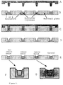
- Figure 1 illustrates the methodology to create a solid support where the acrylamide-enmeshed liposomes are part of a multiwell microtiter plate. Formation of a transparent polyacrylamide layer in the wells of a multiwell plate is carried out as follows : (A). a 10% acrylamide-methylene-bis-acrylamide solution (as) is placed in the wells (w) of a plastic multiwell plate (mwp).
- a plastic mold (pm) is made such that it can fit into the wells; (B) polymerization of the acrylamide (as) with the mold (pm) in place; (C) withdrawal of the mold resulting in formation of transparent polyacrylamide layers in the wells (diagonal lines, top left to bottom right), ready for the introduction of the acrylamide-enmeshed liposomes.
- Figure 2 illustrates formation of the acrylamide-enmeshed liposome wells and the appearance of the wells on reacting with immune and non-immune sera.
- the mold A is introduced to give an ensemble as shown in C.
- the black dots represent the calcein fluorescence that is very high, because the calcein is released to the external medium of the liposomes where it is diluted and loses its self-quenching.
- non-immune serum not containing antibodies to the hapten present at the liposome surface
- polyacrylamide-enmeshed liposomes containing N-trinitrophenyl-phosphatidyl-ethanolamine(TNP-PE) may be prepared by reacting trinitro-benzenesulfonic acid either with non-enmeshed (procedure 2.1) or with polyacrylamide-enmeshed liposomes (procedure 2.2).
- Example 3 Direct immunoassay of anti-trinitrobenzene antibodies in immune serum in the presence of complement
- the suspension was gently incubated at 37°C and, after the appropriate time (30 min), 100 ⁇ l of Hepes-K+ buffer were added, the suspension was sedimented and the calcein in the supernatant was measured by its fluorescence at 485 nm excitation, 515 nm emission.
- the total calcein present in the liposomes was determined by treating an equivalent fraction of the liposome gel with Triton X-100, 0.1% final, instead of the antibody. The results are shown in Table 1.
- the 9 fold enhancement obtained here with non-enmeshed liposomes is very high and due to the fact that the liposomes were always kept within a dialysis sack.
- the usual enhancement factor is about 5 to 6.
- the liposome lysis is proportional to the antibody concentration in the serum; dilution of the serum with lower antibody concentration leads to a fall in the lysis and less fluorescence is measured.
- the liposomes were prepared by reverse-phase evaporation according to Szoka Jr., F., and Papahadjopoulos, D., (1978) Proc.Nat.Acad.Sci. USA 75 : 4194-4198. In this procedure, aqueous buffer is introduced into a solution of lipids in ether, an emulsion is created by sonication and the ether is evaporated to form large unilamellar liposomes (LUVs). The external solutes are removed by dialysis and the liposomes are then enmeshed in polyacrylamide gels.
- LUVs large unilamellar liposomes
- Example 5 Preparation of hapten-attached polyacrylamide-enmeshed liposomes with occluded horseradish peroxidase
- polyacrylamide-enmeshed liposomes having as hapten attached to the outer liposomal surface moieties of 1-(succinimidyl)propionyl-3-dithio-2-pyridine(SPDP) were prepared as follows:
- Polyacrylamide-enmeshed liposomes containing occluded horseradish peroxidase of Example 4 were reacted with a 1.2 molar excess (with respect to the amino groups present at the liposome surface) of 1-(succinimidyl)propionyl-3-dithio-2-pyridine (SPDP, Pharmacia).
- SPDP 1-(succinimidyl)propionyl-3-dithio-2-pyridine
- the reaction was performed in Hepes-K+buffer, pH 8.5.
- the excess SPDP was removed by washing in Hepes-K+ buffer, pH 7.4.
- the antigen linked to a protein carrier, FITC-soybean trypsin inhibitor was also reacted with SPDP under the same conditions and immediately before use, was treated with 0.1 mM of dithiothreitol, and passed through a Sephadex G-25 column to remove the excess dithiothreitol.
- the FITC-soybean trypsin inhibitor antigen containing a free thiol group derived from the SDPD was mixed with the polyacrylamide-enmeshed liposomes and the reaction was followed by the liberation of 2-thiopyridone. When the reaction was complete (30 min), the liposomes entrapped in the acrylamide gel were washed with Hepes-K+ buffer, pH 7.4.
- the polyacrylamide-enmeshed liposomes (20 ⁇ l) containing the surface-bound FITC-soybean trypsin inhibitor of Example 5 were placed at the bottom of three conical tubes. To the first were added 20 ⁇ l of normal serum, to the second 20 ⁇ l of the test serum containing anti-FITC antibodies, and to the third 20 ⁇ l of the test serum containing anti-FITC antibodies and, in addition, an excess of the free FITC-soybean trypsin inhibitor.
- the tubes were left at room temperature for 15 min and then 50 ⁇ l of 50 mM Hepes,100 mM K2SO4, pH 6.0 containing 0.025 mM 2,2'-azino-bis(3-ethylbenzthiazoline-6-sulfonic acid) as a substrate for the peroxidase, and 0.25 mM hydrogen peroxide, were added and the components were incubated for 15 min at room temperature. The color developed was read at 412 nm. The results are given in Table 3.
- Example 7 Antibody immunoassay with a solid support of polyacrylamide-enmeshed liposomes in a multiwell microtiter plate
- Example 8 Immunoassay of an antigen of hapten with polyacrylamide-enmeshed liposomes with occluded fluorophore in the presence of a cytolytic agent
- CH-1 a melittin homolog of the formula with molecular weight of 2,000 daltons
- hapten a peptide epitope or other
- the [CH-1-hapten] conjugate is reacted with a monoclonal antibody such that the antibody and the [CH-1-hapten] conjugate form a 1:1 complex. Since the antibody has a molecular weight of 150,000 daltons, the complex will have a molecular weight of about 152,000. At this size, the complex is not able to penetrate through the acrylamide gel matrix.
- the free [CH-1-hapten] conjugate when released from the interaction with the monoclonal antibody, readily penetrates the gel matrix. It then associates to form a channel through the liposome membrane that releases the occluded fluorophore into the medium.
- the [monoclonal antibody-CH-1-hapten] complex is incubated with serum or plasma containing the antigen to be determined, the antigen being similar in structure to the hapten attached to the CH-1. Because of this similarity in structure, the hapten displaces the [CH-1-hapten] conjugate from the monoclonal antibody.
- Polyacrylamide-enmeshed liposomes containing a fluorophore are washed by sedimentation, to reduce any interference by the fluorophore and then added to the sample.
- the [CH-1-hapten] conjugate diffuses into the acrylamide gel matrix, lyses the liposomes and releases the fluorophore, that is measured.
- Phospholipase molecular weight 12,000 daltons
- This [phospholipase-hapten] conjugate is mixed with a monoclonal antibody to give a 1:1 complex. Since the antibody has a molecular weight of 150,000 daltons, the complex has a molecular weight of about 162,000, and is not able to penetrate into the polyacrylamide gel matrix. However, the free [phospholipase-hapten] conjugate readily penetrates the gel matrix, when released from the interaction with the monoclonal antibody, thus lysing the liposomes to release the fluorophore.
- the [monoclonal antibody-phospholipase-hapten] complex is incubated in the presence of albumin with serum or plasma containing the antigen to be determined, the antigen being similar in structure to the hapten attached to the phospholipase. Because of this similarity in structure, the hapten displaces the [phospholipase-hapten] conjugate from the monoclonal antibody. Polyacrylamide-enmeshed liposomes containing a fluorophore are washed (to remove free fluorophore) and are then added to the sample. The free [phospholipase-hapten] conjugate diffuses into the gel and lyses the liposomes releasing the fluorophore, that is measured.
Landscapes
- Life Sciences & Earth Sciences (AREA)
- Health & Medical Sciences (AREA)
- Engineering & Computer Science (AREA)
- Urology & Nephrology (AREA)
- Cell Biology (AREA)
- Biomedical Technology (AREA)
- Hematology (AREA)
- Immunology (AREA)
- Chemical & Material Sciences (AREA)
- Molecular Biology (AREA)
- Biotechnology (AREA)
- Microbiology (AREA)
- Food Science & Technology (AREA)
- Medicinal Chemistry (AREA)
- Physics & Mathematics (AREA)
- Analytical Chemistry (AREA)
- Biochemistry (AREA)
- General Health & Medical Sciences (AREA)
- General Physics & Mathematics (AREA)
- Pathology (AREA)
- Medicines Containing Antibodies Or Antigens For Use As Internal Diagnostic Agents (AREA)
- Medicinal Preparation (AREA)
- Peptides Or Proteins (AREA)
Applications Claiming Priority (2)
| Application Number | Priority Date | Filing Date | Title |
|---|---|---|---|
| IL98473 | 1991-06-12 | ||
| IL9847391A IL98473A (en) | 1991-06-12 | 1991-06-12 | Liposome immunoassays for detection of antigens antibodies and haptens |
Publications (3)
| Publication Number | Publication Date |
|---|---|
| EP0518319A2 true EP0518319A2 (de) | 1992-12-16 |
| EP0518319A3 EP0518319A3 (en) | 1993-01-13 |
| EP0518319B1 EP0518319B1 (de) | 1999-03-24 |
Family
ID=11062538
Family Applications (1)
| Application Number | Title | Priority Date | Filing Date |
|---|---|---|---|
| EP92109828A Expired - Lifetime EP0518319B1 (de) | 1991-06-12 | 1992-06-11 | Liposome-Immunoassay zum Nachweis von Antigenen, Antikörpern und Haptenen |
Country Status (4)
| Country | Link |
|---|---|
| EP (1) | EP0518319B1 (de) |
| JP (1) | JPH06160389A (de) |
| DE (1) | DE69228720D1 (de) |
| IL (1) | IL98473A (de) |
Cited By (2)
| Publication number | Priority date | Publication date | Assignee | Title |
|---|---|---|---|---|
| WO1998008097A1 (en) * | 1996-08-22 | 1998-02-26 | Biovation Limited | Signal amplification method |
| EP0683397A4 (de) * | 1993-02-03 | 2000-09-27 | Nissui Pharm Co Ltd | Reagenz zur immunoagglutinierung und immunoanalytisches verfahren |
Families Citing this family (2)
| Publication number | Priority date | Publication date | Assignee | Title |
|---|---|---|---|---|
| US5738868A (en) * | 1995-07-18 | 1998-04-14 | Lipogenics Ltd. | Liposome compositions and kits therefor |
| JP2001208754A (ja) * | 2000-01-26 | 2001-08-03 | Kazunori Kataoka | 生物学的な被検体を検出するための組成物 |
Family Cites Families (6)
| Publication number | Priority date | Publication date | Assignee | Title |
|---|---|---|---|---|
| US4176174A (en) * | 1974-07-19 | 1979-11-27 | Burroughs Wellcome Co. | Viral antibody diagnostic test system |
| FR2538907A1 (fr) * | 1982-09-15 | 1984-07-06 | Braylan Paul | Reactif immunologique constitue par des antigenes ou des anticorps en forme de cellules des particules ou des molecules ancres sur des surfaces solides continues par des unions ioniques ou covalents |
| JPS60231609A (ja) * | 1984-04-28 | 1985-11-18 | Terumo Corp | リポソ−ム製剤 |
| US4874710A (en) * | 1986-02-20 | 1989-10-17 | Becton Dickinson And Company | Assay and product in which binder and liposomes are supported on a solid support |
| JPS6319559A (ja) * | 1986-07-11 | 1988-01-27 | Fuji Photo Film Co Ltd | 免疫分析方法 |
| EP0301333A3 (de) * | 1987-07-29 | 1992-07-01 | Abbott Laboratories | Auf liposomen basierter, homogener Immunoassay für diagnostische Teste |
-
1991
- 1991-06-12 IL IL9847391A patent/IL98473A/en not_active IP Right Cessation
-
1992
- 1992-06-11 DE DE69228720T patent/DE69228720D1/de not_active Expired - Lifetime
- 1992-06-11 EP EP92109828A patent/EP0518319B1/de not_active Expired - Lifetime
- 1992-06-12 JP JP4195826A patent/JPH06160389A/ja active Pending
Cited By (3)
| Publication number | Priority date | Publication date | Assignee | Title |
|---|---|---|---|---|
| EP0683397A4 (de) * | 1993-02-03 | 2000-09-27 | Nissui Pharm Co Ltd | Reagenz zur immunoagglutinierung und immunoanalytisches verfahren |
| WO1998008097A1 (en) * | 1996-08-22 | 1998-02-26 | Biovation Limited | Signal amplification method |
| US6258528B1 (en) | 1996-08-22 | 2001-07-10 | Scancell Limited | Signal amplification method |
Also Published As
| Publication number | Publication date |
|---|---|
| EP0518319B1 (de) | 1999-03-24 |
| IL98473A0 (en) | 1992-07-15 |
| JPH06160389A (ja) | 1994-06-07 |
| DE69228720D1 (de) | 1999-04-29 |
| EP0518319A3 (en) | 1993-01-13 |
| IL98473A (en) | 1995-08-31 |
Similar Documents
| Publication | Publication Date | Title |
|---|---|---|
| CA1175740A (en) | Immunolabelled liposomes | |
| US4605630A (en) | Large-liposome agglutination reagent and method | |
| US4483921A (en) | Immunoassay with antigen or antibody labeled liposomes sequestering enzyme | |
| FI81914C (fi) | Lipidvesikel innehaollande detekterbart maerkningsaemne och dess anvaendning vid analys. | |
| US4704355A (en) | Assay utilizing ATP encapsulated within liposome particles | |
| CA1264662A (en) | Immunoliposome assay - methods and products | |
| US4814270A (en) | Production of loaded vesicles | |
| EP0091958A1 (de) | Liposom-konjugate sowie diagnoseverfahren die diese verwenden | |
| EP0215027A1 (de) | Immunologisches reagens und verfahrenmit liposomen | |
| EP0212989B1 (de) | Lipid-Vesikel die markierte Spezies enthalten und ihre analytische Benutzung | |
| US4745074A (en) | Blood-fluid composition for cell lysis system | |
| JPS61500182A (ja) | 免疫検定法および組成物 | |
| EP0518319B1 (de) | Liposome-Immunoassay zum Nachweis von Antigenen, Antikörpern und Haptenen | |
| Monroe | Novel liposome immunoassays for detecting antigens, antibodies, and haptens | |
| NAKAMURA et al. | A Liposome Immunoassay Based on a Chemiluminescense Reaction | |
| US4806466A (en) | Cell agglutination reagent comprising conjugates of antibody covalently bound to liposomes | |
| Rongen et al. | Biotinylated and streptavidinylated liposomes as labels in cytokine immunoassays | |
| WO1992007959A1 (en) | Homogeneous membrane lytic immunoassay method utilizing contact sensitive liposome formulations | |
| US4888288A (en) | Vesicles resistant to enzyme lysis and use thereof in an enzyme assay | |
| JPH02115A (ja) | 薬剤の担体としての並びに免疫分析および細胞/分子分別のミクロ浮選装置としての乳脂小球の使用法 | |
| EP0258388A1 (de) | Immunoliposomes testverfahren und erzeugnisse | |
| JP3654732B2 (ja) | 免疫測定法 | |
| Badley et al. | Phospholipids in diagnosis | |
| Kung et al. | Recent Developments in the use of Liposomes in in vitro Diagnostic Assays | |
| Schreier et al. | Prevention of nonspecific lysis in liposomal and erythrocyte immunoassay systems by small lipid vesicles and erythrocyte ghosts |
Legal Events
| Date | Code | Title | Description |
|---|---|---|---|
| PUAI | Public reference made under article 153(3) epc to a published international application that has entered the european phase |
Free format text: ORIGINAL CODE: 0009012 |
|
| PUAL | Search report despatched |
Free format text: ORIGINAL CODE: 0009013 |
|
| AK | Designated contracting states |
Kind code of ref document: A2 Designated state(s): CH DE ES FR GB IT LI NL |
|
| AK | Designated contracting states |
Kind code of ref document: A3 Designated state(s): CH DE ES FR GB IT LI NL |
|
| 17P | Request for examination filed |
Effective date: 19930510 |
|
| 17Q | First examination report despatched |
Effective date: 19950404 |
|
| GRAG | Despatch of communication of intention to grant |
Free format text: ORIGINAL CODE: EPIDOS AGRA |
|
| GRAG | Despatch of communication of intention to grant |
Free format text: ORIGINAL CODE: EPIDOS AGRA |
|
| GRAH | Despatch of communication of intention to grant a patent |
Free format text: ORIGINAL CODE: EPIDOS IGRA |
|
| GRAH | Despatch of communication of intention to grant a patent |
Free format text: ORIGINAL CODE: EPIDOS IGRA |
|
| GRAA | (expected) grant |
Free format text: ORIGINAL CODE: 0009210 |
|
| AK | Designated contracting states |
Kind code of ref document: B1 Designated state(s): CH DE ES FR GB IT LI NL |
|
| PG25 | Lapsed in a contracting state [announced via postgrant information from national office to epo] |
Ref country code: IT Free format text: LAPSE BECAUSE OF FAILURE TO SUBMIT A TRANSLATION OF THE DESCRIPTION OR TO PAY THE FEE WITHIN THE PRESCRIBED TIME-LIMIT;WARNING: LAPSES OF ITALIAN PATENTS WITH EFFECTIVE DATE BEFORE 2007 MAY HAVE OCCURRED AT ANY TIME BEFORE 2007. THE CORRECT EFFECTIVE DATE MAY BE DIFFERENT FROM THE ONE RECORDED. Effective date: 19990324 Ref country code: FR Free format text: LAPSE BECAUSE OF FAILURE TO SUBMIT A TRANSLATION OF THE DESCRIPTION OR TO PAY THE FEE WITHIN THE PRESCRIBED TIME-LIMIT Effective date: 19990324 Ref country code: CH Free format text: LAPSE BECAUSE OF FAILURE TO SUBMIT A TRANSLATION OF THE DESCRIPTION OR TO PAY THE FEE WITHIN THE PRESCRIBED TIME-LIMIT Effective date: 19990324 Ref country code: LI Free format text: LAPSE BECAUSE OF FAILURE TO SUBMIT A TRANSLATION OF THE DESCRIPTION OR TO PAY THE FEE WITHIN THE PRESCRIBED TIME-LIMIT Effective date: 19990324 Ref country code: NL Free format text: LAPSE BECAUSE OF FAILURE TO SUBMIT A TRANSLATION OF THE DESCRIPTION OR TO PAY THE FEE WITHIN THE PRESCRIBED TIME-LIMIT Effective date: 19990324 Ref country code: ES Free format text: THE PATENT HAS BEEN ANNULLED BY A DECISION OF A NATIONAL AUTHORITY Effective date: 19990324 |
|
| REG | Reference to a national code |
Ref country code: CH Ref legal event code: EP |
|
| REF | Corresponds to: |
Ref document number: 69228720 Country of ref document: DE Date of ref document: 19990429 |
|
| PG25 | Lapsed in a contracting state [announced via postgrant information from national office to epo] |
Ref country code: GB Free format text: LAPSE BECAUSE OF NON-PAYMENT OF DUE FEES Effective date: 19990624 |
|
| PG25 | Lapsed in a contracting state [announced via postgrant information from national office to epo] |
Ref country code: DE Free format text: LAPSE BECAUSE OF FAILURE TO SUBMIT A TRANSLATION OF THE DESCRIPTION OR TO PAY THE FEE WITHIN THE PRESCRIBED TIME-LIMIT Effective date: 19990625 |
|
| EN | Fr: translation not filed | ||
| NLV1 | Nl: lapsed or annulled due to failure to fulfill the requirements of art. 29p and 29m of the patents act | ||
| REG | Reference to a national code |
Ref country code: CH Ref legal event code: PL |
|
| PLBE | No opposition filed within time limit |
Free format text: ORIGINAL CODE: 0009261 |
|
| STAA | Information on the status of an ep patent application or granted ep patent |
Free format text: STATUS: NO OPPOSITION FILED WITHIN TIME LIMIT |
|
| GBPC | Gb: european patent ceased through non-payment of renewal fee |
Effective date: 19990624 |
|
| 26N | No opposition filed |