EP0538404B1 - Recepteurs de facteur de croissance fibroblastique - Google Patents
Recepteurs de facteur de croissance fibroblastique Download PDFInfo
- Publication number
- EP0538404B1 EP0538404B1 EP91915163A EP91915163A EP0538404B1 EP 0538404 B1 EP0538404 B1 EP 0538404B1 EP 91915163 A EP91915163 A EP 91915163A EP 91915163 A EP91915163 A EP 91915163A EP 0538404 B1 EP0538404 B1 EP 0538404B1
- Authority
- EP
- European Patent Office
- Prior art keywords
- bek
- flg
- binding
- cells
- protein
- Prior art date
- Legal status (The legal status is an assumption and is not a legal conclusion. Google has not performed a legal analysis and makes no representation as to the accuracy of the status listed.)
- Expired - Lifetime
Links
Images


Classifications
-
- C—CHEMISTRY; METALLURGY
- C07—ORGANIC CHEMISTRY
- C07K—PEPTIDES
- C07K14/00—Peptides having more than 20 amino acids; Gastrins; Somatostatins; Melanotropins; Derivatives thereof
- C07K14/435—Peptides having more than 20 amino acids; Gastrins; Somatostatins; Melanotropins; Derivatives thereof from animals; from humans
- C07K14/705—Receptors; Cell surface antigens; Cell surface determinants
- C07K14/71—Receptors; Cell surface antigens; Cell surface determinants for growth factors; for growth regulators
-
- A—HUMAN NECESSITIES
- A61—MEDICAL OR VETERINARY SCIENCE; HYGIENE
- A61P—SPECIFIC THERAPEUTIC ACTIVITY OF CHEMICAL COMPOUNDS OR MEDICINAL PREPARATIONS
- A61P31/00—Antiinfectives, i.e. antibiotics, antiseptics, chemotherapeutics
-
- A—HUMAN NECESSITIES
- A61—MEDICAL OR VETERINARY SCIENCE; HYGIENE
- A61P—SPECIFIC THERAPEUTIC ACTIVITY OF CHEMICAL COMPOUNDS OR MEDICINAL PREPARATIONS
- A61P31/00—Antiinfectives, i.e. antibiotics, antiseptics, chemotherapeutics
- A61P31/12—Antivirals
-
- A—HUMAN NECESSITIES
- A61—MEDICAL OR VETERINARY SCIENCE; HYGIENE
- A61P—SPECIFIC THERAPEUTIC ACTIVITY OF CHEMICAL COMPOUNDS OR MEDICINAL PREPARATIONS
- A61P31/00—Antiinfectives, i.e. antibiotics, antiseptics, chemotherapeutics
- A61P31/12—Antivirals
- A61P31/20—Antivirals for DNA viruses
- A61P31/22—Antivirals for DNA viruses for herpes viruses
-
- A—HUMAN NECESSITIES
- A61—MEDICAL OR VETERINARY SCIENCE; HYGIENE
- A61P—SPECIFIC THERAPEUTIC ACTIVITY OF CHEMICAL COMPOUNDS OR MEDICINAL PREPARATIONS
- A61P43/00—Drugs for specific purposes, not provided for in groups A61P1/00-A61P41/00
-
- A—HUMAN NECESSITIES
- A61—MEDICAL OR VETERINARY SCIENCE; HYGIENE
- A61P—SPECIFIC THERAPEUTIC ACTIVITY OF CHEMICAL COMPOUNDS OR MEDICINAL PREPARATIONS
- A61P9/00—Drugs for disorders of the cardiovascular system
- A61P9/10—Drugs for disorders of the cardiovascular system for treating ischaemic or atherosclerotic diseases, e.g. antianginal drugs, coronary vasodilators, drugs for myocardial infarction, retinopathy, cerebrovascula insufficiency, renal arteriosclerosis
-
- A—HUMAN NECESSITIES
- A61—MEDICAL OR VETERINARY SCIENCE; HYGIENE
- A61K—PREPARATIONS FOR MEDICAL, DENTAL OR TOILETRY PURPOSES
- A61K38/00—Medicinal preparations containing peptides
Definitions
- This invention relates to a unique class of fibroblast growth factor receptors, nucleic acids encoding same and expression of the growth factor receptors in recombinant systems. This invention also relates to the use of the expressed receptors or fragments thereof in screens for candidate drugs which act as receptor antagonists.
- the fibroblast growth factor (FGF) family consists of seven related heparin-binding proteins, which include acidic FGF (aFGF), basic FGF (bFGF), int-2, hst/kFGF, FGF-5, FGF-6 and KGF.
- the members of the FGF family share approximately 30-55% amino acid sequence identity, similar gene structure, and are capable of transforming cultured cells when overexpressed in transfected cells.
- the prototypic FGFs, aFGF and bFGF were the first to be purified, sequenced and cloned. They are mitogens, in vitro , for a variety of cells of mesenchymal and neuroectodermal origin.
- FGFs can induce the formation of mesoderm in developing Xenopus embryos and possess potent angiogenic activity (reviewed by Burgess and Maciag, Ann. Rev. Biochem. 58: 575-606 (1989)).
- the response of cells to FGFs is mediated by binding and activation of specific cell surface receptors possessing intrinsic tyrosine kinase activity.
- Receptors are proteins, often glycosylated, which serve as integral components of cellular membranes.
- receptors possess an extracellular domain located at the cell surface capable of specific interaction with substances known as ligands.
- the fibroblast growth factors are examples of ligands.
- a second region of the receptor located on the intracellular surface of the membrane i.e. the cytoplasmic domain
- the cytoplasmic domain comprises a catalytic region, that is a region possessing enzymatic activity.
- the catalytic domain is a protein tyrosine kinase.
- the substrate of this kinase can be the receptor itself (i.e. autophosphorylation) or other intracellular proteins such as phospholipase C- ⁇
- the kinase domain of certain receptors can be interupted by insertion of up to 100 mostly hydrophilic amino acid residues. This insert may act to modulate receptor interaction with certain cellular substrates and effector proteins.
- the cytoplasmic domain terminates with a COOH-terminal tail region. This sequence is the most divergent among all known RTKs.
- a receptor acts as a molecular transducer, translating an extracellular event (ligand binding) into an intracellular response (cytoplasmic enzymatic activity).
- the substance which is bound by the receptor is known as the ligand; a term which is definitionally meaningful only by reference to its counterpart receptor. Accordingly, the term “ligand” does not imply any particular molecule size, structural or compositional feature other than that the substance is capable of binding or otherwise interacting with the receptor in such a manner that the receptor conveys information about the presence of the ligand to an intracellular target molecule. Such a functional definition necessarily excludes substances which may bind to an extracellular domain but fail to affect receptor activation. Stated directly not all substances capable of binding receptors are ligands, but all ligands are capable of binding a receptor.
- receptors have been identified that have assayable biological activity dependent on ligand interaction. Generally, the activity is enzymatic and is localized in the cytoplasmic domain.
- One group of receptors relevant to this invention possess intrinsic protein tyrosine kinase (PTK) activity.
- PTK protein tyrosine kinase
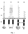
- FIG 1 presents a schematic representation of several known growth factor receptors that bear PTK activity.
- Growth factor receptors with PTK activity or receptor tyrosine kinases (RTKs)
- RTKs receptor tyrosine kinases
- RTKs can be envisioned as membrane-associated allosteric enzymes. Unlike water-soluble allosteric enzymes, RTK topology dictates that the ligand binding domain and PTK activity are separated by the plasma membrane. Therefore, receptor activation due to extracellular ligand binding must be translated across the membrane barrier into activation of intracellular domain functions.
- Structural features characteristic of the four subclasses include two cysteine-rich repeat sequences in the extracellular domain of monomeric subclass I receptors, disulfide-linked heterotetrameric ⁇ 2 ⁇ 2 structures with similar cysteine-rich sequences in subclass II RTKs, and five or three immunoglobulin-like repeats in the extracellular domains of subclass III and IV TRKs, respectively.
- the tyrosine kinase domain of the latter two subclasses is interrupted by hydrophilic insertion sequences of varying length.
- the availability of RTK cDNA clones has made it possible to initiate detailed structure/function analyses of the mechanisms of action of RTK family members. (Reviewed by Ullrich & Schlessinger, Cell 61: 203-221 (1990)).
- This invention is predicated on the discovery of a partial human cDNA clone of a f ms- l ike g ene ( flg ) which encoded a protein tyrosine kinase whose kinase domain was interrupted at a position similar to the kinase inserts of the CSF-1 and PDGF receptor tyrosine kinases (Ruta et al ., Oncogene , 3: 9-15 (1988)).
- cDNAs for chicken flg were isolated by cloning with flg -specific oligonucleotide probes (Lee et al ., Science , 245: 57-60 (1989)) and with antiphosphotyrosine antibodies (Pasquale and Singer, Proc. Nat'l. Acad. Sci. USA, 86: 5449-5453 (1989)).
- Flg is an FGF receptor based on four criteria: 1) the receptor purified from a chicken embryo extract by affinity chromatography on immobilized bFGF is the chicken flg gene product, 2) flg anti-peptide antiserum immunoprecipitates [ 125 I]aFGF crosslinked to proteins of 130 and 150 kDa in A204 rhabdomyosarcoma cells, 3) flg -anti-peptide antiserum immunoprecipitates proteins of similar size which are specifically phosphorylated on tyrosine upon treatment of living cells with aFGF or bFGF, and 4) proteins of 130 and 150 kDa immunoprecipitated with flg anti-peptide antiserum from aFGF-treated cell lysates undergo tyrosine phosphorylation in vitro .
- a putative second FGF receptor is the mouse b acterially e xpressed k inase ( bek ) gene product, a partial clone of which was obtained by screening a mouse liver cDNA expression library with anti- phosphotyrosine antibodies (Kornbluth et al ., Mol. Cell. Biol. , 8:5541-5544 (1988)).
- the deduced amino acid sequences of the partial bek clone and the corresponding region of flg are 85% identical.
- bek represents the mouse homologue of the human flg gene or another closely related gene.
- This invention provides full length cDNA clones for both human bek and flg , their complete deduced amino acid sequences, and demonstrates in transfected cells that both bek and flg bind aFGF, bFGF and k-FGF specifically and with high affinity.
- Receptor function has increasingly become the focus of attempts to rationally design drugs. Molecules that affect receptor-ligand binding, receptor activation and/or receptor intracellular target interaction are all potential candidates for therapeutics.
- Large scale screening is limited by conventional methods in which candidate drugs are tested on isolated cells or tissue explants. Tissue samples or isolated cells containing the target receptors are costly to obtain, present in limited quantity and difficult to maintain in a functionally viable state. Additionally, it is often difficult to reliably and reproducibly administer the candidate drug to tissue samples. Screening assays using primary explants in tissue culture are undertaken in larger scale than is possible with tissue samples. However, it is more difficult to assay physiological effect and the assays are subject to interference from many sources, e.g. culture media or cultivation conditions. Finally, assays using receptors isolated from natural materials have the disadvantage that the receptor is subject to natural variability and suitable natural sources may not always be available. It is an object herein to provide receptor molecules in a form amendable for large scale screening protocols.
- This invention provides a receptor protein or fragments thereof, in isolated form, comprising a cytoplasmic domain with protein kinase activity, a kinase insert, a transmembrane domain and an extracellular domain having three immunoglobulin-like repeats and biologically active equivalents thereof.
- Another aspect of this invention is an isolated DNA sequence encoding a receptor protein or fragments thereof, said protein having a cytoplasmic domain with protein kinase activity, a kinase insert, a single transmembrane domain and an extracellular domain having three immunoglobulin-like repeats and biologically equivalents thereof.
- Another aspect of this invention is a vector comprising a cDNA encoding a receptor protein, said protein having a cytoplasmic domain with protein kinase activity, a kinase insert, a single transmembrane domain and an extracellular domain having three immunoglobulin-like repeats.
- Another aspect of this invention is a host cell transformed with the above-described vector.
- Another aspect of this invention is a therapeutic composition
- a therapeutic composition comprising the extracellular domain or fragment thereof of a human receptor protein comprising a cytoplasmic domain having protein kinase activity, a kinase insert, a single transmembrane domain and an extracellular domain having three immunoglobulin-like repeats in an amount effective to inhibit undesirable heparin-binding growth factor mediated cellular responses and a pharmaceutical acceptable carrier.
- Another aspect of this invention is a therapeutuc composition
- a therapeutuc composition comprising the extracellular domain or fragment thereof of a human receptor protein comprising a cytoplasmic domain having protein kinase activity, a kinase insert, a single transmembrane domain and an extracellular domain having three immunoglobulin-like repeats in an amount effective to inhibit the binding of an opportunistic pathogen to human cells bearing said receptor protein and a pharmaceutical acceptable carrier.
- Another aspect of this invention is a method for the treatment of a patient with a disease characterized by an undesirable heparin-binding growth factor mediated cellular response comprising administering to such patient an effective amount of the composition of Claim 16.
- Another aspect of this invention is a method for the treatment of a patient suffering from an opportunistic pathogen infection said pathogen being capable of binding to a heparin-binding growth factor receptor comprising administering to such patient an effective amount of the composition of Claim 18.
- Another aspect of the invention is a method for the screening of drugs that inhibit fibroblast growth factor binding to cellular receptors comprising the steps of:
- a final aspect of the invention is a method for screening of drugs that inhibit the binding of opportunistic pathogens to fibroblast growth factor receptors comprising the steps of:
- bek and flg denotes fibroblast growth factor receptors or their fragments produced by cell or cell-free culture systems, in bioactive forms having the capacity to influence cellular growth, differentiation and response in vitro as does native bek and flg .
- bek and flg may exist in nature. These variations may be characterized by differences in the nucleotide sequence of the structural gene coding for proteins of identical biological function. It is possible to produce analogs having single or multiple amino acid substitutions, deletions, additions or replacements.
- a first embodiment involves the cloning and identification of full length cDNAs encoding human bek receptor protein.
- a second embodiment constitutes the expression of human bek gene products in recombinant host cells and a third embodiment constitutes the use of the recombinantly derived products for, inter alia , bek receptor analysis and drug screening.
- Genes coding for various polypeptides may be cloned by incorporating a DNA fragment coding for the polypeptide in a recombinant DNA vehicle, e.g., bacterial or viral vectors, and transforming a suitable host.
- a recombinant DNA vehicle e.g., bacterial or viral vectors
- This host is typically an Escherichia coli ( E . coli ) strain, however, depending upon the desired product, eukaryotic hosts may be utilized.
- Clones incorporating the recombinant vectors are isolated and may be grown and used to produce the desired polypeptide on a large scale.
- mRNA messenger RNA
- cDNA single-stranded complementary DNA
- Reverse transcriptase synthesizes DNA in the 5'-3' direction, utilizes deoxyribonucleoside 5'-triphosphates as precursors, and requires both a template and a primer strand, the latter of which must have a free 3-hydroxyl terminus.
- Reverse transcriptase products whether partial or complete copies of the mRNA template, often possess short, partially double-stranded hairpins ("loops") at their 3' termini. In the second reaction, these "hairpin loops" can be exploited as primers for DNA polymerases. Preformed DNA is required both as a template and as a primer in the action of DNA polymerase.
- the DNA polymerase requires the presence of a DNA strand having a free 3'-hydroxyl group, to which new nucleotides are added to extend the chain in the 5'-3' direction.
- the products of such sequential reverse transcriptase and DNA polymerase reactions still possess a loop at one end.
- the apex of the loop or "fold-point" of the double-stranded DNA, which has thus been created, is substantially a single-strand segment.
- this single-strand segment is cleaved with the single-strand specific nuclease S1 to generate a "blunt-end" duplex DNA segment.
- This general method is applicable to any mRNA mixture, and is described by Buell, et al ., J. Biol. Chem. , 253: 2483 (1978).
- ds-cDNA double-stranded cDNA mixture
- the cloning vehicle is used to transform a suitable host.
- These cloning vehicles usually impart an antibiotic resistance trait on the host.
- Such hosts are generally prokaryotic cells.
- only a few of the transformed or transfected hosts contain the desired cDNA.
- the sum of all transformed or transfected hosts constitutes a gene "library".
- the overall ds-cDNA library created by this method provides a representative sample of the coding information present in the mRNA mixture used as the starting material.
- an appropriate oligonucleotide sequence it can be used to identify clones of interest in the following manner. Individual transformed or transfected cells are grown as colonies on a nitrocellulose filter paper. These colonies are lysed; the DNA released is bound tightly to the filter paper by heating. The filter paper is then incubated with a labeled oligonucleotide probe which is complementary to the structural gene of interest. The probe hybridizes with the cDNA for which it is complementary, and is identified by autoradiography. The corresponding clones are characterized in order to identify one or a combination of clones which contain all of the structural information for the desired protein. The nucleic acid sequence coding for the protein of interest is isolated and reinserted into an expression vector.
- the expression vector brings the cloned gene under the regulatory control of specific prokaryotic or eukaryotic control elements which allow the efficient expression (transcription and translation) of the ds-cDNA.
- this general technique is only applicable to those proteins for which at least a portion of their amino acid or DNA sequence is known for which an oligonucleotide probe is available. See, generally, Maniatis, et al ., supra.
- PCR Polymerase Chain Reaction
- Flg and bek are similar yet distinct gene products (Fig. 2B), with structural features shared by the PDGF/CSF-1/c-kit family of receptor linked tyrosine kinases (Ullrich and Schlessinger, 1990 supra ).
- Their coding sequences consist of a hydrophobic signal peptide sequence of 21 amino acids, an extracellular domain of 356 ( bek ) and 355 ( flg ) amino acids, a transmembrane domain of 21 amino acids and an cytoplasmic domain of 423 ( bek ) and 425 ( flg ) amino acids.
- the extracellular domains of flg and bek contain 3 "immunoglobulin-like" (Ig) domains of similar size and location.
- N-linked glycosylation sites are located at identical positions in the extracellular domains of flg and bek .
- Flg contains an additional N-linked glycosylation site at amino acid 185.
- Lee, et al . (1989) have noted a region in the extracellular domain of chicken flg with 8 neighboring acidic amino acids. This "acidic box" is also present in human flg and human bek , and consists of 8 and 5 acidic residues, respectively.
- the cytoplasmic domains of bek and flg consist of long juxtamembrane regions followed by conserved catalytic kinase domains which are split by 14 amino acid insertions.
- the kinase domains are followed by divergent carboxy terminal tails.
- the overall identity between bek and flg is 71%, with the region of highest identity (88%) being the kinase domain.
- Flg and bek mRNA expression in various cell lines was determined by Northern blot analysis with bek and flg cDNA probes corresponding to a portion of their non-homologous 3' untranslated regions.
- A204 rhabdomyosarcoma cells two flg transcripts of approximately 4.3 and 4.2 kb are observed, but bek mRNA is undetectable (Fig. 3).
- HUVEC and human teratocarcinoma NTERA2 cells uniquely express the 4.2 and 4.3 kb flg transcripts, respectively.
- the significance of the distinct 4.3 and 4.2 kb flg transcripts is presently unknown. They may be related to flg clones which have been isolated that when compared to Fig.
- the flg and bek ds-cDNAs can be inserted into expression vectors by any one of many known techniques. In general, methods can be found in Maniatis, et al . (1982), supra, and Ausubel, et al . (1987), supra . In general, the vector is linearized by at least one restriction endonuclease, which will produce at least two blunt or cohesive ends. The ds-cDNA is ligated with or joined into the vector insertion site.
- prokaryotic cells or other cells which contain substantial cell wall material are employed, the most common method of transformation with the expression vector is calcium chloride pretreatment as described by Cohen, R.N., et al ., Proc. Nat'l. Acad. Sci. USA , 69: 2110 (1972). If cells without cell wall barriers are used as host cells, transfection is carried out by the calcium phosphate precipitation method described by Graham and Van der Eb, Virology , 52: 456 (1973). Other methods for introducing DNA into cells such as nuclear injection, viral infection, electroporation or protoplast fusion may be successfully used. The cells are then cultured on selective media, and proteins for which the expression vector encodes are produced.
- “Expression vectors” refer to vectors which are capable of transcribing and translating DNA sequences contained therein, where such sequences are linked to other regulatory sequences capable of affecting their expression. These expression vectors must be replicable in the host organisms or systems either as episomes, bacteriophage, or as an integral part of the chromosomal DNA.
- One form of expression vector is the bacteriophage, viruses which normally inhabit and replicate in bacteria. Particularly desirable phage for this purpose are the ⁇ gt 10 and ⁇ gt 11 phage described by Yound and Davis, supra.
- ⁇ gt 11 is a general recombinant DNA expression vector capable of producing polypeptides specified by the inserted DNA.
- foreign proteins or portions thereof are synthesized fused to the prokaryotic protein ⁇ -galactosidase.
- IPTG lactose
- the use of host cells defective in protein degradation pathways may also increase the lifetime of novel proteins produced from the induced ⁇ gt 11 clones. Proper expression of foreign DNA in ⁇ gt 11 clones will depend upon the proper orientation and reading frame of the inserted DNA with respect to the ⁇ -galactosidase promoter and translation initiating codon.
- Another form of expression vector useful in recombinant DNA techniques is the plasmid - a circular unintegrated (extra-chromosomal), double-stranded DNA.
- Any other form of expression vector which serves an equivalent function is suitable for use in the process of this invention.
- Recombinant vectors and methodology disclosed herein are suitable for use in host cells covering a wide range of prokaryotic and eukaryotic organisms. Prokaryotic cells are preferred for the cloning of DNA sequences and in the construction of vectors.
- E . coli K12 strain HB101 ATCC No. 339694
- other microbial strains may be used.
- Vectors containing replication and control sequences which are derived from species compatible with the host cell or system are used in connection with these hosts.
- the vector ordinarily carries an origin of replication, as well as characteristics capable of providing phenotypic selection in transformed cells.
- E . coli can be transformed using the vector pBR322, which contains genes for ampicillin and tetracycline resistance (Bolivar, et al ., Gene. 2:95 (1977)).
- the expression vector may also contain control elements which can be used for the expression of the gene of interest.
- Common prokaryotic control elements used for expression of foreign DNA sequences in E . coli include the promoters and regulatory sequences derived from the ⁇ -galactosidase and tryptophan (trp) operons of E . coli , as well as the pR and pL promoters of bacteriophage ⁇ . Combinations of these elements have also been used (e.g., TAC, which is a fusion of the trp promoter with the lactose operator).
- Other promoters have also been discovered and utilized, and details concerning their nucleotide sequences have been published enabling a skilled worker to combine and exploit them functionally.
- eukaryotic microbes such as yeast cultures
- Saccharomyces cerevisiae or common baker's yeast, is the most commonly used among eukaryotic microorganisms, although a number of other strains are commonly available.
- Yeast promoters suitable for the expression of foreign DNA sequences in yeast include the promoters for 3-phosphoglycerate kinase or other glycolytic enzymes.
- Suitable expression vectors may contain termination signals which provide for the polyadenylation and termination of the mRNA transcript of the cloned gene. Any vector containing a yeast-compatible promoter, origin of replication and appropriate termination sequence is suitable for expression of bek or flg .
- Cell lines derived from multicellular organisms may also be used as hosts.
- any such cell culture is workable, whether from a vertebrate or invertebrate source.
- interest has been greatest in vertebrate cells, and propagation of vertebrate cells in culture (tissue culture) has become a routine procedure in recent years.
- useful hosts are the VERO, HeLa, mouse C127 or 3T3, Chinese hamster ovary (CHO), WI38, BHK, COS-7, and MDCK cell lines.
- Mouse 3T3 and CHO cells are particularly preferred.
- Expression vectors for such cells ordinarily include an origin or replication, a promoter located in front of the gene to be expressed, RNA splice sites (if necessary), and transcriptional termination sequences.
- control functions on the expression vectors are often provided by viral material.
- promoters are derived from polyoma, Adenovirus 2, and most frequently, Simian Virus 40 (SV40).
- Eukaryotic promoters such as the promoter of the murine metallothionein gene (Paulakis and Hamer, Proc. Nat'l, Acad, Sci. , 80: 397-401 (1983)), may also be used.
- eukaryotic enhancer sequences can also be added to the construction. These sequences can be obtained from a variety of animal cells or oncogenic retroviruses such as the mouse sarcoma virus.
- An origin of replication may be provided either by construction of the vector to include an exogenous origin, such as that provided by SV40 or other viral sources, or may be provided by the host cell chromosomal replication mechanism. If the vector is integrated into the host cell chromosome, the latter is often sufficient.
- Host cells can yield bek or flg which can be of a variety of chemical compositions.
- the protein is produced having methionine as its first amino acid. This methionine is present by virtue of the ATG start codon naturally existing at the origin of the structural gene or by being engineered before a segment of the structural gene.
- the protein may also be intracellularly or extracellularly cleaved, giving rise to the amino acid which is found naturally at the amino terminus of the protein.
- the protein may be produced together with either its own or a heterologous signal peptide, the signal polypeptide being specifically cleavable in an intra- or extracellular environment.
- bek or flg may be produced by direct expression in mature form without the necessity of cleaving away any extraneous polypeptide.
- Recombinant host cells refer to cells which have been transformed with vectors constructed using recombinant DNA techniques. As defined herein, bek or flg is produced as a consequence of this transformation. bek or flg or their fragments produced by such cells are referred to as "recombinant bek or flg ".
- over-expressing bek and flg cell lines are useful for investigation of ligand/reception interaction as well as in a screening program for rationally designed drugs.
- Potential sites of action for therapeutics include but are not limited to receptor/ligand binding, signal transduction, receptor/target interaction.
- drugs may be tested for their ability to inhibit natural ligand binding by competitive inhibition or other mechanisms.
- the bek and flg over-producing cells may be used as an immunogen source to produce antibodies, such as monoclonal antibodies, which also may be tested for their ability to affect ligand binding or receptor aggregation.
- the system may also be used to evaluate drugs that affect the kinase/target interactions.
- These drugs known as tyrphostins have as their site of action the phosphorylation of cellular targets by the receptor kinase domain, including the autophosphorylation of the receptor itself.
- this invention contemplates the expression of biologically active fragments of flg and bek .
- the products of the cloning and expression of cDNA encoding the first 377 amino acids of bek or first 376 amino acids of flg (i.e. the extracellular plus transmembrand domains) or COOH terminal 423 amino acids of bek or 425 amino acids of flg may be usefully employed the evaluate ligand and tryphostins respectively.
- Example III illustrates several of these applications.
- the extracellular domain alone or sub-domains thereof may be useful as inhibitors of infection by opportunistic pathogens such as Herpes Simplex Virus type I or other pathogens which use endogenous bek or flg as a means of infecting its target cell.
- pathogens such as Herpes Simplex Virus type I or other pathogens which use endogenous bek or flg as a means of infecting its target cell.
- the over-expressing cell lines may be used to screen inhibitor drugs by measuring reduced virus binding (direct assay) or by reduced infectivity or reduction of virus from media in the presence or absence of candidate drugs such as receptor fragments themselves.
- This invention provides the complete human cDNA sequences for two fibroblast growth factor receptors.
- a partial cDNA sequence of a human protein kinase terminal flg was known (Ruta, et al ., supra , (1988)) as was the partial sequence of the mouse protein kinase bek (Kornbluth, et al ., supra , (1988))
- the complete sequences of the human forms of these genes were not available. Without the complete sequences the extent of the functional ligand binding site and the degree of homology between flg and bek could not be properly evaluated.
- flg and bek are similar but distinct gene products which have high affinity for aFGF, bFGF and k-FGF.
- the bek and flg genes are located on different chromosomes and are differentially expressed in various cell lines and tissues.
- Flg and bek exhibit structural features shared by the PDGF and CSF-1 receptor tyrosine kinases including a split tyrosine kinase domain and an extracellular region consisting of multiple immunoglobulin-like domains.
- the FGF receptors are structurally distinct from the PDGF and CSF-1 receptors in several respects: 1) the extracellular domains of the FGF receptors consist of 3 immunoglobulin (Ig) domains, whereas those of the PDGF and CSF-1 receptors consist of 5 Ig domains.
- the FGF receptors resemble the IL-1 receptor, whose extracellular region consists of three Ig domains (Sims, et al ., Science , 241: 585-589 (1988)); 2) the juxtamembrane region of the FGF receptors, 87 ( flg ) and 89 ( bek ) amino acids, is significantly longer than that of the PDGF and CSF-1 receptors (49- and 51- amino acids respectively); 3) the kinase insert domain of the FGF receptors consists of only 14 amino acids whereas the PDGF and CSF-1 receptor kinase inserts are much larger (104- and 70- amino acids respectively) (Hanks, et al ., Science , 241: 42-52 (1988)).
- a consensus tyrosine residue and potential autophosphorylation site in the kinase domain of the PDGF receptor is conserved in both FGF receptors ( flg residue 654, bek residue 657).
- the presence of common motifs among the receptors for a variety of biologically distinct ligands supports the argument that, for reasons of structure and/or function, then have been subject to strong evolutionary constraints (Ullrich and Schlessinger, Cell 60: 203-221 (1990)).
- NIH 3T3 cells transfected with mammalian expression vectors containing the coding sequences of either flg or bek direct the synthesis of glycoproteins with apparent molecular weights of 150,000 and 135,000 respectively.
- the molecular weight of the major flg protein is 110 kDa while that of bek is 90 kDa.
- Two flg proteins of 90 and 110 kDa had been previously observed in immunoprecipitates of tunicamycin-treated, metabolically labeled human rhabdomyosarcoma cells (Ruta, et al ., Proc. Nat'l. Acad. Sci. USA, 86: 8722-8726 (1989)).
- cDNA clones can be recovered which appear to encode truncated forms of flg and bek from HUVEC and human brain cDNA libraries.
- a variant of flg lacking the first Ig-like domain, but otherwise intact was isolated.
- Other cDNA variants include a truncated version of bek encoding only a single sequence, a single "Ig-like" domain and a stop codon and a variant of flg encoding two "Ig-like" domains. These two clones may represent secreted forms of bek or flg .
- the A204 rhabdomyosarcoma cells express two different flg mRNAs which might represent the alternatively spliced forms, whereas the NTERA-2 and endothelial cells appear to differentially express the alternative flg mRNAs.
- a deposit of biologically pure culture of the following strain was made with the American Type Culture Collection, 12301 Parklawn Drive, Rockville, Maryland, the accession number indicated was assigned after successful viability testing, and the requisite fees were paid. Access to said culture will be available during pendency of the patent application to one determined by the Commissioner to be entitled thereto under 37 C.F.R. ⁇ 1.14 and 35 U.S.C. ⁇ 122. All restriction on availability of said culture to the public will be irrevocably removed upon the granting of a patent based upon the application and said culture will remain permanently available for a term of at least five years after the most recent request for the furnishing of a sample and in any case for a period of at least 30 years after the date of the deposit.
- vectors of the present invention have been deposited in the form of transformed bacterial hosts. It is a matter of routine skill however for a recipient to culture the bacterial cells, recovering the vectors and use same to transform other host cells such as mouse 3T3 cells as described herein.
- This example illustrates the cloning of full length flg and bek cDNAs.
- a partial clone was isolated by screening a cDNA library derived from human endothelial mRNA with a probe based on a retroviral transforming gene product. (See Ruta, et al ., 1988 supra for details). Specifically the V-fms oncogene (the activated form of the CSF-1 tyrosine kinase receptor) was employed as a probe to screen 10 6 plaques under low stringency. Fifteen positive ⁇ gt 11 clones were detected, and five were plaque purified and analyzed further.
- PCR Polymerase Chain Reaction
- oligonucleotide primers were synthesized on an Applied Biosystems 380A DNA Synthesizer using well-known, standard cyanoethyl phosphoramidate chemistry.
- the oligonucleotides have the following sequences:
- Flg cDNA 5' of the internal Eco RI site (5' end of pC51, Ruta, et al ., 1988 supra) was obtained by anchored PCR (Loh, et al ., 1989 supra) using primer Fig-1 to specifically prime first strand cDNA synthesis from 50 ⁇ g human endothelial cell total RNA.
- primer Fig-1 to specifically prime first strand cDNA synthesis from 50 ⁇ g human endothelial cell total RNA.
- PCR amplification (30 cycles) with primers Fig-2, AN, and 10 pmol ANpolyC was performed with cycling times of 94°C for 1.5 minutes, 50°C for 2 minutes and 72°C for 4 minutes.
- a single PCR product was obtained which extended the C51 sequence 170 bp in the 5' direction.
- the new sequence was used to design the two oligonucleotides which were used with ⁇ gt 11 arm-specific oligonucleotides to amplify, by PCR, 5' flg clones contained in the endothelial cell library.
- the second PCR reaction was performed with 2 x 10 6 phage and the oligonucleotides Flg-PE-165non and GT11-R1.
- the third reaction used 5 ⁇ l of the preceding reaction as template for the oligonucleotide primers Flg-PE-141non and GT11-R3.
- the 560 bp product from the third reaction was made blunt ended by incubation with T4 DNA polymerase and cloned into the Sma I site of both pGem-1 (Stratagene) and M13mp19 and sequenced.
- the 5' flg PCR product in pGem-1 was excised, radiolabelled by random hexamer priming (Feinberg & Vogelstein, Anal. Biochem. , 137: 266 (1984)), and used to rescreen the HUVEC cDNA library in order to obtain a cDNA clone free of potential PCR generated artifacts. Only one clone in 2 x 10 6 screened recombinant phage was detected.
- Nucleotide sequence analysis of the cDNA insert of this clone revealed that its sequence was identical to the PCR-generated product and that it terminated at the Eco RI site shared by the partial flg clone pC51.
- This cDNA was digested with Eco RI and Sac I which cuts 50 bp 5' of the translation initiation ATG, and the 650 bp Eco RI/ Sac I fragment was cloned into similarly cleaved pGem-1.
- a full-length flg cDNA was constructed by cloning the 3' Eco RI flg insert from pC51 into the Eco RI site of this 5' flg clone.
- pMJ30 is derived by substitution of a linker containing an Eco RV cloning site for the aFGF insert in p267 (Jaye, et al ., EMBO. J. , 7:963-969 (1988)).
- a one-day-old human brain stem cDNA library in ⁇ gt11 was screened with an antisense 33-base oligonucleotide (3'mbek) complimentary to the 3' end of the partial murine bek coding sequence (Kombluth, et al ., 1988 supra ).
- 3'mbek 33-base oligonucleotide
- Thirty-two positive cDNA clones were isolated from a screen of 1.5 x 10 6 recombinant plaques and the longest clone, ⁇ bek5, was chosen for further analysis.
- ⁇ bek5 was digested with Eco RI and the 5' 2.2 kb and the 3' 1.0 kb Eco RI fragments were subcloned separately into M13mp19 and pGem-1 vectors for further manipulations.
- cDNA clones for the 5' end of human bek were isolated by screening the brainstem library a second time with a 5' proximal 750 bp fragment from ⁇ bek5. The fragment was radiolabelled by nick-translation and used to screen 1.5 x 10 6 plaques. Fifty-four hybridizing plaques were detected and one, ⁇ bek78, overlapped the original cDNA clone and extended 218 bp further in the 5' direction.
- A204 (human rhabdomyosarcoma), U563 (human glioblastoma) and NTERA-2 cl.D1 (human teratocarcinoma) cells were grown in DMEM with 10% fetal calf serum.
- Human umbilical vein endothelial cells (HUVEC) were grown in Medium 199 containing 10% fetal calf serum, 10 U/ml heparin (Upjohn) and 5 ng/ml recombinant aFGF.
- Total RNA was isolated from NTERA2cl.D1 cells with guanidine HCI (Wang, et al ., Dev. Biol.
- RNA was quantitated by its optical absorbance at 260 nm. Samples containing 4 ⁇ g total RNA were fractionated on a 1.25% agarose gel containing formaldehyde, transferred to nitrocellulose and then probed essentially as described (Seed, In: Genetic Engineering , Setlow et al ., eds., Vol 4 pp 91-102, Plenum Press New York, 1982)) using probes generated by random hexamer priming (Feinberg and Vogelstein, Anal.
- the flg probe was a 550 bp Apa I/ Eco RI fragment entirely contained within the 3' non-coding region.
- the bek probe was a 850 bp Tth 111I/ Eco RI fragment entirely contained within the 3' non-coding region.
- This example illustrates the expression of full length flg and bek in a mammalian expression vector.
- pMJ30 As mentioned in the previous Example, the full length flg clone was excised with Sma I/ Apa I, made blunt ended with T4 DNA polymerase, and cloned into an Eco RV site of expression plasmid pMJ30.
- pMJ30 having been derived by linker substitution for the aFGF insert in p267 (Jaye, et al ., EMBO J. 7: 963-969 (1988)). Accordingly, pMJ30 containing the full length flg sequence is denoted as pflgFL24.
- the human bek cDNA was prepared for mammalian expression by amplifying 276 bp fragment from ⁇ Bek78 with the oligonucleotide primers Bek4A and Bek1 B. After digestion with Hin dIII and Bc lI, the 5' 222 bp fragment was joined to the 2.2 kb subclone of ⁇ bek5 at a unique Bcl I site in the region of overlap. The PCR amplified fragment added restriction sites and a favorable translational initiation sequence immediately upstream of the putative initiator ATG codon. The 3' 1.0 kb Eco RI bek cDNA fragment was inserted subsequently.
- pMJ30 For introduction into NIH 3T3 cells, a 2.5 kb fragment containing the entire human bek coding region was subcioned into pMJ30.
- the nucleotide sequence of all clones was determined on both strands by chain termination (Sanger, et al ., Ann. N.Y. Acad. Sci. , 51:660-672 (1977)). Accordingly, pMJ30 containing full length bek sequence is denoted as pGC37.
- NIH 3T3 cells were grown in Dulbecco's Modified Eagle's Medium (DMEM) with 10% calf serum.
- DMEM Dulbecco's Modified Eagle's Medium
- NIH 3T3 cells were transfected with calcium phosphate co-precipitates of either 1 ⁇ g pSV2neo, or 1 ⁇ g pSV2neo with 20 ⁇ g of expression vectors containing the full coding sequence of either flg cDNA or bek cDNA.
- pSV2neo merely provides a selectable marker and any other equivalent co-transfectant system may be employed.
- Individual clones were selected in 500 ⁇ g/ml Geneticin (Gibco) and maintained in media containing 200 ⁇ g/ml Geneticin.
- NIH 3T3 cells were cotransfected with a 1:20 mixture of pSV2neo (Southern and Berg, J. Mol. Appl. Gen. , 1: 327-341 (1982)) and either a bek or flg expression vector, and transfectants were selected by growth in the presence of 500 ⁇ g/ml Geneticin (Gibco). Approximately 50 clones of each were screened for overexpression of FGF receptors by crosslinking to [ 125 I]aFGF.
- NIH 3T3 cells transfected with pSV2neo alone served as controls.
- Clones of both bek - and flg - transfected cells displaying increased binding for aFGF were identified in this way, indicating that bek and flg were both aFGF receptors.
- One bek transfected clone, Nbek8, and one flg transfected clone, Nflg26, were chosen for further analysis based on their increased expression of aFGF receptors.
- the translation products of the flg and bek expression vectors were analyzed by metabolically labeling Nbek8 and Nflg26 cells with [ 35 S]methionine, preparing cell extracts and immunoprecipitating with bek -and flg -specific anti-peptide antisera.
- Peptides corresponding to the COOH terminal 15 amino acids of flg (Fig-1) or the COOH-terminal 17 amino acids of bek (Bek-1) were synthesized and coupled to keyhole limpet hemocyanin using the crosslinking reagent 1-ethyl-3-(3-dimethylaminopropyl) carbodiimide. Rabbits were then immunized with these reagents emulsified in complete Freund's adjuvant to generate the polyclonal antisera Anti Flg-1 and Anti Bek-1.
- the cells were then washed three times with PBS (Gibco) and scraped in 0.5 ml of lysis buffer (20 mM Hepes pH 7.5, 150 mM NaCI, 10% glycerol, 1% Triton X-100, 1.5 mM MgCl 2 , 1 mM EDTA, 1 ⁇ g/ml aprotinin, 1 ⁇ g/ml leupeptin, 1 mM phenylmethylsulfonyl fluoride) and incubated for 15 minutes on ice. The lysates were centrifuged for 15 minutes in an Eppendorf centrifuge at 4°C.
- lysis buffer (20 mM Hepes pH 7.5, 150 mM NaCI, 10% glycerol, 1% Triton X-100, 1.5 mM MgCl 2 , 1 mM EDTA, 1 ⁇ g/ml aprotinin, 1 ⁇ g/ml leupeptin, 1
- the protein A-Sepnarose/antibody complexes were then incubated with the respective clarified cell lysates for 60 minutes in HNTG buffer at 4°C, washed twice with 50 mM Hepes, pH 8.0, 500 mM NaCI, 0.2% Triton X-100 and 5 mM EGTA, twice with 50 mM Hepes, pH 8.0, 150 mM NaCI, 0.1% Triton X-100, 5 mM EGTA and 0.1% SDS and finally twice with 10 mM Tris-HCI pH 8 and 0.1% Triton X-100.
- Laemmli sample buffer (Laemmli, Nature 227: 680-685 (1970)) was added to the washed immunoprecipitates which were then boiled for 4 minutes and separated on a SDS-(7%)polyacrylamide gel. Autoradiograms of the dried gels were made on Kodak X-Omat film (Eastman Kodak Co., Rochester, NY).
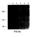
- the immunoprecipitates were subjected to SDS-PAGE followed by autoradiography ( Figure 4).
- a band of 150 kDa was specifically immunoprecipitated from flg -transfected cells with flg -antipeptide antiserum (lane 3) while bek -antipeptide antiserum specifically immunoprecipitated a band of 135 kDa (lane 7) from bek -transfected cells.
- Immunoprecipitation in the presence of the corresponding antigenic peptide completely eliminates the precipitation of flg and bek specific proteins.
- the basal level of endogenous fibroblast growth-factor receptors will vary from cell type to cell type.
- 3T3 cells contain about 5-10,000 such receptors but some 3T3 sub-clones such as 3T3 2.2 cells may contain up to 30,000 receptors per cell.
- the term "over-expressing" cells as used herein refers to cells having about 50,000 receptors or more. Initial transfection of 3T3 cells with the plasmid disclosed above yield cells having about 50,000 receptor/cell. However, over time in culture clones may be selected which expression 300,000 or more receptors/cells. If DHFR-deficient CHO cells are transfected and amplified by methotrexate selection they would be expected to produce up to a million or more receptors per cell.
- Bovine derived aFGF purified as previously described (Burgess, et al ., J. Biol. Chem. , 260: 11389-11392 (1985)), human recombinant bFGF (a kind gift of Dr. Moscatelli) and human recombinant kFGF (a kind gift of Dr. Claudio Basilico) were radioiodinated using the protocol for Enzymobeads (BioRad). The specific activity of the radiolabelled ligand was determined by radioisotope dilution using the respective unlabelled competitor. The concentrations of the unlabelled ligands were determined by amino acid analysis (Jaye, et al ., 1987 supra ).
- the specific activity of the different preparations of [ 125 I]aFGF varied between 130,000 - 760,000 cpm/ng and the specific activity for [ 125 I]bFGF between 50,000 - 500,000 cpm/ng.
- the specific activity of [ 125 I]kFGF varied from 50,000 - 200,000 cpm/ng.
- the binding assay was performed as follows:
- Fibronectin-coated 24 well dishes containing 1 x 10 5 cells/welt were placed on ice, and the wells were rinsed twice with cold binding buffer (DMEM, 1 mg/ml BSA, 5 U/ml heparin, 50 mM Hepes, pH 7.4). Dishes were incubated on ice with 1 ml of ice cold binding buffer for 20 minutes, followed by 2 hours at 4°C with serial dilutions of the [ 125 I]FGF in cold binding buffer without heparin. Nonspecific binding was obtained using the same serial dilutions but in the presence of 100 fold molar excess of non-radioactive aFGF.
- cold binding buffer DMEM, 1 mg/ml BSA, 5 U/ml heparin, 50 mM Hepes, pH 7.4
- Dishes were incubated on ice with 1 ml of ice cold binding buffer for 20 minutes, followed by 2 hours at 4°C with serial dilutions of the [ 125 I]
- the cells were placed on ice and rinsed twice with ice cold binding buffer minus heparin and then solubilized in 0.3N NaOH 37°C, 15 minutes.
- [ 125 I]bFGF was used the cells were placed on ice after incubation, washed three times with PBS, twice with 1 ml 2M NaCI in 20 mM Hepes, pH 7.5 and twice with 1 ml 2M NaCI in 20 mM sodium acetate, pH 4.0.
- the radioactivity released by the acid 2M NaCI wash represents specific binding to high affinity sites (Moscatelli. J. Cell. Physiol. , 131:123-130 (1987)).
- NFIg26, NBek8 and NNeo4 cells were grown in 60 mm tissue culture dishes coated with human fibronectin. At confluency, cells were washed twice with binding buffer (DMEM containing 0.2% BSA and 20 mM Hepes, pH 7.5) and incubated for 90 minutes at 4°C with binding buffer containing 25 ng/ml of [ 125 I]aFGF, [ 125 ]bFGF or [ 125 I]kFGF. After washing once with binding buffer and once with PBS, cells were further incubated for 20 minutes at 4°C with PBS containing 0.3 mM disuccinimidyl suberate (Pierce) as crosslinker (prepared as 30 mM stock solution in DMSO).
- binding buffer DMEM containing 0.2% BSA and 20 mM Hepes, pH 7.5
- binding buffer containing 25 ng/ml of [ 125 I]aFGF, [ 125 ]bFGF or [ 125 I]kFGF After washing
- Cell pellets were lysed in 100 ⁇ l of lysis buffer (20 mM Hepes, pH 7.5, 150 mM NaCl, 1% Triton X-100, 10% glycerol, 1.5 mM MgCl 2 , 1 mM EDTA, 1 ⁇ g/ml aprotinin, 1 ⁇ g/ml leupeptin and 1 mM PMSF), incubated for 15 minutes on ice and centrifuged for 15 minutes at 4°C in an Eppendorf centrifuge.
- lysis buffer 20 mM Hepes, pH 7.5, 150 mM NaCl, 1% Triton X-100, 10% glycerol, 1.5 mM MgCl 2 , 1 mM EDTA, 1 ⁇ g/ml aprotinin, 1 ⁇ g/ml leupeptin and 1 mM PMSF
- a single band of 165 kDa was crosslinked to both aFGF and bFGF in flg overexpressing cell lines (lanes 2 and 5).
- a single band of 150 kDa was crosslinked to both aFGF and bFGF in bek overexpressing cell lines (lanes 3 and 6).
- Acidic and basic FGF were radioiodinated and purified on heparin-Sepharose as described above.
- the dissociation constants of bek and flg for aFGF and bFGF were established by saturation binding analysis of the overexpressing cell lines with the iodinated ligands. Binding of both aFGF and bFGF to bek and flg was specific and saturable (Fig. 6A, inserts). In addition, binding of [ 125 I]aFGF and [ 125 I]bFGF at saturation was completely eliminated in the presence of a 100 fold molar excess of either aFGF or bFGF, indicating that each ligand effectively competed with the other for binding to the same sites.
- Fig. 6A indicates that the flg overexpressing cells bear approximately 55,000 receptors per cell with affinities of 24 pM and 47 pM for aFGF and bFGF respectively.
- the bek -overexpressing cells bear approximately 64,000 receptors per cell with affinities of 47 pM and 82 pM for aFGF and bFGF respectively.
- kFGF binds to flg and bek with affinities of 320 and 80 pM, respectively ( Figure 6B).
- kFGF binds to bek with similar affinity as does bFGF but binds to flg with 4-foldless avidity than does bFGF.
- the data from additional experiments yield apparent Kds of aFGF for flg in the range of 20-80 pM and for bek in the range of 40-100 pM.
- a similar variation in the Kds of bFGF binding to flg (50-150 pM) and bek (80-150 pM) is observed but within any single experiment bFGF always shows approximately 2-fold lower affinity for either flg or bek when compared to aFGF.
- the biologically active transformed cells may be used to screen compounds for their effectiveness in inhibiting the binding of FGFs to their cognant receptors. Having established the parameters of ligand/receptor interaction in the absence of potential inhibitors, it is merely a routine task to preincubate the cells with a test sample containing a compound to be evaluated, added labelled FGF and measure the reduction of FGF binding, if any.
- FGF i.e. an intracellular event
- an intracellular event is protein phosphorylation either of the receptor itself or another intracellular target such as phospholipase C- ⁇ .
- the appearance of phosphorylated protein can be measured by reactivity of such proteins with an anti-phosphotyrosine antibody.
- This invention relates to useful cell lines for drug evaluation, not to any particular assay employed. As mentioned previously, it is not necessary to express the entire receptor molecule in order to provide a useful screening system. For example, it has been discovered that an extracellular domain in which the first Ig-domain and the "acidic box" have been deleted is still capable of binding FGF-ligand. As mentioned above, host cells expressing on the cytoplasmic domain may be used as a convenient source of protein kinase for the testing of candidated tryphostins. E . coli transformed with a plasmid containing only the cytoplasmic domain has been shown to be useful for this purpose.
- Potential therapeutics include but are not limited to small organic molecules, receptor fragments themselves or antibodies that inhibit the binding of ligand to extracellular domain or effect protein kinase activity.
- Compounds with inhibitory activity are potentially useful as therapeutics for the treatment of disease states characterized by undesirable FGF-mediated cellular responses.
- diseases states that have been shown to be characterized by such responses include cancer, specifically certain forms of breast cancer, psoriasis, arthritis, atherosclerosis and benign prostatic hypertrophy.
- the therapeutic agents of this invention may be administered alone or in combination with pharmaceutically acceptable carriers, the proportion of which is determined by the solubility and chemical nature of the compound, chosen route of administration and standard pharmaceutical practice.
- they may be administered orally in the form of tablets or capsules containing such excepients as starch, milk sugar, certain types of clay and so forth.
- They may be administered sublingually in the form of troches or lozenges in which the active ingredient is mixed with sugar and corn syrups, flavoring agents and dyes; and then dehydrated sufficiently to make it suitable for pressing into a solid form.
- They may be administered orally in the form of solutions which may contain coloring and flavoring agents or they may be injected parenterally, that is, intramuscularly, intravenously or subcutaneously.
- parenteral administration may be used in the form of a sterile solution containing other solutes, for example, enough saline or glucose to make the solution isotonic.
- the physician will determine the dosage of the present therapeutic agents which will be most suitable and it will vary with the form of administration and the particular compound chosen, and furthermore, it will vary with the particular patient under treatment. He will generally wish to initiate treatment with small dosages substantially less then the optimum dose of the compound and increase the dosage by small increments until the optimum effect under the circumstances is reached. It will generally be found that when the composition is administered orally, larger quantities of the active agent will be required to produce the same effect as a smaller quantity given parenterally.
Landscapes
- Health & Medical Sciences (AREA)
- Life Sciences & Earth Sciences (AREA)
- Chemical & Material Sciences (AREA)
- Organic Chemistry (AREA)
- Medicinal Chemistry (AREA)
- General Health & Medical Sciences (AREA)
- Animal Behavior & Ethology (AREA)
- General Chemical & Material Sciences (AREA)
- Chemical Kinetics & Catalysis (AREA)
- Nuclear Medicine, Radiotherapy & Molecular Imaging (AREA)
- Pharmacology & Pharmacy (AREA)
- Public Health (AREA)
- Veterinary Medicine (AREA)
- Virology (AREA)
- Engineering & Computer Science (AREA)
- Oncology (AREA)
- Communicable Diseases (AREA)
- Bioinformatics & Cheminformatics (AREA)
- Molecular Biology (AREA)
- Immunology (AREA)
- Cell Biology (AREA)
- Toxicology (AREA)
- Zoology (AREA)
- Gastroenterology & Hepatology (AREA)
- Biochemistry (AREA)
- Biophysics (AREA)
- Genetics & Genomics (AREA)
- Proteomics, Peptides & Aminoacids (AREA)
- Vascular Medicine (AREA)
- Biotechnology (AREA)
- Cardiology (AREA)
- Heart & Thoracic Surgery (AREA)
- Urology & Nephrology (AREA)
- Peptides Or Proteins (AREA)
- Micro-Organisms Or Cultivation Processes Thereof (AREA)
- Medicines That Contain Protein Lipid Enzymes And Other Medicines (AREA)
- Compounds Of Unknown Constitution (AREA)
- Measuring Or Testing Involving Enzymes Or Micro-Organisms (AREA)
- Medicines Containing Material From Animals Or Micro-Organisms (AREA)
- Preparation Of Compounds By Using Micro-Organisms (AREA)
Claims (32)
- Protéine réceptrice bek humaine isolée, comprenant un domaine cytoplasmique présentant une activité de la protéine kinase, une insertion de kinase, un domaine transmembranaire et un domaine extracellulaire ayant trois répétitions de type immunoglobuline et ayant la séquence d'acides aminés comme le montre la Figure 2A.
- Fragment protéique du récepteur selon la revendication 1, n'ayant qu'un domaine extracellulaire et un domaine transmembranaire.
- Fragment protéique du récepteur selon la revendication 1, n'ayant qu'un domaine cytoplasmique.
- Séquence d'ADN ou d'ARNm isolée, codant pour une protéine réceptrice bek humaine, ladite protéine présentant un domaine cytoplasmique présentant une activité de la protéine kinase, une insertion de kinase, un domaine transmembranaire et un domaine extracellulaire ayant trois répétitions de type immunoglobuline, ladite protéine ayant la séquence d'acides aminés comme le montre la Figure 2A.
- ADN selon la revendication 4, ayant la séquence nucléotidique de la Figure 8.
- ADN isolé, codant pour le fragment de la revendication 2.
- ADN isolé, codant pour le fragment de la revendication 3.
- Vecteur comprenant l'ADN selon l'une quelconque des revendications 4 à 7.
- Vecteur selon la revendication 8, caractérisé en ce que ledit vecteur est un vecteur d'expression.
- Cellule hôte recombinante, transformée par le vecteur selon l'une quelconque des revendications 8 ou 9.
- Cellule hôte selon la revendication 10, caractérisée en ce que ladite cellule hôte est une cellule procaryote.
- Cellule hôte selon la revendication 10, caractérisée en ce que ladite cellule hôte est une cellule eucaryote.
- Cellule hôte selon la revendication 11, caractérisée en ce que ledit hôte est Escherichia, Bacillus ou Streptomyces.
- Cellule hôte selon la revendication 12, caractérisée en ce que l'hôte est une cellule de levure ou de vertébré en culture.
- Cellule hôte selon la revendication 14, caractérisée en ce que la cellule hôte est une cellule de souris en culture.
- Composition thérapeutique, comprenant le domaine extracellulaire d'une protéine réceptrice bek humaine présentant la séquence d'acides aminés comme le montre la Figure 2A, dans une quantité efficace pour inhiber les réponses cellulaires indésirables médiées par le facteur de croissance se liant à l'héparine, et un support pharmaceutique acceptable.
- Composition selon la revendication 16, caractérisée en ce que ladite réponse indésirable est choisie dans le groupe constitué d'angiogénèse et de vascularisation de tumeurs stimulées par des facteurs de croissance, d'effets mytogènes pendant le psoriasis, d'arthrite, d'athérosclérose et d'hypertrophie prostatique bénigne.
- Composition thérapeutique comprenant le domaine extracellulaire d'une protéine réceptrice bek humaine, présentant la séquence d'acides aminés comme le montre la Figure 2A, dans une quantité efficace pour inhiber la liaison d'un pathogène opportuniste aux cellules humaines portant ladite protéine réceptrice et un support pharmaceutique acceptable.
- Composition selon la revendication 18, caractérisée en ce que ledit pathogène est un virus.
- Composition selon la revendication 19, caractérisée en ce que ledit virus est le virus de l'herpès simplex.
- Utilisation d'un domaine extracellulaire d'une protéine réceptrice bek humaine, présentant la séquence d'acides aminés comme le montre la Figure 2A, dans une quantité efficace pour inhiber les réponses cellulaires indésirables médiées par le facteur de croissance se liant à l'héparine, pour la préparation d'une composition thérapeutique destinée au traitement d'un patient atteint de la maladie caractérisée par une réponse cellulaire indésirable médiée par le facteur de croissance se liant à l'héparine, caractérisée en ce que ladite réponse indésirable est choisie dans le groupe constitué d'angiogénèse et de vascularisation de tumeurs stimulées par des facteurs de croissance, d'effets mytogènes pendant le psoriasis, d'arthrite, d'athérosclérose et d'hypertrophie prostatique bénigne.
- Utilisation d'un domaine extracellulaire d'une protéine réceptrice bek humaine, présentant la séquence d'acides aminés comme le montre la Figure 2A, dans une quantité efficace pour inhiber la liaison d'un pathogène opportuniste aux cellules humaines portant ladite protéine réceptrice, pour la préparation d'une composition thérapeutique destinée au traitement d'un patient atteint d'une infection à pathogène opportuniste, ledit pathogène étant capable de se lier à un récepteur du facteur de croissance se liant à l'héparine.
- Procédé in vitro de criblage de médicaments inhibant la liaison du facteur de croissance des fibroblastes aux récepteurs bek cellulaires présentant la séquence d'acides aminés comme le montre la Figure 2A et comprenant les étapes consistant à :a) incuber le médicament avec des cellules hôtes susceptibles de surexprimer les récepteurs du facteur de croissance des fibroblastes en présence d'un facteur de croissance des fibroblastes etb) mesurer la capacité du médicament à inhiber la liaison du facteur de croissance des fibroblastes.
- Procédé selon la revendication 23, caractérisé en ce que ladite capacité à inhiber est mesurée en détectant la liaison réduite de FGF marqué par un réactif détectable par l'analyse.
- Procédé selon la revendication 24, caractérisé en ce que ledit réactif est un isotope radioactif.
- Procédé selon la revendication 23, caractérisé en ce que ladite capacité à inhiber est mesurée en détectant une baisse de la réponse intracellulaire médiée par FGF.
- Procédé in vitro de criblage de médicaments inhibant la liaison de pathogènes opportunistes aux récepteurs bek cellulaires présentant la séquence d'acides aminés comme le montre la Figure 2A, comprenant les étapes consistant à :(a) incuber le médicament avec des cellules hôtes susceptibles de surexprimer les récepteurs bek présentant la séquence d'acides aminés comme le montre la Figure 2A, en présence dudit pathogène et(b) mesurer la capacité du médicament à inhiber la liaison du pathogène.
- Procédé selon la revendication 27, caractérisé en ce que ladite capacité à inhiber est mesurée en détectant la liaison réduite d'un pathogène marqué par un réactif détectable par l'analyse.
- Procédé selon la revendication 28, caractérisé en ce que ledit réactif est un isotope radioactif.
- Procédé selon la revendication 27, caractérisé en ce que ladite capacité à inhiber est mesurée en détectant une réduction du pouvoir infectieux du pathogène.
- Procédé selon la revendication 27, caractérisé en ce que ledit pathogène est un virus.
- Procédé selon la revendication 31, caractérisé en ce que ledit virus est le virus de l'herpès simplex.
Priority Applications (1)
| Application Number | Priority Date | Filing Date | Title |
|---|---|---|---|
| EP20030005707 EP1403285A1 (fr) | 1990-07-06 | 1991-07-03 | Récepteurs de facteur de croissance fibroblastique |
Applications Claiming Priority (3)
| Application Number | Priority Date | Filing Date | Title |
|---|---|---|---|
| US54958790A | 1990-07-06 | 1990-07-06 | |
| US549587 | 1990-07-06 | ||
| PCT/US1991/004745 WO1992000999A1 (fr) | 1990-07-06 | 1991-07-03 | Recepteurs de facteur de croissance fibroblastique |
Related Child Applications (1)
| Application Number | Title | Priority Date | Filing Date |
|---|---|---|---|
| EP20030005707 Division EP1403285A1 (fr) | 1990-07-06 | 1991-07-03 | Récepteurs de facteur de croissance fibroblastique |
Publications (3)
| Publication Number | Publication Date |
|---|---|
| EP0538404A1 EP0538404A1 (fr) | 1993-04-28 |
| EP0538404A4 EP0538404A4 (en) | 1993-05-26 |
| EP0538404B1 true EP0538404B1 (fr) | 2003-06-18 |
Family
ID=24193610
Family Applications (2)
| Application Number | Title | Priority Date | Filing Date |
|---|---|---|---|
| EP91915163A Expired - Lifetime EP0538404B1 (fr) | 1990-07-06 | 1991-07-03 | Recepteurs de facteur de croissance fibroblastique |
| EP20030005707 Ceased EP1403285A1 (fr) | 1990-07-06 | 1991-07-03 | Récepteurs de facteur de croissance fibroblastique |
Family Applications After (1)
| Application Number | Title | Priority Date | Filing Date |
|---|---|---|---|
| EP20030005707 Ceased EP1403285A1 (fr) | 1990-07-06 | 1991-07-03 | Récepteurs de facteur de croissance fibroblastique |
Country Status (10)
| Country | Link |
|---|---|
| US (1) | US6344546B1 (fr) |
| EP (2) | EP0538404B1 (fr) |
| JP (2) | JP3615220B2 (fr) |
| AT (1) | ATE243253T1 (fr) |
| AU (1) | AU662107B2 (fr) |
| CA (1) | CA2086425C (fr) |
| DE (1) | DE69133281T2 (fr) |
| DK (1) | DK0538404T3 (fr) |
| ES (1) | ES2204886T3 (fr) |
| WO (1) | WO1992000999A1 (fr) |
Families Citing this family (25)
| Publication number | Priority date | Publication date | Assignee | Title |
|---|---|---|---|---|
| US6228609B1 (en) * | 1992-07-09 | 2001-05-08 | Systemix, Inc. | Soluble Flk-2 sequence |
| JPH11512708A (ja) | 1995-09-11 | 1999-11-02 | オステオアルスリィティス サイエンシズ,インコーポレイテッド | 変形性関節炎を治療するためのプロテインチロシンキナーゼインヒビター |
| US6214795B1 (en) | 1996-11-12 | 2001-04-10 | Praecis Pharmaceuticals, Inc. | Peptide compounds useful for modulating FGF receptor activity |
| AU2596400A (en) * | 1998-12-31 | 2000-07-31 | Advanced Research And Technology Institute, Inc. | Human fibroblast growth factor receptor 1 is a co-receptor for infection by adeno-associated virus |
| US6657078B2 (en) * | 2001-02-07 | 2003-12-02 | Celanese International Corporation | Low energy carbonylation process |
| EP1910542B1 (fr) | 2005-07-22 | 2009-12-02 | Five Prime Therapeutics, Inc. | Compositions et procedes de traitement de maladies a l'aide de proteines de fusion fgfr |
| CL2007003411A1 (es) | 2006-11-28 | 2008-07-04 | Centelion | Proteina fusion que consiste en una region fc de una inmunoglobulina con un fragmento o dominio soluble de un receptor para fgf; polinucleotido que la codifica y vector y celula que lo comprenden; composicion farmaceutica que comprende la proteina fu |
| EP2296690B1 (fr) | 2008-06-04 | 2016-11-30 | Amgen, Inc | Mutants fgf21 et leurs utilisations |
| CN102171343B (zh) | 2008-08-04 | 2017-07-14 | 戊瑞治疗有限公司 | Fgfr细胞外结构域酸性区突变蛋白 |
| JP5878757B2 (ja) | 2008-10-10 | 2016-03-08 | アムジエン・インコーポレーテツド | Fgf21変異体およびその使用 |
| EP2427207B1 (fr) | 2009-05-05 | 2017-08-16 | Amgen, Inc | Mutants de fgf21 et leurs utilisations |
| HRP20240135T1 (hr) | 2009-05-05 | 2024-04-12 | Amgen Inc. | Fgf21 mutanti i njihove upotrebe |
| US8324160B2 (en) * | 2009-06-17 | 2012-12-04 | Amgen Inc. | Chimeric polypeptides and uses thereof |
| WO2011034940A1 (fr) | 2009-09-15 | 2011-03-24 | Five Prime Therapeutics, Inc. | Méthodes de croissance des cheveux utilisant les domaines extracellulaires de fgfr4 |
| AU2010319327B2 (en) | 2009-11-13 | 2015-08-13 | Five Prime Therapeutics, Inc. | Use of FGFR1 extra cellular domain proteins to treat cancers characterized by ligand-dependent activating mutations in FGFR2 |
| EP2679234A3 (fr) * | 2009-12-02 | 2014-04-23 | Amgen Inc. | Protéines de liaison qui se lient au FGFR1C humain, au beta-klotho humain et aux deux ensemble |
| UA109888C2 (uk) * | 2009-12-07 | 2015-10-26 | ІЗОЛЬОВАНЕ АНТИТІЛО АБО ЙОГО ФРАГМЕНТ, ЩО ЗВ'ЯЗУЄТЬСЯ З β-КЛОТО, РЕЦЕПТОРАМИ FGF І ЇХНІМИ КОМПЛЕКСАМИ | |
| EP2512501A4 (fr) | 2009-12-17 | 2014-01-01 | Five Prime Therapeutics Inc | Procédés favorisant la pousse des cheveux à l'aide des domaines extracellulaires de fgfr3 |
| JP2013529076A (ja) | 2010-05-11 | 2013-07-18 | アベオ ファーマシューティカルズ, インコーポレイテッド | 抗fgfr2抗体 |
| JP5945277B2 (ja) | 2010-11-15 | 2016-07-05 | ファイブ プライム セラピューティックス インコーポレイテッド | Fgfr1細胞外ドメイン併用療法 |
| US8481038B2 (en) | 2010-11-15 | 2013-07-09 | Five Prime Therapeutics, Inc. | Treatment of cancer with elevated dosages of soluble FGFR1 fusion proteins |
| US8951972B2 (en) | 2010-12-09 | 2015-02-10 | Five Prime Therapeutics, Inc. | FGFR1 extracellular domain combination therapies for lung cancer |
| JP2015505818A (ja) | 2011-11-14 | 2015-02-26 | ファイブ プライム セラピューティックス インコーポレイテッド | 癌を治療する方法 |
| KR20150123250A (ko) | 2013-03-06 | 2015-11-03 | 제넨테크, 인크. | 암 약물 내성의 치료 및 예방 방법 |
| CN107073121A (zh) | 2014-06-13 | 2017-08-18 | 基因泰克公司 | 治疗及预防癌症药物抗性的方法 |
Citations (1)
| Publication number | Priority date | Publication date | Assignee | Title |
|---|---|---|---|---|
| WO1991011459A1 (fr) * | 1990-01-23 | 1991-08-08 | Farmitalia Carlo Erba S.R.L. | Forme extracellulaire du recepteur de croissance fibroblastique humain |
Family Cites Families (1)
| Publication number | Priority date | Publication date | Assignee | Title |
|---|---|---|---|---|
| JP3039802B2 (ja) * | 1989-07-06 | 2000-05-08 | ザ リージェンツ オブザ ユニバーシティ オブ カリフォルニア | 繊維芽細胞成長因子のための受容体 |
-
1991
- 1991-07-03 CA CA002086425A patent/CA2086425C/fr not_active Expired - Lifetime
- 1991-07-03 WO PCT/US1991/004745 patent/WO1992000999A1/fr not_active Ceased
- 1991-07-03 EP EP91915163A patent/EP0538404B1/fr not_active Expired - Lifetime
- 1991-07-03 JP JP51370991A patent/JP3615220B2/ja not_active Expired - Lifetime
- 1991-07-03 AU AU84273/91A patent/AU662107B2/en not_active Expired
- 1991-07-03 ES ES91915163T patent/ES2204886T3/es not_active Expired - Lifetime
- 1991-07-03 DK DK91915163T patent/DK0538404T3/da active
- 1991-07-03 EP EP20030005707 patent/EP1403285A1/fr not_active Ceased
- 1991-07-03 AT AT91915163T patent/ATE243253T1/de not_active IP Right Cessation
- 1991-07-03 DE DE69133281T patent/DE69133281T2/de not_active Expired - Lifetime
-
1994
- 1994-10-14 US US08/323,430 patent/US6344546B1/en not_active Expired - Lifetime
-
2003
- 2003-04-16 JP JP2003111330A patent/JP3996084B2/ja not_active Expired - Lifetime
Patent Citations (1)
| Publication number | Priority date | Publication date | Assignee | Title |
|---|---|---|---|---|
| WO1991011459A1 (fr) * | 1990-01-23 | 1991-08-08 | Farmitalia Carlo Erba S.R.L. | Forme extracellulaire du recepteur de croissance fibroblastique humain |
Also Published As
| Publication number | Publication date |
|---|---|
| EP0538404A1 (fr) | 1993-04-28 |
| EP0538404A4 (en) | 1993-05-26 |
| US6344546B1 (en) | 2002-02-05 |
| JPH05508550A (ja) | 1993-12-02 |
| WO1992000999A1 (fr) | 1992-01-23 |
| DE69133281T2 (de) | 2004-05-06 |
| ES2204886T3 (es) | 2004-05-01 |
| ATE243253T1 (de) | 2003-07-15 |
| AU8427391A (en) | 1992-02-04 |
| DE69133281D1 (de) | 2003-07-24 |
| CA2086425C (fr) | 2006-01-17 |
| CA2086425A1 (fr) | 1992-01-07 |
| DK0538404T3 (da) | 2003-10-13 |
| EP1403285A1 (fr) | 2004-03-31 |
| AU662107B2 (en) | 1995-08-24 |
| JP2004041176A (ja) | 2004-02-12 |
| JP3615220B2 (ja) | 2005-02-02 |
| JP3996084B2 (ja) | 2007-10-24 |
Similar Documents
| Publication | Publication Date | Title |
|---|---|---|
| EP0538404B1 (fr) | Recepteurs de facteur de croissance fibroblastique | |
| US6355440B1 (en) | Assays using receptors for fibroblast growth factors | |
| US6316217B1 (en) | Activin receptor-like kinases, proteins having serine threonine kinase domains and polynucleotides encoding same | |
| US6692925B1 (en) | Proteins having serine/threonine kinase domains, corresponding nucleic acid molecules, and their use | |
| US5916755A (en) | Methods of characterizing ligands for the erbB-3 receptor, methods of influencing erbB-3 activities and methods of diagnosing erbB-3-related neoplasm | |
| US5863888A (en) | Human Bek Fibroblast growth factor receptor | |
| JPH08508161A (ja) | 血管内皮細胞増殖因子阻害剤 | |
| JPH025865A (ja) | ヒトの血小板由来の成長因子受容体 | |
| CA2354846A1 (fr) | Technique permettant d'accroitre l'activite biologique de ligands | |
| HUT74838A (en) | Dna sequence coding for a bmp receptor | |
| US6475782B1 (en) | Human platelet-derived growth factor receptor | |
| US20060008846A1 (en) | Antagonism of TGF-beta superfamily receptor signaling | |
| Park et al. | The Eek receptor, a member of the Eph family of tyrosine protein kinases, can be activated by three different Eph family ligands | |
| JP2001527418A (ja) | グリア細胞系由来の神経栄養因子レセプター | |
| JPH08143597A (ja) | ヒト・ニューロテンシンレセプター蛋白質、その製造法および用途 |
Legal Events
| Date | Code | Title | Description |
|---|---|---|---|
| PUAI | Public reference made under article 153(3) epc to a published international application that has entered the european phase |
Free format text: ORIGINAL CODE: 0009012 |
|
| 17P | Request for examination filed |
Effective date: 19921230 |
|
| AK | Designated contracting states |
Kind code of ref document: A1 Designated state(s): AT BE CH DE DK ES FR GB GR IT LI LU NL SE |
|
| A4 | Supplementary search report drawn up and despatched |
Effective date: 19930405 |
|
| AK | Designated contracting states |
Kind code of ref document: A4 Designated state(s): AT BE CH DE DK ES FR GB GR IT LI LU NL SE |
|
| RAP1 | Party data changed (applicant data changed or rights of an application transferred) |
Owner name: RHONE-POULENC RORER INTERNATIONAL (HOLDINGS) INC. |
|
| 17Q | First examination report despatched |
Effective date: 19980320 |
|
| GRAH | Despatch of communication of intention to grant a patent |
Free format text: ORIGINAL CODE: EPIDOS IGRA |
|
| GRAH | Despatch of communication of intention to grant a patent |
Free format text: ORIGINAL CODE: EPIDOS IGRA |
|
| GRAA | (expected) grant |
Free format text: ORIGINAL CODE: 0009210 |
|
| AK | Designated contracting states |
Designated state(s): AT BE CH DE DK ES FR GB GR IT LI LU NL SE |
|
| REG | Reference to a national code |
Ref country code: GB Ref legal event code: FG4D |
|
| RIC1 | Information provided on ipc code assigned before grant |
Ipc: 7C 12N 15/12 A Ipc: 7C 07K 14/71 B Ipc: 7G 01N 33/53 B Ipc: 7C 12N 5/10 B Ipc: 7A 61K 38/17 B |
|
| REG | Reference to a national code |
Ref country code: CH Ref legal event code: EP |
|
| REF | Corresponds to: |
Ref document number: 69133281 Country of ref document: DE Date of ref document: 20030724 Kind code of ref document: P |
|
| REG | Reference to a national code |
Ref country code: CH Ref legal event code: NV Representative=s name: KEMENY AG PATENTANWALTBUERO |
|
| REG | Reference to a national code |
Ref country code: SE Ref legal event code: TRGR |
|
| REG | Reference to a national code |
Ref country code: GR Ref legal event code: EP Ref document number: 20030403619 Country of ref document: GR |
|
| REG | Reference to a national code |
Ref country code: CH Ref legal event code: PUE Owner name: GENCELL S.A. Free format text: RHONE-POULENC RORER INTERNATIONAL (HOLDINGS) INC.#3711 KENNETT PIKE, SUITE 200#GREENVILLE, DELAWARE 19807 (US) -TRANSFER TO- GENCELL S.A.#72-82 RUE LEON GEFFROY#94400 VITRY-SUR-SEINE (FR) |
|
| RAP2 | Party data changed (patent owner data changed or rights of a patent transferred) |
Owner name: GENCELL S.A. |
|
| REG | Reference to a national code |
Ref country code: GB Ref legal event code: 732E |
|
| NLT2 | Nl: modifications (of names), taken from the european patent patent bulletin |
Owner name: GENCELL S.A. |
|
| PLBE | No opposition filed within time limit |
Free format text: ORIGINAL CODE: 0009261 |
|
| ET | Fr: translation filed | ||
| REG | Reference to a national code |
Ref country code: ES Ref legal event code: FG2A Ref document number: 2204886 Country of ref document: ES Kind code of ref document: T3 |
|
| NLS | Nl: assignments of ep-patents |
Owner name: GENCELL S.A. |
|
| 26N | No opposition filed |
Effective date: 20040319 |
|
| BECN | Be: change of holder's name |
Owner name: *CENTELION S.A.S. Effective date: 20050203 |
|
| REG | Reference to a national code |
Ref country code: CH Ref legal event code: PFA Owner name: CENTELION (OU CENTELION SAS) Free format text: GENCELL SAS#72-82 RUE LEON GEFFROY#94400 VITRY-SUR-SEINE (FR) -TRANSFER TO- CENTELION (OU CENTELION SAS)#72-82 RUE LEON GEFFROY#94400 VITRY-SUR-SEINE (FR) Ref country code: CH Ref legal event code: PFA Owner name: GENCELL SAS Free format text: GENCELL S.A.#72-82 RUE LEON GEFFROY#94400 VITRY-SUR-SEINE (FR) -TRANSFER TO- GENCELL SAS#72-82 RUE LEON GEFFROY#94400 VITRY-SUR-SEINE (FR) |
|
| BECN | Be: change of holder's name |
Owner name: *CENTELION S.A.S. Effective date: 20050203 |
|
| PGFP | Annual fee paid to national office [announced via postgrant information from national office to epo] |
Ref country code: NL Payment date: 20100707 Year of fee payment: 20 Ref country code: ES Payment date: 20100813 Year of fee payment: 20 Ref country code: CH Payment date: 20100714 Year of fee payment: 20 |
|
| PGFP | Annual fee paid to national office [announced via postgrant information from national office to epo] |
Ref country code: IT Payment date: 20100723 Year of fee payment: 20 Ref country code: LU Payment date: 20100716 Year of fee payment: 20 Ref country code: SE Payment date: 20100708 Year of fee payment: 20 Ref country code: AT Payment date: 20100712 Year of fee payment: 20 Ref country code: DE Payment date: 20100630 Year of fee payment: 20 Ref country code: FR Payment date: 20100805 Year of fee payment: 20 Ref country code: GB Payment date: 20100630 Year of fee payment: 20 |
|
| PGFP | Annual fee paid to national office [announced via postgrant information from national office to epo] |
Ref country code: GR Payment date: 20100615 Year of fee payment: 20 |
|
| PGFP | Annual fee paid to national office [announced via postgrant information from national office to epo] |
Ref country code: DK Payment date: 20100712 Year of fee payment: 20 |
|
| PGFP | Annual fee paid to national office [announced via postgrant information from national office to epo] |
Ref country code: BE Payment date: 20100714 Year of fee payment: 20 |
|
| REG | Reference to a national code |
Ref country code: ES Ref legal event code: PC2A Owner name: AVENTIS PHARMA S.A. Effective date: 20110323 |
|
| REG | Reference to a national code |
Ref country code: DE Ref legal event code: R071 Ref document number: 69133281 Country of ref document: DE |
|
| REG | Reference to a national code |
Ref country code: DE Ref legal event code: R071 Ref document number: 69133281 Country of ref document: DE |
|
| REG | Reference to a national code |
Ref country code: NL Ref legal event code: V4 Effective date: 20110703 |
|
| REG | Reference to a national code |
Ref country code: CH Ref legal event code: PL |
|
| REG | Reference to a national code |
Ref country code: DK Ref legal event code: EUP |
|
| REG | Reference to a national code |
Ref country code: GB Ref legal event code: PE20 Expiry date: 20110702 |
|
| BE20 | Be: patent expired |
Owner name: AVENTIS PHARMA S.A. Effective date: 20110703 |
|
| REG | Reference to a national code |
Ref country code: SE Ref legal event code: EUG |
|
| PG25 | Lapsed in a contracting state [announced via postgrant information from national office to epo] |
Ref country code: GB Free format text: LAPSE BECAUSE OF EXPIRATION OF PROTECTION Effective date: 20110702 |
|
| PG25 | Lapsed in a contracting state [announced via postgrant information from national office to epo] |
Ref country code: NL Free format text: LAPSE BECAUSE OF EXPIRATION OF PROTECTION Effective date: 20110703 |
|
| PG25 | Lapsed in a contracting state [announced via postgrant information from national office to epo] |
Ref country code: DE Free format text: LAPSE BECAUSE OF EXPIRATION OF PROTECTION Effective date: 20110704 |
|
| REG | Reference to a national code |
Ref country code: ES Ref legal event code: FD2A Effective date: 20130802 |
|
| PG25 | Lapsed in a contracting state [announced via postgrant information from national office to epo] |
Ref country code: ES Free format text: LAPSE BECAUSE OF EXPIRATION OF PROTECTION Effective date: 20110704 |