EP0538659A2 - Appareil de thérapie extracorporelle - Google Patents
Appareil de thérapie extracorporelle Download PDFInfo
- Publication number
- EP0538659A2 EP0538659A2 EP92116850A EP92116850A EP0538659A2 EP 0538659 A2 EP0538659 A2 EP 0538659A2 EP 92116850 A EP92116850 A EP 92116850A EP 92116850 A EP92116850 A EP 92116850A EP 0538659 A2 EP0538659 A2 EP 0538659A2
- Authority
- EP
- European Patent Office
- Prior art keywords
- ray
- target
- therapy
- therapy device
- ray tube
- Prior art date
- Legal status (The legal status is an assumption and is not a legal conclusion. Google has not performed a legal analysis and makes no representation as to the accuracy of the status listed.)
- Ceased
Links
Images



Classifications
-
- A—HUMAN NECESSITIES
- A61—MEDICAL OR VETERINARY SCIENCE; HYGIENE
- A61B—DIAGNOSIS; SURGERY; IDENTIFICATION
- A61B17/00—Surgical instruments, devices or methods
- A61B17/22—Implements for squeezing-off ulcers or the like on inner organs of the body; Implements for scraping-out cavities of body organs, e.g. bones; for invasive removal or destruction of calculus using mechanical vibrations; for removing obstructions in blood vessels, not otherwise provided for
- A61B17/225—Implements for squeezing-off ulcers or the like on inner organs of the body; Implements for scraping-out cavities of body organs, e.g. bones; for invasive removal or destruction of calculus using mechanical vibrations; for removing obstructions in blood vessels, not otherwise provided for for extracorporeal shock wave lithotripsy [ESWL], e.g. by using ultrasonic waves
- A61B17/2255—Means for positioning patient, shock wave apparatus or locating means, e.g. mechanical aspects, patient beds, support arms or aiming means
-
- A—HUMAN NECESSITIES
- A61—MEDICAL OR VETERINARY SCIENCE; HYGIENE
- A61B—DIAGNOSIS; SURGERY; IDENTIFICATION
- A61B17/00—Surgical instruments, devices or methods
- A61B2017/00831—Material properties
- A61B2017/00902—Material properties transparent or translucent
Definitions
- the invention is based on an extracorporeal therapy device for treating concrements and tissue by means of sound waves in the body of a patient with a generator device for generating the sound waves, with a target device and using an X-ray device that can be provided to locate a therapy object.
- EP-OS 0 402 584 describes a therapy device of the above type in the form of a lithotriptor.
- a target device is mounted on the outside and directly on the head of a shock wave generator, which consists of a carrier made of X-ray negative material and several target marks made of X-ray positive material embedded therein.
- Four target marks or reference points are provided, and in each case two target marks of the carrier mentioned each define a target straight line, so that two imaginary target straight lines are defined.
- This shock wave generator equipped in this way is used in conjunction with an X-ray device of the C-arm type which can be provided.
- the two home straight lines intersect at a point that is aligned with the focal point of the shock wave generator must be and in which the patient's subsequent therapy object is set.
- this target device facilitates the setting of the x-ray device to the focus of the shock wave generator and thus to a concretion in the body of a patient
- the setting and thus the location with this target device are still cumbersome and not safe.
- the beam of rays of the X-ray device must first be brought into line with the first straight line of the target device. Then a fictitious focus point of the shock wave generator on a first straight line, which corresponds to the first straight line, must be determined on the screen of a monitor and then the therapy object of the patient, which is also visible on the screen, must be set to the fictitious focus. Several X-rays are taken to obtain this result.
- the third spatial coordinate of the therapy point of the patient in relation to the fictitious focus can be seen by pivoting the x-ray device into the second target line of the target device. Due to the usual adjustment of the table on which the patient is located, the patient's therapy object is now set to the final fictitious focus point, as a result of which the true focus of the shock wave generator now actually corresponds to the therapy object. Several x-rays are also taken for this result. Overall, the X-ray exposure is relatively high. Furthermore, the target device is unprotected and can itself be easily adjusted, e.g. by bumping.
- DE-OS 40 07 669 describes a lithotriptor whose shock wave generator is provided with an X-ray device, the beam axis of which coincides with the focus of the generator and which consists of an X-ray tube and the actual X-ray source.
- Another lithotriptor is described in DE-OS 39 16 093, which is also permanently connected to an X-ray device of the C-arm support type, which can be set in three spatial coordinates.
- These lithotriptors have a special C-arm for the X-ray device, which is permanently connected to the shock wave generator.
- the object of the invention is to improve an extracorporeal therapy device of the type mentioned in the introduction in such a way that the positioning of an X-ray device which can be provided in relation to the therapy device for the purpose of faster X-ray location of the patient's therapy object is possible quickly, safely and easily.
- the target marks of the target device always appear overlapping on the observation monitor when the X-ray device is set correctly, and the therapy object, which is also visible on the monitor, can be easily set into the target marks shown using the patient table that can be set in the three spatial coordinates X, Y and Z. Due to the inventive arrangement of the target device protected inside the X-ray tube, the X-ray beam of the X-ray device that can be provided is aligned with its main axis in a simple manner to the focus of the shock wave generator, so that the target device, after adjustment and fixation of the X-ray device on the shock wave generator, exactly the geometric location of the focus in every projection direction of the shock wave generator on the X-ray monitor.
- a preferred embodiment of the aiming device consists of two disks made of X-ray negative material spaced apart and parallel to one another in the interior of the X-ray tube and of target marks made of X-ray positive material provided on or in the disks, the centers of the disks being on the central axis of the X-ray tube.
- the target marks themselves can e.g. consist of channels formed in the disks, representing geometrical figures, and of an X-ray positive liquid that can be pumped through the channels. This liquid can be a mixture of alcohol and X-ray contrast media.
- This aiming device has a simple construction and can be masked out by pumping said liquid out of the channels again.
- Figure 1 shows an extracorporeal therapy device, generally designated 1, for generating sound waves for the purpose of treating a patient's therapy object, e.g. of concretions or tissues.
- the treatment with sound waves for concretions is usually carried out using ultrasonic shock waves and for tissue, e.g. Tumors, by means of transient high-energy ultrasound impulses for mechanical tissue tearing or by means of high-energy ultrasound in continuous or burst operation for thermal tissue destruction.
- the therapy device 1 comprises a known dome-type sound wave generator 2, e.g. for ultrasonic shock waves.
- an X-ray tube 4 is firmly connected to the generator.
- This X-ray tube is preferably designed to be adjustable in length, as specified, for example, in DE-A-40 07 669, so that a more detailed explanation of its construction is unnecessary.
- the lower region of the X-ray tube 4 is sealed airtight by a closure device 5, which consists of a bayonet mount part 6 fastened to the generator 2 and a closure plate part 7 arranged removably thereon.
- the plate part 7 is transparent to X-rays.
- the longitudinal axis 4a of the X-ray tube 4 coincides with the central axis 3 of the generator 2.
- the lower end region of the X-ray tube 4 facing the generator 2 is provided with a target device 8 defining a straight line 8a, which is shown in more detail in FIGS. 2 to 5.
- an X-ray device 9 of the C-arm type which can be seen better in FIGS. 6 and 8, is positioned and fixed when X-ray localization of a patient's concrement is to be carried out.
- the shock wave generator 2 in FIG. 1 is only partially shown, ie the flexible, upper space limitation and the liquid medium for the transmission of the sound waves destroying the concrement are missing.
- the X-ray device 9, also partially shown in FIG. 1, shows in this figure only one holder 10 and one X-ray emitter 11 mounted therein.
- the X-ray device 9 of the C-arm type can be detachably connected below the shock wave generator 2, in such a way that the X-ray emitter 11 of the device 9 is aligned with its central radiation axis 11a in line with the target straight line 8a of the target device 8.
- the shock wave generator 2 is provided with holding, adjusting and fixing means 41, 42 indicated schematically in FIG. 8, so that the x-ray emitter 11 can be adjusted relative to the generator 2 and then fixed. After fixation, the x-ray device 9 and the shock wave generator 2 form a unit and can be pivoted together confocally around the focus of the shock wave generator.
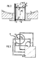
- FIGS. 2 and 3 show a first embodiment of the aiming device 8. It consists of two disks 12 and 13 made of X-ray negative material, for example made of plexiglass, which are provided in the lower end region of the X-ray tube 4 and are spaced apart and parallel to one another, and target marks 15, 16 made of x-ray positive material.
- the target marks can consist, for example, of channels which are formed within the panes and represent geometric figures, such that these figures define the course and position of an imaginary straight line 8a, which coincides with the central longitudinal axis 4a of the x-ray tube 4, which in turn , as already mentioned, coincides with the central axis 3 of the shock wave generator 2.
- the latter need not necessarily be the case, as shown in FIG. 8, if only that the axis 4a of the X-ray tube 4 runs through the focus of the generator 2.
- the geometric figures of the target marks consist of a circle 15 for the upper disc 12 and a cross 16 for the lower disc 13. This can be clearly seen from FIG. Instead of a cross, a circle can also be provided. Furthermore, other figures can also be provided for the target marks. It can also be seen that the channels representing the target marks 15 and 16 are connected to a pump device 18 via lines 17 for supplying and removing the X-ray positive liquid.
- the x-ray positive liquid is pumped into the channels forming the target marks when the x-ray emitter 11 of the x-ray device 9 is to be aligned with the x-ray tube 4 and the x-ray location of the therapy object is to be carried out.
- the target line 8a is predetermined by the target device 8, so that the X-ray emitter 11 can be set quickly and easily for this purpose, i.e. the center of the bundle of rays of the X-ray emitter is brought into line with the target straight line or the longitudinal center line 4a of the X-ray tube 4 and is thus aligned with the focus of the shock wave generator.
- the x-ray positive liquid can consist of a known x-ray contrast medium or also of a mixture of alcohol and an x-ray contrast medium.
- mercury can also be used, using the thermometer principle. The mercury is preferably introduced and removed by heating or cooling using a Peltier element.
- x-ray positive liquid By inserting and removing the x-ray positive liquid into or out of the channels, they are faded into the x-ray tube 4, as it were, in the application, or faded out afterwards.
- a suitable gas can also be used.
- FIG. 4 shows that in the lower end region of the X-ray tube 4 a metal ring 19 is provided as the first target, the center of which coincides with the longitudinal axis 4a of the tube 4 and which, according to FIG. 4, is designed to be rotatable about a horizontal axis.
- a motor 20 is provided outside the tube 4, which causes the metal ring 19 to pivot via a traction mechanism drive 21.
- a metal part 22 in the form of a target cross is provided as the second target mark, which can be adjusted and adjusted horizontally by means of a drive rod 23 by means of a lifting device 24, for example a lifting magnet.
- these targets 19 and 22 can be faded in and out if necessary.
- the two target marks 19 and 22 are spaced apart and arranged parallel to one another and form geometric target figures of the desired shape.
- FIG. 6 shows that the therapy device 1 is mounted on a frame 27 by means of an arm 25 so that it can pivot about a horizontal axis 26.
- the horizontal axis 26 runs through the focus 28 of the shock wave generator 2.
- the coupling balloon of the generator 2 is designated 29.
- the balloon 29 is shown extended in the left part of the shock wave generator 2.
- the patient table 30 is also shown in a lowered and in an elevated position.
- the X-ray device 9 which can be provided comprises a C-shaped support arm 31, one end of which has the X-ray emitter already mentioned 11 and the other end of which has an image intensifier 32 which is aligned with the X-ray emitter.
- the height of the C-shaped support arm can be adjusted according to the double arrow 33.
- the x-ray device 9 is set so that its horizontal pivot axis 34 coincides with the horizontal pivot axis 26 of the shock wave generator 2.
- the pivot axis 34 thus also runs through the focus 28 of the generator 2, as a result of which the x-ray device 9 can also be pivoted confocally about the focus 28.
- the patient table 30 can best be seen from FIG.
- This table can be adjusted in the usual way in the three spatial coordinates X, Y and Z indicated in FIG.
- the setting device for this consists of two lower, flat construction, adjustable in the horizontal X-Y coordinates adjusting units 35 and 36, which rest on a base 37, and a lifting unit 38 arranged on the upper unit 35, which is connected to the table top 39 at the top.
- the construction of this adjustment device i.e. the arrangement of the units 35, 36 in the plane below the lifting unit 38 makes it possible to provide a relatively thin tabletop, which results in an increased free space below the tabletop. This in turn results in a larger swivel angle range for the therapy device below the table top 39.
- FIG. 8 shows that the X-ray tube 4 can also be arranged outside the central axis 3 of the shock wave generator 2. Its central longitudinal axis 4a is at an angle to the axis 3 mentioned.
- a further locating device 40 can be provided which works according to the continuous ultrasound locating principle. This device is also arranged with its longitudinal axis at an angle to the central axis 3. This training of the therapy device with an additional ultrasound location system also makes a cranial / caudal X-ray location possible, e.g. at an angle of 15 ° to the central axis 3.
- FIG. 9 shows a modified embodiment of the therapy device for locating the therapy object.
- an ultrasound location can be used.
- the x-ray device 9, the plate part 7 and the target device 8 (FIG. 1) are removed and an ultrasound location scanner 43 is inserted into the x-ray tube 4 and mounted on the mounting part 6.
- the scanner 43 is rotatable on the mounting part 6 and adjustable in height, i.e. about and in the direction of the longitudinal axis 4a of the X-ray tube 4. These movements can be carried out by motor.
- the X-ray tube 4 has a flexible end part 4b, e.g. Latex on.
- the scanner 43 is ultrasonically coupled to the patient via a gel mass.
- the ultrasound generator 43 is used to treat the object, e.g. a fabric made.
Landscapes
- Health & Medical Sciences (AREA)
- Surgery (AREA)
- Life Sciences & Earth Sciences (AREA)
- Heart & Thoracic Surgery (AREA)
- Molecular Biology (AREA)
- Vascular Medicine (AREA)
- Engineering & Computer Science (AREA)
- Biomedical Technology (AREA)
- Orthopedic Medicine & Surgery (AREA)
- Medical Informatics (AREA)
- Nuclear Medicine, Radiotherapy & Molecular Imaging (AREA)
- Animal Behavior & Ethology (AREA)
- General Health & Medical Sciences (AREA)
- Public Health (AREA)
- Veterinary Medicine (AREA)
- Surgical Instruments (AREA)
- Radiation-Therapy Devices (AREA)
Applications Claiming Priority (2)
| Application Number | Priority Date | Filing Date | Title |
|---|---|---|---|
| DE4135328 | 1991-10-25 | ||
| DE4135328A DE4135328C2 (de) | 1991-10-25 | 1991-10-25 | Extrakorporales Therapiegerät |
Publications (2)
| Publication Number | Publication Date |
|---|---|
| EP0538659A2 true EP0538659A2 (fr) | 1993-04-28 |
| EP0538659A3 EP0538659A3 (en) | 1993-06-09 |
Family
ID=6443463
Family Applications (1)
| Application Number | Title | Priority Date | Filing Date |
|---|---|---|---|
| EP19920116850 Ceased EP0538659A3 (en) | 1991-10-25 | 1992-10-01 | Extracorporal therapy device |
Country Status (5)
| Country | Link |
|---|---|
| US (1) | US5388581A (fr) |
| EP (1) | EP0538659A3 (fr) |
| CA (1) | CA2081076A1 (fr) |
| DE (1) | DE4135328C2 (fr) |
| IL (1) | IL103438A (fr) |
Cited By (4)
| Publication number | Priority date | Publication date | Assignee | Title |
|---|---|---|---|---|
| EP0606548A1 (fr) * | 1993-01-14 | 1994-07-20 | Richard Wolf GmbH | Appareil de thérapie extracorporelle |
| FR2708191A1 (fr) * | 1993-07-27 | 1995-02-03 | Siemens Ag | Appareil de thérapie pour le traitement au moyen d'ondes acoustiques. |
| EP0715831A1 (fr) * | 1994-12-07 | 1996-06-12 | Philips Patentverwaltung GmbH | Ensemble de lithotripsie avec une unité de thérapie |
| EP1405604A1 (fr) * | 2002-10-04 | 2004-04-07 | HMT High Medical Technologies AG | Dispositif pour localisation et/ou diagnostic d'une zone de traitement d'un corps humain ou animal |
Families Citing this family (11)
| Publication number | Priority date | Publication date | Assignee | Title |
|---|---|---|---|---|
| US5732122A (en) * | 1996-10-17 | 1998-03-24 | Eastman Kodak Company | X-ray marker apparatus and method |
| US20050228319A1 (en) * | 1996-12-30 | 2005-10-13 | Kenny Daniele J | Neoplasm cell destruction device |
| US20090318839A1 (en) * | 1996-12-30 | 2009-12-24 | Daniele Kenny | Neoplastic cell destruction device and method utilizing low frequency sound waves to disrupt or displace cellular materials |
| US20090318840A1 (en) * | 1996-12-30 | 2009-12-24 | Daniele Kenny | Neoplastic cell destruction device and method utilizing low frequency sound waves to disrupt or displace cellular materials |
| US20090318838A1 (en) * | 1996-12-30 | 2009-12-24 | Daniele Kenny | Neoplastic cell destruction device and method utilizing low frequency sound waves to disrupt or displace cellular materials |
| US6684097B1 (en) * | 1999-04-22 | 2004-01-27 | University Of Miami | Intraoperative monitoring of temperature-induced tissue changes with a high-resolution digital x-ray system during thermotherapy |
| DE102011082680B4 (de) * | 2011-09-14 | 2019-12-24 | Siemens Healthcare Gmbh | Anlage |
| ITRE20130076A1 (it) * | 2013-10-23 | 2015-04-24 | Giovanni Borasi | Fus/rt: apparecchiatura per il trattamento di neoplasie |
| US10639503B2 (en) * | 2014-08-27 | 2020-05-05 | Fusmobile Inc. | Handheld devices for projecting focused ultrasound and related methods |
| US12383768B2 (en) | 2015-08-10 | 2025-08-12 | Fus Mobile Inc. | Image guided focused ultrasound treatment device and aiming apparatus |
| EP3199106B1 (fr) * | 2017-04-26 | 2020-09-09 | Siemens Healthcare GmbH | Procédé et dispositif destinés à l'examen par ultrasons |
Family Cites Families (8)
| Publication number | Priority date | Publication date | Assignee | Title |
|---|---|---|---|---|
| DE3840077A1 (de) * | 1988-11-28 | 1990-05-31 | Wolf Gmbh Richard | Lithotriptor |
| EP0372119B2 (fr) * | 1988-12-09 | 1997-08-27 | Siemens Aktiengesellschaft | Lithotripteur |
| US4930509A (en) * | 1989-01-23 | 1990-06-05 | Northgate Research, Inc. | X-ray aiming fixture |
| DE3916093A1 (de) * | 1989-05-17 | 1990-11-22 | Wolf Gmbh Richard | Lithotriptor |
| DE3919083C1 (fr) * | 1989-06-10 | 1990-06-21 | Dornier Medizintechnik Gmbh, 8000 Muenchen, De | |
| DE3932364A1 (de) * | 1989-09-28 | 1991-04-11 | Wolf Gmbh Richard | Vorrichtung zur raeumlichen ortung und zur zerstoerung von koerperinneren objekten |
| DE4007669C3 (de) * | 1990-03-10 | 1997-11-13 | Wolf Gmbh Richard | Vorrichtung zur Stoßwellenbehandlung |
| DE9017444U1 (de) * | 1990-12-24 | 1991-03-14 | Dornier Medizintechnik GmbH, 8000 München | Zieleinrichtung für Lithotripter |
-
1991
- 1991-10-25 DE DE4135328A patent/DE4135328C2/de not_active Expired - Fee Related
-
1992
- 1992-10-01 EP EP19920116850 patent/EP0538659A3/de not_active Ceased
- 1992-10-15 IL IL10343892A patent/IL103438A/en not_active IP Right Cessation
- 1992-10-21 CA CA002081076A patent/CA2081076A1/fr not_active Abandoned
-
1994
- 1994-06-08 US US08/255,578 patent/US5388581A/en not_active Expired - Fee Related
Cited By (5)
| Publication number | Priority date | Publication date | Assignee | Title |
|---|---|---|---|---|
| EP0606548A1 (fr) * | 1993-01-14 | 1994-07-20 | Richard Wolf GmbH | Appareil de thérapie extracorporelle |
| FR2708191A1 (fr) * | 1993-07-27 | 1995-02-03 | Siemens Ag | Appareil de thérapie pour le traitement au moyen d'ondes acoustiques. |
| EP0715831A1 (fr) * | 1994-12-07 | 1996-06-12 | Philips Patentverwaltung GmbH | Ensemble de lithotripsie avec une unité de thérapie |
| US5836898A (en) * | 1994-12-07 | 1998-11-17 | U.S. Philips Corporation | Lithotripsy combination comprising a therapy unit |
| EP1405604A1 (fr) * | 2002-10-04 | 2004-04-07 | HMT High Medical Technologies AG | Dispositif pour localisation et/ou diagnostic d'une zone de traitement d'un corps humain ou animal |
Also Published As
| Publication number | Publication date |
|---|---|
| IL103438A (en) | 1996-11-14 |
| DE4135328A1 (de) | 1993-04-29 |
| IL103438A0 (en) | 1993-03-15 |
| US5388581A (en) | 1995-02-14 |
| EP0538659A3 (en) | 1993-06-09 |
| CA2081076A1 (fr) | 1993-04-26 |
| DE4135328C2 (de) | 1995-01-19 |
Similar Documents
| Publication | Publication Date | Title |
|---|---|---|
| DE69319354T2 (de) | Rahmen für stereotaktische radiochirurgie mit befestigung am knochengerüst | |
| DE4135328C2 (de) | Extrakorporales Therapiegerät | |
| DE19746956C2 (de) | Medizinisches System aufweisend ein Röntgengerät und ein Therapiegerät mit einer Quelle fokussierter akustischer Wellen | |
| EP0397980B1 (fr) | Lithotriteur | |
| EP0402584B1 (fr) | Dispositif de visée pour lithotripteur | |
| EP0221325B1 (fr) | Unité de traitement par lithotritie | |
| EP0532981A1 (fr) | Appareil pour le traitement d'un être vivant par ondes acoustiques | |
| DE3714397C2 (fr) | ||
| EP0405282A1 (fr) | Appareil pour le traitement d'un être vivant avec des ondes de choc focalisées | |
| DE3840077A1 (de) | Lithotriptor | |
| EP0715831A1 (fr) | Ensemble de lithotripsie avec une unité de thérapie | |
| EP0419872B1 (fr) | Dispositif pour la localisation spatiale et la destruction des objets dans le corps | |
| EP0265742A1 (fr) | Lithotripteur avec moyens de localisation | |
| EP0372119A1 (fr) | Lithotripteur | |
| DE4306460A1 (de) | Therapieeinrichtung zur Behandlung mit fokussierten akustischen Wellen | |
| DE4003350C1 (en) | C-bow X=ray appts. setter for lithotriptor - uses metal sphere on support as reference element for position of therapy focus | |
| EP0400196A1 (fr) | Transducteur d'ondes de choc pour destruction de concrétions | |
| DE2902331A1 (de) | Geraet zur transkutanen, unblutigen veroedung von kleinen retikulaeren und besenreiser-varicen | |
| EP1720466B1 (fr) | Equipement pour un traitement medical non invasif | |
| DE19511106C1 (de) | Lithotripsievorrichtung | |
| DE4325212A1 (de) | Medizinisches Gerät | |
| DE4329167A1 (de) | Medizinisches Gerät mit einem Geräteteil | |
| DE102017216017B4 (de) | Medizinisch-therapeutisches System | |
| DE10302612B4 (de) | Urologischer Arbeitsplatz | |
| DE29824080U1 (de) | Stoßwellentherapiesystem und Ortungssystem in einem isozentrischem System |
Legal Events
| Date | Code | Title | Description |
|---|---|---|---|
| PUAI | Public reference made under article 153(3) epc to a published international application that has entered the european phase |
Free format text: ORIGINAL CODE: 0009012 |
|
| PUAL | Search report despatched |
Free format text: ORIGINAL CODE: 0009013 |
|
| AK | Designated contracting states |
Kind code of ref document: A2 Designated state(s): BE DE FR GB |
|
| AK | Designated contracting states |
Kind code of ref document: A3 Designated state(s): BE DE FR GB |
|
| 17P | Request for examination filed |
Effective date: 19931202 |
|
| 17Q | First examination report despatched |
Effective date: 19940621 |
|
| GRAG | Despatch of communication of intention to grant |
Free format text: ORIGINAL CODE: EPIDOS AGRA |
|
| 18R | Application refused |
Effective date: 19960927 |