EP0554670A2 - Spezifische Antikörper gegen aktivierte Plättchen, ihre Herstellung und ihre Verwendung in Diagnose und Therapie - Google Patents
Spezifische Antikörper gegen aktivierte Plättchen, ihre Herstellung und ihre Verwendung in Diagnose und Therapie Download PDFInfo
- Publication number
- EP0554670A2 EP0554670A2 EP93100339A EP93100339A EP0554670A2 EP 0554670 A2 EP0554670 A2 EP 0554670A2 EP 93100339 A EP93100339 A EP 93100339A EP 93100339 A EP93100339 A EP 93100339A EP 0554670 A2 EP0554670 A2 EP 0554670A2
- Authority
- EP
- European Patent Office
- Prior art keywords
- monoclonal antibody
- mak
- recognized
- host cell
- polypeptide
- Prior art date
- Legal status (The legal status is an assumption and is not a legal conclusion. Google has not performed a legal analysis and makes no representation as to the accuracy of the status listed.)
- Granted
Links
- 0 C1C2=C1CC*2 Chemical compound C1C2=C1CC*2 0.000 description 3
Images







Classifications
-
- C—CHEMISTRY; METALLURGY
- C07—ORGANIC CHEMISTRY
- C07K—PEPTIDES
- C07K14/00—Peptides having more than 20 amino acids; Gastrins; Somatostatins; Melanotropins; Derivatives thereof
- C07K14/435—Peptides having more than 20 amino acids; Gastrins; Somatostatins; Melanotropins; Derivatives thereof from animals; from humans
- C07K14/78—Connective tissue peptides, e.g. collagen, elastin, laminin, fibronectin, vitronectin or cold insoluble globulin [CIG]
-
- A—HUMAN NECESSITIES
- A61—MEDICAL OR VETERINARY SCIENCE; HYGIENE
- A61P—SPECIFIC THERAPEUTIC ACTIVITY OF CHEMICAL COMPOUNDS OR MEDICINAL PREPARATIONS
- A61P7/00—Drugs for disorders of the blood or the extracellular fluid
- A61P7/02—Antithrombotic agents; Anticoagulants; Platelet aggregation inhibitors
-
- C—CHEMISTRY; METALLURGY
- C07—ORGANIC CHEMISTRY
- C07K—PEPTIDES
- C07K14/00—Peptides having more than 20 amino acids; Gastrins; Somatostatins; Melanotropins; Derivatives thereof
- C07K14/435—Peptides having more than 20 amino acids; Gastrins; Somatostatins; Melanotropins; Derivatives thereof from animals; from humans
- C07K14/705—Receptors; Cell surface antigens; Cell surface determinants
-
- C—CHEMISTRY; METALLURGY
- C07—ORGANIC CHEMISTRY
- C07K—PEPTIDES
- C07K16/00—Immunoglobulins [IG], e.g. monoclonal or polyclonal antibodies
- C07K16/18—Immunoglobulins [IG], e.g. monoclonal or polyclonal antibodies against material from animals or humans
-
- C—CHEMISTRY; METALLURGY
- C07—ORGANIC CHEMISTRY
- C07K—PEPTIDES
- C07K16/00—Immunoglobulins [IG], e.g. monoclonal or polyclonal antibodies
- C07K16/18—Immunoglobulins [IG], e.g. monoclonal or polyclonal antibodies against material from animals or humans
- C07K16/28—Immunoglobulins [IG], e.g. monoclonal or polyclonal antibodies against material from animals or humans against receptors, cell surface antigens or cell surface determinants
-
- A—HUMAN NECESSITIES
- A61—MEDICAL OR VETERINARY SCIENCE; HYGIENE
- A61K—PREPARATIONS FOR MEDICAL, DENTAL OR TOILETRY PURPOSES
- A61K38/00—Medicinal preparations containing peptides
-
- C—CHEMISTRY; METALLURGY
- C07—ORGANIC CHEMISTRY
- C07K—PEPTIDES
- C07K2317/00—Immunoglobulins specific features
- C07K2317/20—Immunoglobulins specific features characterized by taxonomic origin
- C07K2317/24—Immunoglobulins specific features characterized by taxonomic origin containing regions, domains or residues from different species, e.g. chimeric, humanized or veneered
-
- C—CHEMISTRY; METALLURGY
- C07—ORGANIC CHEMISTRY
- C07K—PEPTIDES
- C07K2319/00—Fusion polypeptide
-
- C—CHEMISTRY; METALLURGY
- C07—ORGANIC CHEMISTRY
- C07K—PEPTIDES
- C07K2319/00—Fusion polypeptide
- C07K2319/33—Fusion polypeptide fusions for targeting to specific cell types, e.g. tissue specific targeting, targeting of a bacterial subspecies
Definitions
- Hybridoma technology makes it possible to produce selective monoclonal antibodies (MAK) against any epitope on defined or undefined antigens.
- MAK monoclonal antibodies
- highly purified, defined antigens or also antigen mixtures, cells or tissues can be used as immunogens, a suitable immunogen also having to be found in order to find highly selective MACs.
- MAK monoclonal antibodies
- the experimental error limits when determining the isoelectric point are approximately ⁇ 10-15%, in particular approximately ⁇ 5-10%.
- a monoclonal antibody characterized in that it is derived from the hybridoma 2128 (DSM ACC2024) (BW 2128).
- Monoclonal antibodies or parts thereof that bind to an epitope recognized by a 2128 hybridoma monoclonal antibody Monoclonal antibodies or parts thereof that bind to an epitope recognized by a 2128 hybridoma monoclonal antibody.
- preferential binding means that there are 3-1000 times and in particular 10-100 times more epitopes that are recognized by the BW 2128 on activated platelets than on non-activated platelets.
- the invention furthermore relates to monoclonal antibodies or parts thereof which are chimeric, humanized, bi- or oligospecific.
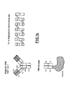
- this includes an mru fragment, a single domain fragment, a single chain fragment, an F (ab) fragment or an F (ab ') 2 fragment with one or more hinge regions, such as in FIGS. 1 and 1 b shown.
- Humanized antibodies are particularly preferred examples thereof.
- a mru minimal recognition unit is a polypeptide derived from a CDR (Complementarity Determining Region), which has the property of targeting the epitope recognized by the specific MAK tie.
- the subject of the invention also includes mimetics which bind to an epitope which is recognized by the monoclonal antibody BW 2128.
- mimetics which bind to an epitope which is recognized by the monoclonal antibody BW 2128.
- Small organic chemical compounds with a high binding potential to the epitope defined by BW 2128 are suitable mimetics.
- the subject of the invention includes polypeptides containing an amino acid sequence according to Tab. La and / or Tab. Ib or any allelic variant or mutant thereof which has the biological property to an epitope which is recognized by the monoclonal antibody BW 2128, to tie.
- the monoclonal antibodies according to the invention or parts thereof or the mimetics according to the invention are used as fusion proteins which contain an immunoglobulin part according to the invention and a part not belonging to the immunoglobulin family.
- the protein not belonging to the immunoglobulin family should be a fibrinolytic protein or parts thereof, for example the fibrinolytic protein streptokinase, urokinase, tPA, tPA mutants or parts thereof.
- This also includes vectors, in particular the expression vectors L1 and L2 and suitable host cells, in particular COS, CHO or BHK cells, preferably BHK cells.
- suitable host cells in particular COS, CHO or BHK cells, preferably BHK cells.
- the invention furthermore relates to polynucleotides which contain one or more nucleic acid sequences which code for a polypeptide which has the biological property to bind to an epitope which is recognized by the monoclonal antibody BW 2128.
- nucleic acids according to Tab. La and Ib are included, their degenerate codons and nucleic acids which hybridize against the nucleic acids according to Tab. La and Ib under generally known hybridization conditions.
- Preferred hybridization conditions are stringent conditions as described in Sambrook, J., Fritsch, EF, Maniatis, T. (1982), Molecular cloning: a laboratory manual, Cold Spring Harbor Laboratory Sambrook, J., Fritsch, EF, Maniatis, T. (1989 ), Second Edition, Molecular cloning: A Laboratory Manual, Cold Spring Harbor Laboratory Press.
- Another object of the invention is a method for producing a polypeptide according to the invention, wherein a prokaryotic or eukaryotic host cell is transformed or transfected with a polynucleotide according to the invention so that the host cell expresses the polypeptide mentioned.
- the polypeptide can then be isolated from the culture by methods known to those skilled in the art.
- the subject of the invention includes an antigen or immunologically reactive parts thereof, which is specifically recognized by one of the antibodies according to the invention, in particular by MAK BW 2128.
- polypeptides according to the invention can also be prepared chemically by methods of peptide synthesis known to the person skilled in the art.
- polypeptide means proteins or amino acid sequences including their spatial configuration and post-translational modifications, such as glycosylation or phosphorylation.
- the invention is particularly suitable in the in vivo presentation or therapy of diseases in which activated platelets are involved, such as e.g. Thromboses, especially during heart attacks or strokes, during thrombolytic therapy, deep-seated venous thrombosis, pulmonary embolism, vascular injuries, atherosclerotic plaques, pyogenic infections and intestinal ischemia.
- diseases in which activated platelets are involved such as e.g. Thromboses, especially during heart attacks or strokes, during thrombolytic therapy, deep-seated venous thrombosis, pulmonary embolism, vascular injuries, atherosclerotic plaques, pyogenic infections and intestinal ischemia.
- the MAK precipitates an antigen which, under reducing conditions, has a molecular weight which is smaller than that of TSP and a relative molecular mass of approximately 150,000 to 170,000, preferably approximately 160,000, but has an isoelectric point, such as TSP, which is indistinguishable from the experimental error limits, preferably in the range of approximately 5.5-6.5 (TSP had a molecular weight of 180 KDa and an isoelectric point of 5.5 to 6 in a comparative experiment , 5).
- TSP had a molecular weight of 180 KDa and an isoelectric point of 5.5 to 6 in a comparative experiment , 5).
- This new antigen is associated with TSP, whereby both physical and chemical binding is possible and is preferentially expressed on activated platelets and is therefore generally referred to as platelet antigen.
- the antigen also preferably shows the tissue distribution shown in Table 11.
- the antigen which can be isolated, for example, by means of immunoaffinity chromatography, preferably with the aid of MAK BW 2128, is particularly well suited for the production or checking of antibodies equivalent to MAK BW 2128 or immunologically reactive parts thereof and for the production of mimetics.
- the antibodies according to the invention can be labeled with radioactive isotopes, especially with Tc-99m (Schwarz, A., Steinstraesser, A. (1987) J. Nucl. Med. 28: 721) if they are of the IgG1 isotype like the MAK BW 2128 Another possibility is the labeling with paramagnetic compounds.
- the antibodies and mimetics according to the invention are therefore particularly well suited for the immunoscintigraphic display of vascular occlusions.
- the hybridoma 2128 was deposited with the DSM, German Collection of Microorganisms and Cell Cultures GmbH (Mascheroder Weg 1 B, 3300 Braunschweig) according to the Budapest contract under the deposit number DSM ACC 2024. Furthermore, the V genes of the heavy and light chains of the MAK BW 2128 were those of Orlandi, R., Güssow, D., Jones, P.T., Winter, G. (1989). Proc. Nat. Acad. Sci. USA 86: 3833-3837 isolated and according to the method described by Sanger, F., Nicklen, S., Coulson, A.R. (1977). Proc. Nat. Acad. Sci. USA 74: 5463-5467 determines the nucleic acid sequence of the essential regions of the V gene exon (Tab. La, b).
- the cloned V genes were expressed as chimeric MAK BW Chi 2128 with human truncated IgG3 Fc part (IgG3 ⁇ ) and human C-kappa in BHK cells (Wirth, M., Bode, J., Zettlmeissl, G., Hauser, H (1988). Gene 73: 419-426) to confirm the identity of the cloned V genes (Examples A-N).
- the pronounced BW Chi 2128 MAK showed the same antigen binding specificity as the mouse MAK. Furthermore, e.g. after polypeptide synthesis of the CDRs or of parts or more defined CDRs, the mrus (minimal recognition units) are determined and used as less immunogenic specific peptides for the in vivo localization of activated platelets. Furthermore, according to the method described by Saragovi, H.U., Fitzpatrick, D., Raktabutr, A., Nakanishi, H., Kahn, M., Greene, M.I. (1991). Science 253: 792-795 described method using organic chemical synthesis a mimetic with high specificity and avidity against the epitope defined by MAK BW 2128 are generated.
- the humanized V region of the MAK BW 2128 can be recombinantly e.g. are linked to nucleotide sequences which code for fibrinolytic proteins such as streptokinase, urokinase, tPA, but above all tPA mutants.
- Preferred tPA mutants can be produced, for example, according to EP-A1-0 387 380.
- German patent application P 41 06 389.9 (Ma 876) using the example of a humanized aCEA MAK and human ⁇ -glucuronidase, these constructs can be linked at the DNA level and expressed as functional fusion proteins.
- the fusion proteins mentioned are efficient and low-side-effect fibrinolytics (Haber, E., Quertermous, Th., Matsueda, GR, Runge, MS, Bode, Ch. (1989. Jap. Circulation J. 54: 345-353).
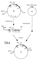
- the plasmid clone 54.1.24 which carries the human IgG 3 C gene (DE 38 25 615 A1, Ma 706, Fig. 2) was cleaved with Pstl. The resulting vector was ligated with the largest of the resulting Pst1 insert fragments and transformed into bacteria.
- the plasmid clone A which carries a human IgG 3 C gene in which the H1, H2 and H3 exons have been deleted, was identified by restriction analysis and nucleic acid sequence determination (FIG. 2).
- the plasmid clone A was cleaved with HindIII and EcoRI, the ends filled in with Klenow polymerase, the IgG 3 insert isolated and ligated into a pUC19 vector cleaved with Sstl and blunt-ended with the aid of T 4 polymerase.
- a plasmid clone B was identified by restriction mapping and nucleic acid sequence analysis, in which the IgG 3 gene is oriented in such a way that the HindIII at the 5 'end and the EcoRI interface of the pUC19 polylinker are at the 3' end (FIG. 3).
- Plasmid clone B was cleaved with EcoRI and HindIII, the IgG 3 insert isolated and ligated into a KS + phasmid vector (pBluescriptII KS +; Stratagene, La Jolla, CA), which was also cleaved with HindIII and EcoRI.
- the phasmid clone C was isolated, in which the IgG 3 gene is flanked at the 5 'and at the 3' end by a BamHI interface (FIG. 4).
- Phasmid clone C was cleaved with BamHl, the IgG 3 insert isolated and ligated into the expression vector pABStop (Wirth et al. Supra), which was also cleaved with BamHl.
- the expression plasmid D was identified, which contains the IgG 3 C gene in the orientation shown in the formula. In this cloning, the pABStop vector loses the SV40 stop and polyadenylation signal located between the two BamHI interfaces (FIG. 5).
- the expression plasmid D was partially cleaved with BamHl, the ends filled in with Klenow polymerase and religated.
- the expression plasmid E was isolated, in which the BamHI site 3 'is destroyed by the IgG 3 gene (FIG. 6).
- the human C-kappa gene (Hieter, P.A., Mainzel, J.V., Jr., Leder, P. (1982), The Journal of Biological Chemistry, 257: 1516-1522) was cloned as an EcoRI fragment in pBR 322.
- the pBR322 vector was digested with EcoRI, the EcoRI sites filled in, the C-kappa insert isolated and ligated into a pUC19 vector digested with Smal.
- the plasmid clone F was isolated, in which the C-kappa gene is flanked at the 5 'end by a HindIII and a BamHI interface and at the 3' end by an EcoRI interface (FIG. 7).
- the plasmid clone F was cleaved with HindIII and EcoRI, the C-kappa insert isolated and cloned into a HindIII / EcoRI cleaved KS + phasmid.
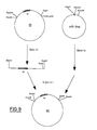
- the phasmid clone G was isolated, in which the C-kappa insert is flanked at the 5 'and at the 3' end by a BamHI interface (FIG. 8).
- Phasmid clone G was cleaved with BamHI, the C-kappa insert isolated and cloned into a pAB Stop vector cleaved with BamHI. Restriction mapping and nucleic acid sequence analysis identified the clone H in which the C-kappa gene is oriented such that the HindIII site of the pAB stop vector is located at its 5 'end (FIG. 9).
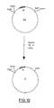
- Clone H was partially cleaved with BamHI, the restriction ends filled in and religated. Restriction mapping was used to identify clone I in which the BamHI site 3 'of the C-kappa gene had been destroyed (FIG. 10).
- V H and V K genes of the MAK BW 2128 were developed according to Orlandi et al. (supra) amplified by means of PCR technology and specific oligonucleotides and cloned in KS + vectors (Güssow, D. and Seemann, G. (1992). Methods in Enzymology, Vol. 203, 99-121), which contain irrelevant V H and V K genes with appropriate restriction sites. Clones K1 and K2 were then isolated, in which the irrelevant V H and V L genes were replaced by the V H (K1) and V K (K2) genes of MAK BW 2128 (FIG. 11).
- the nucleic acid sequence of the V H and V K genes of MAK BW 2128 was determined from clones K1 and K2 according to the method of Sanger PNAS, 74, 5463, (1977) (Tab. La, b).
- V H and V K genes of the MAK BW 2128 were cut out from the clones K1 and K2 using the restriction enzymes Hindlll and BamHI, the V-gene inserts were isolated and the expression vectors D (V H ) and I (cut with Hindlll and BamHI were isolated. V K ) cloned.
- Expression vectors L1 (Fig. 12) and L2 (Fig. 13) containing a complete immunoglobulin heavy chain gene (L1) and light chain gene (L2) were isolated.
- the expression vectors L1 and L2 were mediated together with the vector pR11 H140 (Hudziak, RM, Laski, FA, RajBhandary, UL, Sharp, PA, Capecchi, MR (1982), Cell 31: 137-146) of the neomycin resistance (FIG. 14) and the vector psVdhfr (Lee, F., Mulligan, R., Berg, P., Ringold, G. (1981), Nature 294: 228-232), which carries a dihydrofolate reductase gene and mediates resistance to methotrexate (Fig.
- the immunohistochemical specificity analysis of the antibodies according to the invention was carried out using the APAAP technique (Cordell et al., Supra). Table II shows the result of an immunohistochemical specificity analysis of the MAK BW 2128.
- the activation of platelets can be measured by a quantitative determination of the activation marker PF4 using the ELISA described by H. Pelzer and N. Heimburger (supra).
- Table III shows the influence of MAK BW 2128 on platelet activation.
Landscapes
- Health & Medical Sciences (AREA)
- Chemical & Material Sciences (AREA)
- Organic Chemistry (AREA)
- Life Sciences & Earth Sciences (AREA)
- General Health & Medical Sciences (AREA)
- Medicinal Chemistry (AREA)
- Biochemistry (AREA)
- Genetics & Genomics (AREA)
- Biophysics (AREA)
- Molecular Biology (AREA)
- Proteomics, Peptides & Aminoacids (AREA)
- Immunology (AREA)
- Toxicology (AREA)
- Zoology (AREA)
- Gastroenterology & Hepatology (AREA)
- Cell Biology (AREA)
- Engineering & Computer Science (AREA)
- Public Health (AREA)
- Diabetes (AREA)
- Hematology (AREA)
- Chemical Kinetics & Catalysis (AREA)
- General Chemical & Material Sciences (AREA)
- Nuclear Medicine, Radiotherapy & Molecular Imaging (AREA)
- Pharmacology & Pharmacy (AREA)
- Animal Behavior & Ethology (AREA)
- Bioinformatics & Cheminformatics (AREA)
- Veterinary Medicine (AREA)
- Preparation Of Compounds By Using Micro-Organisms (AREA)
- Medicines Containing Antibodies Or Antigens For Use As Internal Diagnostic Agents (AREA)
- Peptides Or Proteins (AREA)
- Micro-Organisms Or Cultivation Processes Thereof (AREA)
- Medicines Containing Material From Animals Or Micro-Organisms (AREA)
- Organic Low-Molecular-Weight Compounds And Preparation Thereof (AREA)
Abstract
Description
- Die Hybridomatechnologie erlaubt es, gegen beliebige Epitope auf definierten oder undefinierten Antigenen selektive monoklonale Antikörper (MAK) herzustellen. Hierzu können als Immunogene hochgereinigte, definierte Antigene oder auch Antigenmischungen, Zellen oder Gewebe verwendet werden, wobei für das Auffinden hochselektiver MAK auch ein geeignetes Immunogen gefunden werden muß. Mit dieser Technologie ist es nun gelungen, MAK zu entwickeln, die in der Lage sind, nach i.v. Injektion z.B. an Tumoren, entzündliche Prozesse und Gefäßverschlüsse im menschlichen Körper zu binden. Diese präferentielle Bindung von MAK an die entsprechenden Zielstrukturen (Tumor, entzündlicher Prozeß, Gefäßverschluß) wird bereits im Rahmen der nuklearmedizinischen Diagnostik genutzt. Die nuklearmedizinische Spezialdisziplin, die sich mit der Injektion radioaktiv markierter MAK zum Nachweis von Zielstrukturen beschäftigt, ist die Immunszintigraphie (Mach,J.P., Buchegger, F., Forni, M., Ritschard, J., Berche, C., Lumbraso, J.-O., Schreyer, M., Girardet, C., Accolla, R.C., Carrel, S. (1981) Immunology today 2:239-249). Ihr stehen z.Z. keine MAK zur Verfügung, die selektiv an Plättchen binden und im wesentlichen nicht an anderes menschliches Normalgewebe. Selbst die MAK, die gegen a-Granula Proteine von Plättchen gerichtet sind, binden zwar präferentiell an aktivierte Plättchen, zeigen aber doch deutliche Bindung an Epitope auf anderen Geweben. So reagieren z.B. MAK, die gegen Thrombospondin (TSP), ein a-Granula Protein (Baenziger, N.L., Brodie, G.N., Majerus, P.W. (1971). Proc. Nat. Acad. Sci. USA 68:240-243; Ganguly, P. (1971). J. Biol. Chem. 246:4286-4290) gerichtet sind, mit Epitopen im Drüsenepithel der Haut und Lunge, den Verbindungsstücken zwischen Dermis und Epidermis (Wight, T.N., Raugi, G.J., Mumby, S.M., Bornstein, D. (1985). J. Histochem. Cytochem. 33:295-302), dem Knorpel (Miller, R.R., McDevitt, C.A. (1988). Biochem. Biophys. Res. Commun. 153:708-714), decalzifiziertem Knochen (Robey, P.G., Young, M.F., Fisher, L.W., McClain, T.D. (1989). J. Cell. Biol. 108:719-727), Nabelschnurarterien (Fauvel-Lafeve, F., Legrand, Y.J. (1988). Thromb. Res. 50:305-316), Typ 11 Pneumozyten (Sage, H., Farin, F.M., Striker, G.E., Fisher, A.B. (1983). Biochemistry 22:2148-2155), Megakaryozyten (Beckstead, J.H., Stenberg, P.E., McEver, R.P., Shuman, M.A., Bainton, D.F. (1986). Blood 67:285-293) sowie proliferierenden Endothelzellen, glatter Muskulatur und Fibroblasten (Mumby, S.M., Abbott-Brown, D., Raugi, G.J., Bornstein, P. (1984). J. Cell. Physiol. 120:280-288; Jaffe, E.A., Ruggiero, J.T., Leung, L.K., Doyle, M.J., McKeown-Lango, P.J., Mosher, D.F. (1983). Proc. Nat. Acad. Sci. USA 80:998-1002). Bedingt durch die Kreuzreaktivitäten mit den eben erwähnten Normalgeweben sind falsch positive Signale beim Versuch, mit bekannten anti TSP-MAK in vivo Gefäßverschlüsse nachzuweisen, im allgemeinen zu beobachten.
- Bei der Herstellung von aTSP-MAK gemäß Jaffe et al. (supra) haben wir überraschenderweise einen spezifischen MAK gegen aktivierte Plättchen (MAK BW 2128) gefunden, der im Westernblot nach Towbin T. & Gordon J. (1979) Proc. Natl. Acad. Sci. USA 76, 4350-4354 weder mit den bekannten a Granula Proteinen wie TSP, v. Willebrand Faktor oder gP 140 reagiert noch mit den oben genannten Zellen oder Geweben. Der MAK immunpräzipitiert ein Antigen, welches mit TSP assoziiert ist, einen innerhalb der experimentellen Fehlergrenzen von TSP nicht unterscheidbaren isoelektrischen Punkt besitzt, aber unter reduzierenden Bedingungen ein deutlich kleineres Molekulargewicht als TSP von ca. 160 kDa hat.
- Die experimentellen Fehlergrenzen bei der Bestimmung des isoelektrischen Punktes betragen ca. ± 10-15 %, insbesondere ca. ± 5-10 %.
- Gegenstand der Erfindung ist daher:
- Ein Hybridom 2128 (DSM ACC2024).
- Ein monoklonaler Antikörper, dadurch gekennzeichnet, daß er von dem Hybridom 2128 (DSM ACC2024) abstammt (BW 2128).
- Monoklonale Antikörper oder Teile davon, die an ein Epitop, das von einem monoklonalen Antikörper des Hybridoms 2128 erkannt wird, binden.
- Monoklonale Antikörper oder Teile davon, die präferentiell an aktivierte humane Plättchen binden und eine Aminosäuresequenz gemäß Tab. la und/oder Tab. Ib oder irgendeine allelische Variante oder Mutante davon, die die biologische Eigenschaft besitzt, an ein Epitop, das von dem monoklonalen Antikörper des Hybridoms 2128 erkannt wird, zu binden, enthalten.
- Präferentielle Bindung bedeutet im Sinne der Erfindung, daß sich auf aktivierten Plättchen 3-1000 mal und insbesondere 10-100 mal mehr Epitope, die vom BW 2128 erkannt werden, befinden als auf nicht aktivierten Plättchen.
- Ferner gehören zum Gegenstand der Erfindung monoklonale Antikörper oder Teile davon die chimärer, humanisierter, bi- oder oligospezifischer Art sind. Insbesondere fallen darunter ein mru-Fragment, ein single domain Fragment, ein single chain Fragment, ein F(ab)-Fragment oder ein F(ab')2-Fragment mit einer oder mehreren Hinge-Regionen, wie beispielsweise in Figur 1 und 1 b dargestellt.
- Humanisierte Antikörper sind besonders bevorzugte Beispiele davon.
- Ein mru (minimalrecognition unit) ist ein von einem CDR (Complementarity Determining Region) abgeleitetes Polypeptid, welches die Eigenschaft besitzt, an das vom spezifischen MAK erkannte Epitop zu binden.
- Auch gehören zum Gegenstand der Erfindung Mimetika, die an ein Epitop, welches von dem monoklonalen Antikörper BW 2128 erkannt wird, binden. Kleine organisch-chemische Verbindungen mit hohem Bindungspotential an das durch BW 2128 definierte Epitop kommen als geeignete Mimetika in Frage.
- Im allgemeinen gehören zum Gegenstand der Erfindung Polypeptide, enthaltend eine Aminosäuresequenz gemäß Tab. la und/oder Tab. Ib oder irgendeine allelische Variante oder Mutante davon, die die biologische Eigenschaft besitzt, an ein Epitop, das von dem monoklonalen Antikörper BW 2128 erkannt wird, zu binden.
- Besonders vorteilhaft ist es, wenn die erfindungsgemäßen monoklonalen Antikörper oder Teile davon oder die erfindungsgemäßen Mimetika als Fusionsproteine verwendet werden, die einen erfindungsgemäßen Immunglobulinanteil und einen nicht zur Immunglobulinfamilie gehörenden Teil enthalten.
- Insbesondere soll das nicht zur Immunglobulinfamilie gehörende Protein ein fibrinolytisches Protein oder Teile davon sein, beispielsweise das fibrinolytische Protein Streptokinase, Urokinase, tPA, tPA-Mutanten oder Teile davon.
- Ferner gehören dazu Vektoren, insbesondere die Expressionsvektoren L1 und L2 und geeignete Wirtszellen, insbesondere COS, CHO oder BHK Zellen, vorzugsweise BHK Zellen.
- Ferner gehören zum Gegenstand der Erfindung Polynukleotide, die eine oder mehrere Nukleinsäuresequenzen enthalten, die für ein Polypeptid kodieren, das die biologische Eigenschaft besitzt, an ein Epitop, das von dem monoklonalen Antikörper BW 2128 erkannt wird, zu binden. Insbesondere fallen Nukleinsäuren gemäß Tab. la und Ib darunter, deren degenerierte Codons sowie Nukleinsäuren die gegen die Nukleinsäuren gemäß Tab. la und Ib unter allgemein bekannten Hybridisierungsbedingungen hybridisieren. Bevorzugte Hybridisierungsbedingungen sind stringente Bedingungen wie bei Sambrook, J., Fritsch, E.F., Maniatis, T. (1982), Molecular cloning: a laboratory manual, Cold Spring Harbor Laboratory Sambrook, J., Fritsch, E.F., Maniatis, T. (1989), Second Edition, Molecular cloning: A Laboratory Manual, Cold Spring Harbor Laboratory Press erläutert.
- Ein weiterer Gegenstand der Erfindung ist ein Verfahren zur Herstellung eines erfindungsgemäßen Polypeptids, wobei eine prokaryotische oder eukaryotische Wirtszelle mit einem erfindungsgemäßen Polynukleotid transformiert oder transfiziert wird, so daß die Wirtszelle das genannte Polypeptid exprimiert. Das Polypeptid kann dann aus der Kultur nach dem Fachmann bekannten Methoden isoliert werden.
- Darüberhinaus gehören zum Gegenstand der Erfindung ein Antigen oder immunologisch reaktive Teile davon, das von einem der erfindungsgemäßen Antikörper insbesondere vom MAK BW 2128 spezifisch erkannt wird.
- Die allgemeinen gentechnischen Methoden sind dem Fachmann bekannt und können, wenn nicht anders, beispielsweise aus Molecular Cloning: A Laboratory Manual; Sambrook et al. (supra) entnommen werden.
- Die erfindungsgemäßen Polypeptide können jedoch auch chemisch nach dem Fachmann bekannten Methoden der Peptidsynthese hergestellt werden.
- Unter Polypeptid versteht man im Sinne der Erfindung Proteine oder Aminosäuresequenzen einschließlich ihrer räumlichen Konfiguration und posttranslationaler Modifikationen, wie Glykosylierung oder Phosphorylierung.
- Im folgenden werden die Figuren kurz beschrieben:
- Schematische Darstellung von Antikörpern, rekombinanten Antikörpern, Antikörperfragmenten und gentechnisch verknüpften Antikörperkonjugaten.
- Schematische Darstellung von Antikörperkonjugaten. Die verschiedenen Antigenbindungsspezifitäten des bispezifischen Antikörpers sind durch unterschiedliche Schraffierung der V-Bereiche verdeutlicht. Beim oligospezifischen Makromolekül sind jeweils die CH1 Domänen über Polypeptidlinker mit den nächsten VH Domänen verknüpft, während die leichten Ketten assoziiert sind. Beim Antikörper/Enzym Konjugat ist das Enzym gepunktet dargestellt.
- Die Erfindung ist besonders geeignet in der in vivo Darstellung bzw. Therapie von Erkrankungen, bei denen aktivierte Plättchen beteiligt sind, wie z.B. Thrombosen, speziell beim Herzinfarkt oder Schlaganfällen, bei der thrombolytischen Therapie, tiefsitzenden Venenthrombosen, Lungenembolien, Gefäßverletzungen, atherosklerotischen Plaques, pyogenen Infektionen und Darmischämien.
- Der MAK BW 2128 kann wie folgt hergestellt werden:
- Humanes TSP wurde, wie von Dixit, V.M., Haverstick, D.M., O'Rourke, K.M., Hennessy, S.W., Grant, G.A., Santoro, S.A., Frazier, W.A. (1985), Biochemistry 24:4270-4275 beschrieben, aus aktivierten humanen Plättchen isoliert und zur Immunisierung von Balb/c Mäusen nach bekanntem Schema benutzt (Bosslet, K., Lüben, G., Schwarz, A., Hundt, E., Harthus, H.P., Seiler, F.R., Muhrer, C., Klöppel, G., Kayser, K., Sedlacek, H.H. (1985), Int. J. Cancer 36: 75-84). (Diese TSP Präparation kann mit anderen Plättchenproteinen kontaminiert sein.) Die Überstände der entstandenen Hybridome wurden auf kryopräservierten menschlichen Geweben mittels der indirekten APAAP-Technik (Cordell, J.L., Falini, B., Erber, W.N. et al. (1984), J. Histochem. Cytochem. 32:219) auf Spezifität untersucht. Im allgemeinen reagierten die Überstände mit aktivierten humanen Plättchen, dem Gangepithel der Leber, Gefäßen in der Niere, Lungenepithel, Milz und Knochenmark (Megakaryozyten). Sie zeigten also die literaturbekannte TSP-Spezifität. Es fand sich jedoch überraschenderweise auch ein Überstand, der spezifisch mit aktivierten humanen Plättchen reagierte aber nicht mit oben erwähnten Normalgeweben. Das Ergebnis war deshalb so überraschend, weil es in der zitierten Literatur keinen Hinweis gibt, der einen für aktivierte humane Plättchen spezifischen MAK erwarten ließ. Die Hybridomazelle, die diesen MAK sezernierte, wurde 3x mittels der "limited dilution" Technik kloniert und in flüssigen Stickstoff eingefroren (Hybridomazelle 2128). Eine detaillierte, immunhistochemische Spezifitätsanalyse ist in Tabelle 11 gezeigt. Über seine hohe Spezifität für Plättchen, die mittels der APAAP-Technik (Cordell et al. supra) nachgewiesen wurde, hinaus reagiert der MAK BW 2128 > 10x stärker mit Thrombin-aktivierten Plättchen als mit nicht aktivierten Plättchen. Formaldehydfixierung von Plättchen inhibiert die Bindung des MAK an Plättchen nur schwach. Zugabe des MAK zu nicht aktivierten humanen Plättchen führt zu keiner signifikanten Aktivierung, während die Zugabe von 0.5 Units Thrombin/ml eine starke Ausschüttung des Aktivierungsmarkers (PF4) bewirkt (Tabelle 111). MAK, spezifisch für Epitope auf Granulozyten (z.B. BW 250) bzw. auf Mykoplasmen (z.B. BW 227) sowie gegen GPla/lla (z.B. BW4) haben ebenso wie der MAK BW 2128 keinen Einfluß auf die Aktivierung ruhender Plättchen (K. Bosslet et al. (1988) 14, 523-528). Die Methodik des ELISAs zur PF4-Messung nach Inkubation von Plättchen unter den in Tabelle III beschriebenen Bedingungen ist bei Pelzer, H., Heimburger, N. (1986), J. Biomed. Mat. Res. 20:1401-1409 sowie Osei-Bonsu, A., Cafourek, G., Reiter, S., Popovic, R., Sinzinger, H. (1987), Wiener klin. Wschr. 99:595-600 beschrieben. Diese Eigenschaften unterscheiden den MAK BW 2128 von den von Dixit, V.M., Haverstick, D.M., O'Rourke, K.M., Hennessy, S.W., Grant, G.A., Santoro, S.A., Frazier, W.A. (1985), Biochemistry 24:4270-4275 erzeugten anti TSP MAK sowie dem von Hayward, C.P.M., Warkentin, T.E., Horsewood, P., Kelton, J.G. (1991), Blood 77:2556-2560 beschriebenen anti Multimerin MAK JS-1, die die erwähnten biologischen Eigenschaften beeinflussen. Mittels Immunpräzipitation bzw. Western Blot konnte gezeigt werden, daß das von MAK BW 2128 erkannte Plättchenantigen nicht identisch ist mit gP 140, v. Willebrand Faktor, Fibronektin und Fibrinogen.
- Mit Hilfe von MAK, die 3 unterschiedliche, nicht kreuzreaktive Epitope auf TSP erkennen (MAK BW 2125/8, BW 2126/332, BW 2126/662; das Epitopmapping wurde entsprechend der von Hammarström et al., Cancer Res. 49, 4852-4858, 1989, beschriebenen Methode durchgeführt), konnte gezeigt werden, daß obige MAK stark mit gereinigtem TSP reagieren, nicht aber an das mittels des MAK BW 2128 gereinigte Plättchenantigen binden. Diese Befunde wurden sowohl mittels eines Standard-Festphasen ELISAs als auch durch Western blotting-Analyse unter nicht reduzierenden Bedingungen erhoben. Diese Experimente zeigen, daß das durch den MAK BW 2128 definierte Plättchenantigen nicht die 3 Epitope trägt, welche von den oben erwähnten anti TSP MAK auf TSP erkannt werden, Zusätzlich war es möglich mittels 2D-Gelelektrophorese (O'Farrell, P.H. (1975), J. Biol. Chem. 250, 4007-4021) zu zeigen, daß der MAK ein Antigen präzipitiert, welches unter reduzierenden Bedingungen ein Molekulargewicht besitzt, welches kleiner ist als das von TSP und eine relative Molekularmasse von ungefähr 150.000 bis 170.000, vorzugsweise von ungefähr 160.000 besitzt, aber einen innerhalb der experimentellen Fehlergrenzen nicht unterscheidbaren isoelektrischen Punkt wie TSP besitzt, vorzugsweise im Bereich von ungefähr 5,5-6,5 (TSP hatte in einem Vergleichsexperiment ein Molekulargewicht von 180 KDa und einen isoelektrischen Punkt von 5,5 bis 6,5). Dieses neue Antigen ist mit TSP assoziiert, wobei sowohl eine physikalische wie auch eine chemische Bindung möglich ist und wird präferentiell auf aktivierten Plättchen exprimiert und daher im allgemeinen als Plättchenantigen bezeichnet. Das Antigen zeigt auch vorzugsweise die in Tabelle 11 aufgeführte Gewebeverteilung.
- Das beispielsweise mittels Immunaffinitätschromatographie vorzugsweise mit Hilfe des MAK BW 2128 isolierbare Antigen eignet sich besonders gut für die Herstellung bzw. Überprüfung von zum MAK BW 2128 äquivalenten Antikörpern oder immunologisch reaktiven Teilen davon und für die Herstellung von Mimetika.
- Die erfindungsgemäßen Antikörper lassen sich mit radioaktiven Isotopen markieren, insbesondere mit Tc-99m (Schwarz, A., Steinstraesser, A. (1987). J. Nucl. Med. 28:721) wenn sie vom IgG1 Isotyp sind wie der MAK BW 2128. Eine weitere Möglichkeit stellt die Markierung mit paramagnetischen Verbindungen dar. Die erfindungsgemäßen Antikörper und Mimetika eignen sich demzufolge besonders gut für die Immunszintigraphische Darstellung von Gefäßverschlüssen.
- Das Hybridom 2128 wurde bei der DSM, Deutsche Sammlung von Mikroorganismen und Zellkulturen GmbH (Mascheroder Weg 1 B, 3300 Braunschweig) gemäß dem Budapester Vertrag unter der Hinterlegungsnummer DSM ACC 2024 hinterlegt. Des weiteren wurden die V-Gene der schweren und leichten Ketten des MAK BW 2128 nach der von Orlandi, R., Güssow, D., Jones, P.T., Winter, G. (1989). Proc. Nat. Acad. Sci. USA 86:3833-3837 beschriebenen Methode isoliert und nach der von Sanger, F., Nicklen, S., Coulson, A.R. (1977). Proc. Nat. Acad. Sci. USA 74:5463-5467 beschriebenen Methode die Nukleinsäuresequenz der essentiellen Bereiche des V-Gen Exons bestimmt (Tab. la, b).
- Die klonierten V-Gene wurden als chimäre MAK BW Chi 2128 mit humanem verkürztem IgG3 Fc Teil (IgG3Δ) und humanem C-Kappa in BHK-Zellen ausgeprägt (Wirth, M., Bode, J., Zettlmeissl, G., Hauser, H. (1988). Gene 73: 419-426), um die Identität der klonierten V-Gene zu bestätigen (Beispiele A - N).
- Die ausgeprägten BW Chi 2128 MAK zeigten die gleiche Antigenbindungsspezifität wie der Maus MAK. Des weiteren kann z.B. nach Polypeptidsynthese der CDRs bzw. von Teilen oder mehreren definierten CDRs die mrus (minimal recognition units) bestimmt und als wenig immunogene spezifische Peptide zur in vivo Lokalisation von aktivierten Plättchen eingesetzt werden. Des weiteren kann nach der von Saragovi, H.U., Fitzpatrick, D., Raktabutr, A., Nakanishi, H., Kahn, M., Greene, M.I. (1991). Science 253:792-795 beschriebenen Methode mittels organisch-chemischer Synthese ein Mimetikum mit hoher Spezifität und Avidität gegen das von MAK BW 2128 definierte Epitop erzeugt werden. Die humanisierte V-Region des MAK BW 2128 kann rekombinant z.B. mit Nukleotidsequenzen verknüpft werden, die für fibrinolytische Proteine wie Streptokinase, Urokinase, tPA, vor allem aber tPA-Mutanten kodieren. Bevorzugte tPA-Mutanten können beispielsweise nach EP-A1-0 387 380 hergestellt werden. Diese Konstrukte können wie in der deutschen Patentanmeldung P 41 06 389.9 (Ma 876) am Beispiel eines humanisierten aCEA MAK und der humanen ß-Glucuronidase gezeigt, auf der Ebene der DNS verknüpft und als funktionelle Fusionsproteine exprimiert werden. Im allgemeinen stellen die genannten Fusionsproteine aufgrund der Kombination einer spezifischen hochaffinen Binderegion für aktivierte Plättchen in Gefäßverschlüssen und einer funktionell aktiven Region im gleichen Molekül effiziente und nebenwirkungsarme Fibrinolytika dar (Haber, E., Quertermous, Th., Matsueda, G.R., Runge, M.S., Bode, Ch. (1989). Jap. Circulation J. 54:345-353).
- Der Plasmidklon 54.1.24, der das humane IgG3 C-Gen trägt (DE 38 25 615 A1, Ma 706, Fig. 2) wurde mit Pstl gespalten. Der dabei entstehende Vektor wurde mit dem größten der entstehenden Pst1 Insert Fragmente ligiert und in Bakterien transformiert. Durch Restriktionsanalyse und Nukleinsäuresequenzbestimmung wurde der Plasmidklon A identifiziert, der ein humanes IgG3 C-Gen trägt, bei welchem die H1, H2 und H3 Exons deletiert sind (Fig. 2).
- Der Plasmidklon A wurde mit Hindlll und EcoRI gespalten, die Enden mit Klenow Polymerase aufgefüllt, das IgG3 Insert isoliert und in einen mit Sstl gespaltenen und mit Hilfe von T4 Polymerase mit glatten Enden versehenen pUC19 Vektor ligiert. Durch Restriktionskartierung und Nukleinsäuresequenzanalyse wurde ein Plasmidklon B identifiziert, bei dem das IgG3 Gen so orientiert ist, daß sich am 5' Ende die Hindlll und am 3' Ende die EcoRI Schnittstelle des pUC19 Polylinkers befinden (Fig. 3).
- Das Plasmidklon B wurde mit EcoRl und Hindlll gespalten, das IgG3 Insert isoliert und in einen ebenfalls Hindlll und EcoRl gespaltenen KS + Phasmidvektor (pBluescriptll KS+; Stratagene, La Jolla, CA) ligiert. Es wurde der Phasmidklon C isoliert, bei dem das IgG3 Gen am 5' und am 3' Ende von einer BamHl Schnittstelle flankiert wird (Fig. 4).
- Der Phasmidklon C wurde mit BamHl gespalten, das IgG3 Insert isoliert und in den ebenfalls mit BamHl gespaltenen Expressionsvektor pABStop (Wirth et al. supra) ligiert. Es wurde das Expressionsplasmid D identifiziert, welches das IgG3 C-Gen in der in der Formel gezeigten Orientierung enthält. Bei dieser Klonierung verliert der pABStop Vektor das zwischen den beiden BamHl Schnittstellen gelegene SV40 Stop und Polyadenylierungssignal (Fig. 5).
- Das Expressionsplasmid D wurde mit BamHl partiell gespalten, die Enden mit Klenow Polymerase aufgefüllt und religiert. Es wurde das Expressionsplasmid E isoliert, bei dem die BamHl Schnittstelle 3' vom IgG3 Gen zerstört ist (Fig. 6).
- Das humane C-Kappa Gen (Hieter, P.A., Mainzel, J.V., Jr., Leder, P. (1982), The Journal of Biological Chemistry, 257:1516-1522) wurde als EcoRl Fragment in pBR 322 kloniert. Der pBR322 Vektor wurde mit EcoRI gespalten, die EcoRI Schnittstellen aufgefüllt, das C-Kappa Insert isoliert und in einen mit Smal gespaltenen pUC19 Vektor ligiert. Es wurde der Plasmidklon F isoliert, bei dem das C-Kappa Gen am 5' Ende von einer Hindlll und einer BamHl Schnittstelle und am 3' Ende von einer EcoRI Schnittstelle flankiert wird (Fig. 7).
- Der Plasmidklon F wurde mit Hindlll und EcoRI gespalten, das C-Kappa Insert isoliert und in einen Hindlll/EcoRl gespaltenen KS+ Phasmid kloniert. Es wurde der Phasmidklon G isoliert, bei dem das C-Kappa Insert am 5' und am 3' Ende von einer BamHl Schnittstelle flankiert wird (Fig. 8).
- Der Phasmidklon G wurde mit BamHl gespalten, das C-Kappa Insert isoliert und in einen mit BamHI gespaltenen pAB Stop Vektor kloniert. Durch Restriktionskartierung und Nukleinsäuresequenzanalyse wurde der Klon H identifiziert, bei dem das C-Kappa Gen so orientiert ist, daß an seinem 5' Ende die Hindlll Schnittstelle des pAB Stop Vektors liegt (Fig. 9).
- Der Klon H wurde mit BamHI partiell gespalten, die Restriktionsenden aufgefüllt und religiert. Durch Restriktionskartierung wurde der Klon I identifiziert, bei dem die BamHI Schnittstelle 3' des C-Kappa Gens zerstört ist (Fig. 10).
- Die VH und VK Gene des MAK BW 2128 wurden nach Orlandi et al. (supra) mittels PCR Technik und spezifischer Oligonukleotide amplifiziert und in KS+ Vektoren kloniert (Güssow, D. und Seemann, G. (1992). Methods in Enzymology, Vol. 203, 99-121), welche unrelevante VH und VK Gene mit geeigneten Restriktionsschnittstellen enthielten. Es wurden anschließend die Klone K1 und K2 isoliert, bei denen die unrelevanten VH und VL Gene gegen die VH (K1) und VK (K2) Gene des MAK BW 2128 ausgetauscht sind (Fig. 11).
- Aus den Klonen K1 und K2 wurde die Nukleinsäuresequenz der VH und VK Gene des MAK BW 2128 nach der Methode von Sanger PNAS, 74, 5463, (1977) bestimmt (Tab. la, b).
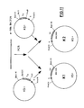
- Aus den Klonen K1 und K2 wurden die VH und VK Gene des MAK BW 2128 mit Hilfe der Restriktionsenzyme Hindlll und BamHI ausgeschnitten, die V-Gen Inserts isoliert und in die mit Hindlll und BamHI geschnittenen Expressionsvektoren D(VH) und I(VK) kloniert. Es wurden die Expressionsvektoren L1 (Fig. 12) und L2 (Fig. 13) isoliert, die ein vollständiges Immunglobulin schwere Ketten Gen (L1) bzw. leichte Ketten Gen (L2) enthalten.
- Die Expressionsvektoren L1 und L2 wurden zusammen mit dem Vektor pR11 H140 (Hudziak, R.M., Laski, F.A., RajBhandary, U.L., Sharp, P.A., Capecchi, M.R. (1982), Cell 31:137-146) der Neomycin Resistenz vermittelt (Fig. 14) und dem Vektor psVdhfr (Lee, F., Mulligan, R., Berg, P., Ringold, G. (1981), Nature 294:228-232), welcher ein Dihydrofolatreduktasegen trägt und Resistenz gegen Methotrexat vermittelt (Fig. 15), in BHK-Zellen transfiziert (Zettlmeißl, G., Wirth, M., Hauser, H., Küpper, H.A. (1988). Behring Inst. Mitt. 82:26-34) und der ausgeprägte Antikörper auf Antigenbindungseigenschaften untersucht, um die Identität der VH und VK Gene des MAK BW 2128 zu bestätigen.
- Die immunhistochemische Spezifitätsanalyse der erfindungsgemäßen Antikörper erfolgte nach der APAAP-Technik (Cordell et al., supra). Tabelle II zeigt das Ergebnis einer immunhistochemischen Spezifitätsanalyse des MAK BW 2128.
- Die Plättchenaktivierung kann über eine quantitative Bestimmung des Aktivierungsmarkers PF4 mit Hilfe des bei H. Pelzer und N. Heimburger (supra) beschriebenen ELISAs gemessen werden. Tabelle III zeigt den Einfluß des MAK BW 2128 auf die Plättchenaktivierung.


Claims (29)
Applications Claiming Priority (2)
| Application Number | Priority Date | Filing Date | Title |
|---|---|---|---|
| DE4203545A DE4203545A1 (de) | 1992-02-07 | 1992-02-07 | Spezifische antikoerper gegen aktivierte plaettchen, ihre herstellung und ihre verwendung in diagnose und therapie |
| DE4203545 | 1992-02-07 |
Publications (3)
| Publication Number | Publication Date |
|---|---|
| EP0554670A2 true EP0554670A2 (de) | 1993-08-11 |
| EP0554670A3 EP0554670A3 (en) | 1994-07-13 |
| EP0554670B1 EP0554670B1 (de) | 2001-09-05 |
Family
ID=6451181
Family Applications (1)
| Application Number | Title | Priority Date | Filing Date |
|---|---|---|---|
| EP93100339A Expired - Lifetime EP0554670B1 (de) | 1992-02-07 | 1993-01-12 | Spezifische Antikörper gegen aktivierte Plättchen, ihre Herstellung und ihre Verwendung in Diagnose und Therapie |
Country Status (9)
| Country | Link |
|---|---|
| US (1) | US5686583A (de) |
| EP (1) | EP0554670B1 (de) |
| JP (1) | JP3821858B2 (de) |
| AT (1) | ATE205255T1 (de) |
| AU (1) | AU669870B2 (de) |
| CA (1) | CA2088889C (de) |
| DE (2) | DE4203545A1 (de) |
| ES (1) | ES2161698T3 (de) |
| NO (1) | NO930406L (de) |
Families Citing this family (3)
| Publication number | Priority date | Publication date | Assignee | Title |
|---|---|---|---|---|
| US7538195B2 (en) * | 2002-06-14 | 2009-05-26 | Immunogen Inc. | Anti-IGF-I receptor antibody |
| US8034904B2 (en) * | 2002-06-14 | 2011-10-11 | Immunogen Inc. | Anti-IGF-I receptor antibody |
| US7655411B2 (en) * | 2002-08-23 | 2010-02-02 | W2 Holdings, Inc. | Thrombospondin fragments and binding agents in the detection, diagnosis and evaluation of cancer |
Family Cites Families (7)
| Publication number | Priority date | Publication date | Assignee | Title |
|---|---|---|---|---|
| US4610960A (en) * | 1983-12-21 | 1986-09-09 | Wisconsin Alumni Research Foundation | Monoclonal antibody to thrombospondin and method for assaying for and isolating thrombospondin |
| EP0387380A1 (de) * | 1989-03-17 | 1990-09-19 | BEHRINGWERKE Aktiengesellschaft | Mutanten von Plasminogenaktivator aus Urin, ihre Herstellung und Verwendung |
| WO1991006858A1 (en) * | 1989-11-03 | 1991-05-16 | The United States Of America, As Represented By The Secretary, U.S. Department Of Commerce | Novel monoclonal antibody against human platelets |
| US5192744A (en) * | 1990-01-12 | 1993-03-09 | Northwestern University | Method of inhibiting angiogenesis of tumors |
| US5200397A (en) * | 1990-02-22 | 1993-04-06 | W. R. Grace & Co.-Conn. | Use of peptide analogs of thrombospondin for the inhibition of angiogenic activity |
| WO1992017499A1 (en) * | 1991-04-08 | 1992-10-15 | Cornell Research Foundation, Inc. | A unique hexapeptide derived from thrombospondin and its uses |
| CA2063055A1 (en) * | 1991-05-22 | 1992-11-23 | Jacob Eyal | Peptides having thrombospondin-like activity and their therapeutic use |
-
1992
- 1992-02-07 DE DE4203545A patent/DE4203545A1/de not_active Withdrawn
-
1993
- 1993-01-12 ES ES93100339T patent/ES2161698T3/es not_active Expired - Lifetime
- 1993-01-12 EP EP93100339A patent/EP0554670B1/de not_active Expired - Lifetime
- 1993-01-12 AT AT93100339T patent/ATE205255T1/de active
- 1993-01-12 DE DE59310204T patent/DE59310204D1/de not_active Expired - Lifetime
- 1993-02-05 AU AU32836/93A patent/AU669870B2/en not_active Ceased
- 1993-02-05 CA CA002088889A patent/CA2088889C/en not_active Expired - Fee Related
- 1993-02-05 NO NO93930406A patent/NO930406L/no unknown
- 1993-02-08 JP JP04317293A patent/JP3821858B2/ja not_active Expired - Fee Related
-
1995
- 1995-06-06 US US08/467,393 patent/US5686583A/en not_active Expired - Lifetime
Also Published As
| Publication number | Publication date |
|---|---|
| ES2161698T3 (es) | 2001-12-16 |
| JP3821858B2 (ja) | 2006-09-13 |
| EP0554670A3 (en) | 1994-07-13 |
| JPH06133768A (ja) | 1994-05-17 |
| ATE205255T1 (de) | 2001-09-15 |
| AU669870B2 (en) | 1996-06-27 |
| NO930406D0 (no) | 1993-02-05 |
| EP0554670B1 (de) | 2001-09-05 |
| CA2088889C (en) | 2009-08-25 |
| DE59310204D1 (de) | 2001-10-11 |
| NO930406L (no) | 1993-08-09 |
| US5686583A (en) | 1997-11-11 |
| DE4203545A1 (de) | 1993-08-12 |
| CA2088889A1 (en) | 1993-08-08 |
| AU3283693A (en) | 1993-08-12 |
Similar Documents
| Publication | Publication Date | Title |
|---|---|---|
| DE69531679T2 (de) | Für e-selectin und p-selectin spezifische kreuzreaktive monoklonale antikörper | |
| DE69233153T2 (de) | Humanisierte monoklonale antikörper | |
| DE68929061T2 (de) | Herstellung humanähnlicher Immunoglobuline und entsprechender Polynukleotide | |
| DE69534235T2 (de) | Cytokin lerk-7 | |
| DE69129896T2 (de) | Gegen cd3 gerichtete antikörper | |
| DE69133326T2 (de) | Verbesserte humanähnlich gemachte immunglobuline | |
| DE69735596T3 (de) | Humanisiertes immunglobulin gegen den alpha4beta7-integrin | |
| DE69605181T3 (de) | Antikörper gegen cd30, die proteolytische spaltung und abgabe des membrangebundenen cd30 antigens verhindern | |
| DE69233482T2 (de) | Verfahren zur Verminderung der Immunogenität der variablen Antikörperdomänen | |
| DE68921364T2 (de) | Verfahren zur Herstellung von Proteinen mittels rekombinanter DNA. | |
| DE3587524T2 (de) | Herstellung von chimären antikörpern. | |
| DE69530763T2 (de) | Antikörper gegen e-selektin | |
| DE69525593T2 (de) | Humanisierte und chimäre monoklonale Antikörper die den Rezeptor für epidermalen Wachstumfaktor (EGF-R) erkennen; diagnostische und therapeutische Verwendung | |
| DE69419721T2 (de) | Rekombinante anti-vla4 antikörpermoleküle | |
| DE69535243T2 (de) | Gegen menschliches interleukin-8 gerichteter, rekonstituierter menschlicher antikörper | |
| DE3853636T3 (de) | Antigene epitope, die sich ausschliesslich auf ige-tragenden b-lymphocyten befinden. | |
| DE69535133T2 (de) | Humanisierte antikörper gegen das leukozytenadhäsionsmolekül vla-4 | |
| EP0538754B1 (de) | Verwendung von Antikörper enthaltenden Präparationen zur Immunsuppression | |
| DE4118120A1 (de) | Tetravalente bispezifische rezeptoren, ihre herstellung und verwendung | |
| DE69331175T2 (de) | Interleukin-2 Rezeptor Gamma-Kette | |
| DE69233480T2 (de) | CD4-gamma2- und CD4-IgG2-Chimären | |
| DE3850654T2 (de) | Immunglobulin-e-kompetitor. | |
| DE68929380T2 (de) | Plättchen-spezifisches chimärisches immunglobulin | |
| DE69020112T2 (de) | Spezifische bindende Wirkstoffe. | |
| DE4332256C1 (de) | Monoklonale Antikörper gegen Leukozyten-spezifische G-Protein-gekoppelte Rezeptoren |
Legal Events
| Date | Code | Title | Description |
|---|---|---|---|
| PUAI | Public reference made under article 153(3) epc to a published international application that has entered the european phase |
Free format text: ORIGINAL CODE: 0009012 |
|
| AK | Designated contracting states |
Kind code of ref document: A2 Designated state(s): AT BE CH DE DK ES FR GB GR IE IT LI LU NL PT SE |
|
| PUAL | Search report despatched |
Free format text: ORIGINAL CODE: 0009013 |
|
| AK | Designated contracting states |
Kind code of ref document: A3 Designated state(s): AT BE CH DE DK ES FR GB GR IE IT LI LU NL PT SE |
|
| 17P | Request for examination filed |
Effective date: 19950113 |
|
| 17Q | First examination report despatched |
Effective date: 19970918 |
|
| RAP1 | Party data changed (applicant data changed or rights of an application transferred) |
Owner name: DADE BEHRING MARBURG GMBH |
|
| GRAG | Despatch of communication of intention to grant |
Free format text: ORIGINAL CODE: EPIDOS AGRA |
|
| RIC1 | Information provided on ipc code assigned before grant |
Free format text: 7C 12N 15/13 A, 7C 12N 5/20 B, 7C 12N 15/62 B, 7C 12P 21/08 B, 7A 61K 39/395 B, 7C 07K 16/28 B, 7G 01N 33/577 B |
|
| GRAG | Despatch of communication of intention to grant |
Free format text: ORIGINAL CODE: EPIDOS AGRA |
|
| GRAH | Despatch of communication of intention to grant a patent |
Free format text: ORIGINAL CODE: EPIDOS IGRA |
|
| GRAH | Despatch of communication of intention to grant a patent |
Free format text: ORIGINAL CODE: EPIDOS IGRA |
|
| GRAA | (expected) grant |
Free format text: ORIGINAL CODE: 0009210 |
|
| AK | Designated contracting states |
Kind code of ref document: B1 Designated state(s): AT BE CH DE DK ES FR GB GR IE IT LI LU NL PT SE |
|
| PG25 | Lapsed in a contracting state [announced via postgrant information from national office to epo] |
Ref country code: IE Free format text: LAPSE BECAUSE OF FAILURE TO SUBMIT A TRANSLATION OF THE DESCRIPTION OR TO PAY THE FEE WITHIN THE PRESCRIBED TIME-LIMIT Effective date: 20010905 |
|
| REF | Corresponds to: |
Ref document number: 205255 Country of ref document: AT Date of ref document: 20010915 Kind code of ref document: T |
|
| RIN1 | Information on inventor provided before grant (corrected) |
Inventor name: SEEMANN, GERHARD Inventor name: KEHREL, BEATE Inventor name: BOSSLET, KLAUS |
|
| REG | Reference to a national code |
Ref country code: CH Ref legal event code: EP |
|
| REF | Corresponds to: |
Ref document number: 59310204 Country of ref document: DE Date of ref document: 20011011 |
|
| REG | Reference to a national code |
Ref country code: IE Ref legal event code: FG4D Free format text: GERMAN |
|
| PG25 | Lapsed in a contracting state [announced via postgrant information from national office to epo] |
Ref country code: PT Free format text: LAPSE BECAUSE OF FAILURE TO SUBMIT A TRANSLATION OF THE DESCRIPTION OR TO PAY THE FEE WITHIN THE PRESCRIBED TIME-LIMIT Effective date: 20011205 Ref country code: DK Free format text: LAPSE BECAUSE OF FAILURE TO SUBMIT A TRANSLATION OF THE DESCRIPTION OR TO PAY THE FEE WITHIN THE PRESCRIBED TIME-LIMIT Effective date: 20011205 |
|
| ET | Fr: translation filed | ||
| PG25 | Lapsed in a contracting state [announced via postgrant information from national office to epo] |
Ref country code: GR Free format text: LAPSE BECAUSE OF FAILURE TO SUBMIT A TRANSLATION OF THE DESCRIPTION OR TO PAY THE FEE WITHIN THE PRESCRIBED TIME-LIMIT Effective date: 20011207 |
|
| REG | Reference to a national code |
Ref country code: ES Ref legal event code: FG2A Ref document number: 2161698 Country of ref document: ES Kind code of ref document: T3 |
|
| REG | Reference to a national code |
Ref country code: GB Ref legal event code: IF02 |
|
| GBT | Gb: translation of ep patent filed (gb section 77(6)(a)/1977) |
Effective date: 20011201 |
|
| REG | Reference to a national code |
Ref country code: IE Ref legal event code: FD4D |
|
| PLBE | No opposition filed within time limit |
Free format text: ORIGINAL CODE: 0009261 |
|
| STAA | Information on the status of an ep patent application or granted ep patent |
Free format text: STATUS: NO OPPOSITION FILED WITHIN TIME LIMIT |
|
| 26N | No opposition filed | ||
| REG | Reference to a national code |
Ref country code: CH Ref legal event code: PFA Owner name: SIEMENS HEALTHCARE DIAGNOSTICS PRODUCTS GMBH Free format text: DADE BEHRING MARBURG GMBH#POSTFACH 11 49#35001 MARBURG (DE) -TRANSFER TO- SIEMENS HEALTHCARE DIAGNOSTICS PRODUCTS GMBH#EMIL-VON-BEHRING-STRASSE 76#35041 MARBURG (DE) Ref country code: CH Ref legal event code: NV Representative=s name: SIEMENS SCHWEIZ AG |
|
| NLT1 | Nl: modifications of names registered in virtue of documents presented to the patent office pursuant to art. 16 a, paragraph 1 |
Owner name: SIEMENS HEALTHCARE DIAGNOSTICS PRODUCTS GMBH |
|
| REG | Reference to a national code |
Ref country code: FR Ref legal event code: CD |
|
| PGFP | Annual fee paid to national office [announced via postgrant information from national office to epo] |
Ref country code: ES Payment date: 20100222 Year of fee payment: 18 |
|
| PGFP | Annual fee paid to national office [announced via postgrant information from national office to epo] |
Ref country code: BE Payment date: 20100204 Year of fee payment: 18 |
|
| PGFP | Annual fee paid to national office [announced via postgrant information from national office to epo] |
Ref country code: DE Payment date: 20100325 Year of fee payment: 18 |
|
| PGFP | Annual fee paid to national office [announced via postgrant information from national office to epo] |
Ref country code: CH Payment date: 20100408 Year of fee payment: 18 |
|
| PGFP | Annual fee paid to national office [announced via postgrant information from national office to epo] |
Ref country code: LU Payment date: 20110110 Year of fee payment: 19 |
|
| PGFP | Annual fee paid to national office [announced via postgrant information from national office to epo] |
Ref country code: FR Payment date: 20110120 Year of fee payment: 19 Ref country code: IT Payment date: 20110126 Year of fee payment: 19 Ref country code: NL Payment date: 20110125 Year of fee payment: 19 Ref country code: SE Payment date: 20110107 Year of fee payment: 19 Ref country code: AT Payment date: 20101209 Year of fee payment: 19 |
|
| PGFP | Annual fee paid to national office [announced via postgrant information from national office to epo] |
Ref country code: GB Payment date: 20110110 Year of fee payment: 19 |
|
| BERE | Be: lapsed |
Owner name: *DADE BEHRING MARBURG G.M.B.H. Effective date: 20110131 |
|
| REG | Reference to a national code |
Ref country code: CH Ref legal event code: PL |
|
| PG25 | Lapsed in a contracting state [announced via postgrant information from national office to epo] |
Ref country code: CH Free format text: LAPSE BECAUSE OF NON-PAYMENT OF DUE FEES Effective date: 20110131 Ref country code: LI Free format text: LAPSE BECAUSE OF NON-PAYMENT OF DUE FEES Effective date: 20110131 |
|
| PG25 | Lapsed in a contracting state [announced via postgrant information from national office to epo] |
Ref country code: BE Free format text: LAPSE BECAUSE OF NON-PAYMENT OF DUE FEES Effective date: 20110131 |
|
| REG | Reference to a national code |
Ref country code: DE Ref legal event code: R119 Ref document number: 59310204 Country of ref document: DE Effective date: 20110802 |
|
| REG | Reference to a national code |
Ref country code: ES Ref legal event code: FD2A Effective date: 20120220 |
|
| PG25 | Lapsed in a contracting state [announced via postgrant information from national office to epo] |
Ref country code: ES Free format text: LAPSE BECAUSE OF NON-PAYMENT OF DUE FEES Effective date: 20110113 |
|
| REG | Reference to a national code |
Ref country code: NL Ref legal event code: V1 Effective date: 20120801 |
|
| REG | Reference to a national code |
Ref country code: SE Ref legal event code: EUG |
|
| GBPC | Gb: european patent ceased through non-payment of renewal fee |
Effective date: 20120112 |
|
| REG | Reference to a national code |
Ref country code: FR Ref legal event code: ST Effective date: 20120928 |
|
| PG25 | Lapsed in a contracting state [announced via postgrant information from national office to epo] |
Ref country code: SE Free format text: LAPSE BECAUSE OF NON-PAYMENT OF DUE FEES Effective date: 20120113 Ref country code: GB Free format text: LAPSE BECAUSE OF NON-PAYMENT OF DUE FEES Effective date: 20120112 |
|
| PG25 | Lapsed in a contracting state [announced via postgrant information from national office to epo] |
Ref country code: IT Free format text: LAPSE BECAUSE OF NON-PAYMENT OF DUE FEES Effective date: 20120112 |
|
| REG | Reference to a national code |
Ref country code: AT Ref legal event code: MM01 Ref document number: 205255 Country of ref document: AT Kind code of ref document: T Effective date: 20120112 |
|
| PG25 | Lapsed in a contracting state [announced via postgrant information from national office to epo] |
Ref country code: FR Free format text: LAPSE BECAUSE OF NON-PAYMENT OF DUE FEES Effective date: 20120131 |
|
| PG25 | Lapsed in a contracting state [announced via postgrant information from national office to epo] |
Ref country code: NL Free format text: LAPSE BECAUSE OF NON-PAYMENT OF DUE FEES Effective date: 20120801 Ref country code: AT Free format text: LAPSE BECAUSE OF NON-PAYMENT OF DUE FEES Effective date: 20120112 |
|
| PG25 | Lapsed in a contracting state [announced via postgrant information from national office to epo] |
Ref country code: DE Free format text: LAPSE BECAUSE OF NON-PAYMENT OF DUE FEES Effective date: 20110802 |
|
| PG25 | Lapsed in a contracting state [announced via postgrant information from national office to epo] |
Ref country code: LU Free format text: LAPSE BECAUSE OF NON-PAYMENT OF DUE FEES Effective date: 20120112 |