EP0582143B1 - Verfahren zum Nachweis von Alzheimer-Krankheit - Google Patents
Verfahren zum Nachweis von Alzheimer-Krankheit Download PDFInfo
- Publication number
- EP0582143B1 EP0582143B1 EP93111756A EP93111756A EP0582143B1 EP 0582143 B1 EP0582143 B1 EP 0582143B1 EP 93111756 A EP93111756 A EP 93111756A EP 93111756 A EP93111756 A EP 93111756A EP 0582143 B1 EP0582143 B1 EP 0582143B1
- Authority
- EP
- European Patent Office
- Prior art keywords
- kda
- μcanp
- isoform
- disease
- alzheimer
- Prior art date
- Legal status (The legal status is an assumption and is not a legal conclusion. Google has not performed a legal analysis and makes no representation as to the accuracy of the status listed.)
- Expired - Lifetime
Links
Images




Classifications
-
- C—CHEMISTRY; METALLURGY
- C12—BIOCHEMISTRY; BEER; SPIRITS; WINE; VINEGAR; MICROBIOLOGY; ENZYMOLOGY; MUTATION OR GENETIC ENGINEERING
- C12N—MICROORGANISMS OR ENZYMES; COMPOSITIONS THEREOF; PROPAGATING, PRESERVING, OR MAINTAINING MICROORGANISMS; MUTATION OR GENETIC ENGINEERING; CULTURE MEDIA
- C12N9/00—Enzymes; Proenzymes; Compositions thereof; Processes for preparing, activating, inhibiting, separating or purifying enzymes
- C12N9/14—Hydrolases (3)
- C12N9/48—Hydrolases (3) acting on peptide bonds (3.4)
- C12N9/50—Proteinases, e.g. Endopeptidases (3.4.21-3.4.25)
- C12N9/64—Proteinases, e.g. Endopeptidases (3.4.21-3.4.25) derived from animal tissue
- C12N9/6421—Proteinases, e.g. Endopeptidases (3.4.21-3.4.25) derived from animal tissue from mammals
- C12N9/6472—Cysteine endopeptidases (3.4.22)
-
- A—HUMAN NECESSITIES
- A61—MEDICAL OR VETERINARY SCIENCE; HYGIENE
- A61K—PREPARATIONS FOR MEDICAL, DENTAL OR TOILETRY PURPOSES
- A61K31/00—Medicinal preparations containing organic active ingredients
- A61K31/185—Acids; Anhydrides, halides or salts thereof, e.g. sulfur acids, imidic, hydrazonic or hydroximic acids
- A61K31/19—Carboxylic acids, e.g. valproic acid
- A61K31/195—Carboxylic acids, e.g. valproic acid having an amino group
-
- A—HUMAN NECESSITIES
- A61—MEDICAL OR VETERINARY SCIENCE; HYGIENE
- A61K—PREPARATIONS FOR MEDICAL, DENTAL OR TOILETRY PURPOSES
- A61K31/00—Medicinal preparations containing organic active ingredients
- A61K31/33—Heterocyclic compounds
- A61K31/335—Heterocyclic compounds having oxygen as the only ring hetero atom, e.g. fungichromin
-
- A—HUMAN NECESSITIES
- A61—MEDICAL OR VETERINARY SCIENCE; HYGIENE
- A61K—PREPARATIONS FOR MEDICAL, DENTAL OR TOILETRY PURPOSES
- A61K38/00—Medicinal preparations containing peptides
- A61K38/16—Peptides having more than 20 amino acids; Gastrins; Somatostatins; Melanotropins; Derivatives thereof
- A61K38/55—Protease inhibitors
-
- C—CHEMISTRY; METALLURGY
- C07—ORGANIC CHEMISTRY
- C07K—PEPTIDES
- C07K16/00—Immunoglobulins [IG], e.g. monoclonal or polyclonal antibodies
- C07K16/40—Immunoglobulins [IG], e.g. monoclonal or polyclonal antibodies against enzymes
-
- G—PHYSICS
- G01—MEASURING; TESTING
- G01N—INVESTIGATING OR ANALYSING MATERIALS BY DETERMINING THEIR CHEMICAL OR PHYSICAL PROPERTIES
- G01N33/00—Investigating or analysing materials by specific methods not covered by groups G01N1/00 - G01N31/00
- G01N33/48—Biological material, e.g. blood, urine; Haemocytometers
- G01N33/50—Chemical analysis of biological material, e.g. blood, urine; Testing involving biospecific ligand binding methods; Immunological testing
- G01N33/68—Chemical analysis of biological material, e.g. blood, urine; Testing involving biospecific ligand binding methods; Immunological testing involving proteins, peptides or amino acids
- G01N33/6893—Chemical analysis of biological material, e.g. blood, urine; Testing involving biospecific ligand binding methods; Immunological testing involving proteins, peptides or amino acids related to diseases not provided for elsewhere
- G01N33/6896—Neurological disorders, e.g. Alzheimer's disease
-
- G—PHYSICS
- G01—MEASURING; TESTING
- G01N—INVESTIGATING OR ANALYSING MATERIALS BY DETERMINING THEIR CHEMICAL OR PHYSICAL PROPERTIES
- G01N2800/00—Detection or diagnosis of diseases
- G01N2800/28—Neurological disorders
- G01N2800/2814—Dementia; Cognitive disorders
- G01N2800/2821—Alzheimer
Definitions
- the present invention relates to the field of medical diagnostics employing antibodies to screen patients for Alzheimer's disease.
- Calcium-activated neutral proteinases are a family of proteases implicated in regulating aspects of signal transduction (Pontremoli, S., et al., Proc. Natl. Acad. Sci. USA 87:3705-3707 (1990); Piggott, M.A., et al., Brain Res . 565 :42-47 (1991); Adunsky, A., et al ., J . Neuroimmunology 33 :167-172 (1991); Suzuki & Ohno, Cell Struct . Funct . 15 :1-6 (1990); Murachi, T., Biochem . Internatl . 18(2) :263-294 (1989)).
- CANPs Limited proteolysis by CANPs is involved in regulating important enzymes, including calcium-dependent protein kinases and protein phosphatases and neurotransmitter enzymes, and in modifying the function of structural proteins of the membrane and membrane skeleton (Togari, A., et al ., Biochem . Biophys. Res . Commun . 134(2) :749-754 (1968); Kishimoto, A., et al ., J . Biol . Chem . 258 :1156-1164 (1983); Tallant, E.A., et al ., Biochem . 27 :2205-2211 (1988); Nixon, R.A., Ann . N . Y . Acad . Sci .
- CANP exists in cells principally as an inactive precursor isoform (Suzuki & Ohno, Cell Struct . Funct . 15 :1-6 (1990); Suzuki, K., et al ., FEBS Lett . 220 :271 (1987)), which is activated by autoproteolytic cleavage of an amino-terminal sequence in the presence of calcium (Suzuki, K., et al ., J . Biochem . 90 :1787-1793 (1981); Inomata, M., et al ., J . Biochem . 98 :407-416 (1985); Zimmerman & Schlaepfer, Biochim . Biophys .
- the invention relates to the discovery that the ratio of precursor to activated isoforms of ⁇ CANP in tissues provides an index of CANP function in vivo .
- the invention relates to a method for the detection of Alzheimer's disease in an individual, comprising
- the invention also relates to a method for the detection of Alzheimer's disease in an individual, comprising
- the invention also relates to a kit which is useful for detecting Alzheimer's disease in a individual suspected of having the disease, comprising a carrier means having in close confinement therein one or more container means, wherein a first container means contains a detectable antibody that is specific for the active 76-kDa isoform of ⁇ CANP and the 80-kDa isoform of ⁇ CANP. Further container means may contain a detectably labelled antibody that is immunoreactive with said detectable antibody that is specific for the active 76-kDa isoform of ⁇ CANP and the 80-kDa isoform of ⁇ CANP. In addition, further container means may contain a means for separating the 76-kDa isoform of ⁇ CANP and the 80-kDa isoform of ⁇ CANP.
- the invention also relates to a kit which is useful for detecting Alzheimer's disease in a individual suspected of having the disease, comprising a carrier means having in close confinement therein two or more container means, wherein a first container means contains a first detectable antibody that is immunoreactive with the 80-kDa isoform of ⁇ CANP and is not immunoreactive with the 76-kDa and 78-kDa isoforms of ⁇ CANP and a second container means contains a second detectable antibody that is immunoreactive with the 76-kDa, 78-kDa and 80-kDa isoforms of ⁇ CANP.
- the first and second detectable antibodies are detectably labelled with different labels that can be discriminated on detection.
- the invention also relates to a method for screening a drug for use in the treatment or prevention of Alzheimer's disease, comprising
- the invention also relates to a method for screening a drug for use in the treatment or prevention of Alzheimer's disease, comprising
- Fig. 1(A) depicts an immunoblot analysis of ⁇ CANP in prefrontal cortex from two control (CONT) and two Alzheimer (AD) patients.
- Fig. 1(B) depicts a gel electrophoresis showing the autolysis of ⁇ CANP from human erythrocytes in vitro .
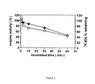
- Fig. 2 depicts a graph showing the relationship between enzyme activity and ⁇ CANP content.
- Fig. 3 depicts an immunoblot analysis of ⁇ CANP autolysis in human brain slices (prefrontal cortex).
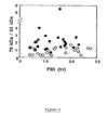
- Fig. 4 depicts a graph showing the ratio of 76 kDa/80 kDa isoforms of ⁇ CANP in prefrontal cortex from control individuals (O) and AD patients ( ⁇ ), plotted against postmortem interval (PMI).
- Fig. 5 depicts bar graphs showing the ⁇ CANP isoform ratios (76 kDa/80 kDa) in brain regions from control, Alzheimer and Huntington disease (HD) brains.
- A Prefrontal cortex.
- B Putamen.
- C Cerebellum.
- ⁇ CANP calcium-activated neutral proteinases
- ⁇ CANP micromolar calcium levels
- the ratios of the activated isoform of ⁇ CANP to the precursor isoforms were measured immunochemically in regions of postmortem human brain. This ratio was elevated 3-fold in the prefrontal cortex from patients with Alzheimer disease but not from those with Huntington disease. Other regions of Alzheimer brain (putamen and cerebellum) where neuronal degeneration is considered minimal in Alzheimer disease also displayed significantly increased ⁇ CANP activation.
- the invention relates to the discovery that elevated levels of the active isoform of ⁇ CANP is associated with Alzheimer's disease.
- the invention relates to a method for the detection of Alzheimer's disease in an individual, comprising
- the tissue sample may be a brain biopsy or, more preferably, a fibroblast sample. Where a brain biopsy sample is obtained, it preferably obtained from the frontal cortex, putamen or cerebellum. When obtained from the frontal cortex, it is possible to discriminate between Alzheimer's disease and Huntington's disease. When nervous system tissue is analyzed according to the present invention, about 0.1 to about 0.5 g of tissue is needed to carry out the analysis.
- a sample of fibroblasts is obtained from an individual suspected of having Alzheimer's disease.
- Fibroblasts may be obtained from individuals according to the procedure described by Willers, I. et al., Pathobiology 59:357-360 (1991). See also Ham and McKeehan, In Vitro 14 :11-22 (1978), who teach methods for growing fibroblasts in cell culture. About 0.05 to about 0.1 g of fibroblasts is needed to carry out the analysis.
- the tissue sample After obtaining the tissue sample, it is homogenized in a physiological buffer, centrifuged and the supernatant tested for the 76-kDa and 80-kDa isoforms of ⁇ CANP.
- the supernatant is first subjected to fractionation to remove potential interfering substances. This may be carried out by applying the supernatant to a DEAE-cellulose column, and eluting the column with a buffer containing increase amounts of a salt such as potassium chloride.
- the fractions eluting off the column may be subject to electrophoresis to separate the 76-kDa, 78-kDa and 80-kDa isoforms, followed by electrotransfer to a membrane (e.g., nitrocellulose) and immunoblot analysis with detectable antibodies that are immunoreactive with one or more of the 76-kDa, 78-kDa and 80-kDa isoforms.
- the detectable antibody may be detectably labelled as described herein or may be detected by contacting with a second antibody that is immunoreactive therewith, e.g. where the first antibody is from a first animal species and the second antibody is from a second animal species and is immunoreactive with the antibodies from the first animal species.
- Antibodies which are immunoreactive with the 76-kDa, 78-kDa and 80-kDa isoforms are taught, for example, by Samis, J.A., et al., Biochem. J. 246 :481-488 (1987); and Elce, J.S., et al., Biochem. J. 261 :1039-1042 (1989). It is also possible to employ antibodies that are selective for only the 76-kDa isoform. See Saido, T.C. et al., J. Biochem . 111 :81-86 (1992).
- the ratio of 76-kDa and 80-kDa isoforms is determined with detectably labelled antibodies that discriminate between the 80-kDa isoform and the 76-kDa and 78-kDa isoforms.
- the 80-kDa isoform is converted to the 76-kDa and 78-kDa isoforms by proteolysis, the N-terminus of the 80-kDa isoform is lost.
- a second antibody which is immunochemically reactive with the 76-kDa, 78-kDa and 80-kDa isoforms can be used to quantitate total ⁇ CANP. Since the amount of the 78-kDa isoform does not change significantly in Alzheimer diseased brain, the ratio of the second antibody to the first antibody correlates to the ratio of the 76-kDa to 80-kDa isoforms.
- the first antibody is raised against a region in the first 21 N-terminal amino acids of ⁇ CANP.
- the amino acid sequence of ⁇ CANP is taught by Imajoh, S. et al., Biochem. 27:8122-8128 (1988). Given the amino acid sequence of ⁇ CANP, it is possible to obtain N-terminal fragments of ⁇ CANP and prepare monoclonal or polyclonal antibodies thereto. For example, one may prepare peptide segments of 8 amino acids or more by means of solid phase synthesis, conjugate the peptide segments to an immunogenic carrier, an immunize an animal therewith. Larger peptide fragments are capable of inducing, per se, an immune response.
- immunogenic carriers include, but are not limited to, proteins such as bovine serum albumin, diphtheria toxoid, tetanus toxoid, edestin, keyhole-limpet hemocyanin, Pseudomonal Toxin A, choleragenoid, cholera toxin, pertussis toxin, viral proteins, and eukaryotic proteins such as interferons, interleukins, or tumor necrosis factor.
- proteins may be obtained from natural or recombinant sources according to methods known to those skilled in the art.
- the immunogenic carrier may comprise a protein fragment comprising at least the immunogenic portion of the molecule.
- immunogenic macromolecules which may be used in the practice of the invention include, but are not limited to, polysaccharides, tRNA, nonmetabolizable synthetic polymers such as polyvinylamine, polymethacrylic acid, polyvinylpyrrolidone, mixed polycondensates (with relatively high molecular weight) of 4'4'-diaminodiphenyl-methane-3,3'-dicarboxylic acid and 4-nitro-2-aminobenzoic acid (See Sela, M., Science 166 :1365-1374 (1969)) or glycolipids, lipids or carbohydrates.
- the immunogenic carrier is a protein.
- the animal immunized with the N-terminal segment of ⁇ CANP may then be bled and the polyclonal sera isolated. See Vitto and Nixon, J . Neurochem . 47 :1039-1051 (1986).
- monoclonal antibodies may be prepared according to the well known methods of Khöler and Milstein, Nature 256 :495-497 (1975) where the spleen of the immunized animal is removed, and the cells fused with an immortal cell line to give hybridoma cells.
- Hybridomas secreting the desired antibodies may be obtained by subcloning of the hybridomas in HAT selection media and screening of the resultant clones for immunogenic activity with the 80-kDa isoform and lack of immunoreactivity with the 76-kDa and 78-kDa isoforms.
- N-terminal segment of ⁇ CANP is the first 21 amino acids having the following amino acid sequence [SEQ ID NO. 1]:
- the antibodies employed in the practice of the present invention may be detectably labelled in a manner known per se.
- labels and methods of labeling known to those of ordinary skill in the art.
- Examples of the types of labels which can be used in the present invention include, but are not limited to, enzymes, radioisotopes, dyes, fluorescent compounds, chemiluminescent compounds, bioluminescent compounds and metal chelates.
- Those of ordinary skill in the art will know of other suitable labels for binding to the antibodies, or will be able to ascertain the same by the use of routine experimentation.
- the binding of these labels to the antibodies can be accomplished using standard techniques commonly known to those of ordinary skill in the art.
- One of the ways in which the antibodies can be detectably labeled is by linking the same to an enzyme.
- This enzyme when later exposed to its substrate, will react with the substrate in such a manner as to produce a chemical moiety which can be detected as, for example, by spectrophotometric, fluorometric or by visual means.
- Enzymes which can be used to detectably label antibodies include, but are not limited to, malate dehydrogenase, staphylococcal nuclease, delta-V-steroid isomerase, yeast alcohol dehydrogenase, alpha-glycerophosphate dehydrogenase, triose phosphate isomerase, horseradish peroxidase, alkaline phosphatase, asparaginase, glucose oxidase, beta-galactosidase, ribonuclease, urease, catalase, glucose-VI-phosphate dehydrogenase, glucoamylase and acetylcholine esterase.
- the antibodies may also be labeled with a radioactive isotope which can be determined by such means as the use of a gamma counter or a scintillation counter or by autoradiography.
- Isotopes which are particularly useful for the purpose of the present invention include: 3 H, 125 I, 131 I, 32 P, 35 S, 14 C or the radioisotopes of lead, mercury, thallium, technetium or indium ( 203 Pb, 198 Hg, 201 Tl, 99m Tc, 111 In).
- fluorescent labelling compounds are fluorescein isothiocyanate, rhodamine, phycoerythrin, phycocyanin, allophycocyanin, o-phthaldehyde and fluorescamine.
- the antibodies can also be detectably labeled using fluorescent emitting metals such as 152 Eu, or others of the lanthanide series. These metals can be attached to the antibodies using such metal chelating groups as diethylenetriaminepentaacetic acid (DTPA) or ethylenediamine-tetraacetic acid (EDTA).
- DTPA diethylenetriaminepentaacetic acid
- EDTA ethylenediamine-tetraacetic acid
- Labelling is also possible by means of a paramagnetic contrast agent which is detectable in a MRI (magnetic resonance imaging) system. It is possible to use gadolinium, cobalt, nickel, manganese or iron complexes by means of which conjugates may be provided as diagnostic agents which are detectable in a MRI system. A strong magnetic field is used in such systems in order to adjust the nuclear spin vectors of the atoms in the organism. Then the field is destroyed which causes the nuclei to return to their initial state. This process is observed and recorded.
- MRI magnetic resonance imaging
- the antibodies also can be detectably labeled by coupling them to a chemiluminescent compound.
- the presence of the chemiluminescent-tagged antibody is then determined by detecting the presence of luminescence that arises during the course of a chemical reaction.
- particularly useful chemiluminescent labeling compounds are luminol, isoluminol, theromatic acridinium ester, imidazole, acridinium salt and oxalate ester.
- Bioluminescence is a type of chemiluminescence found in biological systems in which a catalytic protein increases the efficiency of the chemiluminescent reaction. The presence of a bioluminescent protein is determined by detecting the presence of luminescence. Important bioluminescent compounds for purposes of labeling are luciferin, luciferase and aequorin.
- Another technique which may also result in greater sensitivity when used in conjunction with the present invention consists of coupling the antibody to low molecular weight haptens.
- the haptens can then be specifically detected by means of a second reaction.
- Amplification strategies may be readily applied to these and other labels.
- the detectable antibodies are ideally suited for the preparation of a kit.
- a kit may comprise a carrier means being compartmentalized to receive in close confinement therein one or more container means such as vials, tubes and the like, each of said container means comprising the separate elements of the assay.
- container means such as vials, tubes and the like
- each of said container means comprising the separate elements of the assay.
- there may be a container means containing a detectable antibody specific for all isoforms of ⁇ CANP, and further container means containing, for example, standard serial dilutions of the 76-kDa, 78-kDa and 80-kDa isoforms of ⁇ CANP in solution.
- the detectable antibody may be detectably labelled.
- a further container means may contain a means for carrying out an electrophoretic separation of the 76-kDa, 78-kDa and 80-kDa isoforms of ⁇ CANP.
- the standard solutions of ⁇ CANP may be used as standards for the electrophoretic separation of the ⁇ CANP isoforms of a tissue sample and to prepare standard curves with the concentration of the 76-kDa, 78-kDa and 80-kDa isoforms of ⁇ CANP plotted on the abscissa and the detection signal on the ordinate.
- results obtained from the supernatant of a tissue sample such as brain cells or fibroblasts may be interpolated from such a plot to give the concentration of the 76-kDa, 78-kDa and 80-kDa isoforms of ⁇ CANP and, thus, an indication of whether the individual has Alzheimer's disease.
- Further container means may comprise a detectably labelled antibody that is immunoreactive with the detectable antibody that is specific for the active 76-kDa isoform of ⁇ CANP and the 80-kDa isoform of ⁇ CANP.
- the kit may comprise a carrier means being compartmentalized to receive in close confinement therein a container means containing a first detectable antibody specific for only the 80-kDa isoform of ⁇ CANP and a second detectable antibody which is specific for all isoforms of ⁇ CANP.
- the first and second detectable antibodies are labelled with different labels which can be discriminated by different detection methods.
- Further container means may contain standard serial dilutions of the 76-kDa, 78-kDa and 80-kDa isoforms of ⁇ CANP in solution.
- the kit may comprise a carrier means being compartmentalized to receive in close confinement therein a container means containing a first detectable antibody specific for only the 80-kDa isoform of ⁇ CANP and a second detectable antibody which is specific for the 76-kDa isoform of ⁇ CANP.
- the first and second detectable antibodies are labelled with different labels which can be discriminated by different detection methods.
- Further container means may contain standard serial dilutions of the 76-kDa and 80-kDa isoforms of ⁇ CANP in solution.
- the means for separating/identifying the ⁇ CANP isoforms may include slab gels that can be used to electrophoretically separate the ⁇ CANP isoforms.
- slab gels are sodium dodecyl sulfate (SDS)-polyacrylamide gels.
- the specific concentrations of the isoforms, the temperature and time of incubation, as well as other assay conditions may be varied, depending on various factors including the concentration of the ⁇ CANP isoforms, the nature of the sample, and the like. Those skilled in the art will be able to determine operative and optimal assay conditions by employing routine experimentation.
- Detection of the detectable antibodies and, thus, the isoforms of ⁇ CANP may be accomplished with a scintillation counter if, for example, the detectable antibodies are labelled with a radioactive gamma emitter, or by a fluorometer if the detectable label is a fluorescence emitter.
- the detection can be accomplished by colorimetric methods which employ a substrate for the enzyme. Detection may be accomplished by comparison with the isoform standards.
- first and second antibodies as described herein, are employed to determine the relative ratio of the 76-kDa ⁇ 80-kDa isoforms
- the two antibodies may be detectably labelled with two different labels with may be separately detected.
- the ratio may be obtained from a single incubation with both antibodies.
- the sample comprising the ⁇ CANP isoforms may be immobilized to a solid phase carrier such as nitrocellulose or polyvinylidene difluoride membrane (Immobilon-P), contacted with the first and second detectably labelled antibodies, and the amount of bound label determined.
- Other steps such as washing, stirring, shaking, filtering and the like may be added to the assay as is customary or necessary for the particular situation.
- the invention also relates to a method for screening a drug for use in the treatment or prevention of Alzheimer's disease, comprising
- the first and second tissue samples are obtained premortem from the brain of an animal or postmortem from an individual having Alzheimer's disease.
- the invention relates to a method for screening a drug for use in the treatment or prevention of Alzheimer's disease, comprising
- the tissue sample may be first homogenized in a buffer and centrifuged to give a supernatant and then the relative ratio of the active 76-kDa isoform of ⁇ CANP to the 80-kDa isoform in the supernatant is then determined.
- the relative ratio of the active 76-kDa isoform of ⁇ CANP to the 80-kDa isoform may be determined by
- the relative ratio of the active 76-kDa isoform of ⁇ CANP to the 80-kDa isoform may be determined by
- step (c) the tissue sample is first incubated with calcium and the drug for a time sufficient to alter the 76-kDa/80-kDa ratio.
- the drug and calcium are contacted with the tissue sample for about 5 to about 60 min.
- the concentration of calcium may range from 0-5 mM.
- the concentration of the drug may range from 0.001 to about 1000 ⁇ M.
- the tissue sample is first incubated with the drug at about 4°C and is then incubated at about 30 °C with calcium and the drug.
- a calcium ionophore such as A23187 is employed at a concentration range of 1-100 ⁇ M.
- the tissue sample is then homogenized in a buffer and centrifuged to give a supernatant, and then the relative ratio of the active 76-kDa isoform of ⁇ CANP to the 80-kDa isoform in said supernatant is determined.
- a monoclonal antibody raised against human ⁇ CANP (Samis, J.A., et al., Biochem . J. 246: 481-488 (1987); and Elce, J.S., et al., Biochem . J. 261 :1039-1042 (1989) may be used to detect by Western blot analysis the three isoforms of the catalytic subunit of ⁇ CANP in human brain extracts. These isoenzymes exhibit molecular masses on SDS polyacrylamide gels of 80 kDa, 78 kDa and 76 kDa identical to those in human erythrocytes (Samis, J.A., et al., Biochem . J.
- Antiserum prepared against this 34-mer specifically cross-reacted with the 80-kDa isoform of ⁇ CANP on Western blot analyses of 30,000 xg supernatant extract from human brain.
- the three ⁇ CANP isoforms were quantitated by immunoassay of extracts of brain tissue from 22 individuals who had premortem diagnoses of dementia and met neuropathological criteria for Alzheimer disease, and 17 individuals who were free of clinical neurologic disease (Khachaturian, Z.S., Arch. Neurol. 42 :1097-1105 (1985)) and neuropathological evidence (Mirra, S.S., et al., Neurology 41 :479-486 (1991)) for Alzheimer disease.
- the Alzheimer and control groups were matched with respect to age of the individual (75.8 ⁇ 2.0 years vs. 68.9 ⁇ 2.6 years, respectively), and postmortem interval for brain samples (12.2 ⁇ 1.3 hr vs. 12.0 ⁇ 1.8 hr, respectively).
- the content of cytosolic 80-kDa ⁇ CANP was substantially lower in the Alzheimer group than in controls (22.7 ⁇ 1.5% vs. 37.2 ⁇ 2.2%, p ⁇ 0.001, unpaired t-test).
- autolytically activated (76 kDa) ⁇ CANP was increased nearly two-fold in Alzheimer brain (41.2 ⁇ 1.6%) than in controls (26.6 ⁇ 2.2%) (p ⁇ 0.001).
- the 78-kDa ⁇ CANP isoform was unchanged.
- the ratio of 76-kDa to 80-kDa ⁇ CANP for individual brain samples was nearly 3-fold higher in the Alzheimer group (2.20 ⁇ 0.39 vs. 0.81 ⁇ 0.10).
- the sum of the three ⁇ CANP isoforms was not changed (Table 1).
- the degree of abnormal ⁇ CANP activation was comparable in Alzheimer brains at all postmortem intervals.
- the possibility cannot be excluded, however, that isoform ratios change significantly within the first few hours after death.
- the degree of ⁇ CANP activation also did not significantly correlate with age within the range studied for any group.
- ⁇ CANP activation was increased 50% (p ⁇ 0.05) in samples of putamen from Huntington patients (HD) (Vonsattel, J.-P., et al ., J . Neuropathol . Exp . Neurol . 44 :559-577 (1985); Roos, R.A.C., et al ., J . Neurol . Neurosurg . Psychiatry 48 :422-425 (1985)), where neuronal cell loss is considerable (Fig. 5b).
- ⁇ CANP activation is not a consequence simply of end-stage neuronal degeneration but reflects a more widespread metabolic alteration that precedes and possibly contributes to neuronal degeneration.
- ⁇ CANP activation in brain is consistent with evidence in fibroblasts of Alzheimer patients for increased degradation of spectrin (Peterson, C., et al ., Neurosci . Lett . 121 :239-243 (1991)) and diminished content of protein kinase C (Saitoh, T., et al ., Lab . Invest . 64 :596-616 (1991)) which are two preferred substrates of CANPs.
- Brain spectrin breakdown is also increased in cortex and hippocampus of Alzheimer brain (Masliah, E., et al ., Brain Res . 531 :36-44 (1990)).
- abnormal ⁇ CANP activation may have additional consequences in neurons that bear on the development of neurofibrillary pathology and ⁇ -amyloid formation.
- These include the abnormal processing of cytoskeletal proteins and other sensitive CANP substrates, which include the amyloid precursor protein (APP) (Siman et al ., in Neurotoxicity of Excitatory Amino Acids , A. Guidotti, Ed. (Raven Press, New York, 1990), pp. 145-161; Lee et al ., Proc . Natl . Acad .
- APP amyloid precursor protein
- Fig. 1(A) depicts the immunoblot analysis of ⁇ CANP in prefrontal cortex from two control (CONT) and two Alzheimer (AD) patients.
- Prefrontal cortex 0.5 g
- Prefrontal cortex 0.5 g
- Prefrontal cortex 0.5 g
- the homogenate was centrifuged at 15,000 x g for 30 min at 4°C and the supernatant was applied to a column (1.0 x 15 cm) of DEAE-cellulose (DE-52, Whatman®) equilibrated with Buffer A.
- the column extensively washed with Buffer A, was eluted with stepwise increments of KCI from 0 to 3 mM.
- ⁇ CANP was eluted from the column by 0.15 M KCI in Buffer A. These fractions were subjected to 7.5% SDS-PAGE according to the method of Laemmli, Nature 227 :680-685 (1970). Electroblotting was performed according to Towbin et al., Proc. Natl . Acad. Sci.
- Fig. 1(B) depicts the immunoblot analysis of the autolysis products of ⁇ CANP from human erythrocytes in vitro.
- Human erythrocyte ⁇ CANP was purified from normal adult blood as described before (Inomata, M., et al., J. Biochem.
- Fig. 2 shows the relationship between enzyme activity and ⁇ CANP content.
- 1 mM EGTA was added to stop the reaction.
- Half of each sample was applied to SDS/PAGE and immunoreactivity of each ⁇ CANP isoform was quantified using a Bio-Rad Video Densitometer Model 620. The other half was used to measure enzyme activity as previously described (Takeuchi, K.S., et al ., J . Neurochem . 92 :526-532 (1992)) by adding [ 14 C]azocacein as substrate and 4 mM CaCl 2 (final free calcium concentration was 125 ⁇ M). Note that the 78 kDa isoform completely disappears by 1 min of incubation. O: protease activity.
- ⁇ ⁇ CANP 76 kDa content.
- Fig. 3 shows ⁇ CANP autolysis in human brain slices. Sections of fresh postmortem human brain (prefrontal cortex) (200 ⁇ M) were incubated at 37°C for 60 min with Buffer A with or without CaCl 2 (final concentration 5 mM). Slices were then rinsed with Buffer A (1 ml x 2), homogenized, and centrifuged 15,000 x g for 30 min at 4°C. The supernatant was subjected to immunoblot analysis as described above.
- Human erythrocyte ⁇ CANP (lane 1); human brain ⁇ CANP from unincubated slices (lane 2) or slices incubated in the absence of exogenous calcium (lane 3), 5 mM CaCl 2 (lane 4), or with 5 mM CaCl 2 plus 10 ⁇ M leupeptin (lane 5).
- Fig. 4 depicts a graph showing the ratio of 76 kDa/80 kDa isoforms of ⁇ CANP in prefrontal cortex from control individuals (O) and AD patients ( ⁇ ), plotted against postmortem interval (PMI). Immunoassay of individual isoforms (Fig. 1) was performed as described in the legend to Table 1.
- Fig. 5 depicts the ⁇ CANP isoform ratios (76 kDa/80 kDa) in brain regions from control, Alzheimer and Huntington disease (HD) brains measured by Western blot immunoassay as described in Example 3.
- B Putamen: The mean values ⁇ S.E.M.
- fibroblasts from FAD cohorts were carried out in collaboration with Dr. Jesse Sisken (University of Kentucky Medical School), who provided fibroblast samples from control (C) and AD-affected individuals.
- Stocks were maintained in DMEM (Gibco) supplemented with 15% FBS (Sigma).
- Cells were set up on Corning 100-mm culture plates in DMEM (Gibco) supplemented with 17% FBS (Gibco). After 1-2 days, the cultures were considered “subconfluent" and were harvested. The cells were gently scraped off the surface with a sterile rubber policeman.
- the cells were then transferred to a centrifuge tube, centrifuged, and the cell pellet was resuspended in medium with FBS, transferred to a cryovial, recentrifuged and frozen. Alternatively, cells were harvested by addition of 0.05% trypsin-EDTA, neutralization by medium with FBS. Immunoassay of the individual isoforms was performed as described in the legend to Table 1. The results are depicted in Fig. 6 (panels A and B).
Landscapes
- Health & Medical Sciences (AREA)
- Life Sciences & Earth Sciences (AREA)
- Chemical & Material Sciences (AREA)
- Engineering & Computer Science (AREA)
- Medicinal Chemistry (AREA)
- General Health & Medical Sciences (AREA)
- Biomedical Technology (AREA)
- Molecular Biology (AREA)
- Organic Chemistry (AREA)
- Immunology (AREA)
- Proteomics, Peptides & Aminoacids (AREA)
- Genetics & Genomics (AREA)
- Veterinary Medicine (AREA)
- Biochemistry (AREA)
- Public Health (AREA)
- Animal Behavior & Ethology (AREA)
- Bioinformatics & Cheminformatics (AREA)
- Pharmacology & Pharmacy (AREA)
- Epidemiology (AREA)
- Microbiology (AREA)
- Hematology (AREA)
- Zoology (AREA)
- Urology & Nephrology (AREA)
- Wood Science & Technology (AREA)
- Biotechnology (AREA)
- Biophysics (AREA)
- General Physics & Mathematics (AREA)
- Neurosurgery (AREA)
- Cell Biology (AREA)
- General Engineering & Computer Science (AREA)
- Gastroenterology & Hepatology (AREA)
- Food Science & Technology (AREA)
- Physics & Mathematics (AREA)
- Analytical Chemistry (AREA)
- Neurology (AREA)
- Pathology (AREA)
- Peptides Or Proteins (AREA)
- Medicines That Contain Protein Lipid Enzymes And Other Medicines (AREA)
- Investigating Or Analysing Biological Materials (AREA)
- Measuring Or Testing Involving Enzymes Or Micro-Organisms (AREA)
Claims (43)
- Verfahren zur Feststellung der Alzheimer-Krankheit in einem Individuum, umfassend(a) Erhalten einer Gewebeprobe von einem Individuum, von dem angenommen wird, daß es die Alzheimer-Krankheit hat, und(b) Bestimmen des relativen Verhältnisses der aktiven 76-kDa µCANP-Isoform zu der 80-kDA Isoform in der Gewebeprobe, wobei ein erhöhtes 76-kDa/80-kDA Verhältnis im Vergleich zu einer Kontrollgruppe von Individuen, die nicht die Alzheimer-Krankheit haben, bestätigt, daß das Individuum, von dem angenommen wird, daß es die Alzheimer-Krankheit hat, die Alzheimer-Krankheit hat.
- Verfahren nach Anspruch 1, worin die Gewebeprobe von dem Gehirn des Individuums erhalten wird.
- Verfahren nach Anspruch 1, worin die Gewebeprobe von der Stirnlappenkortex des Individuums erhalten wird.
- Verfahren nach Anspruch 1, worin die Gewebeprobe eine von dem Individuum erhaltene Fibroblasten-Gewebeprobe ist.
- Verfahren nach Anspruch 1, worin in Schritt (b) die Gewebeprobe zunächst unter Erhalt eines Überstandes in einem Puffer homogenisiert und zentrifugiert wird und anschließend das relative Verhältnis der aktiven 76-kDa µCANP-Isoform zu der 80-kDA Isoform in dem Überstand bestimmt wird.
- Verfahren nach Anspruch 5, worin das relative Verhältnis der aktiven 76-kDa µCANP-Isoform zu der 80-kDA Isoform bestimmt wird durch(c) Trennen der µCANP-Isoformen durch Gelelektrophorese,(d) Kontaktieren der getrennten 76-kDa µCANP-Isoform und der 80-kDA µCANP-Isoform mit mindestens einem nachweisbaren Antikörper, der mit diesen immunoreaktiv ist,(e) Feststellen der Menge des nachweisbaren an die 76-kDa und die 80-kDA µCANP-Isoformen gebundenen Antikörpers, um ein relatives Signal für jede Isoform zu erhalten, und(e) Dividieren des relativen Signals entsprechend der 76-kDa Isoform durch das relative Signal entsprechend der 80-kDA Isoform, um das Verhältnis zu erhalten.
- Verfahren nach Anspruch 6, worin der nachweisbare Antikörper feststellbar markiert wird, und die Menge des nachweisbar markierten Antikörpers durch Feststellen der Markierung bestimmt wird.
- Verfahren nach Anspruch 6, worin der nachweisbare Antikörper durch Kontaktieren des nachweisbaren Antikörpers mit einem zweiten nachweisbar markierten Antikörper, der mit diesem nachweisbaren Antikörper immunoreaktiv ist, festgestellt wird.
- Verfahren zur Feststellung der Alzheimer-Krankheit in einem Individuum, umfassend(a) Erhalten einer Gewebeprobe von einem Individuum, von dem angenommen wird, daß es die Alzheimer-Krankheit hat, und(b) Bestimmen des relativen Verhältnisses der aktiven 76-kDa µCANP-Isoform zu den Gesamtmengen der 76-kDA, 78-kDA und 80-kDA Isoformen, wobei ein erhöhtes Verhältnis von 76-kDa/76-kDa + 78-kDa ± 80-kDA im Vergleich zu einer Kontrollgruppe von Individuen, die nicht die Alzheimer-Krankheit haben, bestätigt, daß das Individuum, von dem angenommen wird, daß es die Alzheimer-Krankheit hat, die Alzheimer-Krankheit hat.
- Verfahren nach Anspruch 9, worin die Gewebeprobe von dem Gehirn des Individuums erhalten wird.
- Verfahren nach Anspruch 9, worin die Gewebeprobe von der Stirnlappenkortex des Individuums erhalten wird.
- Verfahren nach Anspruch 9, worin die Gewebeprobe eine von dem Individuum erhaltene Fibroblasten-Gewebeprobe ist.
- Verfahren nach Anspruch 9, worin in Schritt (b) die Gewebeprobe zunächst unter Erhalt eines Überstandes in einem Puffer homogenisiert und zentrifugiert wird und anschließend das relative Verhältnis der aktiven 76-kDa µCANP-Isoform zu den 76-kDA, 78-kDA und 80-kDA Isoformen in dem Überstand bestimmt wird.
- Verfahren nach Anspruch 13, worin das relative Verhältnis der aktiven 76-kDA µCANP-Isoform zu den 76-kDA, 78-kDA und 80-kDA Isoformen bestimmt wird durch(c) Kontaktieren des Überstandes mit einem ersten und einem zweiten nachweisbar markierten Antikörper, wobei der erste nachweisbare Antikörper nur mit der 80-kDA µCANP-Isoform und der zweite nachweisbare Antikörper mit den 76-kDA, 78-kDA und 80-kDA µCANP-Isoformen immunoreaktiv ist,(d) Feststellen der Menge des nachweisbaren ersten und zweiten Antikörpers, der an die jeweiligen µCANP-Isoformen bindet, um ein relatives Signal zu erhalten, und(e) Dividieren des relativen Signals entsprechend den 76-kDA, 78-kDA und 80-kDA Isoformen durch das relative Signal entsprechend der 80-kDA Isoform, um das Verhältnis zu erhalten.
- Verfahren nach Anspruch 14, worin der erste und zweite nachweisbare Antikörper feststellbar markiert werden, und die Menge der nachweisbar markierten Antikörper durch Feststellen der jeweiligen Markierungen bestimmt wird.
- Verfahren nach Anspruch 14, worin der erste und zweite nachweisbare Antikörper festgestellt werden durch Kontaktieren derselben mit mindestens einem nachweisbar markierten Antikörper, der mit dem ersten und zweiten nachweisbaren Antikörper immunoreaktiv ist, und Feststellen der Markierung,.
- Kit, das zur Feststellung der Alzheimer-Krankheit in einem Individuum, von dem angenommen wird, daß es die Alzheimer-Krankheit hat, nützlich ist, umfassend eine Trägereinrichtung, die einen oder mehrere Behältereinrichtungen mit enger Begrenzung hat, worin der erste Behälter einen nachweisbaren Antikörper enthält, der mit der aktiven 76-kDa µCANP-Isoform und der 80-kDa µCANP-Isoform immunoreaktiv ist.
- Kit nach Anspruch 17, worin der nachweisbare Antikörper feststellbar markiert ist.
- Kit nach Anspruch 17, außerdem umfassend einen zweiten Behälter, der einen nachweisbaren Antikörper enthält, der mit dem nachweisbaren Antikörper immunoreaktiv ist, der mit der aktiven 76-kDa µCANP-Isoform und der 80-kDa µCANP-Isoform immunoreaktiv ist.
- Kit nach Anspruch 17, außerdem umfassend einen dritten Behälter, der ein Mittel zum Trennen der 76-kDa µCANP-Isoform und der 80-kDa µCANP-Isoform enthält.
- Kit, das zur Feststellung der Alzheimer-Krankheit in einem Individuum, von dem angenommen wird, daß es die Alzheimer-Krankheit hat, nützlich ist, umfassend eine Trägereinrichtung, die zwei oder mehrere Behältereinrichtungen mit enger Begrenzung hat, worin der erste Behälter einen ersten nachweisbaren Antikörper enthält, der mit der 80-kDa µCANP-Isoform und nicht mit den 76-kDa und 80-kDa µCANP-Isoformen immunoreaktiv ist, und ein zweiter Behälter einen zweiten nachweisbaren Antikörper enthält, der mit den 76-kDa, 78-kDa und 80-kDa µCANP-Isoformen immunoreaktiv ist.
- Kit nach Anspruch 21, worin der erste und zweite nachweisbare Antikörper mit verschiedenen Markierungen, die beim Nachweis unterschieden werden können, nachweisbar markiert sind.
- Kit, das zur Feststellung der Alzheimer-Krankheit in einem Individuum, von dem angenommen wird, daß es die Alzheimer-Krankheit hat, nützlich ist, umfassend eine unterteilte Trägereinrichtung, die mit enger Begrenzung eine Behältereinrichtung enthält, die einen ersten nachweisbaren Antikörper, der nur für die 80-kDa µCANP-Isoform spezifisch ist und einen zweiten nachweisbaren Antikörper, der nur für die 76-kDa µCANP-Isoform spezifisch ist, enthält.
- Kit nach Anspruch 23, worin der erste und zweite nachweisbare Antikörper mit verschiedenen Markierungen, die beim Nachweis unterschieden werden können, nachweisbar markiert sind.
- Kit nach Anspruch 23, außerdem umfassend einen Behälter, der Standard-Serienverdünnungen der 76-kDa und 80-kDa µCANP-Isoformen in Lösung enthält.
- Verfahren zum Screenen eines Medikaments zur Verwendung bei der Behandlung oder Vorbeugung der Alzheimer-Krankheit, umfassendwobei bestätigt wird, daß das Medikament zum Behandeln oder Vorbeugen der Alzheimer-Krankheit nützlich ist, wenn sich das 76-kDa/80-kDA Verhältnis der Tierhirnprobe, die mit dem Medikament kontaktiert wird, wie in Schritt (d) bestimmt, im Vergleich zu dem in Schritt (b) erhaltenen 76-kDa/80-kDA Verhältnis verringert.(a) Erhalten einer ersten Tierhirnprobe,(b) Bestimmen des relativen Verhältnisses der aktiven 76-kDa µCANP-Isoform zu der 80-kDa Isoform in der ersten Tierhirnprobe,(c) Inkubieren einer zweiten Tierhirnprobe mit einer wäßrigen Lösung aus Ca++ und einem Medikament, von dem angenommen wird, daß es beim Behandeln oder Vorbeugen der Alzheimer-Krankheit nützlich ist;(d) Bestimmen des relativen Verhältnisses der aktiven 76-kDa µCANP-Isoform zu der 80-kDa Isoform in der in Schritt (c) erhaltenen Gewebeprobe,
- Verfahren nach 26, worin die erste und zweite Hirnprobe Corticohirnscheiben sind, die post-mortem von dem Tierhirn unmittelbar erhalten werden.
- Verfahren nach 26, worin die erste und zweite Hirnprobe Corticohirnscheiben sind, die von einem Individuum mit Alzheimer-Krankheit post-mortem erhalten werden.
- Verfahren nach Anspruch 26, worin in Schritt (b) die Hirnprobe zunächst unter Erhalt eines Überstandes in einem Puffer homogenisiert und zentrifugiert wird und anschließend das relative Verhältnis der aktiven 76-kDa µCANP-Isoform zu der 80-kDa Isoform in dem Überstand bestimmt wird.
- Verfahren nach Anspruch 29, worin das relative Verhältnis der aktiven 76-kDa µCANP-Isoform zu der 80-kDa Isoform bestimmt wird durch(e) Trennen der µCANP-Isoformen in dem Überstand durch Gelelektrophorese,(f) Kontaktieren der getrennten 76-kDa µCANP-Isoform und der 80-kDA µCANP-Isoform mit mindestens einem nachweisbaren Antikörper, der mit diesen immunoreaktiv ist,(g) Feststellen der Menge des nachweisbaren Antikörpers, der an die 76-kDa und die 80-kDA µCANP-Isoformen gebunden ist, um ein relatives Signal für jede Isoform zu erhalten, und(h) Dividieren des relativen Signals entsprechend der 76-kDa Isoform durch das relative Signal entsprechend der 80-kDA Isoform, um das Verhältnis zu erhalten.
- Verfahren nach Anspruch 30, worin der nachweisbare Antikörper feststellbar markiert wird, und die Menge des nachweisbaren Antikörpers in Schritt (g) durch Feststellen der Markierung bestimmt wird.
- Verfahren nach Anspruch 30, worin der nachweisbare Antikörper in Schritt (g) festgestellt wird durch Kontaktieren des nachweisbaren Antikörpers mit einem zweiten nachweisbar markierten Antikörper, der mit dem nachweisbaren Antikörper immunoreaktiv ist, und Feststellen der Markierung.
- Verfahren nach Anspruch 29, worin das relative Verhältnis der aktiven 76-kDa µCANP-Isoform zu der 80-kDa Isoform bestimmt wird durch(e) Kontaktieren des Überstandes mit einem ersten und einem zweiten nachweisbar markierten Antikörper, wobei der erste nachweisbare Antikörper nur mit der 80-kDA µCANP-Isoform und der zweite nachweisbare Antikörper mit den 76-kDA, 78-kDA und 80-kDA µCANP-Isoformen immunoreaktiv ist,(f) Feststellen der Menge des nachweisbaren ersten und zweiten Antikörpers, der an die jeweiligen µCANP-Isoformen bindet, um ein relatives Signal zu erhalten, und(g) Dividieren des relativen Signals entsprechend den 76-kDA, 78-kDA und 80-kDA Isoformen durch das relative Signal entsprechend der 80-kDA Isoform, um das Verhältnis zu erhalten.
- Verfahren nach Anspruch 33, worin der nachweisbare Antikörper feststellbar markiert wird, und die Menge des nachweisbaren Antikörper in Schritt (f) durch Feststellen der Markierung bestimmt wird.
- Verfahren nach Anspruch 33, worin der erste und zweite nachweisbare Antikörper mit zwei verschiedenen Markierungen feststellbar markiert werden.
- Verfahren zum Screenen eines Medikaments zur Verwendung bei der Behandlung oder Vorbeugung der Alzheimer-Krankheit, umfassendwobei bestätigt wird, daß das Medikament zum Behandeln oder Vorbeugen der Alzheimer-Krankheit nützlich ist, wenn sich das 76-kDa/80-kDA Verhältnis der Fibroblastenprobe, die mit dem Medikament kontaktiert wird, wie in Schritt (d) bestimmt, im Vergleich zu dem in Schritt (b) erhaltenen 76-kDa/80-kDA Verhältnis verringert.(a) Erhalten einer ersten Fibroblastenprobe von einem Tier,(b) Bestimmen des relativen Verhältnisses der aktiven 76-kDa µCANP-Isoform zu der 80-kDa Isoform in der ersten Fibroblastenprobe,(c) Inkubieren einer zweiten Fibroblastenprobe mit einer wäßrigen Lösung aus Ca++, einem Calciumionophor und einem Medikament, von dem angenommen wird, daß es beim Behandeln oder Vorbeugen der Alzheimer-Krankheit nützlich ist;(d) Bestimmen des relativen Verhältnisses der aktiven 76-kDa µCANP-Isoform zu der 80-kDa Isoform in der in Schritt (c) erhaltenen Fibroblastenprobe,
- Verfahren nach Anspruch 36, worin in Schritt (b) die Fibroblastenprobe zunächst unter Erhalt eines Überstandes in einem Puffer homogenisiert und zentrifugiert wird und anschließend das relative Verhältnis der aktiven 76-kDa µCANP-Isoform zu der 80-kDa Isoform in dem Überstand bestimmt wird.
- Verfahren nach Anspruch 37, worin das relative Verhältnis der aktiven 76-kDa µCANP-Isoform zu der 80-kDa Isoform bestimmt wird durch(e) Trennen der µCANP-Isoformen in dem Überstand durch Gelelektrophorese,(f) Kontaktieren der getrennten 76-kDa µCANP-Isoform und der 80-kDA µCANP-Isoform mit mindestens einem nachweisbaren Antikörper, der mit diesen immunoreaktiv ist,(g) Feststellen der Menge des nachweisbaren Antikörpers, der an die 76-kDa und 80-kDA µCANP-Isoformen gebunden ist, unter Erhalt eines relativen Signals für jede Isoform, und(h) Dividieren des relativen Signals entsprechend der 76-kDa Isoform durch das relative Signal entsprechend der 80-kDA Isoform, um das Verhältnis zu erhalten.
- Verfahren nach Anspruch 38, worin der nachweisbare Antikörper feststellbar markiert ist, und die Menge des nachweisbar markierten Antikörpers in Schritt (g) durch Feststellen der Markierung bestimmt wird.
- Verfahren nach Anspruch 38, worin der nachweisbare Antikörper in Schritt (g) festgestellt wird durch Kontaktieren des nachweisbaren Antikörpers mit einem zweiten nachweisbar markierten Antikörper, der mit dem nachweisbaren Antikörper immunoreaktiv ist, und Feststellen der Markierung.
- Verfahren nach Anspruch 37, worin das relative Verhältnis der aktiven 76-kDa µCANP-Isoform zu der 80-kDa Isoform bestimmt wird durch(e) Kontaktieren des Überstandes mit einem ersten und einem zweiten nachweisbaren Antikörper, wobei der erste nachweisbare Antikörper nur mit der 80-kDA µCANP-Isoform und der zweite nachweisbare Antikörper mit den 76-kDA, 78-kDA und 80-kDA µCANP-Isoformen immunoreaktiv ist,(f) Feststellen der Menge des nachweisbaren ersten und zweiten Antikörpers, der an die jeweiligen µCANP-Isoformen gebunden ist, um ein relatives Signal zu erhalten, und(g) Dividieren des relativen Signals entsprechend den 76-kDA, 78-kDA und 80-kDA Isoformen durch das relative Signal entsprechend der 80-kDA Isoform, um das Verhältnis zu erhalten.
- Verfahren nach Anspruch 41, worin der nachweisbare Antikörper feststellbar markiert wird, und die Menge des nachweisbar markierten Antikörpers in Schritt (f) durch Feststellen der Markierung bestimmt wird.
- Verfahren nach Anspruch 41, worin der erste und zweite nachweisbare Antikörper mit zwei verschiedenen Markierungen feststellbar markiert werden.
Applications Claiming Priority (2)
| Application Number | Priority Date | Filing Date | Title |
|---|---|---|---|
| US92559492A | 1992-07-22 | 1992-07-22 | |
| US925594 | 1992-07-22 |
Publications (2)
| Publication Number | Publication Date |
|---|---|
| EP0582143A1 EP0582143A1 (de) | 1994-02-09 |
| EP0582143B1 true EP0582143B1 (de) | 1998-11-04 |
Family
ID=25451973
Family Applications (1)
| Application Number | Title | Priority Date | Filing Date |
|---|---|---|---|
| EP93111756A Expired - Lifetime EP0582143B1 (de) | 1992-07-22 | 1993-07-22 | Verfahren zum Nachweis von Alzheimer-Krankheit |
Country Status (5)
| Country | Link |
|---|---|
| EP (1) | EP0582143B1 (de) |
| JP (1) | JPH07225231A (de) |
| AT (1) | ATE173088T1 (de) |
| CA (1) | CA2101117A1 (de) |
| DE (1) | DE69321893T2 (de) |
Families Citing this family (3)
| Publication number | Priority date | Publication date | Assignee | Title |
|---|---|---|---|---|
| US5804560A (en) * | 1995-01-06 | 1998-09-08 | Sibia Neurosciences, Inc. | Peptide and peptide analog protease inhibitors |
| US6017887A (en) * | 1995-01-06 | 2000-01-25 | Sibia Neurosciences, Inc. | Peptide, peptide analog and amino acid analog protease inhibitors |
| WO2001090758A2 (en) * | 2000-05-23 | 2001-11-29 | The University Of Southern California | Diagnosis of alzheimer's disease ldl receptor protein-1 |
Family Cites Families (1)
| Publication number | Priority date | Publication date | Assignee | Title |
|---|---|---|---|---|
| US5340922B1 (en) * | 1988-05-31 | 1999-11-02 | Mclean Hospital Corp | Neural calcium-activated neutral proteinase inhibitors |
-
1993
- 1993-07-22 AT AT93111756T patent/ATE173088T1/de active
- 1993-07-22 JP JP5181273A patent/JPH07225231A/ja active Pending
- 1993-07-22 CA CA002101117A patent/CA2101117A1/en not_active Abandoned
- 1993-07-22 EP EP93111756A patent/EP0582143B1/de not_active Expired - Lifetime
- 1993-07-22 DE DE69321893T patent/DE69321893T2/de not_active Expired - Fee Related
Also Published As
| Publication number | Publication date |
|---|---|
| DE69321893D1 (de) | 1998-12-10 |
| CA2101117A1 (en) | 1994-01-23 |
| ATE173088T1 (de) | 1998-11-15 |
| DE69321893T2 (de) | 1999-05-12 |
| EP0582143A1 (de) | 1994-02-09 |
| JPH07225231A (ja) | 1995-08-22 |
Similar Documents
| Publication | Publication Date | Title |
|---|---|---|
| Fagerhol et al. | Immunological studies on human antithrombin III: Influence of age, sex and use of oral contraceptives on serum concentration | |
| Berrettini et al. | Detection of in vitro and in vivo cleavage of high molecular weight kininogen in human plasma by immunoblotting with monoclonal antibodies | |
| US6265551B1 (en) | Form of dipeptidylpeptidase IV (CD26) found in human serum, antibodies thereto, and uses thereof | |
| Hanissian et al. | Expression of a Ca2+/calmodulin-dependent protein kinase, CaM kinase-Gr, in human T lymphocytes. Regulation of kinase activity by T cell receptor signaling. | |
| Moir et al. | Relative increase in Alzheimer's disease of soluble forms of cerebral Aβ amyloid protein precursor containing the Kunitz protease inhibitory domain | |
| US5369008A (en) | Methods for the detection of BCR-ABL and abnormal ABL proteins in leukemia patients | |
| EP0527823A1 (de) | Reinigung, nachweis und verfahren zur verwendung der protease-nexin-2 | |
| EP0540591B1 (de) | Zellnekrosnachweis durch prüfung von spektrin und seine abbauprodukte | |
| EP0800528B1 (de) | Protease hemmende peptide und peptid-analogen | |
| Hasty et al. | Heterogeneity among human collagenases demonstrated by monoclonal antibody that selectively recognizes and inhibits human neutrophil collagenase. | |
| WO1996038550A9 (en) | A novel form of dipeptidylpeptidase iv (cd26) found in human serum, antibodies thereto, and uses therefor | |
| WO1996038550A1 (en) | A novel form of dipeptidylpeptidase iv (cd26) found in human serum, antibodies thereto, and uses therefor | |
| Sasaki et al. | A new serum component which specifically inhibits thiol proteinases | |
| CA2086165A1 (en) | Diagnostic assay for alzheimer's disease based on the proteolysis of alzheimer's precursor protein | |
| JP2559537B2 (ja) | プロテインcに対するモノクローナル抗体 | |
| US5213962A (en) | Purification, detection and methods of use of protease Nexin-2 | |
| US5624807A (en) | Methods for detecting Alzheimer's disease by measuring ratios of calcium-activated neutral protease isoforms | |
| Vitto et al. | Calcium‐activated neutral proteinase of human brain: subunit structure and enzymatic properties of multiple molecular forms | |
| Rosenblatt et al. | Protease nexin I immunostaining in Alzheimer's disease | |
| US7439025B2 (en) | Antibodies binding a gamma carboxyglutamic acid displaying epitope | |
| CA2200354A1 (en) | Cloning of an insulin-dependent membrane aminopeptidase from glut-4 vesicles | |
| US5340922A (en) | Neural calcium-activated neutral proteinase inhibitors | |
| EP0455733A1 (de) | Schnelltest für funktionelle alpha1-proteinase-inhibitoren | |
| Ichimaru et al. | Characterization of hemoglobin-hydrolyzing acidic proteinases in human and rat neutrophils | |
| EP0582143A1 (de) | Verfahren zum Nachweis von Alzheimer-Krankheit |
Legal Events
| Date | Code | Title | Description |
|---|---|---|---|
| PUAI | Public reference made under article 153(3) epc to a published international application that has entered the european phase |
Free format text: ORIGINAL CODE: 0009012 |
|
| AK | Designated contracting states |
Kind code of ref document: A1 Designated state(s): AT BE CH DE DK ES FR GB GR IE IT LI LU MC NL PT SE |
|
| 17P | Request for examination filed |
Effective date: 19940706 |
|
| 17Q | First examination report despatched |
Effective date: 19970128 |
|
| GRAG | Despatch of communication of intention to grant |
Free format text: ORIGINAL CODE: EPIDOS AGRA |
|
| GRAG | Despatch of communication of intention to grant |
Free format text: ORIGINAL CODE: EPIDOS AGRA |
|
| GRAH | Despatch of communication of intention to grant a patent |
Free format text: ORIGINAL CODE: EPIDOS IGRA |
|
| GRAH | Despatch of communication of intention to grant a patent |
Free format text: ORIGINAL CODE: EPIDOS IGRA |
|
| GRAA | (expected) grant |
Free format text: ORIGINAL CODE: 0009210 |
|
| AK | Designated contracting states |
Kind code of ref document: B1 Designated state(s): AT BE CH DE DK ES FR GB GR IE IT LI LU MC NL PT SE |
|
| PG25 | Lapsed in a contracting state [announced via postgrant information from national office to epo] |
Ref country code: SE Free format text: THE PATENT HAS BEEN ANNULLED BY A DECISION OF A NATIONAL AUTHORITY Effective date: 19981104 Ref country code: NL Free format text: LAPSE BECAUSE OF FAILURE TO SUBMIT A TRANSLATION OF THE DESCRIPTION OR TO PAY THE FEE WITHIN THE PRESCRIBED TIME-LIMIT Effective date: 19981104 Ref country code: LI Free format text: LAPSE BECAUSE OF FAILURE TO SUBMIT A TRANSLATION OF THE DESCRIPTION OR TO PAY THE FEE WITHIN THE PRESCRIBED TIME-LIMIT Effective date: 19981104 Ref country code: IT Free format text: LAPSE BECAUSE OF FAILURE TO SUBMIT A TRANSLATION OF THE DESCRIPTION OR TO PAY THE FEE WITHIN THE PRE;WARNING: LAPSES OF ITALIAN PATENTS WITH EFFECTIVE DATE BEFORE 2007 MAY HAVE OCCURRED AT ANY TIME BEFORE 2007. THE CORRECT EFFECTIVE DATE MAY BE DIFFERENT FROM THE ONE RECORDED.SCRIBED TIME-LIMIT Effective date: 19981104 Ref country code: GR Free format text: LAPSE BECAUSE OF NON-PAYMENT OF DUE FEES Effective date: 19981104 Ref country code: ES Free format text: THE PATENT HAS BEEN ANNULLED BY A DECISION OF A NATIONAL AUTHORITY Effective date: 19981104 Ref country code: CH Free format text: LAPSE BECAUSE OF FAILURE TO SUBMIT A TRANSLATION OF THE DESCRIPTION OR TO PAY THE FEE WITHIN THE PRESCRIBED TIME-LIMIT Effective date: 19981104 Ref country code: BE Free format text: LAPSE BECAUSE OF FAILURE TO SUBMIT A TRANSLATION OF THE DESCRIPTION OR TO PAY THE FEE WITHIN THE PRESCRIBED TIME-LIMIT Effective date: 19981104 Ref country code: AT Free format text: LAPSE BECAUSE OF FAILURE TO SUBMIT A TRANSLATION OF THE DESCRIPTION OR TO PAY THE FEE WITHIN THE PRESCRIBED TIME-LIMIT Effective date: 19981104 |
|
| REF | Corresponds to: |
Ref document number: 173088 Country of ref document: AT Date of ref document: 19981115 Kind code of ref document: T |
|
| REG | Reference to a national code |
Ref country code: CH Ref legal event code: EP |
|
| REF | Corresponds to: |
Ref document number: 69321893 Country of ref document: DE Date of ref document: 19981210 |
|
| REG | Reference to a national code |
Ref country code: IE Ref legal event code: FG4D |
|
| ET | Fr: translation filed | ||
| PG25 | Lapsed in a contracting state [announced via postgrant information from national office to epo] |
Ref country code: PT Free format text: LAPSE BECAUSE OF FAILURE TO SUBMIT A TRANSLATION OF THE DESCRIPTION OR TO PAY THE FEE WITHIN THE PRESCRIBED TIME-LIMIT Effective date: 19990204 Ref country code: DK Free format text: LAPSE BECAUSE OF FAILURE TO SUBMIT A TRANSLATION OF THE DESCRIPTION OR TO PAY THE FEE WITHIN THE PRESCRIBED TIME-LIMIT Effective date: 19990204 |
|
| NLV1 | Nl: lapsed or annulled due to failure to fulfill the requirements of art. 29p and 29m of the patents act | ||
| REG | Reference to a national code |
Ref country code: CH Ref legal event code: PL |
|
| PG25 | Lapsed in a contracting state [announced via postgrant information from national office to epo] |
Ref country code: LU Free format text: LAPSE BECAUSE OF NON-PAYMENT OF DUE FEES Effective date: 19990722 Ref country code: IE Free format text: LAPSE BECAUSE OF NON-PAYMENT OF DUE FEES Effective date: 19990722 |
|
| PLBE | No opposition filed within time limit |
Free format text: ORIGINAL CODE: 0009261 |
|
| STAA | Information on the status of an ep patent application or granted ep patent |
Free format text: STATUS: NO OPPOSITION FILED WITHIN TIME LIMIT |
|
| 26N | No opposition filed | ||
| PG25 | Lapsed in a contracting state [announced via postgrant information from national office to epo] |
Ref country code: MC Free format text: LAPSE BECAUSE OF NON-PAYMENT OF DUE FEES Effective date: 20000131 |
|
| REG | Reference to a national code |
Ref country code: IE Ref legal event code: MM4A |
|
| PGFP | Annual fee paid to national office [announced via postgrant information from national office to epo] |
Ref country code: GB Payment date: 20010614 Year of fee payment: 9 |
|
| PGFP | Annual fee paid to national office [announced via postgrant information from national office to epo] |
Ref country code: FR Payment date: 20010702 Year of fee payment: 9 |
|
| PGFP | Annual fee paid to national office [announced via postgrant information from national office to epo] |
Ref country code: DE Payment date: 20010731 Year of fee payment: 9 |
|
| REG | Reference to a national code |
Ref country code: GB Ref legal event code: IF02 |
|
| PG25 | Lapsed in a contracting state [announced via postgrant information from national office to epo] |
Ref country code: GB Free format text: LAPSE BECAUSE OF NON-PAYMENT OF DUE FEES Effective date: 20020722 |
|
| PG25 | Lapsed in a contracting state [announced via postgrant information from national office to epo] |
Ref country code: DE Free format text: LAPSE BECAUSE OF NON-PAYMENT OF DUE FEES Effective date: 20030201 |
|
| GBPC | Gb: european patent ceased through non-payment of renewal fee |
Effective date: 20020722 |
|
| PG25 | Lapsed in a contracting state [announced via postgrant information from national office to epo] |
Ref country code: FR Free format text: LAPSE BECAUSE OF NON-PAYMENT OF DUE FEES Effective date: 20030331 |
|
| REG | Reference to a national code |
Ref country code: FR Ref legal event code: ST |