EP0612845A2 - Récepteur d'opioides purifié - Google Patents
Récepteur d'opioides purifié Download PDFInfo
- Publication number
- EP0612845A2 EP0612845A2 EP94101968A EP94101968A EP0612845A2 EP 0612845 A2 EP0612845 A2 EP 0612845A2 EP 94101968 A EP94101968 A EP 94101968A EP 94101968 A EP94101968 A EP 94101968A EP 0612845 A2 EP0612845 A2 EP 0612845A2
- Authority
- EP
- European Patent Office
- Prior art keywords
- receptor
- endorphin
- binding
- ligand
- opioid
- Prior art date
- Legal status (The legal status is an assumption and is not a legal conclusion. Google has not performed a legal analysis and makes no representation as to the accuracy of the status listed.)
- Withdrawn
Links
- 102000003840 Opioid Receptors Human genes 0.000 title claims abstract description 69
- 108090000137 Opioid Receptors Proteins 0.000 title claims abstract description 69
- 108020003175 receptors Proteins 0.000 claims abstract description 125
- 102000005962 receptors Human genes 0.000 claims abstract description 124
- 230000027455 binding Effects 0.000 claims description 75
- 238000009739 binding Methods 0.000 claims description 73
- 239000003446 ligand Substances 0.000 claims description 62
- 239000012528 membrane Substances 0.000 claims description 44
- 238000010828 elution Methods 0.000 claims description 24
- 238000000034 method Methods 0.000 claims description 24
- 210000004027 cell Anatomy 0.000 claims description 18
- 150000001413 amino acids Chemical group 0.000 claims description 17
- 239000012634 fragment Substances 0.000 claims description 15
- 239000013598 vector Substances 0.000 claims description 10
- 238000009396 hybridization Methods 0.000 claims description 9
- 108090001090 Lectins Proteins 0.000 claims description 6
- 102000004856 Lectins Human genes 0.000 claims description 6
- 108091028043 Nucleic acid sequence Proteins 0.000 claims description 6
- 239000002523 lectin Substances 0.000 claims description 6
- 239000000758 substrate Substances 0.000 claims description 6
- 239000013604 expression vector Substances 0.000 claims description 3
- 239000013612 plasmid Substances 0.000 claims description 3
- 102100027467 Pro-opiomelanocortin Human genes 0.000 claims description 2
- 150000007523 nucleic acids Chemical group 0.000 claims 4
- 108091006629 SLC13A2 Proteins 0.000 claims 2
- 238000012258 culturing Methods 0.000 claims 2
- 101800005049 Beta-endorphin Proteins 0.000 claims 1
- WOPZMFQRCBYPJU-NTXHZHDSSA-N beta-endorphin Chemical compound C([C@@H](C(=O)N[C@@H](CCCCN)C(=O)N[C@@H](CC(N)=O)C(=O)N[C@@H](C)C(=O)N[C@@H]([C@@H](C)CC)C(=O)N[C@@H]([C@@H](C)CC)C(=O)N[C@@H](CCCCN)C(=O)N[C@@H](CC(N)=O)C(=O)N[C@@H](C)C(=O)N[C@@H](CC=1C=CC(O)=CC=1)C(=O)N[C@@H](CCCCN)C(=O)N[C@@H](CCCCN)C(=O)NCC(=O)N[C@@H](CCC(N)=O)C(O)=O)NC(=O)[C@H](CC(C)C)NC(=O)[C@@H](NC(=O)[C@@H](NC(=O)[C@H](CC(C)C)NC(=O)[C@H]1N(CCC1)C(=O)[C@@H](NC(=O)[C@H](CCC(N)=O)NC(=O)[C@H](CO)NC(=O)[C@H](CCCCN)NC(=O)[C@H](CCC(N)=O)NC(=O)[C@H](CO)NC(=O)[C@@H](NC(=O)[C@H](CCSC)NC(=O)[C@H](CC=1C=CC=CC=1)NC(=O)CNC(=O)CNC(=O)[C@@H](N)CC=1C=CC(O)=CC=1)[C@@H](C)O)[C@@H](C)O)C(C)C)[C@@H](C)O)C1=CC=CC=C1 WOPZMFQRCBYPJU-NTXHZHDSSA-N 0.000 claims 1
- 210000004748 cultured cell Anatomy 0.000 claims 1
- 230000003381 solubilizing effect Effects 0.000 claims 1
- 239000002773 nucleotide Substances 0.000 abstract description 7
- 125000003729 nucleotide group Chemical group 0.000 abstract description 7
- FAPWRFPIFSIZLT-UHFFFAOYSA-M Sodium chloride Chemical compound [Na+].[Cl-] FAPWRFPIFSIZLT-UHFFFAOYSA-M 0.000 description 77
- 239000011780 sodium chloride Substances 0.000 description 38
- 108090000623 proteins and genes Proteins 0.000 description 37
- 210000004556 brain Anatomy 0.000 description 32
- 102000004169 proteins and genes Human genes 0.000 description 32
- 241000700159 Rattus Species 0.000 description 31
- XOFLBQFBSOEHOG-UUOKFMHZSA-N γS-GTP Chemical compound C1=2NC(N)=NC(=O)C=2N=CN1[C@@H]1O[C@H](COP(O)(=O)OP(O)(=O)OP(O)(O)=S)[C@@H](O)[C@H]1O XOFLBQFBSOEHOG-UUOKFMHZSA-N 0.000 description 28
- 238000000746 purification Methods 0.000 description 23
- 108010051423 streptavidin-agarose Proteins 0.000 description 23
- 238000002474 experimental method Methods 0.000 description 20
- 239000000463 material Substances 0.000 description 20
- 108090000765 processed proteins & peptides Proteins 0.000 description 17
- 239000006228 supernatant Substances 0.000 description 17
- 108090000288 Glycoproteins Proteins 0.000 description 15
- 102000003886 Glycoproteins Human genes 0.000 description 15
- 238000011534 incubation Methods 0.000 description 15
- 239000000203 mixture Substances 0.000 description 15
- 239000002299 complementary DNA Substances 0.000 description 14
- HPZJMUBDEAMBFI-WTNAPCKOSA-N (D-Ala(2)-mephe(4)-gly-ol(5))enkephalin Chemical compound C([C@H](N)C(=O)N[C@H](C)C(=O)NCC(=O)N(C)[C@@H](CC=1C=CC=CC=1)C(=O)NCCO)C1=CC=C(O)C=C1 HPZJMUBDEAMBFI-WTNAPCKOSA-N 0.000 description 13
- UZHSEJADLWPNLE-GRGSLBFTSA-N naloxone Chemical compound O=C([C@@H]1O2)CC[C@@]3(O)[C@H]4CC5=CC=C(O)C2=C5[C@@]13CCN4CC=C UZHSEJADLWPNLE-GRGSLBFTSA-N 0.000 description 13
- 229960004127 naloxone Drugs 0.000 description 13
- 239000000523 sample Substances 0.000 description 13
- 229920000936 Agarose Polymers 0.000 description 12
- 238000004458 analytical method Methods 0.000 description 12
- 238000010494 dissociation reaction Methods 0.000 description 12
- 230000005593 dissociations Effects 0.000 description 12
- 241000894007 species Species 0.000 description 12
- IAZDPXIOMUYVGZ-UHFFFAOYSA-N Dimethylsulphoxide Chemical compound CS(C)=O IAZDPXIOMUYVGZ-UHFFFAOYSA-N 0.000 description 11
- 108010090804 Streptavidin Proteins 0.000 description 11
- 108020004999 messenger RNA Proteins 0.000 description 11
- 229940127240 opiate Drugs 0.000 description 11
- 239000008188 pellet Substances 0.000 description 10
- 239000000137 peptide hydrolase inhibitor Substances 0.000 description 10
- YBYRMVIVWMBXKQ-UHFFFAOYSA-N phenylmethanesulfonyl fluoride Chemical compound FS(=O)(=O)CC1=CC=CC=C1 YBYRMVIVWMBXKQ-UHFFFAOYSA-N 0.000 description 10
- XLYOFNOQVPJJNP-UHFFFAOYSA-N water Chemical compound O XLYOFNOQVPJJNP-UHFFFAOYSA-N 0.000 description 10
- 108091006027 G proteins Proteins 0.000 description 9
- 102000030782 GTP binding Human genes 0.000 description 9
- 108091000058 GTP-Binding Proteins 0.000 description 9
- 238000003556 assay Methods 0.000 description 9
- 150000003839 salts Chemical class 0.000 description 9
- 238000002415 sodium dodecyl sulfate polyacrylamide gel electrophoresis Methods 0.000 description 9
- 238000002955 isolation Methods 0.000 description 8
- 229940005483 opioid analgesics Drugs 0.000 description 8
- 238000012163 sequencing technique Methods 0.000 description 8
- 239000011537 solubilization buffer Substances 0.000 description 8
- 108700022183 Ala(2)-MePhe(4)-Gly(5)- Enkephalin Proteins 0.000 description 7
- 238000001042 affinity chromatography Methods 0.000 description 7
- 238000005119 centrifugation Methods 0.000 description 7
- 150000001875 compounds Chemical class 0.000 description 7
- 238000005406 washing Methods 0.000 description 7
- 108010004028 Leucine-2-Alanine Enkephalin Proteins 0.000 description 6
- 239000007983 Tris buffer Substances 0.000 description 6
- 239000012148 binding buffer Substances 0.000 description 6
- 230000000903 blocking effect Effects 0.000 description 6
- 230000000694 effects Effects 0.000 description 6
- 230000003993 interaction Effects 0.000 description 6
- 230000000144 pharmacologic effect Effects 0.000 description 6
- 230000036515 potency Effects 0.000 description 6
- 238000002360 preparation method Methods 0.000 description 6
- 102000004196 processed proteins & peptides Human genes 0.000 description 6
- 239000002287 radioligand Substances 0.000 description 6
- 238000011084 recovery Methods 0.000 description 6
- LENZDBCJOHFCAS-UHFFFAOYSA-N tris Chemical compound OCC(N)(CO)CO LENZDBCJOHFCAS-UHFFFAOYSA-N 0.000 description 6
- ZHUJMSMQIPIPTF-IBURTVSXSA-N (2r)-2-[[(2s)-2-[[2-[[(2r)-2-[[(2s)-2-amino-3-(4-hydroxyphenyl)propanoyl]amino]propanoyl]amino]acetyl]amino]-3-phenylpropanoyl]amino]-4-methylpentanoic acid Chemical compound C([C@@H](C(=O)N[C@H](CC(C)C)C(O)=O)NC(=O)CNC(=O)[C@@H](C)NC(=O)[C@@H](N)CC=1C=CC(O)=CC=1)C1=CC=CC=C1 ZHUJMSMQIPIPTF-IBURTVSXSA-N 0.000 description 5
- 108091032973 (ribonucleotides)n+m Proteins 0.000 description 5
- 108700026244 Open Reading Frames Proteins 0.000 description 5
- 229940124158 Protease/peptidase inhibitor Drugs 0.000 description 5
- 239000003599 detergent Substances 0.000 description 5
- 229940042399 direct acting antivirals protease inhibitors Drugs 0.000 description 5
- 239000002609 medium Substances 0.000 description 5
- 210000002569 neuron Anatomy 0.000 description 5
- 239000000047 product Substances 0.000 description 5
- 239000011347 resin Substances 0.000 description 5
- 229920005989 resin Polymers 0.000 description 5
- 230000007928 solubilization Effects 0.000 description 5
- 238000005063 solubilization Methods 0.000 description 5
- YFGBQHOOROIVKG-BHDDXSALSA-N (2R)-2-[[(2R)-2-[[2-[[2-[[(2S)-2-amino-3-(4-hydroxyphenyl)propanoyl]amino]acetyl]amino]acetyl]amino]-3-phenylpropanoyl]amino]-4-methylsulfanylbutanoic acid Chemical compound C([C@H](C(=O)N[C@H](CCSC)C(O)=O)NC(=O)CNC(=O)CNC(=O)[C@@H](N)CC=1C=CC(O)=CC=1)C1=CC=CC=C1 YFGBQHOOROIVKG-BHDDXSALSA-N 0.000 description 4
- 101710132383 66 kDa protein Proteins 0.000 description 4
- ZHNUHDYFZUAESO-UHFFFAOYSA-N Formamide Chemical compound NC=O ZHNUHDYFZUAESO-UHFFFAOYSA-N 0.000 description 4
- 102400000988 Met-enkephalin Human genes 0.000 description 4
- 108010042237 Methionine Enkephalin Proteins 0.000 description 4
- 108091034117 Oligonucleotide Proteins 0.000 description 4
- 108010046516 Wheat Germ Agglutinins Proteins 0.000 description 4
- 230000001419 dependent effect Effects 0.000 description 4
- 238000000265 homogenisation Methods 0.000 description 4
- BQJCRHHNABKAKU-KBQPJGBKSA-N morphine Chemical compound O([C@H]1[C@H](C=C[C@H]23)O)C4=C5[C@@]12CCN(C)[C@@H]3CC5=CC=C4O BQJCRHHNABKAKU-KBQPJGBKSA-N 0.000 description 4
- 230000036961 partial effect Effects 0.000 description 4
- 238000001179 sorption measurement Methods 0.000 description 4
- 239000000126 substance Substances 0.000 description 4
- 210000001103 thalamus Anatomy 0.000 description 4
- RYCNUMLMNKHWPZ-SNVBAGLBSA-N 1-acetyl-sn-glycero-3-phosphocholine Chemical compound CC(=O)OC[C@@H](O)COP([O-])(=O)OCC[N+](C)(C)C RYCNUMLMNKHWPZ-SNVBAGLBSA-N 0.000 description 3
- WEVYAHXRMPXWCK-UHFFFAOYSA-N Acetonitrile Chemical compound CC#N WEVYAHXRMPXWCK-UHFFFAOYSA-N 0.000 description 3
- 108091003079 Bovine Serum Albumin Proteins 0.000 description 3
- 108091028026 C-DNA Proteins 0.000 description 3
- 108020004635 Complementary DNA Proteins 0.000 description 3
- 108700022182 D-Penicillamine (2,5)- Enkephalin Proteins 0.000 description 3
- MCMMCRYPQBNCPH-WMIMKTLMSA-N DPDPE Chemical compound C([C@H](N)C(=O)N[C@@H]1C(C)(C)SSC([C@@H](NC(=O)[C@H](CC=2C=CC=CC=2)NC(=O)CNC1=O)C(O)=O)(C)C)C1=CC=C(O)C=C1 MCMMCRYPQBNCPH-WMIMKTLMSA-N 0.000 description 3
- KCXVZYZYPLLWCC-UHFFFAOYSA-N EDTA Chemical compound OC(=O)CN(CC(O)=O)CCN(CC(O)=O)CC(O)=O KCXVZYZYPLLWCC-UHFFFAOYSA-N 0.000 description 3
- 108010092674 Enkephalins Proteins 0.000 description 3
- PEDCQBHIVMGVHV-UHFFFAOYSA-N Glycerine Chemical compound OCC(O)CO PEDCQBHIVMGVHV-UHFFFAOYSA-N 0.000 description 3
- URLZCHNOLZSCCA-VABKMULXSA-N Leu-enkephalin Chemical compound C([C@@H](C(=O)N[C@@H](CC(C)C)C(O)=O)NC(=O)CNC(=O)CNC(=O)[C@@H](N)CC=1C=CC(O)=CC=1)C1=CC=CC=C1 URLZCHNOLZSCCA-VABKMULXSA-N 0.000 description 3
- 102000018697 Membrane Proteins Human genes 0.000 description 3
- 108010052285 Membrane Proteins Proteins 0.000 description 3
- 101000633010 Rattus norvegicus Somatostatin Proteins 0.000 description 3
- 241000209140 Triticum Species 0.000 description 3
- 235000021307 Triticum Nutrition 0.000 description 3
- 230000009471 action Effects 0.000 description 3
- 125000000539 amino acid group Chemical group 0.000 description 3
- 238000000211 autoradiogram Methods 0.000 description 3
- 230000015572 biosynthetic process Effects 0.000 description 3
- 125000004057 biotinyl group Chemical group [H]N1C(=O)N([H])[C@]2([H])[C@@]([H])(SC([H])([H])[C@]12[H])C([H])([H])C([H])([H])C([H])([H])C([H])([H])C(*)=O 0.000 description 3
- 229940098773 bovine serum albumin Drugs 0.000 description 3
- 235000013339 cereals Nutrition 0.000 description 3
- 210000003710 cerebral cortex Anatomy 0.000 description 3
- 238000012512 characterization method Methods 0.000 description 3
- 238000010367 cloning Methods 0.000 description 3
- 230000000295 complement effect Effects 0.000 description 3
- 230000003247 decreasing effect Effects 0.000 description 3
- 229940009976 deoxycholate Drugs 0.000 description 3
- KXGVEGMKQFWNSR-LLQZFEROSA-N deoxycholic acid Chemical compound C([C@H]1CC2)[C@H](O)CC[C@]1(C)[C@@H]1[C@@H]2[C@@H]2CC[C@H]([C@@H](CCC(O)=O)C)[C@@]2(C)[C@@H](O)C1 KXGVEGMKQFWNSR-LLQZFEROSA-N 0.000 description 3
- 238000001378 electrochemiluminescence detection Methods 0.000 description 3
- 239000000839 emulsion Substances 0.000 description 3
- 238000007901 in situ hybridization Methods 0.000 description 3
- 230000007246 mechanism Effects 0.000 description 3
- 102000051367 mu Opioid Receptors Human genes 0.000 description 3
- 239000013615 primer Substances 0.000 description 3
- 239000011550 stock solution Substances 0.000 description 3
- 238000003786 synthesis reaction Methods 0.000 description 3
- 108020001612 μ-opioid receptors Proteins 0.000 description 3
- NCYCYZXNIZJOKI-IOUUIBBYSA-N 11-cis-retinal Chemical compound O=C/C=C(\C)/C=C\C=C(/C)\C=C\C1=C(C)CCCC1(C)C NCYCYZXNIZJOKI-IOUUIBBYSA-N 0.000 description 2
- 208000002109 Argyria Diseases 0.000 description 2
- 108090001008 Avidin Proteins 0.000 description 2
- 108010001478 Bacitracin Proteins 0.000 description 2
- 108010051696 Growth Hormone Proteins 0.000 description 2
- 239000012741 Laemmli sample buffer Substances 0.000 description 2
- CSNNHWWHGAXBCP-UHFFFAOYSA-L Magnesium sulfate Chemical compound [Mg+2].[O-][S+2]([O-])([O-])[O-] CSNNHWWHGAXBCP-UHFFFAOYSA-L 0.000 description 2
- 206010028980 Neoplasm Diseases 0.000 description 2
- 239000004677 Nylon Substances 0.000 description 2
- 238000012408 PCR amplification Methods 0.000 description 2
- 108700003572 Pen(2,5)-4-chloro-Phe(4)- enkephalin Proteins 0.000 description 2
- 239000004743 Polypropylene Substances 0.000 description 2
- 101100244562 Pseudomonas aeruginosa (strain ATCC 15692 / DSM 22644 / CIP 104116 / JCM 14847 / LMG 12228 / 1C / PRS 101 / PAO1) oprD gene Proteins 0.000 description 2
- 102100040756 Rhodopsin Human genes 0.000 description 2
- 108090000820 Rhodopsin Proteins 0.000 description 2
- 102000006382 Ribonucleases Human genes 0.000 description 2
- 108010083644 Ribonucleases Proteins 0.000 description 2
- 102100029329 Somatostatin receptor type 1 Human genes 0.000 description 2
- 102100038803 Somatotropin Human genes 0.000 description 2
- DTQVDTLACAAQTR-UHFFFAOYSA-N Trifluoroacetic acid Chemical compound OC(=O)C(F)(F)F DTQVDTLACAAQTR-UHFFFAOYSA-N 0.000 description 2
- 229930013930 alkaloid Natural products 0.000 description 2
- 230000036592 analgesia Effects 0.000 description 2
- 229960003071 bacitracin Drugs 0.000 description 2
- 229930184125 bacitracin Natural products 0.000 description 2
- CLKOFPXJLQSYAH-ABRJDSQDSA-N bacitracin A Chemical compound C1SC([C@@H](N)[C@@H](C)CC)=N[C@@H]1C(=O)N[C@@H](CC(C)C)C(=O)N[C@H](CCC(O)=O)C(=O)N[C@@H]([C@@H](C)CC)C(=O)N[C@@H]1C(=O)N[C@H](CCCN)C(=O)N[C@@H]([C@@H](C)CC)C(=O)N[C@H](CC=2C=CC=CC=2)C(=O)N[C@@H](CC=2N=CNC=2)C(=O)N[C@H](CC(O)=O)C(=O)N[C@@H](CC(N)=O)C(=O)NCCCC1 CLKOFPXJLQSYAH-ABRJDSQDSA-N 0.000 description 2
- 210000004958 brain cell Anatomy 0.000 description 2
- 239000000872 buffer Substances 0.000 description 2
- 210000001638 cerebellum Anatomy 0.000 description 2
- 238000004587 chromatography analysis Methods 0.000 description 2
- 230000009137 competitive binding Effects 0.000 description 2
- 108700023159 delta Opioid Receptors Proteins 0.000 description 2
- 102000048124 delta Opioid Receptors Human genes 0.000 description 2
- JAQUASYNZVUNQP-PVAVHDDUSA-N dextrorphan Chemical class C1C2=CC=C(O)C=C2[C@@]23CCN(C)[C@@H]1[C@H]2CCCC3 JAQUASYNZVUNQP-PVAVHDDUSA-N 0.000 description 2
- 210000002451 diencephalon Anatomy 0.000 description 2
- 230000029087 digestion Effects 0.000 description 2
- 238000010790 dilution Methods 0.000 description 2
- 239000012895 dilution Substances 0.000 description 2
- 238000012377 drug delivery Methods 0.000 description 2
- DEFVIWRASFVYLL-UHFFFAOYSA-N ethylene glycol bis(2-aminoethyl)tetraacetic acid Chemical compound OC(=O)CN(CC(O)=O)CCOCCOCCN(CC(O)=O)CC(O)=O DEFVIWRASFVYLL-UHFFFAOYSA-N 0.000 description 2
- 238000001914 filtration Methods 0.000 description 2
- 239000000122 growth hormone Substances 0.000 description 2
- 230000010243 gut motility Effects 0.000 description 2
- 238000010438 heat treatment Methods 0.000 description 2
- 238000004128 high performance liquid chromatography Methods 0.000 description 2
- 210000001320 hippocampus Anatomy 0.000 description 2
- 210000003016 hypothalamus Anatomy 0.000 description 2
- 238000003384 imaging method Methods 0.000 description 2
- 239000007788 liquid Substances 0.000 description 2
- 210000004185 liver Anatomy 0.000 description 2
- 238000004949 mass spectrometry Methods 0.000 description 2
- 210000001259 mesencephalon Anatomy 0.000 description 2
- 229960005181 morphine Drugs 0.000 description 2
- AJPSBXJNFJCCBI-YOHUGVJRSA-N naloxonazine Chemical compound C([C@@H](N(CC1)CC=C)[C@]2(O)CC\C3=N/N=C4/[C@H]5[C@]67CCN(CC=C)[C@@H]([C@@]7(CC4)O)CC4=CC=C(C(O5)=C46)O)C4=CC=C(O)C5=C4[C@@]21[C@H]3O5 AJPSBXJNFJCCBI-YOHUGVJRSA-N 0.000 description 2
- 230000009871 nonspecific binding Effects 0.000 description 2
- 229920001778 nylon Polymers 0.000 description 2
- 210000002826 placenta Anatomy 0.000 description 2
- -1 polypropylene Polymers 0.000 description 2
- 229920001155 polypropylene Polymers 0.000 description 2
- 230000002829 reductive effect Effects 0.000 description 2
- 230000028327 secretion Effects 0.000 description 2
- JQWHASGSAFIOCM-UHFFFAOYSA-M sodium periodate Chemical compound [Na+].[O-]I(=O)(=O)=O JQWHASGSAFIOCM-UHFFFAOYSA-M 0.000 description 2
- 108010082379 somatostatin receptor type 1 Proteins 0.000 description 2
- 230000009870 specific binding Effects 0.000 description 2
- 210000000278 spinal cord Anatomy 0.000 description 2
- 230000000707 stereoselective effect Effects 0.000 description 2
- LWIHDJKSTIGBAC-UHFFFAOYSA-K tripotassium phosphate Chemical compound [K+].[K+].[K+].[O-]P([O-])([O-])=O LWIHDJKSTIGBAC-UHFFFAOYSA-K 0.000 description 2
- 239000011534 wash buffer Substances 0.000 description 2
- UVGHPGOONBRLCX-NJSLBKSFSA-N (2,5-dioxopyrrolidin-1-yl) 6-[5-[(3as,4s,6ar)-2-oxo-1,3,3a,4,6,6a-hexahydrothieno[3,4-d]imidazol-4-yl]pentanoylamino]hexanoate Chemical compound C([C@H]1[C@H]2NC(=O)N[C@H]2CS1)CCCC(=O)NCCCCCC(=O)ON1C(=O)CCC1=O UVGHPGOONBRLCX-NJSLBKSFSA-N 0.000 description 1
- MRXDGVXSWIXTQL-HYHFHBMOSA-N (2s)-2-[[(1s)-1-(2-amino-1,4,5,6-tetrahydropyrimidin-6-yl)-2-[[(2s)-4-methyl-1-oxo-1-[[(2s)-1-oxo-3-phenylpropan-2-yl]amino]pentan-2-yl]amino]-2-oxoethyl]carbamoylamino]-3-phenylpropanoic acid Chemical compound C([C@H](NC(=O)N[C@H](C(=O)N[C@@H](CC(C)C)C(=O)N[C@@H](CC=1C=CC=CC=1)C=O)C1NC(N)=NCC1)C(O)=O)C1=CC=CC=C1 MRXDGVXSWIXTQL-HYHFHBMOSA-N 0.000 description 1
- UZHSEJADLWPNLE-PIKADFDJSA-N (4s,4ar,7as,12br)-4a,9-dihydroxy-3-prop-2-enyl-2,4,5,6,7a,13-hexahydro-1h-4,12-methanobenzofuro[3,2-e]isoquinoline-7-one Chemical compound O=C([C@H]1O2)CC[C@]3(O)[C@@H]4CC5=CC=C(O)C2=C5[C@]13CCN4CC=C UZHSEJADLWPNLE-PIKADFDJSA-N 0.000 description 1
- VTVUYFFDQHWCTJ-WMIMKTLMSA-N (4s,7s,13s)-13-[[(2s)-2-amino-3-(4-hydroxyphenyl)propanoyl]amino]-7-[(4-chlorophenyl)methyl]-3,3,14,14-tetramethyl-6,9,12-trioxo-1,2-dithia-5,8,11-triazacyclotetradecane-4-carboxylic acid Chemical compound C([C@H](N)C(=O)N[C@@H]1C(C)(C)SSC([C@@H](NC(=O)[C@H](CC=2C=CC(Cl)=CC=2)NC(=O)CNC1=O)C(O)=O)(C)C)C1=CC=C(O)C=C1 VTVUYFFDQHWCTJ-WMIMKTLMSA-N 0.000 description 1
- 125000003088 (fluoren-9-ylmethoxy)carbonyl group Chemical group 0.000 description 1
- 101800003405 15 kDa peptide Proteins 0.000 description 1
- PLNDUXDDRBNHFX-CICBKQEESA-N 2-[3-[(3aR,6S,6aS)-3-hydroxy-2-oxo-3a,4,6,6a-tetrahydro-1H-thieno[3,4-d]imidazol-6-yl]propyl]-8-amino-2-(2,5-dioxopyrrolidin-1-yl)-3-oxooctanoic acid Chemical compound ON1[C@H]2CS[C@@H](CCCC(C(O)=O)(C(CCCCCN)=O)N3C(CCC3=O)=O)[C@H]2NC1=O PLNDUXDDRBNHFX-CICBKQEESA-N 0.000 description 1
- JKMHFZQWWAIEOD-UHFFFAOYSA-N 2-[4-(2-hydroxyethyl)piperazin-1-yl]ethanesulfonic acid Chemical compound OCC[NH+]1CCN(CCS([O-])(=O)=O)CC1 JKMHFZQWWAIEOD-UHFFFAOYSA-N 0.000 description 1
- ZHUJMSMQIPIPTF-UHFFFAOYSA-N 2-[[2-[[2-[2-[[2-azaniumyl-3-(4-hydroxyphenyl)propanoyl]amino]propanoylamino]acetyl]amino]-3-phenylpropanoyl]amino]-4-methylpentanoate Chemical compound C=1C=C(O)C=CC=1CC(N)C(=O)NC(C)C(=O)NCC(=O)NC(C(=O)NC(CC(C)C)C(O)=O)CC1=CC=CC=C1 ZHUJMSMQIPIPTF-UHFFFAOYSA-N 0.000 description 1
- RVCMQOUQLRUENR-XSFUTMHBSA-N 4-[[(4r,4ar,7s,7ar,12bs)-9-hydroxy-3-methyl-2,4,4a,7,7a,13-hexahydro-1h-4,12-methanobenzofuro[3,2-e]isoquinoline-7-yl]oxy]-4-oxobutanoic acid Chemical compound O([C@H]1[C@H](C=C[C@H]23)OC(=O)CCC(O)=O)C4=C5[C@@]12CCN(C)[C@@H]3CC5=CC=C4O RVCMQOUQLRUENR-XSFUTMHBSA-N 0.000 description 1
- 241000894006 Bacteria Species 0.000 description 1
- 229920002799 BoPET Polymers 0.000 description 1
- 241000283690 Bos taurus Species 0.000 description 1
- 238000009010 Bradford assay Methods 0.000 description 1
- CURLTUGMZLYLDI-UHFFFAOYSA-N Carbon dioxide Chemical compound O=C=O CURLTUGMZLYLDI-UHFFFAOYSA-N 0.000 description 1
- 102000014914 Carrier Proteins Human genes 0.000 description 1
- 102000017063 Catecholamine Receptors Human genes 0.000 description 1
- 108010013659 Catecholamine Receptors Proteins 0.000 description 1
- OLVPQBGMUGIKIW-UHFFFAOYSA-N Chymostatin Natural products C=1C=CC=CC=1CC(C=O)NC(=O)C(C(C)CC)NC(=O)C(C1NC(N)=NCC1)NC(=O)NC(C(O)=O)CC1=CC=CC=C1 OLVPQBGMUGIKIW-UHFFFAOYSA-N 0.000 description 1
- 108091035707 Consensus sequence Proteins 0.000 description 1
- 102000008130 Cyclic AMP-Dependent Protein Kinases Human genes 0.000 description 1
- 108010049894 Cyclic AMP-Dependent Protein Kinases Proteins 0.000 description 1
- KDXKERNSBIXSRK-RXMQYKEDSA-N D-lysine Chemical compound NCCCC[C@@H](N)C(O)=O KDXKERNSBIXSRK-RXMQYKEDSA-N 0.000 description 1
- 108020004414 DNA Proteins 0.000 description 1
- 239000003155 DNA primer Substances 0.000 description 1
- 238000001712 DNA sequencing Methods 0.000 description 1
- 102100031817 Delta-type opioid receptor Human genes 0.000 description 1
- 101710121791 Delta-type opioid receptor Proteins 0.000 description 1
- 206010012335 Dependence Diseases 0.000 description 1
- 239000006144 Dulbecco’s modified Eagle's medium Substances 0.000 description 1
- 108010049140 Endorphins Proteins 0.000 description 1
- 102000009025 Endorphins Human genes 0.000 description 1
- 241000283074 Equus asinus Species 0.000 description 1
- 241000588724 Escherichia coli Species 0.000 description 1
- 229920001917 Ficoll Polymers 0.000 description 1
- SXRSQZLOMIGNAQ-UHFFFAOYSA-N Glutaraldehyde Chemical compound O=CCCCC=O SXRSQZLOMIGNAQ-UHFFFAOYSA-N 0.000 description 1
- 239000007995 HEPES buffer Substances 0.000 description 1
- 241000238631 Hexapoda Species 0.000 description 1
- 101001018097 Homo sapiens L-selectin Proteins 0.000 description 1
- 102100033467 L-selectin Human genes 0.000 description 1
- 102400000243 Leu-enkephalin Human genes 0.000 description 1
- 108010022337 Leucine Enkephalin Proteins 0.000 description 1
- GDBQQVLCIARPGH-UHFFFAOYSA-N Leupeptin Natural products CC(C)CC(NC(C)=O)C(=O)NC(CC(C)C)C(=O)NC(C=O)CCCN=C(N)N GDBQQVLCIARPGH-UHFFFAOYSA-N 0.000 description 1
- JAQUASYNZVUNQP-USXIJHARSA-N Levorphanol Chemical class C1C2=CC=C(O)C=C2[C@]23CCN(C)[C@H]1[C@@H]2CCCC3 JAQUASYNZVUNQP-USXIJHARSA-N 0.000 description 1
- 101001018085 Lysobacter enzymogenes Lysyl endopeptidase Proteins 0.000 description 1
- 241001465754 Metazoa Species 0.000 description 1
- 241001529936 Murinae Species 0.000 description 1
- 239000005041 Mylar™ Substances 0.000 description 1
- 230000004988 N-glycosylation Effects 0.000 description 1
- 206010028813 Nausea Diseases 0.000 description 1
- 102000028517 Neuropeptide receptor Human genes 0.000 description 1
- 108070000018 Neuropeptide receptor Proteins 0.000 description 1
- 108090000189 Neuropeptides Proteins 0.000 description 1
- 108020005187 Oligonucleotide Probes Proteins 0.000 description 1
- 229940127450 Opioid Agonists Drugs 0.000 description 1
- 108010093625 Opioid Peptides Proteins 0.000 description 1
- 102000001490 Opioid Peptides Human genes 0.000 description 1
- 241000283973 Oryctolagus cuniculus Species 0.000 description 1
- 102000016979 Other receptors Human genes 0.000 description 1
- 229930040373 Paraformaldehyde Natural products 0.000 description 1
- 208000007913 Pituitary Neoplasms Diseases 0.000 description 1
- 229920012196 Polyoxymethylene Copolymer Polymers 0.000 description 1
- 108010069820 Pro-Opiomelanocortin Proteins 0.000 description 1
- 101710118538 Protease Proteins 0.000 description 1
- 101000959433 Rattus norvegicus Beta-2 adrenergic receptor Proteins 0.000 description 1
- 101500027533 Rattus norvegicus Neurokinin-B Proteins 0.000 description 1
- 101000721029 Rattus norvegicus Rhodopsin Proteins 0.000 description 1
- 208000004756 Respiratory Insufficiency Diseases 0.000 description 1
- 206010038678 Respiratory depression Diseases 0.000 description 1
- 241001021910 Rhizomys sumatrensis Species 0.000 description 1
- 240000004808 Saccharomyces cerevisiae Species 0.000 description 1
- 229920002684 Sepharose Polymers 0.000 description 1
- 238000012300 Sequence Analysis Methods 0.000 description 1
- 108010056088 Somatostatin Proteins 0.000 description 1
- 102000005157 Somatostatin Human genes 0.000 description 1
- 108050001286 Somatostatin Receptor Proteins 0.000 description 1
- 102000011096 Somatostatin receptor Human genes 0.000 description 1
- 206010041349 Somnolence Diseases 0.000 description 1
- 208000037065 Subacute sclerosing leukoencephalitis Diseases 0.000 description 1
- 206010042297 Subacute sclerosing panencephalitis Diseases 0.000 description 1
- 108010006886 Vitrogen Proteins 0.000 description 1
- 206010047700 Vomiting Diseases 0.000 description 1
- JLCPHMBAVCMARE-UHFFFAOYSA-N [3-[[3-[[3-[[3-[[3-[[3-[[3-[[3-[[3-[[3-[[3-[[5-(2-amino-6-oxo-1H-purin-9-yl)-3-[[3-[[3-[[3-[[3-[[3-[[5-(2-amino-6-oxo-1H-purin-9-yl)-3-[[5-(2-amino-6-oxo-1H-purin-9-yl)-3-hydroxyoxolan-2-yl]methoxy-hydroxyphosphoryl]oxyoxolan-2-yl]methoxy-hydroxyphosphoryl]oxy-5-(5-methyl-2,4-dioxopyrimidin-1-yl)oxolan-2-yl]methoxy-hydroxyphosphoryl]oxy-5-(6-aminopurin-9-yl)oxolan-2-yl]methoxy-hydroxyphosphoryl]oxy-5-(6-aminopurin-9-yl)oxolan-2-yl]methoxy-hydroxyphosphoryl]oxy-5-(6-aminopurin-9-yl)oxolan-2-yl]methoxy-hydroxyphosphoryl]oxy-5-(6-aminopurin-9-yl)oxolan-2-yl]methoxy-hydroxyphosphoryl]oxyoxolan-2-yl]methoxy-hydroxyphosphoryl]oxy-5-(5-methyl-2,4-dioxopyrimidin-1-yl)oxolan-2-yl]methoxy-hydroxyphosphoryl]oxy-5-(4-amino-2-oxopyrimidin-1-yl)oxolan-2-yl]methoxy-hydroxyphosphoryl]oxy-5-(5-methyl-2,4-dioxopyrimidin-1-yl)oxolan-2-yl]methoxy-hydroxyphosphoryl]oxy-5-(5-methyl-2,4-dioxopyrimidin-1-yl)oxolan-2-yl]methoxy-hydroxyphosphoryl]oxy-5-(6-aminopurin-9-yl)oxolan-2-yl]methoxy-hydroxyphosphoryl]oxy-5-(6-aminopurin-9-yl)oxolan-2-yl]methoxy-hydroxyphosphoryl]oxy-5-(4-amino-2-oxopyrimidin-1-yl)oxolan-2-yl]methoxy-hydroxyphosphoryl]oxy-5-(4-amino-2-oxopyrimidin-1-yl)oxolan-2-yl]methoxy-hydroxyphosphoryl]oxy-5-(4-amino-2-oxopyrimidin-1-yl)oxolan-2-yl]methoxy-hydroxyphosphoryl]oxy-5-(6-aminopurin-9-yl)oxolan-2-yl]methoxy-hydroxyphosphoryl]oxy-5-(4-amino-2-oxopyrimidin-1-yl)oxolan-2-yl]methyl [5-(6-aminopurin-9-yl)-2-(hydroxymethyl)oxolan-3-yl] hydrogen phosphate Polymers Cc1cn(C2CC(OP(O)(=O)OCC3OC(CC3OP(O)(=O)OCC3OC(CC3O)n3cnc4c3nc(N)[nH]c4=O)n3cnc4c3nc(N)[nH]c4=O)C(COP(O)(=O)OC3CC(OC3COP(O)(=O)OC3CC(OC3COP(O)(=O)OC3CC(OC3COP(O)(=O)OC3CC(OC3COP(O)(=O)OC3CC(OC3COP(O)(=O)OC3CC(OC3COP(O)(=O)OC3CC(OC3COP(O)(=O)OC3CC(OC3COP(O)(=O)OC3CC(OC3COP(O)(=O)OC3CC(OC3COP(O)(=O)OC3CC(OC3COP(O)(=O)OC3CC(OC3COP(O)(=O)OC3CC(OC3COP(O)(=O)OC3CC(OC3COP(O)(=O)OC3CC(OC3COP(O)(=O)OC3CC(OC3COP(O)(=O)OC3CC(OC3CO)n3cnc4c(N)ncnc34)n3ccc(N)nc3=O)n3cnc4c(N)ncnc34)n3ccc(N)nc3=O)n3ccc(N)nc3=O)n3ccc(N)nc3=O)n3cnc4c(N)ncnc34)n3cnc4c(N)ncnc34)n3cc(C)c(=O)[nH]c3=O)n3cc(C)c(=O)[nH]c3=O)n3ccc(N)nc3=O)n3cc(C)c(=O)[nH]c3=O)n3cnc4c3nc(N)[nH]c4=O)n3cnc4c(N)ncnc34)n3cnc4c(N)ncnc34)n3cnc4c(N)ncnc34)n3cnc4c(N)ncnc34)O2)c(=O)[nH]c1=O JLCPHMBAVCMARE-UHFFFAOYSA-N 0.000 description 1
- 230000021736 acetylation Effects 0.000 description 1
- 238000006640 acetylation reaction Methods 0.000 description 1
- 102000030621 adenylate cyclase Human genes 0.000 description 1
- 108060000200 adenylate cyclase Proteins 0.000 description 1
- 230000004075 alteration Effects 0.000 description 1
- LSTJLLHJASXKIV-UHFFFAOYSA-N amino hexanoate Chemical compound CCCCCC(=O)ON LSTJLLHJASXKIV-UHFFFAOYSA-N 0.000 description 1
- 230000005875 antibody response Effects 0.000 description 1
- 210000003403 autonomic nervous system Anatomy 0.000 description 1
- 238000000376 autoradiography Methods 0.000 description 1
- PXXJHWLDUBFPOL-UHFFFAOYSA-N benzamidine Chemical compound NC(=N)C1=CC=CC=C1 PXXJHWLDUBFPOL-UHFFFAOYSA-N 0.000 description 1
- 102000016966 beta-2 Adrenergic Receptors Human genes 0.000 description 1
- 108010014499 beta-2 Adrenergic Receptors Proteins 0.000 description 1
- WZZVUHWLNMNWLW-VFCSDQTKSA-N beta-D-GlcpNAc-(1->4)-beta-D-GlcpNAc-(1->4)-beta-D-GlcpNAc Chemical compound O[C@@H]1[C@@H](NC(=O)C)[C@H](O)O[C@H](CO)[C@H]1O[C@H]1[C@H](NC(C)=O)[C@@H](O)[C@H](O[C@H]2[C@@H]([C@@H](O)[C@H](O)[C@@H](CO)O2)NC(C)=O)[C@@H](CO)O1 WZZVUHWLNMNWLW-VFCSDQTKSA-N 0.000 description 1
- 239000003833 bile salt Substances 0.000 description 1
- 108091008324 binding proteins Proteins 0.000 description 1
- 230000006287 biotinylation Effects 0.000 description 1
- 238000007413 biotinylation Methods 0.000 description 1
- ZDXGFIXMPOUDFF-XLIONFOSSA-N bremazocine Chemical compound C([C@]1(C2=CC(O)=CC=C2C[C@@H]2C1(C)C)CC)CN2CC1(O)CC1 ZDXGFIXMPOUDFF-XLIONFOSSA-N 0.000 description 1
- 229950008841 bremazocine Drugs 0.000 description 1
- 210000004899 c-terminal region Anatomy 0.000 description 1
- 150000001720 carbohydrates Chemical class 0.000 description 1
- 235000011089 carbon dioxide Nutrition 0.000 description 1
- 210000000170 cell membrane Anatomy 0.000 description 1
- 230000001413 cellular effect Effects 0.000 description 1
- 230000019522 cellular metabolic process Effects 0.000 description 1
- 229920002301 cellulose acetate Polymers 0.000 description 1
- 230000002490 cerebral effect Effects 0.000 description 1
- 108010086192 chymostatin Proteins 0.000 description 1
- 238000003776 cleavage reaction Methods 0.000 description 1
- 239000011248 coating agent Substances 0.000 description 1
- 238000000576 coating method Methods 0.000 description 1
- 238000007796 conventional method Methods 0.000 description 1
- 230000001054 cortical effect Effects 0.000 description 1
- ATDGTVJJHBUTRL-UHFFFAOYSA-N cyanogen bromide Chemical compound BrC#N ATDGTVJJHBUTRL-UHFFFAOYSA-N 0.000 description 1
- 238000013461 design Methods 0.000 description 1
- 238000001514 detection method Methods 0.000 description 1
- 238000011161 development Methods 0.000 description 1
- 230000003292 diminished effect Effects 0.000 description 1
- 238000009826 distribution Methods 0.000 description 1
- 239000003814 drug Substances 0.000 description 1
- 206010013663 drug dependence Diseases 0.000 description 1
- 238000004520 electroporation Methods 0.000 description 1
- 239000012149 elution buffer Substances 0.000 description 1
- 230000002124 endocrine Effects 0.000 description 1
- 210000000750 endocrine system Anatomy 0.000 description 1
- 238000005516 engineering process Methods 0.000 description 1
- 238000011156 evaluation Methods 0.000 description 1
- 230000002349 favourable effect Effects 0.000 description 1
- 239000012467 final product Substances 0.000 description 1
- 230000005176 gastrointestinal motility Effects 0.000 description 1
- 238000001502 gel electrophoresis Methods 0.000 description 1
- 239000011521 glass Substances 0.000 description 1
- 229940094991 herring sperm dna Drugs 0.000 description 1
- CLJCCYBXXQEZBO-UHFFFAOYSA-M hybromet Chemical compound [Br-].[Hg+2].C1=CC(OCC(C)OC)=CC=C1CCC(C)(O)C1C(C2C34CCN(C)C5CC=6C4=C(C(=CC=6)OC)O2)(OC)C=CC53C1 CLJCCYBXXQEZBO-UHFFFAOYSA-M 0.000 description 1
- 230000002209 hydrophobic effect Effects 0.000 description 1
- 238000000338 in vitro Methods 0.000 description 1
- 239000003112 inhibitor Substances 0.000 description 1
- 239000002198 insoluble material Substances 0.000 description 1
- 230000003834 intracellular effect Effects 0.000 description 1
- URLZCHNOLZSCCA-UHFFFAOYSA-N leu-enkephalin Chemical compound C=1C=C(O)C=CC=1CC(N)C(=O)NCC(=O)NCC(=O)NC(C(=O)NC(CC(C)C)C(O)=O)CC1=CC=CC=C1 URLZCHNOLZSCCA-UHFFFAOYSA-N 0.000 description 1
- GDBQQVLCIARPGH-ULQDDVLXSA-N leupeptin Chemical compound CC(C)C[C@H](NC(C)=O)C(=O)N[C@@H](CC(C)C)C(=O)N[C@H](C=O)CCCN=C(N)N GDBQQVLCIARPGH-ULQDDVLXSA-N 0.000 description 1
- 108010052968 leupeptin Proteins 0.000 description 1
- 230000000670 limiting effect Effects 0.000 description 1
- HWYHZTIRURJOHG-UHFFFAOYSA-N luminol Chemical compound O=C1NNC(=O)C2=C1C(N)=CC=C2 HWYHZTIRURJOHG-UHFFFAOYSA-N 0.000 description 1
- 229910052943 magnesium sulfate Inorganic materials 0.000 description 1
- 235000019341 magnesium sulphate Nutrition 0.000 description 1
- 210000004962 mammalian cell Anatomy 0.000 description 1
- 238000004519 manufacturing process Methods 0.000 description 1
- 230000001404 mediated effect Effects 0.000 description 1
- 238000000329 molecular dynamics simulation Methods 0.000 description 1
- 230000036651 mood Effects 0.000 description 1
- WPEIOODEGJJMDJ-LOJOQKOYSA-N n-[(4r,4as,7s,7ar,12br)-7,9-dihydroxy-3-methyl-1,2,4,7,7a,13-hexahydro-4,12-methanobenzofuro[3,2-e]isoquinoline-4a-yl]-2-bromoacetamide Chemical compound O([C@H]1[C@H](C=C[C@]23NC(=O)CBr)O)C4=C5[C@@]12CCN(C)[C@@H]3CC5=CC=C4O WPEIOODEGJJMDJ-LOJOQKOYSA-N 0.000 description 1
- 230000008693 nausea Effects 0.000 description 1
- 210000001577 neostriatum Anatomy 0.000 description 1
- 239000002751 oligonucleotide probe Substances 0.000 description 1
- 150000002482 oligosaccharides Polymers 0.000 description 1
- 239000003402 opiate agonist Substances 0.000 description 1
- 239000003399 opiate peptide Substances 0.000 description 1
- 229940124636 opioid drug Drugs 0.000 description 1
- 230000003647 oxidation Effects 0.000 description 1
- 238000007254 oxidation reaction Methods 0.000 description 1
- 229920002866 paraformaldehyde Polymers 0.000 description 1
- 239000013618 particulate matter Substances 0.000 description 1
- 108010091212 pepstatin Proteins 0.000 description 1
- FAXGPCHRFPCXOO-LXTPJMTPSA-N pepstatin A Chemical compound OC(=O)C[C@H](O)[C@H](CC(C)C)NC(=O)[C@H](C)NC(=O)C[C@H](O)[C@H](CC(C)C)NC(=O)[C@H](C(C)C)NC(=O)[C@H](C(C)C)NC(=O)CC(C)C FAXGPCHRFPCXOO-LXTPJMTPSA-N 0.000 description 1
- 230000002093 peripheral effect Effects 0.000 description 1
- 102000013415 peroxidase activity proteins Human genes 0.000 description 1
- 108040007629 peroxidase activity proteins Proteins 0.000 description 1
- 238000005220 pharmaceutical analysis Methods 0.000 description 1
- 230000026731 phosphorylation Effects 0.000 description 1
- 238000006366 phosphorylation reaction Methods 0.000 description 1
- 230000001766 physiological effect Effects 0.000 description 1
- 230000001817 pituitary effect Effects 0.000 description 1
- 208000010916 pituitary tumor Diseases 0.000 description 1
- 229920000036 polyvinylpyrrolidone Polymers 0.000 description 1
- 239000001267 polyvinylpyrrolidone Substances 0.000 description 1
- 235000013855 polyvinylpyrrolidone Nutrition 0.000 description 1
- 230000004481 post-translational protein modification Effects 0.000 description 1
- 229910000160 potassium phosphate Inorganic materials 0.000 description 1
- 235000011009 potassium phosphates Nutrition 0.000 description 1
- 238000002953 preparative HPLC Methods 0.000 description 1
- 230000037452 priming Effects 0.000 description 1
- 238000012545 processing Methods 0.000 description 1
- 238000000734 protein sequencing Methods 0.000 description 1
- 239000013014 purified material Substances 0.000 description 1
- 230000002285 radioactive effect Effects 0.000 description 1
- 238000003653 radioligand binding assay Methods 0.000 description 1
- 230000009467 reduction Effects 0.000 description 1
- 230000001105 regulatory effect Effects 0.000 description 1
- 238000011160 research Methods 0.000 description 1
- 238000004007 reversed phase HPLC Methods 0.000 description 1
- 108010038196 saccharide-binding proteins Proteins 0.000 description 1
- 229920006395 saturated elastomer Polymers 0.000 description 1
- 238000013391 scatchard analysis Methods 0.000 description 1
- 230000007017 scission Effects 0.000 description 1
- 238000007790 scraping Methods 0.000 description 1
- 238000012216 screening Methods 0.000 description 1
- 238000007423 screening assay Methods 0.000 description 1
- 230000035945 sensitivity Effects 0.000 description 1
- 230000007727 signaling mechanism Effects 0.000 description 1
- 239000007790 solid phase Substances 0.000 description 1
- 239000000243 solution Substances 0.000 description 1
- NHXLMOGPVYXJNR-ATOGVRKGSA-N somatostatin Chemical compound C([C@H]1C(=O)N[C@H](C(N[C@@H](CO)C(=O)N[C@@H](CSSC[C@@H](C(=O)N[C@@H](CCCCN)C(=O)N[C@@H](CC(N)=O)C(=O)N[C@@H](CC=2C=CC=CC=2)C(=O)N[C@@H](CC=2C=CC=CC=2)C(=O)N[C@@H](CC=2C3=CC=CC=C3NC=2)C(=O)N[C@@H](CCCCN)C(=O)N[C@H](C(=O)N1)[C@@H](C)O)NC(=O)CNC(=O)[C@H](C)N)C(O)=O)=O)[C@H](O)C)C1=CC=CC=C1 NHXLMOGPVYXJNR-ATOGVRKGSA-N 0.000 description 1
- 229960000553 somatostatin Drugs 0.000 description 1
- GGYTXJNZMFRSLX-DFTNLTQTSA-N somatostatin-28 Chemical compound N([C@@H](C)C(=O)N[C@@H](CCSC)C(=O)N[C@@H](C)C(=O)N1[C@@H](CCC1)C(=O)N[C@@H](CCCNC(N)=N)C(=O)N[C@@H](CCC(O)=O)C(=O)N[C@@H](CCCNC(N)=N)C(=O)N[C@@H](CCCCN)C(=O)N[C@@H](C)C(=O)NCC(=O)N[C@@H]1C(N[C@@H](CCCCN)C(=O)N[C@@H](CC(N)=O)C(=O)N[C@@H](CC=2C=CC=CC=2)C(=O)N[C@@H](CC=2C=CC=CC=2)C(=O)N[C@@H](CC=2C3=CC=CC=C3NC=2)C(=O)N[C@@H](CCCCN)C(=O)N[C@H](C(=O)N[C@@H](CC=2C=CC=CC=2)C(=O)N[C@H](C(=O)N[C@@H](CO)C(=O)N[C@@H](CSSC1)C(O)=O)[C@@H](C)O)[C@@H](C)O)=O)C(=O)[C@@H]1CCCN1C(=O)[C@H](CC(N)=O)NC(=O)[C@H](CO)NC(=O)[C@H](CC(N)=O)NC(=O)[C@H](C)NC(=O)[C@@H](N)CO GGYTXJNZMFRSLX-DFTNLTQTSA-N 0.000 description 1
- NHXLMOGPVYXJNR-UHFFFAOYSA-N srif Chemical compound N1C(=O)C(C(C)O)NC(=O)C(CCCCN)NC(=O)C(CC=2C3=CC=CC=C3NC=2)NC(=O)C(CC=2C=CC=CC=2)NC(=O)C(CC=2C=CC=CC=2)NC(=O)C(CC(N)=O)NC(=O)C(CCCCN)NC(=O)C(NC(=O)CNC(=O)C(C)N)CSSCC(C(O)=O)NC(=O)C(CO)NC(=O)C(C(O)C)NC(=O)C1CC1=CC=CC=C1 NHXLMOGPVYXJNR-UHFFFAOYSA-N 0.000 description 1
- 238000010186 staining Methods 0.000 description 1
- 239000007858 starting material Substances 0.000 description 1
- 238000003756 stirring Methods 0.000 description 1
- 208000011117 substance-related disease Diseases 0.000 description 1
- 230000002195 synergetic effect Effects 0.000 description 1
- 238000012360 testing method Methods 0.000 description 1
- 230000000542 thalamic effect Effects 0.000 description 1
- 210000004001 thalamic nuclei Anatomy 0.000 description 1
- 238000013519 translation Methods 0.000 description 1
- 210000004881 tumor cell Anatomy 0.000 description 1
- 210000001177 vas deferen Anatomy 0.000 description 1
- 210000005172 vertebrate brain Anatomy 0.000 description 1
- 230000008673 vomiting Effects 0.000 description 1
- 238000001262 western blot Methods 0.000 description 1
Images


Classifications
-
- C—CHEMISTRY; METALLURGY
- C07—ORGANIC CHEMISTRY
- C07K—PEPTIDES
- C07K14/00—Peptides having more than 20 amino acids; Gastrins; Somatostatins; Melanotropins; Derivatives thereof
- C07K14/435—Peptides having more than 20 amino acids; Gastrins; Somatostatins; Melanotropins; Derivatives thereof from animals; from humans
- C07K14/705—Receptors; Cell surface antigens; Cell surface determinants
- C07K14/70571—Receptors; Cell surface antigens; Cell surface determinants for neuromediators, e.g. serotonin receptor, dopamine receptor
-
- A—HUMAN NECESSITIES
- A61—MEDICAL OR VETERINARY SCIENCE; HYGIENE
- A61P—SPECIFIC THERAPEUTIC ACTIVITY OF CHEMICAL COMPOUNDS OR MEDICINAL PREPARATIONS
- A61P25/00—Drugs for disorders of the nervous system
Definitions
- the present invention relates to substantially pure opioid receptors, and the nucleotide sequence encoding the opioid receptor.
- Opioids are a chemically diverse group of compounds which includes naturally occurring peptides and alkaloids as well as a large number of synthetic analogs.
- the physiological effects of opioid agonists include analgesia, drowsiness, changes in mood, respiratory depression, decreased gastrointestinal motility, nausea, vomiting and alterations in the endocrine and autonomic nervous systems (Jaffe and Martin, in The Pharmacological Basis of Therapeutics, Gilman, A.G. et al. , eds.; MacMillan, New York, pages 491-531, 1985).
- opioids The physiological actions of opioids are mediated through specific receptors that exist in the responsive tissues. In vitro characterization of these receptors by binding of radiolabelled opioid alkaloids to brain membranes was first described in 1973 by three independent groups of investigators was used to searchfor endogenous opioids in vertebrate brain. This search led to the discovery of met- and leu-enkephalin, two opioid pentapeptides, in 1975. In that same year, other opioid peptides (derived from the gene later designated as "POMC”) were discovered in the pituitary.
- POMC opioid peptides
- opiate receptors The best characterized classes of opiate receptors are the mu (m), delta (w) and kappa (k) classes, based on clear differences in their ligand selectivities and pharmacological effects (Lord et al. , Nature, 267 :495-499, 1977). Sigma (s) (Jaffe and Martin, supra ), and epsilon e (Schulz et al. , J. Pharmacol. Exp. Ther. 216 :604-606, 1981) are also thought to exist, based on differential pharmacology and ligand binding. There is also evidence for receptor subtypes within these major classes (Jaffe and Martin, supra ).
- the present invention relates to a substantially pure opioid receptor protein, and biologically active fragments thereof.
- the invention relates to the nucleotide sequence encodes that for the opioid receptor.
- substantially pure as used throughout the present specification and claims, means a protein free of other non-opioid receptor cellular proteins with which it would normally be associated in its membrane-bound state. Such a protein is essential in order to successfully obtain accurate sequence information.
- a purified opioid receptor is isolatable by binding a biotinylated opioid ligand with membranes derived from an appropriate tissue source, i.e., one expected to express opioid receptors, to form a receptor:ligand complex.
- the membranes are then solubilized in a bile-salt like detergent composition, and contacted with an avidin or streptavidin containing affinity substrate, to which the biotinylated receptor:ligand complex will bind.
- the receptor is eluted from the bound complex by contact with an eluant containing GTP and NaCl or NaCl alone.
- the eluate is then contacted with a lectin affinity column which specifically binds glycoproteins.
- a receptor is identified by its binding a b-endorphin ligand.
- three species are identifiable by this characteristic in the method described.
- a primary species has a molecular weight of about 66,000, while two minor species have molecular weights of 140-160,000 and 50-55,000. Based on the affinity for b-endorphin, and other pharmacological data, these species are believed to represent a m opioid receptor type.
- the purified receptor in addition to its use in sequencing and ultimate cloning of the receptor gene, the purified receptor, or biologically active fragments thereof, can be used in production of monoclonal or polyclonal anti-receptor antibodies and to identify patterns of post translational modifications and to Elucidate associated G. proteins.
- "Biologically active” in the present context refers not only to fragments which retain ligand binding activity, but also refers to fragments capable of raising an antibody response when injected into a host animal.
- Such antibodies poly- or monoclonal
- Such antibodies can be used in manipulation of peripheral opioid receptors involved in gut motility and growth hormone secretion.
- Such antibodies can also be utilized in drug delivery to specific tissues or for tumor imaging.
- Receptor clones isolated utilizing sequence information obtained from the purified protein are useful in identifying other receptor subtypes, in screening for new opioid ligands, and for understanding mechanisms of opioid action, for example, drug addiction.
- FIG. 1 Purification of Biotinylated b-Endorphin .
- FIG. 2 Relative Receptor Binding Activities of Biotinylated and Nonbiotinylated b-endorphins .
- Rat brain membranes (30 mg per well) are incubated with [125I]b-endorphin (100,000 cpm per well) in the microtiter plate assay as described in Experimental Procedures.
- 2.A Binding incubations are for 1 hour.
- FIG. 3 Efficacy of Biotinyl-b-Endorphin in Receptor Purification. Rat brain membranes (each containing 30 mg of protein) are incubated with: (a) No ligand, (b) 100 nM biotinyl-b-endorphin F1 or (c) 100 nm biotinyl-b-endorphin F2 and solubilized in 0.15% D:L (all procedures as in Experimental Methods. 1.-3.: One exception in this particular experiment is that the binding and wash step is done in 50 mM potassium phosphate [pH 7.4] + 0.1% BSA).
- the eluates are then incubated with WGA-agarose and processed for analysis by SDS-PAGE of glycoproteins (proteins bound to WGA-A) and nonglycoproteins (proteins not bound to WGA-A) as described in Experimental Procedures. 6. and 7.
- FIG. 4 Immunoreactivity of 30-40,000 MW Material Eluted by GTP from Streptavidin-Agarose Previously Incubated with R:L Complexes Made with Biotinylated Somatostatin or b-Endorphin.
- WGA-A nonbound fractions of GTP eluates from streptavidin from receptor purifications done with biotinyl-somatostatin 28 (bio-S28) or biotinyl-b-endorphin are separated by SDS-PAGE and analyzed by Western blotting. The electroblotted samples are first reacted with an anti-G ia rabbit antiserum (1:400 dilution).
- the second antibody is a peroxidase-coupled, donkey antirabbit Ig antiserum (1:104 dilution).
- the final complex is detected by enhanced chemiluminscence (ECL; Whitehead, T.P. et al. , Nature 305 :158-159, 1983).
- ECL is based on the peroxidase-catalyzed oxidation of luminol and subsequence enhanced chemiluminescence where the probe is bound (Amersham Life Science Products catalog, 1989/90 edition, page 5).
- Lanes A-C contain 200, 100 and 50 ng of recombinant G ia2 .
- Lanes D and E are from a purification of SRIF receptor by the method of Eppler et al. (as described in U.S. Ser. No. 07/677,003) with 20 nM bio-s28, Lane E, binding step done with 20 nM bio-S28 + 20 mM S14.
- Lanes F-I are from a purification of opioid receptor by the method described herein.
- F and G are eluates with 500 mM NaCl.
- H and I are eluates with 100 mM GTP-g-S.
- F and H are from a sample without ligand in the binding step.
- G and I are from a sample with 100 nM biotinyl-b-endorphin in the binding step.
- FIG. 5 Effects of Competition by Non-Biotinylated Opioid Ligands on Purification of Receptor Bands by Bio-b-Endorphin.
- Competition with biotinyl-b-endorphin in the binding step is by 50 mM b-endorphin + 50 mM met-enkephalin (A) or by 40 mM naloxone (B).
- A 50 mM met-enkephalin
- B 40 mM naloxone
- 20 mM naloxone is also added to the 100,000 x g supernatant before incubation with streptavidin-agarose because of the relatively high rate of dissociation of naloxone from opioid receptors.
- Elution from SA-A is with 500 mM NaCl.
- Eluates are processed by adsorption to WGA, eluted from WGA by TAC and analyzed by SDS-PAGE as described in Experimental Procedures 6.
- FIG. 7 GTP-Na+ Interactions in Elution of Opioid Receptor from Streptavidin-Agarose. II. Receptor purification is carried out as described for Figure 6 up to the point of washing the SA-A columns. Elution is by 100 mM GTP-100 mM NaCl (1) and then by 500 mM NaCl (2). Further processing of the eluates is as described for Figure 6.
- FIG. 8 Competitive Binding of Mu and Delta Receptor-Specific Peptide with b-Endorphin for Binding with Opioid Receptor.
- This 11% SDS-PAGE gel illustrates the mu-subtype identity of the isolated receptor protein.
- Lanes 3 and 4 demonstrate the ability of a peptide having preferential binding with a mu-subtype receptor to competitively inhibit binding of a biotinylated b-endorphin to the receptor, thereby preventing isolation of the 66 kDa protein from rat membranes using the biotin-avidin affinity chromatography.
- Lanes 5, 6 and 7 represent competitive binding using a delta-subtype specific peptide which permits recovery of the 66 kDa protein.
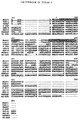
- FIG. 9 Amino acid sequence of the rat brain u-opiate receptor predicted from the sequence of the cDNA RC8-I, and comparisons of sequence of other G-linked receptor family members .
- RC8-1 is subject to double stranded sequencing by automated and manual means, and the translation product open reading frame aligned with those of the mouse w-opioid receptor (DOR-1), rat somatostain receptor (SOMAT), human N-formylmethlonine receptor (F-PEP), human opioid hinding protein (OPB-R), rat neuromedin K recptor (NEU-K), rat rhodopsin (RHODOP), and rat beta2 adrenergic receptors (B-2ADR).
- DOR-1 mouse w-opioid receptor
- SOMAT rat somatostain receptor
- F-PEP human N-formylmethlonine receptor
- OOB-R human opioid hinding protein
- NU-K rat
- FIG. 10 Saturation analyses of binding of [3H]DAMGO (top) and [3H]DADLE (bottom) of membranes prepared from COS cells transfected three days before assay with pcDNA1RC8-1-1. Receptor binding is carried out with 100 nM naloxone added to parallel incubations to estimate nonspecific binding.
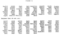
- Figure 11 Nucleotide Sequence of m receptor and predicted amino acid sequence of m opiate receptor C-DNA, open reading frame analyzed for plasmid CDNA.
- FIG. 12 Expression of m-opiate receptor RC8-1 mRNA.
- A Northern analyses and phosphoimager autoradiogram of m-opiate receptor mRNA hybridization to radiolabeled kRC8-1 cDNA in 20 mg total RNA extracted from rat thalamus (lanes 1 and 2), or hypothaiamus (lane 4). Size markers (lane 3) suggest a 10.5 kb mRNA size.
- the purified-opioid receptors are isolated by a receptor purification method disclosed in U.S. Serial No. 07/677,003, the contents of which are incorporated herein by reference.
- the purified opioid receptors are described in U.S. Serial No. 07/026,140 the contents which are incorporated herein by reference.
- Opioid receptors can be found in a wide variety of tissue types (Jaffe and Martin, supra , the contents of which is incorporated herein by reference).
- the w, m, k, and s classes of receptors are found in brain, as well as other tissues; the e type is found in vas deferens and the k type is also plentiful in placenta (Ahmed et al. , supra ).
- the opiate receptor is isolated initially as a complex with its associated G proteins.
- a number of opiate or opioid analogs are commercially available that can be used for receptor binding.
- Research Biochemicals, Incorporated, 1991 Catalog, page XV identifies a number of opioid ligands by their subtype specificity.
- the ligand used will generally be selected based on its affinity for a particular receptor subtype.
- a biotinylated opiate analog is used.
- the ligand used for isolation of receptor is a biotinylated b-endorphin.
- the ligand is first bound to intact cell membranes, thereby forming a receptor:ligand (R:L) complex.
- the membranes are solubilized in detergent and intact receptor:ligand complexes are obtained.
- a useful detergent for this purpose is a combination of deoxycholate and lysophosphatidylcholine in a 1:1 ratio, preferably at a concentration of 0.2% w/v or less.
- the complex consists of the receptor and its associated G protein subunits. The association of the receptor with G proteins is confirmed by the rapid dissociation of the complex in the presence of a stable GTP analog.
- the solubilized complex is then contacted with an appropriate high affinity binding column.
- the column used is preferably streptavidin-agarose (SA-A), whereby the biotinylated portion of the R:L complex will tightly bind to the streptavidin.
- Streptavidin is preferred, due to its lower non-specific binding; however, free and immobilized avidin is also available (Pierce, Vector) and may be suitable for some purposes.
- the column is eluted with a GTP analog, such as GTP-q-S.
- the GTP analog serves to dissociate G protein subunits from the receptor, thereby lowering the affinity of the receptor for its ligand, and thus indirectly causing dissociation from the ligand.
- the elution with GTP analog is combined with elution with at least 25 mM NaCl, preferably 50-100 mM, up to a maximum of about 500 mM NaCl.
- dissociation will occur with GTP alone, it occurs at a relatively low level (about 30%), and the use of NaCl enhances this dissociation.
- a high level i.e., 500 mM of salt can be used alone.
- the eluate from the streptavidin column is then incubated with a lectin affinity chromatography substrate, such as wheat germ agluttinin (WGA)-agarose, which will separate glycoproteins from nonglycoproteins.
- WGA wheat germ agluttinin
- the eluate containing the glycosylated material shows a protein with a molecular weight of about 66,000; this protein is also seen in material eluted by GTP-q-S and/or with NaCl, but is not seen in eluates from samples not previously bound with the biotinylated b-endorphin, indicating its ligand dependence.
- This band appears to represent an opioid receptor, presumably a "mu” or “delta” type opioid receptor, based on b-endorphin's known preferential binding to "mu” or “delta” receptor types, and the pharmacological data discussed below.
- the purified 66 kDa glycoprotein is subjected to Lys-C endoprotease digestion, SDS polyacrylamide gel electrophoresis and electroblotting, producing a 15 kDa peptide band.
- This peptide yields 20 cycles of high quality amino acid sequence.
- the N-terminus of this band overlaps by 4 amino acid residues with a 7-amino acid residue sequence obtained from a bond of about 3 kDa from a cyanogen bromide digest, giving a total sequence length of 23 amino acid residues.
- the sequence (Sequence ID No. 1) obtained is as follows: This sequence is quite similar to a region of the SSTR1 somatostatin receptor, spanning parts of intracellular loop III and transmembrane region VI.
- Pharmacological evaluation of the purified protein indicates that it is a mu-subtype receptor, and that the difference between the repacted delta subtype receptor and the present receptor is not attributable to a simple species difference, but is due to a known mu-specific peptide capable of blocking the binding of b-endorphin to the isolated receptor.
- the novel sequence information obtained provides the basis for isolation and cloning of the corresponding gene encoding the receptor.
- the delta opioid sequence in this region is nearly identical to the same region of SSTR1, and seems to be highly conserved in a set of 5 or 6 receptors, indicating homology in the mu receptor as well.
- the purified receptor, or biologically active fragments thereof, are useful for a number of purposes.
- the purified material in glycosylated or nonglycosylated form, is used to create monoclonal or polyclonal antibodies having specificity for the opioid receptor.
- the technology for creation of monoclonal antibodies is well known in the art (see, e.g., Goding, Monoclonal Antibodies: Principle and Practice, 2nd Ed., 1986).
- Such antibodies have utility in manipulating purified opioid receptors involved in gut motility and growth hormone secretion, or in drug delivery to specific tissues or for tumor imaging.
- General techniques for preparing anti-receptor antibodies are found in U.S. Patent No. 4,857,637, the contents of which are incorporated herein by reference.
- the isolated receptor protein itself, and protein expressed from the cloned opiate receptor CDNA is useful in screening assays to identify compounds that act as analogs.
- the receptor protein is immobilized by any means which does not interfere with opiate binding activity.
- the immobilized receptor is then contacted with a specific compound or mixture and its ability to compete with radiolabelled opiate for binding to the receptor is evaluated. Variations on this method are apparent to those skilled in the art.
- the present invention encompasses the opiate receptor protein and its biologically active fragments produced by any means, whether synthetically, recombinantly, or by purification of the native protein.
- the isolated opiate receptor as described above, is used in protein sequencing procedures.
- the protein sequence in turn is used to design oligonucleotide probes used to screen ggt10 libraries containing the relevant cDNA (copies of RNA), e.g., from brain cells.
- Hybridization of oligos with the library identifies the clone(s) containing the SRIF receptor gene or portions thereof.
- the gene or gene fragments are isolated from the clones, the whole gene reconstructed and then ligated into an appropriate vector by known methods.
- the vector is chosen based upon the choice of preferred host cell.
- the host cell is prokaryotic, e.g., E. coli or other bacteria; or eukaryotic, e.g., yeast, insect, or mammalian cells.
- rat brains frozen in liquid N2 are purchased from Pel-Freez (Rogers, AR). All procedures for membrane preparation are carried out at a temperature of 2-6 o C.
- the brains are homogenized in a Waring blender in a buffer containing 1 mM Na-bicarbonate (pH 7.2), 1 mM EDTA, 1 mM EGTA (all chemicals from Sigma Chemical, St. Louis, MO) and 0.7% (vol./vol.) of the 100X 4Pase protease inhibitor mixture (see “Protease Inhibitors” below).
- the ratio of tissue/homogenization medium is from 25-35 gm of brain/500 ml.
- the blender is controlled through a variable output rheostat (Staco Energy Products, Dayton, OH; type 3PN1010) at a setting of 40.
- the homogenate is centrifuged for 10 minutes at 1,000 x g pellet is rehomeginized in 500 ml of homogenization medium and recentrifuged for 10 minutes at 1,000 x g.
- the 1,000 x g pellet is discarded.
- the 1,000 x g supernatants are combined and centrifuged for 30 minutes at 20,000 x g.
- the 20,000 x g membrane pellet is washed by being resuspended with a Dounce homogenizer in 500 ml of homogenization medium supplemented with 10 mM EDTA (pH readjusted to 7.4) and then washed twice by being resuspended in 25 mM Tris buffer (Sigma Chemical Co.; pH 7.4) and centrifuged for 25 minutes at 20,000 x g.
- the final membrane pellet is resuspended in 25 mM inhibitor mixture to a protein concentration of 4-12 mg/ml.
- the resuspended membranes are aliquoted, frozen on dry ice and stored at -90 o C.
- Binding of [125I]-labelled b-endorphin, b-endorphin and other b-endorphin analogs and opioids is done in a binding buffer containing 50 mM HEPES (Sigma; pH 7.4; pHed with KOH), 0.1% (w/v) bovine serum albumin (Miles Laboratories, Elkhart, IN) and protease inhibitors as specified below for specific applications. All binding incubations are carried out at room temperature (20-23 o C).
- This step is carried out in a solubilization buffer containing 25 mM Tris (pH 8.0) and 10% glycerol. All procedures are at 4 o C or on ice.
- Protease inhibitors 100X 4Pase; 1% vol./vol.
- rat brain membranes are diluted out into this medium to a protein concentration of 0.5 mg/ml.
- the samples are centrifuged for 30 minutes at 100,000 x g.
- the 100,000 x g supernatants are aspirated out of the centrifuge tubes as far as possible without disturbing the pellets of insoluble material.
- the remaining supernatant is poured out of the tubes and filtered through a 0.2 m cellulose acetate or nylon filter unit (Corning Inc., Corning, NY) to remove particulate matter dislodged from the pellet. This filtered supernatant is then combined with the material removed by aspiration.
- protease inhibitors Three mixtures of protease inhibitors are used in these procedures.
- 100X 4Pase 5 mg pepstatin A, 15 mg chymostatin, 38 mg leupeptin and 73 mg phenylmethylsulfonylfluoride (PMSF; all compounds from Bachem, Torrance, CA) are dissolved per 5 ml of dimethylsulfoxide (DMSO; Aldrich Chemicals). Aliquots are stored frozen at 4o C.
- PMSF/Baci 2 mg of PMSF and 2 mg of bacitracin (Sigma) are dissolved per ml of DMSO. Aliquots are stored frozen.
- C. 400X P/B/Bz 20 mg of PMSF, 20 mg of bacitracin and 20 mg of benzamidine (Sigma) are dissolved per ml of DMSO. Aliquots are stored frozen.
- Elution is by a gradient of acetonitrile mixed in water/0.1% trifluoroacetic acid. Two closely spaced product peaks are eluted from the column. These two peptide fractions are lyophilized and solubilized in water at 1 mg/ml. Aliquots are stored frozen at -90 o C.
- the resin is washed with 20 bed volumes of solubilization buffer + 0.15% D:L + 1/500 volume of the 100X 4Pase protease inhibitor mixture.
- the eluates from the SA-A columns are incubated overnight (12-15 hours) with 1/200 to 1/400 volumes of immobilized wheat germ agglutinin (WGA-agarose or WGA-A; Vector Labs, Burlingame, CA).
- the WGA-A is pelleted by centrifugation, washed twice with 50-100 volumes of solubilization buffer + 0.15% D:L (after removing the supernatants containing material not bound to WGA) and then either: (A) eluted with 8 mM triacetylchitotriose (TAC; Sigma) in solubilization buffer + 0.15% D:L (3 sequential elutions where resin is mixed with 2 volumes of elution buffer at room temperature for 15-20 minutes, pelleted by centrifugation and supernatant removed and saved) or B. solubilized directly by addition of 1X Laemmli sample buffer and heating at 90 o C for 10-15 minutes. These samples are analyzed by SDS-PAGE and silver staining. The nonbound supernatants from the WGA-binding step are concentrated, solubilized in 1X Laemmli sample buffer and analyzed by SDS-PAGE and silver staining.
- Hybridization is performed at 30 o C in buffer contained 29% formamide and 6 x SSC, washing is at 50 o C in 0.4 x SSC/0.1% SDS, and 2 days' autoradiographic exposure is used.
- One clone, termed RC8-1 is subjected to complete sequencing of both strands using automated and manual methods, and subcloned into pcDNA1 (InVitrogen) to yield pcDNA1RC8-1.
- DNA sequences are analyzed using conventional methods.
- COS cells are transfected by electroporation with 20 ug/107 cells pcDNA1RC8-1-1 or pcDNA1 vector.
- Transfected COS cells are plated in DMEM containing 10% FBS, cultured for 2-3 days, and tested for opiate receptor expression by radioligand binding.
- Radioligands include [H]DAMGO [D-Ala2,N- methyl-Phc4,Glyo15] enkephalin; 60 Ci/mmole, Amersham), [H]DPDPEpCl [D-Pen2,4'-Cl-Phe4,D-Pen5]enkephalin; 51 Ci/mmole, NEN), [H]DALE (D-Ala2,D-Leu5 enkephalin: 37 Ci/mmole, NEN), and [H]U-69,593 (57 Ci/mmole Amersham). Radioactivity is assesed in a Beckman liquid scintillation counter at 40 efficiency.
- RNA is prepared from rat tissues that are rapidly dissected and frozen at -70 o C. 20 mg of total RNA is prepared and electrophoresed along with molecular weight standards (BRL) and transferred to nylon membranes. Blots are hybridized with the 2.2 kb [32P]-random-primed insert of pcDNA1RC8-1 in 5 x SSPE/1% SDS/150% formamide/2.5 x Denhardt/200 mg/ml herring sperm DNA at 42 o C overnight, washed twice in 0.4x SSC/0.5% SDS for 30 min at 52 o C, and radioactive patterns identified using a phosphoimaging molecular dynamics device following overnight exposures.
- Hybridization at 37 o C overnight in a complexbuffer is followed by washing at 50 o C and emulsion autoradiography with 2 week exposures, emulsion development, tissue section staining, and analyses. Grain densities overlying individual neurons are counted and analyzed. with positively-hydridizing neurons identified as those with densities more than five times background autoradiographs values. Neurons are identified based on size, shape, nuclear profiles, and frequent presence of nucleoli.
- the assay for R:L complex exploits the well known glycoprotein nature of receptors which, like most cell surface proteins, contain covalently linked carbohydrate.
- the ligand, b-endorphin is not glycosylated and will not bind to a carbohydrate-binding lectin, such as wheat germ agglutinin (WGA). Binding of the radioligand, solubilized after the binding step, to immobilized WGA is considered to reflect binding of the R:L complex to WGA via oligosaccharide groups on the receptor.
- Table 1 Binding of Solubilized [125I]b-Endorphin: Opioid Receptor Complex to WGA-Agarose and Dissociation of the Complex by GTP-g-S and NaCl.
- Rat brain membranes are incubated with [125I]b-endorphin as previously described. The "total” binding sample is incubated with only [125I]b-endorphin. The "nonspecific” binding sample is incubated with [125I]b-endorphin plus 10 ⁇ 6 M nonlabelled b-endorphin. After the binding step, the membranes are solubilized as described previously. CPM of [125I]b-endorphin in the 100,000 x g supernatant are counted as described previously.
- WGA-agarose immobilized wheat germ agglutinin
- Vector Labs, Burlingame, CA immobilized wheat germ agglutinin
- the WGA-agarose is pelleted by centrifugation, the supernatants are removed and the WGA-agarose is washed once in solubilization buffer + 0.15% D:L and counted for radioactivity.
- Table 2 Binding of solubilized [125I]b-endorphin: opioid receptor complex to WGA-agarose and dissociation of the complex by GTP-g-S and NaCl. II. All steps are done essentially as in Table 1 above except that here GTP-q-S is tested either alone or in the presence of different concentrations of NaCl. Samples of 1.5 ml volume are mixed with 0.35 ml of WGA-agarose Table 2 CPM of [125I]b-Endorphin Bound to WGA-Agarose Sample A. Total B.
- the membrane bound complex between [125I]b-endorphin and its receptor is solubilized mostly in intact form. This is shown by the adsorption of a high proportion of the solubilized [125I]b-endorphin to immobilized WGA. Not only is a high proportion of the specifically bound radioligand adsorbed to WGA, as would be expected if it is bound to the receptor, but WGA selects for specifically bound material. This is shown by the large increase in the ratio of total cpm/nonspecific cpm in the WGA-bound material. Also, the soluble R:L complex is stable enough to be separated from free ligand in a step taking 2-3 hours.
- the two fractions of biotinylated b-endorphin are assayed for binding to rat brain opioid receptor by competition with [125I]b-endorphin.
- the IC50s for reduction of radioligand binding by competition with cold ligand are: b-endorphin, 1 nM; biotinyl-b-endorphin (F1), 1 nM; and biotinyl-b-endorphin (F2), 5 nM.
- both fractions of biotinylated b-endorphin show high affinity binding to opioid receptor.
- the F1 fraction consists of two peptides with molecular masses, identified by mass spectroscopy, of 3816 and 3875 daltons.
- the F1 fraction contains only the 3816 dalton species, the expected mass for biotinyl-b-endorphin. What is shown here is that heating the F1 fraction for 5 min. at 50 o C eliminates the 3875 dalton species. Thus there is only one species of biotinyl-b- endorphin, with a mass of 3816 daltons, by mass spectrometry. Before further use, the F1 fraction is heated at 50 o C for 5 min. This material has been reanalyzed for binding to receptors in rat brain membranes by competition vs. [125I]b-endorphin and it binds with a protency very similar to that of b-endorphin. The IC508 are 1.2 nM for b-endorphin and 1.8 nM for biotinyl-b-endorphin.
- Samples of brain membranes are incubated either with or without the F1 and F2 fractions of biotinyl-b-endorphin and carried through the procedure of solubilization, adsorption with immobilized streptavidin, elution and protein analysis by SDS-PAGE.
- the WGA bound glycoprotein (WGA+) fractions of the eluates primarily contain a protein with MW about 66,000. Small amounts of this protein are seen in the material eluted by GTP-q-S and much larger amounts elute with the subsequent elution with 500 mM NaCl. The appearance of this band is ligand-dependent because it does not appear in eluates from the samples done without prior binding of biotinyl-b-endorphin.
- the nonglycosylated (WGA-) fractions show that GTP-q-S alone elutes nonglycosylated bands in the 30-40,000 MK (30-40K) range.
- the elution of the 66K glycoprotein correlates with the effects of GTP-q-S on stability of the soluble R:L complex.
- 100 mM GTP-q-S gives only partial dissociation of the soluble R:L complex and partial elution of the 66K glycoprotein.
- This band is considered to be the opioid receptor and will be referred to as such. It will also be referred to as "66K glycoprotein".
- Both purifications employ essentially the same steps: binding of biotinylated ligand to intact membranes (from GH4C1 pituitary tumor cells and brain); solubilization of intact R:(bio)L complex; binding of R:(bio)L complex to streptavidin-immunoreactive material in the 40K size range only with the samples where receptor is complexed with biotinyl-ligand. Samples where the receptor is unoccupied or occupied by non-biotinyl ligand show no evidence of G ia .
- the ligand specificity of the 66K glycoprotein is further tested by blocking binding of the biotinylated b-endorphin with a large molar excess of nonbiotinylated ligand.
- 100 nM biotinyl-b-endorphin (1:1 F1 + F2) is competed with a combination of 50 mM b-endorphin + 50 mM met-enkephalin, the yield of the 66K glycoprotein is greatly diminished.
- 40 mM naloxone effectively competes with 60 nM biotinyl-b-endorphin to nearly eliminate the recovery of 66K glycoprotein (Figure 5B).
- the SA-A column is first washed with 1 mM EDTA + 1 mM EGTA and then with 100 mM NaCl prior to elution with 500 mM NaCl. Since these wash steps carried out with very little loss of the 66K receptor band, they are incorporated into further procedures.
- Two different peptides one known to exhibit mu-receptor selective binding ([D-Ala2, N-MePhe4, glyol5]enkephalin or DAGO; Bachem; 300 fold selectivity for mu over delta) and the other known to exhibit delta receptor selective binding ([D-Pen 2,5 , pCL-Phe4]enkephalin or pCl-DPDPE; 500-fold selectivity for delta over mu) are used to block binding of biotinyl-b-endorphin to rat brain membranes.
- This pair of ligands is appropriate because their affinities for their respective receptors are very similar (approximately 1 mM K D ). Each incubation contains 3 nM biotinyl-b-endorphin, and the blocking peptides are included at 50, 500, and 5000 nM. The ligand mixtures are incubated with unsolubilized membranes for one hour at room temperature and then purification of the receptor proceeds as described herein. A summary of the condition is provided in Table 3.
- the ability of the respective peptides to block b-endorphin binding is determined by observing the relative recovery of biotinylated b-endorphin bound 66 kDa protein from each sample. It can be seen that the 66 kDa protein is recovered in about the same amounts from the control as when the pCl-DPDPE is used as a competitor. In contrast, DAGO blocked recovery of receptor almost completely at 500 nM and completely at 5000 nM, thereby confirming the identity of the protein as a mu-subtype opioid receptor.
- This cDNA display 70% nucleotide sequence identity to the rat m-opioid receptor cDNA and homology with cDNA sequences of other G-linked receptor.
- One open reading frame of the pPCR4A sequence matches each of the 23 amino acids sequenced from a m-opioid receptor protein preparation.
- COS cell expression of RC8-1 in the expression vector pcDNA1 yield naloxone-blockable, high affinity specific binding of [2H] DAMGO and [3H] DADLE binding saturation experiments are most consistent with a single population of binding sites for each ligand, with KK values of 0.4 and 0.5 nM, respectively.
- [2H] DAMGO binding is reduced by addition of Na+ or GTP to incubations, but not by adding ATP (Fig. 3).
- Mg++ addition increases binding by COS cell expression of RC8-1 in the expression vector pcDNA1 yielded naloxone-blockable, high effinity specific binding of [3H]DAMGO and [3H]DADLE, with no appreciable specific recognition of [3H]DPDPE or [3H]U 69,593, that is not present in cells transfected with vector alone.
- Scatchard analysis of [3H]DAMGO and [3H]DADLE binding saturation experiments are most consistent with a single population of high affinity binding sites for each ligand, with Kn values on 0.4 and 0.5 nM respectively.
- [3H]DAMGO binding is reduced by addition of Na+ of GTP to incubations.
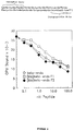
- [3H]DAMGO binding is displaced by a number of opioid compounds in stereoselective fashion.
- Pharamcologicallyactive (-) naloxone and dextrorphan isomers display substantially greater potency than pharmacologically less active (+) naloxone and dextrorphan isomers.
- Morphine, DADLE, (-) naloxone, naloxonazine, ethylketocyclazocino and bromazocino displace binding of [3H]DAMGO with high potency (Table 1).
- DPDPE and p-C1-DEDPE, relatively 8-selective, U 50,488 and U 69,593, relatively k selective, display substantially less potency.
- U-50,488, relatively -selective, displayed substantially loss potencies.
Landscapes
- Health & Medical Sciences (AREA)
- Chemical & Material Sciences (AREA)
- Life Sciences & Earth Sciences (AREA)
- Organic Chemistry (AREA)
- General Health & Medical Sciences (AREA)
- Medicinal Chemistry (AREA)
- Neurology (AREA)
- Genetics & Genomics (AREA)
- Molecular Biology (AREA)
- Immunology (AREA)
- Biophysics (AREA)
- Biomedical Technology (AREA)
- Biochemistry (AREA)
- Proteomics, Peptides & Aminoacids (AREA)
- Gastroenterology & Hepatology (AREA)
- Engineering & Computer Science (AREA)
- Zoology (AREA)
- Toxicology (AREA)
- Cell Biology (AREA)
- General Chemical & Material Sciences (AREA)
- Veterinary Medicine (AREA)
- Public Health (AREA)
- Animal Behavior & Ethology (AREA)
- Pharmacology & Pharmacy (AREA)
- Nuclear Medicine, Radiotherapy & Molecular Imaging (AREA)
- Chemical Kinetics & Catalysis (AREA)
- Neurosurgery (AREA)
- Bioinformatics & Cheminformatics (AREA)
- Peptides Or Proteins (AREA)
- Preparation Of Compounds By Using Micro-Organisms (AREA)
- Micro-Organisms Or Cultivation Processes Thereof (AREA)
- Medicines That Contain Protein Lipid Enzymes And Other Medicines (AREA)
- Medicines Containing Antibodies Or Antigens For Use As Internal Diagnostic Agents (AREA)
Applications Claiming Priority (2)
| Application Number | Priority Date | Filing Date | Title |
|---|---|---|---|
| US2614093A | 1993-02-26 | 1993-02-26 | |
| US26140 | 1993-02-26 |
Publications (2)
| Publication Number | Publication Date |
|---|---|
| EP0612845A2 true EP0612845A2 (fr) | 1994-08-31 |
| EP0612845A3 EP0612845A3 (en) | 1994-09-21 |
Family
ID=21830132
Family Applications (1)
| Application Number | Title | Priority Date | Filing Date |
|---|---|---|---|
| EP19940101968 Withdrawn EP0612845A3 (en) | 1993-02-26 | 1994-02-09 | Purified opioid receptor. |
Country Status (5)
| Country | Link |
|---|---|
| EP (1) | EP0612845A3 (fr) |
| JP (1) | JPH0795889A (fr) |
| KR (1) | KR940019861A (fr) |
| AU (1) | AU5640494A (fr) |
| IL (1) | IL108734A0 (fr) |
Cited By (11)
| Publication number | Priority date | Publication date | Assignee | Title |
|---|---|---|---|---|
| WO1995007983A1 (fr) * | 1993-09-13 | 1995-03-23 | Indiana University Foundation | Recepteur mu humain d'opioides |
| WO1994028132A3 (fr) * | 1993-05-20 | 1995-05-04 | Arch Dev Corp | Recepteurs d'opioides: compositions et procedes |
| US5753516A (en) * | 1995-02-03 | 1998-05-19 | Heagy; Wyrta E. | Screening method for ligands of the EBI-1 receptor |
| US6103492A (en) * | 1993-03-08 | 2000-08-15 | Indiana University | Polynucleotide encoding mu opioid receptor |
| US6326156B1 (en) | 1996-06-13 | 2001-12-04 | Hoffmann-La Roche Inc. | Method of identifying compounds having antiepileptic anticonvulsant or anxiolytic activities |
| WO2001036471A3 (fr) * | 1999-11-17 | 2002-01-03 | Arena Pharm Inc | Versions endogenes et non-endogenes de recepteurs couples a la proteine g humaine |
| US6555339B1 (en) | 1997-04-14 | 2003-04-29 | Arena Pharmaceuticals, Inc. | Non-endogenous, constitutively activated human protein-coupled receptors |
| US6902902B2 (en) | 2001-11-27 | 2005-06-07 | Arena Pharmaceuticals, Inc. | Human G protein-coupled receptors and modulators thereof for the treatment of metabolic-related disorders |
| US7097988B1 (en) | 1993-03-08 | 2006-08-29 | Advanced Research And Technology Institute | Methods for screening for substances that bind opioid receptors |
| US7119190B2 (en) | 1997-04-14 | 2006-10-10 | Arena Pharmaceuticals, Inc. | Endogenous and non-endogenous versions of human G protein-coupled receptors |
| US7235366B1 (en) | 1993-05-20 | 2007-06-26 | Arch Development Corporation | Methods of identifying agonists and antagonists of opioid receptors |
Family Cites Families (1)
| Publication number | Priority date | Publication date | Assignee | Title |
|---|---|---|---|---|
| WO1993019086A1 (fr) * | 1992-03-23 | 1993-09-30 | American Cyanamid Company | Recepteur d'opioides purifie |
-
1994
- 1994-02-09 EP EP19940101968 patent/EP0612845A3/en not_active Withdrawn
- 1994-02-22 IL IL10873494A patent/IL108734A0/xx unknown
- 1994-02-25 JP JP6051088A patent/JPH0795889A/ja active Pending
- 1994-02-25 KR KR1019940003640A patent/KR940019861A/ko not_active Withdrawn
- 1994-02-25 AU AU56404/94A patent/AU5640494A/en not_active Abandoned
Cited By (14)
| Publication number | Priority date | Publication date | Assignee | Title |
|---|---|---|---|---|
| US7097988B1 (en) | 1993-03-08 | 2006-08-29 | Advanced Research And Technology Institute | Methods for screening for substances that bind opioid receptors |
| US6103492A (en) * | 1993-03-08 | 2000-08-15 | Indiana University | Polynucleotide encoding mu opioid receptor |
| WO1994028132A3 (fr) * | 1993-05-20 | 1995-05-04 | Arch Dev Corp | Recepteurs d'opioides: compositions et procedes |
| US6319686B1 (en) | 1993-05-20 | 2001-11-20 | Arch Development Corporation | Nucleic acids encoding kappa opioid receptors |
| US7235366B1 (en) | 1993-05-20 | 2007-06-26 | Arch Development Corporation | Methods of identifying agonists and antagonists of opioid receptors |
| WO1995007983A1 (fr) * | 1993-09-13 | 1995-03-23 | Indiana University Foundation | Recepteur mu humain d'opioides |
| US5753516A (en) * | 1995-02-03 | 1998-05-19 | Heagy; Wyrta E. | Screening method for ligands of the EBI-1 receptor |
| US6326156B1 (en) | 1996-06-13 | 2001-12-04 | Hoffmann-La Roche Inc. | Method of identifying compounds having antiepileptic anticonvulsant or anxiolytic activities |
| US7410777B2 (en) | 1997-04-14 | 2008-08-12 | Arena Pharmaceuticals, Inc. | Non-endogenous, constitutively activated human G protein-coupled receptors |
| US7119190B2 (en) | 1997-04-14 | 2006-10-10 | Arena Pharmaceuticals, Inc. | Endogenous and non-endogenous versions of human G protein-coupled receptors |
| US6555339B1 (en) | 1997-04-14 | 2003-04-29 | Arena Pharmaceuticals, Inc. | Non-endogenous, constitutively activated human protein-coupled receptors |
| WO2001036471A3 (fr) * | 1999-11-17 | 2002-01-03 | Arena Pharm Inc | Versions endogenes et non-endogenes de recepteurs couples a la proteine g humaine |
| US6902902B2 (en) | 2001-11-27 | 2005-06-07 | Arena Pharmaceuticals, Inc. | Human G protein-coupled receptors and modulators thereof for the treatment of metabolic-related disorders |
| US7829298B2 (en) | 2001-11-27 | 2010-11-09 | Arena Pharmaceuticals, Inc. | Human G protein-coupled receptors for metabolic-related disorders |
Also Published As
| Publication number | Publication date |
|---|---|
| KR940019861A (ko) | 1994-09-15 |
| AU5640494A (en) | 1994-09-01 |
| EP0612845A3 (en) | 1994-09-21 |
| JPH0795889A (ja) | 1995-04-11 |
| IL108734A0 (en) | 1994-05-30 |
Similar Documents
| Publication | Publication Date | Title |
|---|---|---|
| Pisegna et al. | Molecular cloning of the human brain and gastric cholecystokinin receptor: structure, functional expression and chromosomal localization | |
| Meunier | Nociceptin/orphanin FQ and the opioid receptor-like ORL1 receptor | |
| Schofield et al. | Molecular characterization of a new immunoglobulin superfamily protein with potential roles in opioid binding and cell contact. | |
| CA2107569C (fr) | Recepteur de l'hormone parathyroidienne et adn codant pour ce recepteur | |
| AU2005244540B2 (en) | Endogenous and non-endogenous versions of human G protein-coupled receptors | |
| Lapalu et al. | Comparison of the structure-activity relationships of nociceptin and dynorphin A using chimeric peptides | |
| Bunzow et al. | Characterization and distribution of a cloned rat μ‐opioid receptor | |
| JPH07503611A (ja) | ソマトスタチン受容体 | |
| US6225080B1 (en) | Mu-subtype opioid receptor | |
| EP0612845A2 (fr) | Récepteur d'opioides purifié | |
| WO1994021665A1 (fr) | Recepteurs clones et methodes de criblage associees | |
| EP0769024B1 (fr) | Yrecepteurs de la melatonine de haute affinite et leurs utilisations | |
| EP1097170A1 (fr) | Recepteurs et acides nucleiques en rapport avec un secretagogue d'hormone de croissance | |
| AU704927B2 (en) | Human neuronal nicotinic acetylcholine receptor compositions and methods employing same | |
| CA2066263C (fr) | Recepteur de thyrotropine | |
| US7510846B2 (en) | Assay employing human or rat GAL-R2 galanin receptor | |
| CA2330251A1 (fr) | Clonage et identification du recepteur de la motiline | |
| CA2087844A1 (fr) | Recepteur de cannabinoide | |
| JP2001513653A (ja) | 新規な受容体 | |
| US6660496B1 (en) | Nucleic acid molecules encoding a KOR3 kappa opioid receptor and the methods of producing the encoded receptor | |
| WO1993019086A1 (fr) | Recepteur d'opioides purifie | |
| Miotto et al. | Molecular characterization of opioid receptors | |
| US6576742B1 (en) | DNA sequence encoding a human imidazoline receptor and method for cloning the same | |
| Vincent et al. | Receptors for neuropeptides: receptor isolation studies and molecular biology | |
| NZ248602A (en) | Isolation, characterisation and cloning of growth hormone releasing hormone receptor |
Legal Events
| Date | Code | Title | Description |
|---|---|---|---|
| PUAI | Public reference made under article 153(3) epc to a published international application that has entered the european phase |
Free format text: ORIGINAL CODE: 0009012 |
|
| PUAL | Search report despatched |
Free format text: ORIGINAL CODE: 0009013 |
|
| AK | Designated contracting states |
Kind code of ref document: A2 Designated state(s): AT BE CH DE DK ES FR GB GR IE IT LI LU NL PT SE |
|
| AK | Designated contracting states |
Kind code of ref document: A3 Designated state(s): AT BE CH DE DK ES FR GB GR IE IT LI LU NL PT SE |
|
| 17P | Request for examination filed |
Effective date: 19941111 |
|
| STAA | Information on the status of an ep patent application or granted ep patent |
Free format text: STATUS: THE APPLICATION IS DEEMED TO BE WITHDRAWN |
|
| 18D | Application deemed to be withdrawn |
Effective date: 19970902 |