EP0613944A2 - Verfahren zur Regulierung der Immunantwort durch den Gebrauch von CTLA4-Bindungsmoleküle und IL4-Bindungsmoleküle - Google Patents
Verfahren zur Regulierung der Immunantwort durch den Gebrauch von CTLA4-Bindungsmoleküle und IL4-Bindungsmoleküle Download PDFInfo
- Publication number
- EP0613944A2 EP0613944A2 EP94100882A EP94100882A EP0613944A2 EP 0613944 A2 EP0613944 A2 EP 0613944A2 EP 94100882 A EP94100882 A EP 94100882A EP 94100882 A EP94100882 A EP 94100882A EP 0613944 A2 EP0613944 A2 EP 0613944A2
- Authority
- EP
- European Patent Office
- Prior art keywords
- ctla4
- ctla4ig
- cells
- amino acid
- cell
- Prior art date
- Legal status (The legal status is an assumption and is not a legal conclusion. Google has not performed a legal analysis and makes no representation as to the accuracy of the status listed.)
- Granted
Links
Images


Classifications
-
- C—CHEMISTRY; METALLURGY
- C07—ORGANIC CHEMISTRY
- C07K—PEPTIDES
- C07K16/00—Immunoglobulins [IG], e.g. monoclonal or polyclonal antibodies
- C07K16/18—Immunoglobulins [IG], e.g. monoclonal or polyclonal antibodies against material from animals or humans
- C07K16/28—Immunoglobulins [IG], e.g. monoclonal or polyclonal antibodies against material from animals or humans against receptors, cell surface antigens or cell surface determinants
- C07K16/2803—Immunoglobulins [IG], e.g. monoclonal or polyclonal antibodies against material from animals or humans against receptors, cell surface antigens or cell surface determinants against the immunoglobulin superfamily
- C07K16/2827—Immunoglobulins [IG], e.g. monoclonal or polyclonal antibodies against material from animals or humans against receptors, cell surface antigens or cell surface determinants against the immunoglobulin superfamily against B7 molecules, e.g. CD80, CD86
-
- A—HUMAN NECESSITIES
- A61—MEDICAL OR VETERINARY SCIENCE; HYGIENE
- A61P—SPECIFIC THERAPEUTIC ACTIVITY OF CHEMICAL COMPOUNDS OR MEDICINAL PREPARATIONS
- A61P29/00—Non-central analgesic, antipyretic or antiinflammatory agents, e.g. antirheumatic agents; Non-steroidal antiinflammatory drugs [NSAID]
-
- A—HUMAN NECESSITIES
- A61—MEDICAL OR VETERINARY SCIENCE; HYGIENE
- A61P—SPECIFIC THERAPEUTIC ACTIVITY OF CHEMICAL COMPOUNDS OR MEDICINAL PREPARATIONS
- A61P31/00—Antiinfectives, i.e. antibiotics, antiseptics, chemotherapeutics
- A61P31/12—Antivirals
-
- A—HUMAN NECESSITIES
- A61—MEDICAL OR VETERINARY SCIENCE; HYGIENE
- A61P—SPECIFIC THERAPEUTIC ACTIVITY OF CHEMICAL COMPOUNDS OR MEDICINAL PREPARATIONS
- A61P31/00—Antiinfectives, i.e. antibiotics, antiseptics, chemotherapeutics
- A61P31/12—Antivirals
- A61P31/14—Antivirals for RNA viruses
-
- A—HUMAN NECESSITIES
- A61—MEDICAL OR VETERINARY SCIENCE; HYGIENE
- A61P—SPECIFIC THERAPEUTIC ACTIVITY OF CHEMICAL COMPOUNDS OR MEDICINAL PREPARATIONS
- A61P31/00—Antiinfectives, i.e. antibiotics, antiseptics, chemotherapeutics
- A61P31/12—Antivirals
- A61P31/14—Antivirals for RNA viruses
- A61P31/18—Antivirals for RNA viruses for HIV
-
- A—HUMAN NECESSITIES
- A61—MEDICAL OR VETERINARY SCIENCE; HYGIENE
- A61P—SPECIFIC THERAPEUTIC ACTIVITY OF CHEMICAL COMPOUNDS OR MEDICINAL PREPARATIONS
- A61P35/00—Antineoplastic agents
-
- A—HUMAN NECESSITIES
- A61—MEDICAL OR VETERINARY SCIENCE; HYGIENE
- A61P—SPECIFIC THERAPEUTIC ACTIVITY OF CHEMICAL COMPOUNDS OR MEDICINAL PREPARATIONS
- A61P37/00—Drugs for immunological or allergic disorders
-
- A—HUMAN NECESSITIES
- A61—MEDICAL OR VETERINARY SCIENCE; HYGIENE
- A61P—SPECIFIC THERAPEUTIC ACTIVITY OF CHEMICAL COMPOUNDS OR MEDICINAL PREPARATIONS
- A61P37/00—Drugs for immunological or allergic disorders
- A61P37/02—Immunomodulators
-
- A—HUMAN NECESSITIES
- A61—MEDICAL OR VETERINARY SCIENCE; HYGIENE
- A61P—SPECIFIC THERAPEUTIC ACTIVITY OF CHEMICAL COMPOUNDS OR MEDICINAL PREPARATIONS
- A61P37/00—Drugs for immunological or allergic disorders
- A61P37/02—Immunomodulators
- A61P37/06—Immunosuppressants, e.g. drugs for graft rejection
-
- A—HUMAN NECESSITIES
- A61—MEDICAL OR VETERINARY SCIENCE; HYGIENE
- A61P—SPECIFIC THERAPEUTIC ACTIVITY OF CHEMICAL COMPOUNDS OR MEDICINAL PREPARATIONS
- A61P37/00—Drugs for immunological or allergic disorders
- A61P37/08—Antiallergic agents
-
- C—CHEMISTRY; METALLURGY
- C07—ORGANIC CHEMISTRY
- C07K—PEPTIDES
- C07K14/00—Peptides having more than 20 amino acids; Gastrins; Somatostatins; Melanotropins; Derivatives thereof
- C07K14/435—Peptides having more than 20 amino acids; Gastrins; Somatostatins; Melanotropins; Derivatives thereof from animals; from humans
- C07K14/705—Receptors; Cell surface antigens; Cell surface determinants
- C07K14/70503—Immunoglobulin superfamily
- C07K14/70521—CD28, CD152
-
- C—CHEMISTRY; METALLURGY
- C07—ORGANIC CHEMISTRY
- C07K—PEPTIDES
- C07K14/00—Peptides having more than 20 amino acids; Gastrins; Somatostatins; Melanotropins; Derivatives thereof
- C07K14/435—Peptides having more than 20 amino acids; Gastrins; Somatostatins; Melanotropins; Derivatives thereof from animals; from humans
- C07K14/705—Receptors; Cell surface antigens; Cell surface determinants
- C07K14/715—Receptors; Cell surface antigens; Cell surface determinants for cytokines; for lymphokines; for interferons
- C07K14/7155—Receptors; Cell surface antigens; Cell surface determinants for cytokines; for lymphokines; for interferons for interleukins [IL]
-
- C—CHEMISTRY; METALLURGY
- C07—ORGANIC CHEMISTRY
- C07K—PEPTIDES
- C07K16/00—Immunoglobulins [IG], e.g. monoclonal or polyclonal antibodies
- C07K16/18—Immunoglobulins [IG], e.g. monoclonal or polyclonal antibodies against material from animals or humans
- C07K16/28—Immunoglobulins [IG], e.g. monoclonal or polyclonal antibodies against material from animals or humans against receptors, cell surface antigens or cell surface determinants
- C07K16/2803—Immunoglobulins [IG], e.g. monoclonal or polyclonal antibodies against material from animals or humans against receptors, cell surface antigens or cell surface determinants against the immunoglobulin superfamily
- C07K16/2818—Immunoglobulins [IG], e.g. monoclonal or polyclonal antibodies against material from animals or humans against receptors, cell surface antigens or cell surface determinants against the immunoglobulin superfamily against CD28 or CD152
-
- A—HUMAN NECESSITIES
- A61—MEDICAL OR VETERINARY SCIENCE; HYGIENE
- A61K—PREPARATIONS FOR MEDICAL, DENTAL OR TOILETRY PURPOSES
- A61K38/00—Medicinal preparations containing peptides
-
- C—CHEMISTRY; METALLURGY
- C07—ORGANIC CHEMISTRY
- C07K—PEPTIDES
- C07K2317/00—Immunoglobulins specific features
- C07K2317/70—Immunoglobulins specific features characterized by effect upon binding to a cell or to an antigen
- C07K2317/73—Inducing cell death, e.g. apoptosis, necrosis or inhibition of cell proliferation
-
- C—CHEMISTRY; METALLURGY
- C07—ORGANIC CHEMISTRY
- C07K—PEPTIDES
- C07K2319/00—Fusion polypeptide
- C07K2319/01—Fusion polypeptide containing a localisation/targetting motif
- C07K2319/02—Fusion polypeptide containing a localisation/targetting motif containing a signal sequence
-
- C—CHEMISTRY; METALLURGY
- C07—ORGANIC CHEMISTRY
- C07K—PEPTIDES
- C07K2319/00—Fusion polypeptide
- C07K2319/01—Fusion polypeptide containing a localisation/targetting motif
- C07K2319/036—Fusion polypeptide containing a localisation/targetting motif targeting to the medium outside of the cell, e.g. type III secretion
-
- C—CHEMISTRY; METALLURGY
- C07—ORGANIC CHEMISTRY
- C07K—PEPTIDES
- C07K2319/00—Fusion polypeptide
- C07K2319/30—Non-immunoglobulin-derived peptide or protein having an immunoglobulin constant or Fc region, or a fragment thereof, attached thereto
-
- C—CHEMISTRY; METALLURGY
- C07—ORGANIC CHEMISTRY
- C07K—PEPTIDES
- C07K2319/00—Fusion polypeptide
- C07K2319/32—Fusion polypeptide fusions with soluble part of a cell surface receptor, "decoy receptors"
-
- Y—GENERAL TAGGING OF NEW TECHNOLOGICAL DEVELOPMENTS; GENERAL TAGGING OF CROSS-SECTIONAL TECHNOLOGIES SPANNING OVER SEVERAL SECTIONS OF THE IPC; TECHNICAL SUBJECTS COVERED BY FORMER USPC CROSS-REFERENCE ART COLLECTIONS [XRACs] AND DIGESTS
- Y10—TECHNICAL SUBJECTS COVERED BY FORMER USPC
- Y10S—TECHNICAL SUBJECTS COVERED BY FORMER USPC CROSS-REFERENCE ART COLLECTIONS [XRACs] AND DIGESTS
- Y10S424/00—Drug, bio-affecting and body treating compositions
- Y10S424/81—Drug, bio-affecting and body treating compositions involving autoimmunity, allergy, immediate hypersensitivity, delayed hypersensitivity, immunosuppression, immunotolerance, or anergy
-
- Y—GENERAL TAGGING OF NEW TECHNOLOGICAL DEVELOPMENTS; GENERAL TAGGING OF CROSS-SECTIONAL TECHNOLOGIES SPANNING OVER SEVERAL SECTIONS OF THE IPC; TECHNICAL SUBJECTS COVERED BY FORMER USPC CROSS-REFERENCE ART COLLECTIONS [XRACs] AND DIGESTS
- Y10—TECHNICAL SUBJECTS COVERED BY FORMER USPC
- Y10S—TECHNICAL SUBJECTS COVERED BY FORMER USPC CROSS-REFERENCE ART COLLECTIONS [XRACs] AND DIGESTS
- Y10S530/00—Chemistry: natural resins or derivatives; peptides or proteins; lignins or reaction products thereof
- Y10S530/868—Chemistry: natural resins or derivatives; peptides or proteins; lignins or reaction products thereof involving autoimmunity, allergy, immediate hypersensitivity, delayed hypersensitivity, immunosuppression, or immunotolerance
Definitions
- the present invention relates to expression of the CTLA4 receptor gene, identification of the interaction between the receptor and cells expressing B7 antigen, and to methods for regulating cellular interactions involving the CTLA4 receptor and the B7 antigen.
- T cell-B cell interactions are essential to the immune response. Levels of many cohesive molecules found on T cells and B cells increase during an immune response (Springer et al., (1987), supra ; Shaw and Shimuzu, Current Opinion in Immunology , Eds. Kindt and Long, 1:92-97 (1988)); and Hemler Immunology Today 9:109-113 (1988)).
- Increased levels of these molecules may help explain why activated B cells are more effective at stimulating antigen-specific T cell proliferation than are resting B cells (Kaiuchi et al., J. Immunol. 131:109-114 (1983); Kreiger et al., J. Immunol. 135:2937-2945 (1985); McKenzie, J. Immunol. 141:2907-2911 (1988); and Hawrylowicz and Unanue, J. Immunol. 141:4083-4088 (1988)).
- T cell T lymphocyte
- T cell T lymphocyte
- T cell T lymphocyte
- cytokines or lymphokines soluble immune mediators
- This response is regulated by several T-cell surface receptors, including the T-cell receptor complex (Weiss et al., Ann. Rev. Immunol. 4:593-619 (1986)) and other "accessory" surface molecules (Springer et al., (1987) supra ).
- CD cell surface differentiation
- Antigen-independent intercellular interactions involving lymphocyte accessory molecules are essential for an immune response (Springer et al., (1987), supra ).
- binding of the T cell-associated protein, CD2, to its ligand LFA-3, a widely expressed glycoprotein is important for optimizing antigen-specific T cell activation (Moingeon et al., Nature 339:314 (1988)).
- lymphocytes An important adhesion system involves binding of the LFA-1 glycoprotein found on lymphocytes, macrophages, and granulocytes (Springer et al., (1987), supra ; Shaw and Shimuzu (1988), supra ) to its ligands ICAM-1 (Makgoba et al., Nature 331:86-88 (1988)) and ICAM-2 (Staunton et al., Nature 339:61-64 (1989)).
- the T cell accessory molecules CD8 and CD4 strengthen T cell adhesion by interaction with MHC class I (Norment et al., Nature 336:79-81 (1988)) and class II (Doyle and Strominger, Nature 330:256-259 (1987)) molecules, respectively.
- Homing receptors are important for control of lymphocyte migration (Stoolman, Cell 56:907-910 (1989)).
- VLA glycoproteins are integrins which appear to mediate lymphocyte functions requiring adhesion to extracellular matrix components (Hemler, supra ).
- the CD2/LFA-3, LFA-1/ICAM-1 and ICAM-2, and VLA adhesion systems are distributed on a wide variety of cell types (Springer et al., (1987), supra ; Shaw and Shimuzu, (1988,) supra and Hemler, (1988), supra ).
- cytokines are involved in the generation of alloreactive effector cells.
- membrane bound IL-4 and soluble IL-4 receptor were administered separately to mice and were shown to augment the lymphoproliferative response (William C. Fanslow et al. "Regulation of Alloreactivity in vivo by IL-4 and the soluble Il-4 receptor" J. Immunol. 147 :535-540 (1991)).
- administration of IL-4 to BALB ⁇ c mice resulted in slight augmentation of the lymphoproliferative response.
- the soluble IL-4 receptor suppressed this response to allogeneic cells in a dose dependent manner.
- a neutralizing antibody against IL-4 and another against soluble IL-4 receptor were effective inhibitors of the lymphoproliferative response.
- COS cells transfected with this cDNA have been shown to stain by both labeled mAb B7 and mAb BB-1 (Clark et al., Human Immunol. 16:100-113 (1986); Yokochi et al., J. Immunol. 128:823 (1981)); Freeman et al., (1989) supra ; and Freedman et al., (1987), supra )).
- expression of this antigen has been detected on cells of other lineages, such as monocytes (Freeman et al., supra ).
- T helper cell (T h ) antigenic response are provided by antigen-presenting cells (APC).
- APC antigen-presenting cells
- the first signal is initiated by interaction of the T cell receptor complex (Weiss, J. Clin. Invest. 86:1015 (1990)) with antigen presented in the context of class II major histocompatibility complex (MHC) molecules on the APC (Allen, Immunol. Today 8:270 (1987)).
- MHC major histocompatibility complex
- This antigen-specific signal is not sufficient to generate a full response, and in the absence of a second signal may actually lead to clonal inactivation or anergy (Schwartz, Science 248:1349 (1990)).
- CD28 antigen a homodimeric glycoprotein of the immunoglobulin superfamily (Aruffo and Seed, Proc. Natl. Acad. Sci. 84:8573-8577 (1987)), is an accessory molecule found on most mature human T cells (Damle et al., J. Immunol. 131:2296-2300 (1983)). Current evidence suggests that this molecule functions in an alternative T cell activation pathway distinct from that initiated by the T-cell receptor complex (June et al., Mol. Cell. Biol. 7:4472-4481 (1987)). Monoclonal antibodies (mAbs) reactive with CD28 antigen can augment T cell responses initiated by various polyclonal stimuli (reviewed by June et al., supra ).
- Anti-CD28 mAbs can also have inhibitory effects, i.e., they can block autologous mixed lymphocyte reactions (Damle et al., Proc. Natl. Acad. Sci. 78:5096-6001 (1981)) and activation of antigen-specific T cell clones (Lesslauer et al., Eur. J. Immunol. 16:1289-1296 (1986)).
- B7/BB-1 B cell activation antigen
- T cell stimulation with B7 positive CHO cells also specifically stimulates increased levels of transcripts for IL-2. Additional studies have shown that anti-CD28 mAb inhibited IL-2 production induced in certain T cell leukemia cell lines by cellular interactions with a B cell leukemia line (Kohno et al., Cell. Immunol. 131-1-10 (1990)).
- CD28 has a single extracellular variable region (V)-like domain (Aruffo and Seed, supra ).
- V variable region
- a homologous molecule, CTLA4 has been identified by differential screening of a murine cytolytic-T cell cDNA library (Brunet et al., Nature 328:267-270 (1987)).
- CD4 the receptor for HIV-1
- CD28 and B7 receptors using hybrid fusion molecules consisting of DNA sequences encoding amino acids corresponding to portions of the extracellular domain of CD4 receptor fused to antibody domains (immunoglobulin ⁇ 1 (Capon et al., Nature 337:525-531 (1989) (CD4) and Linsley et al., J. Exp. Med. , supra (CD28 and B7)).
- panimmunosuppressive drugs such as cyclosporine A or monoclonal antibodies (MAbs) to CD3.
- MAbs monoclonal antibodies
- the present invention provides the complete and correct DNA sequence encoding the amino acid sequence corresponding to the CTLA4 receptor protein, and identifies B7 antigen as a natural ligand for the CTLA4 receptor.
- the invention also provides a method for expressing the DNA as a CTLA4 immunoglobulin (Ig) fusion protein product.
- Embodiments of the invention include CTLA4Ig fusion protein, and hybrid fusion proteins including CD28Ig/CTLA4Ig fusion proteins.
- the human CTLA receptor protein of the invention is encoded by 187 amino acids and includes a newly identified N-linked glycosylation site.
- the CTLA4Ig fusion protein of the invention binds the B7 antigen expressed on activated B cells, and cells of other lineages, a ligand for CD28 receptor on T cells.
- the CTLA4Ig binds B7 antigen with significantly higher affinity than B7 binding to the CD28 receptor.
- the CTLA4Ig construct has a first amino acid sequence corresponding to the extracellular domain of the CTLA4 receptor fused to a second amino acid sequence corresponding to the human Ig C ⁇ 1 domain.
- the first amino acid sequence contains amino acid residues from about position 1 to about position 125 of the amino acid sequence corresponding to the extracellular domain of CTLA4 joined to a second amino acid sequence containing amino acid residues corresponding to the hinge, CH2 and CH3 regions of human IgC ⁇ 1.
- the fusion protein is preferably produced in dimeric form. Soluble CTLA4Ig is a potent inhibitor in vitro of T and B lymphocyte responses.
- hybrid fusion proteins such as CD28Ig/CTLA4Ig fusion proteins having a first amino acid sequence corresponding to fragments of the extracellular domain of CD28 joined to a second amino acid sequence corresponding to fragments of the extracellular domain of CTLA4Ig and a third amino acid sequence corresponding to the hinge, CH2 and CH3 regions of human IgC ⁇ 1.
- hybrid fusion proteins is a CD28Ig/CTLA4Ig fusion construct having a first amino acid sequence containing amino acid residues from about position 1 to about position 94 of the amino acid sequence corresponding to the extracellular domain of CD28, joined a second amino acid sequence containing amino acid residues from about position 94 to about position 125 of the amino acid sequence corresponding to the extracellular domain of CTLA4, joined to a third amino acid sequence containing amino acids residues corresponding to the hinge, CH2 and CH3 regions of human IgC ⁇ 1.
- Also included in the invention is a method for regulating T cell interactions with other cells by inhibiting the interaction of CTLA4-positive T cells with B7 positive cells by reacting the T cells with ligands for the CTLA4 receptor.
- the ligands include B7Ig fusion protein, a monoclonal antibody reactive with CTLA4 receptor, and antibody fragments.
- the invention also provides a method for regulating T cell interactions with B7 positive cells, using a ligand for the B7 antigen.
- a ligand is the CTLA4Ig fusion protein of the invention, its fragments or derivatives, the CD28Ig/CTLA4Ig fusion protein hybrid, or a monoclonal antibody reactive with the B7 antigen.
- the invention further includes a method for treating immune system diseases mediated by T cell interactions with B7 positive cells by administering a ligand reactive with B7 antigen to regulate T cell interactions with B7 positive cells.
- the ligand is the CTLA4Ig fusion protein, or the CD28Ig/CTLA4Ig fusion protein hybrid, or a monoclonal antibody reactive with B7 antigen.
- a monoclonal antibody reactive with the CTLA4Ig fusion protein and a monoclonal antibody reactive with CD28Ig/CTLA4Ig fusion protein are described for use in regulating cellular interactions.
- a novel Chinese Hamster Ovary cell line stably expressing the CTLA4Ig fusion protein is also disclosed.
- the present invention provides a method for blocking B7 interaction so as to regulate the immune response.
- This method comprises contacting lymphocytes with a CTLA4-binding molecule and an IL4-binding molecule.
- the present invention provides a method for regulating an immune response which comprises contacting B7-positive lymphocytes with a CTLA4-binding molecule and an IL4-binding molecule.
- the invention provides method for inhibiting tissue transplant rejection by a subject, the subject being a recipient of transplanted tissue.
- This method comprises administering to the subject a CTLA4-binding molecule and an IL4-binding molecule.
- the present invention further provides a method for inhibiting graft versus host disease in a subject which comprises administering to the subject a CTLA4-binding molecule and an IL4-binding molecule.
- Figure 1 is a diagrammatic representation of CTLA4Ig fusion constructs as described in Example 2, infra .
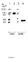
- Figure 2 is a photograph of a gel obtained from SDS-PAGE chromatographic purification of CTLA4Ig as described in Example 2, infra .
- Figure 3 depicts the complete amino acid sequence encoding human CTLA4 receptor (SEQ ID NOs: 13 and 14) fused to the oncostatin M signal peptide (position -25 to -1), and including the newly identified N-linked glycosylation site (position 109-111), as described in Example 3, infra .
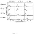
- Figure 4 depicts the results of FACS R analysis of binding of the B7Ig fusion protein to CD28- and CTLA4-transfected COS cells as described in Example 4, infra .
- Figure 5 depicts the results of FACS R analysis of binding of purified CTLA4Ig on B7 antigen-positive (B7+) CHO cells and on a lymphoblastoid cell line (PM LCL) as described in Example 4, infra .
- Figure 6 is a graph illustrating competition binding analysis of 125I labeled B7Ig to immobilized CTLA4Ig as described in Example 4, infra .
- Figure 7 is a graph showing the results of Scatchard analysis of 125I-labeled B7Ig binding to immobilized CTLA4Ig as described in Example 4, infra .
- Figure 8 is a photograph of a gel from SDS-PAGE chromatography of immunoprecipitation analysis of B7 positive CHO cells and PM LCL cells surface-labeled with 125I as described in Example 4, infra .
- Figure 9 is a graph depicting the effects on proliferation of T cells of CTLA4Ig as measured by [3H]-thymidine incorporation as described in Example 4, infra .
- Figure 10 is a bar graph illustrating the effects of CTLA4Ig on helper T cell (T h )-induced immunoglobulin secretion by human B cells as determined by enzyme immunoassay (ELISA) as described in Example 4, infra .
- Figures 11A, 11B, and 11C are line graphs showing the survival of human pancreatic islet xenografts.
- Figures 12A, 12B, 12C, and 12D are photographs of histopathology slides of human islets transplanted under the kidney capsule of B10 mice.
- Figure 13 is a line graph showing the prolongation of islet graft survival with MAb to human B7.
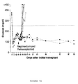
- Figure 14 is a line graph showing induction of donor-specific unresponsiveness to islet graft antigens by CTLA4Ig.
- Figure 15 is a line graph showing antibody serum titer levels of mice injected with sheep red blood cells (SRBC), mAb L6 and rat Ig, mAb L6 and anti-IL4, CTLA4Ig and rat Ig, CTLA4Ig and anti-IL4.
- the X axis measures the antibody-serum titer.
- the Y axis measures time in days.
- the closed box represents mice injected with SRBC at day 0 and day 46.
- the open box represents mice injected with SRBC at day 46.
- the closed circle represents mice injected with mAb L6 and rat immunoglobulin.
- the open circle represents mice injected with mAb L6 and anti-IL4 antibody.
- the closed triangle represents mice injected with CTLA4Ig and rat immunoglobulin.
- the open triangle represents mice injected with CTLA4Ig and anti-IL4 antibody.
- Figure 16 is a line graph showing antibody serum titer levels of mice injected with KLH, mAb L6 and rat Ig, mAb L6 and anti-IL4, CTLA4Ig and rat Ig, CTLA4Ig and anti-IL4.
- the X axis measures the antibody-serum titer.
- the Y axis measures time in days.
- the closed box represents mice injected with keyhole limpet hemocyanin (KLH) at day 46.
- the closed circle represents mice injected with mAb L6 and rat immunoglobulin.
- the open circle represents mice injected with mAb L6 and anti-IL4 antibody.
- the closed triangle represents mice injected with CTLA4Ig and rat immunoglobulin.
- the open triangle represents mice injected with CTLA4Ig and anti-IL4 antibody.
- blocking B7 interaction means to interfere with the binding of the B7 antigen to its ligands such as CD28 and CTLA4 thereby obstructing T cell and B cell interaction.
- CTLA4-binding molecule means any molecule which will bind the B7 antigen.
- an "IL4-binding molecule” means any molecule which will recognize and bind to IL4.
- This invention is directed to the isolation and expression of the human CTLA4 receptor found on T cell surfaces, which binds to the B7 antigen expressed on activated B cells, and cells of other lineages, and to expression of soluble fusion protein products of the CTLA4 receptor gene.
- the invention also provides methods for using the expressed CTLA4 receptor to regulate cellular interactions, including T cell interactions with B7 positive cells.
- the complete and correct DNA sequence encoding the amino acid sequence corresponding to human CTLA4 receptor protein of the invention is cloned using PCR.
- the cDNA containing the complete predicted coding sequence of CTLA4 was assembled from two PCR fragments amplified from H38 RNA, and inserted into the expression vector, CDM8 as described in detail in the Examples, infra . Isolates were transfected into COS cells and tested for binding of B7Ig, a soluble fusion protein having an amino acid sequence corresponding to the extracellular domain of B7 and a human immunoglobulin (Ig) C ⁇ 1 region, as described by Linsley et al., J. Exp. Med. 173:721-730 (1991).
- the DNA sequence of one isolate was then determined and found to correspond exactly to the predicted human CTLA4 sequence, fused at the N-terminus to the signal peptide from oncostatin M.
- the CTLA4 receptor is encoded by 187 amino acids (exclusive of the signal peptide and stop codons) and includes a newly identified N-linked glycosylation site at amino acid positions 109-111 (see Figure 3, infra ).
- the CTLA4 receptor is expressed using the oncostatin M signal peptide.
- soluble forms of the protein product of the CTLA4 receptor gene are prepared using fusion proteins having a first amino acid sequence corresponding to the extracellular domain of CTLA4 and a second amino acid sequence corresponding to the human IgC ⁇ 1 domain.
- Cloning and expression plasmids (CDM8 and ⁇ LN) were constructed containing cDNAs encoding portions of the amino acid sequence corresponding to human CTLA4 receptor based on the cDNA sequence described herein, where the cDNA encoding a first amino acid sequence corresponding to a fragment of the extracellular domain of the CTLA4 receptor gene is joined to DNA encoding a second amino acid sequence corresponding to an IgC region that permits the expression of the CTLA4 receptor gene by altering the solubility of the expressed CTLA4 protein.
- soluble CTLA4Ig fusion protein is encoded by a first amino acid sequence containing amino acid residues from about position 1 to about position 125 of the amino acid sequence corresponding to the extracellular domain of CTLA4 joined to a second amino acid sequence containing amino acid residues corresponding to the hinge, CH2 and CH3 regions of human IgC ⁇ 1.
- the fusion protein is preferably produced in dimeric form. The construct was then transfected into COS or CHO cells, and CTLA4Ig was purified and identified as a dimer.
- CTLA4Ig and the CTL4Ig/CD28 fusion protein hybrid may have amino acid substitutions in the amino acid sequence corresponding to the external domain of CTLA4 so as to produce molecules which would retain the functional property of CTLA4, namely, the molecule having such substitutions will still bind the B7 antigen.
- amino acid substitutions include, but are not necessarily limited to, amino acid substitutions known in the art as "conservative".
- substitutions can frequently be made in a protein without altering either the conformation or the function of the protein.
- Such changes include substituting any of isoleucine (I), valine (V), and leucine (L) for any other of these hydrophobic amino acids; aspartic acid (D) for glutamic acid (E) and vice versa; glutamine (Q) for asparagine (N) and vice versa; and serine (S) for threonine (T) and vice versa.
- Other substitutions can also be considered conservative, depending on the environment of the particular amino acid and its role in the three-dimensional structure of the protein.
- glycine (G) and alanine (A) can frequently be interchangeable, as can alanine and valine (V).
- Methionine (M) which is relatively hydrophobic, can frequently be interchanged with leucine and isoleucine, and sometimes with valine. Lysine (K) and arginine (R) are frequently interchangeable in locations in which the significant feature of the amino acid residue is its charge and the differing pK's of these two amino acid residues are not significant. Still other changes can be considered "conservative" in particular environments.
- mutants of the CTLA4-binding molecule were produced.
- One mutant comprises (1) a sequence beginning with the amino acid at position 1 and ending with the amino acid at position 95 of the CD28 receptor protein (Aruffo and Seed 1987); (2) a sequence beginning with the amino acid at position 95 and ending with amino acid at position 125 of the extracellular domain of CTLA4 (Brunet et al. 1987); and (3) a sequence corresponding to the human IgC ⁇ 1 domain.
- the second mutant comprises (1) a sequence beginning with the amino acid at position 1 and ending with the amino acid at position 95 of the CD28 receptor protein (Aruffo and Seed 1987); (2) a sequence beginning with the amino acid at position 95 and ending with amino acid at position 120 of the extracellular domain of CTLA4 (Brunet et al. 1987); and (3) a sequence corresponding to the human IgC ⁇ 1 domain.
- the present invention provides a method for blocking B7 interaction so as to regulate the immune response which comprises contacting lymphocytes with a CTLA4-binding molecule and an IL4-binding molecule.
- the lymphocytes may be B7 positive lymphocytes.
- the present invention provides a method for regulating an immune response which comprises contacting B7-positive lymphocytes with a CTLA4-binding molecule and an IL4-binding molecule.
- the immune response may be a B cell response resulting in the inhibition of antibody production. Additionally, the immune response may be a T cell response resulting in inhibition of cell mediated immunity. Further, the immune response may be an inhibition of lymphocyte proliferation.
- the present invention provides a method for inhibiting tissue transplant rejection by a subject, the subject being a recipient of transplanted tissue.
- This method can comprise administering to the subject a CTLA4-binding molecule and an IL4-binding molecule.
- the invention further provides a method for inhibiting graft versus host disease in a subject which comprises administering to the subject a CTLA4-binding molecule and an IL4-binding molecule.
- the CTLA4-binding molecule may be a CTLA4Ig fusion protein.
- the CTLA4Ig fusion protein may be a fusion protein having a first amino acid sequence containing amino acid residues from about position 1 to about position 125 of the amino acid sequence corresponding to the extracellular domain of CTLA4 and a second amino acid sequence containing amino acid residues corresponding to the hinge, CH2 and CH3 regions of human immunoglobulin C ⁇ 1.
- the CTLA4-binding molecule may be a CD28Ig/CTLA4Ig fusion protein hybrid.
- the CD28Ig/CTLA4Ig fusion protein hybrid may be a fusion protein hybrid having a first amino acid sequence corresponding to a portion of the extracellular domain of CD28 receptor fused to a second amino acid sequence corresponding to a portion of the extracellular domain of CTLA4 receptor and a third amino acid sequence corresponding to the hinge, CH2 and CH3 regions of human immunoglobulin C ⁇ 1.
- the IL4-binding molecule may be a monoclonal antibody which specifically recognizes and binds to IL4.
- the IL4-binding molecule is a soluble IL4 receptor which recognizes and binds to IL4 (Fanslow et al. 1991).
- the present invention provides the first protein product of CTLA4 transcripts in the form of a soluble fusion protein.
- the CTLA4Ig protein forms a disulfide-linked dimer having two subunits, each of which has an M r of approximately 50,000 indicating that native CTLA4 probably exists on the T cell surface as a disulfide-linked homodimer.
- B7 antigen has been shown to be a ligand for CD28 receptor on T cells (Linsley et al., Proc. Natl. Acad. Sci. USA , supra ).
- the CTLA4 receptor molecule appears functionally and structurally related to the CD28 receptor; both are receptors for the B cell activation antigen, B7, while CTLA4 appears to have higher affinity for B7, among the highest yet reported for lymphoid adhesion systems.
- CTLA4Ig was shown to bind more strongly to B7 positive (B7+) cell lines than CD28Ig.
- CTLA4Ig was shown to bind a single protein on lymphoblastoid cells which is similar in size to the B7 antigen.
- CTLA4Ig inhibited T cell proliferation and inhibited T h -induced IgM production.
- hybrid fusion proteins having amino acid sequences corresponding to fragments of different receptor proteins were constructed.
- amino acid sequences corresponding to selected fragments of the extracellular domains of CD28 and CTLA4 were linked to form CD28Ig/CTLA4Ig hybrid fusion proteins.
- a CD28Ig/CTLA4Ig fusion protein was obtained having a first amino acid sequence containing amino acid residues corresponding to a fragment of the extracellular domain of CD28 joined to a second amino acid sequence corresponding to a fragment of the extracellular domain of CTLA4Ig and to a third amino acid sequence corresponding to the hinge, CH2 and CH3 regions of human IgC ⁇ 1.
- hybrid fusion proteins is a CD28Ig/CTLA4Ig fusion construct having a first amino acid sequence containing amino acid residues from about position 1 to about position 94 of the amino acid sequence corresponding to the extracellular domain of CD28, joined to a second amino acid sequence containing amino acid residues from about position 94 to about position 125 of the amino acid sequence corresponding to the extracellular domain of CTLA4, joined to a third amino acid sequence corresponding to the hinge, CH2 and CH3 regions of human IgC ⁇ 1.
- Fusion protein constructs corresponding to CD28IgC ⁇ 1 and B7IgC ⁇ 1 for characterizing the CTLA4Ig of the present invention, and for preparing CD28Ig/CTLA4Ig fusion hybrids, were prepared as described by Linsley et al., J. Exp. Med. 173:721-730 (1991), incorporated by reference herein.
- cDNA clones may be prepared from RNA obtained from cells expressing B7 antigen and CD28 receptor based on knowledge of the published sequences for these proteins (Aruffo and Seed, and Freeman, supra ) using standard procedures.
- CTLA4Ig fusions consisting of DNA encoding amino acid sequences corresponding to the extracellular domain of CTLA4 and the hinge, CH2 and CH3 regions of human IgC ⁇ 1 were constructed by ligation of PCR fragments.
- the cDNA encoding the amino acid sequences is amplified using the polymerase chain reaction ("PCR") technique (see U.S. Patent Nos. 4,683,195 and 4,683,202 to Mullis et al. and Mullis & Faloona, Methods Enzymol. 154:335-350 (1987)).
- CTLA4Ig fusion polypeptides were obtained having DNA encoding amino acid sequences containing amino acid residues from about position 1 to about position 125 of the amino acid sequence corresponding to the extracellular domain of CTLA4 and DNA encoding amino acid sequences corresponding to the hinge, CH2 and CH3 regions of Ig C ⁇ 1.
- CTLA4 receptor protein in human lymphoid cells has not been previously reported, it was necessary to locate a source of CTLA4 mRNA.
- PCR cDNA made from the total cellular RNA of several human leukemia cell lines was screened, using as primers, oligonucleotides from the published sequence of the CTLA4 gene (Dariavach et al., supra ). Of the cDNA tested, H38 cells (an HTLV II-associated leukemia line) provided the best yield of PCR products having the expected size.
- CTLA4 Since a signal peptide for CTLA4 was not identified in the CTLA4 gene, the N terminus of the predicted sequence of CTLA4 was fused to the signal peptide of oncostatin M (Malik et al., Molec. and Cell. Biol. 9:2847 (1989)) in two steps using oligonucleotides as described in the Examples, infra .
- the product of the PCR reaction was ligated with cDNA encoding the amino acid sequences corresponding to the hinge, CH2 and CH3 regions of Ig C ⁇ 1 into a expression vector, such as CDM8 or ⁇ LN.
- a cDNA encoding the transmembrane and cytoplasmic domains of CTLA4 was obtained by PCR from H38 cells and joined with a fragment from CTLA4Ig, obtained as described above, encoding the oncostatin M signal peptide fused to the N terminus of CTLA4, using oligonucleotide primers as described in the Examples, infra .
- PCR fragments were ligated into the plasmid CDM8, resulting in an expression plasmid encoding the full length CTLA4 gene, and designated OMCTLA4.
- DNA encoding amino acids corresponding to portions of the extracellular domain of one receptor gene is joined to DNA encoding amino acids corresponding to portions of the extracellular domain of another receptor gene, and to DNA encoding the amino acid sequences corresponding to the hinge, CH2 and CH3 regions of human IgC ⁇ 1 using procedures as described above for the B7Ig, CD28Ig and CTLA4Ig constructs.
- DNA encoding amino acid residues from about position 1 to about position 94 of the amino acid sequence corresponding to the extracellular domain of the CD28 receptor is joined to DNA encoding amino acid residues from about position 94 to about position 125 of the amino acid sequence corresponding to the extracellular domain of the CTLA4 receptor and to DNA encoding the amino acid sequences corresponding to the hinge, CH2 and CH3 regions of human IgC ⁇ 1.
- vectors containing DNA encoding the fusion constructs of the invention are transformed into suitable host cells, such as the bacterial cell line E. coli strain MC1061/p3 (Invitrogen Corp., San Diego, CA) using standard procedures, and colonies are screened for the appropriate plasmids.
- suitable host cells such as the bacterial cell line E. coli strain MC1061/p3 (Invitrogen Corp., San Diego, CA) using standard procedures, and colonies are screened for the appropriate plasmids.
- transfection is performed using standard techniques appropriate to such cells. For example, transfection into mammalian cells is accomplished using DEAE-dextran mediated transfection, CaP04 co-precipitation, lipofection, electroporation, or protoplast fusion, and other methods known in the art including: lysozyme fusion or erythrocyte fusion, scraping, direct uptake, osmotic or sucrose shock, direct microinjection, indirect microinjection such as via erythrocyte-mediated techniques, and/or by subjecting host cells to electric currents.
- the above list of transfection techniques is not considered to be exhaustive, as other procedures for introducing genetic information into cells will no doubt be developed.
- eukaryotic host cell cultures derived from multicellular organisms are preferred (see Tissue Cultures , Academic Press, Cruz and Patterson, Eds. (1973)). These systems have the additional advantage of the ability to splice out introns and thus can be used directly to express genomic fragments.
- Useful host cell lines include Chinese hamster ovary (CHO), monkey kidney (COS), VERO and HeLa cells. In the present invention, cell lines stably expressing the fusion constructs are preferred.
- Expression vectors for such cells ordinarily include promoters and control sequences compatible with mammalian cells such as, for example, CMV promoter (CDM8 vector) and avian sarcoma virus (ASV) ( ⁇ LN vector).
- CMV promoter CDM8 vector
- ASV avian sarcoma virus
- Other commonly used early and late promoters include those from Simian Virus 40 (SV 40) (Fiers, et al., Nature 273:113 (1973)), or other viral promoters such as those derived from polyoma, Adenovirus 2, and bovine papilloma virus.
- SV 40 Simian Virus 40
- the controllable promoter, hMTII Kerin, et al., Nature 299:797-802 (1982) may also be used.
- preferred host cells for expression of the fusion constructs include eukaryotic cells such as COS or CHO cells, other eukaryotic microbes may be used as hosts.
- Laboratory strains of Saccharomyces cerevisiae Baker's yeast, are most used although other strains such as Schizosaccharomyces pombe may be used.
- Control sequences for yeast vectors include promoters for the synthesis of glycolytic enzymes (Hess et al., J. Adv. Enzyme Reg. 7:149 (1968); Holland et al., Biochemistry 17:4900 (1978)). Additional promoters known in the art include the CMV promoter provided in the CDM8 vector (Toyama and Okayama, FEBS 268:217-221 (1990); the promoter for 3-phosphoglycerate kinase (Hitzeman et al., J. Biol. Chem. 255:2073 (1980)), and those for other glycolytic enzymes.
- promoters which have the additional advantage of transcription controlled by growth conditions are the promoter regions for alcohol dehydrogenase 2, isocytochrome C, acid phosphatase, degradative enzymes associated with nitrogen metabolism, and enzymes responsible for maltose and galactose utilization. It is also believed terminator sequences are desirable at the 3' end of the coding sequences. Such terminators are found in the 3' untranslated region following the coding sequences in yeast-derived genes.
- prokaryotic cells may be used as hosts for expression.
- Prokaryotes most frequently are represented by various strains of E. coli ; however, other microbial strains may also be used.
- Commonly used prokaryotic control sequences which are defined herein to include promoters for transcription initiation, optionally with an operator, along with ribosome binding site sequences, include such commonly used promoters as the beta-lactamase (penicillinase) and lactose (lac) promoter systems (Chang et al., Nature 198: 1056 (1977)), the tryptophan (trp) promoter system (Goeddel et al., Nucleic Acids Res. 8:4057 (1980)) and the lambda derived P L promoter and N-gene ribosome binding site (Shimatake et al., Nature 292:128 (1981)).
- the nucleotide sequences encoding CD28Ig and CTLA4Ig proteins, and fusion hybrid proteins such as CD28Ig/CTLA4Ig, may be expressed in a variety of systems as set forth below.
- the cDNA may be excised by suitable restriction enzymes and ligated into suitable prokaryotic or eukaryotic expression vectors for such expression.
- CD28 and CTLA4 receptor proteins occur in nature as dimers, it is believed that successful expression of these proteins requires an expression system which permits these proteins to form as dimers. Truncated versions of these proteins (i.e. formed by introduction of a stop codon into the sequence at a position upstream of the transmembrane region of the protein) appear not to be expressed.
- the expression of CD28 and CTLA4 receptors as fusion proteins permits dimer formation of these proteins.
- expression of CTLA4 protein as a fusion product is preferred in the present invention.
- a stable CHO line of the invention designated Chinese Hamster Ovary Cell Line CTLA4Ig-24, is preferred for expression of CTLA4Ig and has been deposited with the ATCC under the terms of the Budapest Treaty on May 31, 1991, and accorded ATCC accession number 10762.
- CTLA4 receptor of the invention is accomplished transfecting a cell line such as COS cells, and detecting expression by binding of the CTLA4-transfected cells to a ligand for the CTLA4 receptor, for example by testing for binding of the cells to B7Ig fusion protein.
- CD28 and CTLA4 receptor genes are not readily expressed as mature proteins using direct expression of DNA encoding the truncated protein.
- DNA encoding the amino acid sequence corresponding to the extracellular domains of CD28 and CTLA4, and including the codons for a signal sequence such as that of oncostatin M in cells capable of appropriate processing is fused with DNA encoding the amino acid sequence corresponding to the Fc domain of a naturally dimeric protein. Purification of these fusion protein products after secretion from the cells is thus facilitated using antibodies reactive with the anti-immunoglobulin portion of the fusion proteins. When secreted into the medium, the fusion protein product is recovered using standard protein purification techniques, for example by application to protein A columns.
- CTLA4Ig fusion protein and/or fragments of the fusion protein may be used to react with B7 positive cells, such as B cells, to regulate immune responses mediated by T cell interactions with the B7 antigen positive cells.
- CTLA4Ig fusion protein and CTLA4Ig/CD28Ig hybrid proteins, and/or fragments and derivatives of these proteins, may also be used to react with B7 positive cells, including B cells, to regulate immune responses mediated by T cell dependent B cell responses.
- fragment as used herein means a portion of the amino acid sequence encoding the protein referred to as "CTLA4".
- a fragment of the CTLA4Ig fusion protein that may be used is a polypeptide having an amino acid sequence corresponding to some portion of the amino acid sequence corresponding to the CTLA4 receptor used to obtain the CTLA4Ig fusion protein as described herein.
- the B7 antigen expressed on activated B cells and cells of other lineages, and the CD28 receptor expressed on T cells, can directly bind to each other, and this interaction can mediate cell-cell interaction. Such interactions directly trigger the CD28 activation pathway in T cells, leading to cytokine production, T cell proliferation, and B cell differentiation into immunoglobulin producing cells.
- the activation of B cells that occurs can cause increased expression of B7 antigen and further CD28 stimulation, leading to a state of chronic inflammation such as in autoimmune diseases, allograft rejection, graft versus host disease or chronic allergic reactions. Blocking or inhibiting this reaction may be effective in preventing T cell cytokine production and thus preventing or reversing inflammatory reactions.
- CTLA4Ig is shown herein to be a potent inhibitor of in vitro lymphocyte functions requiring T and B cell interaction. This indicates the importance of interactions between the B7 antigen and its counter-receptors, CTLA4 and/or CD28.
- the cytoplasmic domains of murine and human CTLA4 are similar (Dariavach et al., supra , 1988), suggesting that this region has important functional properties.
- the cytoplasmic domains of CD28 and CTLA4 also share homology.
- CTLA4 is a more potent inhibitor in vitro of lymphocyte responses than either anti-BB1, or anti-CD28 mAbs.
- CTLA4Ig does not have direct stimulatory effects on T cell proliferation to counteract its inhibitory effects. Therefore, the CTLA4Ig fusion protein may perform as a better inhibitor in vivo than anti-CD28 monoclonal antibodies.
- the immunosuppressive effects of CTLA4Ig in vitro suggests its use in therapy for treatment of autoimmune disorders involving abnormal T cell activation or Ig production.
- CTLA4Ig fusion protein is expected to exhibit inhibitory properties in vivo .
- CTLA4Ig will act to inhibit T cells in a manner similar to the effects observed for the anti-CD28 antibody, under similar conditions in vivo .
- binding of introduced CTLA4Ig to react with B7 antigen positive cells, for example B cells may interfere, i.e. inhibit, the T cell/B cell interactions resulting in regulation of immune responses.
- CTLA4Ig is expected to be useful in vivo as an inhibitor of T cell activity, over non-specific inhibitors such as cyclosporine and glucosteroids.
- the CTLA4Ig fusion protein or CTLA4Ig/CD28Ig hybrid proteins may be introduced in a suitable pharmaceutical carrier in vivo , i.e. administered into a human subject for treatment of pathological conditions such as immune system diseases or cancer.
- Introduction of the fusion protein in vivo is expected to result in interference with T cell interactions with other cells, such as B cells, as a result of binding of the ligand to B7 positive cells.
- the prevention of normal T cell interactions may result in decreased T cell activity, for example, decreased T cell proliferation.
- administration of the fusion protein in vivo is expected to result in regulation of in vivo levels of cytokines, including, but not limited to, interleukins, e.g. interleukin ("IL")-2, IL-3, IL-4, IL-6, IL-8, growth factors including tumor growth factor (“TGF”), colony stimulating factor (“CSF”), interferons (“IFNs”), and tumor necrosis factor (“TNF”) to promote desired effects in a subject.
- cytokines including, but not limited to, interleukins, e.g. interleukin (“IL")-2, IL-3, IL-4, IL-6, IL-8, growth factors including tumor growth factor (“TGF”), colony stimulating
- the fusion protein when introduced in vivo , it may block production of cytokines, which contribute to malignant growth, for example of tumor cells.
- the fusion protein may also block proliferation of viruses dependent on T cell activation, such as the virus that causes AIDS, HTLV1.
- the effect of administration of the CTLA4Ig fusion protein or its fragments in vivo is inhibitory, resulting from blocking by the fusion protein of the CTLA4 and CD28 triggering resulting from T cell/B cell contact.
- the CTLA4Ig protein may block T cell proliferation.
- Introduction of the CTLA4Ig fusion protein in vivo will thus produce effects on both T and B cell-mediated immune responses.
- the fusion protein may also be administered to a subject in combination with the introduction of cytokines or other therapeutic reagents.
- other reagents including derivatives reactive with the CTLA4Ig fusion protein or the CTLA4 receptor are used to regulate T cell interactions.
- antibodies, and/or antibody fragments reactive with the CTLA4 receptor may be screened to identify those capable of inhibiting the binding of the CTLA4Ig fusion protein to the B7 antigen.
- the antibodies or antibody fragments such as Fab or F(ab')2 fragments, may then be used to react with the T cells, for example, to inhibit T cell proliferation.
- Monoclonal antibodies reactive with CTLA4 receptor may be produced by hybridomas prepared using known procedures, such as those introduced by Kohler and Milstein (see Kohler and Milstein, Nature , 256:495-97 (1975)), and modifications thereof, to regulate cellular interactions.
- the animal which is primed to produce a particular antibody.
- the animal can be primed by injection of an immunogen (e.g. the B7Ig fusion protein, CTLA4Ig fusion protein or CD28Ig/CTLA4Ig hybrid fusion protein) to elicit the desired immune response, i.e. production of antibodies from the primed animal.
- an immunogen e.g. the B7Ig fusion protein, CTLA4Ig fusion protein or CD28Ig/CTLA4Ig hybrid fusion protein
- a primed animal is also one which is expressing a disease. Lymphocytes derived from the lymph nodes, spleens or peripheral blood of primed, diseased animals can be used to search for a particular antibody.
- the lymphocyte chromosomes encoding desired immunoglobulins are immortalized by fusing the lymphocytes with myeloma cells, generally in the presence of a fusing agent such as polyethylene glycol (PEG).
- a fusing agent such as polyethylene glycol (PEG).
- PEG polyethylene glycol
- Any of a number of myeloma cell lines may be used as a fusion partner according to standard techniques; for example, the P3-NS1/1-Ag4-1, P3-x63-Ag8.653, Sp2/0-Ag14, or HL1-653 myeloma lines. These myeloma lines are available from the ATCC, Rockville, Maryland.
- the resulting cells which include the desired hybridomas, are then grown in a selective medium such as HAT medium, in which unfused parental myeloma or lymphocyte cells eventually die. Only the hybridoma cells survive and can be grown under limiting dilution conditions to obtain isolated clones.
- the supernatants of the hybridomas are screened for the presence of the desired specificity, e.g. by immunoassay techniques using the CTLA4Ig protein that has been used for immunization. Positive clones can then be subcloned under limiting dilution conditions, and the monoclonal antibody produced can be isolated.
- the individual cell line may be propagated in vitro , for example, in laboratory culture vessels, and the culture medium containing high concentrations of a single specific monoclonal antibody can be harvested by decantation, filtration, or centrifugation.
- fragments of these antibodies containing the active binding region reactive with the extracellular domain of CTLA4 receptor such as Fab, F(ab')2 and Fv fragments may be produced.
- Such fragments can be produced using techniques well established in the art (see e.g. Rousseaux et al., in Methods Enzymol ., 121:663-69, Academic Press (1986)).
- Anti-B7 monoclonal antibodies prepared as described above may be used to bind to B7 antigen to inhibit interactions of CD28-positive or CTLA4-positive T cells with B7 positive cells.
- Anti-CTLA4 monoclonal antibodies may be used to bind to CTLA4 receptor to inhibit the interaction of CTLA4-positive T cells with other cells.
- the CTLA4Ig fusion protein may be used to identify additional compounds capable of regulating the interaction between CTLA4 and the B7 antigen.
- Such compounds may include small naturally occurring molecules that can be used to react with B cells and/or T cells.
- fermentation broths may be tested for the ability to inhibit CTLA4/B7 interactions.
- derivatives of the CTLA4Ig fusion protein as described above may be used to regulate T cell proliferation.
- the fragments or derivatives may be used to block T cell proliferation in graft versus host (GVH) disease which accompanies allogeneic bone marrow transplantation.
- GVH graft versus host
- the CD28-mediated T cell proliferation pathway is cyclosporine-resistant, in contrast to proliferation driven by the CD3/Ti cell receptor complex (June et al., 1987, supra ). Cyclosporine is relatively ineffective as a treatment for GVH disease (Storb, Blood 68:119-125 (1986)). GVH disease is thought to be mediated by T lymphocytes which express CD28 antigen (Storb and Thomas, Immunol. Rev. 88:215-238 (1985)). Thus, the CTLA4Ig fusion protein may be useful alone, or in combination with immunosuppressants such as cyclosporine, for blocking T cell proliferation in GVH disease.
- Regulation of CTLA4-positive T cell interactions with B7 positive cells, including B cells, by the methods of the invention may thus be used to treat pathological conditions such as autoimmunity, transplantation, infectious diseases and neoplasia.
- CTLA4-binding molecules and IL4-binding molecules described herein may be in a variety of dosage forms which include, but are not limited to, liquid solutions or suspensions, tablets, pills, powders, suppositories, polymeric microcapsules or microvesicles, liposomes, and injectable or infusible solutions.
- dosage forms include, but are not limited to, liquid solutions or suspensions, tablets, pills, powders, suppositories, polymeric microcapsules or microvesicles, liposomes, and injectable or infusible solutions. The preferred form depends upon the mode of administration and the therapeutic application.
- the most effective mode of administration and dosage regimen for the molecules of the present invention depends upon the severity and course of the disease, the subject's health and response to treatment and the judgment of the treating physician. Accordingly, the dosages of the molecules should be titrated to the individual subject.
- Adjustments in the dosage regimen may be made to optimize the growth inhibiting response. Doses may be divided and administered on a daily basis or the dose may be reduced proportionally depending upon the situation. For example, several divided doses may be administered daily or the dose may be proportionally reduced as indicated by the specific therapeutic situation.
- an effective amount for treating a subject may be between about 0.1 and about 10mg/kg body weight of subject. Also, the effective amount may be an amount between about 1 and about 10 mg/kg body weight of subject.
- the subject invention overcomes the problems associated with current therapies directed to preventing the rejection of tissue or organ transplants.
- the present invention affects only immunological responses mediated by B7 interactions.
- the present invention affects the transplant antigen-specific T cells, thus inducing donor-specific and antigen-specific tolerance.
- the binding of CD28 by its ligand, B7/BB1 (B7), during T cell receptor engagement is critical for proper T cell signaling in some systems (M. K. Jenkins, P. S. Taylor, S. D. Norton, K. B. Urdahl, J. Immunol. 147 :2461 (1991); C. H. June, J. A. Ledbetter, P. S. Linsley, C. B. Thompson,Immunol. Today 11 :211 (1990); H. Reiser, G. J. Freeman, Z. Razi-Wolf, C. D. Gimmi, B. Benacerraf, L.
- CTLA4Ig fusion protein binds to both human and murine B7 (with a 20-fold greater affinity than CD28), blocks the binding of CD28 to B7, inhibits T cell activation, and induces T cell unresponsiveness in vitro (F. A. Harding, J. G. McArthur, J. A. Gross, D. H. Raulet, J. P. Allison, Nature 356 :607 (1992); P. S. Linsley et al., J. Exp. Med. 174 :561 (1991).
- the present invention would be useful to obtain expression of a soluble protein product of the heretofore unexpressed CTLA4 gene, and to identify a natural ligand for CTLA4 that is involved in functional responses of T cells.
- the soluble protein product could then be used to regulate T cell responses in vivo to treat pathological conditions.
- Receptor-immunoglobulin C gamma (IgC ⁇ ) fusion proteins B7Ig and CD28Ig were prepared as described by Linsley et al., in J. Exp. Med. 173:721-730 (1991), incorporated by reference herein. Briefly, DNA encoding amino acid sequences corresponding to the respective receptor protein (e.g. B7) was joined to DNA encoding amino acid sequences corresponding to the hinge, CH2 and CH3 regions of human IgC ⁇ 1. This was accomplished as follows.
- PCR Polymerase Chain Reaction
- DNA fragments were amplified using primer pairs as described below for each fusion protein.
- PCR reactions (0.1 ml final volume) were run in Tag polymerase buffer (Stratagene, La Jolla, CA), containing 20 ⁇ moles each of dNTP; 50-100 pmoles of the indicated primers; template (1 ng plasmid or cDNA synthesized from ⁇ 1 ⁇ g total RNA using random hexamer primer, as described by Kawasaki in PCR Protocols, Academic Press, pp. 21-27 (1990), incorporated by reference herein); and Tag polymerase (Stratagene). Reactions were run on a thermocycler (Perkin Elmer Corp., Norwalk, CT) for 16-30 cycles (a typical cycle consisted of steps of 1 min at 94°C, 1-2 min at 50°C and 1-3 min at 72°C).
- Plasmid Construction Expression plasmids containing cDNA encoding CD28, as described by Aruffo and Seed, Proc. Natl. Acad. Sci. USA 84:8573 (1987)), were provided by Drs. Aruffo and Seed (Mass General Hospital, Boston, MA). Plasmids containing cDNA encoding CD5, as described by Aruffo, Cell 61:1303 (1990)), were provided by Dr. Aruffo. Plasmids containing cDNA encoding B7, as described by Freeman et al., J. Immunol. 143:2714 (1989)), were provided by Dr. Freeman (Dana Farber Cancer Institute, Boston, MA).
- OMCD28 and OMB7 constructs were made (OMCD28 and OMB7) as described by Linsley et al., J. Exp. Med. , supra , in which stop codons were introduced upstream of the transmembrane domains and the native signal peptides were replaced with the signal peptide from oncostatin M (Malik et al., Mol. Cell Biol. 9:2847 (1989)). These were made using synthetic oligonucleotides for reconstruction (OMCD28) or as primers (OMB7) for PCR.
- OMCD28 is a CD28 cDNA modified for more efficient expression by replacing the signal peptide with the analogous region from oncostatin M.
- CD28Ig and B7Ig fusion constructs were made in two parts. The 5' portions were made using OMCD28 and OMB7 as templates and the oligonucleotide, CTAGCCACTGAAGCTTCACCATGGGTGTACTGCTCACAC (SEQ ID NO:1), (encoding the amino acid sequence corresponding to the oncostatin M signal peptide) as a forward primer, and either TGGCATGGGCTCCTGATCAGGCTTAGAAGGTCCGGGAAA (SEQ ID NO:2), or, TTTGGGCTCCTGATCAGGAAAATGCTCTTGCTTGGTTGT (SEQ ID NO:3) as reverse primers, respectively.
- Products of the PCR reactions were cleaved with restriction endonucleases (Hind III and BclI) as sites introduced in the PCR primers and gel purified.
- the 3' portion of the fusion constructs corresponding to human IgC ⁇ 1 sequences was made by a coupled reverse transcriptase (from Avian myeloblastosis virus; Life Sciences Associates, Bayport, NY)-PCR reaction using RNA from a myeloma cell line producing human-mouse chimeric mAb L6 (provided by Dr. P. Fell and M. Gayle, Bristol-Myers Squibb Company, Pharmaceutical Research Institute, Seattle, WA) as template.
- the oligonucleotide was used as forward primer, and CTTCGACCAGTCTAGAAGCATCCTCGTGCGACCGCGAGAGC (SEQ ID NO:5) as reverse primer.
- Reaction products were cleaved with BclI and XbaI and gel purified.
- Final constructs were assembled by ligating HindIII/BclI cleaved fragments containing CD28 or B7 sequences together with BclI/XbaI cleaved fragment containing IgC ⁇ 1 sequences into HindIII/XbaI cleaved CDM8.
- Ligation products were transformed into MC1061/p3 E. coli cells and colonies were screened for the appropriate plasmids. Sequences of the resulting constructs were confirmed by DNA sequencing.
- the construct encoding B7 contained DNA encoding amino acids corresponding to amino acid residues from approximately position 1 to approximately position 215 of the extracellular domain of B7.
- the construct encoding CD28 contained DNA encoding amino acids corresponding to amino acid residues from approximately position 1 to approximately position 134 of the extracellular domain of CD28.
- CD5Ig was constructed in identical fashion, using CATTGCACAGTCAAGCTTCCATGCCCATGGGTTCTCTGGCCACCTTG (SEQ ID NO:6), as forward primer and ATCCACAGTGCAGTGATCATTTGGATCCTGGCATGTGAC (SEQ ID NO:7) as reverse primer.
- the PCR product was restriction endonuclease digested and ligated with the IgC ⁇ 1 fragment as described above.
- the resulting construct (CD5Ig) encoded a mature protein having an amino acid sequence containing amino acid residues from position 1 to position 347 of the sequence corresponding to CD5, two amino acids introduced by the construction procedure (amino acids DQ), followed by DNA encoding amino acids corresponding to the IgC ⁇ 1 hinge region.
- COS monkey kidney cells
- COS monkey kidney cells
- expression plasmids expressing CD28 and B7 using a modification of the protocol of Seed and Aruffo (Proc. Natl. Acad. Sci. 84:3365 (1987)), incorporated by reference herein.
- Cells were seeded at 106 per 10 cm diameter culture dish 18-24 h before transfection.
- Plasmid DNA was added (approximately 15 ⁇ g/dish) in a volume of 5 mls of serum-free DMEM containing 0.1 mM chloroquine and 600 ⁇ g/ml DEAE Dextran, and cells were incubated for 3-3.5 h at 37°C.
- Transfected cells were then briefly treated (approximately 2 min) with 10% dimethyl sulfoxide in PBS and incubated at 37°C for 16-24 h in DMEM containing 10% FCS.
- culture medium was removed and replaced with serum-free DMEM (6 ml/dish). Incubation was continued for 3 days at 37°C, at which time the spent medium was collected and fresh serum-free medium was added. After an additional 3 days at 37°C, the spent medium was again collected and cells were discarded.
- CHO cells expressing CD28, CD5 or B7 were isolated as described by Linsley et al., (1991) supra , as follows: Briefly, stable transfectants expressing CD28, CD5, or B7, were isolated following cotransfection of dihydrofolate reductase-deficient Chinese hamster ovary (dhfr CHO) cells with a mixture of the appropriate expression plasmid and the selectable marker, pSV2dhfr (Linsley et al., Proc. Natl. Acad. Sci. USA 87:5031 (1990)), incorporated by reference herein.
- dhfr CHO dihydrofolate reductase-deficient Chinese hamster ovary
- Transfectants were then grown in increasing concentrations of methotrexate to a final level of 1 ⁇ M and were maintained in DMEM supplemented with 10% fetal bovine serum (FBS), 0.2 mM proline and 1 ⁇ M methotrexate.
- CHO lines expressing high levels of CD28 (CD28+ CHO) or B7 (B7+ CHO) were isolated by multiple rounds of fluorescence-activated cell sorting (FACS R ) following indirect immunostaining with mAbs 9.3 or BB-1. Amplified CHO cells negative for surface expression of CD28 or B7 (dhfr+ CHO) were also isolated by FACS R from CD28-transfected populations.
- Transfected CHO or COS cells or activated T cells were analyzed by indirect immunostaining. Before staining, CHO cells were removed from their culture vessels by incubation in PBS containing 10 mM EDTA. Cells were first incubated with murine mAbs 9.3 (Hansen et al., Immunogenetics 10:247 (1980)) or BB-1 (Yokochi et al., J. Immunol. 128:823 (1981)), or with Ig fusion proteins (all at 10 ⁇ g/ml in DMEM containing 10% FCS) for 1-2 h at 4°C.
- FITC-conjugated second step reagent goat anti-mouse Ig serum for murine mAbs, or goat anti-human Ig C ⁇ serum for fusion proteins (Tago, Inc., Burlingame, CA)
- Fluorescence was analyzed on a FACS IV R cell sorter (Becton Dickinson and Co., Mountain View, CA) equipped with a four decade logarithmic amplifier.
- a soluble genetic fusion encoding CTLA4Ig between the extracellular domain of CTLA4 and an IgC ⁇ 1 domain was constructed in a manner similar to that described above for the CD28Ig construct.
- the extracellular domain of the CTLA4 gene was cloned by PCR using synthetic oligonucleotides corresponding to the published sequence (Dariavach et al., Eur. Journ. Immunol. 18:1901-1905 (1988)).
- the oligonucleotide (which encoded the C terminal 15 amino acids from the oncostatin M signal peptide fused to the N terminal 7 amino acids of CTLA4) was used as forward primer, and TTTGGGCTCCTGATCAGAATCTGGGCACGGTTG (SEQ ID NO:9) (encoding amino acid residues 119-125 of the amino acid sequence encoding CTLA4 receptor and containing a Bcl I restriction enzyme site) as reverse primer.
- the template for this step was cDNA synthesized from 1 ⁇ g of total RNA from H38 cells (an HTLV II infected T cell leukemic cell line provided by Drs. Salahudin and Gallo, NCI, Bethesda, MD).
- a portion of the PCR product from the first step was reamplified, using an overlapping forward primer, encoding the N terminal portion of the oncostatin M signal peptide and containing a Hind III restriction endonuclease site, and the same reverse primer.
- the product of the PCR reaction was digested with Hind III and Bcl I and ligated together with a Bcl 1/Xba I cleaved cDNA fragment encoding the amino acid sequences corresponding to the hinge, CH2 and CH3 regions of IgC ⁇ 1 into the Hind III/Xba I cleaved expression vector, CDM8 or Hind III/Xba I cleaved expression vector ⁇ LN (provided by Dr. Aruffo).
- FIG. 1 A map of the resulting CTLA4Ig fusion construct is shown in Figure 1. Sequences displayed in this figure show the junctions between CTLA4 (upper case letters, unshaded regions) and the signal peptide, SP, of oncostatin M (dark shaded regions), and the hinge, H, of IgC ⁇ 1 (stippled regions). The amino acid in parentheses was introduced during construction. Asterisks (*) indicate cysteine to serine mutations introduced in the IgC ⁇ hinge region.
- the immunoglobulin superfamily V-like domain present in CTLA4 is indicated, as are the CH2 and CH3 domains of IgC ⁇ 1.
- Expression plasmids, CDM8, containing CTLA4Ig were then transfected into COS cells using DEAE/dextran transfection by modification (Linsley et al., 1991, supra ) of the protocol described by Seed and Aruffo, 1987, supra .
- Expression plasmid constructs ( ⁇ LN or CDM8) containing cDNA encoding the amino acid sequence of CTLA4Ig, was transfected by lipofection using standard procedures into dhfr CHO lines to obtain novel cell lines stably expressing CTLA4Ig.
- FBS fetal bovine serum
- proline 0.2 mM proline
- methotrexate 1 fetal bovine serum
- CTLA4Ig-24 CHO cell line has been deposited with the ATCC under the Budapest Treaty on May 31, 1991 and has been accorded accession number ATCC 10762.
- CTLA4Ig was purified by protein A chromatography from serum-free conditioned supernatants ( Figure 2). Concentrations of CTLA4Ig were determined assuming an extinction coefficient at 280 nm of 1.6 (experimentally determined by amino acid analysis of a solution of known absorbance). Molecular weight standards (lanes 1 and 3, Figure 2) and samples (1 ⁇ g) of CTLA4Ig (lanes 2 and 4) were subjected to SDS-PAGE (4-12% acrylamide gradient) under non-reducing conditions (- ⁇ ME, lanes 1 and 2) or reducing conditions (+ ⁇ ME, lanes 3 and 4) Proteins were visualized by staining with Coomassie Brilliant Blue.
- CTLA4Ig Under non-reducing conditions, CTLA4Ig migrated as a M r approximately 100,000 species, and under reducing conditions, as a M r approximately 50,000 species ( Figure 2). Because the IgC ⁇ hinge disulfides were eliminated during construction, CTLA4Ig, like CD28Ig, is a dimer presumably joined through a native disulfide linkage.
- cDNA encoding amino acids corresponding to a fragment of the transmembrane and cytoplasmic domains of CTLA4 was cloned by PCR and then joined with cDNA encoding amino acids corresponding to a fragment from CTLA4Ig that corresponded to the oncostatin M signal peptide fused to the N-terminus of CTLA4.
- Procedures for PCR, and cell culture and transfections were as described above in Example 1 using COS cells and DEAE-dextran transfection.
- CTLA4 receptor protein in human lymphoid cells has not been previously reported, it was necessary to locate a source of CTLA4 mRNA.
- the oligonucleotide GCAATGCACGTGGCCCAGCCTGCTGTGGTAGTG (SEQ ID NO:11), (encoding the first 11 amino acids in the predicted coding sequence) was used as a forward primer, and TGATGTAACATGTCTAGATCAATTGATGGGAATAAAATAAGGCTG (SEQ ID NO:12) (homologous to the last 8 amino acids in CTLA4 and containing a Xba I site) as reverse primer.
- the template again was a cDNA synthesized from 1 ⁇ g RNA from H38 cells.
- the resulting construct corresponded to full length CTLA4 (SEQ ID NOs: 13 and 14) and the oncostatin M signal peptide.
- the construct is shown in Figure 3 and was designated OMCTLA4.
- the sequence for CTLA4 shown in Figure 3 differs from the predicted human CTLA4 DNA sequence (Dariavach et al., supra ) by a base change such that the previously reported alanine at amino acid position 111 of the amino acid sequence shown, encodes a threonine. This threonine is part of a newly identified N-linked glycosylation site that may be important for successful expression of the fusion protein.
- Ligation products were transformed into MC1061/p3 E. coli cells and colonies were screened for the appropriate plasmids. Sequences of the resulting constructs were confirmed by DNA sequence analysis.
- CTLA4Ig constructs To characterize the CTLA4Ig constructs, several isolates, CD28Ig, B7Ig, and CD5Ig, were prepared as described above and were transfected into COS cells as described in Examples 2 and 3, and were tested by FACS R analysis for binding of B7Ig.
- mAbs Murine monoclonal antibodies (mAbs) 9.3 (anti-CD28) and G19-4 (anti-CD3), G3-7 (anti-CD7), BB-1 (anti-B7 antigen) and rat mAb 187.1 (anti-mouse K chain) have been described previously (Ledbetter et al., Proc. Natl. Acad. Sci. 84:1384-1388 (1987); Ledbetter et al., Blood 75:1531 (1990); Yokochi et al., supra ) and were purified from ascites before use.
- the hybridoma producing mAb OKT8 was obtained from the ATCC, Rockville, MD, and the mAb was also purified from ascites before use.
- mAb 4G9 (anti-CD19) was provided by Dr. E. Engleman, Stanford University, Palo Alto, CA). Purified human-mouse chimeric mAb L6 (having human C ⁇ 1 Fc portion) was a gift of Dr. P. Fell and M. Gayle (Bristol-Myers Squibb Pharmaceutical Research Institute, Seattle, WA).
- FITC-conjugated anti-mouse and anti-human second step reagents were mixed together before use. Fluorescence on a total of 10,000 cells was then analyzed by FACS R .
- Peripheral Blood Lymphocyte Separation and Stimulation Peripheral blood lymphocytes (PBLs) were isolated by centrifugation through Lymphocyte Separation Medium (Litton Bionetics, Kensington, MD). Alloreactive T cells were isolated by stimulation of PBL in a primary mixed lymphocyte reaction (MLR). PBL were cultured at 106/ml irradiated (5000 rad) T51 LCL. EBV-transformed lymphoblastoid cell lines (LCL), PM (Bristol-Myers Squibb Co.) and T51 (Bristol-Myers Squibb Co.) were maintained in RPMI supplemented with 10% FBS.
- LCL lymphoblastoid cell lines
- PM Bristol-Myers Squibb Co.
- T51 Bristol-Myers Squibb Co.
- PHA-activated T cells were prepared by culturing PBLs with 1 ⁇ g/ml PHA (Wellcome, Charlotte, NC) for five days, and one day in medium lacking PHA. Viable cells were collected by sedimentation through Lymphocyte Separation Medium before use. Cells were stimulated with mAbs or transfected CHO cells for 4-6 hr at 37°C, collected by centrifugation and used to prepare RNA.
- CD4+ T cells were isolated from PBLs by separating PBLs from healthy donors into T and non-T cells using sheep erythrocyte rosetting technique and further separating T cells by panning into CD4+ cells as described by Damle et al., J. Immunol. 139:1501 (1987), incorporated by reference herein. B cells were also purified from peripheral blood by panning as described by Wysocki and Sato, Proc. Natl. Acad. Sci. 75:2844 (1978), incorporated by reference herein, using anti-CD19 mAb 4G9. To measure T h -induced Ig production, 106 CD4+ T cells were mixed with 106 CD19+ B cells in 1 ml of RPMI containing 10% FBS.
- 96-well flat bottom microtiter ELISA plates (Corning, Corning, NY) were coated with 200 ⁇ l/well of sodium carbonate buffer (pH 9.6) containing 10 ⁇ g/ml of affinity-purified goat anti-human IgG or IgM antibody (Tago, Burlingame, CA), incubated overnight at 4°C, and then washed with PBS and wells were further blocked with 2% BSA in PBS (BSA-PBS).
- HRP horseradish peroxidase
- F(ab')2 fraction of affinity-purified goat anti-human IgG or IgM antibody Tago
- the plates were then washed, and 100 ⁇ l/well of o-phenylenediamine (Sigma Chemical Co., St. Louis, MO) solution (0.6 mg/ml in citrate-phosphate buffer with pH 5.5 and 0.045% hydrogen peroxide). Color development was stopped with 2 N sulfuric acid. Absorbance at 490 nm was measured with an automated ELISA plate reader.
- Test and control samples were run in triplicate and the values of absorbance were compared to those obtained with known IgG or IgM standards run simultaneously with the supernatant samples to generate the standard curve using which the concentrations of Ig in the culture supernatant were quantitated. Data are expressed as ng/ml of Ig ⁇ SEM of either triplicate or quadruplicate cultures.
- Binding Assays B7Ig was labeled with 125I to a specific activity of approximately 2 x 10⁶ cpm/pmole.
- a solution containing CTLA4Ig 0.5 ⁇ g in a volume of 0.05 ml of 10 mM Tris, pH 8.
- Wells were blocked with binding buffer (DMEM containing 50 mM BES (Sigma Chemical Co.), pH 6.8, 0.1% BAS, and 10% FCS) before addition of a solution (0.09 ml) containing 125I B7Ig (approximately 5 x 10⁵ cpm) in the presence or absence of competitor.
- binding buffer DMEM containing 50 mM BES (Sigma Chemical Co.), pH 6.8, 0.1% BAS, and 10% FCS
- mAb 9.3 bound to CD28-transfected COS cells, but not to CTLA4-transfected cells.
- the B7Ig fusion protein (but not control CD5Ig fusion protein) bound to both CD28- and CTLA4-transfected cells.
- CD7-transfected COS cells bound neither mAb 9.3 nor either of the fusion proteins. This indicates that CD28 and CTLA4 both bind the B cell activation antigen, B7.
- mAb 9.3 did not detectably bind CTLA4.
- CD28Ig bound to B7+ CHO cells but not to PM LCL, a cell line which expresses relatively low levels of the B7 antigen (Linsley et al., supra , 1990).
- CTLA4Ig bound more strongly to both cell lines than did CD28Ig, suggesting that it bound with higher affinity.
- CD28Ig nor CTLA4Ig bound to CD28+ CHO cells.
- 125I-surface labeled cells were subjected to immunoprecipitation analysis ( Figure 8).
- B7+ CHO and PM LCL cells were surface-labeled with 125I, and extracted with a non-ionic detergent solution as described above. Aliquots of extracts containing approximately 1.5 X 10⁷ cpm in a volume of 0.1 ml were subjected to immunoprecipitation analysis as described above with no addition, or 2 ⁇ g each of CD28Ig, CTLA4Ig or CD5Ig. Washed immunoprecipitates were then analyzed by SDS-PAGE (10-20% acrylamide gradient) under reducing conditions.
- the gel was then dried and subjected to autoradiography.
- the left panel of Figure 8 shows an autoradiogram obtained after a 1 day exposure.
- the right panel of Figure 8 shows an autoradiogram of the same gel after a 10 day exposure.
- the autoradiogram in the center panel of Figure 8 was also exposed for 10 days. Positions of molecular weight standard are also indicated in this figure.
- CTLA4Ig a diffusely migrating (M r approximately 50,000 - 75,000; center at approximately 60,000) radiolabeled protein was immunoprecipitated by CTLA4Ig, but not by CD28Ig or CD5Ig. This molecule co-migrated with B7 immunoprecipitated from B7+ CHO cells by CTLA4Ig, and much more weakly, by CD28Ig.
- MLR Primary mixed lymphocyte reaction
- CTLA4Ig inhibited the MLR reaction in a dose-dependant fashion by a maximum of > 90% with a 1/2 maximal response at approximately 30 ng/ml (approximately 0.8 nM).
- the Fab fragment of mAb 9.3 which previously was shown to be a more potent inhibitor of MLR than whole mAb 9.3 (Damle et al., J. Immunol. 140:1753-1761 (1988)), also inhibited the MLR, but at higher concentrations (approximately 800 ng/ml or approximately 30 nM for 1/2 maximal response).
- B7Ig and CD28Ig did not significantly inhibit the MLR even at higher concentrations.
- addition of B7Ig together with CTLA4Ig partially overcame the inhibition of MLR by CTLA4Ig, indicating that the inhibition was specifically due to interactions with B7 antigen.
- CD4+ T cells stimulated IgM production by allogenic CD19+ B Cells (in the absence of CD4+ T cells, IgM levels were reduced by 93%).
- mAbs 9.3 and BB-1 significantly inhibited T h -induced IgM production (63% and 65% inhibition, respectively).
- CTLA4Ig was even more effective as an inhibitor (89% inhibition) than were these mAbs.
- Inhibition by control Ig molecules, mAb OKT8 and CD5Ig was much less ( ⁇ 30% inhibition). None of these molecules significantly inhibited Ig production measured in the presence of Staphylococcal aureus enterotoxin B. Similar results were obtained with CD4+ T cells and B cells derived from other donors. These results indicate that the inhibition by CTLA4Ig is specific.
- CTLA4 and CD28 receptors are functionally as well as structurally related.
- CTLA4 is also a receptor for the B cell activation antigen, B7.
- CTLA4Ig bound 125I-B7 with an affinity constant, K d , of approximately 12 nM, a value some 20 fold higher than the affinity between CD28 and B7Ig (approximately 200 nM).
- K d affinity constant
- CTLA4 and CD28 may be thought of as high and low affinity receptors, respectively, for the same ligand, the B7 antigen.
- CD28 and B7 The apparent affinity between CD28 and B7 is similar to the affinity reported for binding of soluble alloantigen to the T cell receptor of a murine T cell hybridoma (approximately 100 nM; Schnek et al., Cell 56:47 (1989)), and is higher affinity than interactions between CD2 and LFA3 (Recny et al., J. Biol. Chem. 265:8542 (1990)), or CD4 and MHC class II molecules (Clayton et al., Nature 339:548 (1989)).
- the apparent affinity constant, K d , between CTLA4 and B7 is even greater, and compares favorably with higher affinity mAbs (K d 2-10,000 nM; Alzari et al., Ann. Rev. Immuno. 6:555 (1988)).
- the K d between CTLA4 and B7 is similar to or greater than K d values of integrin receptors and their ligands (10-2000 nM; Hautanen et at., J. Biol. Chem. 264:1437-1442 (1989); Di Minno et al., Blood 61:140-148 (1983); Thiagarajan and Kelley, J. Biol. Chem. 263:035-3038 (1988)).
- the affinity of interaction between CTLA4 and B7 is thus among the highest yet reported for lymphoid adhesion systems.
- CTLA4Ig a fusion construct containing the extracellular domain of CTLA4 fused to an IgC ⁇ 1 domain, forms a disulfide-linked dimer of M r approximately 50,000 subunits ( Figure 1). Because no interchain disulfides would be predicted to form in the Ig portion of this fusion, it seems likely that cysteines from CTLA4 are involved in disulfide bond formation.
- the analogous CD28Ig fusion protein (Linsley et al, supra , 1991) also contains interchain disulfide linkage(s).
- CTLA4 receptor like CD28 (Hansen et al., Immunogenetics 10:247-260 (1980)), exists on the T cell surface as a disulfide linked homodimer.
- CD28 and CTLA4 are highly homologous proteins, they are immunologically distinct, because the anti-CD28 mAb, mAb 9.3, does not recognize CTLA4 ( Figures 4 and 5).
- CTLA4 can activate T cells by a signalling pathway analogous to CD28.
- the cytoplasmic domains of murine and human CTLA4 are identical (Dariavach et al., supra 1988), suggesting that this region has important functional properties.
- the cytoplasmic domains of CD28 and CTLA4 also share homology, although it is unclear if this is sufficient to impart similar signaling properties to the two molecules.
- CTLA4Ig is a potent inhibitor of in vitro lymphocyte functions requiring T cell and B cell collaboration ( Figures 9 and 10). These findings, together with previous studies, indicate the fundamental importance of interactions between B7 antigen and its counter-receptors, CD28 and/or CTLA4, in regulating both T and B lymphocyte responses. CTLA4Ig should be a useful reagent for future investigations on the role of these interactions during immune responses. CTLA4Ig is a more potent inhibitor of in vitro lymphocyte responses than either mAb BB-1 or mAb 9.3 ( Figures 9 and 10). The greater potency of CTLA4Ig over mAb BB-1 is most likely due to the difference in affinities for B7 between these molecules ( Figure 6).
- CTLA4Ig is also more potent than mAb 9.3, probably because, unlike the mAb, it does not also have direct stimulatory effects on T cell proliferation (June et al., Immunology Today 11:211 (1989)) to counteract its inhibitory effects.
- the immunosuppressive effects of CTLA4Ig in vitro suggest that future investigations are warranted into possible therapeutic effects of this molecule for treatment of autoimmune disorders involving aberrant T cell activation or Ig production.
- H-2 d Female BALB/c (H-2 d ) and C57BL/6 (H-2 d )mice, 6 to 8 wk. of age were obtained from The Jackson Laboratory (Bar Harbor, ME).
- B6 or B10 mice treated with streptozocin (175 mg per kilogram of body weight) 3 to 5 days before transplant and exhibiting nonfasting plasma glucose levels of greater than 280 mg/dl (with the majority over 300 mg/ml), were used as recipients.
- Each animal received approximately 800 fresh human islets of 150 ⁇ m in diameter beneath the left renal capsule (D. Faustman and C. Coe, Science 252 :1700 (1991); Y. J. Zeng et al. Transplantation 53 :277 (1992)). Treatment was started immediately after transplantation.
- CTLA4Ig significantly prolongs human islet graft survival in mice in a donor-specific manner thereby providing an approach to immunosuppression
- mice C57BL/6 (B6) or C57BL/10 (B10) mice were treated with streptozotocin to eliminate mouse pancreatic islet B dell function. Diabetic animals were grafted under the kidney capsule, and treatment was started immediately after surgery. Survival of the islet grafts was monitored by the analysis of blood glucose concentrations.
- PBS phosphate-buffered saline
- the grafts were intact, with many islets visible.
- the B cells observed in the human islet tissue produced human insulin ( Figure 12D) and somatostatin.
- the human CTLA4Ig used in this study reacts with both murine and human B7.
- One advantage of the xenogeneic transplant model is the availability of a MAb to human B7 that does not react with mouse B7 (T. Yokochi, R. D. Holly, E. A. Clark, J. Immunol. 128 :823 (1982)).
- APCs antigen-presenting cells
- mice were transplanted as described and then treated with 50 ⁇ g of MAb to human B7 every other day for 14 days after transplant. This treatment prolonged graft survival in treated mice (9 to >50 days) in comparison to that for control mice ( Figure 13).
- the anti-B7 MAb is unable to block rejection as effectively as CTLA4Ig.
- the CTLA4Ig therapy resulted in graft acceptance in the majority of mice. However, the animals may not be tolerant. Transient immunosuppression can lead to permanent islet graft acceptance because of graft adaptation (the loss of immunogenicity as a result of the loss of APC function) (L. Hao, Y. Wang, R. G. Gill, K. J. Lafferty, J. Immunol. 139 :4022 (1987); K. J. Lafferty, S. J. Prowse, M. Simeonovic, Annu. Rev. Immunol. 1 :143 (1983)).
- H-2 d Female BALB/c (H-2 d ) and C57BL/6 (H-2 d )mice, 6 to 8 wk. of age were obtained from The Jackson Laboratory (Bar Harbor, ME).
- Monoclonal antibody 11B11 is a rat IgGl anti-murine IL-4 (Ohara, J., and W. E. Paul, 1985, Production of a monoclonal antibody to and molecular characterization of B-cell stimulatory factor-1. Nature 315 :333) (Verax (Lebanon, NH)).
- mice (five per group) were immunized intravenously with 108 SRBC alone or together with 200 ⁇ g chimeric L6 mAb or human CTLA4Ig fusion protein.
- the indicated groups were treated 2 hrs. prior to injection of SRBCs by intraperitoneal injection of 2 mls of either rat immunoglobulin or rat anti-murine IL-4 mAb 11B11 at 5 mg/ml. Treatment with chimeric L6 mAb or CTLA4Ig was repeated daily for 4 additional days.
- mice were given intravenous injections of SRBCs (Figure 15) or KLH (Figure 16) on day 46.
- the closed circle represents mice who were administered with only SRBC at day 0 and day 46.
- the open circle represents mice administered with only SRBC at day 46.
- the remaining mice represented in Figure 15 were further administered with SRBC at day 46.
- the mice were administered with a different immunogen, KLH, at day 46 only.
- mice Serum concentrations of mice measured as having antibodies directed against SRBCs or KLH were determined by ELISA as described (Linsley et al., Science 1992).
- Serum antibody titers were calculated as the dilution giving an A450 of five times background. Serum antibody titer values from Figure 15 were determined from pooled sera from five mice per group, while serum antibody titer values from Figure 16 represents mean titers of five individual sera. Arrows indicate an SRBC or KLH injection at day 46.
- Figures 15 and 16 show that the immunological response in mice injected concurrently with both CTLA4Ig and anti-IL4 (open triangle) is suppressed in an antigen-specific manner.
- Figure 15 shows that there is no rise in serum antibody titer (i.e. no primary or secondary immunological response) in mice injected concurrently with CTLA4Ig and anti-IL4 and injected with SRBC at day 0 and day 46.
- the combination of CTLA4Ig and anti-IL4 suppresses a primary and secondary immune response and induces long lasting immunological non-responsiveness to SRBC.
- Figure 15 shows that there is no primary immunological response in mice injected concurrently with CTLA4Ig and the control rat Ig (Cappel, Organontecknika, Palo Alto, CA). However, these mice exhibit a secondary immunological response after injection with SRBC at day 46 (closed triangle, Figure 15).
- Figure 16 shows that administration of CTLA4Ig and anti-IL4, followed by a different immunogen, KLH, at day 46 in mice does not suppress a primary immune response to KLH in mice. Instead, these mice exhibited a primary immune response to KLH (open triangle, Figure 16). Thus, mice treated with CTLA4Ig and anti-IL4 exhibited a highly specific immune response depending on the antigen administered therein.
Landscapes
- Health & Medical Sciences (AREA)
- Chemical & Material Sciences (AREA)
- Life Sciences & Earth Sciences (AREA)
- Immunology (AREA)
- Organic Chemistry (AREA)
- Medicinal Chemistry (AREA)
- General Health & Medical Sciences (AREA)
- Molecular Biology (AREA)
- Nuclear Medicine, Radiotherapy & Molecular Imaging (AREA)
- Pharmacology & Pharmacy (AREA)
- Animal Behavior & Ethology (AREA)
- General Chemical & Material Sciences (AREA)
- Public Health (AREA)
- Veterinary Medicine (AREA)
- Chemical Kinetics & Catalysis (AREA)
- Proteomics, Peptides & Aminoacids (AREA)
- Biophysics (AREA)
- Biochemistry (AREA)
- Genetics & Genomics (AREA)
- Engineering & Computer Science (AREA)
- Virology (AREA)
- Bioinformatics & Cheminformatics (AREA)
- Zoology (AREA)
- Cell Biology (AREA)
- Gastroenterology & Hepatology (AREA)
- Toxicology (AREA)
- Communicable Diseases (AREA)
- Oncology (AREA)
- Rheumatology (AREA)
- Transplantation (AREA)
- Pulmonology (AREA)
- Tropical Medicine & Parasitology (AREA)
- Pain & Pain Management (AREA)
- AIDS & HIV (AREA)
- Medicines Containing Antibodies Or Antigens For Use As Internal Diagnostic Agents (AREA)
- Peptides Or Proteins (AREA)
- Steroid Compounds (AREA)
- Medicines That Contain Protein Lipid Enzymes And Other Medicines (AREA)
- Preparation Of Compounds By Using Micro-Organisms (AREA)
- Medicines Containing Material From Animals Or Micro-Organisms (AREA)
Applications Claiming Priority (2)
| Application Number | Priority Date | Filing Date | Title |
|---|---|---|---|
| US08/008,898 US5770197A (en) | 1991-06-27 | 1993-01-22 | Methods for regulating the immune response using B7 binding molecules and IL4-binding molecules |
| US8898 | 1998-01-20 |
Publications (3)
| Publication Number | Publication Date |
|---|---|
| EP0613944A2 true EP0613944A2 (de) | 1994-09-07 |
| EP0613944A3 EP0613944A3 (de) | 1997-03-05 |
| EP0613944B1 EP0613944B1 (de) | 2005-08-24 |
Family
ID=21734327
Family Applications (1)
| Application Number | Title | Priority Date | Filing Date |
|---|---|---|---|
| EP94100882A Expired - Lifetime EP0613944B1 (de) | 1993-01-22 | 1994-01-21 | Verfahren zur Regulierung der Immunantwort durch den Gebrauch von CTLA4-Bindungsmoleküle und IL4-Bindungsmoleküle |
Country Status (13)
| Country | Link |
|---|---|
| US (1) | US5770197A (de) |
| EP (1) | EP0613944B1 (de) |
| JP (1) | JP3833723B2 (de) |
| AT (1) | ATE302846T1 (de) |
| AU (1) | AU682325B2 (de) |
| CA (1) | CA2113744C (de) |
| DE (1) | DE69434465T2 (de) |
| DK (1) | DK0613944T3 (de) |
| ES (1) | ES2247585T3 (de) |
| FI (1) | FI940270L (de) |
| IL (1) | IL108374A (de) |
| NO (1) | NO316223B1 (de) |
| PT (1) | PT613944E (de) |
Cited By (26)
| Publication number | Priority date | Publication date | Assignee | Title |
|---|---|---|---|---|
| WO1995001994A1 (en) * | 1993-07-09 | 1995-01-19 | Synergen, Inc. | Recombinant ctla4 polypeptides and methods for making the same |
| WO1995005464A1 (en) * | 1993-08-16 | 1995-02-23 | Arch Development Corporation | B7-2: ctla4/cd28 counter receptor |
| EP0682039A1 (de) * | 1994-04-15 | 1995-11-15 | Bristol-Myers Squibb Company | CTLA4-Moleküle und IL4-bindende Moleküle und deren Verwendungen |
| WO1996031229A1 (en) * | 1995-04-05 | 1996-10-10 | Beth Israel Hospital Association | Inhibiting rejection of a graft |
| WO1996039514A1 (en) * | 1995-06-05 | 1996-12-12 | The University Of Washington | Method for prolonging the expression of a gene of interest using soluble ctla4 molecules |
| US5773253A (en) * | 1993-01-22 | 1998-06-30 | Bristol-Myers Squibb Company | MYPPPY variants of CTL A4 and uses thereof |
| US5811097A (en) * | 1995-07-25 | 1998-09-22 | The Regents Of The University Of California | Blockade of T lymphocyte down-regulation associated with CTLA-4 signaling |
| US5855887A (en) * | 1995-07-25 | 1999-01-05 | The Regents Of The University Of California | Blockade of lymphocyte down-regulation associated with CTLA-4 signaling |
| US5993800A (en) * | 1995-06-05 | 1999-11-30 | Bristol-Myers Squibb Company | Methods for prolonging the expression of a heterologous gene of interest using soluble CTLA4 molecules and an antiCD40 ligand |
| US6051227A (en) * | 1995-07-25 | 2000-04-18 | The Regents Of The University Of California, Office Of Technology Transfer | Blockade of T lymphocyte down-regulation associated with CTLA-4 signaling |
| WO2000023115A1 (en) * | 1998-10-21 | 2000-04-27 | Opperbas Holding B.V. | TREATMENT OF TRAUMA (I.E. GRAFT REJECTION) WITH LIPOSOMES CONTAINING DNA ENCODING FOR CTLA4Ig OR FOR ANTI-CD40L |
| US6113901A (en) * | 1989-10-27 | 2000-09-05 | Arch Development Corporation | Methods of stimulating or enhancing the immune system with anti-CD3 antibodies |
| US6406696B1 (en) | 1989-10-27 | 2002-06-18 | Tolerance Therapeutics, Inc. | Methods of stimulating the immune system with anti-CD3 antibodies |
| US6491916B1 (en) | 1994-06-01 | 2002-12-10 | Tolerance Therapeutics, Inc. | Methods and materials for modulation of the immunosuppresive activity and toxicity of monoclonal antibodies |
| US6887471B1 (en) | 1991-06-27 | 2005-05-03 | Bristol-Myers Squibb Company | Method to inhibit T cell interactions with soluble B7 |
| US7041634B2 (en) | 1995-09-27 | 2006-05-09 | Emory University | Method of inhibiting immune system destruction of transplanted viable cells |
| US7094874B2 (en) | 2000-05-26 | 2006-08-22 | Bristol-Myers Squibb Co. | Soluble CTLA4 mutant molecules |
| US7304033B2 (en) | 2001-05-23 | 2007-12-04 | Bristol-Myers Squibb Company | Methods for protecting allogeneic islet transplant using soluble CTLA4 mutant molecules |
| US7455835B2 (en) | 2000-07-03 | 2008-11-25 | Bristol-Myers Squibb Company | Methods for treating immune system diseases using a soluble CTLA4 molecule |
| US7605238B2 (en) | 1999-08-24 | 2009-10-20 | Medarex, Inc. | Human CTLA-4 antibodies and their uses |
| US7794718B2 (en) | 2007-11-01 | 2010-09-14 | Perseid Therapeutics, LLC | Immunosuppressive polypeptides and nucleic acids |
| US8142778B2 (en) | 2002-04-12 | 2012-03-27 | Medarex, Inc. | Methods of treatment using CTLA-4 antibodies |
| US8148332B2 (en) | 2000-07-03 | 2012-04-03 | Bristol-Myers Squibb Company | Method for treating a rheumatic disease using a soluble TLA4 molecule |
| US8663634B2 (en) | 2005-07-11 | 2014-03-04 | Macrogenics, Inc. | Methods for the treatment of autoimmune disorders using immunosuppressive monoclonal antibodies with reduced toxicity |
| US9056906B2 (en) | 2006-06-14 | 2015-06-16 | Macrogenics, Inc. | Methods for the treatment of autoimmune disorders using immunosuppressive monoclonal antibodies with reduced toxicity |
| US12565529B2 (en) | 2021-05-24 | 2026-03-03 | Provention Bio, Inc. | Methods for treating type 1 diabetes |
Families Citing this family (52)
| Publication number | Priority date | Publication date | Assignee | Title |
|---|---|---|---|---|
| US6685941B1 (en) * | 1988-11-23 | 2004-02-03 | The Regents Of The University Of Michigan | Methods of treating autoimmune disease via CTLA-4Ig |
| US5637481A (en) * | 1993-02-01 | 1997-06-10 | Bristol-Myers Squibb Company | Expression vectors encoding bispecific fusion proteins and methods of producing biologically active bispecific fusion proteins in a mammalian cell |
| US6084067A (en) * | 1993-07-26 | 2000-07-04 | Dana-Farber Cancer Institute | CTLA4/CD28 ligands and uses therefor |
| US6824779B1 (en) | 1993-07-26 | 2004-11-30 | Dana-Farber Cancer Institute, Inc. | Methods for inhibiting the interaction of B7-2 with its natural ligand |
| US7153508B2 (en) * | 1995-06-07 | 2006-12-26 | Biogen Idec Inc. | Treatment of B cell lymphoma using anti-CD80 antibodies that do not inhibit the binding of CD80 to CTLA-4 |
| US7175847B1 (en) * | 1995-06-07 | 2007-02-13 | Biogen Idec Inc. | Treating intestinal inflammation with anti-CD80 antibodies that do not inhibit CD80 binding to CTLA-4 |
| EP0892643B2 (de) * | 1996-03-20 | 2009-09-02 | Bristol-Myers Squibb Company | Verfahren zur inhibierung der immunreaktion durch blockierung der gp39/cd40 und ctla4/cd28/b7 routen und zusammensetzung zu deren verwendung |
| US6306395B1 (en) | 1996-05-02 | 2001-10-23 | Mochida Pharmaceutical Co., Ltd. | Fas antigen derivatives |
| US20060034844A1 (en) * | 1996-12-04 | 2006-02-16 | The Regents Of The University Of California | Stimulation of T cells against self antigens using CTLA-4 blocking agents |
| US20030219863A1 (en) * | 1997-01-31 | 2003-11-27 | Bristol-Myers Squibb Company | Soluble CTLA4 mutant molecules and uses thereof |
| US7112655B1 (en) * | 1997-02-27 | 2006-09-26 | Japan Tobacco, Inc. | JTT-1 protein and methods of inhibiting lymphocyte activation |
| JP3521382B2 (ja) * | 1997-02-27 | 2004-04-19 | 日本たばこ産業株式会社 | 細胞間接着及びシグナル伝達を媒介する細胞表面分子 |
| NZ511034A (en) | 1998-09-21 | 2004-03-26 | American Nat Red Cross | Methods of downmodulating the immune response to therapeutic proteins |
| US6682736B1 (en) * | 1998-12-23 | 2004-01-27 | Abgenix, Inc. | Human monoclonal antibodies to CTLA-4 |
| US7109003B2 (en) | 1998-12-23 | 2006-09-19 | Abgenix, Inc. | Methods for expressing and recovering human monoclonal antibodies to CTLA-4 |
| CZ302706B6 (cs) | 1998-12-23 | 2011-09-14 | Pfizer Inc. | Lidská monoklonální protilátka, farmaceutická kompozice tuto protilátku obsahující, bunecná linie produkující tuto protilátku, izolovaná molekula kódující težký nebo lehký retezec uvedené protilátky, hostitelská bunka obsahující tuto izolovanou molek |
| EP1212422B1 (de) | 1999-08-24 | 2007-02-21 | Medarex, Inc. | Humane antikörper gegen ctla-4 und deren verwendungen |
| JP4210454B2 (ja) * | 2001-03-27 | 2009-01-21 | 日本たばこ産業株式会社 | 炎症性腸疾患治療剤 |
| JP3871503B2 (ja) | 1999-08-30 | 2007-01-24 | 日本たばこ産業株式会社 | 免疫性疾患治療剤 |
| WO2001068134A2 (en) * | 2000-03-14 | 2001-09-20 | Genetics Institute, Inc. | Therapies that improve graft survival, using antibodies against a b7 antigen |
| JP3597140B2 (ja) | 2000-05-18 | 2004-12-02 | 日本たばこ産業株式会社 | 副刺激伝達分子ailimに対するヒトモノクローナル抗体及びその医薬用途 |
| RU2262511C2 (ru) * | 2000-05-18 | 2005-10-20 | Джапан Тобакко, Инк. | Человеческое моноклональное антитело против ailim, костимулирующей молекулы передачи сигнала, и его фармацевтическое применение |
| US7829084B2 (en) * | 2001-01-17 | 2010-11-09 | Trubion Pharmaceuticals, Inc. | Binding constructs and methods for use thereof |
| US20030133939A1 (en) * | 2001-01-17 | 2003-07-17 | Genecraft, Inc. | Binding domain-immunoglobulin fusion proteins |
| US7754208B2 (en) | 2001-01-17 | 2010-07-13 | Trubion Pharmaceuticals, Inc. | Binding domain-immunoglobulin fusion proteins |
| CA2436139A1 (en) * | 2001-01-26 | 2002-08-01 | Emory University | Methods of inducing organ transplant tolerance and correcting hemoglobinopathies |
| US20080095774A1 (en) * | 2001-02-16 | 2008-04-24 | Wyeth | Agents and Methods for Specifically Blocking CD28-Mediated Signaling |
| JP4212278B2 (ja) * | 2001-03-01 | 2009-01-21 | 日本たばこ産業株式会社 | 移植片拒絶反応抑制剤 |
| US20040058445A1 (en) * | 2001-04-26 | 2004-03-25 | Ledbetter Jeffrey Alan | Activation of tumor-reactive lymphocytes via antibodies or genes recognizing CD3 or 4-1BB |
| WO2003038062A2 (en) * | 2001-10-31 | 2003-05-08 | The Government Of The United States Of America, As Represented By The Secretary Of The Department Of Health And Human Services | Generation of use of tc1 and tc2 cells |
| US20040175827A1 (en) * | 2001-07-02 | 2004-09-09 | Fowler Daniel H. | Methods of generating human cd4+ th2 cells and uses thereof |
| WO2003020904A2 (en) * | 2001-08-31 | 2003-03-13 | The Government Of The United States Of America As Represented By The Secretary Of The Department Of Health And Human Services | Methods of generating human cd4+ th1 cells |
| US7718196B2 (en) * | 2001-07-02 | 2010-05-18 | The United States Of America, As Represented By The Department Of Health And Human Services | Rapamycin-resistant T cells and therapeutic uses thereof |
| US6578724B1 (en) * | 2001-12-29 | 2003-06-17 | United States Can Company | Connector for use in packaging aerosol containers |
| US20030219436A1 (en) * | 2002-03-15 | 2003-11-27 | Ledbetter Jeffrey A. | Compositions and methods to regulate an immune response using CD83 gene expressed in tumors and using soluble CD83-Ig fusion protein |
| JP2005533015A (ja) * | 2002-05-02 | 2005-11-04 | ユニバーシティー オブ コネティカット ヘルス センター | 抗体療法の有効性を増強するための熱ショックタンパク質の使用 |
| MXPA05006523A (es) | 2002-12-23 | 2005-08-26 | Squibb Bristol Myers Co | Procesos de cultivo de celulas de mamiferos para la produccion de proteinas. |
| MXPA05006522A (es) * | 2002-12-23 | 2006-02-17 | Bristol Myers Squibb Co | Mejora en la calidad de producto en procesos de cultivo en celulas de mamiferos para la produccion de proteina. |
| WO2004060911A2 (en) * | 2002-12-30 | 2004-07-22 | Amgen Inc. | Combination therapy with co-stimulatory factors |
| US8453400B2 (en) * | 2003-07-22 | 2013-06-04 | Pedro M. Buarque de Macedo | Prestressed, strong foam glass tiles |
| US7754209B2 (en) | 2003-07-26 | 2010-07-13 | Trubion Pharmaceuticals | Binding constructs and methods for use thereof |
| SG144925A1 (en) * | 2003-08-04 | 2008-08-28 | Bristol Myers Squibb Co | Methods for treating cardiovascular disease using a soluble ctla4 molecule |
| RU2423381C2 (ru) | 2005-07-25 | 2011-07-10 | Трабьон Фармасьютикалз, Инк. | Снижение количества в-клеток с использованием cd37-специфических и cd20-специфических связывающих молекул |
| US8110194B2 (en) | 2005-12-07 | 2012-02-07 | Medarex, Inc. | CTLA-4 antibody dosage escalation regimens |
| CA2646329C (en) | 2006-03-20 | 2018-07-03 | The Regents Of The University Of California | Engineered anti-prostate stem cell antigen (psca) antibodies for cancer targeting |
| NZ573646A (en) | 2006-06-12 | 2012-04-27 | Wyeth Llc | Single-chain multivalent binding proteins with effector function |
| WO2009032949A2 (en) | 2007-09-04 | 2009-03-12 | The Regents Of The University Of California | High affinity anti-prostate stem cell antigen (psca) antibodies for cancer targeting and detection |
| RU2531754C2 (ru) * | 2008-04-11 | 2014-10-27 | ЭМЕРДЖЕНТ ПРОДАКТ ДИВЕЛОПМЕНТ СИЭТЛ,ЭлЭлСи,US | Связывающееся с cd37 иммунотерапевтическое средство и его комбинация с бифункциональным химиотерапевтическим средством |
| US7915222B2 (en) | 2008-05-05 | 2011-03-29 | Bristol-Myers Squibb Company | Method of preventing the development of rheumatoid arthritis in subjects with undifferentiated arthritis |
| CN106432474A (zh) | 2010-03-12 | 2017-02-22 | 艾伯维生物医疗股份有限公司 | Ctla4蛋白和其用途 |
| AU2011309689B2 (en) | 2010-09-28 | 2015-01-15 | Hadasit Medical Research Services & Development Ltd. | Compositions and methods for treatment of hematological malignancies |
| WO2017053469A2 (en) | 2015-09-21 | 2017-03-30 | Aptevo Research And Development Llc | Cd3 binding polypeptides |
Family Cites Families (7)
| Publication number | Priority date | Publication date | Assignee | Title |
|---|---|---|---|---|
| US4399216A (en) * | 1980-02-25 | 1983-08-16 | The Trustees Of Columbia University | Processes for inserting DNA into eucaryotic cells and for producing proteinaceous materials |
| US4683195A (en) * | 1986-01-30 | 1987-07-28 | Cetus Corporation | Process for amplifying, detecting, and/or-cloning nucleic acid sequences |
| US4683202A (en) * | 1985-03-28 | 1987-07-28 | Cetus Corporation | Process for amplifying nucleic acid sequences |
| AU643427B2 (en) * | 1988-10-31 | 1993-11-18 | Immunex Corporation | Interleukin-4 receptors |
| WO1992015671A1 (en) * | 1991-03-08 | 1992-09-17 | Cytomed, Inc. | Soluble cd28 proteins and methods of treatment therewith |
| CA2580812A1 (en) * | 1991-06-27 | 1993-01-07 | Bristol-Myers Squibb Company | Ctla4 receptor, fusion proteins containing it and uses thereof |
| AU5852094A (en) * | 1992-12-29 | 1994-07-19 | Schering Corporation | Monoclonal antibodies against the human interleukin-4 receptor and hybridomas producing the same |
-
1993
- 1993-01-22 US US08/008,898 patent/US5770197A/en not_active Expired - Lifetime
-
1994
- 1994-01-19 FI FI940270A patent/FI940270L/fi not_active Application Discontinuation
- 1994-01-19 IL IL10837494A patent/IL108374A/xx not_active IP Right Cessation
- 1994-01-19 CA CA002113744A patent/CA2113744C/en not_active Expired - Lifetime
- 1994-01-21 DK DK94100882T patent/DK0613944T3/da active
- 1994-01-21 AT AT94100882T patent/ATE302846T1/de active
- 1994-01-21 PT PT94100882T patent/PT613944E/pt unknown
- 1994-01-21 EP EP94100882A patent/EP0613944B1/de not_active Expired - Lifetime
- 1994-01-21 AU AU53901/94A patent/AU682325B2/en not_active Expired
- 1994-01-21 NO NO19940228A patent/NO316223B1/no not_active IP Right Cessation
- 1994-01-21 DE DE69434465T patent/DE69434465T2/de not_active Expired - Lifetime
- 1994-01-21 ES ES94100882T patent/ES2247585T3/es not_active Expired - Lifetime
- 1994-01-24 JP JP02336594A patent/JP3833723B2/ja not_active Expired - Lifetime
Cited By (54)
| Publication number | Priority date | Publication date | Assignee | Title |
|---|---|---|---|---|
| US6113901A (en) * | 1989-10-27 | 2000-09-05 | Arch Development Corporation | Methods of stimulating or enhancing the immune system with anti-CD3 antibodies |
| US6143297A (en) * | 1989-10-27 | 2000-11-07 | Arch Development Corporation | Methods of promoting immunopotentiation and preparing antibodies with anti-CD3 antibodies |
| US6406696B1 (en) | 1989-10-27 | 2002-06-18 | Tolerance Therapeutics, Inc. | Methods of stimulating the immune system with anti-CD3 antibodies |
| US6090914A (en) * | 1991-06-27 | 2000-07-18 | Bristol-Myers Squibb Company | CTLA4/CD28Ig hybrid fusion proteins and uses thereof |
| US7105166B1 (en) | 1991-06-27 | 2006-09-12 | Bristol-Myers Squibb Company | Soluble CTLA4 mutant molecules and uses thereof |
| US7572772B2 (en) | 1991-06-27 | 2009-08-11 | Bristol-Myers Squibb Company | Soluble CTLA4 mutant molecules |
| US6887471B1 (en) | 1991-06-27 | 2005-05-03 | Bristol-Myers Squibb Company | Method to inhibit T cell interactions with soluble B7 |
| US7311910B2 (en) | 1991-06-27 | 2007-12-25 | Bristol-Myers Squibb Company | Methods for treating cancer and infectious disease by blocking CTLA4-B7 interactions |
| US5773253A (en) * | 1993-01-22 | 1998-06-30 | Bristol-Myers Squibb Company | MYPPPY variants of CTL A4 and uses thereof |
| WO1995001994A1 (en) * | 1993-07-09 | 1995-01-19 | Synergen, Inc. | Recombinant ctla4 polypeptides and methods for making the same |
| WO1995005464A1 (en) * | 1993-08-16 | 1995-02-23 | Arch Development Corporation | B7-2: ctla4/cd28 counter receptor |
| EP0682039A1 (de) * | 1994-04-15 | 1995-11-15 | Bristol-Myers Squibb Company | CTLA4-Moleküle und IL4-bindende Moleküle und deren Verwendungen |
| US6491916B1 (en) | 1994-06-01 | 2002-12-10 | Tolerance Therapeutics, Inc. | Methods and materials for modulation of the immunosuppresive activity and toxicity of monoclonal antibodies |
| WO1996031229A1 (en) * | 1995-04-05 | 1996-10-10 | Beth Israel Hospital Association | Inhibiting rejection of a graft |
| US5993800A (en) * | 1995-06-05 | 1999-11-30 | Bristol-Myers Squibb Company | Methods for prolonging the expression of a heterologous gene of interest using soluble CTLA4 molecules and an antiCD40 ligand |
| WO1996039514A1 (en) * | 1995-06-05 | 1996-12-12 | The University Of Washington | Method for prolonging the expression of a gene of interest using soluble ctla4 molecules |
| US5811097A (en) * | 1995-07-25 | 1998-09-22 | The Regents Of The University Of California | Blockade of T lymphocyte down-regulation associated with CTLA-4 signaling |
| US6051227A (en) * | 1995-07-25 | 2000-04-18 | The Regents Of The University Of California, Office Of Technology Transfer | Blockade of T lymphocyte down-regulation associated with CTLA-4 signaling |
| US7229628B1 (en) | 1995-07-25 | 2007-06-12 | The Regents Of The University Of California, Office Of Technology Transfer | Blockade of T lymphocyte down-regulation associated with CTLA-4 signaling |
| US5855887A (en) * | 1995-07-25 | 1999-01-05 | The Regents Of The University Of California | Blockade of lymphocyte down-regulation associated with CTLA-4 signaling |
| US7041634B2 (en) | 1995-09-27 | 2006-05-09 | Emory University | Method of inhibiting immune system destruction of transplanted viable cells |
| WO2000023115A1 (en) * | 1998-10-21 | 2000-04-27 | Opperbas Holding B.V. | TREATMENT OF TRAUMA (I.E. GRAFT REJECTION) WITH LIPOSOMES CONTAINING DNA ENCODING FOR CTLA4Ig OR FOR ANTI-CD40L |
| US7605238B2 (en) | 1999-08-24 | 2009-10-20 | Medarex, Inc. | Human CTLA-4 antibodies and their uses |
| US8318916B2 (en) | 1999-08-24 | 2012-11-27 | Medarex, Inc. | Human CTLA-4 antibodies and their uses |
| US8784815B2 (en) | 1999-08-24 | 2014-07-22 | Medarex, L.L.C. | Human CTLA-4 antibodies and their uses |
| US8017114B2 (en) | 1999-08-24 | 2011-09-13 | Medarex, Inc. | Human CTLA-4 antibodies and their uses |
| US7094874B2 (en) | 2000-05-26 | 2006-08-22 | Bristol-Myers Squibb Co. | Soluble CTLA4 mutant molecules |
| US7700556B2 (en) | 2000-05-26 | 2010-04-20 | Bristol-Myers Squibb Company | Methods of treatment using CTLA4 mutant molecules |
| US10370428B2 (en) | 2000-05-26 | 2019-08-06 | Bristol-Myers Squibb Company | Methods of treatment using CTLA4 mutant molecules |
| US9758565B2 (en) | 2000-05-26 | 2017-09-12 | Bristol-Myers Squibb Company | Methods of treatment using CTLA4 mutant molecules |
| US8785398B2 (en) | 2000-05-26 | 2014-07-22 | Bristol-Myers Squibb Company | Methods of treatment using CTLA4 mutant molecules |
| US7439230B2 (en) | 2000-05-26 | 2008-10-21 | Bristol-Myers Squibb Company | Methods of treatment using CTLA4 mutant molecules |
| US8148332B2 (en) | 2000-07-03 | 2012-04-03 | Bristol-Myers Squibb Company | Method for treating a rheumatic disease using a soluble TLA4 molecule |
| US8703718B2 (en) | 2000-07-03 | 2014-04-22 | Bristol-Myers Squibb Company | Methods for treating juvenile rheumatoid arthritis by administering a soluble CTLA4 molecule |
| US8227420B2 (en) | 2000-07-03 | 2012-07-24 | Bristol-Myers Squibb Company | Method for treating an autoimmune disease using a soluble CTLA4 molecule and a DMARD or NSAID |
| US10052360B2 (en) | 2000-07-03 | 2018-08-21 | Bristol-Myers Squibb Company | Methods for treating dermatomyositis or polymyositis by administering a soluble CTLA4 molecule |
| US9296808B2 (en) | 2000-07-03 | 2016-03-29 | Bristol-Myers Squibb Company | Methods for treating scleroderma by administering a soluble CTLA4 molecule |
| US7455835B2 (en) | 2000-07-03 | 2008-11-25 | Bristol-Myers Squibb Company | Methods for treating immune system diseases using a soluble CTLA4 molecule |
| US8722632B2 (en) | 2000-07-03 | 2014-05-13 | Bristol-Myers Squibb Company | Methods for treating Sjogrens syndrome by administering a soluble CTLA4 molecule |
| US8497247B2 (en) | 2000-07-03 | 2013-07-30 | Bristol-Myers Squibb Company | Methods for treating type I diabetes mellitus by administering a soluble CTLA4 molecule |
| US7829534B2 (en) | 2001-05-23 | 2010-11-09 | Bristol-Myers Squibb Company | Methods for protecting allogeneic islet transplant using soluble CTLA4 mutant molecules |
| US7304033B2 (en) | 2001-05-23 | 2007-12-04 | Bristol-Myers Squibb Company | Methods for protecting allogeneic islet transplant using soluble CTLA4 mutant molecules |
| US8142778B2 (en) | 2002-04-12 | 2012-03-27 | Medarex, Inc. | Methods of treatment using CTLA-4 antibodies |
| US8663634B2 (en) | 2005-07-11 | 2014-03-04 | Macrogenics, Inc. | Methods for the treatment of autoimmune disorders using immunosuppressive monoclonal antibodies with reduced toxicity |
| US9056906B2 (en) | 2006-06-14 | 2015-06-16 | Macrogenics, Inc. | Methods for the treatment of autoimmune disorders using immunosuppressive monoclonal antibodies with reduced toxicity |
| US8496935B2 (en) | 2007-11-01 | 2013-07-30 | Perseid Therapeutics Llc | Immunosuppressive polypeptides and nucleic acids |
| US8491899B2 (en) | 2007-11-01 | 2013-07-23 | Perseid Therapeutics Llc | Immunosuppressive polypeptides and nucleic acids |
| US8445230B2 (en) | 2007-11-01 | 2013-05-21 | Perseid Therapeutics Llc | Immunosuppressive polypeptides and nucleic acids |
| US8318176B2 (en) | 2007-11-01 | 2012-11-27 | Perseid Therapeutics Llc | Immunosuppressive polypeptides and nucleic acids |
| US8071095B2 (en) | 2007-11-01 | 2011-12-06 | Perseid Therapeutics, LLC | Immunosuppressive polypeptides and nucleic acids |
| US8283447B2 (en) | 2007-11-01 | 2012-10-09 | Perseid Therapeutics Llc | Immunosuppressive polypeptides and nucleic acids |
| US8268587B2 (en) | 2007-11-01 | 2012-09-18 | Perseid Therapeutics Llc | Immunosuppressive polypeptides and nucleic acids |
| US7794718B2 (en) | 2007-11-01 | 2010-09-14 | Perseid Therapeutics, LLC | Immunosuppressive polypeptides and nucleic acids |
| US12565529B2 (en) | 2021-05-24 | 2026-03-03 | Provention Bio, Inc. | Methods for treating type 1 diabetes |
Also Published As
| Publication number | Publication date |
|---|---|
| PT613944E (pt) | 2005-11-30 |
| DE69434465D1 (de) | 2005-09-29 |
| FI940270A0 (fi) | 1994-01-19 |
| FI940270A7 (fi) | 1994-07-23 |
| ES2247585T3 (es) | 2006-03-01 |
| IL108374A (en) | 2000-02-29 |
| EP0613944A3 (de) | 1997-03-05 |
| JP3833723B2 (ja) | 2006-10-18 |
| IL108374A0 (en) | 1994-04-12 |
| NO316223B1 (no) | 2003-12-29 |
| CA2113744A1 (en) | 1994-07-23 |
| EP0613944B1 (de) | 2005-08-24 |
| CA2113744C (en) | 2008-12-30 |
| DE69434465T2 (de) | 2006-06-22 |
| FI940270L (fi) | 1994-07-23 |
| NO940228D0 (no) | 1994-01-21 |
| NO940228L (no) | 1994-07-25 |
| AU682325B2 (en) | 1997-10-02 |
| JPH0769914A (ja) | 1995-03-14 |
| DK0613944T3 (da) | 2005-12-05 |
| ATE302846T1 (de) | 2005-09-15 |
| US5770197A (en) | 1998-06-23 |
| AU5390194A (en) | 1994-07-28 |
Similar Documents
| Publication | Publication Date | Title |
|---|---|---|
| US5770197A (en) | Methods for regulating the immune response using B7 binding molecules and IL4-binding molecules | |
| US7572772B2 (en) | Soluble CTLA4 mutant molecules | |
| US5851795A (en) | Soluble CTLA4 molecules and uses thereof | |
| US5844095A (en) | CTLA4 Ig fusion proteins | |
| US6090914A (en) | CTLA4/CD28Ig hybrid fusion proteins and uses thereof | |
| US5434131A (en) | Chimeric CTLA4 receptor and methods for its use | |
| MXPA95001794A (en) | Ctla4 molecules and il-4 link molecules and uses of mis |
Legal Events
| Date | Code | Title | Description |
|---|---|---|---|
| PUAI | Public reference made under article 153(3) epc to a published international application that has entered the european phase |
Free format text: ORIGINAL CODE: 0009012 |
|
| AK | Designated contracting states |
Kind code of ref document: A2 Designated state(s): AT BE CH DE DK ES FR GB GR IE IT LI LU MC NL PT SE |
|
| PUAL | Search report despatched |
Free format text: ORIGINAL CODE: 0009013 |
|
| AK | Designated contracting states |
Kind code of ref document: A3 Designated state(s): AT BE CH DE DK ES FR GB GR IE IT LI LU MC NL PT SE |
|
| 17P | Request for examination filed |
Effective date: 19970702 |
|
| 17Q | First examination report despatched |
Effective date: 19991112 |
|
| GRAP | Despatch of communication of intention to grant a patent |
Free format text: ORIGINAL CODE: EPIDOSNIGR1 |
|
| GRAS | Grant fee paid |
Free format text: ORIGINAL CODE: EPIDOSNIGR3 |
|
| GRAA | (expected) grant |
Free format text: ORIGINAL CODE: 0009210 |
|
| RIC1 | Information provided on ipc code assigned before grant |
Ipc: 7A 61K 39/395 B Ipc: 7C 12P 21/08 B Ipc: 7C 12N 5/10 B Ipc: 7C 12N 15/62 B Ipc: 7C 12N 15/12 A |
|
| AK | Designated contracting states |
Kind code of ref document: B1 Designated state(s): AT BE CH DE DK ES FR GB GR IE IT LI LU MC NL PT SE |
|
| REG | Reference to a national code |
Ref country code: GB Ref legal event code: FG4D |
|
| REG | Reference to a national code |
Ref country code: CH Ref legal event code: EP |
|
| REG | Reference to a national code |
Ref country code: IE Ref legal event code: FG4D |
|
| REF | Corresponds to: |
Ref document number: 69434465 Country of ref document: DE Date of ref document: 20050929 Kind code of ref document: P |
|
| REG | Reference to a national code |
Ref country code: DK Ref legal event code: T3 |
|
| REG | Reference to a national code |
Ref country code: SE Ref legal event code: TRGR |
|
| REG | Reference to a national code |
Ref country code: CH Ref legal event code: NV Representative=s name: BOVARD AG PATENTANWAELTE |
|
| REG | Reference to a national code |
Ref country code: GR Ref legal event code: EP Ref document number: 20050403470 Country of ref document: GR |
|
| REG | Reference to a national code |
Ref country code: ES Ref legal event code: FG2A Ref document number: 2247585 Country of ref document: ES Kind code of ref document: T3 |
|
| ET | Fr: translation filed | ||
| PLBE | No opposition filed within time limit |
Free format text: ORIGINAL CODE: 0009261 |
|
| STAA | Information on the status of an ep patent application or granted ep patent |
Free format text: STATUS: NO OPPOSITION FILED WITHIN TIME LIMIT |
|
| 26N | No opposition filed |
Effective date: 20060526 |
|
| REG | Reference to a national code |
Ref country code: CH Ref legal event code: PFA Owner name: BRISTOL-MYERS SQUIBB COMPANY Free format text: BRISTOL-MYERS SQUIBB COMPANY#345 PARK AVENUE#NEW YORK, N.Y. 10154 (US) -TRANSFER TO- BRISTOL-MYERS SQUIBB COMPANY#345 PARK AVENUE#NEW YORK, N.Y. 10154 (US) |
|
| PGFP | Annual fee paid to national office [announced via postgrant information from national office to epo] |
Ref country code: IT Payment date: 20120113 Year of fee payment: 19 |
|
| PGFP | Annual fee paid to national office [announced via postgrant information from national office to epo] |
Ref country code: MC Payment date: 20121213 Year of fee payment: 20 |
|
| PGFP | Annual fee paid to national office [announced via postgrant information from national office to epo] |
Ref country code: LU Payment date: 20130130 Year of fee payment: 20 Ref country code: GR Payment date: 20121214 Year of fee payment: 20 |
|
| PGFP | Annual fee paid to national office [announced via postgrant information from national office to epo] |
Ref country code: GB Payment date: 20130116 Year of fee payment: 20 Ref country code: DK Payment date: 20130110 Year of fee payment: 20 Ref country code: DE Payment date: 20130116 Year of fee payment: 20 Ref country code: IE Payment date: 20130110 Year of fee payment: 20 Ref country code: ES Payment date: 20130207 Year of fee payment: 20 Ref country code: SE Payment date: 20130114 Year of fee payment: 20 Ref country code: FR Payment date: 20130204 Year of fee payment: 20 Ref country code: CH Payment date: 20130114 Year of fee payment: 20 |
|
| PGFP | Annual fee paid to national office [announced via postgrant information from national office to epo] |
Ref country code: NL Payment date: 20130110 Year of fee payment: 20 Ref country code: BE Payment date: 20130114 Year of fee payment: 20 |
|
| PGFP | Annual fee paid to national office [announced via postgrant information from national office to epo] |
Ref country code: AT Payment date: 20121231 Year of fee payment: 20 Ref country code: PT Payment date: 20130121 Year of fee payment: 20 |
|
| REG | Reference to a national code |
Ref country code: DE Ref legal event code: R071 Ref document number: 69434465 Country of ref document: DE |
|
| REG | Reference to a national code |
Ref country code: DE Ref legal event code: R071 Ref document number: 69434465 Country of ref document: DE |
|
| REG | Reference to a national code |
Ref country code: DK Ref legal event code: EUP Effective date: 20140121 |
|
| REG | Reference to a national code |
Ref country code: NL Ref legal event code: V4 Effective date: 20140121 Ref country code: PT Ref legal event code: MM4A Free format text: MAXIMUM VALIDITY LIMIT REACHED Effective date: 20140121 |
|
| BE20 | Be: patent expired |
Owner name: *BRISTOL-MYERS SQUIBB CY Effective date: 20140121 |
|
| REG | Reference to a national code |
Ref country code: CH Ref legal event code: PL |
|
| REG | Reference to a national code |
Ref country code: NL Ref legal event code: V4 Effective date: 20140121 |
|
| REG | Reference to a national code |
Ref country code: GB Ref legal event code: PE20 Expiry date: 20140120 |
|
| REG | Reference to a national code |
Ref country code: IE Ref legal event code: MK9A |
|
| REG | Reference to a national code |
Ref country code: SE Ref legal event code: EUG |
|
| REG | Reference to a national code |
Ref country code: AT Ref legal event code: MK07 Ref document number: 302846 Country of ref document: AT Kind code of ref document: T Effective date: 20140121 |
|
| REG | Reference to a national code |
Ref country code: GR Ref legal event code: MA Ref document number: 20050403470 Country of ref document: GR Effective date: 20140122 |
|
| REG | Reference to a national code |
Ref country code: ES Ref legal event code: FD2A Effective date: 20140407 |
|
| PG25 | Lapsed in a contracting state [announced via postgrant information from national office to epo] |
Ref country code: IE Free format text: LAPSE BECAUSE OF EXPIRATION OF PROTECTION Effective date: 20140121 Ref country code: GB Free format text: LAPSE BECAUSE OF EXPIRATION OF PROTECTION Effective date: 20140120 Ref country code: DE Free format text: LAPSE BECAUSE OF EXPIRATION OF PROTECTION Effective date: 20140122 |
|
| PG25 | Lapsed in a contracting state [announced via postgrant information from national office to epo] |
Ref country code: PT Free format text: LAPSE BECAUSE OF EXPIRATION OF PROTECTION Effective date: 20140129 |
|
| PG25 | Lapsed in a contracting state [announced via postgrant information from national office to epo] |
Ref country code: ES Free format text: LAPSE BECAUSE OF EXPIRATION OF PROTECTION Effective date: 20140122 |