EP0620847B1 - Präparation von reinkulturen von humanen post-mitotischen neuronen - Google Patents
Präparation von reinkulturen von humanen post-mitotischen neuronen Download PDFInfo
- Publication number
- EP0620847B1 EP0620847B1 EP92917219A EP92917219A EP0620847B1 EP 0620847 B1 EP0620847 B1 EP 0620847B1 EP 92917219 A EP92917219 A EP 92917219A EP 92917219 A EP92917219 A EP 92917219A EP 0620847 B1 EP0620847 B1 EP 0620847B1
- Authority
- EP
- European Patent Office
- Prior art keywords
- cells
- mitotic
- post
- culture
- neurons
- Prior art date
- Legal status (The legal status is an assumption and is not a legal conclusion. Google has not performed a legal analysis and makes no representation as to the accuracy of the status listed.)
- Expired - Lifetime
Links
Images

Classifications
-
- B—PERFORMING OPERATIONS; TRANSPORTING
- B29—WORKING OF PLASTICS; WORKING OF SUBSTANCES IN A PLASTIC STATE IN GENERAL
- B29C—SHAPING OR JOINING OF PLASTICS; SHAPING OF MATERIAL IN A PLASTIC STATE, NOT OTHERWISE PROVIDED FOR; AFTER-TREATMENT OF THE SHAPED PRODUCTS, e.g. REPAIRING
- B29C41/00—Shaping by coating a mould, core or other substrate, i.e. by depositing material and stripping-off the shaped article; Apparatus therefor
- B29C41/02—Shaping by coating a mould, core or other substrate, i.e. by depositing material and stripping-off the shaped article; Apparatus therefor for making articles of definite length, i.e. discrete articles
-
- B—PERFORMING OPERATIONS; TRANSPORTING
- B29—WORKING OF PLASTICS; WORKING OF SUBSTANCES IN A PLASTIC STATE IN GENERAL
- B29C—SHAPING OR JOINING OF PLASTICS; SHAPING OF MATERIAL IN A PLASTIC STATE, NOT OTHERWISE PROVIDED FOR; AFTER-TREATMENT OF THE SHAPED PRODUCTS, e.g. REPAIRING
- B29C41/00—Shaping by coating a mould, core or other substrate, i.e. by depositing material and stripping-off the shaped article; Apparatus therefor
- B29C41/02—Shaping by coating a mould, core or other substrate, i.e. by depositing material and stripping-off the shaped article; Apparatus therefor for making articles of definite length, i.e. discrete articles
- B29C41/12—Spreading-out the material on a substrate, e.g. on the surface of a liquid
-
- B—PERFORMING OPERATIONS; TRANSPORTING
- B29—WORKING OF PLASTICS; WORKING OF SUBSTANCES IN A PLASTIC STATE IN GENERAL
- B29C—SHAPING OR JOINING OF PLASTICS; SHAPING OF MATERIAL IN A PLASTIC STATE, NOT OTHERWISE PROVIDED FOR; AFTER-TREATMENT OF THE SHAPED PRODUCTS, e.g. REPAIRING
- B29C41/00—Shaping by coating a mould, core or other substrate, i.e. by depositing material and stripping-off the shaped article; Apparatus therefor
- B29C41/34—Component parts, details or accessories; Auxiliary operations
- B29C41/36—Feeding the material on to the mould, core or other substrate
-
- C—CHEMISTRY; METALLURGY
- C12—BIOCHEMISTRY; BEER; SPIRITS; WINE; VINEGAR; MICROBIOLOGY; ENZYMOLOGY; MUTATION OR GENETIC ENGINEERING
- C12N—MICROORGANISMS OR ENZYMES; COMPOSITIONS THEREOF; PROPAGATING, PRESERVING, OR MAINTAINING MICROORGANISMS; MUTATION OR GENETIC ENGINEERING; CULTURE MEDIA
- C12N5/00—Undifferentiated human, animal or plant cells, e.g. cell lines; Tissues; Cultivation or maintenance thereof; Culture media therefor
- C12N5/06—Animal cells or tissues; Human cells or tissues
- C12N5/0602—Vertebrate cells
- C12N5/0618—Cells of the nervous system
- C12N5/0619—Neurons
-
- B—PERFORMING OPERATIONS; TRANSPORTING
- B29—WORKING OF PLASTICS; WORKING OF SUBSTANCES IN A PLASTIC STATE IN GENERAL
- B29L—INDEXING SCHEME ASSOCIATED WITH SUBCLASS B29C, RELATING TO PARTICULAR ARTICLES
- B29L2023/00—Tubular articles
-
- C—CHEMISTRY; METALLURGY
- C12—BIOCHEMISTRY; BEER; SPIRITS; WINE; VINEGAR; MICROBIOLOGY; ENZYMOLOGY; MUTATION OR GENETIC ENGINEERING
- C12N—MICROORGANISMS OR ENZYMES; COMPOSITIONS THEREOF; PROPAGATING, PRESERVING, OR MAINTAINING MICROORGANISMS; MUTATION OR GENETIC ENGINEERING; CULTURE MEDIA
- C12N2501/00—Active agents used in cell culture processes, e.g. differentation
- C12N2501/30—Hormones
- C12N2501/38—Hormones with nuclear receptors
- C12N2501/385—Hormones with nuclear receptors of the family of the retinoic acid recptor, e.g. RAR, RXR; Peroxisome proliferator-activated receptor [PPAR]
-
- C—CHEMISTRY; METALLURGY
- C12—BIOCHEMISTRY; BEER; SPIRITS; WINE; VINEGAR; MICROBIOLOGY; ENZYMOLOGY; MUTATION OR GENETIC ENGINEERING
- C12N—MICROORGANISMS OR ENZYMES; COMPOSITIONS THEREOF; PROPAGATING, PRESERVING, OR MAINTAINING MICROORGANISMS; MUTATION OR GENETIC ENGINEERING; CULTURE MEDIA
- C12N2506/00—Differentiation of animal cells from one lineage to another; Differentiation of pluripotent cells
- C12N2506/02—Differentiation of animal cells from one lineage to another; Differentiation of pluripotent cells from embryonic cells
-
- C—CHEMISTRY; METALLURGY
- C12—BIOCHEMISTRY; BEER; SPIRITS; WINE; VINEGAR; MICROBIOLOGY; ENZYMOLOGY; MUTATION OR GENETIC ENGINEERING
- C12N—MICROORGANISMS OR ENZYMES; COMPOSITIONS THEREOF; PROPAGATING, PRESERVING, OR MAINTAINING MICROORGANISMS; MUTATION OR GENETIC ENGINEERING; CULTURE MEDIA
- C12N2506/00—Differentiation of animal cells from one lineage to another; Differentiation of pluripotent cells
- C12N2506/30—Differentiation of animal cells from one lineage to another; Differentiation of pluripotent cells from cancer cells, e.g. reversion of tumour cells
Definitions
- Mature mammalian neurons are incapable of cell division and cannot, with the exception of olfactory neurons, be generated from stem cells in the adult nervous system.
- continuous dividing clonal cell lines with neuronal characteristics have proven to be very useful to neurobiologists studying almost every aspect of the nervous system.
- Such cell lines allow the generation of large numbers of homogeneous cells and the manipulation of these cells through gene transfer to yield novel derivatives expressing foreign gene products.
- PC 12 cells the classic example of a neuronal cell line, revert to their less neuronal, rapidly dividing phenotype upon removal of NGF (Greene and Tischler, 1982, Advances in Cellular Neurobiology, S. Federoff and L. Hertz, eds., Academic Press, New York).
- the ideal cell line for analysis of the processes of neuronal maturation and the intrinsic factors which affect the establishment of the neuronal phenotype would be one that divides rapidly so that it could be grown in large quantities and transfected to produce a stable population of cells expressing exogenous gene products. Upon induction with an agent promoting differentiation, this ideal cell line would leave the cell cycle, undergo an irreversible commitment to a neuronal phenotype, and exist in a stable post-mitotic state. These cells would subsequently elaborate extensive neuritic processes and would mature to a state similar to that of primary neurons in culture.
- Embryonal carcinoma cell lines satisfy some of the above criteria. These cells, which have been derived from both murine and human embryonal tumors, consist of undifferentiated multipotential cells which will differentiate into one or several cell types when placed under certain conditions (usually including treatment with retinoic acid [RA]). This process resembles the actual commitment to different phenotypes which are found in vivo. These cell types frequently include neurons, glial, muscle, and/or endothelial cells at various stages of differentiation. Thus, their usefulness to neurobiologists is limited by their heterogeneity.
- NTera 2/D1 a human teratocarcinoma cell line
- NT2 a human teratocarcinoma cell line
- NT2 a human teratocarcinoma cell line
- the only identifiable phenotype found following RA treatment of NT2 cells are neurons (Andrews, Dev. Biol., 103:285-293 (1984); Andrews et al., Lab. Invest., 50:147-162 (1984); Lee and Andrews, J. Neurosci., 6:514-521 (1986).
- these neurons represented only a small percentage of the cells, and they coexisted with a large unidentified population of dividing large flat cells and a residual number of undifferentiated stem cells (Andrews, 1984).
- Treatment with retinoic acid and primary culture techniques are used to obtain highly purified populations of neurons from a human teratocarcinoma cell line.
- This culture method is capable of yielding highly differentiated post-mitotic cells.
- ⁇ -galactosidase ( ⁇ -gal) expression plasmid When undifferentiated cells were transfected with a ⁇ -galactosidase ( ⁇ -gal) expression plasmid, ⁇ -gal expression was shown to be present in both undifferentiated and post-mitotic cells.
- ⁇ -gal expression plasmids into undifferentiated cells allows the introduction of exogenous gene products into cells which can then be induced to become stable, post-mitotic human neurons.
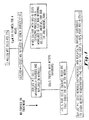
- Figure 1 is a schematic showing the method for generation of pure cultures of NT2-N cells from RA treated NT2 cells.
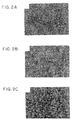
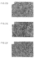
- Figures 2A-F are phase contrast photomicrographs showing the morphologic changes which occur during the course of the method shown in Figure 1: A, untreated NT2 cells; B, NT2 cells following Replate #1 (note the round, phase bright cells in clumps sitting above the adherent cells below); C, 1 day following Replate #2 (note that many cells have begun to elaborate rudimentary processes similar to a number of human neuroblastoma cell lines); D, 7 days following Replate #2 (the non-neuronal cells have begun to die off and the cultures are now dominated by neuron-like NT2-N cells); E , 30 days following Replating #2 (note that most cells exhibit the morphology of neurons and have migrated into large aggregates and that extensive process outgrowth has occurred; F, high power photomicrograph of NT2-N cells showing the typical neuronal morphology of these cells.
- the arrows point to the long thin untapering process resembling an axon emanating from a cell in Figure 2F.
- the arrowheads point to the two major processes which resemble dendrites.
- the bar in E applies to A-E and equals 200 ⁇ m and the bar in F equals 30 ⁇ m.
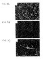
- Figures 3A-C show the results of BrDU labeling of NT2 and NT2-N cells.
- A Undifferentiated NT2 cells labeled with BrDU and stained with BU-1.
- B NT2-N cells labeled with BrDU and stained with BU-1.
- C the same field as B labeled with an anti-NF-L antiserum. The bar in C is 200 ⁇ m.
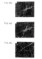
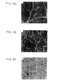
- Figures 4A-F show immunocytochemistry results demonstrating the expression of neuronal markers in NT2-N cells: A and B, double labeling with RMo254, a mouse anti-NF-M mAb, and anti-NF-66, a rabbit antiserum raised against NF-66; C , 1WM 3G5, a mouse anti-MAPlb mAb; D , AP14, a mouse anti-MAP2 mAb; E, A 2 B 5 , a mouse mAb specific for a ganglioside found on many neurons; F , T14, a mouse anti-tau mAb.
- the bar in F is 30 ⁇ m.
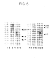
- Figure 5 shows the results of immunoblot analysis of the expression of cytoskeletal markers in NT2-N cells.
- Lanes 1-6 are from 6% SDS-PAGE gels and lanes 7-13 are from 10% SDS-PAGE gels.
- 10 ⁇ m of cytoskeletal extract and 6 human nerve root IF preparation blotted with RMo254, an anti-NF-M mAb.
- FIG. 6 Confocal microscopy of NT2-N cells stained with markers for axons and dendrites.
- a and B show microscopic fields stained with HO14 (green) and RMdO20 (red).
- HO14 is a rat mAb specific for the highly phosphorylated forms of NF-M
- RMdO20 is a mouse mAb specific for the poorly phosphorylated forma of NF-M.
- C shows a high power field of cells stained with HO14 and AP14, a mouse mAb specific for MAP2. These images were generated by collecting separate data for each channel and then merging them and imparting them with computer generated pseudocolor.
- the bar in C is 25 ⁇ m when applied to C, and is 50 ⁇ m when applied to A and B.
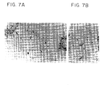
- Figure 7 3 H-uridine labeling of NT2-N cell cultures five weeks after Replate #2.
- a (low power) and B (high power) are photomicrographs of NT2-N cells produced using Hoffman Modulation Contrast optics. The labeled cells and processes appear dark grey or black because of the silver grains from the NTB-2 emulsion autoradiography.
- A shows a field of cells containing labeled cell bodies and dendritic processes (arrowheads) with many unlabeled axonal processes (some examples are shown with arrows) arising from the clumps of cells in the field and elsewhere on the culture dish.
- B shows a higher power micrograph of several cells. The dendritic processes of one of these cells have been indicated using arrowheads.
- the bar in A is 50 ⁇ m when applied to A, and 100 ⁇ m when applied to B.
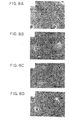
- Figures 8A-D are photographs showing neurite regeneration following Replate #3.
- A NT2-N cells grown on Matrigel for one week following Replate #3.
- B NT2-N cells grown on poly-D-lysine for one week following Replate #3.
- the arrow in Figure 6D points to a flat non-neuronal cell.
- the bar in D refers to A, B, and D and is 200 ⁇ m.
- the bar in C is 30 ⁇ m.
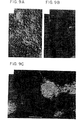
- Figures 9A-C are photographs showing ⁇ -gal expression of NT2-SPUD cells visualized by X-gal histochemistry using Hoffman modulation contrast microscopy.
- A NT2-SPUD cells before RA treatment.
- B and C NT2-SPUD cells after Replate #2.
- the bar in C is 100 ⁇ m when applied to A and C and is 50 ⁇ M when applied to B.
- NTera 2/Cl.D1 or NT2 cells A human teratocarcinoma cell line (NTera 2/Cl.D1 or NT2 cells) was manipulated following treatment with retinoic acid (RA) to yield greater than 95% pure cultures of neuronal cells (NT2-N cells).
- RA retinoic acid
- Replate #1 The derivation, differentiation paradigm, and early morphological appearance of NT2 cells following treatment with RA to this point (i.e., Replate #1) were examined in detail previously (Andrews et al., 1984; Andrews, 1984). Approximately 5% of the cells after Replate #1 are neurons as judged by the presence of neuron specific markers. To further enrich for these neurons, we took advantage of the fact that the small phase bright NT2-N cells were loosely attached to the layer of flat cells after Replate #1 cultures and could be mechanically dislodged and enriched. This treatment (Replate #2), yielded a culture consisting primarily of small rounded phase bright cells, some of which had short processes reminiscent of human neuroblastoma cell lines ( Figure 2C).
- NT2-N cells were treated with a combination of mitotic inhibitors following Replate #2. This treatment had no effect on the NT2-N cells, but it completely suppressed the growth of the flat cells. Hence, nearly all of the flat cells were eliminated, leaving behind a very small number of mitotically poisoned cells with extensive cytoplasm. By two weeks of treatment, about 95% of the cells were differentiated neurons, i.e., NT2-N cells.
- NT2-N cells displayed growth cones typical of primary cultures of neurons from various regions of the nervous system. Another consistent feature of pure NT2-N cells was their motility. Initially, NT2-N cells were evenly dispersed over the entire surface of the culture dish (Figure 2C), but over time they migrated together to form cellular aggregates with large interconnecting fascicles of axons ( Figure 2D). If the cells were plated on poly-D-lysine either alone or with laminin, they formed even larger aggregates that resembled explanted ganglia.
- NT2-N cells are post-mitotic.
- NT2-N cells i.e., cultures derived from Replate #1 - see Figure 1
- These cells were shown previously to express neurofilament proteins (Andrews, 1984; Lee and Andrews, 1986), the antigen recognized by A 2 B 5 (a mAb recognizing a cell surface glycolipid characteristic of neurons and some glial cells), and to possess tetrodotoxin sensitive Na+ channels (Rendt et al., 1989).
- a 2 B 5 a mAb recognizing a cell surface glycolipid characteristic of neurons and some glial cells
- NT2-N cells expressed NF-66 (also known as alpha-internexin; Figure 4B) but not peripherin (Table I), the two more recently described members of the neuronal intermediate filament family. NT2-N cells, therefore, may resemble CNS neurons because NF-66 is abundant only in the CNS, and peripherin is found in virtually all PNS neurons. NT2-N cells also expressed several neuronal microtubule associated proteins (MAPs) i.e., MAP1A, MAP1B, MAP2, and tau (Table I; Figures 4C, D & F).
- MAPs neuronal microtubule associated proteins
- Neuronal membrane or membrane associated antigens were also expressed by NT2-N cells. These included the antigen recognized by A 2 B 5 ( Figure 4E), NCAM (another cell surface molecule frequently expressed by neurons and neuronal neoplasms) and GAP43 (a growth associated protein concentrated in growth cones).
- NT2-N cells expressed markers of secretory activity typical of neurons and neuroendocrine cells (e.g., synaptophysin and chromogranin; Table I).
- synaptophysin is a marker for small transparent synaptic vesicles which store and release classical neurotransmitters
- chromogranin is a marker for larger dense-cored vesicles which are involved in neuropeptide and catecholamine biosynthesis.
- Table I represents the results obtained when NT2-N cells were examined with a number of antibodies specific for the proteins shown in the first column.
- the (+) symbol indicates that the antibody stained and/or blotted the released protein in NT-2N cells.
- the (-) symbol indicates that the relevant antibody does not stain NT2-N cells.
- Markers of the neuronal phenotype of NT2-N cells Protein Antibody Result Reference or Source of Antibody NF-L rabbit anti-NF-L + Trojanowski et al., Am. J. Pathol. 135:747-758 (1989) NF-M RMO254 + Lee et al., J. Neurosci., 7:3474-3488 (1987) NF-H RMO24 + Lee et al., J.
- MAP2b a less phosphorylated form of MAP2, which predominates during development and corresponds to the more rapidly migrating of the two MAP2 polypeptides in the bovine MAP2 preparations.
- Figure 5 also shows that the tau found in NT2-N cells corresponds to the fetal forms rather than the adult forms of tau. Fetal tau is translated from differentially spliced forms of tau mRNA which are predominant during embryonic life.
- MAP1B also known as MAP5
- MAP5 is also an embryonic MAP which persists into adulthood but at greatly reduced levels.
- NT2-N cells express low levels of MAP1A and NF-H (Table I), two proteins which are up regulated in their expression during development, but which only achieve their highest levels in adult nervous systems, including that of humans. This finding, together with the expression of three embryonic MAPs and NF-66, implies that the cytoskeleton of NT2-N cells resembles that of embryonic CNS neurons.
- Neurons typically have a single axon and multiple dendrites which can be distinguished by their morphology, by the organelles they contain and by several differentially distributed molecular markers.
- the cell body and dendrites somato-dendritic domain
- axons axonal domain
- the microtubules in dendrites are oriented in both directions while those in axons have their (+) end oriented distally.
- axons are rich in highly phosphorylated NF proteins and tau, while cell bodies and dendrites have primarily hypophosphorylated variants of NF proteins. These differences are all likely to contribute to the distinct functions of dendrites as primarily post-synaptic processes and of axons as projecting pre-synaptic processes.
- NT2-N cells Because of the highly polarized morphology of NT2-N cells after 3-4 weeks in culture, we decided to examine whether the processes of NT2-N cells can be differentiated into axons and dendrites. Indeed, when we examined the distribution of phospho-isoforms of NF-M in NT2-N cells, using confocal microscopy, we found that highly phosphorylated NF-M is found preferentially in the long thin processes emanating from NT2-N cells while hypophosphorylated NF-M is found in the cell body and short tapering processes (Figures 6A and 6B). MAP2 is also localized exclusively in the cell soma and the short processes of NT2-N cells ( Figure 6C).
- NT2-N cells A more directly functional feature of the dendrites was examined to determine if NT2-N cells possessed functioning dendrites.
- dendrites accumulate ribosomes so that they can respond rapidly to synaptic signals with changes in protein synthesis. This makes it possible to use 3H-uridine to label dendritic processes in neurons in culture.
- This technique was used to label NT2-N cells and found that, following emulsion autoradiography, processes resembling dendrites are indeed darkly labeled with silver grains while long projecting untapered processes remain unlabeled (Figure 7). This demonstrates that NT2-N cells do indeed possess identifiable dendrites and axons as determined by several criteria.
- NT2-N cells may be a model for immature CNS neurons
- pure cultures of NT2-N cells (cells grown for 1-3 weeks following Replate #2) were enzymatically detached and replated again (Replate #3) they rapidly re-extended neurites.
- the rapid elaboration of neurites and the eventual formation of the network of processes was found to be highly dependent on the substratum used. For example, rapid outgrowth occurred on cells grown on poly-D-lysine plus laminin or Matrigel substrates but not on poly-D-lysine alone.
- Figure 8B illustrates cell clumps grown on poly-D-lysine alone. Even after seven days, these cells had very short processes which ended in flat broad growth cones. By twenty hours, the NT2-N cells on Matrigel began to elaborate their processes (Figure 8C). These differential effects of Matrigel (or laminin) and poly-D-lysine on neurite regrowth in NT2-N cells are consistent with the well known neurite-outgrowth promoting effects of laminin on a variety of PNS and CNS neuronal cells in culture. The ability of NT2-N cells to regenerate neurites after multiple replating shows that they retain the plasticity of immature neurons. Furthermore, Replate #3 affords us a means to obtain more pure cultures of NT2-N cells (at least 99%) which will allow us to pursue experiments in the future which depend on the availability of purer cultures.
- Non-neuronal cells played a role in promoting neurite outgrowth; this is most evident in Replate #3 cultures.
- NT2-N cells replated on poly-D-lysine alone were clustered around the extremely rare non-neuronal cells found in these cultures, and that many neurites from the clump of NT2-N cells extended directly toward the non-neuronal cells.
- Figure 8D A dramatic example of this phenomena is shown in Figure 8D in which several processes appear to have changed direction dramatically in order to grow toward the non-neuronal cell. Repeated observation of the cells in Figure 8D revealed that the neurites retracted completely following the death of the non-neuronal cell.
- NT2-N cells To assess the utility of NT2-N cells for gene transfer experiments, we stably transfected undifferentiated NT2 cells with SPUD1, a ⁇ -galactosidase ( ⁇ -gal) expression plasmid. These studies were designed to determine whether NT2-N cells would continue to express the exogenous protein product following differentiation with RA.
- SPUD1 was cotransfected with pSV2neo (used as a selectable marker) into undifferentiated NT2 cells, we derived a G418 resistant population of cells which expressed ⁇ -gal as assayed by histochemical staining (Figure 9A).
- NT2 cells closely approximate the research criteria of an ideal neuronal cell line.
- Our observations on the NT2-N cells and the novel methods developed for their culture show that it is possible to generate pure cultures of post-mitotic, human neurons from rapidly dividing NT2 teratocarcinoma cells following RA treatment.
- These non-dividing NT2-N cells have a stable neuronal phenotype and they can survive in culture for eight to ten weeks without the continuous presence of anyexogenous differentiation promoting or trophic factors other than those in serum.
- NT2-N cells continue to express proteins encoded by plasmids transfected into the undifferentiated NT2 cells.
- This combination of features is unique since, for the first time, cultured human cells with a fully differentiated neuronal phenotype are available in large numbers for the study of basic questions in neurobiology including timed neurite outgrowth, the basis and development of neurite polarity, cytoskeletal maturation and neuronal plasticity.
- NT2 cells are an important system for studying the mechanisms whereby RA can induce the differentiation of stem cells into committed neurons without the need for any other exogenous influences.
- Retinoids especially RA
- RA Retinoids
- RA retinoids
- NT2 cells will provide a valuable in vitro system to study the cellular effects of retinoids and their role in neural induction and differentiation.
- NT2-N cells expressed all of the key neuronal markers we examined. Further, they have specific characteristics which indicate that they are CNS (and not PNS) neurons (i.e., they express the 66 kD NF protein and do not express peripherin [Table I]). Like primary neurons in culture, NT2-N cells have a cytoskeleton dominated by the immature forms of MAPs and NF proteins (e.g., they express primarily fetal tau, MAPlb, MAP2b), but they do synthesize and maintain lower quantities of MAP la and NF-H.
- NT2-N cells Given the stable neuronal phenotype assumed by NT2-N cells and the fact that they can be genetically engineered to express the products of transfected genes, these cells will be extremely useful for examining the cell biology and the functions of neuronal proteins in human neurons.
- the prompt neurite regeneration following replating (see Figure 8) and the expression of well characterized cell adhesion molecules (see Table I) will allow NT2-N cells to be utilized for studying factors which regulate neurite outgrowth. Since NT2-N cells do not divide even in the presence of serum and they represent a reproducible source of highly purified human neuronal cultures, they may be useful cells for transplantation studies into nude mice and other mammals to determine their ability to integrate into the host environment. Indeed, the transfection of trophic factors or other proteins into NT2 cells that are then induced to differentiate into stable, post-mitotic neurons, may be useful as a novel delivery system for bioactive molecules in human neurodegenerative diseases.
- the present invention provides methods for preparing a pure culture of post-mitotic human neurons comprising culturing undifferentiated human teratocarcinoma cells with retinoic acid to obtain a multi-layered culture; dispersing said cultured cells; and culturing said dispersed cells with a mitotic inhibitor or a combination of mitotic inhibitors.
- undifferentiated cells of the above method comprise NTera2/Dl cells.
- the present invention includes this method wherein a combination of mitotic inhibitors comprises cytosine arabinoside, fluorodeoxyuridine and uridine.
- dispersed cells of the above method can be cultured on Matrigel.
- the present invention also provides methods for producing a stable population of post-mitotic human neurons expressing exogenous gene products comprising transfecting one or more plasmids (including a gene encoding an exogenous gene product and a selectable marker) into cultured undifferentiated human teratocarcinoma cells; culturing said undifferentiated human teratocarcinoma cells with retinoic acid to obtain multi-layered culture; dispersing said cultured cells; and culturing said dispersed cells with a mitotic inhibitor or a combination of mitotic inhibitors. It is contemplated that a single selected expression vector may be transfected and expressed in the stable population of post-mitotic cells.
- the selected expression vector may be transfected and expressed in the stable population of post-mitotic cells.
- the selected expression plasmid comprises a ⁇ -galactosidase expression plasmid.
- the undifferentiated cells of this transfection method comprise NTera2/Dl cells.
- the combination of mitotic inhibitors of this transfection method comprise cytosine arabinoside, fluorodeoxyuridine and uridine.
- the dispersed cells of this transfection method can be cultured on Matrigel.
- the present invention also provides stable post-mitotic human neuron cells produced in accordance with the methods described above, including but not limited to stable post-mitotic human neuron cells substantially all of which comprise at least one transfected exogenous gene or stable post-mitotic human neuron cells which are not transfected.
- NT2 cells were maintained in DMEM HG including 10% fetal bovine serum and penicillin/streptomycin as previously described (Andrews, 1984).
- 2 x 10 6 cells were seeded in a 75cm 2 flask and treated with 1 x 10 -2 M RA (a 1 x 10 -2 M stock dissolved in DMSO was prepared fresh monthly) twice a week for four weeks.
- 1 x 10 -2 M RA a 1 x 10 -2 M stock dissolved in DMSO was prepared fresh monthly
- Cytosine arabinoside was continued for the first week of culture and fluorodeoxyuridine and uridine for the first four weeks.
- three week old cultures were enzymatically removed with 0.025% dispase or 0.025% trypsin and replated (Replate #3; Figure 1) on Matrigel, poly-D-lysine (10 ⁇ g/ml), or poly-D-lysine (10 ⁇ g/ml) plus laminin (10 ⁇ g/ml).
- Matrigel is a basement membrane extract containing collagen, laminin, and nidogen (Kleinman et al., 1986).
- Undifferentiated NT2 cells and differentiated NT2-N cells were grown free of mitotic inhibitors for four days and then labeled with 3 mg/ml BrDU for three hours (or up to 20 hours, in some cases). The cells were then washed, fixed and processed for indirect immunofluorescence as described below using BU- 1, a mab which recognizes BrDU incorporated DNA without denaturation.
- PAP immunocytochemistry was performed to visualize low levels of fetal tau because of the great sensitivity of this technique when compared to indirect immunofluorescence.
- the coverslips were fixed as above and blocked for 30 minutes with 0.1M Tris pH 7.0, including 2% calf serum and 0.25% cold water fish gelatin. Following this, the coverslips were processed as described previously in our laboratory for PAP immunocytochemistry (Carden et al., J. Neurosci., 7:3489-3504 (1987).
- MAP-enriched cytoskeletal samples were prepared by extracting the cells at room temperature for 15 minutes with 0.1M MES pH6.8 containing 0.5mM MgSO 4 , 1mm EGTA, 2mM DTT, 2mM GTP, 20 ⁇ M Taxol, 1% Triton X-100 and a cocktail of protease inhibitors.
- the pellets were recovered by centrifugation at 30,000 rpm in a TL100 ultracentrifuge for 30 minutes, solubilized in sample buffer without dye and the protein concentrations of the samples were determined using a Coomassie blue dye binding assay (Pierce). These samples were run on SDS-PAGE gels and then electroblotted to nitrocellulose membranes for probing with antibodies using methods described previously in our laboratory (Lee et al., 1987).
- NT2-N cells 60mm dishes of NT2-N cells were incubated with 50 ⁇ Ci of [5,6- 3 H]uridine (1.70 TBq/mmol) for 16-24 hours. These dishes were then washed with PBS containing 10 ⁇ m unlabelled uridine and fixed with Bouin's fixative. The dishes were then coated with NTB-2 emulsion diluted 1:1 with water, dried overnight and stored at 4°C for four days. The dishes were developed for one minute in Kodak D19 and fixed with Kodak Rapid-Fix.
- Undifferentiated NT2 cells were transfected with 100 ⁇ g SPUD1 and 10 ⁇ g of pSV2neo by lipofection using Lipofectin (Bethesda Research Laboratories). After two days in complete medium, the transfectants were selected with 200 ⁇ g/ml G418 (Gibco) for seven days. Cells were stained for ⁇ -galactosidase activity with 1 mg/ml X-gal, 5 mM potassium ferrocyanide, 5 mM potassium ferricyanide, 2 mM MgCl 2 in PBS after fixation in 2% paraformaldehyde, 0.2% glutaraldehyde in phosphate buffered saline pH7.4.
- SPUD1 (kindly provided by Dr. C. Cepko) is a ⁇ -galactosidase expression vector which utilizes the SV40 promoter and has Moloney murine leukemia virus long terminal repeats upstream and downstream. The cells were photographed using Hoffman modulation contrast to allow the simultaneous visualization of the blue reaction product and the processes.
Landscapes
- Engineering & Computer Science (AREA)
- Health & Medical Sciences (AREA)
- Biomedical Technology (AREA)
- Life Sciences & Earth Sciences (AREA)
- Mechanical Engineering (AREA)
- Neurology (AREA)
- Zoology (AREA)
- Biotechnology (AREA)
- Organic Chemistry (AREA)
- Bioinformatics & Cheminformatics (AREA)
- Genetics & Genomics (AREA)
- Chemical & Material Sciences (AREA)
- Wood Science & Technology (AREA)
- Neurosurgery (AREA)
- Microbiology (AREA)
- Biochemistry (AREA)
- General Engineering & Computer Science (AREA)
- General Health & Medical Sciences (AREA)
- Cell Biology (AREA)
- Micro-Organisms Or Cultivation Processes Thereof (AREA)
- Saccharide Compounds (AREA)
- Preparation Of Compounds By Using Micro-Organisms (AREA)
Claims (12)
- Ein Verfahren zur Herstellung einer Reinkultur von humanen post-mitotischen Neuronen, welches umfaßt:Züchtung von undifferenzierten humanen Teratocarcinom-Zellen mit Retinsäure zur Gewinnung einer mehrschichtigen Kultur;Dispergierung dieser gezüchteten Zellen;Züchtung dieser dispergierten Zellen mit einem mitotischen Inhibitor oder einer Kombination mitotischer Inhibitoren.
- Ein Verfahren zur Herstellung einer stabilen Population von humanen post-mitotischen Neuronen, die exogene Genprodukte exprimieren, welches Verfahren umfaßt:Transfektion mindestens eines Plasmids, welches ein Gen, das für ein exogenes Genprodukt codiert, und einen selektierbaren Marker enthält, in gezüchtete undifferenzierte humane Teratocarcinom-Zellen;Züchtung dieser undifferenzierten humanen Teratocarcinom-Zellen mit Retinsäure zur Gewinnung einer mehrschichtigen Kultur;Dispergierung dieser gezüchteten Zellen; undZüchtung dieser dispergierten Zellen mit einem mitotischen Inhibitor oder einer Kombination mitotischer Inhibitoren.
- Das Verfahren nach Anspruch 1 oder Anspruch 2, bei welchem die genannten undifferenzierten Zellen NTera2/D1-Zellen umfassen.
- Das Verfahren nach einem der vorhergehenden Ansprüche, bei welchem die genannte Kombination von mitotischen Inhibitoren Cytosin-Arabinosid, Fluordeoxyuridin und Uridin umfaßt.
- Das Verfahren nach einem der vorhergehenden Ansprüche, bei welchem die genannten dispergierten Zellen auf Matrigel gezüchtet werden.
- Das Verfahren nach Anspruch 2, bei welchem das genannte Plasmid ein β-Galactosidase-Expressionsplasmid umfaßt.
- Stabile humane post-mitotische Neuronzellen, die gemäß dem Verfahren nach Anspruch 1, Anspruch 2 oder Anspruch 6 hergestellt wurden.
- Stabile humane post-mitotische Neuronzellen, von denen im wesentlichen alle mindestens ein transfiziertes exogenes Gen enthalten.
- Eine Zellkultur, die im wesentlichen aus stabilen homogenen humanen post-mitotischen Neuronzellen besteht.
- Eine Kultur nach Anspruch 9, in welcher im wesentlichen alle Zellen mindesstens ein transfiziertes exogenes Gen enthalten.
- Eine Kultur nach Anspruch 9 oder Anspruch 10, die zu 95 % rein ist.
- Eine Kultur nach Anspruch 9 oder Anspruch 10, die zu 99 % rein ist.
Applications Claiming Priority (3)
| Application Number | Priority Date | Filing Date | Title |
|---|---|---|---|
| US07/780,715 US5175103A (en) | 1991-10-21 | 1991-10-21 | Preparation of pure cultures of post-mitotic human neurons |
| US780715 | 1991-10-21 | ||
| PCT/US1992/006540 WO1993008266A1 (en) | 1991-10-21 | 1992-08-05 | Preparation of pure cultures of post-mitotic human neurons |
Publications (3)
| Publication Number | Publication Date |
|---|---|
| EP0620847A1 EP0620847A1 (de) | 1994-10-26 |
| EP0620847A4 EP0620847A4 (de) | 1995-03-29 |
| EP0620847B1 true EP0620847B1 (de) | 1998-04-29 |
Family
ID=25120453
Family Applications (1)
| Application Number | Title | Priority Date | Filing Date |
|---|---|---|---|
| EP92917219A Expired - Lifetime EP0620847B1 (de) | 1991-10-21 | 1992-08-05 | Präparation von reinkulturen von humanen post-mitotischen neuronen |
Country Status (9)
| Country | Link |
|---|---|
| US (2) | US5175103A (de) |
| EP (1) | EP0620847B1 (de) |
| JP (1) | JP3169611B2 (de) |
| AT (1) | ATE165617T1 (de) |
| CA (1) | CA2120730A1 (de) |
| DE (1) | DE69225332T2 (de) |
| DK (1) | DK0620847T3 (de) |
| ES (1) | ES2117672T3 (de) |
| WO (1) | WO1993008266A1 (de) |
Families Citing this family (58)
| Publication number | Priority date | Publication date | Assignee | Title |
|---|---|---|---|---|
| US5545522A (en) | 1989-09-22 | 1996-08-13 | Van Gelder; Russell N. | Process for amplifying a target polynucleotide sequence using a single primer-promoter complex |
| US7049102B1 (en) | 1989-09-22 | 2006-05-23 | Board Of Trustees Of Leland Stanford University | Multi-gene expression profile |
| US6497872B1 (en) * | 1991-07-08 | 2002-12-24 | Neurospheres Holdings Ltd. | Neural transplantation using proliferated multipotent neural stem cells and their progeny |
| US6071889A (en) * | 1991-07-08 | 2000-06-06 | Neurospheres Holdings Ltd. | In vivo genetic modification of growth factor-responsive neural precursor cells |
| US5980885A (en) * | 1991-07-08 | 1999-11-09 | Neurospheres Holdings Ltd. | Growth factor-induced proliferation of neural precursor cells in vivo |
| US7361505B1 (en) | 1991-07-08 | 2008-04-22 | Neurospheres Holdings Ltd. | Multipotent neural stem cell compositions |
| US6399369B1 (en) * | 1991-07-08 | 2002-06-04 | Neurospheres Holdings Ltd. | Multipotent neural stem cell cDNA libraries |
| US7166277B1 (en) | 1991-07-08 | 2007-01-23 | Neurospheres Holdings Ltd. | Remyelination of neurons using multipotent neural stem cell progeny |
| US6294346B1 (en) | 1991-07-08 | 2001-09-25 | Neurospheres Holdings, Ltd. | Use of multipotent neural stem cells and their progeny for the screening of drugs and other biological agents |
| US5750376A (en) * | 1991-07-08 | 1998-05-12 | Neurospheres Holdings Ltd. | In vitro growth and proliferation of genetically modified multipotent neural stem cells and their progeny |
| US5851832A (en) * | 1991-07-08 | 1998-12-22 | Neurospheres, Ltd. | In vitro growth and proliferation of multipotent neural stem cells and their progeny |
| US6214334B1 (en) * | 1991-10-21 | 2001-04-10 | Trustees Of The University Of Pennsylvania | Compositions and methods for producing and using homogenous neuronal cell transplants to treat neurodegenerative disorders and brain and spinal cord injuries |
| US5175103A (en) * | 1991-10-21 | 1992-12-29 | Trustees Of University Of Pennsylvania | Preparation of pure cultures of post-mitotic human neurons |
| US5792900A (en) * | 1991-10-21 | 1998-08-11 | The Trustees Of The University Of Pennsylvania | Compositions and methods for producing and using homogenous neuronal cell transplants |
| JPH09505054A (ja) * | 1993-11-09 | 1997-05-20 | ザ・トラスティーズ・オブ・ザ・ユニバーシティ・オブ・ペンシルバニア | 均質なニューロン細胞移植片としての組成物とこれらの移植片の製造及び使用方法 |
| US7011827B2 (en) * | 1993-11-09 | 2006-03-14 | Trustees Of The University Of Pennsylvania | Compositions and methods for producing and using homogenous neuronal cell transplants |
| US5449609A (en) * | 1994-01-31 | 1995-09-12 | Children's Hospital Of Philadelphia | Methods for screening for neurotoxicity using a clonal human teratocarcinoma cell line |
| US20060029575A1 (en) * | 1994-11-09 | 2006-02-09 | Lee Virginia M | Compositions and methods for producing and using homogenous neuronal cell transplants |
| FI971956L (fi) * | 1994-11-14 | 1997-07-04 | Neurospheres Holdings Ltd | Hermosolujen lisääntymisen säännöstely |
| US5753505A (en) * | 1995-07-06 | 1998-05-19 | Emory University | Neuronal progenitor cells and uses thereof |
| US7544511B2 (en) * | 1996-09-25 | 2009-06-09 | Neuralstem Biopharmaceuticals Ltd. | Stable neural stem cell line methods |
| US6162428A (en) * | 1997-02-12 | 2000-12-19 | Layton Bioscience, Inc. | hNT-neuron human neuronal cells to replace ganglion cells |
| AU6897498A (en) * | 1997-04-25 | 1998-11-24 | American Home Products Corporation | Human neuronal cell line |
| WO1998057663A1 (en) * | 1997-06-17 | 1998-12-23 | University Technology Corporation | Neural transplantation for treating neurological disease |
| CA2294737A1 (en) * | 1997-07-04 | 1999-01-14 | University Of Utah Research Foundation | Neuron-restricted precursor cells |
| US6734015B1 (en) * | 1997-07-04 | 2004-05-11 | University Of Utah Research Foundation | Isolation of lineage-restricted neuronal precursors |
| US6235527B1 (en) * | 1997-11-29 | 2001-05-22 | University Of Utah Research Foundation | Lineage restricted glial precursors from the central nervous system |
| US5968829A (en) * | 1997-09-05 | 1999-10-19 | Cytotherapeutics, Inc. | Human CNS neural stem cells |
| WO1999013726A1 (en) * | 1997-09-19 | 1999-03-25 | University Of South Florida | Neuronal cells and an immunosuppressant and anti-inflammatory factor |
| WO2000006700A1 (en) * | 1998-07-29 | 2000-02-10 | Layton Bioscience, Inc. | Production and use of dopaminergic cells to treat dopaminergic deficiencies |
| EP1949904A3 (de) | 1999-04-27 | 2008-08-06 | Layton Bioscience, Inc. | Zelltherapie für chronische Apoplexie |
| US20030211085A1 (en) * | 2000-03-16 | 2003-11-13 | Sanberg Paul R. | Cell therapy for chronic stroke |
| DE19928210B4 (de) * | 1999-06-19 | 2005-08-18 | Neuroprogen Gmbh Leipzig | Neuronales Zellmaterial und Verfahren zu dessen Herstellung |
| US6749850B1 (en) | 1999-08-18 | 2004-06-15 | The General Hospital Corporation | Methods, compositions and kits for promoting recovery from damage to the central nervous system |
| WO2001053503A1 (en) * | 2000-01-18 | 2001-07-26 | Cornell Research Foundation, Inc. | Neural progenitor cells from hippocampal tissue and a method for isolating and purifying them |
| WO2001078752A2 (en) | 2000-04-13 | 2001-10-25 | Board Of Regents, The University Of Texas System | Treatment of disorders by implanting stem cells and/or progeny thereof into gastrointestinal organs |
| US7037493B2 (en) | 2000-05-01 | 2006-05-02 | Cornell Research Foundation, Inc. | Method of inducing neuronal production in the brain and spinal cord |
| US6897061B1 (en) * | 2000-06-16 | 2005-05-24 | Spinal Cord Society | Transdifferentiation of glial cells |
| JP4031901B2 (ja) * | 2000-07-19 | 2008-01-09 | 株式会社東芝 | 固体撮像装置 |
| AU2001292350A1 (en) * | 2000-10-05 | 2002-04-15 | Takeda Chemical Industries Ltd. | Promoters for the proliferation and differentiation of stem cells and/or neuron precursor cells |
| AU2002251681A1 (en) * | 2000-12-05 | 2002-08-28 | Layton Bioscience Inc. | Production and use of dopaminergic cells to treat dopaminergic deficiencies |
| EP1397485A2 (de) * | 2001-06-05 | 2004-03-17 | Thomas Rohrmeier | Zellpopulationen zur auffindung neuronaler targets und potentieller wirkstoffe |
| US7576065B2 (en) * | 2002-02-15 | 2009-08-18 | Cornell Research Foundation, Inc. | Enhancing neurotrophin-induced neurogenesis by endogenous neural progenitor cells by concurrent overexpression of brain derived neurotrophic factor and an inhibitor of a pro-gliogenic bone morphogenetic protein |
| GB0217981D0 (en) * | 2002-08-02 | 2002-09-11 | Univ Durham | Method for preparation, purification and differentiation of neurospheres from mammalian stem cells |
| US8293488B2 (en) | 2002-12-09 | 2012-10-23 | Neuralstem, Inc. | Method for screening neurogenic agents |
| US20040185429A1 (en) * | 2002-12-09 | 2004-09-23 | Judith Kelleher-Andersson | Method for discovering neurogenic agents |
| DE10259617A1 (de) * | 2002-12-18 | 2004-07-08 | Cardion Ag | Verfahren zur Inhibierung des Proliferationspotentials von Zellpopulationen und Geweben |
| WO2004098621A1 (en) * | 2003-04-17 | 2004-11-18 | U.S. Department Of Veterans Affairs | Isolated/cloned human nt2 cell lines expressing serotonin and gaba |
| FR2859219B1 (fr) * | 2003-09-02 | 2005-10-14 | Alain Privat | Procede de production de neurones a partir de cellules d'une lignee cellulaire |
| AU2005252434B2 (en) * | 2004-06-09 | 2011-11-24 | The University Court Of The University Of Edinburgh | Neural stem cells |
| US7663614B2 (en) * | 2004-10-05 | 2010-02-16 | Jeff Maynard | Method for storing and comparing computer generated lines |
| WO2006055685A2 (en) | 2004-11-17 | 2006-05-26 | Neuralstem, Inc. | Transplantation of human neural cells for treatment of neurodegenerative conditions |
| US7736641B2 (en) * | 2007-02-21 | 2010-06-15 | Prince Henry's Institute Of Medical Research | Role for SRY in Parkinson's disease |
| US20090136461A1 (en) * | 2007-11-28 | 2009-05-28 | Moon Suk Kim | Neuronal differentiation method of adult stem cells using small molecules |
| CN102325878A (zh) | 2008-12-23 | 2012-01-18 | 斯特姆塞尔思加利福尼亚有限公司 | 少突胶质前体细胞的靶向细胞群及制备与使用方法 |
| US9101570B1 (en) | 2009-02-13 | 2015-08-11 | Endocellutions, Inc. | Adult and neonatal stem cell therapy to treat diabetes through the repair of the gastrointestinal tract |
| SG187226A1 (en) | 2010-07-28 | 2013-03-28 | Neuralstem Inc | Methods for treating and/or reversing neurodegenerative diseases and/or disorders |
| CN107148275A (zh) | 2014-10-20 | 2017-09-08 | 神经干细胞公司 | 包含编码生长因子的外源多核苷酸的稳定的神经干细胞及其使用方法 |
Family Cites Families (6)
| Publication number | Priority date | Publication date | Assignee | Title |
|---|---|---|---|---|
| US4829000A (en) * | 1985-08-30 | 1989-05-09 | The United States Of America As Represented By The Secretary Of The Department Of Health And Human Services | Reconstituted basement membrane complex with biological activity |
| US5032407A (en) * | 1987-01-16 | 1991-07-16 | Ohio University Edison Animal Biotechnology Center | Gene transfer using transformed, neodetermined, embryonic cells |
| US5270191A (en) * | 1988-04-12 | 1993-12-14 | Massachusetts Institute Of Technology | Method for manipulation of the cell types of eukaryotes |
| FR2645866B1 (fr) * | 1989-04-17 | 1991-07-05 | Centre Nat Rech Scient | Nouvelles lipopolyamines, leur preparation et leur emploi |
| US5180820A (en) * | 1989-08-30 | 1993-01-19 | Barde Yves Alain | Brain-derived neurotrophic factor |
| US5175103A (en) * | 1991-10-21 | 1992-12-29 | Trustees Of University Of Pennsylvania | Preparation of pure cultures of post-mitotic human neurons |
-
1991
- 1991-10-21 US US07/780,715 patent/US5175103A/en not_active Expired - Lifetime
-
1992
- 1992-08-05 DK DK92917219T patent/DK0620847T3/da active
- 1992-08-05 DE DE69225332T patent/DE69225332T2/de not_active Expired - Fee Related
- 1992-08-05 EP EP92917219A patent/EP0620847B1/de not_active Expired - Lifetime
- 1992-08-05 ES ES92917219T patent/ES2117672T3/es not_active Expired - Lifetime
- 1992-08-05 WO PCT/US1992/006540 patent/WO1993008266A1/en not_active Ceased
- 1992-08-05 CA CA002120730A patent/CA2120730A1/en not_active Abandoned
- 1992-08-05 JP JP50765893A patent/JP3169611B2/ja not_active Expired - Fee Related
- 1992-08-05 AT AT92917219T patent/ATE165617T1/de not_active IP Right Cessation
-
1996
- 1996-06-10 US US08/660,723 patent/US5654189A/en not_active Expired - Fee Related
Also Published As
| Publication number | Publication date |
|---|---|
| DE69225332T2 (de) | 1998-08-27 |
| US5175103A (en) | 1992-12-29 |
| ES2117672T3 (es) | 1998-08-16 |
| CA2120730A1 (en) | 1993-04-29 |
| DK0620847T3 (da) | 1998-10-07 |
| WO1993008266A1 (en) | 1993-04-29 |
| DE69225332D1 (de) | 1998-06-04 |
| US5654189A (en) | 1997-08-05 |
| EP0620847A1 (de) | 1994-10-26 |
| EP0620847A4 (de) | 1995-03-29 |
| ATE165617T1 (de) | 1998-05-15 |
| JPH07502645A (ja) | 1995-03-23 |
| JP3169611B2 (ja) | 2001-05-28 |
Similar Documents
| Publication | Publication Date | Title |
|---|---|---|
| US5175103A (en) | Preparation of pure cultures of post-mitotic human neurons | |
| Pleasure et al. | Pure, postmitotic, polarized human neurons derived from NTera 2 cells provide a system for expressing exogenous proteins in terminally differentiated neurons | |
| Pittack et al. | Basic fibroblast growth factor induces retinal pigment epithelium to generate neural retina in vitro | |
| Caceres et al. | Immunocytochemical localization of tubulin and microtubule-associated protein 2 during the development of hippocampal neurons in culture | |
| Li et al. | Retinal organoids: cultivation, differentiation, and transplantation | |
| Ma et al. | CNS stem and progenitor cell differentiation into functional neuronal circuits in three-dimensional collagen gels | |
| Steindler et al. | Boundaries during normal and abnormal brain development: in vivo and in vitro studies of glia and glycoconjugates | |
| Van Den Pol et al. | Olfactory ensheathing cells: time lapse imaging of cellular interactions, axonal support, rapid morphologic shifts, and mitosis | |
| US20110044957A1 (en) | Methods and systems for extended in vitro culture of neuronal cells | |
| CA2294737A1 (en) | Neuron-restricted precursor cells | |
| Yamagata et al. | Target-independent diversification and target-specific projection of chemically defined retinal ganglion cell subsets | |
| US7588938B2 (en) | Neural stem cell-collagen-bioreactor system to construct a functional embryonic brain-like tissue | |
| Staines et al. | Retinoic acid treated P19 embryonal carcinoma cells differentiate into oligodendrocytes capable of myelination | |
| Hill et al. | Characterization of the cholinergic neuronal differentiation of the human neuroblastoma cell line LA-N-5 after treatment with retinoic acid | |
| Uchida et al. | Potential functional neural repair with grafted neural stem cells of early embryonic neuroepithelial origin | |
| Sonigra et al. | Spontaneous immortalisation of ensheathing cells from adult rat olfactory nerve | |
| Seeds et al. | Cell junction and ultrastructural development of reaggregated mouse brain cultures | |
| Massa et al. | Cell-cell interactions of isolated and cultured oligodendrocytes: formation of linear occluding junctions and expression of peculiar intramembrane particles | |
| JPH11508142A (ja) | 不死化網膜細胞系およびその適用 | |
| Derouiche et al. | Loss of layer‐specific astrocytic glutamine synthetase immunoreactivity in slice cultures of hippocampus | |
| Peng et al. | Development of presynaptic specializations induced by basic polypeptide‐coated latex beads in spinal cord cultures | |
| Omlin et al. | Differentiation of neuron-like cells in cultured rat optic nerves: a neuron or common neuron-glia progenitor? | |
| US20070248644A1 (en) | Muller Stem Cells | |
| Doering et al. | Isolation and transplantation of oligodendrocyte precursor cells | |
| US20240369537A1 (en) | In Vitro Cell Co-Cultures of Neuronal Cells, Astrocytes, and Oligodendrocytes |
Legal Events
| Date | Code | Title | Description |
|---|---|---|---|
| PUAI | Public reference made under article 153(3) epc to a published international application that has entered the european phase |
Free format text: ORIGINAL CODE: 0009012 |
|
| 17P | Request for examination filed |
Effective date: 19940520 |
|
| AK | Designated contracting states |
Kind code of ref document: A1 Designated state(s): AT BE CH DE DK ES FR GB GR IE IT LI LU MC NL SE |
|
| RHK1 | Main classification (correction) |
Ipc: C12N 5/08 |
|
| A4 | Supplementary search report drawn up and despatched |
Effective date: 19950208 |
|
| AK | Designated contracting states |
Kind code of ref document: A4 Designated state(s): AT BE CH DE DK ES FR GB GR IE IT LI LU MC NL SE |
|
| 17Q | First examination report despatched |
Effective date: 19970327 |
|
| GRAG | Despatch of communication of intention to grant |
Free format text: ORIGINAL CODE: EPIDOS AGRA |
|
| GRAG | Despatch of communication of intention to grant |
Free format text: ORIGINAL CODE: EPIDOS AGRA |
|
| GRAH | Despatch of communication of intention to grant a patent |
Free format text: ORIGINAL CODE: EPIDOS IGRA |
|
| GRAH | Despatch of communication of intention to grant a patent |
Free format text: ORIGINAL CODE: EPIDOS IGRA |
|
| GRAA | (expected) grant |
Free format text: ORIGINAL CODE: 0009210 |
|
| AK | Designated contracting states |
Kind code of ref document: B1 Designated state(s): AT BE CH DE DK ES FR GB GR IE IT LI LU MC NL SE |
|
| REF | Corresponds to: |
Ref document number: 165617 Country of ref document: AT Date of ref document: 19980515 Kind code of ref document: T |
|
| REG | Reference to a national code |
Ref country code: CH Ref legal event code: NV Representative=s name: PATENTANWAELTE SCHAAD, BALASS, MENZL & PARTNER AG Ref country code: CH Ref legal event code: EP |
|
| REF | Corresponds to: |
Ref document number: 69225332 Country of ref document: DE Date of ref document: 19980604 |
|
| ET | Fr: translation filed | ||
| ITF | It: translation for a ep patent filed | ||
| REG | Reference to a national code |
Ref country code: IE Ref legal event code: FG4D Free format text: 80056 |
|
| REG | Reference to a national code |
Ref country code: ES Ref legal event code: FG2A Ref document number: 2117672 Country of ref document: ES Kind code of ref document: T3 |
|
| REG | Reference to a national code |
Ref country code: DK Ref legal event code: T3 |
|
| PLBE | No opposition filed within time limit |
Free format text: ORIGINAL CODE: 0009261 |
|
| 26N | No opposition filed | ||
| REG | Reference to a national code |
Ref country code: GB Ref legal event code: IF02 |
|
| PGFP | Annual fee paid to national office [announced via postgrant information from national office to epo] |
Ref country code: GB Payment date: 20050817 Year of fee payment: 14 |
|
| PGFP | Annual fee paid to national office [announced via postgrant information from national office to epo] |
Ref country code: AT Payment date: 20050914 Year of fee payment: 14 |
|
| PGFP | Annual fee paid to national office [announced via postgrant information from national office to epo] |
Ref country code: DK Payment date: 20050916 Year of fee payment: 14 Ref country code: NL Payment date: 20050916 Year of fee payment: 14 Ref country code: LU Payment date: 20050916 Year of fee payment: 14 Ref country code: IE Payment date: 20050916 Year of fee payment: 14 |
|
| PGFP | Annual fee paid to national office [announced via postgrant information from national office to epo] |
Ref country code: FR Payment date: 20050921 Year of fee payment: 14 |
|
| PGFP | Annual fee paid to national office [announced via postgrant information from national office to epo] |
Ref country code: SE Payment date: 20050926 Year of fee payment: 14 Ref country code: CH Payment date: 20050926 Year of fee payment: 14 |
|
| PGFP | Annual fee paid to national office [announced via postgrant information from national office to epo] |
Ref country code: GR Payment date: 20050927 Year of fee payment: 14 |
|
| PGFP | Annual fee paid to national office [announced via postgrant information from national office to epo] |
Ref country code: ES Payment date: 20050929 Year of fee payment: 14 |
|
| PGFP | Annual fee paid to national office [announced via postgrant information from national office to epo] |
Ref country code: BE Payment date: 20051006 Year of fee payment: 14 |
|
| PGFP | Annual fee paid to national office [announced via postgrant information from national office to epo] |
Ref country code: DE Payment date: 20051031 Year of fee payment: 14 |
|
| PGFP | Annual fee paid to national office [announced via postgrant information from national office to epo] |
Ref country code: MC Payment date: 20051213 Year of fee payment: 14 |
|
| PG25 | Lapsed in a contracting state [announced via postgrant information from national office to epo] |
Ref country code: AT Free format text: LAPSE BECAUSE OF NON-PAYMENT OF DUE FEES Effective date: 20060805 |
|
| PG25 | Lapsed in a contracting state [announced via postgrant information from national office to epo] |
Ref country code: SE Free format text: LAPSE BECAUSE OF NON-PAYMENT OF DUE FEES Effective date: 20060806 |
|
| PG25 | Lapsed in a contracting state [announced via postgrant information from national office to epo] |
Ref country code: IE Free format text: LAPSE BECAUSE OF NON-PAYMENT OF DUE FEES Effective date: 20060807 |
|
| PG25 | Lapsed in a contracting state [announced via postgrant information from national office to epo] |
Ref country code: MC Free format text: LAPSE BECAUSE OF NON-PAYMENT OF DUE FEES Effective date: 20060831 Ref country code: LI Free format text: LAPSE BECAUSE OF NON-PAYMENT OF DUE FEES Effective date: 20060831 Ref country code: DK Free format text: LAPSE BECAUSE OF NON-PAYMENT OF DUE FEES Effective date: 20060831 Ref country code: CH Free format text: LAPSE BECAUSE OF NON-PAYMENT OF DUE FEES Effective date: 20060831 Ref country code: BE Free format text: LAPSE BECAUSE OF NON-PAYMENT OF DUE FEES Effective date: 20060831 |
|
| PGFP | Annual fee paid to national office [announced via postgrant information from national office to epo] |
Ref country code: IT Payment date: 20060831 Year of fee payment: 15 |
|
| PG25 | Lapsed in a contracting state [announced via postgrant information from national office to epo] |
Ref country code: NL Free format text: LAPSE BECAUSE OF NON-PAYMENT OF DUE FEES Effective date: 20070301 Ref country code: DE Free format text: LAPSE BECAUSE OF NON-PAYMENT OF DUE FEES Effective date: 20070301 |
|
| REG | Reference to a national code |
Ref country code: DK Ref legal event code: EBP |
|
| REG | Reference to a national code |
Ref country code: CH Ref legal event code: PL |
|
| EUG | Se: european patent has lapsed | ||
| GBPC | Gb: european patent ceased through non-payment of renewal fee |
Effective date: 20060805 |
|
| NLV4 | Nl: lapsed or anulled due to non-payment of the annual fee |
Effective date: 20070301 |
|
| REG | Reference to a national code |
Ref country code: IE Ref legal event code: MM4A |
|
| REG | Reference to a national code |
Ref country code: FR Ref legal event code: ST Effective date: 20070430 |
|
| REG | Reference to a national code |
Ref country code: ES Ref legal event code: FD2A Effective date: 20060807 |
|
| PG25 | Lapsed in a contracting state [announced via postgrant information from national office to epo] |
Ref country code: GB Free format text: LAPSE BECAUSE OF NON-PAYMENT OF DUE FEES Effective date: 20060805 |
|
| BERE | Be: lapsed |
Owner name: THE *TRUSTEES OF THE UNIVERSITY OF PENNSYLVANIA Effective date: 20060831 |
|
| PG25 | Lapsed in a contracting state [announced via postgrant information from national office to epo] |
Ref country code: ES Free format text: LAPSE BECAUSE OF NON-PAYMENT OF DUE FEES Effective date: 20060807 |
|
| PG25 | Lapsed in a contracting state [announced via postgrant information from national office to epo] |
Ref country code: FR Free format text: LAPSE BECAUSE OF NON-PAYMENT OF DUE FEES Effective date: 20060831 |
|
| PG25 | Lapsed in a contracting state [announced via postgrant information from national office to epo] |
Ref country code: LU Free format text: LAPSE BECAUSE OF NON-PAYMENT OF DUE FEES Effective date: 20060805 |
|
| PG25 | Lapsed in a contracting state [announced via postgrant information from national office to epo] |
Ref country code: GR Free format text: LAPSE BECAUSE OF NON-PAYMENT OF DUE FEES Effective date: 20070302 |
|
| PG25 | Lapsed in a contracting state [announced via postgrant information from national office to epo] |
Ref country code: IT Free format text: LAPSE BECAUSE OF NON-PAYMENT OF DUE FEES Effective date: 20070805 |