EP0638648A2 - Monoklonale Antikörper, die spezifisch mit PAF-Rezeptoren reagieren, Verfahren zu deren Herstellung und die diese Antikörper herstellende Hybridomazellen - Google Patents
Monoklonale Antikörper, die spezifisch mit PAF-Rezeptoren reagieren, Verfahren zu deren Herstellung und die diese Antikörper herstellende Hybridomazellen Download PDFInfo
- Publication number
- EP0638648A2 EP0638648A2 EP94305815A EP94305815A EP0638648A2 EP 0638648 A2 EP0638648 A2 EP 0638648A2 EP 94305815 A EP94305815 A EP 94305815A EP 94305815 A EP94305815 A EP 94305815A EP 0638648 A2 EP0638648 A2 EP 0638648A2
- Authority
- EP
- European Patent Office
- Prior art keywords
- cells
- paf receptor
- paf
- peptide
- monoclonal antibody
- Prior art date
- Legal status (The legal status is an assumption and is not a legal conclusion. Google has not performed a legal analysis and makes no representation as to the accuracy of the status listed.)
- Withdrawn
Links
Images



Classifications
-
- C—CHEMISTRY; METALLURGY
- C07—ORGANIC CHEMISTRY
- C07K—PEPTIDES
- C07K16/00—Immunoglobulins [IG], e.g. monoclonal or polyclonal antibodies
- C07K16/18—Immunoglobulins [IG], e.g. monoclonal or polyclonal antibodies against material from animals or humans
- C07K16/28—Immunoglobulins [IG], e.g. monoclonal or polyclonal antibodies against material from animals or humans against receptors, cell surface antigens or cell surface determinants
Definitions
- the present invention relates to monoclonal antibodies, and more particularly to monoclonal antibodies specifically reacting with a platelet-activating factor receptor (PAF-R) which is a cell surface molecule, a process for producing the monoclonal antibodies, and hybridomas producing the monoclonal antibodies.
- PAF-R platelet-activating factor receptor
- a platelet-activating factor is a phospholipid found as a factor derived from a leukocyte and inducing the activation of platelets, for example, shape change, histamine release and aggregation, and has a structure of 1-O-alkyl-2-acetyl-sn-glycero-3-phosphocholine.
- PAF is a phospholipid autacoid produced in platelets, leukocytes, lung, spleen, kidney, brain, etc., has potent inflaming effect and hypotensive effect in a small amount, and is considered to participate in bronchial asthma, endotoxin shock, anaphylactic shock and the like. More specifically, PAF has not only a platelet-activating effect, but also neutrophil-activating effect, macrophage-activating effect, hypotensive effect, vascular permeability-accelerating effect, smooth muscle-contracting effect, glycogenolysis-accelerating effect in the liver, and the like, and is a factor involved in the development of anaphylaxis, inflammation, allergy and the like in vivo. PAF activates a target cell such as a platelet, neutrophil or macrophage through a receptor on a cell membrane.
- the present inventors have considered that if a monoclonal antibody against the PAF receptor is produced under the above-described circumstances as to the analysis of the PAF receptor at the protein level, the analysis of antibodies against the PAF receptor and the like, the analyses as to reactions caused by the binding of the PAF and PAF receptor and the PAF receptor itself have been advanced, and hence carried out an extensive investigation. As a result, the present inventors have succeeded in acquiring monoclonal antibodies specifically reacting to the PAF receptor and hybridomas producing the monoclonal antibodies. The present invention has been led to completion on the basis of this finding.
- monoclonal antibodies specifically reacting with a PAF receptor which is a cell surface molecule.
- hybridomas separately producing monoclonal antibodies which react specifically with a PAF receptor.
- hybridomas according to the present invention include Hybridoma PAF-R14 deposited as FERM BP-4723 and YM-PAFR deposited as FERM BP-4722 in National Institute of Bioscience and Human-Technology, Agency of Industrial Science and Technology, Japan, on the basis of Budapest Treaty on the International Recognition of the Deposit of Microorganisms for the Purposes of Patent Procedure.
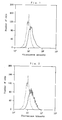
- FIG. 1 is an FACS diagram illustrating that an antibody produced by a hybridoma according to the present invention reacts with cells on which PAF-R molecules have been expressed.
- a dotted curve represents a negative control.
- FIG. 2 is an FACS diagram illustrating that an antibody produced by another hybridoma according to the present invention reacts with cells on which PAF-R molecules have been expressed.
- a dotted curve represents a negative control.
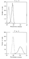
- FIG. 3 is an FACS diagram illustrating that an antibody produced by a further hybridoma according to the present invention reacts with cells on which PAF-R molecules have been expressed.
- a dotted curve represents a negative control.
- FIG. 4 is an FACS diagram illustrating that an antibody produced by a still further hybridoma according to the present invention reacts with cells on which PAF-R molecules have been expressed.
- a dotted curve represents a negative control.
- FIG. 5 is an FACS diagram illustrating that the antibody produced by the hybridoma according to the present invention reacts with eosinophils on which human PAF-R molecules have been expressed.
- a dotted curve represents a negative control.
- FIG. 6 is an FACS diagram illustrating that the antibody produced by the hybridoma according to the present invention reacts with B cells (Raji) on which human PAF-R molecules have been expressed.
- a dotted curve represents a negative control.
- FIG. 7 is an FACS diagram illustrating that the antibody produced by the hybridoma according to the present invention reacts with monocytes (U937) on which human PAF-R molecules have been expressed.
- a dotted curve represents a negative control.
- FIG. 8 is an FACS diagram illustrating that the antibody produced by the hybridoma according to the present invention does not react with T cells (Molt-4) on which human PAF-R molecules have been expressed.
- a dotted curve represents a negative control.
- FIG. 9 illustrates the results of an immuno-precipitation.
- Monoclonal antibodies specifically reacting with a PAF receptor, and hybridomas producing these monoclonal antibodies can be produced in accordance with the following procedure.
- the monoclonal antibody specifically reacting with the PAF receptor reacts with a peptide of the extracellular third or forth domain, or the intracellular fourth domain of the PAF receptor molecule.
- the PAF receptor is composed of 342 amino acid residues, and the molecular weight estimated from its primary structure is 38,982. Hydropathy analysis of the PAF receptor revealed that the PAF receptor has seven lipophilic domains, and it is hence considered to have a seven-times transmembrane structure (Biochemical Pharmacology, Vol. 44, No. 6, 1001-1008, 1992).
- the PAF receptor has 4 domains in its extracellular space, and the second, third and fourth domains thereof each have a loop-type structure.
- the PAF receptor has 4 domains in its intracellular space, and the first, second and third domains thereof each have a loop-type structure.
- the monoclonal antibodies according to the present invention are preferably those reacting with peptides of the extracellular third domain of the PAF receptor.
- four monoclonal antibodies have been found and are classified into 3 antibodies belonging to a class of IgM and having a ⁇ -L chain, and an antibody belonging to a class of IgG and a subclass of IgG2a and having a ⁇ -L chain.
- the monoclonal antibodies according to the present invention react with human eosinophils on which human PAF-R molecules have been expressed.
- the monoclonal antibodies according to the present invention also react with human B cells and monocytes, but do not react with human T cells.
- the monoclonal antibodies according to the present invention react with proteins of about 40 kDa in CHO-PR1 and CHO-WT6'A which are cells transfected with a PAF-R gene, and Raji and Wa which are B cells.
- the present invention includes active fragments of the monoclonal antibodies specifically reacting with the PAF receptor.
- the monoclonal antibodies according to the present invention are useful for immunological researches as to many reactions caused by the binding of PAF and PAF-receptor, immunotherapy, immunodiagnosis, and the like. It is not always necessary to use the whole monoclonal antibody molecule for such applications.
- Its active fragment may be used. As easily understood by those skilled in the art, the use of the active fragment may be preferred in some cases.
- the monoclonal antibodies are homogeneous immunoglobulins recognizing a specific antigenic substance.
- the term "active fragment” means a fragment of a monoclonal antibody active in antigen-antibody reaction. As specific examples thereof, may be mentioned F(ab')2, Fab', Fab, Fv and recombinant Fv. These active fragments may be prepared from the monoclonal antibodies according to the present invention in accordance with a method known per se in the art.
- the F(ab')2 fragment is one of fragments obtained by digesting an immunoglobulin IgG with pepsin.
- IgG is digested with pepsin at a pH near 4.0, it is cleaved at a hinge area of its H chain to produce a fragment having a molecular weight of about 100,000. This cleavage takes place on the C-terminal side away from the disulfide bond between H chains.
- This fragment has two antigen-binding sites and hence can couple to an antigen, thereby undergoing precipitin reaction and agglutination reaction.
- the Fab' fragment is a fragment produced by reducing the F(ab')2 fragment with a reagent such as 2-mercaptoethanol and alkylating the reduced product with monoiodoacetic acid, thereby cleaving a disulfide bond between H chains, and having a molecular weight of about 50,000.
- a Fab fragment is one of fragments obtained by the papain digestion of IgG.
- IgG is digested with papain in the presence of cysteine, its H chain is cleaved at a site on the N-terminal side away from the disulfide bond between H chains in a hinge area, thereby producing two Fab fragments and one Fc fragment (crystallizable fragment).
- the Fab fragment is a fragment in which a Fd fragment corresponding to about a half of the H chain on the N-terminal side (V H domain + C H 1 dome is coupled to an L chain by a disulfide bond, said fragment having a molecular weight of about 45,000 and one antigen-binding site.
- the Fv fragment is an antigen-binding fragment composed of a variable region of immunoglobulin heavy chain (V H ) and a variable region of immunoglobulin light chain (V L ), said variable regions being coupled to each other by a nonconjugate bond.
- the recombinant Fv fragment can be obtained by sequencing a DNA from a hybridoma which produces a monoclonal antibody to determine base sequences which code V H and L H , respectively, and then integrating these DNA fragments in a vector to produce a monovalent active antibody fragment having a structure of V L -Linker-V H .
- V H and L H are coupled to each other by an S-S bond.
- a linker is inserted between V H and L H so as to take the same steric structure as the state coupled by the S-S bond. This fragment may be simply called "Fv” in some cases. It may also be called "SCFv (single chain FV)".
- the recombinant Fv fragment may also be expressed by microorganisms such as Escherichia coli and bacteriophages.
- the monoclonal antibodies according to the present invention against the PAF receptor are useful for explicating various reactions which are considered to be caused by the binding of PAF and PAF-R, and for understanding the mechanisms of PAF and PAF-R in bronchial asthma, endotoxin shock, anaphylactic shock and the like.
- the monoclonal antibodies according to the present invention block the binding of PAF and PAF-R and hence are expected to prevent these various reactions.
- the monoclonal antibodies, production process thereof and hybridomas producing these monoclonal antibodies according to the present invention are useful in fields of genetic engineering, medical science and protein engineering, and industrial fields associated with these fields.
- Peptides of the extracellular third and forth domains, and the intracellular fourth domain of a guinea pig PAF receptor were synthesized by a peptide synthesizer (Model 431A, manufactured by ABI Company), cleaved, purified by HPLC and then lyophilized.
- the guinea pig PAF receptor is composed of 342 amino acid residues.
- the peptide of the extracellular third domain of the PAF receptor has an amino acid sequence of from the 158th to the 173th, Thr-Asn-Val-Val-Ser-Asn-Lys-Ala-Gly-Ser-Gly-Asn-Ile-Thr-Arg-Cys.
- the peptide of the extracellular fourth domain of the guinea pig PAF receptor has a structure that Cys is added to the C-terminal of an amino acid sequence of from the 259th to the 273th, Glu-Leu-Gly-Met-Trp-Pro-Ser-Ser-Asn-His-Gln-Ala-Ile-Asn-Asp.
- the peptide of the intracellular fourth domain of the guinea pig PAF receptor has a structure that Cys is added to the C-terminal of an amino acid sequence of from the 323th to the 337th, Asp-Thr-Gly-Thr-Glu-Met-Ala-IlePro-Ile-Asn-His-Thr-Pro-Val.
- One milligram of the peptide synthesized above was dissolved in 200 ⁇ l of a conjugation buffer (product of Pierce Company). To the resulting solution, 500 ⁇ l of a carrier protein KLH (2 mg/ml) was added to mix them. The mixture was rotationally stirred overnight at 4°C to couple the peptide to KLH, thereby providing the thus- obtained complex as an immunogen.
- a conjugation buffer product of Pierce Company
- the peptide-KLH complex in an amount of 10 ⁇ g in terms of the peptide was intraperitoneally injected into a Balb/c mouse together with FCA (Freund's complete adjuvant) in priming and with a poly A.poly U adjuvant on and after the second week.
- FCA Full's complete adjuvant
- the peptide complex was injected once a week, 15 times in total, thereby immunizing the mouse.
- the spleen Upon boosted time of 4 days after final immunization, the spleen was taken out of the mouse. The spleen was minced and filtered through a mesh and then suspended in an RPMI1640 medium, thereby obtaining 6 x 10⁷ splenocytes. The splenocytes and a mouse-derived 8-azaguanine-resistance myeloma cell strain (hypoxanthine-guanine phosphoribosyl transferase defective strain) P3U1 (1 x 10⁷ cells) were mixed with each other in a proportion of about 6:1, and the resulting mixture was centrifuged (1500 rpm, 5 minutes).
- a mouse-derived 8-azaguanine-resistance myeloma cell strain hyperxanthine-guanine phosphoribosyl transferase defective strain
- HEPES-RPMI1640 solution After the cell fusion, a great amount (25 ml) of an HEPES-RPMI1640 solution was added, and the mixture was centrifuged (1500 rpm, 5 minutes) to remove a supernatant. On the resultant precipitate, 1 ml of a 10% FCS-RPM11640 medium containing hypoxanthine (100 ⁇ M), aminopterine (0.4 ⁇ M) and thymidine (10 ⁇ M) (HAT medium) was placed. After the HAT medium was left at rest for 30 minutes in a CO2-incubator at 37°C, the splenocytes were adjusted to 1 x 10⁶ cells/ml in the incubator.
- hypoxanthine 100 ⁇ M
- aminopterine 0.4 ⁇ M
- thymidine 10 ⁇ M
- the cell suspension prepared in the step 3 was poured in 200- ⁇ l portions into 3 microplates each having 96 wells to culture the cells in a CO2-incubator controlled at 37°C and CO2 concentration of 5%. After a week, it was confirmed that only hybridomas formed colonies and proliferated.
- the culture supernatant of each well was used to detect antibodies reacting with the PAF-R peptide in the following manner.
- Hybridomas proliferated in the wells selected in the step 5 were separately cloned by the limiting dilution technique.
- the culture medium in each well was diluted with a 10% FCS-RPMI1640 medium so as to give a cell concentration of one cell/well.
- the thus-diluted culture medium was poured in portions of 200 ⁇ l/well into wells of a 96-well plate to culture the cell under conditions of 37°C and 5% CO2. After about 1 week, the hybridomas were recognized to proliferate, and colonies became a certain size, and so the detection of antibodies was conducted again in accordance with the process of the step 5.
- the cells were put in portions of 1 x 10⁶ cells into Fischer tubes, into which 200 ⁇ l of the culture supernatant was poured to react them for 20 minutes on an ice bath.
- Each of the reaction mixtures was centrifugally washed with PBS (3000 rpm, 1 minute, 3 times) and added with 100 ⁇ l of FITC-anti-hamster Ig's (product of Cultag Company, diluted to 1:100) to react them for 20 minutes on an ice bath.
- the reaction mixture was centrifugally washed twice with PBS to suspend it in 200 ⁇ l of PBS, thereby conducting measurement by FACScan. As a result, as illustrated in FIGs. 1 to 4, it was found that 4 antibodies reacting with the PAF-R molecule were obtained.
- a histogram indicated by a dotted line represents a negative control free of a primary antibody.
- a histogram indicated by a continuous line was obtained by using, as a primary antibody, various monoclonal antibodies and, as a secondary antibody, a fluorescein (FITC)-labeled antibody commonly specific for all the primary antibodies. Accordingly, if the primary antibody reacts with a cell surface molecule, it becomes a cell population higher in fluorescence intensity than the cell population of the negative control, and so its graph shifts on the right of the graph of the negative control.
- FITC fluorescein
- the subclasses of antibodies produced by the resultant hybridomas were determined by enzyme immunoassay (EIA) in which anti-mouse antibodies (Anti-IgG1, IgG2a, IgG2b, IgG3, IgM, IgA) and anti-mouse L chain antibodies (anti- ⁇ , ⁇ ) (MAB-isotyping kit, Pharmingen) were separately reacted with the anti-PAF-R monoclonal antibodies in the culture supernatants of the hybridomas.
- EIA enzyme immunoassay
- the resultant monoclonal antibodies were able to be classified into three antibodies in which staining was recognized in wells separately containing an anti-mouse antibody (anti-IgM) and an anti-mouse L chain antibody (anti- ⁇ ), and one antibody in which staining was recognized in wells separately containing an anti-mouse antibody (anti-IgG2a) and an anti-mouse L chain antibody (anti- ⁇ ).
- anti-IgM anti-mouse antibody
- anti- ⁇ anti-mouse L chain antibody
- the three monoclonal antibodies belong to a class of IgM and have a ⁇ -L chain, and the other one belongs to a class of IgG and a subclass of IgG2a and has a ⁇ -L chain.
- a hybridoma which produces a monoclonal antibody (FIG. 2; belonging to a class of IgM and having a ⁇ -L chain) reacting with the peptide of the extracellular third domain of PAF-R is Hybridoma PAF-R14 deposited as FERM BP-4723 in National Institute of Bioscience and Human-Technology, Agency of Industrial Science and Technology, Japan.
- a hybridoma which produces a monoclonal antibody (FIG. 3; belonging to a subclass of IgG2a and having a ⁇ -L chain) reacting with the peptide of the extracellular third domain of PAF-R is YM-PAFR deposited as FERM BP-4722 in National Institute of Bioscience and Human-Technology, Agency of Industrial Science and Technology, Japan.
- Example 1 Whether the monoclonal antibody (FIG. 3; belonging to a subclass of IgG2a and having a ⁇ -L chain) obtained in Example 1 reacts with human eosinophils which expressed a human PAF-R molecule, or not was determined by FACS.
- the cells were put in portions of 1 x 10⁶ cells into Fischer tubes, into which 50 ⁇ g/ml of the antibody purified was poured in an amount of 100 ⁇ l to react them for 20 minutes on an ice bath.
- Each of the reaction mixtures was centrifugally washed with PBS (3000 rpm, 1 minute, 3 times) and then added with 100 ⁇ l of FITC-anti-mouse Ig's (product of Caltag Company, diluted to 1:100) to react them for 20 minutes on an ice bath.
- the reaction mixture was centrifugally washed twice with PBS to suspend it in 200 ⁇ l of PBS, thereby conducting measurement by FACScan.
- Example 1 As a result, as illustrated in FIG. 5, it was revealed that the monoclonal antibody obtained in Example 1 also reacts with human PAF-R.
- the respective cell strains of human B cells, T cells and monocytes were used to determine whether these cells express PAF-R or not in the following manner.
- the cells were put in portions of 1 x 10⁶ cells into Fischer tubes, into which 10 ⁇ g/ml of the purified antibody obtained in Example 1 was poured in an amount of 100 ⁇ m to react them for 20 minutes on an ice bath.
- Each of the reaction mixtures was centrifugally washed with PBS (3000 rpm, 1 minute, 3 times) and then added with 100 ⁇ l of FITC-anti-mouse Ig's (product of Cultag Company, diluted to 1:100) to react them for 20 minutes on an ice bath.
- the reaction mixture was centrifugally washed twice with PBS to suspend it in 200 ⁇ l of PBS, thereby conducting measurement by FACScan.
- Example 1 As a result, as illustrated in FIGs. 6 to 8, it was revealed that the monoclonal antibody obtained in Example 1 reacts with the B cells (Raji) and monocytes (U937), but does not react with the T cells (Molt-4). Therefore, it was found that PAF-R is expressed by the B cells and monocytes, but not expressed by the T cells.
Landscapes
- Health & Medical Sciences (AREA)
- Chemical & Material Sciences (AREA)
- Immunology (AREA)
- Organic Chemistry (AREA)
- General Health & Medical Sciences (AREA)
- Biophysics (AREA)
- Biochemistry (AREA)
- Genetics & Genomics (AREA)
- Medicinal Chemistry (AREA)
- Molecular Biology (AREA)
- Proteomics, Peptides & Aminoacids (AREA)
- Life Sciences & Earth Sciences (AREA)
- Preparation Of Compounds By Using Micro-Organisms (AREA)
Applications Claiming Priority (2)
| Application Number | Priority Date | Filing Date | Title |
|---|---|---|---|
| JP217003/93 | 1993-08-09 | ||
| JP21700393 | 1993-08-09 |
Publications (2)
| Publication Number | Publication Date |
|---|---|
| EP0638648A2 true EP0638648A2 (de) | 1995-02-15 |
| EP0638648A3 EP0638648A3 (de) | 1996-05-08 |
Family
ID=16697302
Family Applications (1)
| Application Number | Title | Priority Date | Filing Date |
|---|---|---|---|
| EP94305815A Withdrawn EP0638648A3 (de) | 1993-08-09 | 1994-08-05 | Monoklonale Antikörper, die spezifisch mit PAF-Rezeptoren reagieren, Verfahren zu deren Herstellung und die diese Antikörper herstellende Hybridomazellen. |
Country Status (1)
| Country | Link |
|---|---|
| EP (1) | EP0638648A3 (de) |
Cited By (1)
| Publication number | Priority date | Publication date | Assignee | Title |
|---|---|---|---|---|
| EP0761232A1 (de) * | 1995-09-08 | 1997-03-12 | Sumitomo Electric Industries, Ltd. | Anti-PAF Rezeptor Antikörper enthaltendes Antiallergetikum |
Family Cites Families (2)
| Publication number | Priority date | Publication date | Assignee | Title |
|---|---|---|---|---|
| WO1987005904A1 (en) * | 1986-03-24 | 1987-10-08 | The University Of Sydney | Antigenic analogues of platelet activating factor (paf) |
| ATE195653T1 (de) * | 1990-06-06 | 2000-09-15 | Ruth Korth | Behandlung von erkrankungen mit paf-antagonisten und verfahren zur bestimmung deren wirksamkeit |
-
1994
- 1994-08-05 EP EP94305815A patent/EP0638648A3/de not_active Withdrawn
Cited By (1)
| Publication number | Priority date | Publication date | Assignee | Title |
|---|---|---|---|---|
| EP0761232A1 (de) * | 1995-09-08 | 1997-03-12 | Sumitomo Electric Industries, Ltd. | Anti-PAF Rezeptor Antikörper enthaltendes Antiallergetikum |
Also Published As
| Publication number | Publication date |
|---|---|
| EP0638648A3 (de) | 1996-05-08 |
Similar Documents
| Publication | Publication Date | Title |
|---|---|---|
| DK1625166T3 (en) | Antibodies to MASP-2 | |
| KR0184691B1 (ko) | 다양한 씨디44 표면단백질, 이 단백질을 코딩하는 디엔에이 서열, 이 단백질에 대한 항체와 진단과 치료의 용도 | |
| WO1999012973A1 (en) | Monoclonal antibody inducing apoptosis | |
| JPH08500487A (ja) | L鎖欠落免疫グロブリン | |
| AU2005271892A1 (en) | Methods for discovering antibodies specific to cancer cells and antibodies discovered thereby | |
| US11634487B2 (en) | Diagnostic method | |
| KR20110044714A (ko) | Vcam-1에 특이적으로 결합하고 백혈구와 내피세포의 접착 및 투과를 저해하는 재조합 인간 단일클론항체 | |
| CN115785270A (zh) | 一种针对人cd20蛋白的单克隆抗体、制备方法及其应用 | |
| JP2831852B2 (ja) | ヒト胸腺皮質細胞上に発現する細胞表面タンパク質及びその利用 | |
| JPH10511262A (ja) | ヒトレシピエントにおける異種移植片拒絶または同種移植片拒絶を減少させるためのヒト異種抗体または同種抗体の阻害 | |
| Pawlita et al. | A monoclonal antibody that defines an idiotope with two subsites in galactan-binding myeloma proteins. | |
| JPH0246297A (ja) | モノクローナル抗体、抗体産生ハイブリドーマおよび抗体の製造法 | |
| Rankin et al. | Mouse monoclonal antibodies against the idiotype of human B cell non‐Hodgkin lymphomas: production, characterization and use to monitor the progress of disease | |
| EP0638648A2 (de) | Monoklonale Antikörper, die spezifisch mit PAF-Rezeptoren reagieren, Verfahren zu deren Herstellung und die diese Antikörper herstellende Hybridomazellen | |
| CA2667263C (en) | Antibody against rgd in amino acid sequence of extracellular matrix protein and production method and use of the same | |
| JP4563573B2 (ja) | 抗原およびこの抗原を識別するモノクローナル抗体 | |
| JPH06303990A (ja) | モノクローナル抗体、それを産生するハイブリドーマおよび該抗体の製造方法 | |
| JPH07101998A (ja) | Paf受容体に特異的に反応するモノクローナル抗体、その製造方法、及び該抗体を産生するハイブリドーマ | |
| JPH0779793A (ja) | B70分子に対するモノクローナル抗体 | |
| Loirat et al. | Characterization of new murine monoclonal antibodies directed against glycophorins C and D | |
| KR102949436B1 (ko) | Ct-26 종양 세포 특이적 항체 | |
| CA3070847C (en) | Diagnostic method | |
| Elagib et al. | Characterization and VH sequences of human monoclonal anti-F (ab′) 2 autoantibodies from normals and Sjögren's syndrome patients | |
| Stratieva-Taneeva et al. | Bispecific monoclonal antibodies to human interleukin 2 and horseradish peroxidase | |
| CN121866270A (zh) | 抗gcc抗体、嵌合抗原受体及其用途 |
Legal Events
| Date | Code | Title | Description |
|---|---|---|---|
| PUAI | Public reference made under article 153(3) epc to a published international application that has entered the european phase |
Free format text: ORIGINAL CODE: 0009012 |
|
| AK | Designated contracting states |
Kind code of ref document: A2 Designated state(s): CH DE FR GB IT LI SE |
|
| PUAL | Search report despatched |
Free format text: ORIGINAL CODE: 0009013 |
|
| AK | Designated contracting states |
Kind code of ref document: A3 Designated state(s): CH DE FR GB IT LI SE |
|
| 17P | Request for examination filed |
Effective date: 19961106 |
|
| STAA | Information on the status of an ep patent application or granted ep patent |
Free format text: STATUS: THE APPLICATION HAS BEEN WITHDRAWN |
|
| 18W | Application withdrawn |
Withdrawal date: 20000209 |