EP0643140A1 - Détermination d'acides nucleiques par PCR, mesure du nombre des cellules microbiennes, gènes ou copies de gènes par PCR, et trousse utilisée à cet effet - Google Patents
Détermination d'acides nucleiques par PCR, mesure du nombre des cellules microbiennes, gènes ou copies de gènes par PCR, et trousse utilisée à cet effet Download PDFInfo
- Publication number
- EP0643140A1 EP0643140A1 EP94109568A EP94109568A EP0643140A1 EP 0643140 A1 EP0643140 A1 EP 0643140A1 EP 94109568 A EP94109568 A EP 94109568A EP 94109568 A EP94109568 A EP 94109568A EP 0643140 A1 EP0643140 A1 EP 0643140A1
- Authority
- EP
- European Patent Office
- Prior art keywords
- group
- substituted
- nucleic acid
- unsubstituted
- dye compound
- Prior art date
- Legal status (The legal status is an assumption and is not a legal conclusion. Google has not performed a legal analysis and makes no representation as to the accuracy of the status listed.)
- Granted
Links
- 150000007523 nucleic acids Chemical class 0.000 title claims abstract description 149
- 108020004707 nucleic acids Proteins 0.000 title claims abstract description 148
- 102000039446 nucleic acids Human genes 0.000 title claims abstract description 148
- 108090000623 proteins and genes Proteins 0.000 title claims abstract description 46
- 238000005259 measurement Methods 0.000 title claims abstract description 38
- 230000000813 microbial effect Effects 0.000 title description 11
- 150000001875 compounds Chemical class 0.000 claims abstract description 189
- 238000012408 PCR amplification Methods 0.000 claims abstract description 30
- 238000006243 chemical reaction Methods 0.000 claims description 108
- 238000000034 method Methods 0.000 claims description 107
- 238000010790 dilution Methods 0.000 claims description 89
- 239000012895 dilution Substances 0.000 claims description 89
- 230000003321 amplification Effects 0.000 claims description 73
- 238000003199 nucleic acid amplification method Methods 0.000 claims description 73
- 125000003118 aryl group Chemical group 0.000 claims description 69
- 239000000523 sample Substances 0.000 claims description 57
- 239000000243 solution Substances 0.000 claims description 54
- 244000005700 microbiome Species 0.000 claims description 43
- 125000000623 heterocyclic group Chemical group 0.000 claims description 37
- 239000003153 chemical reaction reagent Substances 0.000 claims description 34
- 150000003839 salts Chemical class 0.000 claims description 29
- 125000000217 alkyl group Chemical group 0.000 claims description 28
- 150000001450 anions Chemical class 0.000 claims description 24
- 125000003277 amino group Chemical group 0.000 claims description 23
- 125000003710 aryl alkyl group Chemical group 0.000 claims description 23
- 125000000753 cycloalkyl group Chemical group 0.000 claims description 23
- 125000004435 hydrogen atom Chemical group [H]* 0.000 claims description 23
- 125000003178 carboxy group Chemical group [H]OC(*)=O 0.000 claims description 22
- 125000004093 cyano group Chemical group *C#N 0.000 claims description 22
- 125000005843 halogen group Chemical group 0.000 claims description 22
- 125000002887 hydroxy group Chemical group [H]O* 0.000 claims description 22
- 125000000449 nitro group Chemical group [O-][N+](*)=O 0.000 claims description 22
- WVIICGIFSIBFOG-UHFFFAOYSA-N pyrylium Chemical group C1=CC=[O+]C=C1 WVIICGIFSIBFOG-UHFFFAOYSA-N 0.000 claims description 22
- 125000005504 styryl group Chemical group 0.000 claims description 22
- 125000001273 sulfonato group Chemical group [O-]S(*)(=O)=O 0.000 claims description 22
- 125000002947 alkylene group Chemical group 0.000 claims description 13
- 125000004043 oxo group Chemical group O=* 0.000 claims description 13
- 239000002904 solvent Substances 0.000 claims description 13
- 229910052717 sulfur Inorganic materials 0.000 claims description 13
- 125000004122 cyclic group Chemical group 0.000 claims description 12
- 229910052760 oxygen Inorganic materials 0.000 claims description 12
- 229910052799 carbon Inorganic materials 0.000 claims description 11
- 125000004432 carbon atom Chemical group C* 0.000 claims description 11
- 229910052711 selenium Inorganic materials 0.000 claims description 11
- XLYOFNOQVPJJNP-UHFFFAOYSA-N water Substances O XLYOFNOQVPJJNP-UHFFFAOYSA-N 0.000 claims description 9
- 239000012470 diluted sample Substances 0.000 claims description 6
- 238000013207 serial dilution Methods 0.000 claims description 6
- 239000012898 sample dilution Substances 0.000 claims description 2
- 239000012736 aqueous medium Substances 0.000 claims 4
- 239000012807 PCR reagent Substances 0.000 claims 2
- HNCZEEIACFGTKE-UHFFFAOYSA-N [(6e)-6-[4-[2-(dimethylamino)phenyl]-6-methylpyran-2-ylidene]cyclohexa-2,4-dien-1-ylidene]-dimethylazanium Chemical class CN(C)C1=CC=CC=C1C(C=C(C)O1)=C\C1=C\1C(=[N+](C)C)C=CC=C/1 HNCZEEIACFGTKE-UHFFFAOYSA-N 0.000 claims 1
- JLGJYDYKIXQKEQ-UHFFFAOYSA-N [6-[4-[2-(dimethylamino)phenyl]-6-methylthiopyran-2-ylidene]cyclohexa-2,4-dien-1-ylidene]-dimethylazanium Chemical class CN(C)C1=CC=CC=C1C(C=C(C)S1)=C\C1=C\1C(=[N+](C)C)C=CC=C/1 JLGJYDYKIXQKEQ-UHFFFAOYSA-N 0.000 claims 1
- 230000001580 bacterial effect Effects 0.000 abstract description 5
- 108020004414 DNA Proteins 0.000 description 112
- 238000003752 polymerase chain reaction Methods 0.000 description 110
- 239000000047 product Substances 0.000 description 78
- 210000004027 cell Anatomy 0.000 description 58
- 239000000975 dye Substances 0.000 description 56
- WEVYAHXRMPXWCK-UHFFFAOYSA-N Acetonitrile Chemical compound CC#N WEVYAHXRMPXWCK-UHFFFAOYSA-N 0.000 description 35
- 238000001514 detection method Methods 0.000 description 29
- 239000000203 mixture Substances 0.000 description 27
- 210000001519 tissue Anatomy 0.000 description 24
- 239000002689 soil Substances 0.000 description 22
- LFQSCWFLJHTTHZ-UHFFFAOYSA-N Ethanol Chemical compound CCO LFQSCWFLJHTTHZ-UHFFFAOYSA-N 0.000 description 19
- ZMMJGEGLRURXTF-UHFFFAOYSA-N ethidium bromide Chemical compound [Br-].C12=CC(N)=CC=C2C2=CC=C(N)C=C2[N+](CC)=C1C1=CC=CC=C1 ZMMJGEGLRURXTF-UHFFFAOYSA-N 0.000 description 19
- 229960005542 ethidium bromide Drugs 0.000 description 19
- BGNGWHSBYQYVRX-UHFFFAOYSA-N 4-(dimethylamino)benzaldehyde Chemical compound CN(C)C1=CC=C(C=O)C=C1 BGNGWHSBYQYVRX-UHFFFAOYSA-N 0.000 description 14
- 229940125904 compound 1 Drugs 0.000 description 13
- 230000005284 excitation Effects 0.000 description 12
- 241000588724 Escherichia coli Species 0.000 description 11
- IAZDPXIOMUYVGZ-UHFFFAOYSA-N Dimethylsulphoxide Chemical compound CS(C)=O IAZDPXIOMUYVGZ-UHFFFAOYSA-N 0.000 description 10
- 206010028980 Neoplasm Diseases 0.000 description 9
- 201000011510 cancer Diseases 0.000 description 9
- 230000000052 comparative effect Effects 0.000 description 9
- 239000000138 intercalating agent Substances 0.000 description 9
- NLKNQRATVPKPDG-UHFFFAOYSA-M potassium iodide Chemical compound [K+].[I-] NLKNQRATVPKPDG-UHFFFAOYSA-M 0.000 description 9
- 239000007858 starting material Substances 0.000 description 9
- KFZMGEQAYNKOFK-UHFFFAOYSA-N Isopropanol Chemical compound CC(C)O KFZMGEQAYNKOFK-UHFFFAOYSA-N 0.000 description 8
- 238000000246 agarose gel electrophoresis Methods 0.000 description 8
- 238000011088 calibration curve Methods 0.000 description 8
- 239000011541 reaction mixture Substances 0.000 description 8
- 239000007853 buffer solution Substances 0.000 description 7
- 238000001502 gel electrophoresis Methods 0.000 description 7
- -1 pyrylium compound salts Chemical class 0.000 description 7
- 125000001424 substituent group Chemical group 0.000 description 7
- WFDIJRYMOXRFFG-UHFFFAOYSA-N Acetic anhydride Chemical compound CC(=O)OC(C)=O WFDIJRYMOXRFFG-UHFFFAOYSA-N 0.000 description 6
- 102000053602 DNA Human genes 0.000 description 6
- GRRMZXFOOGQMFA-UHFFFAOYSA-J YoYo-1 Chemical compound [I-].[I-].[I-].[I-].C12=CC=CC=C2C(C=C2N(C3=CC=CC=C3O2)C)=CC=[N+]1CCC[N+](C)(C)CCC[N+](C)(C)CCC[N+](C1=CC=CC=C11)=CC=C1C=C1N(C)C2=CC=CC=C2O1 GRRMZXFOOGQMFA-UHFFFAOYSA-J 0.000 description 6
- 230000015572 biosynthetic process Effects 0.000 description 6
- 238000005119 centrifugation Methods 0.000 description 6
- 229940125782 compound 2 Drugs 0.000 description 6
- 239000002773 nucleotide Substances 0.000 description 6
- 125000003729 nucleotide group Chemical group 0.000 description 6
- VLTRZXGMWDSKGL-UHFFFAOYSA-M perchlorate Chemical compound [O-]Cl(=O)(=O)=O VLTRZXGMWDSKGL-UHFFFAOYSA-M 0.000 description 6
- 239000002244 precipitate Substances 0.000 description 6
- 230000008569 process Effects 0.000 description 6
- 239000007984 Tris EDTA buffer Substances 0.000 description 5
- 238000001962 electrophoresis Methods 0.000 description 5
- 238000002474 experimental method Methods 0.000 description 5
- XMBWDFGMSWQBCA-UHFFFAOYSA-N hydrogen iodide Chemical compound I XMBWDFGMSWQBCA-UHFFFAOYSA-N 0.000 description 5
- 229910052757 nitrogen Inorganic materials 0.000 description 5
- 239000000725 suspension Substances 0.000 description 5
- 238000012360 testing method Methods 0.000 description 5
- 108020004465 16S ribosomal RNA Proteins 0.000 description 4
- QKNYBSVHEMOAJP-UHFFFAOYSA-N 2-amino-2-(hydroxymethyl)propane-1,3-diol;hydron;chloride Chemical compound Cl.OCC(N)(CO)CO QKNYBSVHEMOAJP-UHFFFAOYSA-N 0.000 description 4
- IKHGUXGNUITLKF-UHFFFAOYSA-N Acetaldehyde Chemical compound CC=O IKHGUXGNUITLKF-UHFFFAOYSA-N 0.000 description 4
- KWOLFJPFCHCOCG-UHFFFAOYSA-N Acetophenone Chemical compound CC(=O)C1=CC=CC=C1 KWOLFJPFCHCOCG-UHFFFAOYSA-N 0.000 description 4
- FAPWRFPIFSIZLT-UHFFFAOYSA-M Sodium chloride Chemical compound [Na+].[Cl-] FAPWRFPIFSIZLT-UHFFFAOYSA-M 0.000 description 4
- 239000000872 buffer Substances 0.000 description 4
- 150000001768 cations Chemical class 0.000 description 4
- 239000002299 complementary DNA Substances 0.000 description 4
- 238000010348 incorporation Methods 0.000 description 4
- 238000011534 incubation Methods 0.000 description 4
- 238000002844 melting Methods 0.000 description 4
- 230000008018 melting Effects 0.000 description 4
- 108020004999 messenger RNA Proteins 0.000 description 4
- 239000008055 phosphate buffer solution Substances 0.000 description 4
- 230000035945 sensitivity Effects 0.000 description 4
- 238000000926 separation method Methods 0.000 description 4
- HYHCSLBZRBJJCH-UHFFFAOYSA-M sodium hydrosulfide Chemical compound [Na+].[SH-] HYHCSLBZRBJJCH-UHFFFAOYSA-M 0.000 description 4
- 125000001140 1,4-phenylene group Chemical group [H]C1=C([H])C([*:2])=C([H])C([H])=C1[*:1] 0.000 description 3
- YMWUJEATGCHHMB-UHFFFAOYSA-N Dichloromethane Chemical compound ClCCl YMWUJEATGCHHMB-UHFFFAOYSA-N 0.000 description 3
- KCXVZYZYPLLWCC-UHFFFAOYSA-N EDTA Chemical compound OC(=O)CN(CC(O)=O)CCN(CC(O)=O)CC(O)=O KCXVZYZYPLLWCC-UHFFFAOYSA-N 0.000 description 3
- 108091028043 Nucleic acid sequence Proteins 0.000 description 3
- 108020004682 Single-Stranded DNA Proteins 0.000 description 3
- 108010006785 Taq Polymerase Proteins 0.000 description 3
- 238000004458 analytical method Methods 0.000 description 3
- 230000006037 cell lysis Effects 0.000 description 3
- 238000000605 extraction Methods 0.000 description 3
- 239000001963 growth medium Substances 0.000 description 3
- 239000007788 liquid Substances 0.000 description 3
- 239000012188 paraffin wax Substances 0.000 description 3
- VLTRZXGMWDSKGL-UHFFFAOYSA-N perchloric acid Chemical compound OCl(=O)(=O)=O VLTRZXGMWDSKGL-UHFFFAOYSA-N 0.000 description 3
- 125000000843 phenylene group Chemical group C1(=C(C=CC=C1)*)* 0.000 description 3
- 238000002360 preparation method Methods 0.000 description 3
- VYZSQJOZEWGVJN-UHFFFAOYSA-M pyrylium iodide Chemical compound [I-].C1=CC=[O+]C=C1 VYZSQJOZEWGVJN-UHFFFAOYSA-M 0.000 description 3
- 238000011160 research Methods 0.000 description 3
- 229920006395 saturated elastomer Polymers 0.000 description 3
- 239000011550 stock solution Substances 0.000 description 3
- 108091032973 (ribonucleotides)n+m Proteins 0.000 description 2
- PRDFBSVERLRRMY-UHFFFAOYSA-N 2'-(4-ethoxyphenyl)-5-(4-methylpiperazin-1-yl)-2,5'-bibenzimidazole Chemical compound C1=CC(OCC)=CC=C1C1=NC2=CC=C(C=3NC4=CC(=CC=C4N=3)N3CCN(C)CC3)C=C2N1 PRDFBSVERLRRMY-UHFFFAOYSA-N 0.000 description 2
- NKBIMLJDOFEBIL-UHFFFAOYSA-N CCN(CC)C1=CC=C(C=O)C(C=CC=2C=CC=CC=2)=C1 Chemical compound CCN(CC)C1=CC=C(C=O)C(C=CC=2C=CC=CC=2)=C1 NKBIMLJDOFEBIL-UHFFFAOYSA-N 0.000 description 2
- 230000004544 DNA amplification Effects 0.000 description 2
- 108010014303 DNA-directed DNA polymerase Proteins 0.000 description 2
- 102000016928 DNA-directed DNA polymerase Human genes 0.000 description 2
- 108090000790 Enzymes Proteins 0.000 description 2
- 102000004190 Enzymes Human genes 0.000 description 2
- CDBYLPFSWZWCQE-UHFFFAOYSA-L Sodium Carbonate Chemical compound [Na+].[Na+].[O-]C([O-])=O CDBYLPFSWZWCQE-UHFFFAOYSA-L 0.000 description 2
- VMHLLURERBWHNL-UHFFFAOYSA-M Sodium acetate Chemical compound [Na+].CC([O-])=O VMHLLURERBWHNL-UHFFFAOYSA-M 0.000 description 2
- UIIMBOGNXHQVGW-UHFFFAOYSA-M Sodium bicarbonate Chemical compound [Na+].OC([O-])=O UIIMBOGNXHQVGW-UHFFFAOYSA-M 0.000 description 2
- 229920004890 Triton X-100 Polymers 0.000 description 2
- 239000013504 Triton X-100 Substances 0.000 description 2
- 238000010521 absorption reaction Methods 0.000 description 2
- 238000000862 absorption spectrum Methods 0.000 description 2
- 239000000061 acid fraction Substances 0.000 description 2
- DPKHZNPWBDQZCN-UHFFFAOYSA-N acridine orange free base Chemical compound C1=CC(N(C)C)=CC2=NC3=CC(N(C)C)=CC=C3C=C21 DPKHZNPWBDQZCN-UHFFFAOYSA-N 0.000 description 2
- 239000007864 aqueous solution Substances 0.000 description 2
- 238000003556 assay Methods 0.000 description 2
- DZBUGLKDJFMEHC-UHFFFAOYSA-N benzoquinolinylidene Natural products C1=CC=CC2=CC3=CC=CC=C3N=C21 DZBUGLKDJFMEHC-UHFFFAOYSA-N 0.000 description 2
- 239000012496 blank sample Substances 0.000 description 2
- 230000008859 change Effects 0.000 description 2
- 229910001914 chlorine tetroxide Inorganic materials 0.000 description 2
- YTRQFSDWAXHJCC-UHFFFAOYSA-N chloroform;phenol Chemical compound ClC(Cl)Cl.OC1=CC=CC=C1 YTRQFSDWAXHJCC-UHFFFAOYSA-N 0.000 description 2
- 230000000295 complement effect Effects 0.000 description 2
- 238000007796 conventional method Methods 0.000 description 2
- 238000001816 cooling Methods 0.000 description 2
- PSLIMVZEAPALCD-UHFFFAOYSA-N ethanol;ethoxyethane Chemical compound CCO.CCOCC PSLIMVZEAPALCD-UHFFFAOYSA-N 0.000 description 2
- 238000001914 filtration Methods 0.000 description 2
- WBJINCZRORDGAQ-UHFFFAOYSA-N formic acid ethyl ester Natural products CCOC=O WBJINCZRORDGAQ-UHFFFAOYSA-N 0.000 description 2
- 239000000499 gel Substances 0.000 description 2
- 229910052736 halogen Inorganic materials 0.000 description 2
- 150000002367 halogens Chemical class 0.000 description 2
- 239000006166 lysate Substances 0.000 description 2
- 239000003960 organic solvent Substances 0.000 description 2
- 125000001997 phenyl group Chemical group [H]C1=C([H])C([H])=C(*)C([H])=C1[H] 0.000 description 2
- 108700004121 sarkosyl Proteins 0.000 description 2
- 239000001632 sodium acetate Substances 0.000 description 2
- 235000017281 sodium acetate Nutrition 0.000 description 2
- 239000011780 sodium chloride Substances 0.000 description 2
- KSAVQLQVUXSOCR-UHFFFAOYSA-M sodium lauroyl sarcosinate Chemical compound [Na+].CCCCCCCCCCCC(=O)N(C)CC([O-])=O KSAVQLQVUXSOCR-UHFFFAOYSA-M 0.000 description 2
- UNHKSXOTUHOTAB-UHFFFAOYSA-N sodium;sulfane Chemical class [Na].S UNHKSXOTUHOTAB-UHFFFAOYSA-N 0.000 description 2
- 238000010186 staining Methods 0.000 description 2
- 239000000126 substance Substances 0.000 description 2
- 239000006228 supernatant Substances 0.000 description 2
- 238000003786 synthesis reaction Methods 0.000 description 2
- OKYDCMQQLGECPI-UHFFFAOYSA-N thiopyrylium Chemical compound C1=CC=[S+]C=C1 OKYDCMQQLGECPI-UHFFFAOYSA-N 0.000 description 2
- 238000012546 transfer Methods 0.000 description 2
- GKASDNZWUGIAMG-UHFFFAOYSA-N triethyl orthoformate Chemical compound CCOC(OCC)OCC GKASDNZWUGIAMG-UHFFFAOYSA-N 0.000 description 2
- DGVVWUTYPXICAM-UHFFFAOYSA-N β‐Mercaptoethanol Chemical compound OCCS DGVVWUTYPXICAM-UHFFFAOYSA-N 0.000 description 2
- ASGMFNBUXDJWJJ-JLCFBVMHSA-N (1R,3R)-3-[[3-bromo-1-[4-(5-methyl-1,3,4-thiadiazol-2-yl)phenyl]pyrazolo[3,4-d]pyrimidin-6-yl]amino]-N,1-dimethylcyclopentane-1-carboxamide Chemical compound BrC1=NN(C2=NC(=NC=C21)N[C@H]1C[C@@](CC1)(C(=O)NC)C)C1=CC=C(C=C1)C=1SC(=NN=1)C ASGMFNBUXDJWJJ-JLCFBVMHSA-N 0.000 description 1
- AOSZTAHDEDLTLQ-AZKQZHLXSA-N (1S,2S,4R,8S,9S,11S,12R,13S,19S)-6-[(3-chlorophenyl)methyl]-12,19-difluoro-11-hydroxy-8-(2-hydroxyacetyl)-9,13-dimethyl-6-azapentacyclo[10.8.0.02,9.04,8.013,18]icosa-14,17-dien-16-one Chemical compound C([C@@H]1C[C@H]2[C@H]3[C@]([C@]4(C=CC(=O)C=C4[C@@H](F)C3)C)(F)[C@@H](O)C[C@@]2([C@@]1(C1)C(=O)CO)C)N1CC1=CC=CC(Cl)=C1 AOSZTAHDEDLTLQ-AZKQZHLXSA-N 0.000 description 1
- GLGNXYJARSMNGJ-VKTIVEEGSA-N (1s,2s,3r,4r)-3-[[5-chloro-2-[(1-ethyl-6-methoxy-2-oxo-4,5-dihydro-3h-1-benzazepin-7-yl)amino]pyrimidin-4-yl]amino]bicyclo[2.2.1]hept-5-ene-2-carboxamide Chemical compound CCN1C(=O)CCCC2=C(OC)C(NC=3N=C(C(=CN=3)Cl)N[C@H]3[C@H]([C@@]4([H])C[C@@]3(C=C4)[H])C(N)=O)=CC=C21 GLGNXYJARSMNGJ-VKTIVEEGSA-N 0.000 description 1
- IUSARDYWEPUTPN-OZBXUNDUSA-N (2r)-n-[(2s,3r)-4-[[(4s)-6-(2,2-dimethylpropyl)spiro[3,4-dihydropyrano[2,3-b]pyridine-2,1'-cyclobutane]-4-yl]amino]-3-hydroxy-1-[3-(1,3-thiazol-2-yl)phenyl]butan-2-yl]-2-methoxypropanamide Chemical compound C([C@H](NC(=O)[C@@H](C)OC)[C@H](O)CN[C@@H]1C2=CC(CC(C)(C)C)=CN=C2OC2(CCC2)C1)C(C=1)=CC=CC=1C1=NC=CS1 IUSARDYWEPUTPN-OZBXUNDUSA-N 0.000 description 1
- YJLIKUSWRSEPSM-WGQQHEPDSA-N (2r,3r,4s,5r)-2-[6-amino-8-[(4-phenylphenyl)methylamino]purin-9-yl]-5-(hydroxymethyl)oxolane-3,4-diol Chemical compound C=1C=C(C=2C=CC=CC=2)C=CC=1CNC1=NC=2C(N)=NC=NC=2N1[C@@H]1O[C@H](CO)[C@@H](O)[C@H]1O YJLIKUSWRSEPSM-WGQQHEPDSA-N 0.000 description 1
- VIJSPAIQWVPKQZ-BLECARSGSA-N (2s)-2-[[(2s)-2-[[(2s)-2-[[(2s)-2-[[(2s)-2-[[(2s)-2-acetamido-5-(diaminomethylideneamino)pentanoyl]amino]-4-methylpentanoyl]amino]-4,4-dimethylpentanoyl]amino]-4-methylpentanoyl]amino]propanoyl]amino]-5-(diaminomethylideneamino)pentanoic acid Chemical compound NC(=N)NCCC[C@@H](C(O)=O)NC(=O)[C@H](C)NC(=O)[C@H](CC(C)C)NC(=O)[C@H](CC(C)(C)C)NC(=O)[C@H](CC(C)C)NC(=O)[C@H](CCCNC(N)=N)NC(C)=O VIJSPAIQWVPKQZ-BLECARSGSA-N 0.000 description 1
- STBLNCCBQMHSRC-BATDWUPUSA-N (2s)-n-[(3s,4s)-5-acetyl-7-cyano-4-methyl-1-[(2-methylnaphthalen-1-yl)methyl]-2-oxo-3,4-dihydro-1,5-benzodiazepin-3-yl]-2-(methylamino)propanamide Chemical compound O=C1[C@@H](NC(=O)[C@H](C)NC)[C@H](C)N(C(C)=O)C2=CC(C#N)=CC=C2N1CC1=C(C)C=CC2=CC=CC=C12 STBLNCCBQMHSRC-BATDWUPUSA-N 0.000 description 1
- QFLWZFQWSBQYPS-AWRAUJHKSA-N (3S)-3-[[(2S)-2-[[(2S)-2-[5-[(3aS,6aR)-2-oxo-1,3,3a,4,6,6a-hexahydrothieno[3,4-d]imidazol-4-yl]pentanoylamino]-3-methylbutanoyl]amino]-3-(4-hydroxyphenyl)propanoyl]amino]-4-[1-bis(4-chlorophenoxy)phosphorylbutylamino]-4-oxobutanoic acid Chemical compound CCCC(NC(=O)[C@H](CC(O)=O)NC(=O)[C@H](Cc1ccc(O)cc1)NC(=O)[C@@H](NC(=O)CCCCC1SC[C@@H]2NC(=O)N[C@H]12)C(C)C)P(=O)(Oc1ccc(Cl)cc1)Oc1ccc(Cl)cc1 QFLWZFQWSBQYPS-AWRAUJHKSA-N 0.000 description 1
- UDQTXCHQKHIQMH-KYGLGHNPSA-N (3ar,5s,6s,7r,7ar)-5-(difluoromethyl)-2-(ethylamino)-5,6,7,7a-tetrahydro-3ah-pyrano[3,2-d][1,3]thiazole-6,7-diol Chemical compound S1C(NCC)=N[C@H]2[C@@H]1O[C@H](C(F)F)[C@@H](O)[C@@H]2O UDQTXCHQKHIQMH-KYGLGHNPSA-N 0.000 description 1
- HUWSZNZAROKDRZ-RRLWZMAJSA-N (3r,4r)-3-azaniumyl-5-[[(2s,3r)-1-[(2s)-2,3-dicarboxypyrrolidin-1-yl]-3-methyl-1-oxopentan-2-yl]amino]-5-oxo-4-sulfanylpentane-1-sulfonate Chemical compound OS(=O)(=O)CC[C@@H](N)[C@@H](S)C(=O)N[C@@H]([C@H](C)CC)C(=O)N1CCC(C(O)=O)[C@H]1C(O)=O HUWSZNZAROKDRZ-RRLWZMAJSA-N 0.000 description 1
- MPDDTAJMJCESGV-CTUHWIOQSA-M (3r,5r)-7-[2-(4-fluorophenyl)-5-[methyl-[(1r)-1-phenylethyl]carbamoyl]-4-propan-2-ylpyrazol-3-yl]-3,5-dihydroxyheptanoate Chemical compound C1([C@@H](C)N(C)C(=O)C2=NN(C(CC[C@@H](O)C[C@@H](O)CC([O-])=O)=C2C(C)C)C=2C=CC(F)=CC=2)=CC=CC=C1 MPDDTAJMJCESGV-CTUHWIOQSA-M 0.000 description 1
- OMBVEVHRIQULKW-DNQXCXABSA-M (3r,5r)-7-[3-(4-fluorophenyl)-8-oxo-7-phenyl-1-propan-2-yl-5,6-dihydro-4h-pyrrolo[2,3-c]azepin-2-yl]-3,5-dihydroxyheptanoate Chemical compound O=C1C=2N(C(C)C)C(CC[C@@H](O)C[C@@H](O)CC([O-])=O)=C(C=3C=CC(F)=CC=3)C=2CCCN1C1=CC=CC=C1 OMBVEVHRIQULKW-DNQXCXABSA-M 0.000 description 1
- YQOLEILXOBUDMU-KRWDZBQOSA-N (4R)-5-[(6-bromo-3-methyl-2-pyrrolidin-1-ylquinoline-4-carbonyl)amino]-4-(2-chlorophenyl)pentanoic acid Chemical compound CC1=C(C2=C(C=CC(=C2)Br)N=C1N3CCCC3)C(=O)NC[C@H](CCC(=O)O)C4=CC=CC=C4Cl YQOLEILXOBUDMU-KRWDZBQOSA-N 0.000 description 1
- 0 *1C=C*C=C1 Chemical compound *1C=C*C=C1 0.000 description 1
- 125000002030 1,2-phenylene group Chemical group [H]C1=C([H])C([*:1])=C([*:2])C([H])=C1[H] 0.000 description 1
- 125000001989 1,3-phenylene group Chemical group [H]C1=C([H])C([*:1])=C([H])C([*:2])=C1[H] 0.000 description 1
- HUDYANRNMZDQGA-UHFFFAOYSA-N 1-[4-(dimethylamino)phenyl]ethanone Chemical compound CN(C)C1=CC=C(C(C)=O)C=C1 HUDYANRNMZDQGA-UHFFFAOYSA-N 0.000 description 1
- FQMZXMVHHKXGTM-UHFFFAOYSA-N 2-(1-adamantyl)-n-[2-[2-(2-hydroxyethylamino)ethylamino]quinolin-5-yl]acetamide Chemical compound C1C(C2)CC(C3)CC2CC13CC(=O)NC1=CC=CC2=NC(NCCNCCO)=CC=C21 FQMZXMVHHKXGTM-UHFFFAOYSA-N 0.000 description 1
- YTMLLNPQZXFMLZ-UHFFFAOYSA-N 2-(4-aminophenyl)-1H-indol-6-amine dihydrochloride Chemical compound Cl.Cl.C1=CC(N)=CC=C1C1=CC2=CC=C(N)C=C2N1 YTMLLNPQZXFMLZ-UHFFFAOYSA-N 0.000 description 1
- PYRKKGOKRMZEIT-UHFFFAOYSA-N 2-[6-(2-cyclopropylethoxy)-9-(2-hydroxy-2-methylpropyl)-1h-phenanthro[9,10-d]imidazol-2-yl]-5-fluorobenzene-1,3-dicarbonitrile Chemical compound C1=C2C3=CC(CC(C)(O)C)=CC=C3C=3NC(C=4C(=CC(F)=CC=4C#N)C#N)=NC=3C2=CC=C1OCCC1CC1 PYRKKGOKRMZEIT-UHFFFAOYSA-N 0.000 description 1
- MDKGOTTVUWLPLW-UHFFFAOYSA-N 2-phenyl-1h-indole-3,4-diamine Chemical compound N1C2=CC=CC(N)=C2C(N)=C1C1=CC=CC=C1 MDKGOTTVUWLPLW-UHFFFAOYSA-N 0.000 description 1
- FWBHETKCLVMNFS-UHFFFAOYSA-N 4',6-Diamino-2-phenylindol Chemical compound C1=CC(C(=N)N)=CC=C1C1=CC2=CC=C(C(N)=N)C=C2N1 FWBHETKCLVMNFS-UHFFFAOYSA-N 0.000 description 1
- RUKJCCIJLIMGEP-ONEGZZNKSA-N 4-dimethylaminocinnamaldehyde Chemical compound CN(C)C1=CC=C(\C=C\C=O)C=C1 RUKJCCIJLIMGEP-ONEGZZNKSA-N 0.000 description 1
- 125000004070 6 membered heterocyclic group Chemical group 0.000 description 1
- 241000972773 Aulopiformes Species 0.000 description 1
- 241000894006 Bacteria Species 0.000 description 1
- 208000035143 Bacterial infection Diseases 0.000 description 1
- CPELXLSAUQHCOX-UHFFFAOYSA-M Bromide Chemical compound [Br-] CPELXLSAUQHCOX-UHFFFAOYSA-M 0.000 description 1
- VEXZGXHMUGYJMC-UHFFFAOYSA-M Chloride anion Chemical compound [Cl-] VEXZGXHMUGYJMC-UHFFFAOYSA-M 0.000 description 1
- 229940126657 Compound 17 Drugs 0.000 description 1
- 229940127007 Compound 39 Drugs 0.000 description 1
- 238000007400 DNA extraction Methods 0.000 description 1
- 108010092160 Dactinomycin Proteins 0.000 description 1
- KRHYYFGTRYWZRS-UHFFFAOYSA-M Fluoride anion Chemical compound [F-] KRHYYFGTRYWZRS-UHFFFAOYSA-M 0.000 description 1
- 208000026350 Inborn Genetic disease Diseases 0.000 description 1
- 108700020796 Oncogene Proteins 0.000 description 1
- ISWSIDIOOBJBQZ-UHFFFAOYSA-N Phenol Chemical compound OC1=CC=CC=C1 ISWSIDIOOBJBQZ-UHFFFAOYSA-N 0.000 description 1
- 208000037062 Polyps Diseases 0.000 description 1
- 241000589517 Pseudomonas aeruginosa Species 0.000 description 1
- 102000006382 Ribonucleases Human genes 0.000 description 1
- 108010083644 Ribonucleases Proteins 0.000 description 1
- 239000007983 Tris buffer Substances 0.000 description 1
- 101100020289 Xenopus laevis koza gene Proteins 0.000 description 1
- SPXSEZMVRJLHQG-XMMPIXPASA-N [(2R)-1-[[4-[(3-phenylmethoxyphenoxy)methyl]phenyl]methyl]pyrrolidin-2-yl]methanol Chemical compound C(C1=CC=CC=C1)OC=1C=C(OCC2=CC=C(CN3[C@H](CCC3)CO)C=C2)C=CC=1 SPXSEZMVRJLHQG-XMMPIXPASA-N 0.000 description 1
- PSLUFJFHTBIXMW-WYEYVKMPSA-N [(3r,4ar,5s,6s,6as,10s,10ar,10bs)-3-ethenyl-10,10b-dihydroxy-3,4a,7,7,10a-pentamethyl-1-oxo-6-(2-pyridin-2-ylethylcarbamoyloxy)-5,6,6a,8,9,10-hexahydro-2h-benzo[f]chromen-5-yl] acetate Chemical compound O([C@@H]1[C@@H]([C@]2(O[C@](C)(CC(=O)[C@]2(O)[C@@]2(C)[C@@H](O)CCC(C)(C)[C@@H]21)C=C)C)OC(=O)C)C(=O)NCCC1=CC=CC=N1 PSLUFJFHTBIXMW-WYEYVKMPSA-N 0.000 description 1
- YWVBTHUCZPXULH-UHFFFAOYSA-M [6-[4-[2-(dimethylamino)phenyl]-6-methylpyran-2-ylidene]cyclohexa-2,4-dien-1-ylidene]-dimethylazanium;iodide Chemical compound [I-].CN(C)C1=CC=CC=C1C(C=C(C)O1)=C\C1=C\1C(=[N+](C)C)C=CC=C/1 YWVBTHUCZPXULH-UHFFFAOYSA-M 0.000 description 1
- 239000000980 acid dye Substances 0.000 description 1
- 229930183665 actinomycin Natural products 0.000 description 1
- 239000011543 agarose gel Substances 0.000 description 1
- 125000001931 aliphatic group Chemical group 0.000 description 1
- 210000004102 animal cell Anatomy 0.000 description 1
- 238000000137 annealing Methods 0.000 description 1
- 125000003828 azulenyl group Chemical group 0.000 description 1
- 208000022362 bacterial infectious disease Diseases 0.000 description 1
- 230000008901 benefit Effects 0.000 description 1
- 238000001574 biopsy Methods 0.000 description 1
- 229940006460 bromide ion Drugs 0.000 description 1
- 229910052794 bromium Inorganic materials 0.000 description 1
- 239000006227 byproduct Substances 0.000 description 1
- 238000010805 cDNA synthesis kit Methods 0.000 description 1
- 230000010261 cell growth Effects 0.000 description 1
- 230000004663 cell proliferation Effects 0.000 description 1
- 239000001913 cellulose Substances 0.000 description 1
- 229920002678 cellulose Polymers 0.000 description 1
- 229910052801 chlorine Inorganic materials 0.000 description 1
- ZYVSOIYQKUDENJ-WKSBCEQHSA-N chromomycin A3 Chemical compound O([C@@H]1C[C@@H](O[C@H](C)[C@@H]1OC(C)=O)OC=1C=C2C=C3C[C@H]([C@@H](C(=O)C3=C(O)C2=C(O)C=1C)O[C@@H]1O[C@H](C)[C@@H](O)[C@H](O[C@@H]2O[C@H](C)[C@@H](O)[C@H](O[C@@H]3O[C@@H](C)[C@H](OC(C)=O)[C@@](C)(O)C3)C2)C1)[C@H](OC)C(=O)[C@@H](O)[C@@H](C)O)[C@@H]1C[C@@H](O)[C@@H](OC)[C@@H](C)O1 ZYVSOIYQKUDENJ-WKSBCEQHSA-N 0.000 description 1
- 238000010367 cloning Methods 0.000 description 1
- 229940125773 compound 10 Drugs 0.000 description 1
- 229940125758 compound 15 Drugs 0.000 description 1
- 229940126214 compound 3 Drugs 0.000 description 1
- 229940125878 compound 36 Drugs 0.000 description 1
- 229940125807 compound 37 Drugs 0.000 description 1
- 229940127573 compound 38 Drugs 0.000 description 1
- 229940126540 compound 41 Drugs 0.000 description 1
- 229940125936 compound 42 Drugs 0.000 description 1
- 229940125844 compound 46 Drugs 0.000 description 1
- 229940127271 compound 49 Drugs 0.000 description 1
- 229940125898 compound 5 Drugs 0.000 description 1
- 238000010276 construction Methods 0.000 description 1
- 229910052802 copper Inorganic materials 0.000 description 1
- 238000012937 correction Methods 0.000 description 1
- 239000013078 crystal Substances 0.000 description 1
- 238000012136 culture method Methods 0.000 description 1
- 239000008367 deionised water Substances 0.000 description 1
- 229910021641 deionized water Inorganic materials 0.000 description 1
- 238000003745 diagnosis Methods 0.000 description 1
- 238000007865 diluting Methods 0.000 description 1
- 239000012153 distilled water Substances 0.000 description 1
- 238000001035 drying Methods 0.000 description 1
- 230000000694 effects Effects 0.000 description 1
- IDGUHHHQCWSQLU-UHFFFAOYSA-N ethanol;hydrate Chemical compound O.CCO IDGUHHHQCWSQLU-UHFFFAOYSA-N 0.000 description 1
- 238000001704 evaporation Methods 0.000 description 1
- 230000008020 evaporation Effects 0.000 description 1
- 239000000706 filtrate Substances 0.000 description 1
- 238000002189 fluorescence spectrum Methods 0.000 description 1
- 238000012757 fluorescence staining Methods 0.000 description 1
- 208000016361 genetic disease Diseases 0.000 description 1
- 230000012010 growth Effects 0.000 description 1
- ZJYYHGLJYGJLLN-UHFFFAOYSA-N guanidinium thiocyanate Chemical compound SC#N.NC(N)=N ZJYYHGLJYGJLLN-UHFFFAOYSA-N 0.000 description 1
- 238000010438 heat treatment Methods 0.000 description 1
- 210000005260 human cell Anatomy 0.000 description 1
- 238000009396 hybridization Methods 0.000 description 1
- 229940071870 hydroiodic acid Drugs 0.000 description 1
- 238000000338 in vitro Methods 0.000 description 1
- 238000002347 injection Methods 0.000 description 1
- 239000007924 injection Substances 0.000 description 1
- 238000003780 insertion Methods 0.000 description 1
- 230000037431 insertion Effects 0.000 description 1
- 230000003993 interaction Effects 0.000 description 1
- 238000011835 investigation Methods 0.000 description 1
- XMBWDFGMSWQBCA-UHFFFAOYSA-M iodide Chemical compound [I-] XMBWDFGMSWQBCA-UHFFFAOYSA-M 0.000 description 1
- 229940006461 iodide ion Drugs 0.000 description 1
- 229910052740 iodine Inorganic materials 0.000 description 1
- 150000002500 ions Chemical class 0.000 description 1
- 210000002429 large intestine Anatomy 0.000 description 1
- 201000011061 large intestine cancer Diseases 0.000 description 1
- 230000031700 light absorption Effects 0.000 description 1
- 230000003211 malignant effect Effects 0.000 description 1
- 239000002609 medium Substances 0.000 description 1
- 239000012046 mixed solvent Substances 0.000 description 1
- 238000002156 mixing Methods 0.000 description 1
- 238000012544 monitoring process Methods 0.000 description 1
- IOMMMLWIABWRKL-WUTDNEBXSA-N nazartinib Chemical compound C1N(C(=O)/C=C/CN(C)C)CCCC[C@H]1N1C2=C(Cl)C=CC=C2N=C1NC(=O)C1=CC=NC(C)=C1 IOMMMLWIABWRKL-WUTDNEBXSA-N 0.000 description 1
- 229910052759 nickel Inorganic materials 0.000 description 1
- 125000004433 nitrogen atom Chemical group N* 0.000 description 1
- 102000042567 non-coding RNA Human genes 0.000 description 1
- PIDFDZJZLOTZTM-KHVQSSSXSA-N ombitasvir Chemical compound COC(=O)N[C@@H](C(C)C)C(=O)N1CCC[C@H]1C(=O)NC1=CC=C([C@H]2N([C@@H](CC2)C=2C=CC(NC(=O)[C@H]3N(CCC3)C(=O)[C@@H](NC(=O)OC)C(C)C)=CC=2)C=2C=CC(=CC=2)C(C)(C)C)C=C1 PIDFDZJZLOTZTM-KHVQSSSXSA-N 0.000 description 1
- 125000004430 oxygen atom Chemical group O* 0.000 description 1
- FXLOVSHXALFLKQ-UHFFFAOYSA-N p-tolualdehyde Chemical compound CC1=CC=C(C=O)C=C1 FXLOVSHXALFLKQ-UHFFFAOYSA-N 0.000 description 1
- 229910052763 palladium Inorganic materials 0.000 description 1
- 244000052769 pathogen Species 0.000 description 1
- 230000001717 pathogenic effect Effects 0.000 description 1
- 150000002989 phenols Chemical class 0.000 description 1
- 239000008363 phosphate buffer Substances 0.000 description 1
- INAAIJLSXJJHOZ-UHFFFAOYSA-N pibenzimol Chemical compound C1CN(C)CCN1C1=CC=C(N=C(N2)C=3C=C4NC(=NC4=CC=3)C=3C=CC(O)=CC=3)C2=C1 INAAIJLSXJJHOZ-UHFFFAOYSA-N 0.000 description 1
- 229910052697 platinum Inorganic materials 0.000 description 1
- 239000000843 powder Substances 0.000 description 1
- 238000011533 pre-incubation Methods 0.000 description 1
- 238000001556 precipitation Methods 0.000 description 1
- NTQLADLBRQMNQJ-UHFFFAOYSA-M pyrylium;perchlorate Chemical compound [O-]Cl(=O)(=O)=O.C1=CC=[O+]C=C1 NTQLADLBRQMNQJ-UHFFFAOYSA-M 0.000 description 1
- 239000012429 reaction media Substances 0.000 description 1
- 230000035484 reaction time Effects 0.000 description 1
- 108091008146 restriction endonucleases Proteins 0.000 description 1
- 235000019515 salmon Nutrition 0.000 description 1
- 238000010898 silica gel chromatography Methods 0.000 description 1
- 238000010583 slow cooling Methods 0.000 description 1
- 229910000030 sodium bicarbonate Inorganic materials 0.000 description 1
- 235000017557 sodium bicarbonate Nutrition 0.000 description 1
- 229910000029 sodium carbonate Inorganic materials 0.000 description 1
- 239000001509 sodium citrate Substances 0.000 description 1
- NLJMYIDDQXHKNR-UHFFFAOYSA-K sodium citrate Chemical compound O.O.[Na+].[Na+].[Na+].[O-]C(=O)CC(O)(CC([O-])=O)C([O-])=O NLJMYIDDQXHKNR-UHFFFAOYSA-K 0.000 description 1
- 229940048181 sodium sulfide nonahydrate Drugs 0.000 description 1
- WMDLZMCDBSJMTM-UHFFFAOYSA-M sodium;sulfanide;nonahydrate Chemical compound O.O.O.O.O.O.O.O.O.[Na+].[SH-] WMDLZMCDBSJMTM-UHFFFAOYSA-M 0.000 description 1
- 238000003756 stirring Methods 0.000 description 1
- 125000004434 sulfur atom Chemical group 0.000 description 1
- QAOWNCQODCNURD-UHFFFAOYSA-N sulfuric acid Substances OS(O)(=O)=O QAOWNCQODCNURD-UHFFFAOYSA-N 0.000 description 1
- 230000002194 synthesizing effect Effects 0.000 description 1
- 230000008685 targeting Effects 0.000 description 1
- ACOJCCLIDPZYJC-UHFFFAOYSA-M thiazole orange Chemical compound CC1=CC=C(S([O-])(=O)=O)C=C1.C1=CC=C2C(C=C3N(C4=CC=CC=C4S3)C)=CC=[N+](C)C2=C1 ACOJCCLIDPZYJC-UHFFFAOYSA-M 0.000 description 1
- 229910052723 transition metal Inorganic materials 0.000 description 1
- 150000003624 transition metals Chemical class 0.000 description 1
- LENZDBCJOHFCAS-UHFFFAOYSA-N tris Chemical compound OCC(N)(CO)CO LENZDBCJOHFCAS-UHFFFAOYSA-N 0.000 description 1
- 235000013311 vegetables Nutrition 0.000 description 1
- 238000005406 washing Methods 0.000 description 1
- 239000001993 wax Substances 0.000 description 1
- 229910052724 xenon Inorganic materials 0.000 description 1
- FHNFHKCVQCLJFQ-UHFFFAOYSA-N xenon atom Chemical compound [Xe] FHNFHKCVQCLJFQ-UHFFFAOYSA-N 0.000 description 1
- 229910052725 zinc Inorganic materials 0.000 description 1
Images



Classifications
-
- G—PHYSICS
- G01—MEASURING; TESTING
- G01N—INVESTIGATING OR ANALYSING MATERIALS BY DETERMINING THEIR CHEMICAL OR PHYSICAL PROPERTIES
- G01N33/00—Investigating or analysing materials by specific methods not covered by groups G01N1/00 - G01N31/00
- G01N33/48—Biological material, e.g. blood, urine; Haemocytometers
- G01N33/50—Chemical analysis of biological material, e.g. blood, urine; Testing involving biospecific ligand binding methods; Immunological testing
- G01N33/52—Use of compounds or compositions for colorimetric, spectrophotometric or fluorometric investigation, e.g. use of reagent paper and including single- and multilayer analytical elements
-
- C—CHEMISTRY; METALLURGY
- C09—DYES; PAINTS; POLISHES; NATURAL RESINS; ADHESIVES; COMPOSITIONS NOT OTHERWISE PROVIDED FOR; APPLICATIONS OF MATERIALS NOT OTHERWISE PROVIDED FOR
- C09B—ORGANIC DYES OR CLOSELY-RELATED COMPOUNDS FOR PRODUCING DYES, e.g. PIGMENTS; MORDANTS; LAKES
- C09B57/00—Other synthetic dyes of known constitution
-
- C—CHEMISTRY; METALLURGY
- C12—BIOCHEMISTRY; BEER; SPIRITS; WINE; VINEGAR; MICROBIOLOGY; ENZYMOLOGY; MUTATION OR GENETIC ENGINEERING
- C12Q—MEASURING OR TESTING PROCESSES INVOLVING ENZYMES, NUCLEIC ACIDS OR MICROORGANISMS; COMPOSITIONS OR TEST PAPERS THEREFOR; PROCESSES OF PREPARING SUCH COMPOSITIONS; CONDITION-RESPONSIVE CONTROL IN MICROBIOLOGICAL OR ENZYMOLOGICAL PROCESSES
- C12Q1/00—Measuring or testing processes involving enzymes, nucleic acids or microorganisms; Compositions therefor; Processes of preparing such compositions
- C12Q1/68—Measuring or testing processes involving enzymes, nucleic acids or microorganisms; Compositions therefor; Processes of preparing such compositions involving nucleic acids
- C12Q1/6844—Nucleic acid amplification reactions
- C12Q1/6851—Quantitative amplification
Definitions
- the present invention relates to a method of determination of nucleic acid by detecting the polymerase chain reaction (PCR) amplification product by use of a dye compound which does not fluoresce in the free state but fluoresces reacting with double-stranded nucleic acid.
- the present invention also relates to a method for measuring the number of microbial cells, the number of specified genes and the number of specified gene copies.
- the present invention further relates to a measuring-kit to be utilized for the above method. The method is useful for measuring the number of cells of a specified microorganism in a solution or in soil.
- the PCR is a method for enzymatically amplifying a specific DNA sequence by using the specified sequence as a template defined with two kinds of primers. This method has become utilized in detection of nucleic acid. Detection is conducted by steps of selecting a specific sequence of the target nucleic acid, preparing a set of primers for amplifying a specific sequence, conducting PCR by utilizing the target nucleic acid as a template, and detecting the amplified specific sequence. According to PCR, the specific sequence which is characteristic of the object to be detected is amplified with a high amplification rate, enabling the detection of even a minute amount of target nucleic acid in a sample. For example, the reaction for several hours can give about 1,000,000-fold amplification. Therefore, even one molecule of nucleic acid can be detected by PCR. This amplification proceeds only when the two primer sequences have complementarity to the template nucleic acid, and no amplification product can be obtained when the complementary sequence is not present.
- PCR which greatly improves the sensitivity of nucleic acid detection, has come to be utilized to detect nucleic acid in various technical fields in place of the hybridization method: particularly, to identify the pathogen of a virus- or bacterial disease in clinical tests, to analyze genes in genetic disorders, and to detect gene markers in cancer diagnosis.
- PCR is in measurement of the cell number of a certain microorganism, which is conducted with combination of PCR with MPN (most probable number) method, [H.O. Halvorson, and N.R. Ziegler: J. Bacteriol., 25 101 (1933)].
- MPN most probable number
- MPN method which is also called the dilution count method
- a sample is serially diluted, a predetermined portion of each sample dilution is inoculated into a culture medium in a test tube, and incubated for a sufficient period, thereafter occurrence of cell growth is observed for each tube, and the statistic treatment of the result gives the most probable cell number of the specimen ("Dojobiseibutsu Jikkenho (Experiment in soil microbiology)", page 45, published by Yokendo Co.).
- This method has disadvantages that the procedure is troublesome and requires many test tools and long incubation period especially for soil microorganism determination.
- the number of the specific microbial cells cannot be determined when two or more kinds of microorganisms are present in the sample, since the MPN method is positive for all the microorganisms grown in the employed culture medium.
- trials have been made to detect a microorganism by the DNA level using DNA amplification.
- One method therefor is a combination of PCR with MPN.
- DNA is extracted from a sample containing the target microorganism, the extracted DNA is serially diluted, the dilutions are subjected to PCR with two kinds of primers to amplify the nucleic acid sequence characteristic of the target microorganism, and the number of the target microbial cells is determined by detecting the amplification product.
- PCR amplification gives the product DNA in an amount of micrograms from picograms of starting DNA in a short time, which enables rapid in vitro amplification of cloned DNA or genome DNA in a large amount.
- the PCR is now being applied to cloning and various detections.
- the target double-stranded DNA is denaturated by heating to give single-stranded DNA, and the primers anneal to each of the single-stranded DNA which serves as the template, and a complementary strand is synthesized by extending the primer with DNA polymerase. Therefore, theoretically, one strand of template DNA is required for amplification.
- the length of the single-stranded template DNA greatly varies depending on the kind of the source organism.
- the human genome DNA has a length of 103 times that of Escherichia coli.
- the number of DNA strands in one picogram differs by several numerical orders depending on the kind of the source organism.
- the MPN-PCR method detects the number of DNA based on the above quantitative characteristics.
- the target DNA is serially diluted to the extent that a certain dilution provides only one molecule of DNA or none in the reaction system, and then the DNA is amplified and detected to determined the number of the template DNA molecule in the original sample by probability technique.
- this MPN-PCR method utilizes the principle of the MPN method of measurement of microbial cell number replacing the microorganism in MPN method with the template DNA, and the growth of the microorganism with PCR.
- a sample containing microbial cells is serially diluted to such a concentration that one cell is present or not present in a certain volume of a dilution, then incubated for cell proliferation detection.
- the number of the cells in the starting sample is estimated from the results using probability technique.
- the MPN-PCR method the microorganism is replaced with template DNA, and the incubation process with PCR. Accordingly, in the MPN-PCR method, the object of the measurement is not the quantity of the target DNA, but the number of DNA molecules.
- DNA extracted from a microorganism-containing sample is serially diluted to prepare decimal dilutions (e.g., dilution rate of 1, 10 ⁇ 1, 10 ⁇ 2, ., 10 ⁇ 9).
- the dilution rate 10 -n means that the sample is diluted to contain 1/10 n of the original sample.
- the respective dilutions (10 dilutions in this case) are subjected to PCR to amplify the target DNA. After the PCR, the reaction solution is subjected to agarose gel electrophoresis to detect the amplification product as an electrophoresis band.
- the band of the amplification product becomes not detectable at a certain dilution and thereafter.
- PCR is carried out for the highest dilution in which the product was detected and two adjacent dilutions, namely the dilutions of 10 ⁇ 4, 10 ⁇ 5, and 10 ⁇ 6, in quintuplicate (15 samples in total), followed by agarose gel electrophoresis to detect the amplification product.
- the number of the samples positive for the amplification product in quintuplicate at the respective dilution is compared with the MPN table shown later (cited from J.
- the number of the template DNA measured by the MPN-PCR method is equal to the number of the cells in the sample from which the sample DNA was prepared.
- the target gene belongs to a genome, however, several copies may exist for one gene.
- the DNA extraction efficiency also needs to be taken into account. Therefore, for a precise measurement result, it is desirable to draw a calibration curve.
- This method is also applicable to the quantitative determination of the cells having plural copies of a specific gene due to the amplification as in the cancer cell.
- the amplification product of PCR is conventionally detected, for example, by developing the PCR reaction mixture by gel electrophoresis to separate the amplification product from the other components such as the template nucleic acid and the primers, identifying the fluorescence- stained band of the amplification product in consideration of its molecular weight, and measuring the fluorescence intensity.
- the PCR reaction mixture contains also the template nucleic acid, excess amounts of primers, etc. Therefore, the separation of the amplification product from other non-target components by gel electrophoresis becomes difficult depending on the nature of the nucleotide sequence of the target DNA. Moreover, when there are many test samples, it is laborious and time-consuming to carry out complicated electrophoresis, which lowers the efficiency of the detection operation. Therefore, the conventional techniques are not sufficient enough for especially gene analysis in the clinical examination where many samples should be treated with high efficiency.
- PCR in order to estimate the precise number of the microbial cells, the samples are serially diluted by ten-fold, each dilution is PCR amplified, and the experiment is repeated for three serial decimal dilutions including the highest dilution in which the amplification product has been detected. Since this method estimates the cell number based on probability, usually at least 5 to 10 repetition for each dilution level are required to meet the conditions for the estimation of the cell number on the basis of probability from the MPN table. Accordingly, at least about 25 PCR products for one sample should be subjected to gel electrophoresis, which means a lot of labor, complicated operations, and time to obtain the result.
- the most troublesome operation in detecting PCR amplification product is the gel electrophoresis to separate the amplification product from the primers added to the reaction solution in large excess.
- Various methods are investigated to omit the separation operation.
- a method employing a fluorescent intercalating agent (an intercalator) which increases fluorescence intensity when bound to double-stranded nucleic acid.
- the fluorescent intercalator reacts with the double-stranded nucleic acid which has been amplified by PCR, and the increase of fluorescence intensity caused by the reaction is measured to detect the amplification product.
- the primer single-stranded DNA
- this method advantageously saves the troublesome separation operation.
- Japanese Patent Application Laid-Open No. 5-237000 discloses use of a dye, as the fluorescent intercalator, such as ethidium bromide, acridine orange, bisbenzimide, diaminophenylindole, actinomycin, thiazole orange, chromomycin, and derivatives thereof for detection of the PCR amplification product.
- a dye such as ethidium bromide, acridine orange, bisbenzimide, diaminophenylindole, actinomycin, thiazole orange, chromomycin, and derivatives thereof for detection of the PCR amplification product.
- ethidium bromide is preferred of which fluorescence intensity increases as much as about 50-fold under UV excitation light when bound to double-stranded nucleic acid, and about 20-fold under visible excitation light, in comparison with that in the free state (before the reaction with the double-stranded nucleic acid).
- the dyes mentioned in the above Japanese Patent Application Laid-Open No. 5-237000 have fluorescence even in the free state.
- the measured fluorescence thus includes both the fluorescence produced by the double-stranded nucleic acid-dye complex and that of the free dye. Therefore, the fluorescence of the free dye should be subtracted as the blank value from the measured value.
- the measured value does not give directly the presence or the quantity of the amplification product, and the blank value should be subtracted therefrom.
- the amount of the amplification product is small, the increase of fluorescence intensity from the blank value may be small, and the sensitivity of the detection becomes inevitably low.
- YOYO-1 Nucleic Acids Research, 20 (11), 2803-2812 (1992)
- YOYO-1 Nucleic Acids Research, 20 (11), 2803-2812 (1992)
- emit little fluorescence in the free state but when intercalated into double stranded DNA, a large increase of fluorescence intensity (about 3000-fold) will occur.
- This dye the above disadvantage of a high blank value can be offset.
- This dye is not practically useful because it decomposes at room temperature.
- this dye also detects the higher-order structure formed between the primers, as mentioned later, which disadvantageously prevents precise quantitative determination.
- the amplification cycle is continued till a sufficient fluorescent intensity is obtained in comparison with the blank value: the number of necessary cycles is in reverse proportion to the initial concentration of the target nucleic acid.
- the change of the fluorescence intensity is monitored in the course of the PCR amplification, and the initial concentration of the target nucleic acid is determined from the cycle number at which the fluorescence intensity changed abruptly.
- Such a method of determination requires a troublesome operation of monitoring the fluorescence intensity in each cycle of the PCR reaction. Moreover, it is sometimes difficult to detect the point of the significant change of the fluorescence intensity. Therefore, the method still has problems in efficiency and sensitivity for quantitative determination of the target nucleic acid.
- ethidium bromide EB
- primers react each other during the PCR to form aggregates (higher order structures having a three-dimensional structure) which is also detected with ethidium bromide or the like. If the amplification product is formed in a much larger amount than the higher-order structured primers, no problems arises. If not, the ratio of the fluorescence due to the higher-order structure matter in the measured fluorescence becomes larger, which prevents precise quantitative determination. The amount of the higher-order structured matter is not steady since it becomes small when the amplification products is formed in overwhelming amount, while it tends to be formed more when a little or no amplification product is formed. Therefore, the correction by comparison with the blank value is not easy.
- the formation of the higher-order structured matter can be confirmed, for example, by conducting PCR in the absence of template nucleic acid, developing the reaction mixture by gel electrophoresis, and observing a cloud-like pattern stained with ethidium bromide in the low molecular weight zone.
- the measurement error caused by the higher-order structure is ascribable to the PCR itself, and is liable to be more significant when the amplification is conducted for more cycles with a lower initial concentration of the target nucleic acid.
- the measurement of microbial cells by combination of MPN with PCR also involves the same problem of formation of the higher-ordered structure matter from the primers, inviting large determination error.
- the present invention intends to provide a method for precise determination of a PCR amplification product in a simple operation.
- the present invention also intends to provide a method for measuring the number of target microorganism or cells, the number of a specified gene, or the copy number of a specified gene.
- the present invention also intends to provide a measuring kit for the above methods.
- the method to determine the nucleic acid quantity of the present invention comprises conducting PCR on the sample nucleic acid using a primer set which is necessary to amplify a specified sequence region of the target nucleic acid, then reacting the double-stranded amplification product which is formed when the target nucleic acid is present in the sample, with a dye compound which does not fluoresce in the free state but fluoresces when reacted with the double-stranded nucleic acid, followed by measuring the fluorescence intensity to determine the quantity of the target nucleic acid in the sample.
- the measuring kit for the nucleic acid determination of the present invention comprises a reactor in which a necessary amount of a dye compound which does not fluoresce in the free state but fluoresces when reacted with double-stranded nucleic acid is placed in a PCR reaction chamber.
- This measuring kit may contain in the reaction chamber a necessary amount of a primer set required for PCR to amplify a specified sequence region of the target nucleic acid.
- the measuring kit may have a PCR reaction chamber and separated from it, a reagent chamber wherein a necessary amount of a dye compound which does not fluoresce in the free state but fluoresces when reacted with double-stranded nucleic acid is placed so as to be fed to the reaction chamber.
- This kit also may contain in the reaction chamber a necessary amount of a primer set required for PCR amplification of the specified sequence region of the target nucleic acid.
- the method for measuring the number of a target microorganism or cells, the number of a specific gene, or the copy number of a specific gene comprises extracting the nucleic acid from a sample containing a microorganism or cells to be detected, preparing serial dilutions of the extracted nucleic acid, conducting PCR on the diluted samples to amplify a sequence which is characteristic of the microorganism or the cells, reacting the resulting amplified double-stranded product with a dye compound which does not fluoresce in the free state but fluoresces in the bonded state to the double-stranded nucleic acid, measuring the intensity of the fluorescence, and deriving the number of the microorganism or the cells, the specified genes, or copies of the specified gene from the dilution where the fluorescence was observed.
- the measuring kit used for the above measurement comprises a reactor having a plurality of reaction chambers for PCR and containing therein a required amount of a dye compound which does not fluoresce in the free state but fluoresces when in the bonded state to double-stranded nucleic acid, the reaction chambers can be employed for serial dilution of nucleic acid sample extracted from a sample containing target microorganism or cells, as well as the PCR of each dilution for amplification of a sequence which is characteristic of the target microorganism or cells.
- the each reaction chamber of the kit may further contain a primer set for PCR of the sequence characteristic of the target microorganism or cells.
- the present invention mentioned above is applicable also to an MPN-PCR method as described later.
- Fig. 1 illustrates an arrangement of samples prepared in Example 1 on a microplate.
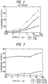
- Fig. 2 shows the relation between the fluorescence intensity and the amount of template DNA in Example 1.
- Fig. 3 shows the relation between the fluorescence intensity and the amount of template DNA in Comparative Example 2.
- Fig. 4 illustrates an arrangement of samples prepared in Example 2 on a microplate.
- Fig. 5 shows the formation of a PCR amplification product in the respective samples on the microplate in Example 2.
- the shadowing shows the wells of which fluorescence intensity was twice or more of the blank.
- Fig. 6 shows correlation between the cell numbers obtained in Example 2 and those obtained in Comparative Example 3.
- Fig. 7 shows the position of the wells of which fluorescence intensity were twice or more that of the blank value in Example 3.
- Fig. 8 shows the results of Example 4 with the sample from the tissue A.
- Figs. 9A to 9C show the construction and the method of use of a nucleic acid-detecting kit of the present invention.
- the dye compound employed in the present invention does not fluoresces in the free state but fluoresces when reacted with double-stranded nucleic acid.
- Reaction of the dye compound with double-stranded nucleic acid means specific insertion of the dye compound into grooves of a stable double helix structure of double-stranded nucleic acid to bind thereto. In that state, the dye compound fluoresces on irradiation of excitation light.
- the dye compound does not fluoresces in the free state, i.e., in the separated state, on irradiation of excitation light.
- the dye compound of the present invention is the one which enters a main groove or a sub groove of a double helix structure and links thereto, producing fluorescence in that state.
- the use of a dye compound having such characteristics enables precise and selective detection of the amplification product by simply adding directly the dye compound to a PCR reaction mixture without separating the amplification product from the primers and template nucleic acid.
- the fluorescence of the dye compound in the free state may be negligibly weak.
- the dye compound is exemplified by DAPI (4',6-diamino-2-phenylindole dihydrochloride), Hoechst 33258 (trade name), Hoechst 33342 (trade name), pyrylium compound salts and other salts analogous thereto represented by the general formula [I] below: where represents a heterocycle, and X is O, S, Se, or Te, the heterocycle including a five-membered and six-membered ring such as a pyrylium ring and pyrylium-analogous ring; R1 and R2 are independently a hydrogen atom, a halogen atom, a sulfonate group, an amino group, a styryl group, a nitro group, a hydroxyl group, a carboxyl group, a cyano group, a substituted or unsubstituted lower alkyl group, a substituted or unsubstituted aryl group, a
- the bivalent group derived from a substituted or unsubstituted aryl group includes a phenylene group and the like, and may have the bonds in any of ortho, meta, and para positions.
- the lower alkylene group includes linear or branched alkylene group having 1 to 4 carbons, the substituent including groups of -L-A.
- the cyclic structure having an oxo group includes a heterocycle, aromatic ring, and aliphatic ring having at least an oxo group.
- the substituent of the phenylene includes the groups mentioned above.
- the heterocycle for R5 includes the ones derived from the groups shown below: where M and N are independently an oxygen atom, a sulfur atom, or a nitrogen atom, and Y ⁇ is an anion.
- the substituent therefor includes a substituted or unsubstituted aryl group.
- the substituted or unsubstituted cycloalkyl group may be saturated or unsaturated and includes the ones derived from the groups below capable of constituting a resonance system:
- the substituted or unsubstituted aromatic ring includes an azulene ring.
- the substituent linked to the above groups includes lower alkyl groups, and substituted or unsubstituted aryl groups.
- the hydrogen atom bonded to the carbon atom which is not linked to R1, R2, or R3 may be substituted by a halogen atom, a sulfonate group, an amino group, a styryl group, a nitro group, a hydroxyl group, a carboxyl group, a cyano group, a substituted or unsubstituted lower alkyl group, a substituted or unsubstituted aryl group, or a substituted or unsubstituted lower aralkyl group.
- Y ⁇ is an anion, including BF4 ⁇ , perchlorate ion, HO3SCH2COO ⁇ , halide ion such as chloride ion, bromide ion, iodide ion, and fluoride ion, a compound functioning as an anion such as aliphatic sulfonates and aromatic sulfonates, and complex ions of transition metals such as Zn, Ni, Cu, Pt, Co, and Pd.
- halide ion such as chloride ion, bromide ion, iodide ion, and fluoride ion
- a compound functioning as an anion such as aliphatic sulfonates and aromatic sulfonates
- complex ions of transition metals such as Zn, Ni, Cu, Pt, Co, and Pd.
- the halogen includes Cl, Br, and I.
- the lower alkyl group may be linear or branched, and is preferably of 1 to 4 carbons.
- the aryl group includes a phenyl group or the like.
- the substituent of the aryl or phenylene group includes an amino group substituted with a lower alkyl group (lower-alkylamino group).
- Such a lower-alkyl amino group include preferably dimethylamino, diethylamino, or the like at a para position.
- the lower aralkyl group includes lower alkyl groups substituted by the aforementioned substituted or unsubstituted aryl.
- the X-containing heterocycle is preferably substituted by two or more substituted or unsubstituted aryl groups.
- the examples of such compounds having six-membered heterocycle are:
- the dye compound employed in the present invention reacts selectively with the stable double-stranded helix DNA to produce fluorescence. Therefore, even if the dye compound is incorporated in a higher-order structure formed by the aforementioned reaction from primers, the compound does not fluoresce because the double-stranded portion of the higher-order structure is not a stable double-stranded structure.
- the compound of General Formula [I] is introduced specifically into the double-stranded structure in a ratio of one molecule to 20 to 30 base pairs, the compound is advantageously not liable to be inserted into the higher-order structure derived from primers as mentioned above. Further, when the compound of General Formula [I] is incorporated into a double-stranded nucleic acid segment having a sequence to be amplified, the incorporation density is much lower than that of ethidium bromide or the like, so that the elevation of melting temperature (Tm) of the double-stranded nucleic acid hardly occurs not affecting the PCR. Because of the low incorporation density, when the PCR product is analyzed by gel electrophoresis, the mobility of the double-stranded nucleic acid is hardly affected by the incorporated dye compound, thus enabling precise size measurement.
- Tm melting temperature
- a conventionally used fluorescent intercalator such as ethidium bromide, acridine orange, and YOYO-1 is inserted into the double-stranded nucleic acid at a ratio of one molecule to several base pairs (about 2 to about 5 base pairs).
- These dyes are incorporated into the double-strand portion of the aforementioned higher-order structure formed from the primers, which causes increase of undesired fluorescence. Also these dyes greatly affect Tm of the double-stranded nucleic acid and the mobility thereof in gel electrophoresis because of the high incorporation rate into the target double-stranded nucleic acid.
- the amplification product is desired to form a stable double helix structure. Therefore the present invention is useful when the amplification products have a length of 100 base pairs or more, for example, preferably 300 base pairs.
- the maximum length of the amplification product is not particularly limited.
- the velocity of incorporation of the nucleotide in PCR is said to 35 to 100 nucleotides per second, for example, at 72°C. The rate depends on the reaction conditions such as pH and salt concentration of the reaction medium, and the base sequence of the target nucleic acid. Accordingly, when the reaction time of each cycle of PCR is one minutes, the length is preferably not more than 2000 base pairs.
- the length is not more than 1000 base pairs ("PCR Protocols", edited by Michael A. Innis, David H. Gelfand, John J. Sninsky, and Thomas J. White, 1990, (Academic Press Inc., San Diego, Calfornia 92101)).
- a primer set which is capable of defining the intended specific region of the target nucleic acid is used according to the purpose.
- the primer is required to have a sufficient length for recognizing the specific sequence at the end of the specific region to be amplified of the template nucleic acid, but should not be unnecessarily long, since excessively long primers tend to form locally a double-stranded structure between the primers. Therefore, the length of the primer is less than about 30 base pairs, preferably less than about 28 base pairs.
- the minimum of the primer length is not specially limited, but is required to have a sufficient length for recognizing the terminal portion of the specific region to be amplified: e.g., not less than 14 base pairs, preferably not less than 18 base pairs.
- nucleic acid of the present invention In the quantitative determination of nucleic acid of the present invention, firstly a sequence region is specified which is characteristic of the target nucleic acid to be determined. Secondly, PCR is conducted using a primer set necessary for amplification of the specified sequence region and the target nucleic acid as the template. Subsequently, the resulting amplified product is reacted with a dye compound having the aforementioned properties, and the intensity of the fluorescence is measured with irradiation of excitation light. The intensity of the fluorescence is proportional to the initial concentration of the template (the concentration before the PCR) provided that the concentrations of the reaction components are selected suitably. Therefore, the target nucleic acid in an unknown sample can be determined by conducting PCR and then measuring the fluorescence intensity by reference to a calibration curve prepared preliminarily for the fluorescence intensity as a function of the template concentration.
- the quantitative determination can be conducted simply by use of a calibration curve as mentioned above with a reaction system for which the optimum condition has been established.
- the reaction conditions however, especially the amount of the target nucleic acid, the amount of the primers, the reaction temperature, etc. depend on the respective reaction systems. Therefore, when the optimum reaction conditions for calibration curve preparation are unknown, complicated operations are necessary for establishing the conditions.
- the approximate quantity of the target nucleic acid can be measured by simply preparing serial dilutions of the nucleic acid of an unknown sample, conducting PCR with the diluted samples, and determining the target nucleic acid segment by use of an MPN-PCR method which gives probability of detection of the target nucleic acid segments.
- the conditions for preparing the calibration curve can be obtained easily.
- the number of the template DNA in the sample is 1.1 ⁇ 105, and at 10 ⁇ 5 dilution, three samples out of the five samples are positive. In this case, at the concentrations of 10 ⁇ 4 dilution or higher, all of the five tests of each concentration will be positive.
- the concentration range including the critical dilution of the template DNA is determined according to the presence or absence of the amplification product, and then detailed MPN investigation is conducted on the concentration region.
- the detection is conducted in an ON/OFF manner, the presence or absence of the band, and even when the DNA is quantitatively detected by fluorescence staining, the quantitation is not sufficient in spite of the complicated operations.
- the method of the present invention employing an MPN-PCR method as described later makes it practicable to check the formation of amplification product for all over the dilutions, by use of a tool such as a microplate which can provide many reaction zones (chambers).
- the number of the template DNA and the quantity thereof can be determined quantitatively from this calibration line since the number of the template DNA is known in this MPN-PCR.
- the method of the present invention enables direct and selective measurement of the fluorescence which is produced only when the amplified product reacted with a dye compound added to the PCR mixture. Therefore, the separation of the amplified product from the primer and the template is unnecessary and even when the higher-order structure are formed between the primers, the influence of it on the measurement result is negligible. Accordingly, the fluorescence intensity obtained by directly adding the dye compound to the reaction mixture can be used for calculation of the precise quantitative determination. When ethidium bromide or a similar substance which fluoresces even in the free state is used for the quantitative determination, the blank value has to be subtracted from the measured value.
- the determination conditions have to be decided in consideration of the relation of the blank value to the measured value for each of measurement conditions, which is quite troublesome.
- such blank value consideration or a set up of determination conditions is not necessary, differing from with ethidium bromide, since the dye compound of the present invention produces no or negligible fluorescence in the free state.
- the fluorescence intensity need not be monitored during the PCR process as described in Japanese Patent Application Laid-Open No. 5-237000.
- a microplate may conveniently be used for the detection by transferring the PCR reaction mixture from the reaction vessel to the microplate well and measuring the fluorescence by use of a microplate fluorescence detector (reader), conveniently requiring a small amount of a reagent and a short measurement time.
- the PCR process itself may be conducted on the microplate, which more simplifies the determination operation.
- a nucleic acid determining kit can be provided by employing a reactor like a microplate having many wells as reaction chambers and placing a necessary amount of a dye compound of the present invention by applying a solution thereof to each of the reaction chambers, followed by evaporation or drying or the like. Such a constitution is practicable owing to the stability of the dye compound at room temperature.
- kit is also useful in which reagent chambers are provided separately from the PCR reaction chambers, and the dye compound of the present invention placed in the reagent chambers is added to the reaction chambers in the PCR step.
- the dye compound may be placed in the reagent chamber as a solution. If the dye compound is water-soluble, it may be solubilized in a suitable buffer solution, and if the compound is soluble in an organic solvent, the compound may be placed as a solvent solution.
- the reagent chamber may contain a buffer solution, nucleotides, the enzyme, etc. necessary for the PCR together with the dye compound.
- the organic solvent for the water-insoluble dye compound is preferably not more than 1 % based on the aqueous solution.
- Figs. 9A to 9C show an example of constitution of the kit having such a reactor.
- primer 3 is placed by liquid application or a like method, and further a container 2 is placed in it as the reagent chamber which is made of paraffin-coated paper or the like to be liquid-tight, and in which packed is a solution containing the enzyme, nucleotides, a buffer solution, etc in an amount necessary for the PCR (see Fig. 9A and Fig. 9B).
- PCR is conducted by injecting a solution of template nucleic acid into the reaction chamber 5 by a suitable means such as a Pipetman, concomitantly breaking through the container 2 to feed the contents therein to the reaction chamber 5, and mixing the reaction components to start PCR (see Fig. 9C).
- the dye compound for detection of the amplified product may be added after the PCR, or may be placed preliminarily in the reaction chamber. Otherwise, the dye compound may be contained in the solution in the reagent chamber 2.
- Two or more reagent chambers may be provided for storing separately the reaction components.
- the dye compound may be added to the reaction chamber for PCR prior to or after the PCR.
- another reagent chamber may be provided in which a necessary amount of the dye compound is held, and the dye compound may be added to the reaction chamber after the PCR from the reagent chamber.
- water-insoluble ones remain undissolved, even if it is placed in the reaction chamber during the PCR, and the compound is allowed to react after the PCR by addition of a suitable solvent to the reaction chamber.
- the water-insolubility of the compound eliminates the undesirable influence of the compound on PCR, even though in general such effect is inherently small for the compounds of General Formula [I].
- the suitable solvent used therefor includes acetonitrile, ethanol, dimethylsulfoxide (DMSO), etc.
- DMSO dimethylsulfoxide
- the detection of the amplification product of PCR with the dye compound of the present invention is suitably conducted by MPN utilizing PCR (MPN-PCR method).
- MPN-PCR method enables measurement of the number of individuals of microorganisms, animal cells, human cells, vegetable cells, etc. (the number of cells, the number of bacteria, the number of mycelia, etc.), and the number of specific genes or the number of copies of the specific gene of the above individuals with greatly simplified operations.
- a soil sample is treated to extract the nucleic acid of microorganisms in soil.
- a soil sample (1 g)
- a phosphate buffer solution (1 ml)
- the mixture is agitated by a vortex mixer twice for 20 seconds each.
- 1/10 volume of 10% SDS was added, and the mixture is voltexed again.
- the resulting mixture is kept at 70°C for one hour for cell lysis.
- the soil is removed by centrifugation to collect the supernatant as the nucleic acid fraction.
- 1/5 volume of 7.5M sodium acetate is added, and the mixture is left standing at 4°C for 5 minutes.
- the supernatant (1 ml) is recovered, and thereto, isopropanol (4/5 volume) is added.
- the nucleic acid fraction is recovered by centrifugation, and dissolved in a TE buffer solution (0.1 ml). DNA is recovered therefrom after RNase treatment.
- An aliquot of the recovered DNA is diluted 100-fold.
- a series of dilutions was prepared to make 10 ⁇ 1 to 10 ⁇ 8 dilutions. These dilutions are placed in the sample block A on the microplate. More specifically, the 10 ⁇ 1-dilution of the starting DNA solution (100-fold diluted soil extract), is placed in A1 to A5 wells, the 10 ⁇ 2-dilution solution at the wells B1 to B5, and so forth, and 10 ⁇ 8-dilution solution at the wells H1 to H5.
- reaction solution containing reagents a nucleotide, a primer salt, a Taq polymerase, etc.
- reagents a nucleotide, a primer salt, a Taq polymerase, etc.
- the fluorescence intensity of respective wells are measured by use of a dye compound.
- the well which fluoresces at an intensity of twice or more that of the blank value is defined to be positive, and the dilution limit for the positive fluorescence is derived.
- the critical dilution for the positive fluorescence is 10 ⁇ 5.
- the numbers of the positive samples (5, 3, and 0) at these dilution degrees are respectively applied to P1, P2, and P3 shown in the MPN table shown later (cited partially, for serial decimal dilutions in quintuplicate, from J. Bacteriol. 25 101 (1933), page 400, "III.
- MPN (Most Probable Number) Table"
- This number of the template DNA itself is the number of the microorganism individuals (cell number) in 1 gram of the soil provided that the sequence of the template DNA is characteristic of the target microorganism in the soil and the sequence exists only one in number in one microbial cell.
- the highest dilution at which all the wells fluoresces reflects the number of the template nucleic acid.
- a calibration curve for the fluorescence intensity as the function of the number of the template DNA (or number of the individual microorganism) can be made.
- the numbers in parentheses are of high reliability (probability L is 0.05 or lower).
- the MPN table cited as Table 1 is a part of the MPN table of the aforementioned literature. If an experimental result falls outside Table 1, corresponding part of the original MPN table of the literature should be referred to.
- the literature includes MPN tables for various experimental conditions, which may be used according to the experiment conditions.
- the respective dilution solutions are subjected to the first PCR in a conventional manner, and the presence of the amplification product is detected by agarose gel electrophoresis to find that the dilutions are positive to the 10 ⁇ 7 dilution degree. Then 5 samples of the respective dilutions at 10 ⁇ 6, 10 ⁇ 7, and 10 ⁇ 8 dilution are again subjected to PCR. The positive numbers at each dilution are 5/5 for 10 ⁇ 6 dilution, 3/5 for 10 ⁇ 7 dilution, and 0/5 for 10 ⁇ 8 dilution. Therefore the value 0.79 is obtained from the MPN table in the same manner as above.
- the number of the template DNA in the soil is derived from this value multiplied by the reciprocal of the dilution degree of the dilution limit (0.79 ⁇ 107).
- This method requires two series of operation of PCR and electrophoresis, and is troublesome.
- the method of the present invention gives the results directly by conducting one PCR operation without the electrophoresis operation, thereby the measurement operation being simplified greatly.
- the aforementioned operations and conditions can be employed in the determination of the PCR amplification product, as well as the aforementioned kit.
- the MPN-PCR method use of a tool having a plurality of reaction chambers (wells) such as a microplate is preferred since simultaneous treatment of serially diluted samples is required for statistical treatment of the result. Even when PCR is conducted in different vessels, the microplate is preferably used for the detection of the amplification product with the dye compound.
- This method for measuring the number of the template DNA is applicable effectively to measure the cell number of the target microorganism in a liquid or in soil, the number of microbial cells in various samples, the number of cells in various samples, the number of cells in various tissues, the number of a specified gene and copy number thereof in various living body samples, and so forth.
- the concentration of template DNA is estimated by weight based on the light absorption intensity.
- the number of the copies is frequently of interest. In such a case, the method of the present invention is useful.
- the number of copies of a specified gene in a tissue can be estimated according to the present invention, irrespectively of the amount of the cells from which the nucleic acid is extracted, by measuring the number of a common gene in the same manner as described above, then measuring the number of a specified gene such as a oncogene, and deriving a ratio of the number of the specified gene and the number of the common gene which is common to normal cells and cancer cells.
- This method is also useful for judging the presence of cancer cells in a human tissue. For this purpose, for example, the number of a specified gene common to all the cells constituting the tissue is estimated by this method, and the number of a specific gene characteristic to cancer cells to be detected is estimated for the same tissue sample.
- ⁇ represents a p-phenylene group, or a phenyl group.
- Compound 7 was prepared by synthesizing Compound [i] according to the method described by W. Foerst et al. ("New Methods of Preparative Organic Chemistry", Acad. Press (1964)), reacting it with p-N,N-dimethylaminobenzaldehyde (formula shown below): and by reacting the resulting compound with a desired anion.
- Compound 17 was prepared by reacting Compound [i] with p-diethylaminostyrylbenzaldehyde (formula shown below): and reaction the resulting product with a desired anion.
- Compound [ii] was prepared by reacting Compound [i] with sodium hydrogen sulfide. From this Compound [ii], Compounds 8 and 18 were prepared in the same manner as Compounds 7 and 17.
- Compound [iii] was synthesized from acetophenone and acetaldehyde according to the method described by R. Wizinger (Helv. Chim. Acta, 39 217 (1956)) through the route shown below: Compound 5 was prepared by reacting Compound [iii] with p-dimethylaminobenzaldehyde, and further reacting the resulting compound with a desired anion.
- Compound 15 was prepared in the same manner by using p-diethylaminostyrylbenzaldehyde.
- Compound 9 was prepared in the same manner by using p-dimethylaminocinnamaldehyde.
- Compound 11 was prepared in the same manner by using the compound below: Compound [iv] was obtained by reacting Compound [iii] with sodium hydrogen sulfide: Compounds 6, 16, 10, and 12 were prepared respectively in the same manner as Compounds 5, 15, 9, and 11 except that Compound [iv] was used in place of Compound [iii].
- Compound 3 was synthesized in the same manner as Compound [iii] except that p-dimethylaminobenzaldehyde was used in place of acetaldehyde as a starting material.
- Compound 4 was prepared by reacting the above resulting compound with sodium hydrogen sulfide, and further with a desired anion.
- Compound [v] was prepared from p-methylbenzaldehyde and acetophenone in the same manner: Compound [vi] was prepared by reacting Compound [v] with sodium hydrogen sulfide: Compounds 13 and 14 were prepared respectively by reacting Compound [v] and Compound [vi] respectively with p-dimethylaminobenzaldehyde, and the resulting compound with a desired anion.
- Compounds 19, 20, and 21 were prepared respectively by reacting Compound [i] or [ii] with the cation portion of Compound 1 or 2, and the compound of the formula below: and reacting the product further with a desired anion.
- Compounds 22, 23, and 24 were prepared respectively by reacting Compound [i] or [ii] with the cation portion of Compound 1 or 2, and the compound of the formula below: and reacting the product further with a desired anion.
- Compounds 25 and 26 were prepared respectively by reacting Compound [i] or [ii] with the compound of the formula below: and reacting the product further with a desired anion.
- Compounds 27, 28, and 29 were prepared respectively by reacting Compound [i] or [ii] with the cation portion of Compound 1 or 2, and ethyl orthoformate [HC(OC2H5)3], and reacting the product further with a desired anion.
- Compounds 30, 31, and 32 were prepared respectively by reacting Compound [iii] or [iv] with a p-dimethylamino derivative of Compound [iii] or [iv] derived in the same manner as Compound [iii] or [iv], and ethyl orthoformate, and reacting the product further with a desired anion.
- Compound 40 was synthesized in the same manner as Compound 36 except for using the compound in place of as a starting material.
- Compound 41 was synthesized in the same manner as Compound 37 except for using the compound in place of as a starting material.
- Compound 42 was synthesized in the same manner as Compound 38 except for using the compound in place of as a starting material.
- Compound 43 was synthesized in the same manner as Compound 39 except for using the compound in place of as a starting material.
- Compound 48 was synthesized in the same manner as Compound 44 except for using the compound in place of as a starting material.
- Compound 49 was synthesized in the same manner as Compound 45 except for using the compound in place of as a starting material.
- Compound 50 was synthesized in the same manner as Compound 46 except for using the compound in place of as a starting material.
- Compound 51 was synthesized in the same manner as Compound 47 except for using the compound in place of as a starting material.
- Example I Compound 1 obtained in Reference Example 1 was dissolved in acetonitrile to make a stock solution, to which a phosphate buffer was added to the final concentration of 10 mM, followed by dilution with water containing 10% acetonitrile to the final concentration of Compound 1 of 3 ⁇ 10 ⁇ 5 M. This solution is referred to as "Sample I”.
- the absorption spectrum of Sample I was measured with a spectrophotometer in a conventional manner.
- Example II Salmon sperm DNA (made by Sigma Co.) was dissolved in a TE buffer solution (10 mM Tris - 1mM EDTA), and was purified by phenol extraction. The purified DNA was further digested with restriction enzyme EcoRI for easy handling. An aliquot of this DNA solution was mixed with the stock solution of Compound I to give a DNA concentration of 50 ⁇ g/ml and a Compound 1 concentration of 3 ⁇ 10 ⁇ 5 M. This solution containing 10% acetonitrile is referred to as "Sample II". The absorption spectrum of Sample II was measured with a spectrophotometer in a conventional manner. In Sample II, the absorption peak of Compound 1 shifted by 20 to 30 nm to longer wavelength owing to its interaction with DNA, which is typical for an intercalator.
- Solutions were prepared which contain Compound 1 at a concentration of 5 ⁇ 10 ⁇ 6 M and DNA at various concentrations by using the above-prepared DNA solution and the above-prepared 10mM phosphate buffer solution containing compound 1 and 10% acetonitrile.
- the fluorescence intensities of the solutions were measured in a conventional manner to determine the relation to the DNA concentration.
- the fluorescence intensity increased in proportion to the DNA concentration, the maximum intensity being about 400 times the value in the absence of DNA.
- the excitation light was emitted from a xenon lamp through a low-cut filter of 480 nm to eliminate ultraviolet light.
- Table 2 were subjected to fluorescence intensity measurement in the same manner as above. Typical examples are shown in Table 3.
- Table 3 Compound No.
- ⁇ max Maximum absorption wavelength Fluorescence intensity Absence of DNA Presence of DNA ⁇ em Increase 1 540 nm 560 nm 650 nm 100-fold 2 580 nm 620 nm 700 nm 60-fold 3 535 nm 570 nm 640 nm 13.6-fold 4 575 nm 610 nm 705 nm 10-fold 6 660 nm 690 nm 800 nm 7-fold 8 650 nm 670 nm weak - 9 660 nm 720 nm 750 nm 16-fold 11 625 nm 660 nm 735 nm 10-fold 15 670 nm 680 nm 820 nm 5-fold 16 690 nm 720 nm 825 nm 5-fold 17 690 nm 720 nm weak -
- 16S rRNA gene 16S ribosomal RNA gene of Pseudomonas aeruginosa as the target, and the amplification product was detected by use of 2,4-bis(4-N,N-dimethylaminophenyl)-6-methylpyrylium salt.
- the entire DNA of P . aeruginosa was prepared as follows. After overnight incubation in a 2 ⁇ YT culture medium, the cells of 2 ml culture were collected by centrifugation. The collected cells were suspended in 0.5 ml of 0.1M phosphate buffer solution (pH: 8.0), to which 0.05 ml of 10% SDS solution was added. The suspension was mixed well, and was kept standing at 70°C for one hour. Then the suspension was vortexed to cause complete cell lysis. To this lysate, an equal amount of phenol-chloroform was added, and mixed. Then the mixture was centrifuged, and the upper layer was collected. Thereto ethanol of twice volume was added to recover DNA as precipitate. The DNA was dissolved in 100 ⁇ l of TE buffer (pH: 8.0). This DNA was used as the template DNA.
- 0.1M phosphate buffer solution pH: 8.0
- Two primers were used for PCR.
- primers were synthesized by means of DNA Synthesizer 381A (made by ABI Co.). The reagents and the techniques for the synthesis are based on the protocol of ABI Co.
- composition of reaction solution (total volume: 50 ⁇ l): 10 ⁇ buffer*: 5 ⁇ l dNTPs: 5 ⁇ l Primers 1 and 2: 10 pmol respectively aaq DNA polymerase: 0.5 unit Template DNA: 500 pg, 100 pg, 10 pg, 1 pg, 100 fg, or 10 fg *(100 mM tris-HCl(pH 9.0), 500 mM KCl, 1% Triton X-100, 25 mM MgCL2 )
- the components were mixed and thereto sterilized water was added to the total volume of 50 ⁇ l.
- the mixture was allowed to react in a Thin-Walled Gene Amp tube (entire volume: 0.5 ml, made by Takara Shuzo Co., Ltd.).
- a sample was prepared in the same manner as above except that the template DNA was not added.
- the required amount of the 10 ⁇ buffer 100 mM tris-HCl(pH 9.0), 500 mM KCl, 1% Triton X-100, 25 mM MgCL2 ) and the dNTPs were supplied with the polymerase.
- the PCR apparatus employed was Gene PCR System 9600 made by Perkin-Elmer Inc.
- the amplification product was detected as follows: After the reaction, respective samples are diluted 2-fold, 10-fold, and 20-fold with TE buffer (10 mM Tris-HCl (pH: 8.0) - 1 mM EDTA), to the final volume of 50 ⁇ l on a 96-well microtiter plate (Falcon Assay Plate 3911 (U-bottomed well) made by Becton-Dickinson Co.) (See Fig. 1).
- TE buffer 10 mM Tris-HCl (pH: 8.0) - 1 mM EDTA
- Fig. 2 shows the fluorescence intensity at 645 nm of the respective dilutions as a function of the amount of the template DNA in the reaction.
- Example 2 The same operation was conducted as in Example 1 except that EB was used in place of 2,4-bis(4-N,N-dimethylaminophenyl)-6-methylpyrylium salt with the following operation conditions.
- EB was used in place of 2,4-bis(4-N,N-dimethylaminophenyl)-6-methylpyrylium salt with the following operation conditions.
- the fluorescence intensities were measured by means of Millipore Fluorescence Apparatus with a 485nm-light transmitting excitation filter and a 620nm-light transmitting emission filter. Consequently, red fluorescence was observed at all the wells including control wells.
- Example 2 The same operation was conducted as in Example 1 except that YOYO-1 (made by Molecular Probe Co.) was used in place of 2,4-bis(4-N,N-dimethylaminophenyl)-6-methylpyrylium salt with the following operation conditions.
- the YOYO-1 solution was prepared by diluting 1 mM stock solution 120-fold, and 1 ⁇ l of the diluted YOYO-1 solution was added to each of the wells, and mixed well by pipetting. The mixture was left standing at room temperature for 5 minutes.
- the fluorescence intensities were measured by means of Millipore Fluorescence Apparatus with a 485nm-light transmitting excitation filter and a 530 nm-light transmitting emission filter.
- Fig. 3 shows the results.
- the fluorescence intensity increased in proportion to the amount of the template DNA, but the increase rate is low.
- the blank wells also produced fluorescence at approximately the same intensity level as those containing the template DNA.
- the fluorescence of the blank is probably due to the higher-order structured product formed from the primers. Accordingly, in the quantitative determination with this dye, quantitation is not satisfactory because of the small difference in the fluorescence intensity between the reaction mixture and the blank mixture and the low increase rate of the fluorescence intensity in proportion to the amount of the template DNA.
- Example 1 From the results of Example 1 and Comparative Examples 1 and 2, the quantitative determination can be conducted precisely by utilizing the fluorescence 2,4-bis(4-N,N-dimethylaminophenyl-6-methyl)pyrylium iodide not influenced by the higher-order structured product formed from the primers.
- This result is well consistent with the result of EB staining of agarose gel electrophoresis of the reaction solutions in Example 1.
- the fluorescence saturated at the template DNA levels of 500 pg and 100 pg apparently showing the presence of the same quantity of the amplification products of the template DNA levels of 100 pg or higher.
- Example 2 Measurement of bacterial cell number by PCR employing a microplate ]
- E. coli JM109 strain An overnight culture (2 ⁇ YT medium) of E. coli JM109 strain was added to soil. From the soil, DNA was extracted in a conventional manner, and subjected to PCR targeting 16S rRNA gene of E. coli as the template, to determine the number of bacterial cells by PCR-MPN method.
- E. coli was added to the soil to give cell numbers per gram of soil of about 107, about 106, about 105, and about 104 (corresponding to Samples A, B, C, and D in Fig. 4, respectively), and the soil samples were stirred.
- One gram of the soil sample containing E. coli was respectively suspended in 0.5 ml of 1M phosphate buffer solution (pH 8.0).
- 0.05 ml of 10% SDS solution was added.
- the mixture after being sufficiently stirred, was left standing at 70°C for one hour.
- the suspension was mixed with a vortex mixer for complete cell lysis.
- To the lysate an equal amount of a phenol-chloroform mixture was mixed. After centrifugation, the upper layer was collected. Thereto, twice the amount of ethanol was added to recover DNA as the precipitate.
- This precipitate was dissolved in 100 ⁇ l of a TE buffer solution (pH 8.0) to form a template DNA solution for PCR.
- PCR was conducted with the extracted DNA as the template and the primers under the same conditions as in Example 1.
- the PCR was conducted in the wells (U-bottomed) of a 96-well microtiter plate (Falcon assay plate 3911 made by Becton-Dickinson Inc.).
- Each well of the microplate contains preliminarily applied and dried 1 ⁇ l of a solution of 2,4-bis(N,N-dimethylaminophenyl)-6-methylpyrylium iodide in acetonitrile of the same concentration as in Example 1.
- the PCR reaction apparatus was Model PTC-100-96 made by MJ Research Inc.
- Fig. 5 shows the results.
- the wells producing fluorescence of twice or more that of the blank wells were regarded to contain a PCR amplification product (shadowed wells in Fig. 5). From the results, the numbers of E. coli in the respective soil samples were estimated to be 4.9 ⁇ 106, 7.9 ⁇ 105, 2.2 ⁇ 105, and 7.9 ⁇ 103 by reference to the table for MPN.
- the numbers of E. coli cells in the same soil samples prepared in Example 2 were measured by plate count method in which a predetermined amount of the sample was inoculated in an E coli culture plate and the number of colonies were measured as the viable cell number. The results agreed well with the results obtained in Example 2 as shown in Fig. 6.
- the PCR method of the present invention enables measurement of the number of the cells with simple operations giving well agreed results with that obtained by conventional plate count method.
- DNA of E. coli was extracted in the same manner as in Example 2, and the extract was diluted 100-fold to obtain a template DNA solution.
- This template DNA solution was serially diluted in the same manner as in Example 2. The dilutions were added by 1 ⁇ l each to the wells of the microplate as shown in Fig. 5. To each of the wells, the PCR reaction solution was added which contains one Ampli Wax PCR Gem 100 (made by Perkin-Elmer) and the components below: 10 ⁇ buffer: 5 ⁇ l dNTPs: 5 ⁇ l Taq DNA polymerase: 0.5 unit Further, to each of the wells, sterilized water was added to the total volume of 50 ⁇ l. Then PCR was carried out with a PCR apparatus, Model PTC-100-96 (made by MJ Research Inc.), under the same conditions as in Example 1.
- Fig. 7 shows the results.
- the wells producing fluorescence of twice or more that of the blank wells were regarded to contain the PCR amplification product (shadowed wells in Fig. 5). From the results, the number of the template DNA molecules was estimated to be 3.5 ⁇ 107 by reference to the table for MPN. In consideration of the initial dilution by the factor of 100, the number of the template DNA molecules was estimated to be 3.5 ⁇ 109.
- Example 3 The number of the bacterial cells in the 2 ml of E. coli culture used in Example 3 was determined by plate count method. Consequently, the number was found to be 4 ⁇ 109, which agrees approximately with the results obtained in Example 3.
- the desirable dilution degree of a DNA template and the desirable concentration range thereof for MPN detection can be decided, and the sample concentration can be set according to the results of Examples 2 and 3. Therefore calibration curves can be prepared for the number of the cells, the copy number, and the amount of template DNA.
- tissue samples of 5 mm cube were taken out respectively from two cancer-suspected sites of the large intestine of a patient by biopsy.
- the samples are hereinafter referred to as "Tissue A” and "Tissue B”.
- tissue samples mRNA was extracted by a conventional manner as follows (by reference to "Shin Seikagaku Jikken Koza (New Library of Experiments in Biochemistry)" vol.
- the mixture was shaken by means of a Vortex mixer for 10 seconds, cooled with ice for 15 minutes, and subjected to centrifugation at 4°C at 10000 ⁇ g for 20 minutes. After the centrifugation, to the aqueous layer, an equal amount of isopropyl alcohol was added, and the mixture was left standing at -20°C overnight. The mixture was then centrifuged as above and 0.6 ml of D Solution was added to dissolve the precipitate. Thereto an equal amount of isopropyl alcohol was added. The mixture was cooled at -20°C for one hour, and centrifuged at 4°C at 10000 ⁇ g for 20 minutes.
- the obtained precipitate was suspended in 75% ethanol, and the suspension was centrifuged again at 10000 x g, at 4°C for 20 minutes.
- the precipitate was dried as a crude RNA fraction, which was heated at 65°C for 5 minutes, then cooled rapidly to room temperature, and was washed with an equal amount of a 2x TNEL buffer solution [20 mM Tris-hydrochloric acid buffer solution (pH 7.6), 0.5M sodium chloride, 1mM EDTA, and 0.1% sodium N-lauroylsarcosinate].
- RNA fraction was applied to an oligo(dT)-cellulose column made by Pharmacia) equilibrated with a TNEL buffer solution, and mRNA was eluted with an extraction solution (TNEL buffer solution without 0.5M sodium chloride).
- Double-stranded cDNA was prepared from the above mRNA by means of TimeSaverTM cDNA Synthesis Kit.
- the primers used for detection of large intestine cancer have following nucleotide sequences.
- Primer 1 and Primer 2 were applied to an amount of 10 pmoles respectively.
- container 2 prepared from paraffin-coated paper, the components below were packed.
- dNTPs 5 ⁇ l Taq polymerase: 0.5 unit 150 ⁇ g/ml 2,4-bis(4-N,N-dimethylaminophenyl)-6-methylpyrylium iodide solution in acetonitrile: 1 ⁇ l 10 ⁇ buffer: 5 ⁇ l Distilled water: 39 ⁇ l (freeze stored)
- the container 2 was set in the vessel 1 as shown in Fig. 9B to make up a kit.
- the cDNA samples obtained above from each of the aforementioned Tissue A and Tissue B were serially diluted, 1-fold (no dilution), 2-fold, 10-fold, 50-fold, 100-fold, 1000-fold, and 10000-fold.
- Each dilution (including non-diluted sample) was injected with a Pipetman to the bottom of vessel 1 of the kit, breaking through the paraffin-coated paper container 2.
- the vessels were centrifuged to transfer the contents of the container 2 completely to the reaction zone 5 containing the primers.
- the broken empty containers 2 were taken out from the vessel 1. Thereto, one Ampli Wax, PCR Gen 100 (made by Perkin-Elmer Co.) was added.
- PCR was conducted in the same manner as in Example 1.
- the samples in the vessels were subjected to fluorescence measurement without further dilution.
- the results regarding Tissue A are shown in Fig. 8.
- the target gene amplification was observed with Tissue A, and the amplification product was formed quantitatively corresponding to the dilution degree of the template DNA. On the contrary, no fluorescence was observed with Tissue B.
- DNA was recovered from the samples by precipitation with ethanol. The recovered matter was examined by agarose gel electrophoresis. The DNA from Tissue A gave a band at 317 bp at the 1-fold (not diluted) to 1000-fold dilutions, whereas no band was observed with Tissue A at 10000-fold dilution, the blank samples containing primers only, and the samples derived from Tissue B.
- Tissue A and Tissue B were examined by a conventional culture method.
- Tissue A exhibited multiplication, and was diagnosed as a malignant cancer, whereas Tissue B did not exhibit multiplication, and was diagnosed as benign polyp.
- the dye compounds which reacts selectively with double-stranded nucleic acid is used for detection of the PCR amplification products, enabling simplification of detection, quantitative determination, and measurement of the number of the cells with precision.
- a PCR amplification product is detected, in quantitative determination of nucleic acid and measurement of the number of bacterial cells or specific genes, by addition of a dye compound which does not fluoresce in the free state but fluoresces in the bonded state to a double-stranded nucleic acid.
Landscapes
- Chemical & Material Sciences (AREA)
- Health & Medical Sciences (AREA)
- Life Sciences & Earth Sciences (AREA)
- Engineering & Computer Science (AREA)
- Organic Chemistry (AREA)
- Immunology (AREA)
- Hematology (AREA)
- Molecular Biology (AREA)
- Biomedical Technology (AREA)
- General Health & Medical Sciences (AREA)
- Analytical Chemistry (AREA)
- Microbiology (AREA)
- Urology & Nephrology (AREA)
- Physics & Mathematics (AREA)
- Wood Science & Technology (AREA)
- Zoology (AREA)
- Biochemistry (AREA)
- Proteomics, Peptides & Aminoacids (AREA)
- Biotechnology (AREA)
- General Engineering & Computer Science (AREA)
- Bioinformatics & Cheminformatics (AREA)
- Chemical Kinetics & Catalysis (AREA)
- Genetics & Genomics (AREA)
- Cell Biology (AREA)
- Biophysics (AREA)
- Food Science & Technology (AREA)
- Medicinal Chemistry (AREA)
- General Physics & Mathematics (AREA)
- Pathology (AREA)
- Measuring Or Testing Involving Enzymes Or Micro-Organisms (AREA)
Applications Claiming Priority (6)
| Application Number | Priority Date | Filing Date | Title |
|---|---|---|---|
| JP22720493 | 1993-09-13 | ||
| JP22720493 | 1993-09-13 | ||
| JP227204/93 | 1993-09-13 | ||
| JP2289594 | 1994-02-21 | ||
| JP22895/94 | 1994-02-21 | ||
| JP02289594A JP3683913B2 (ja) | 1993-03-10 | 1994-02-21 | Pcrによる核酸定量方法、pcrによる微生物または細胞の数、特定遺伝子数及び特定遺伝子コピー数の計測法、ならびにこれらの方法に用いる測定用キット |
Publications (2)
| Publication Number | Publication Date |
|---|---|
| EP0643140A1 true EP0643140A1 (fr) | 1995-03-15 |
| EP0643140B1 EP0643140B1 (fr) | 2001-10-31 |
Family
ID=26360189
Family Applications (1)
| Application Number | Title | Priority Date | Filing Date |
|---|---|---|---|
| EP94109568A Expired - Lifetime EP0643140B1 (fr) | 1993-09-13 | 1994-06-21 | Détermination d'acides nucleiques par PCR, mesure du nombre des cellules microbiennes, gènes ou copies de gènes par PCR, et trousse utilisée à cet effet |
Country Status (5)
| Country | Link |
|---|---|
| US (1) | US5670315A (fr) |
| EP (1) | EP0643140B1 (fr) |
| AT (1) | ATE207966T1 (fr) |
| CA (1) | CA2126391C (fr) |
| DE (1) | DE69428855T2 (fr) |
Cited By (11)
| Publication number | Priority date | Publication date | Assignee | Title |
|---|---|---|---|---|
| EP0684239A1 (fr) * | 1994-05-26 | 1995-11-29 | Canon Kabushiki Kaisha | Méthode de détection de substance, cible dans un échantillon, utilisant un composé de pyrylium |
| WO1997046714A1 (fr) | 1996-06-04 | 1997-12-11 | University Of Utah Research Foundation | Controle de l'hybridation pendant la pcr |
| EP0837141A3 (fr) * | 1996-10-03 | 1998-05-06 | Canon Kabushiki Kaisha | Procédé pour détecter un acide nucléique ciblé, procédé pour sa détermination quantitative et composés pyryliums pour l'analyse en chimioluminescence |
| WO1998036096A1 (fr) * | 1997-02-14 | 1998-08-20 | E.I. Du Pont De Nemours And Company | Detection d'adn en double brin dans une solution homogene |
| US6124342A (en) * | 1993-12-27 | 2000-09-26 | Canon Kk | Pharmaceutical compositions containing pyrylium compounds, pyrylium salts and process for manufacturing a medicament containing the aforesaid compounds |
| US6440706B1 (en) | 1999-08-02 | 2002-08-27 | Johns Hopkins University | Digital amplification |
| US7081226B1 (en) | 1996-06-04 | 2006-07-25 | University Of Utah Research Foundation | System and method for fluorescence monitoring |
| US7273749B1 (en) | 1990-06-04 | 2007-09-25 | University Of Utah Research Foundation | Container for carrying out and monitoring biological processes |
| WO2009011674A1 (fr) * | 2007-07-13 | 2009-01-22 | Western Michigan University Research Foundation | Dérivés de trans-1,2-diphényléthylène et nanodétecteurs obtenus à partir de ceux-ci |
| US7972778B2 (en) | 1997-04-17 | 2011-07-05 | Applied Biosystems, Llc | Method for detecting the presence of a single target nucleic acid in a sample |
| CN110499378A (zh) * | 2019-09-12 | 2019-11-26 | 卓源健康科技有限公司 | 一种从临床血液中检测大肠埃希菌病原菌的方法 |
Families Citing this family (33)
| Publication number | Priority date | Publication date | Assignee | Title |
|---|---|---|---|---|
| US5955268A (en) * | 1996-04-26 | 1999-09-21 | Abbott Laboratories | Method and reagent for detecting multiple nucleic acid sequences in a test sample |
| JPH11341989A (ja) * | 1998-03-31 | 1999-12-14 | Sanyo Electric Co Ltd | Dna断片増幅方法、dna断片増幅装置、微生物群測定方法、微生物群分析方法および汚染物質測定方法 |
| US6960432B2 (en) * | 1999-07-26 | 2005-11-01 | Canon Kabushiki Kaisha | Detection/quantification of targeted nucleotide chains, and detection/quantification of multi-stranded nucleotide chains by fluorescence |
| JP3596868B2 (ja) * | 2000-11-24 | 2004-12-02 | キヤノン株式会社 | 末端標識プローブアレイのプローブの量の評価方法、及び、該標識プローブアレイを用いた標的物質量の評価方法 |
| US20040028665A1 (en) * | 2002-01-08 | 2004-02-12 | Garner Bryan E. | Compositions and methods for inhibiting pathogenic growth |
| US7291326B2 (en) * | 2003-01-06 | 2007-11-06 | Nutrition Physiology Corporation | Compositions and methods for reducing the pathogen content of meat and meat products |
| JP2004069677A (ja) | 2002-06-13 | 2004-03-04 | Canon Inc | 免疫学的測定方法、免疫学的測定用試薬及びその製造方法 |
| JP4458327B2 (ja) * | 2003-08-28 | 2010-04-28 | キヤノン株式会社 | 標的物質の定量方法および該方法に用いるプローブ担体 |
| US20050048515A1 (en) * | 2003-08-29 | 2005-03-03 | Garner Bryan E. | Methods for detecting and quantifying specific probiotic microorganisms in animal feed |
| AU2004269388B2 (en) * | 2003-08-29 | 2009-09-17 | Nutrition Physiology Company, Llc. | Methods for detecting and quantifying specific microorganisms |
| WO2005059086A1 (fr) * | 2003-12-16 | 2005-06-30 | Canon Kabushiki Kaisha | Feuille de transfert de substance biologiquement active, kit pour culture cellulaire forme d'une plaque de culture cellulaire et d'une feuille de transfert de substance biologiquement active, procede de production y relatif, et procede de criblage d'etats de culture cellulaire utilisant cette feuille |
| JP4632400B2 (ja) * | 2003-12-16 | 2011-02-16 | キヤノン株式会社 | 細胞培養用基板、その製造方法、それを用いた細胞スクリーニング法 |
| JP4541731B2 (ja) * | 2004-03-12 | 2010-09-08 | キヤノン株式会社 | 核酸検出方法 |
| JP4147235B2 (ja) * | 2004-09-27 | 2008-09-10 | キヤノン株式会社 | 吐出用液体、吐出方法、液滴化方法、液体吐出カートリッジ及び吐出装置 |
| JP2006094760A (ja) * | 2004-09-29 | 2006-04-13 | Canon Inc | 細胞のスクリーニング方法および口腔癌細胞検出方法 |
| US8535914B2 (en) * | 2005-01-21 | 2013-09-17 | Canon Kabushiki Kaisha | Probe, probe set and information acquisition method using the same |
| JP2006204255A (ja) * | 2005-01-31 | 2006-08-10 | Canon Inc | アセチル−CoAアシルトランスフェラーゼ遺伝子破壊ポリヒドロキシアルカノエート生産菌、またこれを利用したポリヒドロキシアルカノエート生産方法 |
| JP2006204258A (ja) * | 2005-01-31 | 2006-08-10 | Canon Inc | 新規ポリヒドロキシアルカノエート合成微生物及びそれを用いたポリヒドロキシアルカノエートの製造方法 |
| JP2006204257A (ja) | 2005-01-31 | 2006-08-10 | Canon Inc | ポリヒドロキシアルカノエート合成酵素遺伝子の破壊されたポリヒドロキシアルカノエート生産菌の同質遺伝子系統、またこれを利用したポリヒドロキシアルカノエート生産方法 |
| JP4689340B2 (ja) * | 2005-05-02 | 2011-05-25 | キヤノン株式会社 | 吐出用液体医薬組成物 |
| JP2007010413A (ja) * | 2005-06-29 | 2007-01-18 | Canon Inc | 核酸ハイブリッドの融点測定方法及びそのための装置 |
| US7642086B2 (en) * | 2005-08-09 | 2010-01-05 | Canon Kabushiki Kaisha | Labeled probe bound object, method for producing the same and method for using the same |
| JP2007163185A (ja) * | 2005-12-09 | 2007-06-28 | Canon Inc | 酵素電極 |
| JP2007163268A (ja) * | 2005-12-13 | 2007-06-28 | Canon Inc | 酵素電極 |
| US20090011413A1 (en) * | 2005-12-14 | 2009-01-08 | Canon Kabushiki Kaisha | Method for screening colon cancer cells and gene set used for examination of colon cancer |
| WO2007114516A1 (fr) * | 2006-03-31 | 2007-10-11 | Canon Kabushiki Kaisha | Sonde, ensemble sonde, porteur immobilisé par la sonde et méthode de dépistage génétique |
| US8187803B2 (en) * | 2006-03-31 | 2012-05-29 | Canon Kabushiki Kaisha | Probe, probe set, probe-immobilized carrier, and genetic testing method |
| US8093060B2 (en) * | 2008-02-28 | 2012-01-10 | Canon Kabushiki Kaisha | Multisite phosphorylated peptide (protein) recognizing compound and detection method, imaging method, alzheimer's disease diagnosing method and reagent kit using the same |
| WO2010074326A1 (fr) | 2008-12-25 | 2010-07-01 | Canon Kabushiki Kaisha | Sonde pour échantillon biologique, procédé de marquage et procédé de criblage utilisant la sonde |
| JP5729904B2 (ja) | 2009-06-02 | 2015-06-03 | キヤノン株式会社 | 細胞から蛋白質、dna、rnaを調製する方法 |
| EP2465934A1 (fr) | 2010-12-20 | 2012-06-20 | Steffen Mergemeier | Extraction d'acides nucléiques |
| CN110015967B (zh) * | 2018-01-10 | 2020-10-23 | 中国科学院化学研究所 | 乙烯酮类化合物及其制备方法和应用 |
| JP2021534196A (ja) | 2018-08-23 | 2021-12-09 | シージェン インコーポレイテッド | 抗tigit抗体 |
Citations (3)
| Publication number | Priority date | Publication date | Assignee | Title |
|---|---|---|---|---|
| US4555396A (en) * | 1982-12-22 | 1985-11-26 | Eastman Kodak Company | Use of pyrylium and thiapyrylium compounds as biological stains |
| EP0487218A1 (fr) * | 1990-10-31 | 1992-05-27 | Tosoh Corporation | Procédé pour la détection ou détermination quantitative d'acides nucléiques cibles |
| EP0512334A2 (fr) * | 1991-05-02 | 1992-11-11 | F. Hoffmann-La Roche Ag | Procédé pour la détection d'ADN dans un échantillon |
Family Cites Families (10)
| Publication number | Priority date | Publication date | Assignee | Title |
|---|---|---|---|---|
| US4840784A (en) * | 1982-12-22 | 1989-06-20 | Eastman Kodak Company | Use of pyrylium and thiapyrylium compounds as biological stains |
| US4820630A (en) * | 1984-11-23 | 1989-04-11 | Digene Diagnostics, Incorporated | Assay for nucleic acid sequences, particularly genetic lesions, using interactive labels |
| US5011769A (en) * | 1985-12-05 | 1991-04-30 | Meiogenics U.S. Limited Partnership | Methods for detecting nucleic acid sequences |
| CA1273552A (fr) * | 1985-12-23 | 1990-09-04 | Michael J. Heller | Sondes fluorescentes a decalage de stoke pour les determinations d'hybridation des polynucleotides |
| ATE88761T1 (de) * | 1986-01-10 | 1993-05-15 | Amoco Corp | Kompetitiver homogener test. |
| CA1323293C (fr) * | 1987-12-11 | 1993-10-19 | Keith C. Backman | Essai utilisant la reorganisation d'une sonde a l'acide nucleique dependant d'une matrice |
| US5229297A (en) * | 1989-02-03 | 1993-07-20 | Eastman Kodak Company | Containment cuvette for PCR and method of use |
| CA2033692A1 (fr) * | 1990-01-25 | 1991-07-26 | Wilhelm Bannwarth | Systemes de transfert d'energie |
| US5102784A (en) * | 1990-05-04 | 1992-04-07 | Oncor, Inc. | Restriction amplification assay |
| JP3247001B2 (ja) * | 1992-12-21 | 2002-01-15 | キヤノン株式会社 | ピリリウム化合物を用いた2本鎖核酸の検出方法、ピリリウム化合物を含有するプローブ及びそれを用いた標的核酸の検出方法、新規ピリリウム化合物 |
-
1994
- 1994-06-21 US US08/263,072 patent/US5670315A/en not_active Expired - Lifetime
- 1994-06-21 EP EP94109568A patent/EP0643140B1/fr not_active Expired - Lifetime
- 1994-06-21 DE DE69428855T patent/DE69428855T2/de not_active Expired - Lifetime
- 1994-06-21 AT AT94109568T patent/ATE207966T1/de not_active IP Right Cessation
- 1994-06-21 CA CA002126391A patent/CA2126391C/fr not_active Expired - Fee Related
Patent Citations (3)
| Publication number | Priority date | Publication date | Assignee | Title |
|---|---|---|---|---|
| US4555396A (en) * | 1982-12-22 | 1985-11-26 | Eastman Kodak Company | Use of pyrylium and thiapyrylium compounds as biological stains |
| EP0487218A1 (fr) * | 1990-10-31 | 1992-05-27 | Tosoh Corporation | Procédé pour la détection ou détermination quantitative d'acides nucléiques cibles |
| EP0512334A2 (fr) * | 1991-05-02 | 1992-11-11 | F. Hoffmann-La Roche Ag | Procédé pour la détection d'ADN dans un échantillon |
Non-Patent Citations (3)
| Title |
|---|
| D. BASTING ET AL.: "New Laser Dyes", APPLIED PHYSICS, vol. 3, 1974, pages 81 - 88 * |
| G. HAUCKE ET AL: "Absorption and fluorescence of Pyrylium Salts.", BER. BUNSENGES. PHYS. CHEM., vol. 96, no. 7, 1992, pages 880 - 886 * |
| S. A. LATT ET AL.: "New Fluorochromes, Compatible With High Wavelength Excitation, for Flow Cytometric Analysis of Cellular Nucleic Acids.", CYTOMETRY, vol. 5, no. 4, July 1984 (1984-07-01), NEW YORK, pages 339 - 347, XP000946333, DOI: doi:10.1002/cyto.990050409 * |
Cited By (36)
| Publication number | Priority date | Publication date | Assignee | Title |
|---|---|---|---|---|
| US7745205B2 (en) | 1990-06-04 | 2010-06-29 | University Of Utah Research Foundation | Container for carrying out and monitoring biological processes |
| US7273749B1 (en) | 1990-06-04 | 2007-09-25 | University Of Utah Research Foundation | Container for carrying out and monitoring biological processes |
| US6242477B1 (en) | 1993-12-27 | 2001-06-05 | Canon Kabushiki Kaisha | Pharmaceutical compositions containing pyrylium compounds, pyrylium salts and process for manufacturing a medicament containing the aforesaid compounds |
| US6124342A (en) * | 1993-12-27 | 2000-09-26 | Canon Kk | Pharmaceutical compositions containing pyrylium compounds, pyrylium salts and process for manufacturing a medicament containing the aforesaid compounds |
| EP0684239A1 (fr) * | 1994-05-26 | 1995-11-29 | Canon Kabushiki Kaisha | Méthode de détection de substance, cible dans un échantillon, utilisant un composé de pyrylium |
| US6156506A (en) * | 1994-05-26 | 2000-12-05 | Canon Kabushiki Kaisha | Method for detecting a target substance in a sample, utilizing pyrylium compound |
| US6245514B1 (en) | 1996-06-04 | 2001-06-12 | University Of Utah Research Foundation | Fluorescent donor-acceptor pair with low spectral overlap |
| WO1997046714A1 (fr) | 1996-06-04 | 1997-12-11 | University Of Utah Research Foundation | Controle de l'hybridation pendant la pcr |
| US6174670B1 (en) | 1996-06-04 | 2001-01-16 | University Of Utah Research Foundation | Monitoring amplification of DNA during PCR |
| US6232079B1 (en) | 1996-06-04 | 2001-05-15 | University Of Utah Research Foundation | PCR method for nucleic acid quantification utilizing second or third order rate constants |
| US7670832B2 (en) | 1996-06-04 | 2010-03-02 | University Of Utah Research Foundation | System for fluorescence monitoring |
| EP1179600A1 (fr) * | 1996-06-04 | 2002-02-13 | University Of Utah Research Foundation | Contrôle de l'hybridation pendant la PCR |
| US7081226B1 (en) | 1996-06-04 | 2006-07-25 | University Of Utah Research Foundation | System and method for fluorescence monitoring |
| US6569627B2 (en) | 1996-06-04 | 2003-05-27 | University Of Utah Research Foundation | Monitoring hybridization during PCR using SYBR™ Green I |
| EP0837141A3 (fr) * | 1996-10-03 | 1998-05-06 | Canon Kabushiki Kaisha | Procédé pour détecter un acide nucléique ciblé, procédé pour sa détermination quantitative et composés pyryliums pour l'analyse en chimioluminescence |
| US6297008B1 (en) | 1996-10-03 | 2001-10-02 | Canon Kabushiki Kaisha | Process for detecting target nucleic acid, process for quantifying the same, and pyrylium compound for chemiluminescence analysis |
| WO1998036096A1 (fr) * | 1997-02-14 | 1998-08-20 | E.I. Du Pont De Nemours And Company | Detection d'adn en double brin dans une solution homogene |
| US8563275B2 (en) | 1997-04-17 | 2013-10-22 | Applied Biosystems, Llc | Method and device for detecting the presence of a single target nucleic acid in a sample |
| US8822183B2 (en) | 1997-04-17 | 2014-09-02 | Applied Biosystems, Llc | Device for amplifying target nucleic acid |
| US9506105B2 (en) | 1997-04-17 | 2016-11-29 | Applied Biosystems, Llc | Device and method for amplifying target nucleic acid |
| US8859204B2 (en) | 1997-04-17 | 2014-10-14 | Applied Biosystems, Llc | Method for detecting the presence of a target nucleic acid sequence in a sample |
| US8551698B2 (en) | 1997-04-17 | 2013-10-08 | Applied Biosystems, Llc | Method of loading sample into a microfluidic device |
| US8278071B2 (en) | 1997-04-17 | 2012-10-02 | Applied Biosystems, Llc | Method for detecting the presence of a single target nucleic acid in a sample |
| US8257925B2 (en) | 1997-04-17 | 2012-09-04 | Applied Biosystems, Llc | Method for detecting the presence of a single target nucleic acid in a sample |
| US7972778B2 (en) | 1997-04-17 | 2011-07-05 | Applied Biosystems, Llc | Method for detecting the presence of a single target nucleic acid in a sample |
| US8067159B2 (en) | 1997-04-17 | 2011-11-29 | Applied Biosystems, Llc | Methods of detecting amplified product |
| US7915015B2 (en) | 1999-08-02 | 2011-03-29 | The Johns Hopkins University | Digital amplification |
| US7824889B2 (en) | 1999-08-02 | 2010-11-02 | The Johns Hopkins University | Digital amplification |
| US6753147B2 (en) | 1999-08-02 | 2004-06-22 | The Johns Hopkins University | Digital amplification |
| WO2001009386A3 (fr) * | 1999-08-02 | 2002-09-12 | Univ Johns Hopkins | Amplification numerique |
| US6440706B1 (en) | 1999-08-02 | 2002-08-27 | Johns Hopkins University | Digital amplification |
| AU781440B2 (en) * | 1999-08-02 | 2005-05-26 | Johns Hopkins University, The | Digital amplification |
| US8859206B2 (en) | 1999-08-02 | 2014-10-14 | The Johns Hopkins University | Digital amplification |
| US9970058B2 (en) | 1999-08-02 | 2018-05-15 | The Johns Hopkins University | Digital amplification |
| WO2009011674A1 (fr) * | 2007-07-13 | 2009-01-22 | Western Michigan University Research Foundation | Dérivés de trans-1,2-diphényléthylène et nanodétecteurs obtenus à partir de ceux-ci |
| CN110499378A (zh) * | 2019-09-12 | 2019-11-26 | 卓源健康科技有限公司 | 一种从临床血液中检测大肠埃希菌病原菌的方法 |
Also Published As
| Publication number | Publication date |
|---|---|
| CA2126391C (fr) | 2002-01-08 |
| DE69428855T2 (de) | 2002-05-02 |
| EP0643140B1 (fr) | 2001-10-31 |
| ATE207966T1 (de) | 2001-11-15 |
| US5670315A (en) | 1997-09-23 |
| DE69428855D1 (de) | 2001-12-06 |
| CA2126391A1 (fr) | 1995-03-14 |
Similar Documents
| Publication | Publication Date | Title |
|---|---|---|
| EP0643140B1 (fr) | Détermination d'acides nucleiques par PCR, mesure du nombre des cellules microbiennes, gènes ou copies de gènes par PCR, et trousse utilisée à cet effet | |
| Xu et al. | Nonenzymatic autoligation in direct three-color detection of RNA and DNA point mutations | |
| JP4438110B2 (ja) | 標的核酸の定量方法 | |
| US5814447A (en) | Method of detecting specific nucleic acid sequences | |
| US6030115A (en) | Method of measuring melting temperature of nucleic acid | |
| Hövelmann et al. | Single Labeled DNA FIT Probes for Avoiding False‐Positive Signaling in the Detection of DNA/RNA in qPCR or Cell Media | |
| CA2607530C (fr) | Nouvelles strategies d'etiquetage pour detection sensible d'analytes | |
| EP0720659A1 (fr) | Detection de l'acide nucleique par transfert d'energie | |
| EP3526208B1 (fr) | Nouveaux composés et usages correspondants pour détecter des molécules cibles dans un échantillon | |
| CN110691854A (zh) | 一种核酸测序方法以及一种核酸测序试剂盒 | |
| US20220119873A1 (en) | Karyotyping assay | |
| ZA200107390B (en) | Method of labeling a nucleic acid amplicon with simultaneous contamination prevention. | |
| EP1384789B1 (fr) | Sondes d'hybridation fluorescentes ayant un bruit de fond réduit | |
| WO1990008838A1 (fr) | Marquage d'acides nucleiques a l'aide de marqueurs fluorescents | |
| JP3683913B2 (ja) | Pcrによる核酸定量方法、pcrによる微生物または細胞の数、特定遺伝子数及び特定遺伝子コピー数の計測法、ならびにこれらの方法に用いる測定用キット | |
| WO2018043724A1 (fr) | Procédé d'amplification d'adn méthylé, procédé de détermination de méthylation d'adn, et procédé de détermination de l'apparition d'un cancer | |
| EP3633049A1 (fr) | Nouvelle sonde d'extinction de fluorescence pour mesurer un acide nucléique | |
| EP1585832B1 (fr) | Compositions et procedes de detection de polynucleotides | |
| US20100105028A1 (en) | Probe for detecting nucleic acids | |
| Hesse et al. | Fluorescent labelling of in situ hybridisation probes through the copper-catalysed azide-alkyne cycloaddition reaction | |
| EP1035214B1 (fr) | Sonde d'acide nucléique, 3'-modifiée, apte pour l'hybridisation et pour la détection par fluorescence | |
| Deligeorgiev et al. | Cyanine dyes as fluorescent non-covalent labels for nucleic acid research | |
| KR20080011314A (ko) | 분석물의 감응성 검출을 위한 신규한 표지화 전략 | |
| EP2013363A1 (fr) | Balises moléculaires pour photographie d'adn | |
| JPH1175897A (ja) | 遺伝子工学用色素含有剤 |
Legal Events
| Date | Code | Title | Description |
|---|---|---|---|
| PUAI | Public reference made under article 153(3) epc to a published international application that has entered the european phase |
Free format text: ORIGINAL CODE: 0009012 |
|
| AK | Designated contracting states |
Kind code of ref document: A1 Designated state(s): AT BE CH DE DK ES FR GB GR IE IT LI LU MC NL PT SE |
|
| 17P | Request for examination filed |
Effective date: 19950801 |
|
| 17Q | First examination report despatched |
Effective date: 19991118 |
|
| GRAG | Despatch of communication of intention to grant |
Free format text: ORIGINAL CODE: EPIDOS AGRA |
|
| GRAG | Despatch of communication of intention to grant |
Free format text: ORIGINAL CODE: EPIDOS AGRA |
|
| GRAH | Despatch of communication of intention to grant a patent |
Free format text: ORIGINAL CODE: EPIDOS IGRA |
|
| GRAH | Despatch of communication of intention to grant a patent |
Free format text: ORIGINAL CODE: EPIDOS IGRA |
|
| GRAA | (expected) grant |
Free format text: ORIGINAL CODE: 0009210 |
|
| AK | Designated contracting states |
Kind code of ref document: B1 Designated state(s): AT BE CH DE DK ES FR GB GR IE IT LI LU MC NL PT SE |
|
| PG25 | Lapsed in a contracting state [announced via postgrant information from national office to epo] |
Ref country code: BE Free format text: LAPSE BECAUSE OF FAILURE TO SUBMIT A TRANSLATION OF THE DESCRIPTION OR TO PAY THE FEE WITHIN THE PRESCRIBED TIME-LIMIT Effective date: 20011031 Ref country code: AT Free format text: LAPSE BECAUSE OF FAILURE TO SUBMIT A TRANSLATION OF THE DESCRIPTION OR TO PAY THE FEE WITHIN THE PRESCRIBED TIME-LIMIT Effective date: 20011031 |
|
| REF | Corresponds to: |
Ref document number: 207966 Country of ref document: AT Date of ref document: 20011115 Kind code of ref document: T |
|
| REG | Reference to a national code |
Ref country code: CH Ref legal event code: EP |
|
| REG | Reference to a national code |
Ref country code: CH Ref legal event code: NV Representative=s name: BOVARD AG PATENTANWAELTE |
|
| REF | Corresponds to: |
Ref document number: 69428855 Country of ref document: DE Date of ref document: 20011206 |
|
| REG | Reference to a national code |
Ref country code: IE Ref legal event code: FG4D |
|
| REG | Reference to a national code |
Ref country code: GB Ref legal event code: IF02 |
|
| PG25 | Lapsed in a contracting state [announced via postgrant information from national office to epo] |
Ref country code: SE Free format text: LAPSE BECAUSE OF FAILURE TO SUBMIT A TRANSLATION OF THE DESCRIPTION OR TO PAY THE FEE WITHIN THE PRESCRIBED TIME-LIMIT Effective date: 20020131 Ref country code: PT Free format text: LAPSE BECAUSE OF FAILURE TO SUBMIT A TRANSLATION OF THE DESCRIPTION OR TO PAY THE FEE WITHIN THE PRESCRIBED TIME-LIMIT Effective date: 20020131 Ref country code: DK Free format text: LAPSE BECAUSE OF FAILURE TO SUBMIT A TRANSLATION OF THE DESCRIPTION OR TO PAY THE FEE WITHIN THE PRESCRIBED TIME-LIMIT Effective date: 20020131 |
|
| PG25 | Lapsed in a contracting state [announced via postgrant information from national office to epo] |
Ref country code: GR Free format text: LAPSE BECAUSE OF FAILURE TO SUBMIT A TRANSLATION OF THE DESCRIPTION OR TO PAY THE FEE WITHIN THE PRESCRIBED TIME-LIMIT Effective date: 20020201 |
|
| ET | Fr: translation filed | ||
| PG25 | Lapsed in a contracting state [announced via postgrant information from national office to epo] |
Ref country code: ES Free format text: LAPSE BECAUSE OF FAILURE TO SUBMIT A TRANSLATION OF THE DESCRIPTION OR TO PAY THE FEE WITHIN THE PRESCRIBED TIME-LIMIT Effective date: 20020430 |
|
| PG25 | Lapsed in a contracting state [announced via postgrant information from national office to epo] |
Ref country code: MC Free format text: LAPSE BECAUSE OF NON-PAYMENT OF DUE FEES Effective date: 20020621 Ref country code: LU Free format text: LAPSE BECAUSE OF NON-PAYMENT OF DUE FEES Effective date: 20020621 Ref country code: IE Free format text: LAPSE BECAUSE OF NON-PAYMENT OF DUE FEES Effective date: 20020621 |
|
| PLBE | No opposition filed within time limit |
Free format text: ORIGINAL CODE: 0009261 |
|
| STAA | Information on the status of an ep patent application or granted ep patent |
Free format text: STATUS: NO OPPOSITION FILED WITHIN TIME LIMIT |
|
| 26N | No opposition filed | ||
| REG | Reference to a national code |
Ref country code: IE Ref legal event code: MM4A |
|
| PGFP | Annual fee paid to national office [announced via postgrant information from national office to epo] |
Ref country code: NL Payment date: 20090616 Year of fee payment: 16 |
|
| PGFP | Annual fee paid to national office [announced via postgrant information from national office to epo] |
Ref country code: IT Payment date: 20090620 Year of fee payment: 16 |
|
| PGFP | Annual fee paid to national office [announced via postgrant information from national office to epo] |
Ref country code: CH Payment date: 20090611 Year of fee payment: 16 |
|
| REG | Reference to a national code |
Ref country code: NL Ref legal event code: V1 Effective date: 20110101 |
|
| REG | Reference to a national code |
Ref country code: CH Ref legal event code: PL |
|
| REG | Reference to a national code |
Ref country code: FR Ref legal event code: ST Effective date: 20110228 |
|
| PG25 | Lapsed in a contracting state [announced via postgrant information from national office to epo] |
Ref country code: IT Free format text: LAPSE BECAUSE OF NON-PAYMENT OF DUE FEES Effective date: 20100621 |
|
| PG25 | Lapsed in a contracting state [announced via postgrant information from national office to epo] |
Ref country code: LI Free format text: LAPSE BECAUSE OF NON-PAYMENT OF DUE FEES Effective date: 20100630 Ref country code: CH Free format text: LAPSE BECAUSE OF NON-PAYMENT OF DUE FEES Effective date: 20100630 |
|
| PG25 | Lapsed in a contracting state [announced via postgrant information from national office to epo] |
Ref country code: FR Free format text: LAPSE BECAUSE OF NON-PAYMENT OF DUE FEES Effective date: 20100630 Ref country code: NL Free format text: LAPSE BECAUSE OF NON-PAYMENT OF DUE FEES Effective date: 20110101 |
|
| PGFP | Annual fee paid to national office [announced via postgrant information from national office to epo] |
Ref country code: DE Payment date: 20120630 Year of fee payment: 19 |
|
| PGFP | Annual fee paid to national office [announced via postgrant information from national office to epo] |
Ref country code: GB Payment date: 20120626 Year of fee payment: 19 |
|
| GBPC | Gb: european patent ceased through non-payment of renewal fee |
Effective date: 20130621 |
|
| REG | Reference to a national code |
Ref country code: DE Ref legal event code: R119 Ref document number: 69428855 Country of ref document: DE Effective date: 20140101 |
|
| PG25 | Lapsed in a contracting state [announced via postgrant information from national office to epo] |
Ref country code: GB Free format text: LAPSE BECAUSE OF NON-PAYMENT OF DUE FEES Effective date: 20130621 Ref country code: DE Free format text: LAPSE BECAUSE OF NON-PAYMENT OF DUE FEES Effective date: 20140101 |
|
| PGFP | Annual fee paid to national office [announced via postgrant information from national office to epo] |
Ref country code: FR Payment date: 20090624 Year of fee payment: 16 |