EP0653492B1 - Procédé pour l'obtention de microorganismes individuels et applications du procédé - Google Patents
Procédé pour l'obtention de microorganismes individuels et applications du procédé Download PDFInfo
- Publication number
- EP0653492B1 EP0653492B1 EP19940308364 EP94308364A EP0653492B1 EP 0653492 B1 EP0653492 B1 EP 0653492B1 EP 19940308364 EP19940308364 EP 19940308364 EP 94308364 A EP94308364 A EP 94308364A EP 0653492 B1 EP0653492 B1 EP 0653492B1
- Authority
- EP
- European Patent Office
- Prior art keywords
- microorganisms
- microorganism
- suspension
- fluorescent
- antibody
- Prior art date
- Legal status (The legal status is an assumption and is not a legal conclusion. Google has not performed a legal analysis and makes no representation as to the accuracy of the status listed.)
- Expired - Lifetime
Links
- 244000005700 microbiome Species 0.000 title claims description 304
- 238000000034 method Methods 0.000 title claims description 103
- 230000008569 process Effects 0.000 title claims description 74
- 102000004190 Enzymes Human genes 0.000 claims description 72
- 108090000790 Enzymes Proteins 0.000 claims description 72
- 229940088598 enzyme Drugs 0.000 claims description 72
- 239000000725 suspension Substances 0.000 claims description 70
- 239000002689 soil Substances 0.000 claims description 59
- 239000007850 fluorescent dye Substances 0.000 claims description 43
- 239000002245 particle Substances 0.000 claims description 37
- 108010059892 Cellulase Proteins 0.000 claims description 31
- 239000007788 liquid Substances 0.000 claims description 31
- 229940106157 cellulase Drugs 0.000 claims description 30
- 239000006228 supernatant Substances 0.000 claims description 29
- 230000005484 gravity Effects 0.000 claims description 24
- 239000000975 dye Substances 0.000 claims description 20
- XLYOFNOQVPJJNP-UHFFFAOYSA-N water Substances O XLYOFNOQVPJJNP-UHFFFAOYSA-N 0.000 claims description 20
- 239000000203 mixture Substances 0.000 claims description 19
- 239000002244 precipitate Substances 0.000 claims description 17
- 108091005804 Peptidases Proteins 0.000 claims description 15
- 239000004365 Protease Substances 0.000 claims description 15
- 102100037486 Reverse transcriptase/ribonuclease H Human genes 0.000 claims description 15
- 150000007523 nucleic acids Chemical class 0.000 claims description 15
- 102000039446 nucleic acids Human genes 0.000 claims description 15
- 108020004707 nucleic acids Proteins 0.000 claims description 15
- 150000001875 compounds Chemical class 0.000 claims description 14
- 239000010802 sludge Substances 0.000 claims description 11
- CZMRCDWAGMRECN-UGDNZRGBSA-N Sucrose Chemical compound O[C@H]1[C@H](O)[C@@H](CO)O[C@@]1(CO)O[C@@H]1[C@H](O)[C@@H](O)[C@H](O)[C@@H](CO)O1 CZMRCDWAGMRECN-UGDNZRGBSA-N 0.000 claims description 10
- 229930006000 Sucrose Natural products 0.000 claims description 10
- 239000005720 sucrose Substances 0.000 claims description 10
- 238000009826 distribution Methods 0.000 claims description 9
- 239000004382 Amylase Substances 0.000 claims description 8
- 108010065511 Amylases Proteins 0.000 claims description 8
- 102000013142 Amylases Human genes 0.000 claims description 8
- 102000053187 Glucuronidase Human genes 0.000 claims description 8
- 108010060309 Glucuronidase Proteins 0.000 claims description 8
- 235000019418 amylase Nutrition 0.000 claims description 8
- 238000007670 refining Methods 0.000 claims description 8
- 108090000623 proteins and genes Proteins 0.000 claims description 7
- 239000000227 bioadhesive Substances 0.000 claims description 6
- 102000004169 proteins and genes Human genes 0.000 claims description 6
- AIYUHDOJVYHVIT-UHFFFAOYSA-M caesium chloride Chemical compound [Cl-].[Cs+] AIYUHDOJVYHVIT-UHFFFAOYSA-M 0.000 claims description 5
- 108010059820 Polygalacturonase Proteins 0.000 claims description 4
- 108010093305 exopolygalacturonase Proteins 0.000 claims description 4
- 238000004043 dyeing Methods 0.000 claims description 3
- 229940059442 hemicellulase Drugs 0.000 claims description 3
- 108010002430 hemicellulase Proteins 0.000 claims description 3
- 150000004676 glycans Chemical class 0.000 claims description 2
- 239000001814 pectin Substances 0.000 claims description 2
- 229920001277 pectin Polymers 0.000 claims description 2
- 235000010987 pectin Nutrition 0.000 claims description 2
- 229920001282 polysaccharide Polymers 0.000 claims description 2
- 239000005017 polysaccharide Substances 0.000 claims description 2
- 238000012545 processing Methods 0.000 claims description 2
- 238000003113 dilution method Methods 0.000 claims 1
- 239000011541 reaction mixture Substances 0.000 claims 1
- 150000003839 salts Chemical class 0.000 claims 1
- 239000000523 sample Substances 0.000 description 59
- 239000000243 solution Substances 0.000 description 45
- 241000894006 Bacteria Species 0.000 description 35
- 238000012360 testing method Methods 0.000 description 25
- 238000000926 separation method Methods 0.000 description 23
- 239000007787 solid Substances 0.000 description 18
- 238000005119 centrifugation Methods 0.000 description 16
- 239000012153 distilled water Substances 0.000 description 16
- 238000001962 electrophoresis Methods 0.000 description 16
- 238000005259 measurement Methods 0.000 description 13
- 238000012258 culturing Methods 0.000 description 12
- 239000012535 impurity Substances 0.000 description 12
- ZMMJGEGLRURXTF-UHFFFAOYSA-N ethidium bromide Chemical compound [Br-].C12=CC(N)=CC=C2C2=CC=C(N)C=C2[N+](CC)=C1C1=CC=CC=C1 ZMMJGEGLRURXTF-UHFFFAOYSA-N 0.000 description 11
- 244000144992 flock Species 0.000 description 11
- 239000000463 material Substances 0.000 description 11
- WSFSSNUMVMOOMR-UHFFFAOYSA-N Formaldehyde Chemical compound O=C WSFSSNUMVMOOMR-UHFFFAOYSA-N 0.000 description 10
- 210000004027 cell Anatomy 0.000 description 10
- 238000006243 chemical reaction Methods 0.000 description 10
- 241000589513 Burkholderia cepacia Species 0.000 description 9
- 230000000694 effects Effects 0.000 description 9
- 229960005542 ethidium bromide Drugs 0.000 description 9
- 108091008146 restriction endonucleases Proteins 0.000 description 9
- 108010004729 Phycoerythrin Proteins 0.000 description 8
- 238000013019 agitation Methods 0.000 description 8
- 239000007853 buffer solution Substances 0.000 description 8
- LOKCTEFSRHRXRJ-UHFFFAOYSA-I dipotassium trisodium dihydrogen phosphate hydrogen phosphate dichloride Chemical compound P(=O)(O)(O)[O-].[K+].P(=O)(O)([O-])[O-].[Na+].[Na+].[Cl-].[K+].[Cl-].[Na+] LOKCTEFSRHRXRJ-UHFFFAOYSA-I 0.000 description 8
- 239000002953 phosphate buffered saline Substances 0.000 description 8
- BASFCYQUMIYNBI-UHFFFAOYSA-N platinum Chemical compound [Pt] BASFCYQUMIYNBI-UHFFFAOYSA-N 0.000 description 8
- 239000001963 growth medium Substances 0.000 description 7
- 239000011859 microparticle Substances 0.000 description 7
- 241000894007 species Species 0.000 description 7
- 239000000126 substance Substances 0.000 description 7
- 229920001817 Agar Polymers 0.000 description 6
- HEDRZPFGACZZDS-UHFFFAOYSA-N Chloroform Chemical compound ClC(Cl)Cl HEDRZPFGACZZDS-UHFFFAOYSA-N 0.000 description 6
- LFQSCWFLJHTTHZ-UHFFFAOYSA-N Ethanol Chemical compound CCO LFQSCWFLJHTTHZ-UHFFFAOYSA-N 0.000 description 6
- 239000008272 agar Substances 0.000 description 6
- -1 collaginase Proteins 0.000 description 6
- 230000000052 comparative effect Effects 0.000 description 6
- 230000029087 digestion Effects 0.000 description 6
- 241000589776 Pseudomonas putida Species 0.000 description 5
- 230000008901 benefit Effects 0.000 description 5
- 238000002474 experimental method Methods 0.000 description 5
- 230000012010 growth Effects 0.000 description 5
- 239000000843 powder Substances 0.000 description 5
- 239000011343 solid material Substances 0.000 description 5
- 238000003756 stirring Methods 0.000 description 5
- 108091003079 Bovine Serum Albumin Proteins 0.000 description 4
- 229910021536 Zeolite Inorganic materials 0.000 description 4
- 229940098773 bovine serum albumin Drugs 0.000 description 4
- 230000008859 change Effects 0.000 description 4
- 238000000354 decomposition reaction Methods 0.000 description 4
- HNPSIPDUKPIQMN-UHFFFAOYSA-N dioxosilane;oxo(oxoalumanyloxy)alumane Chemical compound O=[Si]=O.O=[Al]O[Al]=O HNPSIPDUKPIQMN-UHFFFAOYSA-N 0.000 description 4
- 238000001914 filtration Methods 0.000 description 4
- 238000002523 gelfiltration Methods 0.000 description 4
- 238000002372 labelling Methods 0.000 description 4
- 239000002609 medium Substances 0.000 description 4
- 230000003287 optical effect Effects 0.000 description 4
- 229910052697 platinum Inorganic materials 0.000 description 4
- 229920000642 polymer Polymers 0.000 description 4
- 229920006395 saturated elastomer Polymers 0.000 description 4
- 239000010457 zeolite Substances 0.000 description 4
- OBYNJKLOYWCXEP-UHFFFAOYSA-N 2-[3-(dimethylamino)-6-dimethylazaniumylidenexanthen-9-yl]-4-isothiocyanatobenzoate Chemical compound C=12C=CC(=[N+](C)C)C=C2OC2=CC(N(C)C)=CC=C2C=1C1=CC(N=C=S)=CC=C1C([O-])=O OBYNJKLOYWCXEP-UHFFFAOYSA-N 0.000 description 3
- QTBSBXVTEAMEQO-UHFFFAOYSA-N Acetic acid Chemical compound CC(O)=O QTBSBXVTEAMEQO-UHFFFAOYSA-N 0.000 description 3
- OKKJLVBELUTLKV-UHFFFAOYSA-N Methanol Chemical compound OC OKKJLVBELUTLKV-UHFFFAOYSA-N 0.000 description 3
- 101710180012 Protease 7 Proteins 0.000 description 3
- 239000002253 acid Substances 0.000 description 3
- 239000007864 aqueous solution Substances 0.000 description 3
- KRKNYBCHXYNGOX-UHFFFAOYSA-N citric acid Chemical compound OC(=O)CC(O)(C(O)=O)CC(O)=O KRKNYBCHXYNGOX-UHFFFAOYSA-N 0.000 description 3
- 230000003247 decreasing effect Effects 0.000 description 3
- 238000001514 detection method Methods 0.000 description 3
- 238000010790 dilution Methods 0.000 description 3
- 239000012895 dilution Substances 0.000 description 3
- 238000002955 isolation Methods 0.000 description 3
- 230000035484 reaction time Effects 0.000 description 3
- 238000005070 sampling Methods 0.000 description 3
- 238000004062 sedimentation Methods 0.000 description 3
- 239000012064 sodium phosphate buffer Substances 0.000 description 3
- 238000010186 staining Methods 0.000 description 3
- ACOJCCLIDPZYJC-UHFFFAOYSA-M thiazole orange Chemical compound CC1=CC=C(S([O-])(=O)=O)C=C1.C1=CC=C2C(C=C3N(C4=CC=CC=C4S3)C)=CC=[N+](C)C2=C1 ACOJCCLIDPZYJC-UHFFFAOYSA-M 0.000 description 3
- SGAOZXGJGQEBHA-UHFFFAOYSA-N 82344-98-7 Chemical compound C1CCN2CCCC(C=C3C4(OC(C5=CC(=CC=C54)N=C=S)=O)C4=C5)=C2C1=C3OC4=C1CCCN2CCCC5=C12 SGAOZXGJGQEBHA-UHFFFAOYSA-N 0.000 description 2
- CSCPPACGZOOCGX-UHFFFAOYSA-N Acetone Chemical compound CC(C)=O CSCPPACGZOOCGX-UHFFFAOYSA-N 0.000 description 2
- XKRFYHLGVUSROY-UHFFFAOYSA-N Argon Chemical compound [Ar] XKRFYHLGVUSROY-UHFFFAOYSA-N 0.000 description 2
- 108090001008 Avidin Proteins 0.000 description 2
- 241000233866 Fungi Species 0.000 description 2
- 241001465754 Metazoa Species 0.000 description 2
- 241001275944 Misgurnus anguillicaudatus Species 0.000 description 2
- ISWSIDIOOBJBQZ-UHFFFAOYSA-N Phenol Chemical compound OC1=CC=CC=C1 ISWSIDIOOBJBQZ-UHFFFAOYSA-N 0.000 description 2
- NBIIXXVUZAFLBC-UHFFFAOYSA-N Phosphoric acid Chemical compound OP(O)(O)=O NBIIXXVUZAFLBC-UHFFFAOYSA-N 0.000 description 2
- 239000002202 Polyethylene glycol Substances 0.000 description 2
- 240000004808 Saccharomyces cerevisiae Species 0.000 description 2
- FAPWRFPIFSIZLT-UHFFFAOYSA-M Sodium chloride Chemical compound [Na+].[Cl-] FAPWRFPIFSIZLT-UHFFFAOYSA-M 0.000 description 2
- 238000010521 absorption reaction Methods 0.000 description 2
- 150000007513 acids Chemical class 0.000 description 2
- DPKHZNPWBDQZCN-UHFFFAOYSA-N acridine orange free base Chemical compound C1=CC(N(C)C)=CC2=NC3=CC(N(C)C)=CC=C3C=C21 DPKHZNPWBDQZCN-UHFFFAOYSA-N 0.000 description 2
- 239000011543 agarose gel Substances 0.000 description 2
- DZBUGLKDJFMEHC-UHFFFAOYSA-N benzoquinolinylidene Natural products C1=CC=CC2=CC3=CC=CC=C3N=C21 DZBUGLKDJFMEHC-UHFFFAOYSA-N 0.000 description 2
- 238000004364 calculation method Methods 0.000 description 2
- 238000007796 conventional method Methods 0.000 description 2
- BNIILDVGGAEEIG-UHFFFAOYSA-L disodium hydrogen phosphate Chemical compound [Na+].[Na+].OP([O-])([O-])=O BNIILDVGGAEEIG-UHFFFAOYSA-L 0.000 description 2
- 239000000428 dust Substances 0.000 description 2
- 230000005684 electric field Effects 0.000 description 2
- 239000000706 filtrate Substances 0.000 description 2
- 238000007667 floating Methods 0.000 description 2
- 238000000684 flow cytometry Methods 0.000 description 2
- 150000002632 lipids Chemical class 0.000 description 2
- 239000006194 liquid suspension Substances 0.000 description 2
- 230000007246 mechanism Effects 0.000 description 2
- 230000000813 microbial effect Effects 0.000 description 2
- 238000002156 mixing Methods 0.000 description 2
- 229910000403 monosodium phosphate Inorganic materials 0.000 description 2
- 235000019799 monosodium phosphate Nutrition 0.000 description 2
- 239000005445 natural material Substances 0.000 description 2
- 229920000620 organic polymer Polymers 0.000 description 2
- 229920001223 polyethylene glycol Polymers 0.000 description 2
- XJMOSONTPMZWPB-UHFFFAOYSA-M propidium iodide Chemical compound [I-].[I-].C12=CC(N)=CC=C2C2=CC=C(N)C=C2[N+](CCC[N+](C)(CC)CC)=C1C1=CC=CC=C1 XJMOSONTPMZWPB-UHFFFAOYSA-M 0.000 description 2
- 238000000746 purification Methods 0.000 description 2
- 238000011084 recovery Methods 0.000 description 2
- AJPJDKMHJJGVTQ-UHFFFAOYSA-M sodium dihydrogen phosphate Chemical compound [Na+].OP(O)([O-])=O AJPJDKMHJJGVTQ-UHFFFAOYSA-M 0.000 description 2
- 239000000758 substrate Substances 0.000 description 2
- 108010075550 termamyl Proteins 0.000 description 2
- MPLHNVLQVRSVEE-UHFFFAOYSA-N texas red Chemical compound [O-]S(=O)(=O)C1=CC(S(Cl)(=O)=O)=CC=C1C(C1=CC=2CCCN3CCCC(C=23)=C1O1)=C2C1=C(CCC1)C3=[N+]1CCCC3=C2 MPLHNVLQVRSVEE-UHFFFAOYSA-N 0.000 description 2
- 239000002351 wastewater Substances 0.000 description 2
- LORKUZBPMQEQET-UHFFFAOYSA-M (2e)-1,3,3-trimethyl-2-[(2z)-2-(1-methyl-2-phenylindol-1-ium-3-ylidene)ethylidene]indole;chloride Chemical compound [Cl-].CC1(C)C2=CC=CC=C2N(C)\C1=C/C=C(C1=CC=CC=C1[N+]=1C)/C=1C1=CC=CC=C1 LORKUZBPMQEQET-UHFFFAOYSA-M 0.000 description 1
- DUFUXAHBRPMOFG-UHFFFAOYSA-N 1-(4-anilinonaphthalen-1-yl)pyrrole-2,5-dione Chemical compound O=C1C=CC(=O)N1C(C1=CC=CC=C11)=CC=C1NC1=CC=CC=C1 DUFUXAHBRPMOFG-UHFFFAOYSA-N 0.000 description 1
- QAMCXJOYXRSXDU-UHFFFAOYSA-N 2,4-dimethoxy-n-[2-(1,3,3-trimethylindol-1-ium-2-yl)ethenyl]aniline;chloride Chemical compound [Cl-].COC1=CC(OC)=CC=C1NC=CC1=[N+](C)C2=CC=CC=C2C1(C)C QAMCXJOYXRSXDU-UHFFFAOYSA-N 0.000 description 1
- YSCNMFDFYJUPEF-OWOJBTEDSA-N 4,4'-diisothiocyano-trans-stilbene-2,2'-disulfonic acid Chemical compound OS(=O)(=O)C1=CC(N=C=S)=CC=C1\C=C\C1=CC=C(N=C=S)C=C1S(O)(=O)=O YSCNMFDFYJUPEF-OWOJBTEDSA-N 0.000 description 1
- SPOOTYDNKDDLDS-UHFFFAOYSA-M 4-[2-(3-ethylcyclopenta[e]thiazin-2-ium-2-yl)ethenyl]-n,n-dimethylaniline;iodide Chemical compound [I-].CCC=1C=C2C=CC=C2S[N+]=1C=CC1=CC=C(N(C)C)C=C1 SPOOTYDNKDDLDS-UHFFFAOYSA-M 0.000 description 1
- LIAYSAAULSOYTG-UHFFFAOYSA-M 4-[2-[6-[2-[4-(dimethylamino)phenyl]ethenyl]-1-ethylpyridin-1-ium-2-yl]ethenyl]-n,n-dimethylaniline;iodide Chemical compound [I-].C1=CC=C(C=CC=2C=CC(=CC=2)N(C)C)[N+](CC)=C1C=CC1=CC=C(N(C)C)C=C1 LIAYSAAULSOYTG-UHFFFAOYSA-M 0.000 description 1
- YNOLSYTTZCDRPB-UHFFFAOYSA-N 4-[4-[2-[4-[4-(dimethylamino)phenyl]but-3-enyl]pyridin-1-ium-4-yl]but-1-enyl]-n,n-dimethylaniline;iodide Chemical compound [I-].C1=CC(N(C)C)=CC=C1C=CCCC1=CC=[NH+]C(CCC=CC=2C=CC(=CC=2)N(C)C)=C1 YNOLSYTTZCDRPB-UHFFFAOYSA-N 0.000 description 1
- NOWKCMXCCJGMRR-UHFFFAOYSA-N Aziridine Chemical compound C1CN1 NOWKCMXCCJGMRR-UHFFFAOYSA-N 0.000 description 1
- CPELXLSAUQHCOX-UHFFFAOYSA-M Bromide Chemical compound [Br-] CPELXLSAUQHCOX-UHFFFAOYSA-M 0.000 description 1
- 241000195493 Cryptophyta Species 0.000 description 1
- 238000007400 DNA extraction Methods 0.000 description 1
- XPDXVDYUQZHFPV-UHFFFAOYSA-N Dansyl Chloride Chemical compound C1=CC=C2C(N(C)C)=CC=CC2=C1S(Cl)(=O)=O XPDXVDYUQZHFPV-UHFFFAOYSA-N 0.000 description 1
- 102000016911 Deoxyribonucleases Human genes 0.000 description 1
- 108010053770 Deoxyribonucleases Proteins 0.000 description 1
- KCXVZYZYPLLWCC-UHFFFAOYSA-N EDTA Chemical compound OC(=O)CN(CC(O)=O)CCN(CC(O)=O)CC(O)=O KCXVZYZYPLLWCC-UHFFFAOYSA-N 0.000 description 1
- 101710121765 Endo-1,4-beta-xylanase Proteins 0.000 description 1
- IMROMDMJAWUWLK-UHFFFAOYSA-N Ethenol Chemical compound OC=C IMROMDMJAWUWLK-UHFFFAOYSA-N 0.000 description 1
- 229920000219 Ethylene vinyl alcohol Polymers 0.000 description 1
- 229920001917 Ficoll Polymers 0.000 description 1
- MURGITYSBWUQTI-UHFFFAOYSA-N Fluorescin Natural products OC(=O)C1=CC=CC=C1C1C2=CC=C(O)C=C2OC2=CC(O)=CC=C21 MURGITYSBWUQTI-UHFFFAOYSA-N 0.000 description 1
- 229920002488 Hemicellulose Polymers 0.000 description 1
- VEXZGXHMUGYJMC-UHFFFAOYSA-N Hydrochloric acid Chemical compound Cl VEXZGXHMUGYJMC-UHFFFAOYSA-N 0.000 description 1
- NPPQSCRMBWNHMW-UHFFFAOYSA-N Meprobamate Chemical compound NC(=O)OCC(C)(CCC)COC(N)=O NPPQSCRMBWNHMW-UHFFFAOYSA-N 0.000 description 1
- 108010029182 Pectin lyase Proteins 0.000 description 1
- WDVSHHCDHLJJJR-UHFFFAOYSA-N Proflavine Chemical compound C1=CC(N)=CC2=NC3=CC(N)=CC=C3C=C21 WDVSHHCDHLJJJR-UHFFFAOYSA-N 0.000 description 1
- 101710180319 Protease 1 Proteins 0.000 description 1
- 101710180316 Protease 2 Proteins 0.000 description 1
- 102000006382 Ribonucleases Human genes 0.000 description 1
- 108010083644 Ribonucleases Proteins 0.000 description 1
- 229920005654 Sephadex Polymers 0.000 description 1
- 239000012507 Sephadex™ Substances 0.000 description 1
- PJANXHGTPQOBST-VAWYXSNFSA-N Stilbene Natural products C=1C=CC=CC=1/C=C/C1=CC=CC=C1 PJANXHGTPQOBST-VAWYXSNFSA-N 0.000 description 1
- 101710137710 Thioesterase 1/protease 1/lysophospholipase L1 Proteins 0.000 description 1
- 241000223261 Trichoderma viride Species 0.000 description 1
- 239000007983 Tris buffer Substances 0.000 description 1
- IVHDZUFNZLETBM-IWSIBTJSSA-N acridine red 3B Chemical compound [Cl-].C1=C\C(=[NH+]/C)C=C2OC3=CC(NC)=CC=C3C=C21 IVHDZUFNZLETBM-IWSIBTJSSA-N 0.000 description 1
- 230000009471 action Effects 0.000 description 1
- 239000004480 active ingredient Substances 0.000 description 1
- 239000003513 alkali Substances 0.000 description 1
- 229910000147 aluminium phosphate Inorganic materials 0.000 description 1
- 230000003698 anagen phase Effects 0.000 description 1
- 239000000427 antigen Substances 0.000 description 1
- 102000036639 antigens Human genes 0.000 description 1
- 108091007433 antigens Proteins 0.000 description 1
- 239000008346 aqueous phase Substances 0.000 description 1
- 229910052786 argon Inorganic materials 0.000 description 1
- 238000000149 argon plasma sintering Methods 0.000 description 1
- KSCQDDRPFHTIRL-UHFFFAOYSA-N auramine O Chemical compound [H+].[Cl-].C1=CC(N(C)C)=CC=C1C(=N)C1=CC=C(N(C)C)C=C1 KSCQDDRPFHTIRL-UHFFFAOYSA-N 0.000 description 1
- 230000001580 bacterial effect Effects 0.000 description 1
- 230000003851 biochemical process Effects 0.000 description 1
- 230000015572 biosynthetic process Effects 0.000 description 1
- 239000007767 bonding agent Substances 0.000 description 1
- 239000000872 buffer Substances 0.000 description 1
- FLJPGEWQYJVDPF-UHFFFAOYSA-L caesium sulfate Chemical compound [Cs+].[Cs+].[O-]S([O-])(=O)=O FLJPGEWQYJVDPF-UHFFFAOYSA-L 0.000 description 1
- 238000011088 calibration curve Methods 0.000 description 1
- 230000000711 cancerogenic effect Effects 0.000 description 1
- BVKZGUZCCUSVTD-UHFFFAOYSA-N carbonic acid Chemical compound OC(O)=O BVKZGUZCCUSVTD-UHFFFAOYSA-N 0.000 description 1
- 231100000357 carcinogen Toxicity 0.000 description 1
- 239000003183 carcinogenic agent Substances 0.000 description 1
- 239000001913 cellulose Substances 0.000 description 1
- 229920002678 cellulose Polymers 0.000 description 1
- 229920002301 cellulose acetate Polymers 0.000 description 1
- CYDMQBQPVICBEU-UHFFFAOYSA-N chlorotetracycline Natural products C1=CC(Cl)=C2C(O)(C)C3CC4C(N(C)C)C(O)=C(C(N)=O)C(=O)C4(O)C(O)=C3C(=O)C2=C1O CYDMQBQPVICBEU-UHFFFAOYSA-N 0.000 description 1
- CYDMQBQPVICBEU-XRNKAMNCSA-N chlortetracycline Chemical compound C1=CC(Cl)=C2[C@](O)(C)[C@H]3C[C@H]4[C@H](N(C)C)C(O)=C(C(N)=O)C(=O)[C@@]4(O)C(O)=C3C(=O)C2=C1O CYDMQBQPVICBEU-XRNKAMNCSA-N 0.000 description 1
- 238000004140 cleaning Methods 0.000 description 1
- 239000000356 contaminant Substances 0.000 description 1
- 239000013068 control sample Substances 0.000 description 1
- 230000008878 coupling Effects 0.000 description 1
- 238000010168 coupling process Methods 0.000 description 1
- 238000005859 coupling reaction Methods 0.000 description 1
- 238000010586 diagram Methods 0.000 description 1
- 229940079919 digestives enzyme preparation Drugs 0.000 description 1
- 238000007865 diluting Methods 0.000 description 1
- SQNZJJAZBFDUTD-UHFFFAOYSA-N durene Chemical compound CC1=CC(C)=C(C)C=C1C SQNZJJAZBFDUTD-UHFFFAOYSA-N 0.000 description 1
- SEACYXSIPDVVMV-UHFFFAOYSA-L eosin Y Chemical compound [Na+].[Na+].[O-]C(=O)C1=CC=CC=C1C1=C2C=C(Br)C(=O)C(Br)=C2OC2=C(Br)C([O-])=C(Br)C=C21 SEACYXSIPDVVMV-UHFFFAOYSA-L 0.000 description 1
- 238000011156 evaluation Methods 0.000 description 1
- 238000000605 extraction Methods 0.000 description 1
- 238000005188 flotation Methods 0.000 description 1
- 239000012530 fluid Substances 0.000 description 1
- ZFKJVJIDPQDDFY-UHFFFAOYSA-N fluorescamine Chemical compound C12=CC=CC=C2C(=O)OC1(C1=O)OC=C1C1=CC=CC=C1 ZFKJVJIDPQDDFY-UHFFFAOYSA-N 0.000 description 1
- 238000001215 fluorescent labelling Methods 0.000 description 1
- 239000000499 gel Substances 0.000 description 1
- 238000010353 genetic engineering Methods 0.000 description 1
- 229910052734 helium Inorganic materials 0.000 description 1
- 239000001307 helium Substances 0.000 description 1
- SWQJXJOGLNCZEY-UHFFFAOYSA-N helium atom Chemical compound [He] SWQJXJOGLNCZEY-UHFFFAOYSA-N 0.000 description 1
- 239000003864 humus Substances 0.000 description 1
- 238000009396 hybridization Methods 0.000 description 1
- XMBWDFGMSWQBCA-UHFFFAOYSA-N hydrogen iodide Chemical compound I XMBWDFGMSWQBCA-UHFFFAOYSA-N 0.000 description 1
- 229910052588 hydroxylapatite Inorganic materials 0.000 description 1
- 206010020718 hyperplasia Diseases 0.000 description 1
- 210000002865 immune cell Anatomy 0.000 description 1
- 230000036039 immunity Effects 0.000 description 1
- 150000002540 isothiocyanates Chemical class 0.000 description 1
- 238000009630 liquid culture Methods 0.000 description 1
- 239000002075 main ingredient Substances 0.000 description 1
- 238000001000 micrograph Methods 0.000 description 1
- 239000011259 mixed solution Substances 0.000 description 1
- 238000010369 molecular cloning Methods 0.000 description 1
- PKALEBFTSZCHGU-UHFFFAOYSA-M n,n-diethyl-4-[2-(1,3,3-trimethylindol-1-ium-2-yl)ethenyl]aniline;chloride Chemical compound [Cl-].C1=CC(N(CC)CC)=CC=C1\C=C\C1=[N+](C)C2=CC=CC=C2C1(C)C PKALEBFTSZCHGU-UHFFFAOYSA-M 0.000 description 1
- HSEVJGUFKSTHMH-UHFFFAOYSA-N n-(2-chloroethyl)-n-ethyl-3-methyl-4-[2-(1,3,3-trimethylindol-1-ium-2-yl)ethenyl]aniline Chemical compound CC1=CC(N(CCCl)CC)=CC=C1C=CC1=[N+](C)C2=CC=CC=C2C1(C)C HSEVJGUFKSTHMH-UHFFFAOYSA-N 0.000 description 1
- 229910052754 neon Inorganic materials 0.000 description 1
- GKAOGPIIYCISHV-UHFFFAOYSA-N neon atom Chemical compound [Ne] GKAOGPIIYCISHV-UHFFFAOYSA-N 0.000 description 1
- PGSADBUBUOPOJS-UHFFFAOYSA-N neutral red Chemical compound Cl.C1=C(C)C(N)=CC2=NC3=CC(N(C)C)=CC=C3N=C21 PGSADBUBUOPOJS-UHFFFAOYSA-N 0.000 description 1
- XJCPMUIIBDVFDM-UHFFFAOYSA-M nile blue A Chemical compound [Cl-].C1=CC=C2C3=NC4=CC=C(N(CC)CC)C=C4[O+]=C3C=C(N)C2=C1 XJCPMUIIBDVFDM-UHFFFAOYSA-M 0.000 description 1
- 150000002894 organic compounds Chemical class 0.000 description 1
- 239000011368 organic material Substances 0.000 description 1
- 238000010422 painting Methods 0.000 description 1
- XYJRXVWERLGGKC-UHFFFAOYSA-D pentacalcium;hydroxide;triphosphate Chemical compound [OH-].[Ca+2].[Ca+2].[Ca+2].[Ca+2].[Ca+2].[O-]P([O-])([O-])=O.[O-]P([O-])([O-])=O.[O-]P([O-])([O-])=O XYJRXVWERLGGKC-UHFFFAOYSA-D 0.000 description 1
- VLTRZXGMWDSKGL-UHFFFAOYSA-M perchlorate Inorganic materials [O-]Cl(=O)(=O)=O VLTRZXGMWDSKGL-UHFFFAOYSA-M 0.000 description 1
- VLTRZXGMWDSKGL-UHFFFAOYSA-N perchloric acid Chemical compound OCl(=O)(=O)=O VLTRZXGMWDSKGL-UHFFFAOYSA-N 0.000 description 1
- 230000000144 pharmacologic effect Effects 0.000 description 1
- GVKCHTBDSMQENH-UHFFFAOYSA-L phloxine B Chemical compound [Na+].[Na+].[O-]C(=O)C1=C(Cl)C(Cl)=C(Cl)C(Cl)=C1C1=C2C=C(Br)C(=O)C(Br)=C2OC2=C(Br)C([O-])=C(Br)C=C21 GVKCHTBDSMQENH-UHFFFAOYSA-L 0.000 description 1
- 239000008055 phosphate buffer solution Substances 0.000 description 1
- 239000007981 phosphate-citrate buffer Substances 0.000 description 1
- 238000005375 photometry Methods 0.000 description 1
- 238000003752 polymerase chain reaction Methods 0.000 description 1
- 238000001556 precipitation Methods 0.000 description 1
- 238000002360 preparation method Methods 0.000 description 1
- 229960000286 proflavine Drugs 0.000 description 1
- 230000002035 prolonged effect Effects 0.000 description 1
- 125000004368 propenyl group Chemical group C(=CC)* 0.000 description 1
- 230000005855 radiation Effects 0.000 description 1
- 230000001105 regulatory effect Effects 0.000 description 1
- 238000001226 reprecipitation Methods 0.000 description 1
- PYWVYCXTNDRMGF-UHFFFAOYSA-N rhodamine B Chemical compound [Cl-].C=12C=CC(=[N+](CC)CC)C=C2OC2=CC(N(CC)CC)=CC=C2C=1C1=CC=CC=C1C(O)=O PYWVYCXTNDRMGF-UHFFFAOYSA-N 0.000 description 1
- 238000012216 screening Methods 0.000 description 1
- 230000028327 secretion Effects 0.000 description 1
- 239000004065 semiconductor Substances 0.000 description 1
- 230000035945 sensitivity Effects 0.000 description 1
- 239000010865 sewage Substances 0.000 description 1
- 238000010008 shearing Methods 0.000 description 1
- OSQUFVVXNRMSHL-LTHRDKTGSA-M sodium;3-[(2z)-2-[(e)-4-(1,3-dibutyl-4,6-dioxo-2-sulfanylidene-1,3-diazinan-5-ylidene)but-2-enylidene]-1,3-benzoxazol-3-yl]propane-1-sulfonate Chemical compound [Na+].O=C1N(CCCC)C(=S)N(CCCC)C(=O)C1=C\C=C\C=C/1N(CCCS([O-])(=O)=O)C2=CC=CC=C2O\1 OSQUFVVXNRMSHL-LTHRDKTGSA-M 0.000 description 1
- 239000002904 solvent Substances 0.000 description 1
- 238000007447 staining method Methods 0.000 description 1
- 239000007858 starting material Substances 0.000 description 1
- 238000007619 statistical method Methods 0.000 description 1
- PJANXHGTPQOBST-UHFFFAOYSA-N stilbene Chemical compound C=1C=CC=CC=1C=CC1=CC=CC=C1 PJANXHGTPQOBST-UHFFFAOYSA-N 0.000 description 1
- 235000021286 stilbenes Nutrition 0.000 description 1
- 238000000967 suction filtration Methods 0.000 description 1
- 239000004094 surface-active agent Substances 0.000 description 1
- 230000031068 symbiosis, encompassing mutualism through parasitism Effects 0.000 description 1
- LENZDBCJOHFCAS-UHFFFAOYSA-N tris Chemical compound OCC(N)(CO)CO LENZDBCJOHFCAS-UHFFFAOYSA-N 0.000 description 1
- 241001446247 uncultured actinomycete Species 0.000 description 1
Images


Classifications
-
- G—PHYSICS
- G01—MEASURING; TESTING
- G01N—INVESTIGATING OR ANALYSING MATERIALS BY DETERMINING THEIR CHEMICAL OR PHYSICAL PROPERTIES
- G01N33/00—Investigating or analysing materials by specific methods not covered by groups G01N1/00 - G01N31/00
- G01N33/48—Biological material, e.g. blood, urine; Haemocytometers
- G01N33/50—Chemical analysis of biological material, e.g. blood, urine; Testing involving biospecific ligand binding methods; Immunological testing
- G01N33/53—Immunoassay; Biospecific binding assay; Materials therefor
- G01N33/569—Immunoassay; Biospecific binding assay; Materials therefor for microorganisms, e.g. protozoa, bacteria, viruses
-
- C—CHEMISTRY; METALLURGY
- C12—BIOCHEMISTRY; BEER; SPIRITS; WINE; VINEGAR; MICROBIOLOGY; ENZYMOLOGY; MUTATION OR GENETIC ENGINEERING
- C12Q—MEASURING OR TESTING PROCESSES INVOLVING ENZYMES, NUCLEIC ACIDS OR MICROORGANISMS; COMPOSITIONS OR TEST PAPERS THEREFOR; PROCESSES OF PREPARING SUCH COMPOSITIONS; CONDITION-RESPONSIVE CONTROL IN MICROBIOLOGICAL OR ENZYMOLOGICAL PROCESSES
- C12Q1/00—Measuring or testing processes involving enzymes, nucleic acids or microorganisms; Compositions therefor; Processes of preparing such compositions
- C12Q1/02—Measuring or testing processes involving enzymes, nucleic acids or microorganisms; Compositions therefor; Processes of preparing such compositions involving viable microorganisms
- C12Q1/04—Determining presence or kind of microorganism; Use of selective media for testing antibiotics or bacteriocides; Compositions containing a chemical indicator therefor
-
- C—CHEMISTRY; METALLURGY
- C12—BIOCHEMISTRY; BEER; SPIRITS; WINE; VINEGAR; MICROBIOLOGY; ENZYMOLOGY; MUTATION OR GENETIC ENGINEERING
- C12Q—MEASURING OR TESTING PROCESSES INVOLVING ENZYMES, NUCLEIC ACIDS OR MICROORGANISMS; COMPOSITIONS OR TEST PAPERS THEREFOR; PROCESSES OF PREPARING SUCH COMPOSITIONS; CONDITION-RESPONSIVE CONTROL IN MICROBIOLOGICAL OR ENZYMOLOGICAL PROCESSES
- C12Q1/00—Measuring or testing processes involving enzymes, nucleic acids or microorganisms; Compositions therefor; Processes of preparing such compositions
- C12Q1/02—Measuring or testing processes involving enzymes, nucleic acids or microorganisms; Compositions therefor; Processes of preparing such compositions involving viable microorganisms
- C12Q1/24—Methods of sampling, or inoculating or spreading a sample; Methods of physically isolating an intact microorganisms
-
- C—CHEMISTRY; METALLURGY
- C12—BIOCHEMISTRY; BEER; SPIRITS; WINE; VINEGAR; MICROBIOLOGY; ENZYMOLOGY; MUTATION OR GENETIC ENGINEERING
- C12Q—MEASURING OR TESTING PROCESSES INVOLVING ENZYMES, NUCLEIC ACIDS OR MICROORGANISMS; COMPOSITIONS OR TEST PAPERS THEREFOR; PROCESSES OF PREPARING SUCH COMPOSITIONS; CONDITION-RESPONSIVE CONTROL IN MICROBIOLOGICAL OR ENZYMOLOGICAL PROCESSES
- C12Q1/00—Measuring or testing processes involving enzymes, nucleic acids or microorganisms; Compositions therefor; Processes of preparing such compositions
- C12Q1/68—Measuring or testing processes involving enzymes, nucleic acids or microorganisms; Compositions therefor; Processes of preparing such compositions involving nucleic acids
Definitions
- the present invention relates to a process for separating into individuals microorganism populations adhered to a carrier or to other microorganisms.
- the invention further comprises collecting separated microorganisms either as a precipitate or suspended in a liquid, and performing various operations on these microorganisms. Such operations can include counting the total microorganisms present, estimating the population of a particular type of microorganism as a proportion of the total, and separating DNA material from the individual microorganisms.
- the process which has been mainly used up until now to bring about microorganism separation has involved forming a suspension of the microorganism-containing material in a suitable buffer solution and painting the suspension onto an agar culture medium containing appropriate growth materials.
- the plate is cultured and after growth has occurred, a colony of the microorganism which it is intended to study is selected, further cultured to a high concentration and then precipitated by centrifugal separation and collected.
- a useful microorganism present in a suspension being sampled it may not be successfully recovered by the above procedure.
- microorganisms have been separated from a suspension of soil, activated sludge or mud at the bottom of the tank directly without any intermediate culturing process.
- a microorganism-containing liquid is different from the liquid culture medium in which the microorganism is cultured and it contains various solids in addition to the microorganisms of interest.
- collection of microorganisms from the liquid directly by filtration or centrifugal sedimentation is difficult.
- a microorganism produces secretions that either adhere it to a solid substance or adhere individual microorganisms together to form a flock, it is difficult to collect individual microorganisms by filtration or by centrifugal sedimentation.
- the Jones-Molison process is described in "Dojo Biseibutsujikkenho (Soil Microorganism Experimentation)", edited by Dojo Biseibutsukenkyukai (Society for the Study of Soil Microorganisms), published by Yokensha, page 143-154.
- This process has the advantage that numbers of microorganisms can be counted even though it is not known what conditions would be needed to culture them. However, the results obtained will lack validity if the microorganisms are insufficiently separated and only a fraction of them is collected.
- dilution plate counting can be used.
- the suspension to be counted is suitably diluted, the diluted liquid is painted onto an agar culture medium, and the number of colonies of the target microorganism which have grown are counted.
- the growth phase can be carried out using a petri dish, and a thermoregulator, and it is easier than the Jones-Molison method where counting is done directly through a microscope.
- this process has the problem that it may take several weeks to carry out the culturing step, and it is only available where suitable culturing conditions for the target microorganism are known.
- Japanese Laid Open Patent Application JP-A2-170053 discloses a process for rapidly determining the presence of a test microorganism in a sample.
- One portion of the sample is treated with a tagged antibody which can bind to the target microorganism if present in the sample.
- fluorescence measurements are carried out, and appropriate statistical calculations are carried out.
- a second portion of the test sample is not reacted with the antibody, and appropriate spectroscopic measurements are carried out after which the measured data is again statistically analysed. The results of the two calculations are compared to determine whether or not the target microorganism is present.
- This process has the advantage that the target microorganism can be detected easily and without the need to carry out a microorganism culturing step.
- This invention is based on the realisation that microorganisms which are adhered to solid substances or cohered to each other to form a flock can be separated into free individuals without any culturing process by a treatment which decomposes the natural material which binds the microorganism to the solid substance or to the other microorganisms, and thereby the individual microorganisms can be separated and collected from liquid suspension at high yield and in high purity.
- a process for separating individual microorganisms which are adhered to a carrier or to other microorganisms which comprises:
- the process of the invention is applicable to a wide range of microorganisms having different growth habits, including bacteria, actinomycete, yeast, moulds, fungi, micro-algae and protozoa, and is applicable to microorganisms existing in or isolated from nature and microorganisms used in biochemical processes or obtained by the application of biotechnology. It is applicable both to microorganisms which have become adhered to a carrier, and to microorganisms living in soil, those living in activated sludge of waste water disposal tanks, and living in mud at the bottom of rivers, lakes or seas.
- the living microorganisms are generally attached to particles of soil or other carrier particles.
- the invention is also applicable to microscopic samples taken from a living or dead animal or plant, and where the microorganisms commonly cohere to each other to form a flock. It is also applicable to microorganisms which are both adhered to one another to form a flock and attached to a carrier.
- bioreactors microorganisms are commonly attached to a solid carrier, and such microorganisms can also be separated by the method of the invention. The method is applicable to the treatment of living microorganisms and after the separation process has been carried out the individual microorganisms remain alive in suspension.
- the decomposition enzyme can be added to the original microorganic system, but it is preferred to do so under conditions in which the population of microorganisms does not increase or decrease and the proportion of the individual microorganisms present does not change.
- the enzymes used and the operational procedures will depend upon the sample being studied and the microorganism(s) present.
- One form of the separation process usable in the invention involves the following steps:
- reaction time of the enzyme is too short, the natural adhesive will be insufficiently digested and the individual microorganisms will be incompletely released.
- the population of microorganisms may increase or may decrease, and the individual microorganisms may be destroyed. Selection of the appropriate reaction condition is a matter of trial and error in each individual case. After the reaction and separation of the microorganisms is complete, it is desirable to proceed immediately to the next process, e.g. counting the individual microorganisms present.
- the enzyme system which may be used in the process of the above invention may be a single enzyme or a mixture of enzymes. These may be selected from polysaccharide-digesting enzymes such as cellulase, hemicellulase, glucuronidase, amylase, glucanase and xylanase and protein-digesting enzymes, e.g. protease, collaginase, and enzymes for digesting pectin, e.g. pectinase and pectin transeliminase.
- the most preferred enzymes are cellulase, hemicellulase, glucuronidase, amylase, protease and pectinase.
- One particularly effective mixture of enzymes comprises cellulase as the main ingredient together zylanase and optionally one or more further enzymes.
- a further preferred mixture of enzymes comprises cellulase together with glucuronidase or cellulase together with protease.
- the amount of enzyme to be added to the sample will differ depending upon the nature of the enzyme, the species of the microorganisms believed to be present and/or the amount of living microorganisms to be present. However, in the case of cellulase the amount to be added to the diluted suspension is usually 1000 U/ml or above of cellulase activity, preferably 10000 U/ml or above.
- the amount to be added to the diluted suspension normally provides 100 U/ml or above of protease activity, preferably 1000 U/ml or above.
- the two enzymes may be used as a mixture, as explained above.
- the amount present will normally provide 10 mg/ml or above of the crude enzyme, preferably 100 mg/ml or above. If the amount of the enzyme is insufficient, the individual microorganisms will be incompletely liberated by decomposition of the natural adhesive. There is no upper limit on the amount of enzyme, but excessive amounts of enzyme do not lead to a corresponding increase in the degree of decomposition.
- the present process is believed to produce a separation into free individual microorganisms of about 10 to 90% by number of all those present in the soil.
- the process enables all the species of microorganism which are present in the sample to be collected, so that in principle the proportion of the different species of microorganism in the sample should not be changed from those present in the original.
- Microorganisms which have been cohered as a flock can be separated into individuals according to the invention, which facilitates the isolation of a target species from the mixture of microorganisms by only one process of screening and to culture of it under conditions appropriate to that microorganism.
- the centrifugation process is based on the concept of suspending the microorganisms in a medium having an identical or closely similar specific gravity.
- Other suspended solids will normally have specific gravities which are either greater or less than that of the microorganism and the surrounding medium, so that the effects of centrifugation will be to cause them to separate either by flotation or by sinking.
- the inventors have found that the specific gravities of microorganisms in soil, activated sludge, mud from rivers, lakes and seas is in the range 1.2 - 1.3, whereas solids which are also present may have two or more different specific gravities which lie outside the aforesaid ranges.
- the specific gravity of the suspension it is therefore preferred to adjust the specific gravity of the suspension to lie in the range 1.2 - 1.5, preferably about 1.3. If the specific gravity of the medium lies outside this range, the effectiveness and reliability of the microorganism separation is reduced. Centrifugation is normally such as to apply a force of 200,000G or more, because although solid particles normally have a higher specific gravity than that of the solution, their sedimentation velocity is low because the particles may be of submicron size.
- the solution in which the materials are suspended for centrifugation may contain a wide range of solutes, other than strong acids and strong alkali, but weak acids for example citric acid may be acceptable.
- Representative materials include sucrose, cesium chloride, cesium sulphate and Ficoll (Registered Trade Mark).
- Sucrose has the advantage that it is inexpensive and it does not change the pH of the material in which it is dissolved.
- Cesium chloride has the advantage that it is dense and can easily be made up into a highly concentrated solution.
- a representative centrifugation process involves the following specific steps.
- a sample containing 4 ml or more of material is introduced into a centrifuge tube made of polyallomer, and the tube is centrifuged at 10,000 - 50,000 rpm, preferably 50,000 rpm for 1 to 10 hours, preferably 1 to 2 hours to bring about precipitation of compounds having high specific weight.
- the speed of revolution and the time are preferably selected to be the minimum for effective removal of the microparticles, and the conditions required in any instance can be determined by routine trial.
- Solutions of sucrose can be viscous so that they may need a slightly longer centrifugation period, and the temperature at which centrifugation is carried out is room temperature or above for optimum purity of the refined and collected microorganisms.
- the supernatant liquor is diluted to 5-10 times, e.g. by means of distilled water, to adjust the density to about 1.05 g/ml.
- the supernatant liquor is then centrifuged e.g. at about 5000 - 15000 rpm for 10 - 30 minutes to precipitate the microorganisms.
- the precipitate may be resuspended and recentrifuged several times to bring about further purification as required.
- microorganisms e.g. 10 4 - 10 5
- PEG polyethylene glycol
- PEG polyethylene glycol
- the microorganisms may be diluted with an appropriate volume of distilled water.
- An alternative collection and purification process involves placing electrodes in the microorganism suspension and applying a voltage between the electrodes with the result that the microorganism will move under the influence of the applied electric field.
- Such a process involves the steps of diluting the enzyme treated suspension, described above, with an appropriate volume of distilled water and pouring it into a tank containing the electrodes; applying a DC voltage between the electrodes to bring about microorganism movement towards one or both of the electrodes; and allowing solids in the suspension to precipitate under the influence of gravity while the DC voltage is maintained so as to bring about separation of the microorganisms, and subsequently collecting the microorganisms.
- the concentration of microorganisms in the suspension to be separated by the electric field the operating conditions such as the applied voltage and the like are selected depending on the characteristics of the microorganism to be collected.
- the applied voltage may have a value of several tens to several hundred volts, preferably 20 - 150 volts for a period of several hours.
- the electrodes are preferably located at an upper part of a water tank whose depth is preferably about 1-10 cm.
- the soil particles present in the suspension precipitate under the influence of gravity so that a separation between the microorganism and the soil particles is easy to obtain.
- the volume of the tank may preferably be about several hundred ml not including the sample volume and is maintained at a generally constant temperature during the electrophoresis operation.
- Microorganisms collected by electrophoresis may be further purified by centrifugation with appropriate resuspension or dilution in distilled water and appropriate adjustment of the specific gravity of the suspension, and the precipitated microorganisms can be collected relatively easily.
- the separated microorganisms may be counted by the dyeing method or by the dilution plate counting mechanism.
- the dyeing method microorganism-containing liquid collected and refined by the procedure described above is concentrated as required to give, for example, a concentration of about 10 7 bacteria/ml. Then the liquid suspension of bacteria is treated with an equal volume of formalin (about 4%) in order to fix the microorganisms.
- the microorganisms are dyed or stained with a dye which is fluoroescent under ultraviolet light, and impurities are removed as necessary.
- the liquor containing the dyed microorganisms is dropped into a counting chamber and observed by a fluorescence microscope which enables the number of individual microorganisms to be counted.
- a variety of dyes may be used to stain the microorganism for counting.
- the sample Prior to counting, the sample preferaby has particles of size 100 ⁇ m or more removed therefrom by a filter, and also particles of lower specific weight than the microorganisms removed therefrom.
- the sample is preferably diluted or concentrated to bring the microorganism concentration into the range of 1 x 10 2 - 1 x 10 9 cells/ml.
- a fluorescent dye is bonded selectively to a target microorganism, or a fluorescent compound is bonded to all the microorganisms unspecifically. Specific bonding of a dye to a microorganism can be achieved using an antibody which bonds selectively to an antigen of the target microorganism and which is a carrier for the dye.
- Monoclonal or polyclonal antibodies may be used, but monoclonal antibodies are preferred because they have greater selectivity.
- Microorganisms generally have a plurality of epitopes, so that it is possible to use a mixture of monoclonal antibodies in order to decrease the antiselectivity.
- an antibody A is attached to a target microorganism, after which if necessary an antibody B having a specificity to the antibody A is attached to the antibody A. If the antibody A is labelled it is not necessary to use the antibody B, and where the antibody B is labelled, it is not necessary to label the antibody A but the second stage reaction is required.
- the antibody B is normally referred to as a secondary antibody.
- Figure A An example in which a fluorescent dye is carried by a specific microorganism by directly reacting with a fluorescent labelled antibody is shown in Figure A.
- Figure 4B is an example where a fluorescent labelled secondary antibody is attached to a target microorganism
- Figure 4C is an example in which a bitonated secondary antibody and fluorescent tagged avidin are attached indirectly to the target microorganism.
- the monoclonal antibodies may be obtained by conventional methods. An animal is subjected to microorganisms to give rise to antibodies depending on the actigen of the microorganism. Immune cells making the required antibody recovered from the organism are fused with other cells to make an immortal cell line. There may also be employed a process or processes in which a gene is expressed which codes for the desired antibody.
- Dyes for labelling the antibody may be of known fluorescent types. Examples include fluorescin isothiocyanate (FITC), rhodamine X isothiocyanate (XRITC), tetramethylrhodamine isothiocyanate (TRITC), phycoerythrin (PE), Texas Red, chlorotetracycline, a cynanin dye, a merocyanin dye such as merocyanin 540, dansyl chloride eosim, an oxonal dye, fluorescamine, anzil aziridine, 5-iodacetoamidefluorescin, N-(1-anilinonaphthyl-4)-maleimide and eosin-5-iodoacetonamido.
- FITC fluorescin isothiocyanate
- XRITC rhodamine X isothiocyanate
- TRITC tetramethylrhodamine isothio
- FITC, XRITC, TRITC, PE and Texas Red are preferred in view of the wavelength of their maximum fluorescence and/or their fluorescent strength.
- the dye and its conditions of use may be selected depending on its absorption characteristics and fluorescent wavelength and also depending upon the light which is available for irradiation of the dye.
- Antibodies may be tagged with the fluorescent compounds by known processes. For example, where an antibody is to be tagged with FITC a solution containing the antibody is adjusted to pH 8 - pH 9 FITC powder is added to the solution and reaction is allowed to take place over a period of four hours at 5°C, after which unreacted fluorescent dye is removed by gel filtration to give the desired fluorescent labelled antibody.
- the coupling ratio of the fluorescent dye to the antibody may be 0.1-20, preferably 1-6.
- the fluorescent dye used may be any compound which stains the microorganism but does not have specificity for other particles present.
- One impossible staining method is for the compound to bind to a protein located at the surface of the microorganism.
- staining dyes include 4,4-diisothiocyano-2,2'-disulfonic acid stilbene(DIDS), hematoporphryn, etc.
- DIDS 4,4-diisothiocyano-2,2'-disulfonic acid stilbene
- hematoporphryn etc.
- fluorescent compounds are preferred which form a bond to the nucleic acid present in the microorganism, and such fluorescent compounds may be used singly or in a mixture of two or more.
- Preferred such compounds include thiazole orange, proflavin eripsin, donomycin (doxorobicin hydrochloride), acronol (phloxine FES), 3,3'-dimethylthiocarbocyanin, 3,3'-diethylthiocarbocyanin, 3,3'-diethyl-9-methylthiocarbocyanin bromide, 2-[ ⁇ -1'-ethyl-4',5'-benzothiazolyliden propenyl]-3-ethyl-4,5-benzothiazolum iodide, astrazon red 6B, basic violet 16, 2-(p-dimethylaminostyryl)-3-ethyl-4,5-benzothiazoliumiodide, 2,4-bis-(p-dimethylaminostyryl-1-ethyl) pyridinium iodide, 2,6-bis-(p-dimethylaminostyryl)-1-ethyl-pyridinium i
- ethydium bromide propidium iodide, acridine orange and thiazole orange are particularly preferred because their fluorescent strength increases when they bond to the nucleic acid of a microorganism.
- Those dyes may be selected which have a desired absorption wavelength and fluorescent wavelength depending upon the light which is to be used to irradiate them.
- the wavelength distributions of the dye and the fluorescent compound are preferably arranged to have minimal overlap. If there is a significant overlap of their fluorescence bands, it is difficult to filter unwanted fluorescent light and the S/N ratio is decreased.
- the fluorescent dye to be used preferably has a maximum fluorescent wavelength ( ⁇ max) which differs from that of the fluorescent compound by 20 nm or above, preferably 30 nm or above.
- the two steps in which firstly a target microorganism is selectively labelled by means of a fluorescent tagged antibody and the step in which a fluorescent compound is non-specifically bonded to the microorganisms present may be carried out in any desired order, or they may be carried out at the same time.
- both the fluorescent tagged antibody and the fluorescent dye may be added at the same time to a suspension containing the organisms and allowed to react.
- the amount of fluorescent tagged antibody to be added is adjusted depending on the amount and type of the target microorganism and the intended reaction time. However, it is preferred that the antibody is added in large amount, for example 0.01-2 mg/ml, preferably 0.05-0.8 mg/ml.
- the amount of the fluorescent compound is also adjusted depending on the concentration of microorganisms in the sample and the intended reaction time.
- the fluorescent compound is preferably added in large amount, for example in the range of 5-500 ⁇ g/ml, particularly 10-100 ⁇ g/ml. If the amount of fluorescent compound is too low, the number of microorganisms which can be counted is decreased, whereas if the amount is too great, the S/N ratio is deteriorated because background fluorescent light appears.
- 2-4% of formalin solution and 90-100% ethanol or methanol can be added to the sample suspension in order to fix it.
- An appropriate amount of 0.05-2 M phosphate buffer solution or citrate buffer solution can be added to adjust the sample pH to a suitable value.
- a suspension containing microorganisms tagged with the fluorescent dye and stained with the fluorescent compound is irradiated by light from an argon, helium, neon, semiconductor or SHG-YAG laser, and the fluorescent light emitted by the dye and the compound are measured.
- the fluorescent light and the fluorescent compound are selected so that their wavelengths match. Photometry using light having two different wavelengths or three different wavelengths by using two or more different light sources having different wavelength distributions are preferably used in order to increase the sensitivity of the measurement or the number of different microorganisms that can be counted using antibodies having respective different fluorescence properties.
- the generation and detection of the fluorescent light may be carried out using a batch-type photometer in which the suspension to be sampled is placed in an optical cell. Separate calibration curves are made relating to the amount of fluorescent light from the target microorganism and the amount of fluorescent light from the whole population of microorganisms in relation to the concentration of the microorganism in question. Then measurement of the strength of fluorescent light gives directly the concentration of the target microorganism and the concentration of all the microorganisms present in the sample. Signals may be obtained measuring forward scattered light, side scattered light, forward fluorescent light and side scattering fluorescent light using a plurality of optical detectors.
- the suspension being sampled it is preferred to cause the suspension being sampled to flow through an optical cell irradiated with light and to detect a signal from each particle and carry out statistical analysis of the detected signal in order to determine the number of organisms present.
- the counting of the target microorganism can be improved in accuracy by counting each signal in which both the fluorescent light from the dye and the fluorescent light from the staining compound are detected together. Apparatus capable of such a measurement is based on flow cytometry and is preferably used.
- the flow cytometer is an apparatus in which the particles are caused to flow one by one in a current of liquid past a sampling position in which they are irradiated with laser light or ultraviolet light to produce an optical reaction such as light scattering or fluorescence, and the size and/or form and/or characteristics of the particles are detected from the scattered or fluorescent light.
- the fluorescent light is preferably filtered into discrete components corresponding to each fluorescence peak and detected individually by using a respective photodetector.
- Commercially available apparatus for carrying out the above measurement includes the FACS-CAN machine manufactured by Becton & Dickinson Co., Ltd., and the Epicsprofile 11 machine manufactured by Coulter.
- the fluorescent light from the sample is divided into three components which are FL1 (530 ⁇ 15 nm), FL2 (585 ⁇ 21 nm) and FL3 (650 nm or above).
- Three photodetectors are provided which each detect a respective one of the above three fluorescent light components.
- the individual microorganisms produce counts in the various counting channels, and the relative intensities can be formed into a histogram which can be used to decide what signals in that channel shall be accepted. Then counts within the channels determine the number of microorganisms detected.
- a detector which is sensitive to forward scattering light and a detector which is sensitive to side scattered light can provide information about particle size etc., and the accuracy of the count can be improved by disregarding the signals from oversized particles.
- a trigger or gate is set for the forward scattered light and/or the side scattered light, the number of particles can be measured by detecting the light scattered from the particles. Signals whose strength is less than a threshold are eliminated by setting a trigger, and a gate is set so that only signals falling within a desired range of intensity are accepted. Furthermore, in the flow cytometry method, particles are caused to flow one by one past the sampling point.
- the count will not be accurate.
- the strength of the fluorescent light is increased so that the aggregate can be detected from the strength of the forward scattered light.
- the aggregate is preferably separated in advance because that facilitates the counting step.
- Such a separation may be carried out using vibrational energy e.g. from cavitation by means of a supersonic wave or shearing energy which is the result of stirring, and energy sufficiently strong to break up the microorganism is not required.
- Suitable supersonic wave treatment may be at 0.1-10W and 20 kHz over a period of 1-10 seconds.
- the fluorescence measurement can be carried out effectively in a flow cytometer using the following procedure.
- a sample is aspirated and introduced into a sheath flow cell and permitted to flow for an appropriate period, until a required volume of the sample has passed through the cell.
- the number of particles is counted while flow is in progress.
- the particles to be counted are microparticles having a diameter of 5 ⁇ m or less, such as bacteria, or where there are significant impurities having the same size as the microorganisms to be measured, e.g. proteins, lipids, dust or other particles, it is difficult to count the number of microorganisms simply by setting a trigger based on the forward scattered light or side scattered light which are conventionally used because the non-microorganism impurities generate noise.
- the noise signal can be cut by setting the trigger or gate based on a fluorescent light signal emitted from a dye which bonds to the nucleic acid. Almost all the impurities will be free of nucleic acid or have a very low content of nucleic acid.
- a signal whose fluorescent strength is below that of a trigger or threshold is disregarded because it will come from impurities other than microorganisms.
- the work involved in data processing can be decreased and the time required can be reduced and both the total number of microorganisms present and the number of target microorganisms present can be counted readily by counting the signal set with a trigger or gate.
- the sensitive wavelength is preferably adjusted to near the maximum fluorescence wavelength of the fluorescent compound which bonds to all of the microorganisms. For example, wavelengths of 10-60 nm to either side of the maximum fluorescent wavelength can be accepted.
- the level of the trigger or gate is then adjusted appropriately depending on the strength of the fluorescent light emitted by the fluorescent compound at the wavelength of the trigger or gate. However, it may be preferable to set the trigger level depending on the signal strength obtained by measuring a known microorganism tagged in advance with a fluorescent compound.
- the use of a gate set as described above enables signals coming from microorganisms to be distinguished from those coming from impurities.
- a fluorescent signal which comes from a fluorescent dye which is specifically bonded to a target microorganism can be arranged to be accepted when there is also present a signal which appears through the trigger or gate of the channel for the fluorescent compound so that there will be little noise from impurities and the like when the number of the target microorganism is being counted. It is preferable to gate the fluorescent light from the dye to be within a desired range which can be selected so that almost all the signal based on the fluorscent dye is accepted, but other signals from impurities are rejected.
- the width of the gate is preferably adjusted depending upon the distribution of fluorscent light intensity emitted by a known microorganism tagged with a fluorescent compound.
- the above-described operation can be carried out even where there is a plurality of antibodies tagged with fluorescent dye which bond to a plurality of target microorganisms specifically, the bonding being either direct or indirect.
- Embodiments of the invention can provide suspensions of microorganisms which are sufficiently pure that it is possible to collect nucleic acid with a sufficient degree of purity that it can be digested by a restriction enzyme without the need for cesium chloride equilibrium density-gradiant centrifugation or gel filtration which have been indispensible up to now.
- a precipitated microorganism obtained by the methods described above is treated with a surfactant such as SBS to destroy the microorganism, after which the DNA is extracted into the aqueous phase using phenol or formalin, nucleic acids are precipitated using ethanol to collect to microbial nucleic acid.
- a surfactant such as SBS
- nucleic acids are precipitated using ethanol to collect to microbial nucleic acid.
- RNase RNA
- the suspension may instead be treated with DNase.
- a powdered enzyme whose main active ingredient is believed to be cellulase and which is available from Meiji Seika Kaisha Ltd. under the name Meiji Cellulase TP was dissolved in distilled water in a proportion of 20 mg of enzyme to 1 ml of distilled water, giving an activity of cellulase of 12000 units.
- the resulting solution was filtered through a filter having a diameter of 0.45 ⁇ m to remove residual suspended solids.
- the solution was left for one hour to permit any micro-soil particles remaining in the supernatant liquor to precipitate, after which the solution was suitably diluted e.g. 100 times and introduced dropwise into a hemocytometer. Observation of the liquid in the hemocytometer by means of a fluorescence microscope confirmed that the microorganisms present were not attached one to another but were floating freely without formation of flock, and the number of individual microorganisms present was counted. As a result it was confirmed that about 3 x 10 8 of bacteria could be collected from 1 g (wet weight) of soil.
- the glucuronidase used is available under the trade name Abalone Aceton powder from Sigma Chemical Company and was dissolved in a proportion of 100 mg of the enzyme to 1 ml of distilled water.
- the protease is available under the name Protease A Amano from Amano Pharmaceutical Co. Ltd. and was used in a proportion of 50 mg to 1 ml of distilled water giving a protease activity of 500 units.
- the amylase used in Example 4 is available under the trade name Termamyl from Novo Nordisk Bioindustry and a 500 ⁇ l sample of the filtered enzyme was used together with 20 mg of the cellulose acetate solution employed in Example 1.
- Example 5 20 mg portion of the cellulase solution used in Example 1 was employed together with 50 mg of protease which is available under the name Protease A. Amano from Amano Pharmaceutical Co. Ltd. In each of the above examples, after the described procedure had been carried out groups of bacteria were not detected, and the bacterial counts were made by a fluorescence microscope as previously described.
- Example 1 The procedure of Example 1 was repeated except that no enzymes were introduced into the test tube. Observation of the fixed and stained supernatant liquor by means of a fluorescence microscope showed that groups of bacteria were present and about 3 x 10 7 of bacteria had been collected from 1 g (wet weight) of soil which is only 10% of the number observed in Example 1, 6% of the number observed in Example 2 and 15% of the number observed in Example 3.
- Example 2 The procedure of Example 1 was repeated except that prior to addition into the test tube the cellulase was deactivated by being maintained at a pressure of two atmospheres and at a temperature of 120°C for 30 minutes. Observation of the dyed suspension by means of a fluorescence microscope indicated the presence of groups of bacteria. It was estimated that about 4 x 10 7 of bacteria were collected from a 1 g wet weight soil sample, but this was only 13% of the number of bacteria observed in Example 1 and 7% of the number observed in Example 2.
- porous zeolite of diameter about 1 mm (trade name Rainbow Sand, manufactured by Sanko Kenso K.K.) were introduced into a bioreactor of volume about 300 ml which was then filled up with 200 ml of a culture liquid. The bioreactor was operated for about ten days, after which it was apparent that a large number of microorganisms had become attached to the zeolite.
- the resulting suspension was dyed and the number of individual microorganisms was counted.
- the bacteria were found to be present as individuals and not in groups, and about 5 x 10 8 of bacteria were found to be present in 1 ml of the sample, giving a total of 1 x 10 10 for the bacteria in the reactor.
- Example 6 A second 10 ml sample of the suspension in the bioreactor of Example 6 was treated as described in that Example except that the cellulase was not added. Microscopic observation revealed that the bacteria were present in groups, and that about 8 x 10 6 bacteria in 1 ml of sample had been collected which was only about 2% of the number in Example 6.
- Example 6 A third 10 ml sample was removed from the bioreactor of Example 6 and treated with cellulase as described except that agitation in a thermoregulated room at 40°C was continued for 13 hours. Thereafter the procedure was as described in Example 6. Microscopic observation revealed that the bacteria were present in groups and that about 4 x 10 3 bacteria per 1ml of sample had been collected which was only 0.008% of the number observed in Example 6.
- sucrose-containing medium Use of a sucrose-containing medium
- sucrose 3 g were introduced into a test tube of volume 5 ml which contained 1 ml of a soil suspension as described in Example 1.
- the test tube was maintained in a thermoregulated room at 40°C under agitation which was maintained until the supernatant liquor was saturated.
- the specific gravity of the liquor was 1.3 or above.
- an aqueous solution of sucrose having a specific gravity of 1.3 was added to the solution so as to bring its volume to about 4 ml with thorough stirring.
- the test tube was then centrifuged at 50000 rpm for 2 hours, after which 3 ml of the supernatant sucrose-saturated liquor was collected and diluted with about 20 ml of distilled water.
- the diluted solution was centrifuged at 10000 rpm for 10 minutes to precipitate microorganisms present.
- the collected precipitate was resuspended in 50 ml of distilled water to form a concentrated liquid containing soil microorganisms.
- This concentrated liquid was fixed with 50 ml of 4% formalin and stained with ethidiumbromide which emits fluorescent light on irradiation with ultraviolet radiation. After the solution had been left for one hour it was stirred thoroughly and diluted suitably (e.g. 100 times). A sample was then observed in a fluorescence microscope from which it was apparent that the microorganisms had been suspended as individuals without grouping into flocks and that there were no soil particles present.
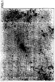
- Fig. 1 is a photomicrograph of the liquid sample in which there can be distinguished microorganisms A which because of the high density of liquid had not become collapsed. Apart from the microorganisms, there were found to be present only a few microparticles of size about 0.1 ⁇ m, identified by the reference letter B.
- Example 1 1 ml of the same soil suspension as in Example 1 was introduced into a test tube followed by 0.5 ml of a filtered enzyme solution which contained 40 mg per ml of the above-mentioned cellulase and 100 mg (1000 U) of the protease.
- the enzyme-containing solution was maintained in a thermoregulated room at 40°C for 2 hours with agitation, after which sucrose was introduced into the mixture in the same manner as in Example 7 to bring the specific gravity to 1.3.
- the resulting mixture was centrifuged and the supernatant liquor was collected as in Example 1, after which the supernatant was diluted with distilled water and centrifuged to collect the precipitated microorganism.
- the precipitate was resuspended in 500 ⁇ l of 1% SDS solution which was the maintained at 70°C for 1 hour to bacterialise the microorganisms.
- DNA was extracted from the resulting liquid by means of chloroform, after which the DNA was precipitated from the chloroform by ethenol and collected.
- the collected DNA was dissolved in distilled water to form a solution containing the DNA present in the soil microorganisms. This DNA solution was subjected to electrophoresis in an agarose gel and about 5000 ng of DNA which had a length 20 kbp or more was collected.
- the collected DNA was treated with BamHI and EcoRI which are typical restriction enzymes and maintained at 37°C for 2 hours, after which the solution was subjected to electrophoresis. It was found that the length of the DNA had been reduced to about 5-10 kbp, indicating that the restriction enzymes had successfully digested the DNA.
- the conditions in the digestion step are as set out in Table 1 below: Conditions for digesting the DNA Collected DNA aqueous solution 2 ⁇ l BamHI/EcoRI 1 ⁇ l H. Buffer solution for restriction enzyme 1 ⁇ l Distilled water 6 ⁇ l 10 ⁇ l
- test tubes having lids were each filled with 6 ml of the soil suspension of Example 1, followed by the previously described filtered solution of cellulase (Meiji Cellulase TP).
- the test tubes were maintained in a thermoregulated room at 40°C for 2 hours, after which the soil suspension was introduced into a 250 ml electrophoresis tank (See Figure 2) containing about 200 ml of buffer solution having the composition shown in Table 2. Electrophoresis was then carried out by applying 100 V (about 50 mA) of direct voltage between platinum electrodes.
- Composition of the buffer solution for electrophoresis Tris 4.08g EDTA.2Na 0.74g Acetic Acid 1.14ml H 2 O 1000ml
- the DNA was digested with BamHI and EcORI in the same manner as in Example 8 and then subjected to electrophoresis, as a result of which it was found that the DNA had been digested and its length had been shortened to about 5-20 KbP. It is therefore apparent that the microorganisms can be refined by electrophoresis, after which they can be collected and the DNA extracted therefrom to give a sample having sufficient purity for digestion by restriction enzymes without additional refining.
- Example 1 1 ml of the same soil suspension as in Example 1 was introduced into a test tube having a lid followed by cellulase (Meiji Cellulase TP), after which the enzyme-containing solution was maintained in a thermoregulated room at 40°C with agitation.
- the test tube was then centrifuged at 5000 rpm for 30 seconds to precipitate soil particles and 1.5 ml of the supernatant liquor was collected and again centrifuged at 1500 rpm for 5 minutes. The resulting precipitate was collected, introduced into a 1.5 ml volume microtube, and a soil microorganism DNA solution was formed in the same manner as in Example 8.
- This DNA solution was subjected to electrophoresis on agarose gel and about 150ng of DNA having a length of 20KbP or above was collected. Attempted digestion of the DNA with BamHI and EcoRI was carried out in the same manner as described in Example 8 and the digested solution was subjected to electrophoresis. However, it was found that the digested DNA had almost the same length as the DNA before digestion, indicating that the restriction enzymes had not worked. It is apparent from this experiment that even where a microorganism has been separated into free individuals using an enzyme, the collected DNA will not be digested by restriction enzyme unless impurities have been sufficiently removed in the refining step, e.g. by extraction into chloroform and then re-precipitation.
- Example 8 The procedure of Example 8 was repeated using the following enzyme preparations:
- Example 8 The same results as in Example 8 were obtained, and in particular the number of separated individual microorganisms was high in the examples where the mixtures of enzymes were used. On the other hand, if the experiments were repeated in the absence of enzymes or using deactivated enzymes, the number of separate individual microorganisms was only 5 to 10% of those reported in the previous examples.
- a mixed solution of sodium phosphate buffer, soil and enzyme was made as described in Example 1, after which the solution was shaken for 2 hours at 40°C in a thermoregulated room. Then 3g of sucrose was added to the solution which was stirred until the supernatant liquor had become saturated. The liquor had a specific gravity of 1.3 or above. Then an aqueous solution of sucrose having a specific gravity of 1.3 was added to the original solution to bring its volume to about 4 ml, with thorough stirring. The test tube containing solution was centrifuged at 50000 rpm for 2 hours after which a 3 ml of supernatant sucrose-saturated liquor was collected and diluted with 20 ml of distilled water.
- the diluted solution was centrifuged at 10000 rpm for 10 minutes and the resulting precipitate was collected.
- the precipitate was suspended in 500 ⁇ l of balanced saline solution.
- a suspension of soil microorganisms was made by resuspending the resulting precipitate in 500 ⁇ l of balanced saline solution which contained 1% of bovine serum albumin buffered with phosphoric acid.
- the suspension was then treated with 0.5 ml of a solution containing 0.2 mg/ml of an anti P. Cepacia monoclonal antibody tagged with FITC.
- the solution was maintained for 1 hour at 0°C to permit the antigen-antibody reaction to take place.
- Cepacia monoclonal antibody was produced by selecting a mouse of the KK01 strain (FERM BP-4235), immunising the mouse by conventional methods, and fusing antibody-producing mouse cells again by convention methods [(see: Japan J. Med. Sei. Biolo., 37, pp 151-159 (1984)].
- the resulting monoclonal antibody was tagged with FITC (maximum fluorescent wavelength is 530 nm) by adding FITC and the antibody to a carbonic acid buffer solution at a pH of 9.5, permitting them to react for 1 hour and then removing free FITC using a column of Sephadex G-25 (Registered Trade Mark) (see “MENEKISEIKAGAKUKENKYUHO” [Immunity Biochemical Experimental Method], 1986, [edited by NIPPON SEIKAGAKUKAI (Society of Biochemistry), published by Tokyo Kagaku Dojinsha K.K.] pp 103-105).
- the bonding ratio of fluorescent dye to antibody protein was 4.2).
- Fig. 3(a) is a graph showing forward scatter against side scatter.
- 3(b) is a histogram of fluorescent light FL1 at 530 nm corresponding to the fluorescence of FITC in the labelled antibody.
- FL3 corresponds to the fluorescence of the ethidium bromide used as a stain for the bacteria and is at 650 nm. The sample was allowed to flow through the counter for 1 minute at a rate of 12 ⁇ l per minute, and observations were made when particles passed the counter.
- the signals FL3 come both from stained bacteria and from impurities which do not contain nucleic acid such as proteins, lipids or microparticles such as dust present.
- a trigger or gate was set in the channel which corresponds to the fluorescent light FL3 of the ethidium bromide stain, and this appears as an arrow mark in Fig. 3(c).
- the channel sensitive to the FL1 fluorescence at 530 nm detects the separate fluorescent light corresponding to FITC, and this signal is gated with a desired width of signal level as is shown in Fig. 3(b).
- the width of the gate is arranged to detect fluorescent light of the strength coming from the FITC labelled bacteria. Pulses produced by the counter during the 1 minute timing interval represent the total number of bacteria found. As a result it was determined that the total number of bacteria was 3.7 x 10 8 , and the estimated number of P. Cepacia was 4.1 x 10 6 , both in 1 gm of soil.
- Example 10 Six test tubes with lids were provided and 6 ml of the same soil suspension as in Example 10 was introduced into each of the 6 test tubes.
- the cellulase enzyme solution of Example 1 was introduced into each test tube, and they were maintained with agitation for 2 hours in a room thermoregulated to 40°C as previously described.
- the resulting soil suspension was introduced into a 250 ml electrophoresis tank containing 200 ml of buffer solution having the composition shown in Table 2 above, and electrophoresis was carried out by applying a 100 volts (about 50 mA) of DC across platinum electrodes. After 4 hours it was apparent that the soil particles had sufficiently precipitated and the supernatant liquor was transparent.
- An anti P. putida monoclonal antibody tagged with PE was provided by mixing PE (manufactured by PolySciences Company) and with N-succinimide-3-(2-pyridyldithio) propionic acid thiol groups and with the anti P. putida monoclonal antibody with which the thiolated PE couples.
- the F/P ratio was 1.7.
- the anti P. Cepacia monoclonal antibody used as starting material was made by the same procedure as described in Example 11.
- Cepacia bacteria labelled with FITC (FL1:530 ⁇ 15 nm), a second of which counts P.putida bacteria labelled by PE (FL2:585 ⁇ 21 nm) and the third of which (FL3) detects fluorescent light from ethidium bromide (FL3:650 nm or above).
- the sample was allowed to flow at a rate of 12 ⁇ l/min. for 1 minute while pulses in the respective channels FL1 and FL2 were counted to provide an estimate of the number of the two different types of bacteria detected. By this means it was determined that a total number of bacteria recovered was 3.5 x 10 8 and that the population of P. Cepacia was 4.0 x 10 6 whilst that of P. putida was 6.9 x 10 6 each in 1 gm of soil.
- Example 2 10 g of the soil sample was processed in the same way as in Example 1 to produce a soil suspension, after which the suspension was filtered using a filter having holes of diameter 15 ⁇ m to remove large suspended soil particles. There was no enzyme treatment, or separation and collection of microorganisms. 50 ⁇ l of the filtrate was centrifuged at 10,000 rpm for ten minutes and the precipitate was collected. Phosphate buffered saline (PBS) containing 1% bovine serum albumin (BSA) was added to the precipitate to give a volume of 500 ⁇ l, after which the precipitate was resuspended to form a soil-containing liquid. The liquid was treated with anti P. Cepacia antibody and the two were allowed to react.
- PBS Phosphate buffered saline
- BSA bovine serum albumin
- Example 10 A sample stained with ethidium bromide was produced. Thereafter a measurement procedure was carried out, all of the steps being as described in Example 10. As a result, the total number of bacteria recovered was found to be 4.1 x 10 6 and 4.4 x 10 4 of P. Cepacia were detected. However, the amount of bacteria detected was about 100th of that in Example 1 which is believed to be because almost all of them remained adhered to the soil particles, or were cohered to one another and were therefore not collected.
- an enzyme can be used to decompose insoluble organic polymer secreted by microorganisms, which polymer may cause the microorganism to adhere either to a carrier or to another microorganism.
- Decomposition of the polymer which acts as a bonding agent enables individual microorganisms to be separated and collected from a suspension or from a liquid in a bioreactor very easily in a short time with high yields and high purity without the need for their particular culturing conditions to be known or for specific culturing steps to be carried out.
- the polymer bonding compound which holds the microorganism to the carrier or holds the microorganisms together may comprise a range of polysaccharides or insoluble proteins depending on the species of microorganism.
- the separation efficiency can be improved by selecting an enzyme, or mixture of enzymes, which can decompose the particular bonding compounds in question.
- Such enzymes may be selected from cellulose, hemicellulose, glucuronidase, amilase, protease and pectinase.
- the process of the invention can be used on soil or on material recovered from a bioreactor for example. It enables useful microorganisms adhered to a carrier, or other microorganisms which may exist in the soil or on the bioreactor, to be separated into individual microorganisms which can be collected.
Landscapes
- Health & Medical Sciences (AREA)
- Life Sciences & Earth Sciences (AREA)
- Chemical & Material Sciences (AREA)
- Organic Chemistry (AREA)
- Engineering & Computer Science (AREA)
- Immunology (AREA)
- Zoology (AREA)
- Wood Science & Technology (AREA)
- Proteomics, Peptides & Aminoacids (AREA)
- Molecular Biology (AREA)
- Biochemistry (AREA)
- Biotechnology (AREA)
- Analytical Chemistry (AREA)
- Physics & Mathematics (AREA)
- Microbiology (AREA)
- General Health & Medical Sciences (AREA)
- Genetics & Genomics (AREA)
- Biophysics (AREA)
- Bioinformatics & Cheminformatics (AREA)
- General Engineering & Computer Science (AREA)
- Hematology (AREA)
- Pathology (AREA)
- Biomedical Technology (AREA)
- Urology & Nephrology (AREA)
- Tropical Medicine & Parasitology (AREA)
- Virology (AREA)
- Cell Biology (AREA)
- Toxicology (AREA)
- Food Science & Technology (AREA)
- Medicinal Chemistry (AREA)
- General Physics & Mathematics (AREA)
- Measuring Or Testing Involving Enzymes Or Micro-Organisms (AREA)
Claims (28)
- Procédé d'obtention de micro-organismes individuels à partir d'un système de micro-organismes dans lequel des micro-organismes individuels adhèrent à un support ou à d'autres micro-organismes individuels au moyen d'un adhésif naturel sécrété par les micro-organismes individuels, caractérisé en ce que ledit procédé comprend l'étape consistant à traiter le système de micro-organismes avec au moins une enzyme choisie dans l'ensemble constitué par des enzymes qui décomposent les polysaccharides, des enzymes qui décomposent les protéines et des enzymes qui décomposent la pectine, séparant ainsi les micro-organismes individuels à partir du support ou d'autres micro-organismes individuels.
- Procédé selon la revendication 1, dans lequel l'enzyme comprend une cellulase, une hémicellulase, une glucuronidase, une amylase, une protéase ou une pectinase ou un mélange de celles-ci.
- Procédé selon la revendication 1 ou la revendication 2, dans lequel le système de micro-organismes comprend une suspension de terre, une suspension de boue activée ou une suspension de boue provenant des rivières, des lacs ou des mers.
- Procédé selon la revendication 1 ou la revendication 2, dans lequel le système de micro-organismes comprend un réactif liquide qui contient un support pour supporter les micro-organismes dans un bioréacteur.
- Procédé selon l'une quelconque des revendications précédentes, dans lequel les micro-organismes séparés sont purifiés et recueillis.
- Procédé selon la revendication 5, dans lequel on purifie et on recueille les micro-organismes en formant une suspension contenant les micro-organismes séparés et ayant un poids volumique analogue à celui des micro-organismes, et en centrifugeant la suspension et en séparant celle-ci en une solution surnageante contenant les micro-organismes et en un précipité en utilisant la différence de poids volumique entre le milieu dispersif et les particules à séparer.
- Procédé selon la revendication 6, dans lequel le mode opératoire pour purifier et recueillir le microorganisme comprend les étapes consistant à :ajuster le poids volumique de la suspension traitée avec une enzyme jusqu'à ce qu'il soit égal au poids volumique du micro-organisme, ou jusqu'à ce qu'il soit supérieur au poids volumique du micro-organisme, ou jusqu'à ce qu'il soit inférieur au poids volumique du support ;centrifuger la suspension dont le poids volumique a été ajusté de façon à faire précipiter le support et à obtenir une solution surnageante dans laquelle les micro-organismes sont en suspension ; etrecueillir le micro-organisme à partir de la solution surnageante.
- Procédé selon la revendication 7, dans lequel on ajuste le poids volumique de la suspension entre 1,2 et 1,5.
- Procédé selon la revendication 7 ou 8, dans lequel on ajuste le poids volumique de la suspension en ajoutant un soluté.
- Procédé selon la revendication 9, dans lequel le soluté comprend du saccharose.
- Procédé selon la revendication 9, dans lequel le soluté comprend du chlorure de césium.
- Procédé selon la revendication 5, dans lequel on purifie le micro-organisme au moyen de sa mobilité électrophorétique et on le recueille.
- Procédé selon la revendication 12, dans lequel l'étape de purification et de collecte du micro-organisme en utilisant sa mobilité électrophorétique comprend les étapes consistant à :appliquer une tension continue à une suspension du micro-organisme après l'avoir traitée avec l'enzyme ; etrecueillir le liquide contenant le micro-organisme qui a été déplacé de façon contigué à l'électrode positive et/ou à l'électrode négative.
- Procédé pour compter un nombre de micro-organismes individuels présents dans un échantillon, comprenant les étapes consistant à recueillir un liquide qui contient des micro-organismes individuels séparés au moyen du procédé selon l'une quelconque des revendications 1 à 4 et à compter le nombre de micro-organismes présents dans ledit liquide.
- Procédé selon la revendication 14, dans lequel les micro-organismes sont séparés, purifiés et recueillis selon l'une quelconque des revendications 6 à 13.
- Procédé selon la revendication 15, dans lequel on effectue le comptage en utilisant le procédé de dilution sur plaque ou le procédé de coloration.
- Procédé pour recueillir un acide nucléique, comprenant les étapes consistant à extraire et à recueillir un acide nucléique d'un micro-organisme à partir d'un liquide contenant le micro-organisme qu'on a recueilli au moyen du procédé défini dans l'une quelconque des revendications 6 à 13.
- Procédé de comptage de la distribution d'un micro-organisme spécifique dans un système de micro-organismes comprenant une pluralité de types de micro-organismes, dans lequel au moins un micro-organisme spécifique adhère à des particules présentes dans le système ou adhère à d'autres micro-organismes, ledit procédé comprenant les étapes consistant à :obtenir une suspension de micro-organismes séparés au moyen du procédé selon l'une quelconque des revendications 1 à 13 et recueillir les micro-organismes séparés à partir de la suspension ;lier au micro-organisme spécifique un anticorps marqué par un colorant fluorescent ;lier aux micro-organismes présents un composé fluorescent dont la longueur d'onde maximale de fluorescence est différente de la longueur d'onde du colorant fluorescent ; etmesurer la fluorescence émise par le colorant fluorescent et par le composé fluorescent.
- Procédé selon la revendication 18, dans lequel on mesure la fluorescence émise par le colorant fluorescent et par le composé fluorescent en :formant un flux de la suspension et en détectant dans ce flux la fluorescence émise par le colorant et la fluorescence émise par le composé pour chaque particule traversant le flux ; ettraitant statistiquement les données détectées pour en tirer une information sur ladite distribution.
- Procédé selon la revendication 19, dans lequel on insère un déclencheur ou une porte responsable de la fluorescence par rapport audit composé de fluorescence de façon à faire passer le signal seulement quand il dépasse une valeur seuil.
- Procédé selon l'une quelconque des revendications 18, 19 ou 20, comprenant, en outre, l'étape consistant à soumettre la suspension à une énergie vibratoire ou a une énergie de cisaillement avant de mesurer la fluorescence émise par le colorant fluorescent et par le composé fluorescent de façon à séparer des groupes de micro-organismes agglutinés par l'anticorps.
- Procédé selon l'une quelconque des revendications 18 à 21, dans lequel la longueur d'onde maximale de fluorescence du colorant fluorescent est séparée de la longueur d'onde maximale de fluorescence du composé fluorescent de 20 nm ou plus.
- Procédé selon l'une quelconque des revendications 18 à 22, dans lequel le composé fluorescent est un colorant qui se lie à un acide nucléique de micro-organismes.
- Procédé selon l'une quelconque des revendications 18 à 23, dans lequel l'étape de marquage de l'anticorps comprend la liaison d'une pluralité d'anticorps différents marqués par un colorant fluorescent à une pluralité de différents types de micro-organismes.
- Procédé selon l'une quelconque des revendications 18 à 24, dans lequel la liaison à l'anticorps est directe.
- Procédé selon l'une quelconque des revendications 18 à 24, dans lequel la liaison à l'anticorps est indirecte.
- Procédé selon l'une quelconque des revendications 18 à 26, dans lequel le système de micro-organismes comprend de la terre, une boue activée ou de la boue marine provenant d'eau fraíche, d'eau de marée ou d'eau salée.
- Procédé selon l'une quelconque des revendications 18 à 27, dans lequel le système de micro-organismes comprend un mélange de réaction provenant d'un bioréacteur contenant un support en suspension qui supporte un micro-organisme.
Applications Claiming Priority (18)
| Application Number | Priority Date | Filing Date | Title |
|---|---|---|---|
| JP284719/93 | 1993-11-15 | ||
| JP30709693 | 1993-11-15 | ||
| JP28471993 | 1993-11-15 | ||
| JP30703493A JP3542366B2 (ja) | 1993-11-15 | 1993-11-15 | 微生物の分離方法および微生物個体数の計測方法 |
| JP307079/93 | 1993-11-15 | ||
| JP30709693A JPH07135962A (ja) | 1993-11-15 | 1993-11-15 | 微生物の精製回収方法 |
| JP30707993A JP3542367B2 (ja) | 1993-11-15 | 1993-11-15 | 微生物の分離精製回収方法、微生物個体数の計測方法および微生物の核酸回収方法 |
| JP307096/93 | 1993-11-15 | ||
| JP30707993 | 1993-11-15 | ||
| JP307034/93 | 1993-11-15 | ||
| JP28471993A JPH07140148A (ja) | 1993-11-15 | 1993-11-15 | 微生物計数方法 |
| JP30703493 | 1993-11-15 | ||
| JP316581/93 | 1993-12-16 | ||
| JP31658193A JP3548211B2 (ja) | 1993-12-16 | 1993-12-16 | 微生物の選別回収方法 |
| JP31658193 | 1993-12-16 | ||
| JP334994 | 1994-01-18 | ||
| JP3349/94 | 1994-01-18 | ||
| JP00334994A JP3441779B2 (ja) | 1994-01-18 | 1994-01-18 | 微生物の計数方法 |
Publications (3)
| Publication Number | Publication Date |
|---|---|
| EP0653492A2 EP0653492A2 (fr) | 1995-05-17 |
| EP0653492A3 EP0653492A3 (fr) | 1995-09-13 |
| EP0653492B1 true EP0653492B1 (fr) | 2000-09-27 |
Family
ID=27547765
Family Applications (1)
| Application Number | Title | Priority Date | Filing Date |
|---|---|---|---|
| EP19940308364 Expired - Lifetime EP0653492B1 (fr) | 1993-11-15 | 1994-11-11 | Procédé pour l'obtention de microorganismes individuels et applications du procédé |
Country Status (2)
| Country | Link |
|---|---|
| EP (1) | EP0653492B1 (fr) |
| DE (1) | DE69426008T2 (fr) |
Families Citing this family (5)
| Publication number | Priority date | Publication date | Assignee | Title |
|---|---|---|---|---|
| ATE354791T1 (de) * | 2001-10-10 | 2007-03-15 | Borealis Tech Oy | Teilchendatenanalyse |
| JP6238046B2 (ja) * | 2013-05-29 | 2017-11-29 | 株式会社サタケ | 微生物の検査方法 |
| WO2018004501A1 (fr) * | 2016-07-01 | 2018-01-04 | Keskinkan Olcayto | Matériau de coloration contenant de l'atrazon rouge fbl, du rouge basique 46 (c.i. 110825) pour l'identification de bactéries filamenteuses dans une boue activée d'installations de traitement biologique |
| CN109959643B (zh) * | 2019-04-10 | 2021-06-01 | 中国环境科学研究院 | 一种评估毒性物质对活性污泥传质性能影响的方法 |
| CN115166192B (zh) * | 2022-09-05 | 2022-11-22 | 山东蓝色海洋科技股份有限公司 | 一种海洋牧场用溶解氧检测机器人 |
Family Cites Families (6)
| Publication number | Priority date | Publication date | Assignee | Title |
|---|---|---|---|---|
| EP0266881A3 (fr) * | 1986-09-30 | 1990-04-04 | Astromed Limited | Procédé et appareil pour essai optique multiple |
| US5081030A (en) * | 1989-04-25 | 1992-01-14 | The Johns Hopkins University | Release of cells from affinity matrices |
| CA2066761C (fr) * | 1989-09-20 | 1999-11-16 | Patrick Soon-Shiong | Invention pour la separation des cellules |
| GB9108047D0 (en) * | 1991-04-16 | 1991-06-05 | Mellors Alan | The use of pasteurella haemolytica clycoproteinase for human bone marrow haematopoietic primitive stem-cell isolation |
| GB9208357D0 (en) * | 1992-04-16 | 1992-06-03 | British Tech Group | Apparatus for separating a mixture |
| EP0636184B1 (fr) * | 1993-01-15 | 2000-08-30 | Baxter International Inc. | Milieux pour l'isolation et la stabilisation de cellules |
-
1994
- 1994-11-11 DE DE1994626008 patent/DE69426008T2/de not_active Expired - Lifetime
- 1994-11-11 EP EP19940308364 patent/EP0653492B1/fr not_active Expired - Lifetime
Also Published As
| Publication number | Publication date |
|---|---|
| EP0653492A2 (fr) | 1995-05-17 |
| DE69426008D1 (de) | 2000-11-02 |
| EP0653492A3 (fr) | 1995-09-13 |
| DE69426008T2 (de) | 2001-05-17 |
Similar Documents
| Publication | Publication Date | Title |
|---|---|---|
| US5225332A (en) | Process for manipulation of non-aqueous surrounded microdroplets | |
| US5055390A (en) | Process for chemical manipulation of non-aqueous surrounded microdroplets | |
| US4677067A (en) | Magnetotactic bacteria in clinical assay, immunoassay, and cell separation procedures and the like | |
| US4959301A (en) | Process for rapidly enumerating viable entities | |
| Caron et al. | Assessment of bacterial viability status by flow cytometry and single cell sorting | |
| EP0070318B1 (fr) | Procede d'isolation d'un materiau presentant une activite microbiologique | |
| US5821066A (en) | Simple, rapid method for the detection, identification and enumeration of specific viable microorganisms | |
| EP3209793B1 (fr) | Methode et dispositifs de traitement d'echantillons biologiques | |
| Vesey et al. | Detection of specific microorganisms in environmental samples using flow cytometry | |
| WO1989010566A1 (fr) | Procede pour former et pour utiliser des microgouttes | |
| EP2996789B1 (fr) | Libération et collecte de particules | |
| Reynolds et al. | Detection of Cryptosporidium oocysts in water: techniques for generating precise recovery data | |
| DE68914871T2 (de) | Verfahren zur Bestimmung von mikro-organismen. | |
| AU785026B2 (en) | Facs assisted methods for introducing individual chromosomes into cells | |
| EP0653492A2 (fr) | Procédé pour la séparation de microorganismes particuliers et applications du procédé | |
| US4503149A (en) | Method of microbial assay | |
| JP3441779B2 (ja) | 微生物の計数方法 | |
| EP1404875A1 (fr) | Procede et systeme de comptage de cellules provenant d'une pluralite d'especes | |
| WO1989010786A2 (fr) | Procede de formation et d'utilisation de microgouttelettes | |
| US6010869A (en) | Method to collect and recover microorganisms from environmental samples | |
| JP3542367B2 (ja) | 微生物の分離精製回収方法、微生物個体数の計測方法および微生物の核酸回収方法 | |
| JP3444946B2 (ja) | 微生物特性測定法 | |
| JP3542366B2 (ja) | 微生物の分離方法および微生物個体数の計測方法 | |
| JPH07140148A (ja) | 微生物計数方法 | |
| Mekada et al. | An attempt to separate mononuclear cells fused with human red blood cell-ghosts from a cell mixture treated with HVJ (Sendai virus) using a fluorescence activated cell sorter (FACS II). |
Legal Events
| Date | Code | Title | Description |
|---|---|---|---|
| PUAI | Public reference made under article 153(3) epc to a published international application that has entered the european phase |
Free format text: ORIGINAL CODE: 0009012 |
|
| AK | Designated contracting states |
Kind code of ref document: A2 Designated state(s): DE GB NL |
|
| PUAL | Search report despatched |
Free format text: ORIGINAL CODE: 0009013 |
|
| AK | Designated contracting states |
Kind code of ref document: A3 Designated state(s): DE GB NL |
|
| 17P | Request for examination filed |
Effective date: 19951006 |
|
| 17Q | First examination report despatched |
Effective date: 19980924 |
|
| GRAG | Despatch of communication of intention to grant |
Free format text: ORIGINAL CODE: EPIDOS AGRA |
|
| RTI1 | Title (correction) |
Free format text: PROCESS FOR OBTAINING INDIVIDUAL MICROORGANISMS, AND APPLICATIONS OF THAT PROCESS |
|
| GRAG | Despatch of communication of intention to grant |
Free format text: ORIGINAL CODE: EPIDOS AGRA |
|
| GRAH | Despatch of communication of intention to grant a patent |
Free format text: ORIGINAL CODE: EPIDOS IGRA |
|
| GRAH | Despatch of communication of intention to grant a patent |
Free format text: ORIGINAL CODE: EPIDOS IGRA |
|
| GRAA | (expected) grant |
Free format text: ORIGINAL CODE: 0009210 |
|
| AK | Designated contracting states |
Kind code of ref document: B1 Designated state(s): DE GB NL |
|
| REF | Corresponds to: |
Ref document number: 69426008 Country of ref document: DE Date of ref document: 20001102 |
|
| EN | Fr: translation not filed | ||
| PLBE | No opposition filed within time limit |
Free format text: ORIGINAL CODE: 0009261 |
|
| STAA | Information on the status of an ep patent application or granted ep patent |
Free format text: STATUS: NO OPPOSITION FILED WITHIN TIME LIMIT |
|
| 26N | No opposition filed | ||
| REG | Reference to a national code |
Ref country code: GB Ref legal event code: IF02 |
|
| PGFP | Annual fee paid to national office [announced via postgrant information from national office to epo] |
Ref country code: NL Payment date: 20091118 Year of fee payment: 16 |
|
| PGFP | Annual fee paid to national office [announced via postgrant information from national office to epo] |
Ref country code: GB Payment date: 20091204 Year of fee payment: 16 |
|
| PGFP | Annual fee paid to national office [announced via postgrant information from national office to epo] |
Ref country code: DE Payment date: 20091130 Year of fee payment: 16 |
|
| REG | Reference to a national code |
Ref country code: NL Ref legal event code: V1 Effective date: 20110601 |
|
| GBPC | Gb: european patent ceased through non-payment of renewal fee |
Effective date: 20101111 |
|
| PG25 | Lapsed in a contracting state [announced via postgrant information from national office to epo] |
Ref country code: NL Free format text: LAPSE BECAUSE OF NON-PAYMENT OF DUE FEES Effective date: 20110601 |
|
| REG | Reference to a national code |
Ref country code: DE Ref legal event code: R119 Ref document number: 69426008 Country of ref document: DE Effective date: 20110601 Ref country code: DE Ref legal event code: R119 Ref document number: 69426008 Country of ref document: DE Effective date: 20110531 |
|
| PG25 | Lapsed in a contracting state [announced via postgrant information from national office to epo] |
Ref country code: DE Free format text: LAPSE BECAUSE OF NON-PAYMENT OF DUE FEES Effective date: 20110531 |
|
| PG25 | Lapsed in a contracting state [announced via postgrant information from national office to epo] |
Ref country code: GB Free format text: LAPSE BECAUSE OF NON-PAYMENT OF DUE FEES Effective date: 20101111 |