EP0704458A2 - Anticorps contre le collagène du type IV et leur utilisation - Google Patents
Anticorps contre le collagène du type IV et leur utilisation Download PDFInfo
- Publication number
- EP0704458A2 EP0704458A2 EP95306948A EP95306948A EP0704458A2 EP 0704458 A2 EP0704458 A2 EP 0704458A2 EP 95306948 A EP95306948 A EP 95306948A EP 95306948 A EP95306948 A EP 95306948A EP 0704458 A2 EP0704458 A2 EP 0704458A2
- Authority
- EP
- European Patent Office
- Prior art keywords
- collagen
- human type
- monoclonal antibody
- type
- carrier
- Prior art date
- Legal status (The legal status is an assumption and is not a legal conclusion. Google has not performed a legal analysis and makes no representation as to the accuracy of the status listed.)
- Withdrawn
Links
Images


Classifications
-
- C—CHEMISTRY; METALLURGY
- C07—ORGANIC CHEMISTRY
- C07K—PEPTIDES
- C07K16/00—Immunoglobulins [IG], e.g. monoclonal or polyclonal antibodies
- C07K16/18—Immunoglobulins [IG], e.g. monoclonal or polyclonal antibodies against material from animals or humans
-
- G—PHYSICS
- G01—MEASURING; TESTING
- G01N—INVESTIGATING OR ANALYSING MATERIALS BY DETERMINING THEIR CHEMICAL OR PHYSICAL PROPERTIES
- G01N33/00—Investigating or analysing materials by specific methods not covered by groups G01N1/00 - G01N31/00
- G01N33/48—Biological material, e.g. blood, urine; Haemocytometers
- G01N33/50—Chemical analysis of biological material, e.g. blood, urine; Testing involving biospecific ligand binding methods; Immunological testing
- G01N33/68—Chemical analysis of biological material, e.g. blood, urine; Testing involving biospecific ligand binding methods; Immunological testing involving proteins, peptides or amino acids
- G01N33/6887—Chemical analysis of biological material, e.g. blood, urine; Testing involving biospecific ligand binding methods; Immunological testing involving proteins, peptides or amino acids from muscle, cartilage or connective tissue
-
- Y—GENERAL TAGGING OF NEW TECHNOLOGICAL DEVELOPMENTS; GENERAL TAGGING OF CROSS-SECTIONAL TECHNOLOGIES SPANNING OVER SEVERAL SECTIONS OF THE IPC; TECHNICAL SUBJECTS COVERED BY FORMER USPC CROSS-REFERENCE ART COLLECTIONS [XRACs] AND DIGESTS
- Y10—TECHNICAL SUBJECTS COVERED BY FORMER USPC
- Y10S—TECHNICAL SUBJECTS COVERED BY FORMER USPC CROSS-REFERENCE ART COLLECTIONS [XRACs] AND DIGESTS
- Y10S435/00—Chemistry: molecular biology and microbiology
- Y10S435/975—Kit
Definitions
- the present invention relates to monoclonal antibodies against human type IV collagen, and to an immunoassay method and kit for measuring human type IV collagen using said monoclonal antibody.
- Collagen is present in connective tissues, and to date 15 genetically different types have been reported, including type I, II, III, IV, etc. These types have been found to have organ specificities, with type I and III collagen plentiful in the bones and skin, and type IV collagen contained in the basement membranes of the blood vessel skin, digestive organs, muscles, nerve fibers, nephrogiomeruli, lungs, etc.
- Type IV collagen is composed of 4 parts, the amino terminal 75 domain, the carboxyl terminal NC1 and NC2 domains, and a triple helical domain (TH domain) wherein the intermediate peptides are combined in a helix form.
- Hepatic fibrosis has been reported to occur in an early stage of liver cirrhosis (Molecular Medicine, Vol.31, No.2, 1994; Special issue: Hepatic fibrosis and Cytokines, etc.).
- hepatic fibrosis markers have emerged into clinical application for liver cirrhosis, and thus it has become possible to take continuous, frequent measurements in a non-invasive manner without liver biopsy, with great usefulness for the analysis of liver disease.
- the major hepatic fibrosis marker is the extracellular matrix (ECM), and the related enzymes and degradation products may be used as markers reflecting the degree of fibrosis.
- ECM extracellular matrix
- type IV collagen is produced and secreted out by endothelial cells and liver cells, especially Ito cells, from the early stages of hepatopathy, its increase is taken to reflect hepatic fibrosis.
- a portion of the type IV collagen produced in the liver enters the blood vessels and therefore type IV collagen in the blood serum may be used as an index of hepatic fibrosis.
- the first method is a competitive method using a radioactive isotope, wherein the 7S domain of type IV collagen obtained by purifying placenta-extracted type IV collagen by collagenase treatment is used as the antigen, and this 7S domain is labelled with 15I as the labelled antigen while the 7S domain is injected into rabbits to prepare antibodies against the 7S domain of type IV collagen (type IV collagen/7S kit manual: Nippon DPC Corp.; Yamada, et al., Clin. Biochem. Vol.25, p.467-470, 1992; Risteli, et al., Eur. J. Biochem. Vol. 108, p.239-250, 1980).
- the second method is the so-called one-step sandwich enzyme immunoassay (EIA) method which uses a monoclonal antibody against pepsin-solubilized human type IV collagen and a monoclonal antibody against the 7S domain of human type IV collagen (Manual for "Panassay IV/C" for blood serum type IV collagen measurement; Daiichi Pure Chemicals; Japanese Unexamined Patent Publication No. 2-1553; Clinical Chimica Acta 181, p.293-304, 1989).
- EIA sandwich enzyme immunoassay
- radioimmuno assay method described above requires careful handling of radio isotope and complicated procedures. For example, special equipment and facilities meeting the legal regulation are necessary, which restricts the locations permitting the measurement. The disposal of waste liquid is also a matter of concern and attention has to be paid for avoiding hazardous irradiation effects on human health. Furthermore, commercial kits for this type of assay involve these problems as well as the fact that a reaction time as long as 16-24 hours is required, and 200 ⁇ l of sample is required per test, which is substantially larger than for common blood tests.
- the inventors established a novel method for determining human type IV collagen, by which the results of measurement reflect the condition of patients, the handling of reagents and the measuring procedure are simple, there are no problems of effects on the body or disposal of waste liquid, and the measurement may be carried out with a small amount of sample in a short period of time.
- the present invention provides an method for immunoassay of human type IV collagen in a sample, which is characterized by using a monoclonal antibody against human type IV collagen and a labelled polyclonal antibody against human type IV collagen.
- the present invention also provides a monoclonal antibody against human type IV collagen for use in the above-mentioned immunoassay method, which monoclonal antibody has the following properties:
- the present invention further provides a monoclonal antibody with the following properties:
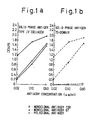
- Fig. 1a is a graph showing the results of testing the binding sites of the monoclonal antibodies 238 and 67 and polyclonal antibody of the present invention, on the human type IV collagen molecule.
- Fig. 1b is a graph showing the results of testing the binding sites of the monoclonal antibodies 238 and 67 and polyclonal antibody of the present invention, on the 7S domain of human type IV collagen molecule.
- Fig. 2 is a graph showing the results of analyzing the identities of the binding sites of the monoclonal antibodies 238 and 67 of the present invention on the human type IV collagen molecule.
- Fig. 3 is a graph showing the results of analyzing the identities of the binding sites of the monoclonal antibodies 238 and 67 of the present invention on the human type IV collagen molecule.
- Fig. 4 is an example of a calibration curve for an assay of human type IV collagen using monoclonal antibody 67 as the immobilized antibody (primary antibody).
- Fig. 5 is an example of a calibration curve for an assay of human type IV collagen using monoclonal antibody 238 as the immobilized antibody (primary antibody).
- the method for measuring human type IV collagen in samples does not use a radioactive isotope, and consequently does not require special equipment or devices which impose limitations on the location where the measurement may be made, while disposal of the waste water is simple and there is no effect on the body.
- the reaction time is as short as 40 minutes.
- the method of the present invention gives results which accurately reflect the condition of patients. That is, there is close correlation between the presence of hepatic fibrosis observed in liver biopsy and the human type IV collagen examination results obtained by the method of the present invention, and more so than with examination using commercially available kits.
- type IV collagen also exists in the serum in the form of fragments produced by body proteases such as collagenase, and the 7S domain is relatively stable in the body among these fragments.
- the results of a measurements which reflect the conditions of patients may be obtained by using a monoclonal antibody specific to the 7S domain of human type IV collagen as the primary antibody binding to the carrier, to capture the human type IV collagen or fragments thereof containing the 7S domain, and then detecting the captured human type IV collagen or fragments thereof containing the 7S domain using a polyclonal antibody (secondary antibody) with a relatively wide specificity range which binds to the human type IV collagen or fragments thereof containing the captured 7S domain.
- a monoclonal antibody specific to the 7S domain of human type IV collagen as the primary antibody binding to the carrier, to capture the human type IV collagen or fragments thereof containing the 7S domain, and then detecting the captured human type IV collagen or fragments thereof containing the 7S domain using a polyclonal antibody (secondary antibody) with a relatively wide specificity range which binds to the human type IV collagen or fragments thereof containing the captured 7S domain.
- a monoclonal antibody is used as the secondary antibody, sometimes only a portion of the type IV collagen or fragments thereof captured by the primary antibody will be detected.
- the polyclonal antibody and monoclonal antibody of the present invention may be prepared according to conventional methods. As examples of concrete methods, a method of producing polyclonal antibodies is given in Example 1, and a method of producing monoclonal antibodies is given in Example 2.
- a monoclonal antibody specific to the 7S domain of human type IV collagen is immobilized on a carrier to be used as the primary antibody.
- a solid carrier is preferred as the carrier, and any desired solid carrier which is common in immunoassays may be used including high molecular weight carriers such as styrene and polystyrene molded into a desired size and shape, as well as inner walls of reaction containers formed such appropriate materials.
- the immobilization of the monoclonal antibody on the carrier may be carried out by a common method, such as by contacting the monoclonal antibody with the carrier in a buffer solution, for example a boric acid buffer solution.
- a buffer solution for example a boric acid buffer solution.
- Another method for example, is one in which an antibody against the monoclonal antibody to be used is first immobilized on a carrier, and this is then contacted with the monoclonal antibody.
- the monoclonal antibody used as the primary antibody is preferably an antibody which binds specifically to the 7S domain of human type IV collagen, for the reasons explained above under "Effect of the Invention", and in concrete terms, the monoclonal antibody 67 produced by the hybridoma COL-IV-67 (FERM P-14561) is preferred.
- the hybridoma CDL-IV-67 was deposited with National Institute of Bioscience and Human-Technology Agency of Industrial Science of Technology, 1-3, Higashi 1-chome, Tsukuba-shi, Ibaraki, Japan, as FERN P-14561 on September 27, 1994, and transferred to an international deposition under the Budapest treaty as FERM BP-5240 on September 25, 1995.
- the polyclonal antibody against human type IV collagen is labelled, preferably with a non-radioactive labelling system, as the secondary antibody.
- the non-radioactive labelling used may be enzyme labelling, fluorescent labelling or optical labelling such as luminescent labelling. Enzyme labelling is preferred, and for example alkaline phosphatase (ALP), ⁇ -D-galactosidase, horseradish peroxidase, or the like may be used as the labelling enzyme.
- the detection of these enzymes may be accomplished using their respective enzyme substrates, namely 4-methylumbelliferyl phosphate (4-MUP), 2-nitrophenyl- ⁇ -D-galactoside, a combination of hydrogen peroxide and 3,3'-5,5'-tetramethylbenzidine, etc.
- 4-MUP 4-methylumbelliferyl phosphate
- 2-nitrophenyl- ⁇ -D-galactoside 2-nitrophenyl- ⁇ -D-galactoside
- a combination of hydrogen peroxide and 3,3'-5,5'-tetramethylbenzidine etc.
- a common method may be used to conjugate the secondary antibody and the enzyme label, and for example, a commercial thiol-based cross-linking reagent may be used to introduce thiol groups into both the labelling substance and the antibody, which are then combined together by S-S bond.
- the above-mentioned carrier with primary monoclonal antibody immobilized, the above-mentioned labelled secondary polyclonal antibody and the testing sample are mixed together and incubated, to bind the human type IV collagen molecules in the testing sample to the primary monoclonal antibody immobilized on the carrier, and to bind the labelled secondary polyclonal antibody to the collagen molecules.
- the labelled secondary polyclonal antibody is immobilized on the carrier through the primary antibody immobilized on the carrier and the type IV collagen in the sample, in an amount which reflects the amount of type IV collagen in the sample.
- a buffer solution for example Tris-HCl or phosphate buffer solution, for 10 to 120 minutes, preferably 10 to 40 minutes, at 15°C to 40°C, preferably 20 to 37°C, or at room temperature, for example.
- the bound labelled carrier and the unbound labelled carrier are separated.
- this separation may be easily accomplished by solid-liquid separation.
- the labelling which is either bound or unbound to the carrier, or both may be measured.
- an arbitrary amount (unknown and in excess) of the labelled antibody has been used, then the amount of labelling which has bound to the carrier may be detected and measured.
- the detection of the labelling bound to the carrier is preferably accomplished by a detection reaction after washing the carrier with a washing solution, for example distilled water containing a surfactant, etc. or an appropriate buffer solution, to remove the unbound labelled antibody.
- the detection may be carried out according to a common method, depending on the type of labelling.
- the present invention also provides an assay kit for type IV collagen.
- the kit includes at least a carrier on which monoclonal antibody against human type IV collagen has been immobilized, or the monoclonal antibody and a carrier for its immobilization, and a polyclonal antibody against human type IV collagen which has been labelled.
- the monoclonal antibody-immobilized carrier and the labelled polyclonal antibody are the same as described in detail above, and specific cases will be given in the following examples.
- the kit may also include a standard solution of human type IV collagen for calibrating the assay together with directions for its handling.
- Type IV collagen was prepared from human placenta following the method of Sage, et al. described in J. Biol. Chem., 254 , 9893-9900 (1980). That is, human placenta pre-washed successively with water, 50 mM Tris-HCl buffer solution (pH 7.5) containing 1.0 M NaCl, and 0.5 M acetic acid, was homogenized and suspended in 0.5 M acetic acid, and then pepsin treatment was performed at 4°C for 24 hours.
- Tris-HCl buffer solution pH 7.5
- pepsin treatment was performed at 4°C for 24 hours.
- This mixture is centrifuged, the supernatant is collected and the NaCl concentration is adjusted to 1.0 M, after which the solution is stirred for 24 hours and centrifuged, the supernatant is collected and the NaCl concentration is further adjusted to 1.8 M, and after further stirring for 24 hours and centrifugation a precipitate is collected.
- This precipitate was dissolved in 50 mM Tris-HCl buffer solution (pH 7.5) containing 1.0 M NaCl, the pH was adjusted to 7.5 with 1.0 M sodium hydroxide for dissolution, after the insoluble material was removed by centrifugation the NaCl concentration was raised to 2.0 M, the solution was stirred for 24 hours and centrifuged to collect a precipitate which was then dissolved in 0.1 M acetic acid to a concentration of 1.0 mg/ml. Solid NaCl was added to 0.7 M and the solution was stirred for 24 hours and centrifuged, then the supernatant was collected, and the NaCl concentration was increased to 1.8 M.
- the solution was further stirred for 24 hours and centrifuged, the precipitate was collected and dissolved in 0.1 M acetic acid to a concentration of 0.1 mg/ml, the NaCl concentration was adjusted to 0.7 M, the solution was stirred for 24 hours and centrifuged, and then the precipitate was collected and purified with DEAE Sepharose to obtain human type IV collagen.
- the human type IV collagen prepared in (a) above was mixed with Freund's complete adjuvant (product of DIFCO Co.) and subcutaneously injected into rabbits at 1.0 mg per rabbit for immunization.
- Booster shots were given at 4 weeks after the initial immunization by the same method, and blood was taken while monitoring the antibody titer.
- Blood was first taken when the antibody titer began to increase, and blood was taken every week thereafter for a total period over one year while booster shots were given every 3 months.
- the antibody was purified from the serum by affinity chromatography on a column of immobilized human type IV collagen and then it was further purified on a protein A-Sepharose CL-4B column to obtain specific anti-human type IV collagen antibody (polyclonal antibody).
- the fused cells were suspended in ERDF medium (product of Kyokuto Pharmaceuticals Co.) containing 15% fetal calf serum (product of M.A. Bioproducts Co.), and cultured on a 96-well plate (product of Falcon Co.). On day 2 of the culture, the medium was changed to ERDF medium containing 100 ⁇ M hypoxanthine, 0.4 ⁇ M aminopterin, 16 ⁇ M thymidine and 15% fetal calf serum (hereunder, "HAT medium”), and HAT medium was further added after 3 days.
- ERDF medium product of Kyokuto Pharmaceuticals Co.
- fetal calf serum product of M.A. Bioproducts Co.
- HAT medium fetal calf serum
- the HAT medium was replaced with the medium lacking aminopterin (hereunder, "HT medium"), and HT medium was replaced every 3-4 days thereafter.
- HT medium medium lacking aminopterin
- the anti-human type IV collagen antibody titer in the culture supernatant was measured by the enzyme immunoassay method described below, and cells producing the anti-human type IV collagen antibody were screened.
- the cells in the wells confirmed to be producing anti-human type IV collagen antibody through screening by the enzyme immunoassay method in (c) above were then cloned by the limiting dilution method as follows.
- the cells were dispensed into a 96-well plate at cell counts of 3, 1 and 0.3 per well, and were cultured in HT medium.
- the anti-human type IV collagen antibody titer in each of the culture supernatants was determined by enzyme immunoassay in the same manner as in (c) above.
- the cells in the wells which were found producing anti-human type IV collagen antibody were repeatedly re-cloned, until production of anti-human type IV collagen antibody was finally confirmed in all of the wells.
- each of the hybridoma lines was cultured in the abdominal cavity of BALB/c mice which had been previously administered pristane (2,6,10,14-tetramethylpentadecane) intraperitoneally at 0.5 ml per mouse.
- pristane 2,6,10,14-tetramethylpentadecane
- the administration of 5 ⁇ 106 hybridoma cells into mouse intraperitoneal cavity resulted in the antibody production of 1-10 mg/ml in the ascites fluid by the 10th to 14th day of post-injection.
- the monoclonal antibodies in the ascites fluid obtained in (e) above were diluted to two-fold with 1.5 M glycine-Na buffer (pH 8.5) containing 3.0 M NaCl, and applied to a protein A-Sepharose CL-4B column which had been equilibrated with 1.5 M glycine-Na buffer (pH 8.5) containing 3.0 M NaCl. After washing the column with the equilibration buffer, the monoclonal antibody bound on the column was eluted with 0.1 M citric acid-Na buffer solution (pH 4.0).
- the hybridomas producing monoclonal antibodies 67 and 238 in Table 2 were named COL IV-67 and COL IV-238, respectively. Both of which had been deposited at the National Institute of Bioscience and Human Technology on September 27, 1994 as FERN P-14561 and FERN P-14560, respectively, and transferred to an international deposition under the Budapest treaty as FERM BP-5240 and FERM BP-5239, respectively on September 25, 1995.
- the monoclonal antibody produced by the hybridoma COL IV-67 will be referred to simply as monoclonal antibody 67
- the monoclonal antibody produced by the hybridoma COL IV-238 will be referred to simply as monoclonal antibody 238.
- the 7S long form of human type IV collagen was prepared from human placenta following the method of Risteli, et al. described in Eur. J. Biochem., 108 , 239-250 (1980).
- monoclonal antibody 67 and monoclonal antibody 238 recognize different sites on human type IV collagen; that is, considering the results in Table 2 and Figs. 1-a and 1-b together monoclonal antibody 67 recognizes the 7S domain of human type IV collagen and monoclonal antibody 238 recognizes a site other than the 7S domain.
- boric acid buffered solution 50 mM boric acid, 0.15M Nacl, pH 8.5
- monoclonal antibody 67 or 238 at the concentration of 62.5 ⁇ g/ml
- the mixture was gently stirred overnight at room temperature to immobilize the antibody on the surface of the beads.
- Tris-HCl buffer solution pH 8.0
- Tris-HCl buffer solution pH 8.0
- BSA bovine serum albumin
- alkaline phosphatase (ALP) labelled polyclonal antibody 6 mg of the polyclonal antibody prepared in Example 1 and 6 mg of commercially available bovine intestinal ALP (product of Biozyme) were used, and these were connected by S-S bonding and subjected to gel filtration (column: G3000SW, product of TOSOH CORP.; elution: 50 mM phosphate buffer solution (pH 7) containing 150 mM NaCl) to isolate the enzyme-antibody complex (Ishikawa, E. et al., Enzyme Immunoassays, 3rd Edition, Igaku Shoin, p.117-).
- ALP alkaline phosphatase
- the ALP-labeled polyclonal antibody conjugate was diluted to have an absorbance (ABS) of 0.0005 to 0.005 at 280 nm with a 0.1 M Tris-HCl buffer solution (pH 7.5) containing 10% BSA, 1 mM MgCl2 and 0.1 mM ZnCl2.
- Standard collagen solutions were prepared in the following manner.
- human type IV collagen was prepared according to the method described in Example 1(a). The protein concentration was measured according to Lowry et. al. and found to be 590 ⁇ g/ml.
- the diluting medium free from type IV collagen was prepared in the following manner. That is, a polyclonal antibody against the human type IV collagen from Example 1 was immobilized on CNBr-activated CL-4B to prepare a column through which normal human blood serum was passed to adsorb and remove the human type IV collagen and fragments thereof from the blood serum, and thus human blood serum free of human type IV collagen was obtained.
- the above-mentioned collagen was diluted with this serum, to prepare collagen standard solutions containing 5-640 ng/ml of human type IV collagen. Serum containing no collagen was used as a control sample.
- Immunoassay was performed using the monoclonal antibody-immobilized beads, the ALP enzyme-labelled polyclonal antibody and the human type IV collagen standard solution.
- the assay was performed with a commercially available fully automated imnuno analyzer (AIA-1200, product of TOSOH CORP.).
- Collagen conc is the concentration of standard human type IV collagen in ng/ml
- S.D is the standard deviation of duplicate measurements
- CV% is the coefficient of variation
- Rate-I and Rate-2 are two measurements of the rate of increase of 4MU.
- Ten human serum samples (A-J) were measured in duplicate by each of the methods of the present invention (using monoclonal antibody 67 as the immobilized antibody and using monoclonal antibody 238 as the immobilized antibody), and averages, standard deviations and CV percentages were calculated based on the results.
- Table 5 shows the results obtained based on the calibration curve in Fig. 4 which is constructed using the data of Table 3, using monoclonal antibody 67 as the immobilized antibody in the method of the present invention
- Table 6 shows the results obtained based the calibration curve in Fig. 5 which is constructed using the data of Table 4, using monoclonal antibody 238 as the immobilized antibody in the method of the present invention.
- Cons.-1 and “Conc.-2" in the tables are the results of two measurements.
- Blood serum was taken from 48 patients diagnosed with liver cirrhosis who were conclusively diagnosed so based on examinations on GPT, GOT, etc. and/or on inquiry by a physician, and the serum was assayed for type IV collagen for determining positivity according to the method of the present invention.
- the [average + 2 ⁇ S.D.] value determined with sera from 33 healthy volunteers (shown in Table 7) were used as the cutoff value, against which higher values were defined as positive and lower values were defined as negative.
- the cutoff was performed with the values indicated in the instructions of each kit, i.e. 5 ng/ml for kit 1 and 140 ng/ml for kit 2.
- the measurement by the present invention was made as described in Example 4 and for kits 1 and 2 the measurement was made according to the instructions.
- liver biopsy Forty patients diagnosed with hepatitis who had undergone liver biopsy were divided into groups based on the results of the liver biopsy (HAI score category 4, Knodell, et al., Hepatology, Vol.1, 1981, p.431-435), into a first group (patients with no fibrosis and patients with mild fibrous portal expansion: 21, group 0 and group 1 in terms of HAI score) and a second group (patients with portal bridging fibrosis and patients with liver cirrhosis: 19, group 3 and group 4 in terms of HAI score).
- HAI score category 4 Knodell, et al., Hepatology, Vol.1, 1981, p.431-435
- Tables 12-15 show the results obtained from the method of the present invention using monoclonal antibody 67 as the immobilized antibody
- Table 13 shows the results obtained from the method of the present invention using monoclonal antibody 238 as the immobilized antibody
- Table 14 shows the results using the commercial kit 1
- Table 15 shows the results using the commercial kit 2.
Landscapes
- Health & Medical Sciences (AREA)
- Life Sciences & Earth Sciences (AREA)
- Chemical & Material Sciences (AREA)
- Engineering & Computer Science (AREA)
- Molecular Biology (AREA)
- Immunology (AREA)
- Biomedical Technology (AREA)
- General Health & Medical Sciences (AREA)
- Hematology (AREA)
- Urology & Nephrology (AREA)
- Proteomics, Peptides & Aminoacids (AREA)
- Organic Chemistry (AREA)
- Medicinal Chemistry (AREA)
- Biochemistry (AREA)
- Biophysics (AREA)
- Cell Biology (AREA)
- Biotechnology (AREA)
- Microbiology (AREA)
- Genetics & Genomics (AREA)
- Food Science & Technology (AREA)
- Physics & Mathematics (AREA)
- Analytical Chemistry (AREA)
- General Physics & Mathematics (AREA)
- Pathology (AREA)
- Preparation Of Compounds By Using Micro-Organisms (AREA)
- Micro-Organisms Or Cultivation Processes Thereof (AREA)
- Peptides Or Proteins (AREA)
Applications Claiming Priority (2)
| Application Number | Priority Date | Filing Date | Title |
|---|---|---|---|
| JP6237962A JPH08100000A (ja) | 1994-09-30 | 1994-09-30 | ヒトiv型コラーゲンに対する抗体及びその利用 |
| JP237962/94 | 1994-09-30 |
Publications (2)
| Publication Number | Publication Date |
|---|---|
| EP0704458A2 true EP0704458A2 (fr) | 1996-04-03 |
| EP0704458A3 EP0704458A3 (fr) | 1996-06-05 |
Family
ID=17023051
Family Applications (1)
| Application Number | Title | Priority Date | Filing Date |
|---|---|---|---|
| EP95306948A Withdrawn EP0704458A3 (fr) | 1994-09-30 | 1995-09-29 | Anticorps contre le collagène du type IV et leur utilisation |
Country Status (3)
| Country | Link |
|---|---|
| US (1) | US5741652A (fr) |
| EP (1) | EP0704458A3 (fr) |
| JP (1) | JPH08100000A (fr) |
Cited By (6)
| Publication number | Priority date | Publication date | Assignee | Title |
|---|---|---|---|---|
| EP0949507A4 (fr) * | 1996-12-26 | 2000-03-22 | Fuji Yakuhin Kogyo Kk | Agent pour traiter les echantillons d'urine |
| WO2000058733A3 (fr) * | 1999-03-29 | 2001-02-15 | Isis Innovation | Essai therapeutique |
| US6300083B1 (en) | 1996-08-22 | 2001-10-09 | Osteometer Biotech A/S | Assaying D-amino acids in body fluids |
| EP0911343A3 (fr) * | 1997-10-03 | 2003-01-08 | Tosoh Corporation | Collagène du type IV d'une masse moleculaire élevée, procédé pour leur production et leur utilisation diagnostique |
| US6660481B2 (en) | 1996-12-09 | 2003-12-09 | Osteometer Biotech A/S | Sandwich assays for collagen type I fragments |
| CN115280146A (zh) * | 2020-03-25 | 2022-11-01 | 富士瑞必欧株式会社 | 包含人iv型胶原7s结构域的片段的测定方法以及用于该测定方法的试剂盒 |
Families Citing this family (6)
| Publication number | Priority date | Publication date | Assignee | Title |
|---|---|---|---|---|
| US8114607B2 (en) | 2006-06-12 | 2012-02-14 | Tsukao Yokoyama | Type IV collagen-like immunoreactive peptide |
| WO2009153783A1 (fr) * | 2008-06-16 | 2009-12-23 | Mor Research Applications Ltd. | Surveillance de produits du métabolisme cutané pour l'évaluation d'une brûlure |
| ES2529101T3 (es) * | 2009-03-30 | 2015-02-16 | Nordic Bioscience A/S | Ensayo de biomarcadores para fibrosis |
| US8956859B1 (en) | 2010-08-13 | 2015-02-17 | Aviex Technologies Llc | Compositions and methods for determining successful immunization by one or more vaccines |
| CN111122875A (zh) * | 2020-01-02 | 2020-05-08 | 四川纳海川生物科技有限公司 | 一种ⅳ型胶原蛋白试剂检测盒及其制备方法 |
| CN114031686A (zh) * | 2021-12-23 | 2022-02-11 | 杭州百凌生物科技有限公司 | 一种四型胶原蛋白ɑ5的抗体、检测试剂盒及其应用 |
Family Cites Families (7)
| Publication number | Priority date | Publication date | Assignee | Title |
|---|---|---|---|---|
| US4391904A (en) * | 1979-12-26 | 1983-07-05 | Syva Company | Test strip kits in immunoassays and compositions therein |
| JPS6325663A (ja) * | 1986-07-18 | 1988-02-03 | Toyo Ink Mfg Co Ltd | マイクロカプセルトナ−の製造方法 |
| US5316914A (en) * | 1986-09-04 | 1994-05-31 | Fuji Yakuhin Kogyo Kabushiki Kaisha | Method for determining human collagen peptides by way of enzyme immunoassay |
| JPH0638081B2 (ja) * | 1986-09-04 | 1994-05-18 | 富士薬品工業株式会社 | ヒトコラーゲンペプチドの酵素免疫測定法 |
| JPH0677017B2 (ja) * | 1988-02-19 | 1994-09-28 | 富士薬品工業株式会社 | ヒト▲iv▼型コラーゲンのサンドイツチ酵素免疫学的定量法 |
| DE68922846T2 (de) * | 1988-02-19 | 1995-11-16 | Fuji Yakuhin Kogyo Kk | Enzymimmuntest für menschliches typiv-collagen gemäss dem sandwichverfahren. |
| JPH04502363A (ja) * | 1988-12-12 | 1992-04-23 | バード ダイアグノスティック サイエンシズ,インコーポレイティド | 癌及びその他の疾病の診断としての基底膜成分の検出 |
-
1994
- 1994-09-30 JP JP6237962A patent/JPH08100000A/ja active Pending
-
1995
- 1995-09-29 US US08/536,740 patent/US5741652A/en not_active Expired - Fee Related
- 1995-09-29 EP EP95306948A patent/EP0704458A3/fr not_active Withdrawn
Cited By (8)
| Publication number | Priority date | Publication date | Assignee | Title |
|---|---|---|---|---|
| US6300083B1 (en) | 1996-08-22 | 2001-10-09 | Osteometer Biotech A/S | Assaying D-amino acids in body fluids |
| US6660481B2 (en) | 1996-12-09 | 2003-12-09 | Osteometer Biotech A/S | Sandwich assays for collagen type I fragments |
| EP0949507A4 (fr) * | 1996-12-26 | 2000-03-22 | Fuji Yakuhin Kogyo Kk | Agent pour traiter les echantillons d'urine |
| EP0911343A3 (fr) * | 1997-10-03 | 2003-01-08 | Tosoh Corporation | Collagène du type IV d'une masse moleculaire élevée, procédé pour leur production et leur utilisation diagnostique |
| WO2000058733A3 (fr) * | 1999-03-29 | 2001-02-15 | Isis Innovation | Essai therapeutique |
| CN115280146A (zh) * | 2020-03-25 | 2022-11-01 | 富士瑞必欧株式会社 | 包含人iv型胶原7s结构域的片段的测定方法以及用于该测定方法的试剂盒 |
| EP4130740A4 (fr) * | 2020-03-25 | 2024-04-17 | Fujirebio Inc. | Procédé de mesure pour fragment comprenant le domaine 7s du collagène de type iv humain, et kit à utiliser pour celui-ci |
| CN115280146B (zh) * | 2020-03-25 | 2025-04-18 | 富士瑞必欧株式会社 | 包含人iv型胶原7s结构域的片段的测定方法以及用于该测定方法的试剂盒 |
Also Published As
| Publication number | Publication date |
|---|---|
| EP0704458A3 (fr) | 1996-06-05 |
| JPH08100000A (ja) | 1996-04-16 |
| US5741652A (en) | 1998-04-21 |
Similar Documents
| Publication | Publication Date | Title |
|---|---|---|
| US4935343A (en) | Monoclonal antibodies for interleukin-1β | |
| EP0122478B2 (fr) | Procédé de préparation d'un anticorps monoclonal spécifique pour des dérivés de fibrine réticulés et procédé d'essai utilisant cet anticorps | |
| JP3423720B2 (ja) | 体液中のコラーゲン断片を測定する方法、該方法を実施するためのテストキット及び手段、並びにコラーゲンの代謝に関連する疾患の存在を診断するために該方法を使用する方法・用途 | |
| JP4268684B2 (ja) | 骨代謝異常症の診断方法 | |
| US5468846A (en) | Monoclonal antibodies and method for the determination of human G-CSF | |
| JPWO1999015691A1 (ja) | 骨代謝異常症の診断方法 | |
| US5741652A (en) | Anti-human type IV collagen antibodies and use thereof | |
| EP0605410B1 (fr) | Diagnostic immunologique de la polyarthrite rhumatoide | |
| US6010903A (en) | Anti-human ceruloplasmin monoclonal antibody | |
| US5973123A (en) | Immunoassay for the detection of MIA | |
| EP0721105A1 (fr) | Essai immunologique sandwich pour N-peptide | |
| AU1688600A (en) | Monoclonal antibody against apolipoprotein a-i | |
| KR960008672B1 (ko) | 심방 나트륨이뇨성 폴리펩티드를 인지하는 단일클론성 항체 | |
| Jackson et al. | Aberrant synthesis of antibodies directed at the Fab fragment of IgA in patients with IgA nephropathies | |
| CA2281262C (fr) | Anticorps monoclonal anti-medullasine humaine, processus de production et dosage immunologique utilisant cet anticorps moinoclonal | |
| EP0393640B1 (fr) | Anticorps monoclonaux reconnaissant l'extrémité C-terminale de l'ANP | |
| EP0575906A2 (fr) | Essai immunologique à liaison double de bêta-N-acétylglucosaminidase et anticorps monoclonal pour l'essai | |
| US5358849A (en) | Monoclonal antibody recognizing membrane phospholipase A2 and immunoassay of membrane phospholipase A2 | |
| JP2712018B2 (ja) | モノクローナル抗体 | |
| JP2878317B2 (ja) | ラミニン測定試薬 | |
| KR100493932B1 (ko) | 레지스틴에 대한 단클론 항체, 이의 제조 방법, 및 용도 | |
| JP2915530B2 (ja) | ラミニン フラグメント | |
| EP0328664B1 (fr) | Procede d'analyse pour la detection des proteines basiques foetales dans l'urine et kit d'analyse relatif | |
| JP2558956B2 (ja) | ヒト・オステオカルシンの免疫学的測定方法、そのための試薬及びキツト、ヒト・オステオカルシンに対する抗体、それを産生するハイブリドーマ及びそれを産生する方法 | |
| JP3023103B2 (ja) | ラミニンフラグメント測定方法 |
Legal Events
| Date | Code | Title | Description |
|---|---|---|---|
| PUAI | Public reference made under article 153(3) epc to a published international application that has entered the european phase |
Free format text: ORIGINAL CODE: 0009012 |
|
| AK | Designated contracting states |
Kind code of ref document: A2 Designated state(s): DE FR GB IT |
|
| PUAL | Search report despatched |
Free format text: ORIGINAL CODE: 0009013 |
|
| AK | Designated contracting states |
Kind code of ref document: A3 Designated state(s): DE FR GB IT |
|
| 17P | Request for examination filed |
Effective date: 19960911 |
|
| 17Q | First examination report despatched |
Effective date: 19990429 |
|
| STAA | Information on the status of an ep patent application or granted ep patent |
Free format text: STATUS: THE APPLICATION IS DEEMED TO BE WITHDRAWN |
|
| 18D | Application deemed to be withdrawn |
Effective date: 20010831 |