EP0705434B1 - Diagnostic precoce de sepsie utilisant des interactions antigene-anticorps amplifiees par chimioluminescence du sang entier - Google Patents
Diagnostic precoce de sepsie utilisant des interactions antigene-anticorps amplifiees par chimioluminescence du sang entier Download PDFInfo
- Publication number
- EP0705434B1 EP0705434B1 EP94918705A EP94918705A EP0705434B1 EP 0705434 B1 EP0705434 B1 EP 0705434B1 EP 94918705 A EP94918705 A EP 94918705A EP 94918705 A EP94918705 A EP 94918705A EP 0705434 B1 EP0705434 B1 EP 0705434B1
- Authority
- EP
- European Patent Office
- Prior art keywords
- antibody
- antigen
- test sample
- amount
- sepsis
- Prior art date
- Legal status (The legal status is an assumption and is not a legal conclusion. Google has not performed a legal analysis and makes no representation as to the accuracy of the status listed.)
- Expired - Lifetime
Links
- 239000008280 blood Substances 0.000 title claims abstract description 181
- 210000004369 blood Anatomy 0.000 title claims abstract description 179
- 206010040047 Sepsis Diseases 0.000 title claims description 90
- 230000009830 antibody antigen interaction Effects 0.000 title description 4
- 238000013399 early diagnosis Methods 0.000 title 1
- 239000000427 antigen Substances 0.000 claims abstract description 112
- 108091007433 antigens Proteins 0.000 claims abstract description 110
- 102000036639 antigens Human genes 0.000 claims abstract description 110
- 239000000523 sample Substances 0.000 claims abstract description 90
- 238000012360 testing method Methods 0.000 claims abstract description 80
- 238000000034 method Methods 0.000 claims abstract description 53
- 239000007800 oxidant agent Substances 0.000 claims abstract description 45
- 210000000265 leukocyte Anatomy 0.000 claims abstract description 44
- 208000015181 infectious disease Diseases 0.000 claims abstract description 40
- 238000004519 manufacturing process Methods 0.000 claims abstract description 29
- 150000001875 compounds Chemical class 0.000 claims abstract description 27
- 241000282414 Homo sapiens Species 0.000 claims abstract description 16
- 239000013068 control sample Substances 0.000 claims abstract description 14
- 241001465754 Metazoa Species 0.000 claims abstract description 9
- 239000002158 endotoxin Substances 0.000 claims description 192
- 229920000392 Zymosan Polymers 0.000 claims description 75
- HWYHZTIRURJOHG-UHFFFAOYSA-N luminol Chemical compound O=C1NNC(=O)C2=C1C(N)=CC=C2 HWYHZTIRURJOHG-UHFFFAOYSA-N 0.000 claims description 37
- 230000000295 complement effect Effects 0.000 claims description 36
- 210000000440 neutrophil Anatomy 0.000 claims description 32
- 241000894006 Bacteria Species 0.000 claims description 16
- 238000009007 Diagnostic Kit Methods 0.000 claims description 11
- 229920000126 latex Polymers 0.000 claims description 11
- 239000004816 latex Substances 0.000 claims description 11
- 239000003550 marker Substances 0.000 claims description 11
- 102000000589 Interleukin-1 Human genes 0.000 claims description 10
- 108010002352 Interleukin-1 Proteins 0.000 claims description 10
- 108050007852 Tumour necrosis factor Proteins 0.000 claims description 10
- 102000018594 Tumour necrosis factor Human genes 0.000 claims description 10
- GZQKNULLWNGMCW-PWQABINMSA-N lipid A (E. coli) Chemical compound O1[C@H](CO)[C@@H](OP(O)(O)=O)[C@H](OC(=O)C[C@@H](CCCCCCCCCCC)OC(=O)CCCCCCCCCCCCC)[C@@H](NC(=O)C[C@@H](CCCCCCCCCCC)OC(=O)CCCCCCCCCCC)[C@@H]1OC[C@@H]1[C@@H](O)[C@H](OC(=O)C[C@H](O)CCCCCCCCCCC)[C@@H](NC(=O)C[C@H](O)CCCCCCCCCCC)[C@@H](OP(O)(O)=O)O1 GZQKNULLWNGMCW-PWQABINMSA-N 0.000 claims description 10
- 241000709721 Hepatovirus A Species 0.000 claims description 8
- 102000004889 Interleukin-6 Human genes 0.000 claims description 8
- 108090001005 Interleukin-6 Proteins 0.000 claims description 8
- 239000011324 bead Substances 0.000 claims description 8
- 230000002757 inflammatory effect Effects 0.000 claims description 8
- 108010069112 Complement System Proteins Proteins 0.000 claims description 7
- 102000000989 Complement System Proteins Human genes 0.000 claims description 7
- 229940100601 interleukin-6 Drugs 0.000 claims description 7
- 241000700605 Viruses Species 0.000 claims description 6
- 230000003612 virological effect Effects 0.000 claims description 6
- 102000004887 Transforming Growth Factor beta Human genes 0.000 claims description 4
- 108090001012 Transforming Growth Factor beta Proteins 0.000 claims description 4
- 210000001124 body fluid Anatomy 0.000 claims description 4
- 239000010839 body fluid Substances 0.000 claims description 4
- 210000001616 monocyte Anatomy 0.000 claims description 4
- ZRKFYGHZFMAOKI-QMGMOQQFSA-N tgfbeta Chemical compound C([C@H](NC(=O)[C@H](C(C)C)NC(=O)CNC(=O)[C@H](CCC(O)=O)NC(=O)[C@H](CCCNC(N)=N)NC(=O)[C@H](CC(N)=O)NC(=O)[C@H](CC(C)C)NC(=O)[C@H]([C@@H](C)O)NC(=O)[C@H](CCC(O)=O)NC(=O)[C@H]([C@@H](C)O)NC(=O)[C@H](CC(C)C)NC(=O)CNC(=O)[C@H](C)NC(=O)[C@H](CO)NC(=O)[C@H](CCC(N)=O)NC(=O)[C@@H](NC(=O)[C@H](C)NC(=O)[C@H](C)NC(=O)[C@@H](NC(=O)[C@H](CC(C)C)NC(=O)[C@@H](N)CCSC)C(C)C)[C@@H](C)CC)C(=O)N[C@@H]([C@@H](C)O)C(=O)N[C@@H](C(C)C)C(=O)N[C@@H](CC=1C=CC=CC=1)C(=O)N[C@@H](C)C(=O)N1[C@@H](CCC1)C(=O)N[C@@H]([C@@H](C)O)C(=O)N[C@@H](CC(N)=O)C(=O)N[C@@H](CCC(O)=O)C(=O)N[C@@H](C)C(=O)N[C@@H](CC=1C=CC=CC=1)C(=O)N[C@@H](CCCNC(N)=N)C(=O)N[C@@H](C)C(=O)N[C@@H](CC(C)C)C(=O)N1[C@@H](CCC1)C(=O)N1[C@@H](CCC1)C(=O)N[C@@H](CCCNC(N)=N)C(=O)N[C@@H](CCC(O)=O)C(=O)N[C@@H](CCCNC(N)=N)C(=O)N[C@@H](CO)C(=O)N[C@@H](CCCNC(N)=N)C(=O)N[C@@H](CC(C)C)C(=O)N[C@@H](CC(C)C)C(O)=O)C1=CC=C(O)C=C1 ZRKFYGHZFMAOKI-QMGMOQQFSA-N 0.000 claims description 4
- 241000192125 Firmicutes Species 0.000 claims description 3
- 241000233866 Fungi Species 0.000 claims description 3
- 102000014150 Interferons Human genes 0.000 claims description 3
- 108010050904 Interferons Proteins 0.000 claims description 3
- 102000004890 Interleukin-8 Human genes 0.000 claims description 3
- 108090001007 Interleukin-8 Proteins 0.000 claims description 3
- 210000004698 lymphocyte Anatomy 0.000 claims description 3
- 230000002596 correlated effect Effects 0.000 claims 2
- 229940079322 interferon Drugs 0.000 claims 2
- 229940096397 interleukin-8 Drugs 0.000 claims 2
- XKTZWUACRZHVAN-VADRZIEHSA-N interleukin-8 Chemical compound C([C@H](NC(=O)[C@H](CC(O)=O)NC(=O)[C@H](CC=1C2=CC=CC=C2NC=1)NC(=O)[C@@H](NC(C)=O)CCSC)C(=O)N[C@@H](CC(O)=O)C(=O)N[C@@H](CC(O)=O)C(=O)N[C@@H](CC(C)C)C(=O)N[C@@H](CC(N)=O)C(=O)N[C@@H](CC=1C=CC=CC=1)C(=O)N[C@@H]([C@@H](C)O)C(=O)NCC(=O)N[C@@H](CCSC)C(=O)N1[C@H](CCC1)C(=O)N1[C@H](CCC1)C(=O)N[C@@H](C)C(=O)N[C@H](CC(O)=O)C(=O)N[C@H](CCC(O)=O)C(=O)N[C@H](CC(O)=O)C(=O)N[C@H](CC=1C=CC(O)=CC=1)C(=O)N[C@H](CO)C(=O)N1[C@H](CCC1)C(N)=O)C1=CC=CC=C1 XKTZWUACRZHVAN-VADRZIEHSA-N 0.000 claims 2
- 210000000601 blood cell Anatomy 0.000 abstract description 4
- 230000004044 response Effects 0.000 description 94
- 229920006008 lipopolysaccharide Polymers 0.000 description 76
- 238000003556 assay Methods 0.000 description 63
- 230000000694 effects Effects 0.000 description 29
- 101710099705 Anti-lipopolysaccharide factor Proteins 0.000 description 27
- 239000000243 solution Substances 0.000 description 27
- 210000004027 cell Anatomy 0.000 description 25
- 230000001590 oxidative effect Effects 0.000 description 25
- 238000006243 chemical reaction Methods 0.000 description 23
- 108010061172 opsonin receptor Proteins 0.000 description 22
- 238000002474 experimental method Methods 0.000 description 20
- 238000010790 dilution Methods 0.000 description 19
- 239000012895 dilution Substances 0.000 description 19
- 238000011050 LAL assay Methods 0.000 description 18
- 238000001514 detection method Methods 0.000 description 17
- 241001529936 Murinae Species 0.000 description 16
- 108010087819 Fc receptors Proteins 0.000 description 15
- 102000009109 Fc receptors Human genes 0.000 description 15
- 239000012981 Hank's balanced salt solution Substances 0.000 description 15
- 230000001965 increasing effect Effects 0.000 description 15
- 230000004913 activation Effects 0.000 description 14
- 230000015572 biosynthetic process Effects 0.000 description 14
- 238000013459 approach Methods 0.000 description 12
- 230000008901 benefit Effects 0.000 description 11
- 210000001539 phagocyte Anatomy 0.000 description 11
- 238000011533 pre-incubation Methods 0.000 description 11
- 230000001225 therapeutic effect Effects 0.000 description 11
- 241000588724 Escherichia coli Species 0.000 description 10
- 102000005962 receptors Human genes 0.000 description 10
- 108020003175 receptors Proteins 0.000 description 10
- 239000000203 mixture Substances 0.000 description 9
- 239000013610 patient sample Substances 0.000 description 9
- 239000011541 reaction mixture Substances 0.000 description 9
- 238000002560 therapeutic procedure Methods 0.000 description 9
- 239000002253 acid Substances 0.000 description 8
- 230000001419 dependent effect Effects 0.000 description 8
- 239000002245 particle Substances 0.000 description 8
- 230000008569 process Effects 0.000 description 8
- 230000019254 respiratory burst Effects 0.000 description 8
- 208000011580 syndromic disease Diseases 0.000 description 8
- 238000011282 treatment Methods 0.000 description 8
- KCXVZYZYPLLWCC-UHFFFAOYSA-N EDTA Chemical compound OC(=O)CN(CC(O)=O)CCN(CC(O)=O)CC(O)=O KCXVZYZYPLLWCC-UHFFFAOYSA-N 0.000 description 7
- 238000004458 analytical method Methods 0.000 description 7
- 238000000338 in vitro Methods 0.000 description 7
- 230000035945 sensitivity Effects 0.000 description 7
- 230000000638 stimulation Effects 0.000 description 7
- 230000020411 cell activation Effects 0.000 description 6
- 239000003795 chemical substances by application Substances 0.000 description 6
- 230000007246 mechanism Effects 0.000 description 6
- 230000004936 stimulating effect Effects 0.000 description 6
- 230000001960 triggered effect Effects 0.000 description 6
- 102000004127 Cytokines Human genes 0.000 description 5
- 108090000695 Cytokines Proteins 0.000 description 5
- 206010053879 Sepsis syndrome Diseases 0.000 description 5
- 206010040070 Septic Shock Diseases 0.000 description 5
- 206010051379 Systemic Inflammatory Response Syndrome Diseases 0.000 description 5
- 239000003153 chemical reaction reagent Substances 0.000 description 5
- 231100000673 dose–response relationship Toxicity 0.000 description 5
- 239000003814 drug Substances 0.000 description 5
- 239000000975 dye Substances 0.000 description 5
- 238000011534 incubation Methods 0.000 description 5
- 230000004054 inflammatory process Effects 0.000 description 5
- 230000000977 initiatory effect Effects 0.000 description 5
- 230000036303 septic shock Effects 0.000 description 5
- MZOFCQQQCNRIBI-VMXHOPILSA-N (3s)-4-[[(2s)-1-[[(2s)-1-[[(1s)-1-carboxy-2-hydroxyethyl]amino]-4-methyl-1-oxopentan-2-yl]amino]-5-(diaminomethylideneamino)-1-oxopentan-2-yl]amino]-3-[[2-[[(2s)-2,6-diaminohexanoyl]amino]acetyl]amino]-4-oxobutanoic acid Chemical compound OC[C@@H](C(O)=O)NC(=O)[C@H](CC(C)C)NC(=O)[C@H](CCCN=C(N)N)NC(=O)[C@H](CC(O)=O)NC(=O)CNC(=O)[C@@H](N)CCCCN MZOFCQQQCNRIBI-VMXHOPILSA-N 0.000 description 4
- 208000001953 Hypotension Diseases 0.000 description 4
- 108060003951 Immunoglobulin Proteins 0.000 description 4
- 206010061218 Inflammation Diseases 0.000 description 4
- 108060008682 Tumor Necrosis Factor Proteins 0.000 description 4
- 102100040247 Tumor necrosis factor Human genes 0.000 description 4
- 230000001133 acceleration Effects 0.000 description 4
- 230000001986 anti-endotoxic effect Effects 0.000 description 4
- 230000024203 complement activation Effects 0.000 description 4
- 230000009089 cytolysis Effects 0.000 description 4
- 238000010438 heat treatment Methods 0.000 description 4
- 102000018358 immunoglobulin Human genes 0.000 description 4
- 238000005259 measurement Methods 0.000 description 4
- VLTRZXGMWDSKGL-UHFFFAOYSA-N perchloric acid Chemical compound OCl(=O)(=O)=O VLTRZXGMWDSKGL-UHFFFAOYSA-N 0.000 description 4
- 230000000241 respiratory effect Effects 0.000 description 4
- 208000013223 septicemia Diseases 0.000 description 4
- 230000035939 shock Effects 0.000 description 4
- 208000024891 symptom Diseases 0.000 description 4
- 229940124597 therapeutic agent Drugs 0.000 description 4
- 108010089414 Anaphylatoxins Proteins 0.000 description 3
- 108010034753 Complement Membrane Attack Complex Proteins 0.000 description 3
- 102000003896 Myeloperoxidases Human genes 0.000 description 3
- 108090000235 Myeloperoxidases Proteins 0.000 description 3
- OUUQCZGPVNCOIJ-UHFFFAOYSA-M Superoxide Chemical compound [O-][O] OUUQCZGPVNCOIJ-UHFFFAOYSA-M 0.000 description 3
- 206010053614 Type III immune complex mediated reaction Diseases 0.000 description 3
- 230000003321 amplification Effects 0.000 description 3
- 239000003242 anti bacterial agent Substances 0.000 description 3
- 229940088710 antibiotic agent Drugs 0.000 description 3
- 230000003190 augmentative effect Effects 0.000 description 3
- 230000001684 chronic effect Effects 0.000 description 3
- 230000004087 circulation Effects 0.000 description 3
- 230000034994 death Effects 0.000 description 3
- 230000003247 decreasing effect Effects 0.000 description 3
- 238000002405 diagnostic procedure Methods 0.000 description 3
- 238000005516 engineering process Methods 0.000 description 3
- 230000005284 excitation Effects 0.000 description 3
- 239000012530 fluid Substances 0.000 description 3
- 208000005252 hepatitis A Diseases 0.000 description 3
- QWPPOHNGKGFGJK-UHFFFAOYSA-N hypochlorous acid Chemical compound ClO QWPPOHNGKGFGJK-UHFFFAOYSA-N 0.000 description 3
- 230000016178 immune complex formation Effects 0.000 description 3
- 230000002401 inhibitory effect Effects 0.000 description 3
- KNJDBYZZKAZQNG-UHFFFAOYSA-N lucigenin Chemical compound [O-][N+]([O-])=O.[O-][N+]([O-])=O.C12=CC=CC=C2[N+](C)=C(C=CC=C2)C2=C1C1=C(C=CC=C2)C2=[N+](C)C2=CC=CC=C12 KNJDBYZZKAZQNG-UHFFFAOYSA-N 0.000 description 3
- 230000001404 mediated effect Effects 0.000 description 3
- 238000003199 nucleic acid amplification method Methods 0.000 description 3
- 230000000242 pagocytic effect Effects 0.000 description 3
- 230000000770 proinflammatory effect Effects 0.000 description 3
- 230000002035 prolonged effect Effects 0.000 description 3
- 102000004169 proteins and genes Human genes 0.000 description 3
- 108090000623 proteins and genes Proteins 0.000 description 3
- 238000012216 screening Methods 0.000 description 3
- 210000002966 serum Anatomy 0.000 description 3
- 239000000126 substance Substances 0.000 description 3
- 230000001629 suppression Effects 0.000 description 3
- 231100000331 toxic Toxicity 0.000 description 3
- 230000002588 toxic effect Effects 0.000 description 3
- 206010003445 Ascites Diseases 0.000 description 2
- 108010007337 Azurin Proteins 0.000 description 2
- 206010057248 Cell death Diseases 0.000 description 2
- 108010078015 Complement C3b Proteins 0.000 description 2
- 206010011409 Cross infection Diseases 0.000 description 2
- 208000037487 Endotoxemia Diseases 0.000 description 2
- 101001046686 Homo sapiens Integrin alpha-M Proteins 0.000 description 2
- 101000935040 Homo sapiens Integrin beta-2 Proteins 0.000 description 2
- 102100022338 Integrin alpha-M Human genes 0.000 description 2
- 102100025390 Integrin beta-2 Human genes 0.000 description 2
- 102000015696 Interleukins Human genes 0.000 description 2
- 108010063738 Interleukins Proteins 0.000 description 2
- 239000012480 LAL reagent Substances 0.000 description 2
- 206010057249 Phagocytosis Diseases 0.000 description 2
- 206010035226 Plasma cell myeloma Diseases 0.000 description 2
- 206010037660 Pyrexia Diseases 0.000 description 2
- PXIPVTKHYLBLMZ-UHFFFAOYSA-N Sodium azide Chemical compound [Na+].[N-]=[N+]=[N-] PXIPVTKHYLBLMZ-UHFFFAOYSA-N 0.000 description 2
- -1 acyl azide Chemical class 0.000 description 2
- 230000003260 anti-sepsis Effects 0.000 description 2
- 238000011948 assay development Methods 0.000 description 2
- QVGXLLKOCUKJST-UHFFFAOYSA-N atomic oxygen Chemical compound [O] QVGXLLKOCUKJST-UHFFFAOYSA-N 0.000 description 2
- 230000003416 augmentation Effects 0.000 description 2
- 230000009286 beneficial effect Effects 0.000 description 2
- 230000002051 biphasic effect Effects 0.000 description 2
- 238000009640 blood culture Methods 0.000 description 2
- 230000001612 cachectic effect Effects 0.000 description 2
- 230000006037 cell lysis Effects 0.000 description 2
- 230000009918 complex formation Effects 0.000 description 2
- 238000004132 cross linking Methods 0.000 description 2
- 230000006378 damage Effects 0.000 description 2
- 238000011161 development Methods 0.000 description 2
- 238000003745 diagnosis Methods 0.000 description 2
- 238000009792 diffusion process Methods 0.000 description 2
- 238000007865 diluting Methods 0.000 description 2
- 201000010099 disease Diseases 0.000 description 2
- 208000037265 diseases, disorders, signs and symptoms Diseases 0.000 description 2
- 208000000718 duodenal ulcer Diseases 0.000 description 2
- 230000001210 effect on neutrophils Effects 0.000 description 2
- 238000011156 evaluation Methods 0.000 description 2
- 230000027950 fever generation Effects 0.000 description 2
- 230000002538 fungal effect Effects 0.000 description 2
- 239000011521 glass Substances 0.000 description 2
- 229920000669 heparin Polymers 0.000 description 2
- ZFGMDIBRIDKWMY-PASTXAENSA-N heparin Chemical compound CC(O)=N[C@@H]1[C@@H](O)[C@H](O)[C@@H](COS(O)(=O)=O)O[C@@H]1O[C@@H]1[C@@H](C(O)=O)O[C@@H](O[C@H]2[C@@H]([C@@H](OS(O)(=O)=O)[C@@H](O[C@@H]3[C@@H](OC(O)[C@H](OS(O)(=O)=O)[C@H]3O)C(O)=O)O[C@@H]2O)CS(O)(=O)=O)[C@H](O)[C@H]1O ZFGMDIBRIDKWMY-PASTXAENSA-N 0.000 description 2
- WQYVRQLZKVEZGA-UHFFFAOYSA-N hypochlorite Chemical compound Cl[O-] WQYVRQLZKVEZGA-UHFFFAOYSA-N 0.000 description 2
- 230000036543 hypotension Effects 0.000 description 2
- 208000021822 hypotensive Diseases 0.000 description 2
- 230000001077 hypotensive effect Effects 0.000 description 2
- 230000028993 immune response Effects 0.000 description 2
- 230000002458 infectious effect Effects 0.000 description 2
- 239000003112 inhibitor Substances 0.000 description 2
- 230000005764 inhibitory process Effects 0.000 description 2
- 208000014674 injury Diseases 0.000 description 2
- 230000003993 interaction Effects 0.000 description 2
- 229940047122 interleukins Drugs 0.000 description 2
- 230000000670 limiting effect Effects 0.000 description 2
- 239000000463 material Substances 0.000 description 2
- 230000004060 metabolic process Effects 0.000 description 2
- 238000012986 modification Methods 0.000 description 2
- 230000004048 modification Effects 0.000 description 2
- 201000000050 myeloid neoplasm Diseases 0.000 description 2
- 230000003287 optical effect Effects 0.000 description 2
- 229910052760 oxygen Inorganic materials 0.000 description 2
- 239000001301 oxygen Substances 0.000 description 2
- 230000008782 phagocytosis Effects 0.000 description 2
- 238000002203 pretreatment Methods 0.000 description 2
- 239000002510 pyrogen Substances 0.000 description 2
- 239000002464 receptor antagonist Substances 0.000 description 2
- 229940044551 receptor antagonist Drugs 0.000 description 2
- 230000009467 reduction Effects 0.000 description 2
- 238000007430 reference method Methods 0.000 description 2
- 238000011160 research Methods 0.000 description 2
- 238000009738 saturating Methods 0.000 description 2
- 238000012289 standard assay Methods 0.000 description 2
- 238000010998 test method Methods 0.000 description 2
- 239000003053 toxin Substances 0.000 description 2
- 231100000765 toxin Toxicity 0.000 description 2
- 108700012359 toxins Proteins 0.000 description 2
- 229920001817 Agar Polymers 0.000 description 1
- 208000031729 Bacteremia Diseases 0.000 description 1
- 208000035143 Bacterial infection Diseases 0.000 description 1
- 231100000699 Bacterial toxin Toxicity 0.000 description 1
- 206010006895 Cachexia Diseases 0.000 description 1
- 241000222120 Candida <Saccharomycetales> Species 0.000 description 1
- 241000557626 Corvus corax Species 0.000 description 1
- 102000018832 Cytochromes Human genes 0.000 description 1
- 108010052832 Cytochromes Proteins 0.000 description 1
- 241001243761 Human hepatitis A virus Species 0.000 description 1
- 206010021138 Hypovolaemic shock Diseases 0.000 description 1
- 102000009490 IgG Receptors Human genes 0.000 description 1
- 108010073807 IgG Receptors Proteins 0.000 description 1
- 206010062016 Immunosuppression Diseases 0.000 description 1
- 102000008070 Interferon-gamma Human genes 0.000 description 1
- 108010074328 Interferon-gamma Proteins 0.000 description 1
- 241000239218 Limulus Species 0.000 description 1
- 102000000490 Mediator Complex Human genes 0.000 description 1
- 108010080991 Mediator Complex Proteins 0.000 description 1
- ACFIXJIJDZMPPO-NNYOXOHSSA-N NADPH Chemical compound C1=CCC(C(=O)N)=CN1[C@H]1[C@H](O)[C@H](O)[C@@H](COP(O)(=O)OP(O)(=O)OC[C@@H]2[C@H]([C@@H](OP(O)(O)=O)[C@@H](O2)N2C3=NC=NC(N)=C3N=C2)O)O1 ACFIXJIJDZMPPO-NNYOXOHSSA-N 0.000 description 1
- 108700022034 Opsonin Proteins Proteins 0.000 description 1
- 102000004316 Oxidoreductases Human genes 0.000 description 1
- 108090000854 Oxidoreductases Proteins 0.000 description 1
- 206010033645 Pancreatitis Diseases 0.000 description 1
- 102000035195 Peptidases Human genes 0.000 description 1
- 108091005804 Peptidases Proteins 0.000 description 1
- 239000004793 Polystyrene Substances 0.000 description 1
- 239000004365 Protease Substances 0.000 description 1
- 241000589516 Pseudomonas Species 0.000 description 1
- FAPWRFPIFSIZLT-UHFFFAOYSA-M Sodium chloride Chemical compound [Na+].[Cl-] FAPWRFPIFSIZLT-UHFFFAOYSA-M 0.000 description 1
- 241000194017 Streptococcus Species 0.000 description 1
- 102000009270 Tumour necrosis factor alpha Human genes 0.000 description 1
- 108050000101 Tumour necrosis factor alpha Proteins 0.000 description 1
- 108010067390 Viral Proteins Proteins 0.000 description 1
- 208000027418 Wounds and injury Diseases 0.000 description 1
- 210000000683 abdominal cavity Anatomy 0.000 description 1
- 238000002835 absorbance Methods 0.000 description 1
- 239000008272 agar Substances 0.000 description 1
- 230000004520 agglutination Effects 0.000 description 1
- 238000009175 antibody therapy Methods 0.000 description 1
- 230000000890 antigenic effect Effects 0.000 description 1
- 108010065183 antilipopolysaccharide antibodies Proteins 0.000 description 1
- 239000004599 antimicrobial Substances 0.000 description 1
- 238000002820 assay format Methods 0.000 description 1
- 238000003149 assay kit Methods 0.000 description 1
- 150000001540 azides Chemical class 0.000 description 1
- 208000022362 bacterial infectious disease Diseases 0.000 description 1
- 244000052616 bacterial pathogen Species 0.000 description 1
- 239000000688 bacterial toxin Substances 0.000 description 1
- 230000003115 biocidal effect Effects 0.000 description 1
- 229960000074 biopharmaceutical Drugs 0.000 description 1
- 238000009534 blood test Methods 0.000 description 1
- 230000022534 cell killing Effects 0.000 description 1
- 238000005119 centrifugation Methods 0.000 description 1
- 239000011248 coating agent Substances 0.000 description 1
- 238000000576 coating method Methods 0.000 description 1
- 108010047295 complement receptors Proteins 0.000 description 1
- 102000006834 complement receptors Human genes 0.000 description 1
- 230000002153 concerted effect Effects 0.000 description 1
- 238000011109 contamination Methods 0.000 description 1
- 238000010219 correlation analysis Methods 0.000 description 1
- 238000012258 culturing Methods 0.000 description 1
- 230000001186 cumulative effect Effects 0.000 description 1
- 230000008260 defense mechanism Effects 0.000 description 1
- 230000004069 differentiation Effects 0.000 description 1
- 238000001378 electrochemiluminescence detection Methods 0.000 description 1
- 108010072542 endotoxin binding proteins Proteins 0.000 description 1
- 230000007613 environmental effect Effects 0.000 description 1
- 230000005281 excited state Effects 0.000 description 1
- 210000001723 extracellular space Anatomy 0.000 description 1
- 238000000605 extraction Methods 0.000 description 1
- 125000002485 formyl group Chemical class [H]C(*)=O 0.000 description 1
- 230000007274 generation of a signal involved in cell-cell signaling Effects 0.000 description 1
- 150000004676 glycans Chemical class 0.000 description 1
- 239000008187 granular material Substances 0.000 description 1
- 230000005283 ground state Effects 0.000 description 1
- 230000012010 growth Effects 0.000 description 1
- 230000036541 health Effects 0.000 description 1
- 230000002949 hemolytic effect Effects 0.000 description 1
- 230000002519 immonomodulatory effect Effects 0.000 description 1
- 230000005934 immune activation Effects 0.000 description 1
- 210000000987 immune system Anatomy 0.000 description 1
- 230000000495 immunoinflammatory effect Effects 0.000 description 1
- 239000000677 immunologic agent Substances 0.000 description 1
- 238000013198 immunometric assay Methods 0.000 description 1
- 230000001506 immunosuppresive effect Effects 0.000 description 1
- 230000001939 inductive effect Effects 0.000 description 1
- 230000003960 inflammatory cascade Effects 0.000 description 1
- 239000003999 initiator Substances 0.000 description 1
- 238000002347 injection Methods 0.000 description 1
- 239000007924 injection Substances 0.000 description 1
- 230000000297 inotrophic effect Effects 0.000 description 1
- 238000007689 inspection Methods 0.000 description 1
- 230000002452 interceptive effect Effects 0.000 description 1
- 229960003130 interferon gamma Drugs 0.000 description 1
- 229940047124 interferons Drugs 0.000 description 1
- 230000003834 intracellular effect Effects 0.000 description 1
- 230000002427 irreversible effect Effects 0.000 description 1
- 238000011031 large-scale manufacturing process Methods 0.000 description 1
- VNYSSYRCGWBHLG-AMOLWHMGSA-N leukotriene B4 Chemical compound CCCCC\C=C/C[C@@H](O)\C=C\C=C\C=C/[C@@H](O)CCCC(O)=O VNYSSYRCGWBHLG-AMOLWHMGSA-N 0.000 description 1
- 239000003446 ligand Substances 0.000 description 1
- 150000002632 lipids Chemical group 0.000 description 1
- 239000007788 liquid Substances 0.000 description 1
- 230000002101 lytic effect Effects 0.000 description 1
- 239000012528 membrane Substances 0.000 description 1
- 230000000813 microbial effect Effects 0.000 description 1
- 244000000010 microbial pathogen Species 0.000 description 1
- 230000002906 microbiologic effect Effects 0.000 description 1
- 238000002625 monoclonal antibody therapy Methods 0.000 description 1
- 230000007935 neutral effect Effects 0.000 description 1
- 229930027945 nicotinamide-adenine dinucleotide Natural products 0.000 description 1
- 230000014207 opsonization Effects 0.000 description 1
- 230000003571 opsonizing effect Effects 0.000 description 1
- 238000005457 optimization Methods 0.000 description 1
- 210000000056 organ Anatomy 0.000 description 1
- 230000003647 oxidation Effects 0.000 description 1
- 238000007254 oxidation reaction Methods 0.000 description 1
- 238000002638 palliative care Methods 0.000 description 1
- 230000036961 partial effect Effects 0.000 description 1
- 239000013618 particulate matter Substances 0.000 description 1
- 230000008506 pathogenesis Effects 0.000 description 1
- 230000037361 pathway Effects 0.000 description 1
- 230000036581 peripheral resistance Effects 0.000 description 1
- 210000000680 phagosome Anatomy 0.000 description 1
- 230000000144 pharmacologic effect Effects 0.000 description 1
- 229940093429 polyethylene glycol 6000 Drugs 0.000 description 1
- 229920001282 polysaccharide Polymers 0.000 description 1
- 239000005017 polysaccharide Substances 0.000 description 1
- 229920002223 polystyrene Polymers 0.000 description 1
- 239000013641 positive control Substances 0.000 description 1
- 230000015323 positive regulation of phagocytosis Effects 0.000 description 1
- 230000002980 postoperative effect Effects 0.000 description 1
- 230000003389 potentiating effect Effects 0.000 description 1
- 238000002360 preparation method Methods 0.000 description 1
- 230000000750 progressive effect Effects 0.000 description 1
- 238000006862 quantum yield reaction Methods 0.000 description 1
- 210000002321 radial artery Anatomy 0.000 description 1
- 239000000376 reactant Substances 0.000 description 1
- 239000000018 receptor agonist Substances 0.000 description 1
- 229940044601 receptor agonist Drugs 0.000 description 1
- 230000000306 recurrent effect Effects 0.000 description 1
- 230000001105 regulatory effect Effects 0.000 description 1
- 230000003252 repetitive effect Effects 0.000 description 1
- 238000012552 review Methods 0.000 description 1
- 238000011896 sensitive detection Methods 0.000 description 1
- 206010040560 shock Diseases 0.000 description 1
- 239000011780 sodium chloride Substances 0.000 description 1
- 239000007787 solid Substances 0.000 description 1
- 241000894007 species Species 0.000 description 1
- 230000002311 subsequent effect Effects 0.000 description 1
- 230000004083 survival effect Effects 0.000 description 1
- 230000009897 systematic effect Effects 0.000 description 1
- 210000001519 tissue Anatomy 0.000 description 1
- 238000012546 transfer Methods 0.000 description 1
- 238000002054 transplantation Methods 0.000 description 1
- 230000008733 trauma Effects 0.000 description 1
- 238000007817 turbidimetric assay Methods 0.000 description 1
- 210000003462 vein Anatomy 0.000 description 1
- 238000012795 verification Methods 0.000 description 1
- 239000002699 waste material Substances 0.000 description 1
- 238000013389 whole blood assay Methods 0.000 description 1
Images




Classifications
-
- G—PHYSICS
- G01—MEASURING; TESTING
- G01N—INVESTIGATING OR ANALYSING MATERIALS BY DETERMINING THEIR CHEMICAL OR PHYSICAL PROPERTIES
- G01N33/00—Investigating or analysing materials by specific methods not covered by groups G01N1/00 - G01N31/00
- G01N33/48—Biological material, e.g. blood, urine; Haemocytometers
- G01N33/50—Chemical analysis of biological material, e.g. blood, urine; Testing involving biospecific ligand binding methods; Immunological testing
- G01N33/53—Immunoassay; Biospecific binding assay; Materials therefor
- G01N33/569—Immunoassay; Biospecific binding assay; Materials therefor for microorganisms, e.g. protozoa, bacteria, viruses
-
- G—PHYSICS
- G01—MEASURING; TESTING
- G01N—INVESTIGATING OR ANALYSING MATERIALS BY DETERMINING THEIR CHEMICAL OR PHYSICAL PROPERTIES
- G01N33/00—Investigating or analysing materials by specific methods not covered by groups G01N1/00 - G01N31/00
- G01N33/48—Biological material, e.g. blood, urine; Haemocytometers
- G01N33/50—Chemical analysis of biological material, e.g. blood, urine; Testing involving biospecific ligand binding methods; Immunological testing
- G01N33/5005—Chemical analysis of biological material, e.g. blood, urine; Testing involving biospecific ligand binding methods; Immunological testing involving human or animal cells
- G01N33/5091—Chemical analysis of biological material, e.g. blood, urine; Testing involving biospecific ligand binding methods; Immunological testing involving human or animal cells for testing the pathological state of an organism
-
- G—PHYSICS
- G01—MEASURING; TESTING
- G01N—INVESTIGATING OR ANALYSING MATERIALS BY DETERMINING THEIR CHEMICAL OR PHYSICAL PROPERTIES
- G01N33/00—Investigating or analysing materials by specific methods not covered by groups G01N1/00 - G01N31/00
- G01N33/48—Biological material, e.g. blood, urine; Haemocytometers
- G01N33/50—Chemical analysis of biological material, e.g. blood, urine; Testing involving biospecific ligand binding methods; Immunological testing
- G01N33/52—Use of compounds or compositions for colorimetric, spectrophotometric or fluorometric investigation, e.g. use of reagent paper and including single- and multilayer analytical elements
-
- G—PHYSICS
- G01—MEASURING; TESTING
- G01N—INVESTIGATING OR ANALYSING MATERIALS BY DETERMINING THEIR CHEMICAL OR PHYSICAL PROPERTIES
- G01N33/00—Investigating or analysing materials by specific methods not covered by groups G01N1/00 - G01N31/00
- G01N33/48—Biological material, e.g. blood, urine; Haemocytometers
- G01N33/50—Chemical analysis of biological material, e.g. blood, urine; Testing involving biospecific ligand binding methods; Immunological testing
- G01N33/68—Chemical analysis of biological material, e.g. blood, urine; Testing involving biospecific ligand binding methods; Immunological testing involving proteins, peptides or amino acids
- G01N33/6863—Cytokines, i.e. immune system proteins modifying a biological response such as cell growth proliferation or differentiation, e.g. TNF, CNF, GM-CSF, lymphotoxin, MIF or their receptors
Definitions
- This invention relates to a rapid diagnostic screening technology to allow biochemical staging of a patient's infection or septic progression so that an appropriate therapeutic intervention strategy can be identified and initiated.
- the endotoxin assay kit purchased from BioWhittaker (Walkerville, MD., U.S.A.) or Seikagaku Kogyo Ltd. (Tokyo, Japan) is a Limulus Amebocyte Lysate Assay technique which may be used as a comparison for the present invention.
- a major clinical and diagnostic challenge is to identify and stage patients, ideally early in the progression of the septic response, or to identify those patients at high risk of developing fulminant sepsis syndrome.
- the same therapeutic agents given at the appropriate stage in the septic process may have more significant beneficial effects since it is clear that an optimal window period may exist for the efficacy of any particular therapeutic agent.
- giving a patient antibodies or receptors directed against gram-negative endotoxins when the patient has no detectable levels of these agents present in the circulation and already has a maximally activated cytokine cascade is a waste of resources and of no benefit to the therapy of the patient.
- the potential market for these antisepsis strategies remains large (about 250,000 cases per year in the USA) and has been limited by the inability to identify and stage patients who could benefit from the appropriate pharmacologic interventions.
- a new rapid diagnostic screening technology which determines biochemical staging of a patient's septic progression so that the appropriate therapeutic intervention strategy can be rapidly identified and initiated utilizing either single or multiple agents.
- Such an approach optimizes the potential cost/benefit ratio by systematically and biochemically identifying patients who would potentially benefit from target directed antisepsis therapy.
- the lack of such rapid testing strategies combined with the high unit dose cost of many of the new therapies ($3,700.00 US per dose for Centocor HA-1A human IgM antibody) has had a seriously limiting effect on FDA approval.
- the present invention's analytical approach provides the early diagnostic information necessary for rapid and rational decision making.
- Sepsis is defined as the presence of pathogenic microorganisms or their toxins in the blood or other tissues.
- Septic shock is shock that may develop as a result of a severe infection. Between 60% to 70% of septic shock cases are caused by Gram-negative aerobic bacilli infections. Since both the incidence and mortality accompanying shock are higher in Gram-negative than Gram-positive infections, most therapy research has been directed to combatting septic shock due to Gram-negative septicemia.
- antibiotics do not prevent the toxic effects of endotoxin (and may stimulate the release of endotoxin from bacteria during cell killing), it has been clearly demonstrated that the judicious administration of antibiotics can significantly increase survival, with up to a 50% reduction in the occurrence of shock.
- Immunomodulating substances are released as part of the non-specific host immunoinflammatory defense mechanism to gram-negative infections. Some of these substances are mediators of the toxic effects of endotoxin and researchers have developed monoclonal antibodies, receptor antagonists and other agents directed to them. Once again, successful therapy with these immunologic agents depends on their timely administration.
- HA-IA Centoxin®
- Centocor Centocor
- murine IgM MAbE5 Xomen®-E5
- Centoxin® and Xomen®-E5 are only effective in gram-negative septicemia, they will be potentially ineffective in almost 50% of the cases in which they are administered if clinical symptoms are the major criterion for use.
- the present invention provides the comprehensive diagnostic test needed to determine the proper therapy for the patient with sepsis, by quantitatively measuring antigen and mediator levels in the blood.
- the present invention is effective for measuring many antigens present in the blood.
- the present invention can measure the amount of antigens to provide staging of an infection.
- the analytical approach of this invention allows the rapid diagnosis and staging of patients who have this devastating syndrome by harnessing the patient's own recognitive systems to help amplify the analytical signal.
- Our diagnostic approach also constitutes a rapid test for evaluating the efficacy of antibodies directed against specific antigens in whole blood.
- This invention takes advantage of the specificity of antigen-antibody interactions and the high sensitivity of chemiluminescent light emission which is triggered in white blood cells through the generation of oxidants and their excitation of a chemilumiphor. A sensitive and generic approach to the detection of sepsis related antigens is thereby provided.
- a method for determining the extent of an infection in a human or animal patient by detecting the amount of an antigen indicative of such infection, the amount of the antigen being detected in a patient blood derived test sample containing while blood cell fractions comprising all complement proteins comprises:
- the antibody/antigen complexes of step i) may be soluble or insoluble in the sample.
- a method for determining the extent of sepsis in a human or animal patient by detecting the amount of sepsis associated markers in a patient blood derived test sample containing white blood cell fractions comprising all complement proteins comprises:
- the antibody/marker complexes of step i) may be soluble or insoluble in the sample.
- Infectious or septic indicators include antigens of gram-negative bacteria, gram-positive bacteria, virus, fungus or inflammatory mediators such as tumour necrosis factor, interleukin 1, 6 or 8 and interferons or transforming growth factor ⁇ .
- the test or control sample may be whole blood or white blood cell fractions including neutrophils, lymphocytes and/or monocytes.
- the addition of zymosan or latex beads and particularly opsonized zymosan or opsonized latex beads enhance the method.
- the chemiluminescent compound may be luminol, lucigenin or pholasin. The generation of a differential chemiluminescence response between the test sample using specific antibody and the control using a non-specific antibody of the same isotype is dependent upon the presence of plasma.
- monoclonal antibody of the IgM class directed against the lipid A portion of the gram-negative endotoxin is incubated with a patient's blood. A solution of luminol and then opsonized zymosan is added and chemiluminescence is measured for at least 10 to 20 minutes. This is compared to the chemiluminescence of the patient's blood with IgM antibody non-specific to antigens or mediators indicative of infection.
- a monoclonal antibody of the IgG class directed against Hepatitis A virus is incubated with a patient's blood.
- a solution of luminol and then opsonized zymosan is added and chemiluminescence is measured for at least 10 to 20 minutes. This is compared to the chemiluminescence of the patient's blood with IgG antibody non-specific to antigens or mediators indicative of infection.
- a diagnostic kit for use in determining the extent of infection in a patient by detecting the presence of antigen indicative of infection or mediators in response to infection, in a patient's blood derived test sample containing white blood cell fractions comprises:
- a method for detecting the amount of an antigen in a human or animal patient's body fluid test sample in the presence of white blood cells and all complement proteins comprises:
- the antibody/antigen complexes of step i) may be soluble or insoluble in the sample.
- Figure 1 is the chemiluminescent response of whole blood with monoclonal antibody and with 100 pg/ml endotoxin and without endotoxin.
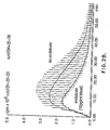
- Figure 2A is the chemiluminescent response with 5 minute pre-incubation with monoclonal antibody with and without 10 pg/ml endotoxins.
- Figure 2B is the same as Figure 2A except that the incubation period with antibody is 60 minutes.
- Figure 3A is the chemiluminescent response using blood from a patient with severe sepsis syndrome who died 6 hours after the sample was taken, as compared to a control antibody of the same class, isotype and concentration but directed against irrelevant epitopes.
- Figure 3B uses blood from a healthy ambulatory volunteer.
- Figure 3C uses blood from a patient with chronic sepsis.
- Figure 3D uses blood from a patient with severe sepsis syndrome which contributed to his death 3 days after the sample was taken. This patient had no evidence of gram-negative endotoxemia or bacteremia.
- Figure 3E uses blood from a patient being weaned from respiratory support and seriously cachectic, but with no clinical evidence of any septic foci.
- Figure 4 is the chemiluminescent response using blood from a patient with a leaky duodenal ulcer.
- Figure 5 is the whole blood chemiluminescence response using varying concentrations of endotoxin with varying concentrations of antibody against the endotoxin.
- the invention is a sensitive, specific and rapid detection method for antigens indicative of infection in whole blood or other body fluids containing white blood cell fractions and all related complement proteins based upon the specificity of antigen-antibody interactions and the high sensitivity of chemiluminescent light emission.
- the invention provides early diagnostic information for determining the amount of antigens indicative of the extent of sepsis and the amount of any mediators which provide information to indicate the extent or stage of sepsis. Results are obtained in minutes which is a great advantage over the previous time consuming methods of blood culturing for determining sepsis.
- the presence of bacteria, viruses or fungi or their related antigenic components in blood is indicative of an infection.
- the immune system's reaction to the presence of these foreign antigens by the production of pro-inflammatory mediators such as interleukin-1, tumour necrosis factor and interleukin-6 is also indicative of an infection.
- the presence and amount of antigens and mediators of sepsis indicates the level or stage of sepsis. For instance, at an early stage of gram-negative sepsis, antibody against gram-negative endotoxin (LPS) in the test of the present invention will detect the presence of LPS at a concentration as low as 5 pg/ml of whole blood.
- LPS gram-negative endotoxin
- TNF- ⁇ tumour necrosis factor
- IL-I interleukin-1
- LPS levels may decrease and TNF- ⁇ be absent, but IL-1 may increase and interleukin-6 (IL-6) may appear.
- IL-6 interleukin-6
- LPS may be present and IL-1 may be at low levels but IL-6 may be at very high levels.
- the rapid measurement of antigens and mediators can provide a measure of the patient's severity and duration of sepsis and ensure that the correct treatment is administered.
- the patient's own antibodies if produced to an antigen indicative of sepsis will not appear until 8 to 14 days after exposure to the antigen and thereby do not affect the present invention's antigen detection system.
- the present invention measures the amount of antigen and mediators in the patient's blood and provides this information rapidly to determine the patient's level of infection. This rapid test and the staging it provides for a septic patient is critical for the administration of appropriate treatment. If an antigen indicative of sepsis is present in a patient's blood, an antibody specific for the antigen indicative of sepsis can form an antigen/antibody complex at the right concentration of antibody. This is especially effective with monoclonal antibody. This antigen/antibody complex activates complement which in turn causes neutrophils and other white blood cells to produce oxidants. The white blood cells can be stimulated with opsonized zymosan resulting in increased production of oxidants.
- the oxidants cause an added chemiluminogenic compound, such as, luminol to release light energy.
- the amount of light emitted over time from a single test can be measured by a luminometer device to indicate the amount of antigen and hence the presence and extent of sepsis.
- the luminometer may be a Berthold model 481 luminometer which is available from Berthold Instruments Inc. in Austria.
- the addition of opsonized zymosan enhances the chemiluminescent response.
- a comparison control sample of the patient's blood is prepared by combining the blood with antibody of the same class non-specific for antigens indicative of sepsis.
- chemiluminescence to detect oxidant production is also described in the Robert C. Allen U.S. Patent 5,108,899.
- the Allen patent measures inflammation of a patient by comparing the extent of opsonin receptor expression on phagocytes with the maximum inducible opsonin receptor expression. The theory is that the less opsonin receptor expression may be induced, the greater the inflammation.
- the Allen patent uses agents which bind to phagocytes and stimulate opsonin receptor expression to give a maximum amount of chemiluminescence with zymosan.
- the present invention adds an antibody specific for an antigen or mediator of sepsis or infection. If an antigen or mediator is present then antibody/antigen or antibody/mediator complex is formed.
- the formation of antigen/antibody complexes in blood results in the activation of the classical complement pathway in the case of IgM complexes via C1q with the formation of complement anaphylatoxins and lytic products of C3, C4 and C5.
- a heat labile element produced during this process is responsible for the activation of white cell oxidant production (see Example XII) which results in the oxidation of luminol with subsequent emission of light.
- IgG immune complexes may modulate white cell oxidant production and chemiluminescence via a different mechanism which could involve binding to white cell Fc ⁇ receptors and subsequent formation of membrane attack complex followed by cell lysis.
- Leukocytes do not have Fc receptors for IgM immune complexes.
- a patient's blood is tested for the level of antigens and mediators which quantities indicate the stage of sepsis.
- Example X demonstrates that activation of white blood cells by immune complex formation is by a mechanism other than increased opsonin receptor expression.
- the Allen patent only indicates how much further opsonin receptor expression may be induced, but unlike the present invention, it does not indicate which antigens or mediators are present and at what levels nor does it provide the stage of sepsis.
- zymosan or latex beads are not a required addition to the test procedure, the measure of immune complexes in the test sample as compared to the control is enhanced by the addition.
- Zymosan and latex beads enhance the chemiluminescent response by stimulating concerted white cell oxidant production and phagocytosis.
- the addition of zymosan or latex acts as an amplification process to increase oxidant production but is not obligatory for the recognition of immune complex formation.
- the enhanced production of white cell oxidants stimulated by IgM immune complexes is not dependent upon increased opsonin receptor expression since the addition of a maximal stimulatory dose of C5a (which upregulates maximal opsonin receptor expression) to the blood during immune complex formation does not significantly affect the ratio of control to specific antibody dependent chemiluminescence when expressed as a light integral or peak count per minute either in the presence or absence of opsonized zymosan (see Example X).
- a sample of the patient's whole blood may be tested for the indication of sepsis.
- the sample of whole blood is mixed preferably with a monoclonal antibody, such as, Xomen®-E5, produced by Xoma in Palo Alto, California, a murine monoclonal IgM pentamer directed against the lipid A component of gram-negative endotoxin.
- a monoclonal antibody such as, Xomen®-E5
- a murine monoclonal IgM pentamer directed against the lipid A component of gram-negative endotoxin.
- Lipid A is the toxic inner core portion of the lipopolysaccharide (LPS) which is highly conserved and thereby is present on all gram-negative bacteria.
- LPS lipopolysaccharide
- the blood which may contain endotoxin, is incubated with the monoclonal antibody.
- a non-specific control murine antibody of the same isotype is incubated with a sample of the blood for comparison.
- luminol solution To the blood-antibody mixture is added luminol solution and then complement activated zymosan or complement opsonized latex beads.
- All analyses are preferably carried out in triplicate and the complete assay mixture in polystyrene or glass cuvettes is placed in a thermostated (37°C) chemiluminometer for repeated multiple readings of light emission intensity.
- a thermostated (37°C) chemiluminometer for repeated multiple readings of light emission intensity.
- the analysis is conducted, in accordance with one aspect of the invention, using 3 different dilutions of specific and control antibody each of which differ from the next highest concentration by one order of magnitude (ie 1:10, 1:100, 1:1000 dilution).
- the presence of antigen of interest, in this case, gram-negative endotoxin is confirmed by a statistically significant increase in integrated light intensity or reaction slope during the first 10 to 20 min. of reaction.
- the three different concentrations of antibody are used to discriminate and semi-quantitate the amount of endotoxin which is present.
- the principle of the triple concentration approach is based on the observation that maximal stimulation of white cell chemiluminescence in whole blood occurs when antigen-antibody equivalence is optimal for the formation of macromolecular crosslinked complexes or aggregates. In the presence of high concentrations of antigen, a high antibody concentration is required to yield such optimal equivalence. Similarly, at intermediate and low concentrations of antigen less concentrated antibody is required for optimal equivalence and macromolecular aggregate formation. This basic principle has been used for years in Ouchterlony diffusion plates and radial diffusion plates for immunometric quantitation of precipitin reactions.
- the whole blood chemiluminescent approach provides a semi-quantitative determination of the antigen concentration in question as high, intermediate or low with analogous concentration ranges (ie. > 100 pg/ml, 20-100 pg/ml, ⁇ 20 pg/ml).
- concentration ranges ie. > 100 pg/ml, 20-100 pg/ml, ⁇ 20 pg/ml.
- a whole blood sample may be used directly in this invention which simplifies the procedure, allowing it to be conducted bedside and provides the desired level of chemiluminescence.
- diluted whole blood or white blood cell fractions may also be used in this invention. Besides being more time consuming, fractionating procedures may result in a loss of subpopulations of neutrophils (Ristola, M. and Repo H., APMIS 503:97 (1989)).
- Using whole blood is much faster, easier and provides the chemiluminescent response above background noise.
- An added potential advantage of the utilization of whole blood rather than plasma or serum as the sample and partial reagent, is that it allows the detection of antigens which are cell associated or bound but still expose accessible epitopes. The use of antibodies against such epitopes may allow antibody binding with the receptor or binding sites on cells permitting a positive analytical signal which would not otherwise be detectable in a plasma or serum sample alone.
- the antibody against an antigen indicative of sepsis or against a mediator can be of the IgM class. Unlike IgG, IgM does not bind to the Fc receptors on the white blood cells. IgM-antigen complexes trigger a reaction sequence which results in the stimulation of white blood cell oxidant production via a heat labile plasma component. Thus white blood cell activation is dependent upon a classical complement pathway activation which is triggered by the formation of antigen-antibody complexes.
- the antibody against an antigen indicative of sepsis or against a mediator can also be of the IgG class.
- IgG binds to the Fc receptors on the white blood cells and can cause cytolysis. Under these circumstances, there is a decrease in the level of white blood cell activation from the normal level since white blood cells are lysed. This then also provides a semiquantitative measure of the level of antigen present and shows an especially dramatic drop in white blood cell activation with high levels of antigen or mediator.
- the chemiluminescent response is measured for at least 10 to 20 minutes to determine the extent of sepsis in comparison to a control. Chemiluminescence may be measured for an hour or longer, but over time the neutrophils appear to be progressively deactivated or their chemiluminescent response is exhausted or an inhibitory luminol metabolite is formed.
- Zymosan is a fungal polysaccharide which is optionally used to stimulate further, phagocytic cells. It is well known that phagocytic cells elicit a maximal phagocytic response when they can ingest particulate matter particularly via receptor mediated mechanisms. Although untreated zymosan can stimulate the phagocytic response, a much more exaggerated response can be achieved by opsonizing the zymosan first.
- the process of opsonization for this invention involves the binding of IgG's and complement factors iC3b and C3b to the zymosan. This process allows the opsonized zymosan to bind to three different classes of receptors on the phagocytic cells.
- Non-opsonized zymosan binds only to a subsite of the CD11b/CD18 integrin receptor which is different from the iC3b-CR 3 binding site. Due to its multiplicity of binding sites opsonized zymosan triggers a much more vigorous respiratory response in phagocytic cells than a comparable concentration of non-opsonized zymosan. In this invention opsonized zymosan is used to maximally elicit the oxidative response of the white blood cells.
- Opsonized zymosan white blood cell activation is dependent upon the concentration of CR 1 and CR 3 receptors on the surface of the phagocytes.
- complement opsonized zymosan minimal IgG
- any type of opsonized zymosan may be used, for example, IgG opsonized or complement and immunoglobulin opsonized. It was also shown that complement opsonized latex beads could be used to maximally elicit the oxidative response of the white blood cells.
- the preferred aspect of this invention uses a small volume of undiluted whole blood (20 ⁇ l or 50 ⁇ l) heparinized ( ⁇ 2 U/ml) or EDTA anticogulated and kept at room temperature.
- the blood sample is incubated with an equal volume of antibody (0.2, 0.02 and 0.002 mg/ml) at 37°C for five to ten minutes. After incubation, 200 ⁇ l of 40 ⁇ M luminol solution is added followed by 50 ⁇ l of complement opsonized zymosan. 2.5 - 3.0 x 10 9 particles/ml.
- the sample is read in thermostated (37°C) luminometer.
- whole blood is first combined with luminol solution and then antibody is added and the mixture incubated at 37°C for five to ten minutes. Zymosan may then be added.
- the examples describe alternatives to these aspects, such as varying the order in which to add the reagents, varying blood dilutions, and omitting zymosan.
- the whole blood sample may instead be sub-fraction of white blood cells, such as neutrophils or lymphocytes or monocytes.
- Another chemiluminescent compound besides luminol may be used, such as, lucigenin or pholasin.
- the quantum yield of this process is of the order of 1%, that is, only 1% of the energy of reaction is emitted as light.
- the major oxidant produced by white cells which is thought in accordance with this invention to excite luminol, has been shown to be HOCl although a superoxide anion which is also an oxidant produced by white cells can also excite the dye predominantly at alkaline pH.
- the primacy of HOCl as the oxidant species which triggers luminol dependent chemiluminescence has been confirmed in whole blood studies using sodium azide as an inhibitor of myeloperoxidase (Allen, R.C. Methods of Enzymology 133:449 (1986). Under these conditions greater than 95% of the chemiluminescent light emission is blocked.
- Other chemiluminescent dyes which produce light as a result of neutrophil oxidant production have also been identified including lucigenin and pholasin.
- the respiratory burst activity provides reducing equivalents for univalent reduction of oxygen via the hexose monophosphate shunt in the form of NADPH which is used as an electron source for the membrane associated oxidase complex which generates superoxide anion O 2 - into the phagolysosome or the extracellular space.
- NADPH hexose monophosphate shunt
- This primary oxidant production in concert with the degranulation of azurophil granules results in the formation of more powerful oxidants such as HOCl which act in concert with neutrophil proteases to kill bacteria and inactivate viral particles.
- the Allen method does not measure the presence of antigens indicative of sepsis or mediators in the blood.
- the present invention does measure the presence of such antigens or mediators by the formation of immune complexes using exogenously administered antibody.
- the Allen method instead measures the degree of phagocyte activation and oxidant production via opsonin receptor expression.
- the present invention uses the white blood cell excitation to measure the presence of antigens indicative of sepsis.
- the antibodies of the murine monoclonal IgM class are directed against gram-negative endotoxin lipid A (a component of lipopolysaccharide (LPS)). These antibodies do not have complementary Fc receptors on neutrophils or any other white cell subtype (Unkeless, J.C., Boros, P., Fein, M., Inflammation, Basic Principles and Clinical Correlates, 2nd Ed., 497-510; Gallin, J.I., Goldstein, R., Synderman, R., Editors Raven Press, N.Y. (1992).
- LPS lipopolysaccharide
- the detection of fluid phase antigen-antibody complexes is based on their ability to fix complement and stimulate white cell oxidant production compared to a control sample containing an irrelevant isotype-matched antibody.
- the subsequent stimulation of the neutrophils with a non-rate limiting dose of zymosan allows an enhanced signal to be generated by maximal stimulation of phagocytosis. In the absence of antigen no such enhanced response is elicited. Complement opsonized zymosan is added to maximize the phagocytic response of neutrophils.
- a control comparison sample is co-analyzed with an identical subclass and concentration of antibody which is directed against an irrelevant epitope not found in human blood.
- chemiluminescent response in the control tube with irrelevant antibody to the tube containing specific antibody against the antigen of interest, it is possible to obtain a semi-quantitative estimate of the concentration of antigen after an assay period of 20 minutes or less.
- the presence of antigen is confirmed by an enhanced chemiluminescence response during the early assay period in the tubes containing specific antibody.
- One of the examples confirms that E. coli bacteria or lipopolysaccharide added to the blood test sample had no detectable effect on neutrophil chemiluminescence during the time course ( ⁇ 60 minutes) of the observed assay in the absence of antibody against LPS.
- antibodies of the murine monoclonal IgG class are directed against Hepatitis A virus. These antibodies do have complementary Fc receptors on white blood cells. The binding of the antigen/antibody complex on the Fc receptors may cause the white blood cells to lyse, thus attenuating their oxidant burst. The attenuation of white blood cell oxidated metabolism at an appropriate antibody concentration due to cell lysis or inhibition of respiratory burst activity may be used to signal the presence of Hepatitis A virus.
- any IgM/antigen complexes will activate complement and cause increased oxidant production of white blood cells, whereas IgG/antigen complexes may bind to Fc receptors on white blood cells and cause them to lyse, thus reducing oxidant production from baseline levels.
- a unique feature of the present invention is that the patient'sown finely tuned immune recognitive systems are used to provide the analytical signal. This approach is unique because the patient's blood is pre-incubated with either control or target antibody for five to ten minutes prior to the addition of luminol and opsonized zymosan and then the chemiluminescent light emission signal is measured for a period of 10 to 20 minutes. A comparison of calculated slopes or integrated light intensity of the response to control or specific antibody then allows a decision to be made regarding the presence or absence of the antigen. In order to guarantee optimal reaction conditions for antibody three concentrations of antibody can be used, each with a matched control.
- Example I Verification of Antibody (Xomen®-E5) Binding to Gram-Negative Endotoxin as Assessed by Turbidimetric Assay.
- Murine monoclonal antibody (Xomen®-E5, stock concentration 2 mg/ml supplied in sterile pyrogen free saline) against gram-negative endotoxin was diluted 10 to 10,000 fold in sterile, pyrogen free Hanks Balanced Salt solution (HBSS) at 10 fold increments. Solid polyethylene glycol 6000 was added to the antibody dilutions to give a final concentration of 6% w/v.
- the Purified gram-negative endotoxin (E. coli serotype 055:B5, grade V, Sigma St. Louis Miss.) was dissolved in HBSS at concentrations of 100, 10, 1 and 0.1 ⁇ g/ml (w/v).
- Table 1 summarizes the turbidimetric antigen-antibody interaction of the monoclonal murine IgM antibody directed against the Lipid A epitope of gram-negative endotoxin with purified E. coli gram-negative endotoxin (LPS).
- LPS E. coli gram-negative endotoxin
- Table 2 illustrates the chemiluminescent response to endotoxin at varying concentrations of anti-LPS antibody.
- Each group of six cuvettes contains the same concentration of antibody ranging from 100 ⁇ g/ml to 0.01 ⁇ g/ml in 10 fold increments.
- the last three tubes in each group contain blood which was supplemented with endotoxin to a concentration of 1 ng/ml.
- the integrals of the cumulative light intensity are used to determine whether a significant difference exists between triplicate tubes with and without endotoxin at any particular antibody concentration.
- the suppression of the CL response at high concentrations of antibody may be due to destruction of neutrophils bound with endotoxin-anti-endotoxin antibody complexes due to complement mediated lysis of the cells or the inhibition of zymosan activation due to blockade of opsonin receptors by antigen-antibody-complement complexes.
- Example III was designed to examine whether endotoxin alone had any effects on CL in the absence of antibody under the conditions of the assay. At a dose of 1000 pg/ml of whole blood the endotoxin had no effect on the chemiluminescence response. In Table 3A none of the samples contained antibody. As indicated in Table 3b the addition of antibody to a final assay concentration of 1.3 ⁇ g/ml had no effect on the shape or magnitude of the CL response in the absence of endotoxin. The presence of endotoxin, however caused an augmentation of the CL response both in terms of increased CL maximum, and increased initial acceleration slope observed during the first 15 minutes of the reaction. These results confirmed the observation that the presence of antigen-antibody complexes could be used to enhance zymosan activated whole blood chemiluminescence and hence detect whether LPS was present in whole blood.
- the final concentration of monoclonal antibody in the reaction mixture was varied from 0.2 ⁇ g/ml to 0.0025 ⁇ g/ml in dilution increments of 3 fold. All assays were analyzed in triplicate and the reactions were initiated by the addition of opsonized zymosan to the reaction mixture. The chemiluminescent response was monitored for 50 minutes at 37°C. Chemiluminescent curve integrals were taken from the time of zymosan addition until 5 minutes of the initial acceleration phase of the reaction for comparison of responses. All integrals were compared to the parallel control containing an equivalent concentration of monoclonal antibody but no endotoxin (Table 4).
- Example IV The possibility that one antibody concentration could span a range of CL response from 0 to 1000 pg/ml of endotoxin was investigated in Example IV.
- the assay results are summarized in Table 4. After careful inspection of the data obtained over a 50 minute assay period it was observed that the best signal to noise ratio was achieved by considering CL curve integrals over the first five minute acceleration phase of the reaction. This data is tabulated in the "Integral" column of Table 4. The starting antibody dilution in this experiment was 0.2 ⁇ g/ml. All subsequent dilutions of antibody were made in three fold steps.
- Figure 1 graphically presents the CL data obtained from the reaction mixtures which gave the largest response ratio for endotoxin at a concentration of 100 pg/ml of whole blood.
- the plotted data emphasizes the difference in the initial slope of the reactions and in the CL maxima. An adequate differentiation of the signals was clearly evidenced after only 5 minutes of reaction emphasizing the rapid diagnostic potential of the assay.
- the standard chemiluminomiter measure emitted luminescent light by use of the standard type of electronic photo counter. Periodically as plotted along the X-axis in minutes the light emission is measured based on the photon counts per minute (cpm). The cpm value is then plotted on the Y-axis whereby over time the respective curves are developed.
- the presence of endotoxin in the sample results in a steeper reaction slope during the acceleration phase of the reaction and a higher CL maximum light emission. In many samples, the time to reach CL maximum is shortened by the presence of endotoxin.
- Example V Chemiluminescent Response of Whole Blood Containing In Vitro Added LPS (Gram-negative Endotoxin) with Addition of Antibodies to LPS but no Zymosan. (ie Chemiluminescent Response in the Absence of an Opsonin Receptor Agonist)
- the intensity of the CL signal with zymosan is approximately 50-100 times higher than without. For this reason opsonized zymosan is used in this invention to increase the magnitude of the response and improve signal to noise characteristics.
- Panel C there was a dose response relationship between endotoxin concentration and the magnitude of CL integral.
- the biphasic nature of the CL response implies that the early peak which occurs in the presence of LPS is due to intracellular oxidant production and the later progressive rise is due to extracellular oxidant release which is temporally later in the sequence of cell activation.
- the general assay protocol may be used without the inclusion of opsonized zymosan to prime and excite a maximal respiratory burst activity.
- Table 6a which summarizes the response to LPS with a short pre-incubation of antibody for 5 minutes
- the best response ratio was achieved at an antibody concentration of 200 ⁇ g/ml which resulted in an almost 2 fold signal enhancement in the presence of 10 pg/ml of endotoxin.
- Table 6b summarizes a parallel experiment except that samples were pre-incubated with anti-LPS antibody for 60 minutes prior to the addition of opsonized zymosan. In this case pre-incubation with antibody for 1 hour resulted in a decreased CL response in the endotoxin containing samples with an antibody concentration of 200 ⁇ g/ml.
- FIG. 2A exemplifies the maximal stimulatory effect observed with the short pre-incubation period.
- Figure 2B graphically illustrates the inhibitory effect which occurs with identical antibody concentrations but a long pre-incubation time. The short pre-incubation strategy is preferred since it allows signal amplification rather than a CL suppression in the presence of LPS.
- Example VII Dose-Response of Whole Blood Chemiluminescence Reaction to Increasing Concentrations of Endotoxin at a Fixed Concentration of Antibody Utilizing Control (Non-Specific Murine Myeloma IgM) and Anti-LPS Murine IgM Antibody
- Example VII examines whether a differential CL dose-response could be achieved under assay conditions which would readily detect small concentrations of endotoxin.
- Table 7 there was not a significant difference in the initial velocities of the CL response in the absence of endotoxin, the mean slope for the linear portion of the control curves was 0.113 x 10 6 cpm/min, for the anti-LPS curves the mean slope was 0.133 x 10 6 cpm/min).
- the mean slope for the anti-LPS antibody curves was 0.159 x 10 6 cpm/min, while the slope for the control antibodies remained at 0.114 x 10 6 cpm/min.
- Figure 3C uses blood from a patient with chronic sepsis.
- Figure 3D uses blood from a patient with severe sepsis syndrome who died 3 days after the sample was taken but had no evidence of gram-negative infection.
- Figure 3E uses blood from a patient with no clinical evidence of any septic foci but with severe cachexia. All blood transfer and reagent dispensing for both assays was accomplished using endotoxin free pipettes and sterile practice.
- the chemiluminescence assay mixture was composed of 200 ⁇ l of luminol solution, 50 ⁇ l of undiluted anti-coagulated whole blood, 50 ⁇ l of antibody (conc. 0.2 mg IgM/ml) and 50 ⁇ l of complement opsonized zymosan.
- Example VIII the response of the CL assay for endotoxin was compared to the standard Limulus Amebocyte Lysate (LAL) assay which is used by the pharmaceutical industry as a reference method for the detection of gram-negative endotoxin.
- LAL Limulus Amebocyte Lysate
- Figure 3A displays the chemiluminescent response of blood taken from the radial artery of a patient with severe sepsis syndrome who died 6 hours after the sample was taken.
- the cause of death was hypotensive shock which was refractory to inotropic support.
- Figure 3B illustrates the CL profile of a healthy ambulatory volunteer and shows no differential response to anti-LPS antibody which was confirmed by LAL assay to indicate the absence of LPS in the blood.
- Figure 3C displays the CL response of a patient with chronic sepsis which was confirmed by blood culture to be primarily due to a beta haemolytic gram positive streptococcus. The CL assay indicated that this patient also had a response consistent with a low level of gram-negative septicemia which was below the limits of detection when assayed by LAL. The limit of detection using the LAL assay was a whole blood concentration of 50 pg/ml LPS.
- FIG. 3D displays the CL response of a patient who had severe sepsis syndrome which ultimately contributed to his death 3 days after the blood sample used for the analysis was taken.
- the CL analysis indicated no evidence of LPS in the blood which was confirmed by LAL assay.
- the microbiological reports on culture material for this patient suggested that he had gram positive sepsis.
- Figure 3E represents the results of a CL assay for LPS conducted on blood obtained from a patient who was being weaned from respiratory support and was previously cachectic, but had no clinical evidence of any septic foci.
- the LAL assay confirmed the absence of endotoxin.
- a preferred approach for testing patient samples for endotoxin is based upon the following assay conditions: 20 microlitres of the patient's blood (EDTA anti-coagulated) is mixed with 20 ⁇ l (microlitre) of antibody (three different dilutions are used, 0.2, 0.02 and 0.002 mg/ml in an endotoxin free assay cuvette. The mixture is incubated for 10 minutes at 37°C and then 200 ⁇ l of luminol solution (40 ⁇ M) is added (pre-equilibrated to a temperature of 37°C) followed by 50 ⁇ l of complement opsonized zymosan 2.5 - 3.0 x 10 9 particles/ml. Measurement of emitted light is then initiated in the chemiluminometer.
- This assay was designed to minimize the amount of antibody necessary to evoke a significant chemiluminescence enhancement in the presence of gram-negative endotoxin. For this reason only patient sample and the antibody are incubated in the first phase of the reaction sequence in order to maximize effective antibody antigen complex formation. This preferred format has been adopted for patient studies.
- Figure 4 demonstrates clearly the difference in the chemiluminescence levels for the patient as compared to the control using an antibody concentrations of 0.2 mg/ml.
- complement anaphylatoxin C5a at a dose which stimulates maximum opsonin receptor expression had no effect on the ratio of integrated light intensity observed in the presence of control antibody versus specific anti-LPS antibody.
- concentration of anti-LPS IgM antibody and the irrelevant IgM antibody are 0.2 mg/ml and the concentration of C5a is 10 pg/sample.
- the patient chosen for this study had high levels of LPS (>700 pg/ml) in his blood as determined independently by LAL assay using the plasma assay procedure protocol of BioWhittaker Inc. (Walkerville MD) or Seikagaku Kogyo (Tokyo, Japan).
- a blood sample from this patient was obtained for assay of endotoxin using control and anti-LPS antibody in the presence and absence of complement opsonized zymosan.
- the addition of C5a at a dose which recruits maximum opsonin receptor expression had no effect on the ratio of integrated light intensity observed with control versus anti-LPS antibody.
- the C5a was added to the blood samples immediately after the 5 minutes incubation of blood with antibody and the samples were then incubated an additional 5 minutes at 37°C prior to the addition of luminol solution.
- C5a In the absence of opsonized zymosan, C5a increased the whole blood chemiluminescence of both control and anti-LPS assay samples but had no effect on the ratio of control versus anti-LPS antibody integrated light intensity. Similarly in the presence of complement opsonized zymosan, C5a had no effect on the same ratio. In addition C5a had a minimal effect on the opsonized zymosan containing assay tubes, causing only a slight increase in chemiluminescent light release. This is indicative of the fact that the patient's blood already had virtually maximal opsonin receptor expression in the absence of C5a. The addition of C5a only increased the chemiluminescent response by about 10% in assay samples containing opsonized zymosan.
- a baseline sample is included containing the specific antibody but no LPS.
- a baseline sample is included at each concentration of antibody used.
- This study was conducted using the standard assay format. Fifty microlitres of EDTA anti-coagulated whole blood was mixed with 50 microlitres of the respective antibody concentration and incubated at 37°C for 5 minutes followed by the addition of 200 microlitres of luminol solution and then 50 microlitres of complement opsonized zymosan. The integrated light intensity was collected for 30 minutes and is expressed on a relative scale in Figure 5. This experiment illustrates the dependence of the chemiluminescence signal on the optimal antigen - antibody equivalence.
- E. Coli was isolated from a patient with gram negative sepsis. An isolate of the bacterium was cultured on agar and suspended in liquid medium at a density of approximately 1 million bacteria per ml. Aliquots of this mixture were added to whole blood and tested for chemiluminescence response in the presence of murine ascites fluid containing an IgM directed against LPS epitopes.
- the source of the monoclonal IgM antibody was QED Laboratories in LaJolla, California, U.S.A. Experiments showed the ability to detect bacteria at concentrations of 10 4 bacteria per ml of whole blood and less using the IgM in murine ascites fluid added to whole blood.
- a murine monoclonal antibody to human Hepatitis A virus of the IgG class was tested for its ability to detect the virus in whole blood at antibody dilutions of 1/50, 1/200 and 1/1,000 against antigen dilutions of 1/50 and 1/500 of the stock viral antigen solution which contained approximately 100 ⁇ g/ml of viral protein.
- the Hepatitis A virus was killed and was at a protein concentration of 100 ⁇ g/ml and the anti-Hepatitis A antibody was a murine IgG at a protein concentration of 1 mg/ml.
- the Hepatitis A virus and antibody were available from ADI of Mississauga, Ontario, Canada. The test procedure was that of Example IX.
- monoclonal antibodies against inflammatory mediators such as, tumour necrosis factor, interleukins 1, 6, 8, Interferon ⁇ and transforming growth factor ⁇ , will indicate the presence and degree of sepsis in this invention.
- An added diagnostic feature of this invention is that it can be used to titrate a patient's therapeutic antibody titre and to evaluate the effectiveness of antibody therapy in reducing endotoxin levels.
- By conducting a simultaneous panel of tests using antibodies against TNF, IL-1, IL-6 and other markers of sepsis it will be possible to establish a "sepsis panel" to indicate the degree and magnitude of sepsis progression.
- a septic panel would use a combination of monoclonal antibodies against bacteria, virus or fungae and inflammatory mediators. Each tube could contain a different monoclonal antibody to test the patient sample for a variety of septic indicators.
- One panel kit could include antibodies to the most common types of bacterial toxins, for example, antibody against gram-negative bacterial endotoxin and antibody against gram-positive bacteria lipotechoic acid. Another panel kit could have antibodies against the viral or fungal antigens which are associated with sepsis. Another panel kit could have antibodies against the inflammatory mediators to determine the patient's immune activation in response to sepsis.
- this invention allows for the diagnosis and staging of patients septic condition and also the evaluation of the efficacy of therapeutic treatments.
- a sepsis panel provides information on a range of sepsis indicators to better indicate the degree of progression of sepsis. Identifying those patients at risk of sepsis is important for initiating effective therapeutic treatment.
- the invention is easily performed, requiring only three reagents and a luminometer. This test can be used to serially monitor a patient, for example by conducting the test three or four times a day or more as clinical conditions or therapeutic interventions warrant.
- LPS (5 pg/ml) Control antibody AntiLPS antibody 0.114 0.159 PANEL C.
- LPS 50 pg/ml Control antibody AntiLPS antibody 0.116 0.219 PANEL D.
- LPS 500 pg/ml Control antibody AntiLPS antibody 0.113 0.226
Landscapes
- Health & Medical Sciences (AREA)
- Life Sciences & Earth Sciences (AREA)
- Engineering & Computer Science (AREA)
- Immunology (AREA)
- Molecular Biology (AREA)
- Hematology (AREA)
- Chemical & Material Sciences (AREA)
- Urology & Nephrology (AREA)
- Biomedical Technology (AREA)
- Cell Biology (AREA)
- Physics & Mathematics (AREA)
- Biochemistry (AREA)
- Pathology (AREA)
- Food Science & Technology (AREA)
- Medicinal Chemistry (AREA)
- Biotechnology (AREA)
- Analytical Chemistry (AREA)
- Microbiology (AREA)
- General Health & Medical Sciences (AREA)
- General Physics & Mathematics (AREA)
- Tropical Medicine & Parasitology (AREA)
- Proteomics, Peptides & Aminoacids (AREA)
- Physiology (AREA)
- Virology (AREA)
- Investigating Or Analysing Materials By The Use Of Chemical Reactions (AREA)
- Investigating Or Analysing Biological Materials (AREA)
Claims (23)
- Procédé pour déterminer l'étendue d'une infection chez un patient animal ou humain en détectant la quantité d'un antigène indicateur d'une telle infection, la quantité dudit antigène étant détectée dans un échantillon de test dérivé du sang du patient contenant des fractions de globules blancs comprenant toutes les protéines de complément, ledit procédé comprenant :i) incuber ledit échantillon de test avec une quantité d'anticorps de test spécifiques audit antigène indicateur d'une infection pour former des complexes anticorps/antigène,ii) permettre audits complexes anticorps/antigène d'interagir avec lesdites fractions de globules blancs, ce qui a pour conséquence la production d'oxydants,iii) introduire, dans soit l'étape i) ou l'étape ii), un composé chimioluminescent dans ledit échantillon de test,iv) lesdits oxydants réagissant avec lesdits composés chimioluminescents pour émettre une lumière luminescente à partir dudit échantillon de test,v) mesurer ladite quantité de lumière émise pendant une période prédéterminée, etvi) corréler l'étendue de l'infection en comparant la quantité mesurée de lumière émise de l'échantillon de test avec la quantité mesurée de lumière émise par un échantillon témoin qui est traité de la même manière que l'échantillon de test pour les étapes i) à v) à l'exception que, dans l'étape i), des anticorps témoins sont utilisés qui sont de la même classe que les anticorps de test mais sont non-spécifiques audits antigènes indicateurs de l'infection.
- Procédé selon la revendication 1, dans lequel ledit échantillon est du sang entier, ou comprend des fractions de globules blancs dérivées du sang entier, lesdites fractions incluant des neutrophiles, des lymphocytes et/ou des monocytes.
- Procédé selon la revendication 1 ou 2, dans lequel du zymosan, du zymosan opsonisé ou des billes de latex opsonisées sont ajoutés audit échantillon avant l'étape iv).
- Procédé selon l'une quelconque des revendications 1-3, dans lequel ledit composé chimioluminescent est choisi parmi le groupe constitué du luminol, du lucigène, et de la pholasine.
- Procédé selon l'une quelconque des revendications 1-4, dans lequel l'échantillon de test et l'échantillon témoin dans l'étape i) sont incubés avec trois rapports d'anticorps vis-à-vis de la solution, lesdits rapports étant 1:10, 1:100 et 1:1000, ladite comparaison entre la lumière mesurée à partir de l'échantillon de test et la lumière mesurée à partir de l'échantillon témoin étant corrélée à la quantité d'antigène avec le rapport qui fournit la plus grande différence dans les émissions de lumière.
- Procédé selon l'une quelconque des revendications 1-5, dans lequel l'anticorps est un anticorps monoclonal choisi dans la classe de IgM et IgG.
- Procédé selon la revendication 6, dans lequel l'antigène est dérivé de bactéries gram-négatives, par exemple l'endotoxine lipide A, de bactéries gram-positives, de virus, par exemple l'antigène viral est le virus de l'hépatite A, ou de champignons.
- Kit de diagnostic utilisable pour déterminer, conformément à un procédé selon l'une quelconque des revendications 3-7, l'étendue d'une infection chez un patient animal ou humain en détectant la quantité d'antigène indicateur d'une infection dans un échantillon de test dérivé du sang du patient contenant des fractions de globules blancs, ledit kit comprenant :i) un premier conteneur d'anticorps spécifique pour l'antigène indicateur de l'infection,ii) un second conteneur de composé chimioluminescent, etiii) un troisième conteneur de zymosan.
- Kit de diagnostic selon la revendication 8, dans lequel ledit anticorps est choisi dans la classe de IgM et IgG.
- Kit de diagnostic selon la revendication 8 ou 9, dans lequel ledit composé chimioluminescent est choisi à partir des composés constitués du luminol, du lucigène, et de la pholasine.
- Kit de diagnostic selon la revendication 9 ou 10, dans lequel ledit anticorps IgM est contre l'endotoxine gram-négative lipide A, ou ledit anticorps IgG est contre le virus de l'hépatite A.
- Kit de diagnostic selon l'une quelconque des revendications 8-11, dans lequel ledit zymosan est opsonisé.
- Procédé pour déterminer l'étendue de la septicémie chez un patient animal ou humain en détectant la quantité de marqueurs associés à la septicémie dans un échantillon de test dérivé du sang du patient contenant des fractions de globules blancs comprenant toutes les protéines de complément, ledit procédé comprenant :i) incuber ledit échantillon de test avec une quantité d'anticorps de test spécifiques à un marqueur associé à la septicémie choisi pour former des complexes anticorps/marqueur,ii) permettre audits complexes anticorps/marqueur d'interagir avec lesdites fractions de globules blancs, ce qui a pour conséquence la production d'oxydants,iii) introduire, dans soit dans l'étape i) ou dans l'étape ii), un composé chimioluminescent dans ledit échantillon de test,iv) lesdits oxydants réagissant avec lesdits composés chimioluminescents pour émettre une lumière luminescente à partir dudit échantillon de test,v) mesurer ladite quantité de lumière émise pendant une période prédéterminée, etvi) corréler l'étendue de la septicémie en comparant la quantité mesurée de lumière émise de l'échantillon de test à la quantité mesurée de lumière émise par un échantillon témoin qui est traité de la même manière que l'échantillon de test pour les étapes i) à v) à l'exception que, dans l'étape i), des anticorps témoins sont utilisés qui sont de la même classe que les anticorps de test mais sont non-spécifiques audits marqueurs associés à la septicémie.
- Procédé selon la revendication 13, dans lequel ledit échantillon est du sang entier, ou comprend des fractions de globules blancs dérivées du sang entier, lesdites fractions incluant des neutrophiles, des lymphocytes et/ou des monocytes.
- Procédé selon la revendication 13 ou 14, dans lequel l'échantillon de test et l'échantillon témoin dans l'étape i) sont incubés avec trois rapports d'anticorps vis-à-vis de la solution, lesdits rapports étant 1:10, 1:100 et 1:1000, ladite comparaison entre la lumière mesurée à partir de l'échantillon de test et la lumière mesurée à partir de l'échantillon témoin étant corrélée à la quantité d'antigène avec le rapport qui fournit la plus grande différence dans les émissions de lumière.
- Procédé selon l'une quelconque des revendications 13-15, dans lequel l'anticorps est un anticorps monoclonal choisi dans la classe de IgM et IgG.
- Procédé selon l'une quelconque des revendications 13-16, dans lequel le marqueur associé à la septicémie est un médiateur inflammatoire, de préférence choisi dans le groupe constitué du facteur de nécrose tumorale, de l'interleukine-1, de l'interleukine-6, de l'interleukine-8, et de l'interféron et du facteur de croissance transformant β.
- Kit de diagnostic utilisable pour déterminer, conformément à un procédé selon l'une quelconque des revendications 13-17, l'étendue de la septicémie chez un patient animal ou humain en détectant la quantité d'un marqueur associé à la septicémie dans un échantillon de test dérivé du sang du patient contenant des fractions de globules blancs, ledit kit comprenant :i) un premier conteneur d'anticorps spécifique pour le marqueur associé à la septicémie,ii) un second conteneur pour un composé chimioluminescent, etiii) un troisième conteneur de zymosan.
- Kit de diagnostic selon la revendication 18, dans lequel ledit marqueur associé à la septicémie est un médiateur inflammatoire, de préférence choisi dans le groupe constitué du facteur de nécrose tumorale, de l'interleukine-1, de l'interleukine-6, de l'interleukine-8, et de l'interféron et du facteur de croissance transformant β.
- Kit de diagnostic selon la revendication 18 ou 19, dans lequel ledit anticorps est choisi dans la classe de IgM et IgG.
- Kit de diagnostic selon l'une quelconque des revendications 18-20, dans lequel ledit composé chimioluminescent est choisi parmi les composés constitués du luminol, du lucigène, et de la pholasine.
- Kit de diagnostic selon l'une quelconque des revendications 18-21, dans lequel ledit zymosan est opsonisé.
- Procédé pour détecter la quantité d'un antigène dans un échantillon de test d'un fluide corporel d'un patient animal ou humain en présence de globules blancs, de préférence des neutrophiles, et de toutes les protéines de complément, ledit procédé comprenant :i) incuber ledit échantillon de test avec une quantité d'anticorps de test spécifiques audit antigène pour former des complexes anticorps/antigène,ii) permettre audits complexes anticorps/antigène d'interagir avec lesdits globules blancs et ledit complément, ce qui a pour conséquence la production d'oxydants,iii) introduire, dans soit l'étape i) ou l'étape ii), un composé chimioluminescent dans ledit échantillon de test,iv) lesdits oxydants réagissant avec lesdits composés chimioluminescents pour émettre une lumière luminescente à partir dudit échantillon de test,v) mesurer ladite quantité de lumière émise pendant une période prédéterminée, etvi) corréler la quantité dudit antigène en comparant la quantité mesurée de lumière émise de l'échantillon de test avec la quantité mesurée de lumière émise par un échantillon témoin qui est traité de la même manière que l'échantillon de test pour les étapes i) à v) à l'exception que, dans l'étape i), des anticorps témoins sont utilisés qui sont de la même classe que les anticorps de test mais sont non-spécifiques audit antigène.
Applications Claiming Priority (3)
| Application Number | Priority Date | Filing Date | Title |
|---|---|---|---|
| CA2097952 | 1993-06-08 | ||
| CA002097952A CA2097952C (fr) | 1993-06-08 | 1993-06-08 | Diagnostic precoce de la sepsie au moyen des interactions antigene-anticorps amplifiees par chimiluminescence du sang total |
| PCT/CA1994/000325 WO1994029728A1 (fr) | 1993-06-08 | 1994-06-08 | Diagnostic precoce de sepsie utilisant des interactions antigene-anticorps amplifiees par chimioluminescence du sang entier |
Publications (2)
| Publication Number | Publication Date |
|---|---|
| EP0705434A1 EP0705434A1 (fr) | 1996-04-10 |
| EP0705434B1 true EP0705434B1 (fr) | 1998-10-21 |
Family
ID=4151762
Family Applications (1)
| Application Number | Title | Priority Date | Filing Date |
|---|---|---|---|
| EP94918705A Expired - Lifetime EP0705434B1 (fr) | 1993-06-08 | 1994-06-08 | Diagnostic precoce de sepsie utilisant des interactions antigene-anticorps amplifiees par chimioluminescence du sang entier |
Country Status (6)
| Country | Link |
|---|---|
| EP (1) | EP0705434B1 (fr) |
| AT (1) | ATE172543T1 (fr) |
| CA (1) | CA2097952C (fr) |
| DE (1) | DE69414110T2 (fr) |
| ES (1) | ES2122292T3 (fr) |
| WO (1) | WO1994029728A1 (fr) |
Families Citing this family (8)
| Publication number | Priority date | Publication date | Assignee | Title |
|---|---|---|---|---|
| US6306614B1 (en) * | 1994-06-08 | 2001-10-23 | Sepsis, Inc. | Measurement of analytes in whole blood |
| US6203997B1 (en) | 1994-06-08 | 2001-03-20 | Sepsis, Inc. | Quantitation of analytes in whole blood |
| US5804370A (en) * | 1994-06-08 | 1998-09-08 | Critichem Medical Products Limited | Early diagnosis of sepsis utilizing antigen-antibody interactions amplified by whole blood chemiluminescence |
| US6159683A (en) * | 1997-12-16 | 2000-12-12 | Spectral Diagnostics, Inc. | Method of determining stage of sepsis |
| FI115165B (fi) | 2001-06-04 | 2005-03-15 | Aboatech Ab Oy | Menetelmä infektion laadun määrittämiseksi |
| DE102017003312A1 (de) * | 2017-04-05 | 2018-10-11 | Rolf Günther | Nachweisverfahren und Vorrichtung |
| WO2022136850A1 (fr) * | 2020-12-22 | 2022-06-30 | Seroxo Limited | Méthodes pour diagnostiquer et surveiller une sepsie |
| CN114612799B (zh) * | 2022-03-11 | 2022-09-16 | 应急管理部国家自然灾害防治研究院 | 基于滑坡/非滑坡面积比的空间自适应正负样本生成方法及系统 |
Family Cites Families (8)
| Publication number | Priority date | Publication date | Assignee | Title |
|---|---|---|---|---|
| CA1210328A (fr) * | 1982-12-14 | 1986-08-26 | James J. Capone | Methode et compose pour l'evaluation de la phagocytose |
| JPS60200167A (ja) * | 1984-03-24 | 1985-10-09 | Toyobo Co Ltd | 化学発光法による過酸化水素の定量法 |
| US4959302A (en) * | 1987-08-20 | 1990-09-25 | Endogen, Inc. | Method of evaluating immune system response to allografts, viral infection and immunosuppressive therapy |
| US5223395A (en) * | 1988-12-01 | 1993-06-29 | Centocor, Inc. | Immunometric assays for tumor necrosis factor-alpha and methods for preventing the loss of biological activity of tumor necrosis factor-alpha in biological samples |
| US5294541A (en) * | 1989-09-21 | 1994-03-15 | The United States Of America As Represented By The Secretary Of The Department Of Health And Human Services | Real-time monitoring of oxidative products from in vitro cell-biomaterial interaction using chemiluminescence |
| US5108899A (en) * | 1989-10-31 | 1992-04-28 | Exoxemis, Inc. | Chemiluminescence assay of in vivo inflammation |
| WO1992003734A1 (fr) * | 1990-08-20 | 1992-03-05 | Alain De Weck | Procede de mesure des reactions des lymphoctytes t par analyses chimioluminescentes |
| CA2736076A1 (fr) * | 1991-03-18 | 1992-09-19 | New York University | Anticorps monoclonaux et chimeriques specifiques du facteur de necrose tumorale humain |
-
1993
- 1993-06-08 CA CA002097952A patent/CA2097952C/fr not_active Expired - Lifetime
-
1994
- 1994-06-08 ES ES94918705T patent/ES2122292T3/es not_active Expired - Lifetime
- 1994-06-08 AT AT94918705T patent/ATE172543T1/de not_active IP Right Cessation
- 1994-06-08 DE DE69414110T patent/DE69414110T2/de not_active Expired - Lifetime
- 1994-06-08 WO PCT/CA1994/000325 patent/WO1994029728A1/fr not_active Ceased
- 1994-06-08 EP EP94918705A patent/EP0705434B1/fr not_active Expired - Lifetime
Also Published As
| Publication number | Publication date |
|---|---|
| AU6992394A (en) | 1995-01-03 |
| DE69414110T2 (de) | 1999-06-10 |
| WO1994029728A1 (fr) | 1994-12-22 |
| DE69414110D1 (de) | 1998-11-26 |
| ES2122292T3 (es) | 1998-12-16 |
| ATE172543T1 (de) | 1998-11-15 |
| EP0705434A1 (fr) | 1996-04-10 |
| AU684291B2 (en) | 1997-12-11 |
| CA2097952C (fr) | 2006-03-14 |
| CA2097952A1 (fr) | 1994-12-09 |
Similar Documents
| Publication | Publication Date | Title |
|---|---|---|
| US5804370A (en) | Early diagnosis of sepsis utilizing antigen-antibody interactions amplified by whole blood chemiluminescence | |
| EP1194779B1 (fr) | Mesure d'analytes dans le sang total | |
| US6159683A (en) | Method of determining stage of sepsis | |
| US5187065A (en) | Method and materials for detecting lyme disease | |
| US5122453A (en) | Method for discriminating surface stained lymphocytes | |
| US4642284A (en) | Method and system for detection of complement pathway activation | |
| US6203997B1 (en) | Quantitation of analytes in whole blood | |
| CA2028605A1 (fr) | Essai de chimioluminescence relatif a l'inflammation in vivo | |
| JPH0782021B2 (ja) | リポ多糖類の非免疫化学的結合およびそのサンドイッチ法による分析方法 | |
| WO1995032426A1 (fr) | Evaluation amelioree de l'acceptation des greffes | |
| CA2097952C (fr) | Diagnostic precoce de la sepsie au moyen des interactions antigene-anticorps amplifiees par chimiluminescence du sang total | |
| Marangoni et al. | Treponema pallidum surface immunofluorescence assay for serologic diagnosis of syphilis | |
| JPH03229153A (ja) | リュウマチ病の診断に有用な特異的抗体または抗原の存在を検出する方法およびそれに用いられるテストキット | |
| IKEGAMI et al. | Early diagnosis of invasive candidiasis and rapid evaluation of antifungal therapy by combined use of conventional chromogenic limulus test and a newly developed endotoxin specific assay | |
| CA2378742C (fr) | Preparation de spheres pour essais diagnostics | |
| AU763916B2 (en) | Lipopolysaccharide immunoassay and test device | |
| FR2529344A1 (fr) | Procede de detection immunologique d'une substance fixante specifique multivalente ou monovalente et suspension aqueuse d'agregats de particules utile dans ce procede | |
| EP0170345B1 (fr) | Cytométrie à écoulement | |
| EP0217845B1 (fr) | Procede de determination immunologique d'une substance biologique dans un echantillon | |
| AU684291C (en) | Early diagnosis of sepsis utilizing antigen-antibody interactions amplified by whole blood chemiluminescence | |
| EP0287655A1 (fr) | Procede de diagnostic de compatibilite sanguine | |
| Follin et al. | Phagocytosis by lipopolysaccharide-primed human neutrophils is associated with increased extracellular release of reactive oxygen metabolites | |
| EP0323909A1 (fr) | Méthode d'essai immunologique | |
| Kumar et al. | Distribution of cells bearing B-cell alloantigen (s) in North Indian rheumatic fever/rheumatic heart disease patients | |
| Wyatt | Antibody‐based detection of microbiological analytes in food: aspects of development |
Legal Events
| Date | Code | Title | Description |
|---|---|---|---|
| PUAI | Public reference made under article 153(3) epc to a published international application that has entered the european phase |
Free format text: ORIGINAL CODE: 0009012 |
|
| 17P | Request for examination filed |
Effective date: 19960207 |
|
| AK | Designated contracting states |
Kind code of ref document: A1 Designated state(s): AT BE CH DE DK ES FR GB GR IE IT LI LU MC NL PT SE |
|
| 17Q | First examination report despatched |
Effective date: 19960927 |
|
| GRAG | Despatch of communication of intention to grant |
Free format text: ORIGINAL CODE: EPIDOS AGRA |
|
| GRAG | Despatch of communication of intention to grant |
Free format text: ORIGINAL CODE: EPIDOS AGRA |
|
| GRAG | Despatch of communication of intention to grant |
Free format text: ORIGINAL CODE: EPIDOS AGRA |
|
| GRAH | Despatch of communication of intention to grant a patent |
Free format text: ORIGINAL CODE: EPIDOS IGRA |
|
| GRAH | Despatch of communication of intention to grant a patent |
Free format text: ORIGINAL CODE: EPIDOS IGRA |
|
| GRAA | (expected) grant |
Free format text: ORIGINAL CODE: 0009210 |
|
| AK | Designated contracting states |
Kind code of ref document: B1 Designated state(s): AT BE CH DE DK ES FR GB GR IE IT LI LU MC NL PT SE |
|
| PG25 | Lapsed in a contracting state [announced via postgrant information from national office to epo] |
Ref country code: GR Free format text: LAPSE BECAUSE OF NON-PAYMENT OF DUE FEES Effective date: 19981021 |
|
| REF | Corresponds to: |
Ref document number: 172543 Country of ref document: AT Date of ref document: 19981115 Kind code of ref document: T |
|
| REG | Reference to a national code |
Ref country code: CH Ref legal event code: EP |
|
| REF | Corresponds to: |
Ref document number: 69414110 Country of ref document: DE Date of ref document: 19981126 |
|
| REG | Reference to a national code |
Ref country code: ES Ref legal event code: FG2A Ref document number: 2122292 Country of ref document: ES Kind code of ref document: T3 |
|
| REG | Reference to a national code |
Ref country code: IE Ref legal event code: FG4D |
|
| ITF | It: translation for a ep patent filed | ||
| PG25 | Lapsed in a contracting state [announced via postgrant information from national office to epo] |
Ref country code: SE Free format text: LAPSE BECAUSE OF FAILURE TO SUBMIT A TRANSLATION OF THE DESCRIPTION OR TO PAY THE FEE WITHIN THE PRESCRIBED TIME-LIMIT Effective date: 19990121 Ref country code: PT Free format text: LAPSE BECAUSE OF FAILURE TO SUBMIT A TRANSLATION OF THE DESCRIPTION OR TO PAY THE FEE WITHIN THE PRESCRIBED TIME-LIMIT Effective date: 19990121 Ref country code: DK Free format text: LAPSE BECAUSE OF FAILURE TO SUBMIT A TRANSLATION OF THE DESCRIPTION OR TO PAY THE FEE WITHIN THE PRESCRIBED TIME-LIMIT Effective date: 19990121 |
|
| ITPR | It: changes in ownership of a european patent |
Owner name: CESSIONE EPO REG. 92;SEPSIS INC. |
|
| REG | Reference to a national code |
Ref country code: CH Ref legal event code: PUE Owner name: CRITICHEM MEDICAL PRODUCTS LIMITED TRANSFER- SEPSI Ref country code: CH Ref legal event code: NV Representative=s name: ISLER & PEDRAZZINI AG |
|
| ET | Fr: translation filed | ||
| REG | Reference to a national code |
Ref country code: FR Ref legal event code: TP |
|
| RAP2 | Party data changed (patent owner data changed or rights of a patent transferred) |
Owner name: SEPSIS, INC. |
|
| REG | Reference to a national code |
Ref country code: GB Ref legal event code: 732E |
|
| NLS | Nl: assignments of ep-patents |
Owner name: SEPSIS, INC. |
|
| NLT2 | Nl: modifications (of names), taken from the european patent patent bulletin |
Owner name: SEPSIS, INC. |
|
| PG25 | Lapsed in a contracting state [announced via postgrant information from national office to epo] |
Ref country code: LU Free format text: LAPSE BECAUSE OF NON-PAYMENT OF DUE FEES Effective date: 19990608 Ref country code: IE Free format text: LAPSE BECAUSE OF NON-PAYMENT OF DUE FEES Effective date: 19990608 Ref country code: AT Free format text: LAPSE BECAUSE OF NON-PAYMENT OF DUE FEES Effective date: 19990608 |
|
| PG25 | Lapsed in a contracting state [announced via postgrant information from national office to epo] |
Ref country code: LI Free format text: LAPSE BECAUSE OF NON-PAYMENT OF DUE FEES Effective date: 19990630 Ref country code: CH Free format text: LAPSE BECAUSE OF NON-PAYMENT OF DUE FEES Effective date: 19990630 Ref country code: BE Free format text: LAPSE BECAUSE OF NON-PAYMENT OF DUE FEES Effective date: 19990630 |
|
| PLBE | No opposition filed within time limit |
Free format text: ORIGINAL CODE: 0009261 |
|
| STAA | Information on the status of an ep patent application or granted ep patent |
Free format text: STATUS: NO OPPOSITION FILED WITHIN TIME LIMIT |
|
| 26N | No opposition filed | ||
| BERE | Be: lapsed |
Owner name: SEPSIS INC. Effective date: 19990630 |
|
| PG25 | Lapsed in a contracting state [announced via postgrant information from national office to epo] |
Ref country code: MC Free format text: LAPSE BECAUSE OF NON-PAYMENT OF DUE FEES Effective date: 19991231 |
|
| PG25 | Lapsed in a contracting state [announced via postgrant information from national office to epo] |
Ref country code: NL Free format text: LAPSE BECAUSE OF NON-PAYMENT OF DUE FEES Effective date: 20000101 |
|
| REG | Reference to a national code |
Ref country code: CH Ref legal event code: PL |
|
| NLV4 | Nl: lapsed or anulled due to non-payment of the annual fee |
Effective date: 20000101 |
|
| REG | Reference to a national code |
Ref country code: IE Ref legal event code: MM4A |
|
| REG | Reference to a national code |
Ref country code: GB Ref legal event code: IF02 |
|
| PGFP | Annual fee paid to national office [announced via postgrant information from national office to epo] |
Ref country code: ES Payment date: 20120626 Year of fee payment: 19 |
|
| PGFP | Annual fee paid to national office [announced via postgrant information from national office to epo] |
Ref country code: GB Payment date: 20130627 Year of fee payment: 20 Ref country code: DE Payment date: 20130627 Year of fee payment: 20 |
|
| PGFP | Annual fee paid to national office [announced via postgrant information from national office to epo] |
Ref country code: IT Payment date: 20130624 Year of fee payment: 20 Ref country code: FR Payment date: 20130702 Year of fee payment: 20 |
|
| REG | Reference to a national code |
Ref country code: DE Ref legal event code: R071 Ref document number: 69414110 Country of ref document: DE |
|
| REG | Reference to a national code |
Ref country code: GB Ref legal event code: PE20 Expiry date: 20140607 |
|
| PG25 | Lapsed in a contracting state [announced via postgrant information from national office to epo] |
Ref country code: GB Free format text: LAPSE BECAUSE OF EXPIRATION OF PROTECTION Effective date: 20140607 |
|
| PG25 | Lapsed in a contracting state [announced via postgrant information from national office to epo] |
Ref country code: DE Free format text: LAPSE BECAUSE OF EXPIRATION OF PROTECTION Effective date: 20140611 |
|
| REG | Reference to a national code |
Ref country code: ES Ref legal event code: FD2A Effective date: 20140926 |
|
| PG25 | Lapsed in a contracting state [announced via postgrant information from national office to epo] |
Ref country code: ES Free format text: LAPSE BECAUSE OF EXPIRATION OF PROTECTION Effective date: 20140609 |