EP0774672A1 - Méthode de détermination de la distribution spatiale/spectrale de la magnétisation nucléaire - Google Patents
Méthode de détermination de la distribution spatiale/spectrale de la magnétisation nucléaire Download PDFInfo
- Publication number
- EP0774672A1 EP0774672A1 EP96203143A EP96203143A EP0774672A1 EP 0774672 A1 EP0774672 A1 EP 0774672A1 EP 96203143 A EP96203143 A EP 96203143A EP 96203143 A EP96203143 A EP 96203143A EP 0774672 A1 EP0774672 A1 EP 0774672A1
- Authority
- EP
- European Patent Office
- Prior art keywords
- areas
- nuclear magnetization
- signals
- gradient
- frequency
- Prior art date
- Legal status (The legal status is an assumption and is not a legal conclusion. Google has not performed a legal analysis and makes no representation as to the accuracy of the status listed.)
- Withdrawn
Links
- 238000009826 distribution Methods 0.000 title claims abstract description 30
- 238000000034 method Methods 0.000 title claims abstract description 22
- 230000005415 magnetization Effects 0.000 title claims description 55
- 230000003595 spectral effect Effects 0.000 title claims description 10
- 230000005284 excitation Effects 0.000 claims description 10
- 238000001514 detection method Methods 0.000 claims description 5
- 230000015572 biosynthetic process Effects 0.000 claims description 4
- 230000002123 temporal effect Effects 0.000 claims description 2
- 230000003993 interaction Effects 0.000 claims 1
- 238000005481 NMR spectroscopy Methods 0.000 abstract 1
- 230000000638 stimulation Effects 0.000 abstract 1
- 238000001228 spectrum Methods 0.000 description 8
- 238000003384 imaging method Methods 0.000 description 7
- 230000009466 transformation Effects 0.000 description 7
- XLYOFNOQVPJJNP-UHFFFAOYSA-N water Substances O XLYOFNOQVPJJNP-UHFFFAOYSA-N 0.000 description 6
- 238000005259 measurement Methods 0.000 description 4
- 238000007796 conventional method Methods 0.000 description 2
- 238000011156 evaluation Methods 0.000 description 2
- 230000002349 favourable effect Effects 0.000 description 2
- 230000002452 interceptive effect Effects 0.000 description 2
- 238000011835 investigation Methods 0.000 description 2
- 238000013507 mapping Methods 0.000 description 2
- 230000001575 pathological effect Effects 0.000 description 2
- 238000004611 spectroscopical analysis Methods 0.000 description 2
- 230000017531 blood circulation Effects 0.000 description 1
- 210000004204 blood vessel Anatomy 0.000 description 1
- 238000000701 chemical imaging Methods 0.000 description 1
- 230000003247 decreasing effect Effects 0.000 description 1
- 238000011161 development Methods 0.000 description 1
- 238000010586 diagram Methods 0.000 description 1
- 238000001727 in vivo Methods 0.000 description 1
- 239000011159 matrix material Substances 0.000 description 1
- 230000010355 oscillation Effects 0.000 description 1
- 238000003672 processing method Methods 0.000 description 1
- 239000000126 substance Substances 0.000 description 1
- 238000000844 transformation Methods 0.000 description 1
Images


Classifications
-
- G—PHYSICS
- G01—MEASURING; TESTING
- G01R—MEASURING ELECTRIC VARIABLES; MEASURING MAGNETIC VARIABLES
- G01R33/00—Arrangements or instruments for measuring magnetic variables
- G01R33/20—Arrangements or instruments for measuring magnetic variables involving magnetic resonance
- G01R33/44—Arrangements or instruments for measuring magnetic variables involving magnetic resonance using nuclear magnetic resonance [NMR]
- G01R33/446—Multifrequency selective RF pulses, e.g. multinuclear acquisition mode
-
- G—PHYSICS
- G01—MEASURING; TESTING
- G01R—MEASURING ELECTRIC VARIABLES; MEASURING MAGNETIC VARIABLES
- G01R33/00—Arrangements or instruments for measuring magnetic variables
- G01R33/20—Arrangements or instruments for measuring magnetic variables involving magnetic resonance
- G01R33/44—Arrangements or instruments for measuring magnetic variables involving magnetic resonance using nuclear magnetic resonance [NMR]
- G01R33/48—NMR imaging systems
- G01R33/4828—Resolving the MR signals of different chemical species, e.g. water-fat imaging
-
- G—PHYSICS
- G01—MEASURING; TESTING
- G01R—MEASURING ELECTRIC VARIABLES; MEASURING MAGNETIC VARIABLES
- G01R33/00—Arrangements or instruments for measuring magnetic variables
- G01R33/20—Arrangements or instruments for measuring magnetic variables involving magnetic resonance
- G01R33/44—Arrangements or instruments for measuring magnetic variables involving magnetic resonance using nuclear magnetic resonance [NMR]
- G01R33/48—NMR imaging systems
- G01R33/483—NMR imaging systems with selection of signals or spectra from particular regions of the volume, e.g. in vivo spectroscopy
- G01R33/4833—NMR imaging systems with selection of signals or spectra from particular regions of the volume, e.g. in vivo spectroscopy using spatially selective excitation of the volume of interest, e.g. selecting non-orthogonal or inclined slices
-
- G—PHYSICS
- G01—MEASURING; TESTING
- G01R—MEASURING ELECTRIC VARIABLES; MEASURING MAGNETIC VARIABLES
- G01R33/00—Arrangements or instruments for measuring magnetic variables
- G01R33/20—Arrangements or instruments for measuring magnetic variables involving magnetic resonance
- G01R33/44—Arrangements or instruments for measuring magnetic variables involving magnetic resonance using nuclear magnetic resonance [NMR]
- G01R33/48—NMR imaging systems
- G01R33/483—NMR imaging systems with selection of signals or spectra from particular regions of the volume, e.g. in vivo spectroscopy
- G01R33/4833—NMR imaging systems with selection of signals or spectra from particular regions of the volume, e.g. in vivo spectroscopy using spatially selective excitation of the volume of interest, e.g. selecting non-orthogonal or inclined slices
- G01R33/4835—NMR imaging systems with selection of signals or spectra from particular regions of the volume, e.g. in vivo spectroscopy using spatially selective excitation of the volume of interest, e.g. selecting non-orthogonal or inclined slices of multiple slices
Definitions
- Multidimensional high-frequency pulses are amplitude- and / or phase-modulated pulses that excite longitudinal magnetization or refocus transverse magnetization in the presence of time-varying magnetic gradient fields.
- Such multidimensional high-frequency pulses are described in J. Magn. Reson. 82, 647-654 (1989).
- the magazine Magn. Reson. Med. 15, 287-304 (1990) describes the generation of spatially and spectrally selective high-frequency pulses with which one of several spatially and frequency-separated areas could be excited. These impulses are also to be understood as multidimensional high-frequency impulses.
- the regions of any shape to be examined are therefore not excited individually (one after the other), but simultaneously.
- the MR signal resulting from such an excitation is composed of signal components from the various areas, which could no longer be easily assigned to the individual areas.
- n different high-frequency pulses are generated, i.e. as many different high frequency pulses as there are separate areas. These are designed in such a way that they can excite the nuclear magnetization in the areas with different coding, so that the nuclear magnetization distribution in the individual areas can be determined by forming a linear combination of the MR signals originating from the different high-frequency pulses.
- the codes of the n high-frequency pulses must be linearly independent (i.e. none of the codes may result from a linear combination of the other codes).
- the advantage of the invention is that - with the same measurement time - there is a better signal-to-noise ratio than if one were to measure the nuclear magnetization in the individual areas one after the other. It is also an advantage. that the measurements in the individual areas take place simultaneously, so that the determined distributions stand better in relation to one another than if they had been measured one after the other.
- the advantage over two or three-dimensional imaging methods, which image the entire object including the areas of no interest, is that the measuring time can be reduced and that the susceptibility to artifacts is reduced because structures susceptible to artifacts are not influenced at all by the high-frequency pulses, so that they too cannot cause artifacts.
- a further development for the investigation of spatially separated areas provides that spatially multi-dimensional high-frequency pulses are used and that the Steps c) and optionally b) are repeated several times, a first magnetic gradient field having a phase encoding gradient running in a first direction, the time integral of which is generated before each renewed acquisition of an MR signal, after each high-frequency pulse and before the acquisition of the resulting MR signal is changed.
- the magnetic gradient field changed from repetition to repetition results in a spatial resolution in the direction of the gradient.
- a magnetic gradient field is effective during the acquisition of the MR signals, the gradient of which is in a third direction perpendicular to the direction of the phase encoding gradient or phases, the spatial distribution of the nuclear magnetization can in the n separate areas.
- the advantage over the conventional methods for the two-dimensional determination of the nuclear magnetization distribution is that, as a rule, the number of repetitions can be lower with the same resolution, so that there is a reduced measuring time.
- steps c) and possibly b) are repeated in several cycles, the time integral over the first gradient field being changed from cycle to cycle, that each cycle in turn comprises several repetitions, whereby Before the resultant MR signal is detected again, a second magnetic gradient field with phase coding running in a second direction is generated, the time integral of which is changed from repetition to repetition.
- a three-dimensional determination in the spatially separated areas is possible, whereby the measuring time can generally be shorter than with conventional methods for three-dimensional determination of the nuclear magnetization distribution.
- spatially multi-dimensional high-frequency pulses are used and that no magnetic gradient fields are effective in order to determine the spectral nuclear magnetization distribution in the areas during the acquisition of the MR signals. This enables a separate determination of the spectral nuclear magnetization distribution in the individual spatial areas, the signal / noise ratio being more favorable than if the individual areas were excited individually and successively by corresponding high-frequency pulses.
- a plurality of spatially separated areas are excited in a number of separate frequency ranges and that the number of encodings or the various high-frequency pulses corresponds to the product of the number of the spatially separated areas and the number of the frequency ranges. This enables, for example, the determination of fat and water images in two separate areas at the same time.
- FIG. 1 denotes a schematically represented main field magnet, which generates a stationary and homogeneous magnetic field with a strength of, for example, 1.5 tesla, running in the z direction, in an examination area (not shown).
- a gradient coil arrangement 2 is provided which can generate magnetic gradient fields with a gradient in the x, y or z direction in the examination area in the z direction.
- the gradient coil arrangement 2 is fed by a gradient amplifier arrangement 3.
- the time course of the magnetic gradient fields is specified by a generator arrangement 4, which is controlled by a control unit 5.
- the control unit 5 interacts with a workstation 6.
- the workstation 6 is provided with a monitor 7 for reproducing MR images. Entries are possible via a keyboard 8 or an interactive input unit 9, for example a light pen.
- the nuclear magnetization in the examination area can be excited by high-frequency pulses from a high-frequency coil 10, which is connected to a high-frequency amplifier 11, which amplifies the output signals of a high-frequency transmitter 12.
- the complex envelopes RF i and RF r of the high-frequency pulse are modulated with two carrier oscillations offset by 90 °, which are supplied by an oscillator 13 whose frequency corresponds to the Larmor frequency (with a main field of 1.5 Tesla approx. 63 MHz).
- the complex envelopes RF i and RF r are loaded by the control unit 5 into a generator 14 which is coupled to the transmitter 12.
- the MR signals generated in the examination area are picked up by a receiving coil 20 and amplified by an amplifier 21.
- the amplified signal is demodulated in a quadrature demodulator 22 and fed via an analog-digital converter 23 to a reconstruction unit 24, which reconstructs an image of the MR distribution from the digitized MR signals. This image can be displayed on the monitor 7.
- MR spectroscopy it is advantageous to use an MR image to determine the areas from which an MR spectrum is to be determined. Since the absolute evaluation of such spectra is often questionable due to the large differences between different patients, it is necessary to use reference values from non-pathological areas.
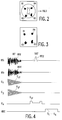
- a layer image created in a conventional manner (FIG. 2), which is displayed on the monitor 7 is reproduced, by means of the interactive input unit 9 four separate, ie spatially non-contiguous areas R1 ... R4, the MR spectrum of which is to be determined.
- the areas to be examined can also be specified automatically with the aid of suitable image processing methods.
- This coding scheme corresponds to a four-point Hadamard transformation.
- the coding could be specified interactively by the user, but it can also be specified automatically by the workstation 6 as soon as the number of areas has been determined.
- any other coding can also be used, provided that they are linearly independent. This also includes the wavelet or Fourier transformation.
- the complex envelopes of four two-dimensional high-frequency pulses can be calculated as excitation pulses in such a way that the sign of the transverse magnetization generated by them in the individual areas corresponds to the specified coding.
- a spatially two-dimensional high-frequency pulse is a pulse that is accompanied by time-varying magnetic gradient fields with gradients running in two mutually perpendicular directions. The calculation of the envelope of such high-frequency pulses is published in the journal J. Magn. Reson. 82, 647-654 (1989).
- FIG. 4 shows the temporal profiles of the complex envelopes RF i1 and FR r1 of one of these four high-frequency pulses in the first two lines, those of cosine or sinusoidal gradient fields with linearly decreasing amplitude and gradients running in the z or y direction G z1 and G y1 are accompanied.
- Such a two-dimensional high-frequency pulse excites the nuclear magnetization in the four areas R1 ... R4, and only in these areas, the phase position of the transverse magnetization in the individual areas corresponding to one of the specified codes.
- the sequence comprises a slice-selective refocusing high-frequency pulse RF i2 , which is accompanied by a magnetic gradient field G x1 with gradients running in the x direction.
- RF i2 a slice-selective refocusing high-frequency pulse
- G x1 a magnetic gradient field
- the investigation is limited to the layer perpendicular to the x direction, the transverse magnetization of which is refocused by the RF i2 high-frequency pulse.
- the MR signal occurring after this high-frequency pulse RF i2 is received and converted into a digital signal by the analog-digital converter 23 implemented.
- the Fourier transform of this signal represents the MR spectrum of all four areas R1 ... R4.
- the sequence shown in Fig. 4 is repeated a second, third and fourth times, however, the control unit loads the envelopes RF i1 and RF r1 of the second, third and fourth two-dimensional high-frequency pulse into the generator 14, so that the signals S2, S3 or S4 result. If necessary, these four sequences can be repeated several times.
- the signal / noise ratio in the described determination of the spectral nuclear magnetization distribution is better by a factor ⁇ n compared to the case in which the MR spectra of the individual regions are separated and determined one after the other; in this case by a factor of 2.
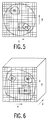
- FIG. 5 shows a two-dimensional MR image.
- Such an image can be created using classic MR imaging methods, in which the entire image field is measured in such a way that it can be imaged on an M x N matrix.
- M is the number the data points in the direction of the reading gradient and N the number of sequences to be generated with a changed phase coding gradient.
- R1 and R2 are of interest in such an image, for example those designated by R1 and R2 in the image.
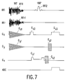
- FIG. 7 shows a single sequence for excitation for simultaneous excitation of the areas R1 and R2 in FIG. 5.
- the sequence according to FIG. 7 largely corresponds to that according to FIG. 1, with only one in the z direction during the reading of the MR signal Magnetic gradient field G z3 is switched on, which is preceded by a magnetic gradient field G z2 , the time integral of which is half as large as that of G z3 , and that between the 180 ° pulse RF i2 and the two-dimensional high-frequency pulse RF i1, a time-varying gradient field G z2 lies.
- phase coding gradient G y2 acting in the y direction is switched on, which must be repeated s times, the phase coding gradient being changed in stages from repeat to repeat.
- the number s results from the larger extent of the two regions in phase coding, divided by the spatial resolution dr, or it indicates how many pixels in the direction of the phase coding gradient comprise the larger area (R2) in this direction. Since the two-dimensional pulse excites the two spatially separated areas R1 and R2 - and only these - the two areas would no longer be distinguishable after the reconstruction in the phase coding direction, and folding artifacts would occur.
- the two areas R1 and R2 are acted upon by two different two-dimensional high-frequency pulses, which code the phase of the nuclear magnetization in these areas as follows: Pulse R1 R2 MR signal 1 + + ⁇ S1 2nd + - ⁇ S2
- the envelope is calculated for these two different two-dimensional high-frequency pulses, as explained in connection with FIG. 4.
- S (R1) S1 + S2
- S (R2) S1 - S2.
- This is equivalent to a two-point Hadamard transformation or a two-point Fourier transformation.
- their Fourier transforms or the images reconstructed from the MR signals S1 or S2 in connection with the phase encodings can also be superimposed in the specified manner.
- the Fourier transform for the two areas or an MR image of the two areas results directly.
- the method described has an advantage over a conventional MR imaging method if the product of the number of areas n and the count s is smaller than the number N of phase encoding steps that would be required in a standard 2DFT method to produce the overall image to reconstruct with the same resolution dr.
- the direction of the reading gradient in the example this is the z direction
- the direction of the phase coding gradient in the example: y
- Fig. 6 shows such a three-dimensional case, the Nuclear magnetization distribution in three unrelated areas R1, R2 and R3 is to be determined.
- two spatial directions (x, y) must be phase-coded, while the reading gradient runs in the third, perpendicular direction.
- R3 may have the largest dimensions of the three regions both in the x and in the y direction, its length in the x direction being t pixels and in the y direction s pixels.
- the reading gradient G z3 and the defocusing gradient G z2 are omitted when the MR signal is acquired, the result is a spatially two-dimensional spectroscopic imaging.
- the invention can also be used in conjunction with MR methods which, like EPI or the like, read out a plurality of MR signals with alternating polarity of the reading gradient following an exciting high-frequency pulse.
- MR methods which, like EPI or the like, read out a plurality of MR signals with alternating polarity of the reading gradient following an exciting high-frequency pulse.
- the phase is changed in a first direction perpendicular to the reading direction by a short pulse ("blip") (for a two-dimensional image).
- blip short pulse
- phase coding is carried out in a second direction, which is perpendicular to the first direction and to the direction of the reading gradient.
- the non-contiguous areas are spatially two- or three-dimensional areas.
- the invention can also be applied to the case of a spatial and a spectral dimension.
- FIG. 9 A sequence suitable for this purpose is given in FIG. 9.
- Magn. Reson describes how to generate high-frequency pulses that selectively excite the nuclear magnetization spatially and spectrally at the same time. Med. 15, 287-304 (1990).
- Such pulses can be used in conjunction with the usual reading gradients (G x ) or phase encoding gradients (G y ) to carry out conventional MR imaging.
- the sequence shown in FIG. 9 must be repeated 4N times, so that there are no advantages in terms of measuring time compared to a method in which fat or water images are determined in succession in the two planes.
- the spatial and spectrally selective high-frequency pulses excite all four areas at the same time, the signal-to-noise ratio is a factor of 2 more favorable.
Landscapes
- Physics & Mathematics (AREA)
- High Energy & Nuclear Physics (AREA)
- Condensed Matter Physics & Semiconductors (AREA)
- General Physics & Mathematics (AREA)
- Magnetic Resonance Imaging Apparatus (AREA)
Applications Claiming Priority (2)
| Application Number | Priority Date | Filing Date | Title |
|---|---|---|---|
| DE19543079A DE19543079A1 (de) | 1995-11-18 | 1995-11-18 | Verfahren zum Bestimmen der räumlichen und/oder spektralen Verteilung der Kernmagnetisierung |
| DE19543079 | 1995-11-18 |
Publications (1)
| Publication Number | Publication Date |
|---|---|
| EP0774672A1 true EP0774672A1 (fr) | 1997-05-21 |
Family
ID=7777823
Family Applications (1)
| Application Number | Title | Priority Date | Filing Date |
|---|---|---|---|
| EP96203143A Withdrawn EP0774672A1 (fr) | 1995-11-18 | 1996-11-11 | Méthode de détermination de la distribution spatiale/spectrale de la magnétisation nucléaire |
Country Status (4)
| Country | Link |
|---|---|
| US (1) | US6005391A (fr) |
| EP (1) | EP0774672A1 (fr) |
| JP (1) | JPH09168528A (fr) |
| DE (1) | DE19543079A1 (fr) |
Families Citing this family (10)
| Publication number | Priority date | Publication date | Assignee | Title |
|---|---|---|---|---|
| DE19743547B4 (de) * | 1997-10-01 | 2005-12-22 | Siemens Ag | Verfahren zur Abbildung von Fett-Plaques mittels Kernspintomographie |
| JP4416221B2 (ja) * | 1999-09-28 | 2010-02-17 | 株式会社日立メディコ | 磁気共鳴画像診断装置 |
| DE102007044463B4 (de) | 2007-09-18 | 2009-05-14 | Bruker Biospin Mri Gmbh | Verfahren zur Bestimmung der räumlichen Verteilung von Magnetresonanzsignalen durch mehrdimensionale HF-Anregungspulse |
| DE102008021736B3 (de) * | 2008-04-30 | 2009-12-10 | Bruker Biospin Mri Gmbh | Verfahren zur Bestimmung der räumlichen Verteilung von Magnetresonanzsignalen beim Einsatz von lokalen ortskodierenden Magnetfeldern |
| DE102008061455B4 (de) * | 2008-12-10 | 2011-03-17 | Siemens Aktiengesellschaft | Verfahren und Vorrichtung zur Bestimmung einer vorbestimmten Signalamplitude bei MR-Messungen |
| US20110043518A1 (en) * | 2009-08-21 | 2011-02-24 | Nicolas Galoppo Von Borries | Techniques to store and retrieve image data |
| JP5730214B2 (ja) * | 2009-11-12 | 2015-06-03 | 株式会社日立メディコ | 磁気共鳴イメージング装置及び2次元励起調整方法 |
| DE102011004432A1 (de) * | 2011-02-20 | 2012-08-23 | Forschungszentrum Jülich GmbH | Magnetresonanzverfahren und Kernspintomograph zur Durchführung des Magnetresonanzverfahrens |
| WO2016123674A1 (fr) * | 2015-02-06 | 2016-08-11 | The University Of Melbourne | Procédé et système pour imagerie par résonance magnétique |
| US10585154B1 (en) * | 2018-01-29 | 2020-03-10 | Quantum Valley Investment Fund LP | Nuclear magnetic resonance diffraction |
Citations (1)
| Publication number | Priority date | Publication date | Assignee | Title |
|---|---|---|---|---|
| EP0361671A2 (fr) * | 1988-09-30 | 1990-04-04 | Kabushiki Kaisha Toshiba | Appareil de résonance magnétique |
Family Cites Families (2)
| Publication number | Priority date | Publication date | Assignee | Title |
|---|---|---|---|---|
| US5545990A (en) * | 1994-03-25 | 1996-08-13 | Siemens Aktiengesellschaft | Averaging of identically phase-encoded MR signals to reduce image degradation caused by spin-spin relaxation |
| US5612619A (en) * | 1995-07-31 | 1997-03-18 | Feinberg David A | Grase-type three-dimensional MR pulse sequences |
-
1995
- 1995-11-18 DE DE19543079A patent/DE19543079A1/de not_active Withdrawn
-
1996
- 1996-11-11 EP EP96203143A patent/EP0774672A1/fr not_active Withdrawn
- 1996-11-14 US US08/749,937 patent/US6005391A/en not_active Expired - Fee Related
- 1996-11-15 JP JP8305351A patent/JPH09168528A/ja active Pending
Patent Citations (1)
| Publication number | Priority date | Publication date | Assignee | Title |
|---|---|---|---|---|
| EP0361671A2 (fr) * | 1988-09-30 | 1990-04-04 | Kabushiki Kaisha Toshiba | Appareil de résonance magnétique |
Non-Patent Citations (3)
| Title |
|---|
| G. GOELMAN, J.S. LEIGH: "Hadamard Spectroscopic Imaging Technique ...", JOURNAL OF MAGNETIC RESONANCE, SERIES A, vol. 105, 1993, pages 78 - 81, XP000402842 * |
| L. BOLINGER, J.S. LEIGH: "Hadamard Spectroscopic Imaging (HSI) for Multivolume Localization", JOURNAL OF MAGNETIC RESONANCE, vol. 80, 1988, pages 162 - 167, XP002026131 * |
| W. DREHER, D. LEIBFRITZ: "Double-Echo Multislice Proton Spectroscopic Imaging ...", MAGNETIC RESONANCE IN MEDICINE, vol. 31, 1994, pages 596 - 600, XP002026130 * |
Also Published As
| Publication number | Publication date |
|---|---|
| JPH09168528A (ja) | 1997-06-30 |
| DE19543079A1 (de) | 1997-05-22 |
| US6005391A (en) | 1999-12-21 |
Similar Documents
| Publication | Publication Date | Title |
|---|---|---|
| EP0074022B1 (fr) | Dispositif de tomographie à spin nucléaire | |
| DE68927107T2 (de) | Verminderung von bewegungsartefakten bei der bilderzeugung mittels magnetischer kernresonanz | |
| DE102009055961B4 (de) | Verfahren zur Erfassung von einer Atembewegung eines Untersuchungsobjektes entsprechender Signaldaten mittels Magnetresonanz | |
| EP0213436B1 (fr) | Méthode de fonctionnement d'un appareil exploitant la résonance magnétique nucléaire | |
| DE4432570B4 (de) | Verfahren und Vorrichtung für die Kernresonanzabbildung physiologischer Funktionsinformation | |
| DE60023161T2 (de) | Verfahren zur abbildung von protonen-quer-relaxationszeiten oder funktionen davon in einem objekt mit lokalisierter bewegung unter verwendung der bildgebenden kernspinresonanz | |
| DE102008021736B3 (de) | Verfahren zur Bestimmung der räumlichen Verteilung von Magnetresonanzsignalen beim Einsatz von lokalen ortskodierenden Magnetfeldern | |
| DE102014206395B4 (de) | Aufnahme und iterative Rekonstruktion einer Parameterkarte eines Zielbereichs | |
| EP3413072A1 (fr) | Procédé de multiplexage de couche en irm | |
| DE3642826A1 (de) | Verfahren zum erzeugen eines nmr-bildes mit verbessertem signal-rausch-verhaeltnis | |
| DE19842937A1 (de) | Mehrschnitt- und Mehrwinkel-Magnetresonanzabbildung unter Verwendung einer Fast-Spin-Echo-Erfassung | |
| DE102018218471B3 (de) | Verfahren zur Magnetresonanzbildgebung mit Zusatzgradientenpulsen, Magnetresonanzeinrichtung, Computerprogramm und elektronisch lesbarer Datenträger | |
| DE3710748A1 (de) | Magnetresonanz-abbildungsanordnung | |
| DE102016219052B3 (de) | Verbesserte Ergänzung mehrerer mittels paralleler Akquisitionstechnik aufgenommener Magnetresonanzdatensätze | |
| DE10106830C2 (de) | Verfahren zur Bilderzeugung mittels magnetischer Resonanz mit mehreren unabhängigen Empfangsantennen | |
| DE10128534C2 (de) | Verfahren zur Bilderzeugung mittels magnetischer Resonanz mit mehreren Empfangsantennen | |
| DE19543079A1 (de) | Verfahren zum Bestimmen der räumlichen und/oder spektralen Verteilung der Kernmagnetisierung | |
| DE19814677A1 (de) | Korrektur einer durch Maxwell-Terme verursachten Verschlechterung eines Axial-Bild-Signals | |
| DE19653212B4 (de) | Verfahren zur Erhöhung der zeitlichen Auflösung bei Magnet-Resonanz-Durchleuchtungsverfahren | |
| DE10144654B4 (de) | Gerät und Verfahren zur Magnet-Resonanz-Bildgebung unter Verwendung einer verbesserten parallelen Akquisition | |
| DE10152734B4 (de) | Gerät und Verfahren zur Magnet-Resonanz-Bildgebung bei gleichzeitiger Messung zweier benachbarter Schichten | |
| DE102020202576B4 (de) | Verfahren zum Erzeugen eines Magnetresonanzbildes | |
| DE69232532T2 (de) | Verfahren und Vorrichtung zur Bilderzeugung mittels magnetischer Resonanz mit der Möglichkeit der Messung von kurzen T2-Signalkomponenten | |
| DE3688981T2 (de) | Nmr-bildformungsverfahren. | |
| EP0413266B1 (fr) | Méthode pour mesurer simultanément des signaux RMN, en particulier pour déterminer les flux sanguins en tomographie RMN, avec l'aide de la méthode Multiple Slice Fourier Flow (= MUFF) |
Legal Events
| Date | Code | Title | Description |
|---|---|---|---|
| PUAI | Public reference made under article 153(3) epc to a published international application that has entered the european phase |
Free format text: ORIGINAL CODE: 0009012 |
|
| AK | Designated contracting states |
Kind code of ref document: A1 Designated state(s): DE FR GB NL |
|
| 17P | Request for examination filed |
Effective date: 19971121 |
|
| RAP3 | Party data changed (applicant data changed or rights of an application transferred) |
Owner name: KONINKLIJKE PHILIPS ELECTRONICS N.V. Owner name: PHILIPS CORPORATE INTELLECTUAL PROPERTY GMBH |
|
| RAP1 | Party data changed (applicant data changed or rights of an application transferred) |
Owner name: KONINKLIJKE PHILIPS ELECTRONICS N.V. Owner name: PHILIPS CORPORATE INTELLECTUAL PROPERTY GMBH |
|
| RAP1 | Party data changed (applicant data changed or rights of an application transferred) |
Owner name: KONINKLIJKE PHILIPS ELECTRONICS N.V. Owner name: PHILIPS INTELLECTUAL PROPERTY & STANDARDS GMBH |
|
| STAA | Information on the status of an ep patent application or granted ep patent |
Free format text: STATUS: THE APPLICATION HAS BEEN WITHDRAWN |
|
| 18W | Application withdrawn |
Effective date: 20030613 |