EP0783894A1 - Antikörper modifizierten bei zwei verschiedenen Orten - Google Patents
Antikörper modifizierten bei zwei verschiedenen Orten Download PDFInfo
- Publication number
- EP0783894A1 EP0783894A1 EP96117696A EP96117696A EP0783894A1 EP 0783894 A1 EP0783894 A1 EP 0783894A1 EP 96117696 A EP96117696 A EP 96117696A EP 96117696 A EP96117696 A EP 96117696A EP 0783894 A1 EP0783894 A1 EP 0783894A1
- Authority
- EP
- European Patent Office
- Prior art keywords
- antibody
- modified
- antibodies
- lym
- mab
- Prior art date
- Legal status (The legal status is an assumption and is not a legal conclusion. Google has not performed a legal analysis and makes no representation as to the accuracy of the status listed.)
- Ceased
Links
Images






Classifications
-
- C—CHEMISTRY; METALLURGY
- C07—ORGANIC CHEMISTRY
- C07K—PEPTIDES
- C07K16/00—Immunoglobulins [IG], e.g. monoclonal or polyclonal antibodies
-
- A—HUMAN NECESSITIES
- A61—MEDICAL OR VETERINARY SCIENCE; HYGIENE
- A61K—PREPARATIONS FOR MEDICAL, DENTAL OR TOILETRY PURPOSES
- A61K47/00—Medicinal preparations characterised by the non-active ingredients used, e.g. carriers or inert additives; Targeting or modifying agents chemically bound to the active ingredient
- A61K47/50—Medicinal preparations characterised by the non-active ingredients used, e.g. carriers or inert additives; Targeting or modifying agents chemically bound to the active ingredient the non-active ingredient being chemically bound to the active ingredient, e.g. polymer-drug conjugates
- A61K47/51—Medicinal preparations characterised by the non-active ingredients used, e.g. carriers or inert additives; Targeting or modifying agents chemically bound to the active ingredient the non-active ingredient being chemically bound to the active ingredient, e.g. polymer-drug conjugates the non-active ingredient being a modifying agent
- A61K47/68—Medicinal preparations characterised by the non-active ingredients used, e.g. carriers or inert additives; Targeting or modifying agents chemically bound to the active ingredient the non-active ingredient being chemically bound to the active ingredient, e.g. polymer-drug conjugates the non-active ingredient being a modifying agent the modifying agent being an antibody, an immunoglobulin or a fragment thereof, e.g. an Fc-fragment
-
- A—HUMAN NECESSITIES
- A61—MEDICAL OR VETERINARY SCIENCE; HYGIENE
- A61K—PREPARATIONS FOR MEDICAL, DENTAL OR TOILETRY PURPOSES
- A61K47/00—Medicinal preparations characterised by the non-active ingredients used, e.g. carriers or inert additives; Targeting or modifying agents chemically bound to the active ingredient
- A61K47/50—Medicinal preparations characterised by the non-active ingredients used, e.g. carriers or inert additives; Targeting or modifying agents chemically bound to the active ingredient the non-active ingredient being chemically bound to the active ingredient, e.g. polymer-drug conjugates
- A61K47/51—Medicinal preparations characterised by the non-active ingredients used, e.g. carriers or inert additives; Targeting or modifying agents chemically bound to the active ingredient the non-active ingredient being chemically bound to the active ingredient, e.g. polymer-drug conjugates the non-active ingredient being a modifying agent
- A61K47/68—Medicinal preparations characterised by the non-active ingredients used, e.g. carriers or inert additives; Targeting or modifying agents chemically bound to the active ingredient the non-active ingredient being chemically bound to the active ingredient, e.g. polymer-drug conjugates the non-active ingredient being a modifying agent the modifying agent being an antibody, an immunoglobulin or a fragment thereof, e.g. an Fc-fragment
- A61K47/6889—Conjugates wherein the antibody being the modifying agent and wherein the linker, binder or spacer confers particular properties to the conjugates, e.g. peptidic enzyme-labile linkers or acid-labile linkers, providing for an acid-labile immuno conjugate wherein the drug may be released from its antibody conjugated part in an acidic, e.g. tumoural or environment
-
- A—HUMAN NECESSITIES
- A61—MEDICAL OR VETERINARY SCIENCE; HYGIENE
- A61K—PREPARATIONS FOR MEDICAL, DENTAL OR TOILETRY PURPOSES
- A61K51/00—Preparations containing radioactive substances for use in therapy or testing in vivo
- A61K51/02—Preparations containing radioactive substances for use in therapy or testing in vivo characterised by the carrier, i.e. characterised by the agent or material covalently linked or complexing the radioactive nucleus
- A61K51/04—Organic compounds
- A61K51/08—Peptides, e.g. proteins, carriers being peptides, polyamino acids, proteins
- A61K51/10—Antibodies or immunoglobulins; Fragments thereof, the carrier being an antibody, an immunoglobulin or a fragment thereof, e.g. a camelised human single domain antibody or the Fc fragment of an antibody
-
- A—HUMAN NECESSITIES
- A61—MEDICAL OR VETERINARY SCIENCE; HYGIENE
- A61K—PREPARATIONS FOR MEDICAL, DENTAL OR TOILETRY PURPOSES
- A61K51/00—Preparations containing radioactive substances for use in therapy or testing in vivo
- A61K51/02—Preparations containing radioactive substances for use in therapy or testing in vivo characterised by the carrier, i.e. characterised by the agent or material covalently linked or complexing the radioactive nucleus
- A61K51/04—Organic compounds
- A61K51/08—Peptides, e.g. proteins, carriers being peptides, polyamino acids, proteins
- A61K51/10—Antibodies or immunoglobulins; Fragments thereof, the carrier being an antibody, an immunoglobulin or a fragment thereof, e.g. a camelised human single domain antibody or the Fc fragment of an antibody
- A61K51/1045—Antibodies or immunoglobulins; Fragments thereof, the carrier being an antibody, an immunoglobulin or a fragment thereof, e.g. a camelised human single domain antibody or the Fc fragment of an antibody against animal or human tumor cells or tumor cell determinants
- A61K51/1063—Antibodies or immunoglobulins; Fragments thereof, the carrier being an antibody, an immunoglobulin or a fragment thereof, e.g. a camelised human single domain antibody or the Fc fragment of an antibody against animal or human tumor cells or tumor cell determinants the tumor cell being from stomach or intestines
-
- A—HUMAN NECESSITIES
- A61—MEDICAL OR VETERINARY SCIENCE; HYGIENE
- A61K—PREPARATIONS FOR MEDICAL, DENTAL OR TOILETRY PURPOSES
- A61K51/00—Preparations containing radioactive substances for use in therapy or testing in vivo
- A61K51/02—Preparations containing radioactive substances for use in therapy or testing in vivo characterised by the carrier, i.e. characterised by the agent or material covalently linked or complexing the radioactive nucleus
- A61K51/04—Organic compounds
- A61K51/08—Peptides, e.g. proteins, carriers being peptides, polyamino acids, proteins
- A61K51/10—Antibodies or immunoglobulins; Fragments thereof, the carrier being an antibody, an immunoglobulin or a fragment thereof, e.g. a camelised human single domain antibody or the Fc fragment of an antibody
- A61K51/1045—Antibodies or immunoglobulins; Fragments thereof, the carrier being an antibody, an immunoglobulin or a fragment thereof, e.g. a camelised human single domain antibody or the Fc fragment of an antibody against animal or human tumor cells or tumor cell determinants
- A61K51/1069—Antibodies or immunoglobulins; Fragments thereof, the carrier being an antibody, an immunoglobulin or a fragment thereof, e.g. a camelised human single domain antibody or the Fc fragment of an antibody against animal or human tumor cells or tumor cell determinants the tumor cell being from blood cells, e.g. the cancer being a myeloma
-
- A—HUMAN NECESSITIES
- A61—MEDICAL OR VETERINARY SCIENCE; HYGIENE
- A61P—SPECIFIC THERAPEUTIC ACTIVITY OF CHEMICAL COMPOUNDS OR MEDICINAL PREPARATIONS
- A61P35/00—Antineoplastic agents
-
- A—HUMAN NECESSITIES
- A61—MEDICAL OR VETERINARY SCIENCE; HYGIENE
- A61P—SPECIFIC THERAPEUTIC ACTIVITY OF CHEMICAL COMPOUNDS OR MEDICINAL PREPARATIONS
- A61P35/00—Antineoplastic agents
- A61P35/02—Antineoplastic agents specific for leukemia
-
- A—HUMAN NECESSITIES
- A61—MEDICAL OR VETERINARY SCIENCE; HYGIENE
- A61P—SPECIFIC THERAPEUTIC ACTIVITY OF CHEMICAL COMPOUNDS OR MEDICINAL PREPARATIONS
- A61P37/00—Drugs for immunological or allergic disorders
-
- C—CHEMISTRY; METALLURGY
- C07—ORGANIC CHEMISTRY
- C07K—PEPTIDES
- C07K16/00—Immunoglobulins [IG], e.g. monoclonal or polyclonal antibodies
- C07K16/18—Immunoglobulins [IG], e.g. monoclonal or polyclonal antibodies against material from animals or humans
- C07K16/28—Immunoglobulins [IG], e.g. monoclonal or polyclonal antibodies against material from animals or humans against receptors, cell surface antigens or cell surface determinants
- C07K16/30—Immunoglobulins [IG], e.g. monoclonal or polyclonal antibodies against material from animals or humans against receptors, cell surface antigens or cell surface determinants from tumour cells
-
- C—CHEMISTRY; METALLURGY
- C07—ORGANIC CHEMISTRY
- C07K—PEPTIDES
- C07K16/00—Immunoglobulins [IG], e.g. monoclonal or polyclonal antibodies
- C07K16/18—Immunoglobulins [IG], e.g. monoclonal or polyclonal antibodies against material from animals or humans
- C07K16/28—Immunoglobulins [IG], e.g. monoclonal or polyclonal antibodies against material from animals or humans against receptors, cell surface antigens or cell surface determinants
- C07K16/30—Immunoglobulins [IG], e.g. monoclonal or polyclonal antibodies against material from animals or humans against receptors, cell surface antigens or cell surface determinants from tumour cells
- C07K16/3046—Stomach, Intestines
-
- C—CHEMISTRY; METALLURGY
- C07—ORGANIC CHEMISTRY
- C07K—PEPTIDES
- C07K16/00—Immunoglobulins [IG], e.g. monoclonal or polyclonal antibodies
- C07K16/18—Immunoglobulins [IG], e.g. monoclonal or polyclonal antibodies against material from animals or humans
- C07K16/28—Immunoglobulins [IG], e.g. monoclonal or polyclonal antibodies against material from animals or humans against receptors, cell surface antigens or cell surface determinants
- C07K16/30—Immunoglobulins [IG], e.g. monoclonal or polyclonal antibodies against material from animals or humans against receptors, cell surface antigens or cell surface determinants from tumour cells
- C07K16/3061—Blood cells
-
- A—HUMAN NECESSITIES
- A61—MEDICAL OR VETERINARY SCIENCE; HYGIENE
- A61K—PREPARATIONS FOR MEDICAL, DENTAL OR TOILETRY PURPOSES
- A61K2121/00—Preparations for use in therapy
-
- A—HUMAN NECESSITIES
- A61—MEDICAL OR VETERINARY SCIENCE; HYGIENE
- A61K—PREPARATIONS FOR MEDICAL, DENTAL OR TOILETRY PURPOSES
- A61K2123/00—Preparations for testing in vivo
Definitions
- This present invention relates to modified antibodies. More specifically, the present invention relates to antibodies which have been modified by chemical conjugation with a heterobifunctional reagent and the use of these modified MAb's in the diagnosis and therapy of cancer and other mammalian disease.
- MAb's monoclonal antibodies
- MAb's specific to tumor cell antigens have been produced. It has also been shown that MAb's may be efficiently coupled to adjuncts such as radionuclides. Such radio-labelled MAb's are useful in providing clinical data, such as tumor imaging from immunoscintography, also known as ⁇ -camera imaging or radioimmunoimaging. In immunoscintography, the MAb's are allowed to bind to the specific tissue or tumor types having the antigen recognized by the MAb's. The radionuclides are then visualized through the use of appropriate technology, such as through the use of a germanium camera. It is the unique specificity of MAb's which enables their application in immunoscintography of tumors and other types of tissues.
- the cytotoxic effect of MAb's can be markedly increased by coupling to radionuclides, drugs or toxins.
- the unique specificity of MAb's has raised hopes of the development of immunotherapy.
- biologically active agents are delivered using MAB's to particular undesirable cell types, such as tumor cells, thereby affecting the undesirable cell types without affecting other cells of the subject.
- immunotherapies require extremely high specificity antibodies in order to avoid affecting healthy tissue.
- a method of increasing the specificity of MAb's would be highly beneficial in achieving the goal of a safe, effective immunotherapy.
- MAb's remain in the circulation for several days following introduction into a subject. This is undesirable for at least two reasons. One reason is that circulating MAb's produces high background levels in immunoscintography. A second reason is that circulating MAb's coupled to radionuclides or other potentially cytotoxic agents may produce undesirable side effects in the subject after prolonged exposure. Thus, there is a need for a method of decreasing the clearance time of MAb's. Of course, too great a decrease would result in MAb's being eliminated before any effective use of the MAb's could be made. Thus, there is a particular need for a method of decreasing the clearance time of MAb's without substantially affecting uptake of MAb's by tumor or other target tissue.
- an "intact" antibody molecule will refer to an unmodified antibody molecule comprised of two heavy chains and two light chains.
- the intact, whole antibody molecule is seen on the reactant side of the chemical equation of Figure 1.
- the intact molecule is divided into the F c and the F ab domains.
- F(ab') 2 the bivalent form of the F ab fragment, may be produced through the digestion of the F c domain with a protease.
- the two heavy chains (designated as "H” in Figure 1) are held together by one or more disulfide bridges. In intact molecules these disulfide bridges are normally protected from reducing agents. It has been found however, that removal of the F c domain allows facile reduction of the disulfide bridges. Thus, F(ab'), the monovalent form, may be produced from F(ab') 2 through the action of a mild reducing agent. Parham, P., On the Fragmentation of Monoclonal IgG1, IgG2a, and IgG2b from BALB/c Mice, J. Immunol.
- F c has been found to be responsible for much of the non-specific binding of antibody molecules. It is also believed that the molecular weight of the fragments is below the threshold for glomerular filtration, thus allowing for rapid elimination of the fragments. Therefore, one approach to increasing clearance time of antibodies for use in radioimaging has been to break down intact antibody into various fragments, such as Fab and its divalent form, F(ab') 2 . As expected, these fragments are cleared from the body so rapidly that their utility is reduced. Moreover, these fragments may result in reduced uptake by tumor or other target tissue relative to intact antibody.
- the absolute concentration of MAb's in the target tissue containing the antigen to which the MAb's will bind has been found to be up to as many as three times or more as much with intact MAb's as with either of the fragments.
- Heterobifunctional reagents are reagents having two groups capable of participating in different reaction.
- succinimidyl 3-(2-pyridyldithio)propionate (SPDP) is heterobifunctional in that its N-hydroxysuccinimide ester group reacts with amino groups and the 2-pyridyl disulphide structure reacts with aliphatic thiols.
- the conjugated MAb's used by Orlandi et al. had on average, 11 PDP groups per molecule. Orlandi et al. found that the modified MAb's increase their binding activity in vitro to an extent that molecules not detected by the unmodified MAb's can be detected. These researchers reported no studies of the use of the conjugated MAb's in vivo . Additionally, these researchers believed that molecules having a very low number of antigenic sites were detected by the conjugated MAb's. Accordingly, the PDP modified MAb's had greatly reduced target-cell specificity relative to the unmodified counterparts.
- Figure 1 shows a schematic representation of the changes believed to occur in a method of producing F(ab') and F(ab') 2 fragments.
- Figure 2 shows whole body retention of different preparations of radio-labeled MAb's Lym-1 in athymic nude mice.
- Figure 3 shows the biodistribution as % of injected dose/gram of MAb's Lym-1 and modified Lym-1 in human lymphoma-bearing nude mice seven days after injection.
- Figure 4 shows the biodistribution as tumor/organ ratios of MAb's Lym-1 and Modified Lym-1 in human lymphoma-bearing nude mice seven days after injection.
- Figure 5 shows the biodistribution as % of injected dose/gram of MAb's Lym-1 F(ab') 2 and Modified Lym-1 in human lymphoma-bearing nude mice five days after injection.
- Figure 6 shows the biodistribution as tumor/organ ratio of MAb's Lym-1 F(ab') 2 and Modified Lym-1 in human lymphoma-bearing nude mice five days after injection.
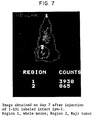
- Figure 7 shows the image obtained on day 7 after injection of I-131 labeled intact Lym-1.
- Figure 8 shows the image obtained on day 7 after injection of I-131 labeled modified Lym-1.
- Figure 9 shows the image obtained on day 7 after injection of I-131 labeled modified Lym-1.
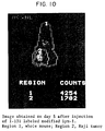
- Figure 10 shows the image obtained on day 5 after injection of I-131 labeled modified Lym-1.
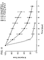
- FIG 11 shows the whole body retention of different preparations of radio-labeled monoclonal antibodies B72.3 in athymic nude mice.
- Figure 12 shows the biodistribution as % of injected dose/gram of MAb's B72.3 and Modified B72.3 in LS174T colon carcinoma-bearing nude mice four days after injection.
- Figure 13 shows the biodistribution as tumor/organ ratio of MAb's B72.3 and Modified B72.3 in LS174T colon carcinoma-bearing nude mice four days after injection.
- Figure 14 shows the image obtained on day 1 after injection of I-131 labeled modified B72.3.
- Figure 15 shows the image obtained on day 4 after injection of I-131 labeled modified B72.3.
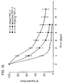
- Figure 16 shows the whole body retention of different preparations of radiolabeled MAb's TNT-1 in athymic nude mice.
- the present invention provides antibodies chemically modified with a heterobifunctional reagent having an in vivo clearance rate between the clearance rates of F(ab') 2 fragments and intact antibodies of the same type.
- the heterobifunctional reagent is SPDP and the antibodies are selected from the group consisting of monoclonal antibodies, human antibodies, genetically engineered antibodies, chimeric antibodies, synthesized antibodies, and polyclonal antibodies.
- the present invention also provides a method of imaging a specific tissue in a mammal comprising obtaining an antibody to the specific tissue, modifying the antibody by conjugation with a heterobifunctional reagent, introducing a label onto the antibody, introducing the antibody into the mammal and producing an image which reveals the label on said antibody.
- the antibody is modified by conjugation with an average of approximately one PDP group per antibody molecule and is labeled with a radionuclide which emits gamma radiation.
- the present invention provides a method of treating a disease state in mammal comprising obtaining an antibody specific to the diseased tissues in the mammal; modifying the antibody by chemical conjugation with a heterobifunctional reagent and with biologically active molecule, and administering the antibody to the mammal.
- the antibodies are modified by chemical conjugation with, on average, one PDP group per antibody molecule and the biologically active molecule is selected from the group consisting of plant toxins, drugs, radionuclides and chelates.
- the present invention also provides pharmaceutical compositions for use with the methods of the present invention.
- the compositions include a modified antibody in a pharmaceutically accepted carrier, excipient or base.
- heterobifunctional reagents including sulfosuccinimidyl 2-(p-azido salicylamido)ethyl1-1,3'-dithiopropionate (SASD), sulfosuccinimidyl 2-(m-azido-o-nitrobenzamido)-ethyl-1,3'-dithiopropionate (SAND), sulfosuccinimidyl (4-azidophenyl-dithio)propionate (sulfo-SADP) and 2-aminothiolane.HCl (Traut's reagent), are believed to provide similar results when conjugated with antibodies in accordance with the present invention, as illustrated herein by conjugation of antibodies with SPDP.
- modification of IgG by the conjugation of one or more PDP groups advantageously, also enhances clearance from normal tissues.
- modification with SPDP leads to fragmentation of the antibody to a form which has a molecular weight below the threshold for glomerular filtration, thus allowing for rapid elimination of the fragments. It is possible that fragmentation of the antibody to the monovalent form of the antibody occurs. Whatever the exact form of the resulting fragments, the elimination of these fragments is, advantageously, not so rapid so as to curtail the diagnostic or therapeutic effectiveness of the modified antibodies.
- modified antibodies of the present invention advantageously, have surprisingly enhanced diagnostic and therapeutic effectiveness relative to fragments of antibodies, such as F(ab') or F(ab') 2 .
- the following example shows an exemplary method for the introduction of, on average, one PDP group to a monoclonal antibody.
- Lym-1 (IgG 2a ), the monoclonal antibody against B cell lymphoma was obtained as in Epstein, A.L. et al., Two New Monoclonal antibodies, Lym-1 and Lym-2, Reactive with Human B-lymphocytes and Derived Tumors, with Immunodiagnostic and Immunoreactive Potential, Cancer Res. 47 : 830-840 (1987), the disclosure of which is hereby incorporated by reference.
- the Lym-1 MAb's were functionalized using SPDP, a heterobifunctional reagent which reacts with free amino groups on antibodies as in Carlson, J.
- the degree of functionalization of Lym-1 with SPDP was determined to be an average of one PDP group per molecule by measurement of release of pyridine-2-thione at 343 nm after reduction of an aliquot of the Lym-1 solution with molar excess of 7 mg dithioerythritol in phosphate buffer saline solution (PBS), pH 7.2., as in Grassetti, D.R. and Murray, J.F., Determination of Sulfhydryl Groups with 2,2'-or 4,4'-dithiodipyridine, Arch. Biochem. Biophys. 119 : 41-49 (1967), the disclosure of which is hereby incorporated by reference.
- Example 2 The modified antibody from Example 1 was analyzed by Fast Protein Liquid Chromatography (FPLC) in order to show that the antibodies remained substantially intact. This analysis is shown in Example 2.
- FPLC Fast Protein Liquid Chromatography
- modified antibody from Example 1 was achieved by Fast protein Liquid Chromatography (FPLC) equipped with a fixed wavelength UV spectrophotometer set at 280 nm. Size exclusion chromatography was performed on a superose-12 column (Pharmacia) with PBS pH 7.2 as the solvent system, eluting at flow rate of 1 mL/min. The modified Lym-1 appeared at a retention time of 690 seconds, identical to the retention time of unlabeled intact Lym-1.
- FPLC Fast protein Liquid Chromatography
- Example 2 shows that the SPDP-modified antibodies behaved virtually identically to the unmodified antibodies in FPLC. This data shows that the modification likely did not lead to breakdown of the intact molecules in vitro.
- radiolabelling of the modified MAb's was performed.
- the radiolabelling is shown in Example 3.
- the reaction was quenched after 3 minutes with 20 ⁇ L of 120 mM solution of sodium metabisulfite.
- the radiolabeled MAb's from Example 3 were subjected to Instant Thin Layer Chromatography (ITLC) in order to determine the purity of the labeled MAb's. This analysis is shown in Example 4.
- ITLC Instant Thin Layer Chromatography
- Modified Lym-1 radiolabeled with 131 I and modified Lym-1 radiolabeled with 125 I via the chloramine T method of Example 3 were analyzed using an analytical ITLC system consisting of silica gel impregnated glass fiber. Strips (2 X 20cm) were activated by heating at 110-1/2°C for 15 minutes prior to use; spotted with 1 ⁇ L of sample; air-dried and eluted with MeOH/H 2 O (80:20) for approximately 12 cm; again air-dried, cut in half and counted, to determine protein-bound and non-protein-bound radioactivity. Both forms of radiolabeled Lym-1 antibodies had an R f value of 0 and showed radiochemical purity of ⁇ 99%. Analysis of intact Lym-1 labeled in the same way as in Example 3 revealed the same purity.
- Example 4 shows that high purity radiolabeled antibodies could be obtained.
- the immunoreactivities of these radiolabeled MAb's were tested by their ability to bind to Raji cells. This analysis is shown in Example 5.
- the in vitro immunoreactivities of the radiolabeled modified Lym-1 and intact Lym-1 were evaluated by conventional live assay of 10 6 Raji cells/tube by the method of Epstein, A.L. et al., supra . Briefly, Raji cells resuspended in 100 ⁇ L of 1% bovine serum albumin in PBS was pipetted into a triplicate set of test tubes. One hundred ⁇ L of the labeled Lym-1 was added to each test tube (100,100 cpm/tube) and incubated for 30 minutes at room temperature with continuous mixing using an orbital shaker.
- the cells were washed three times with 1% bovine serum in PBS by spinning the tubes at 1000 rpm for 5 minutes, decanting the supernatant and resuspending the cells in 200 ⁇ L PBS.
- bound Lym-1 was detected by measuring the radioactivity bound to the cells using a gamma counter. The results showed that the binding activity of the modified Lym-1 was 87%, whereas the intact Lym-1, which served as a standard control, had a binding activity of 80%.
- Example 5 shows that the modified Lym-1 was more immunoreactive in vitro than the unmodified Lym-1.
- modified MAb's were analyzed for their stability in serum, as shown in Example 6.
- Monoclonal antibodies of modified Lym-1 and intact Lym-1 which was labeled directly with I-125 were added to each of several triplicate sets of fresh mouse serum to a final concentration of 100 ⁇ g/mL.
- the tubes were incubated at 37-1/2°C in a humidified incubator maintained in 5% CO 2 in air.
- protein-bound activity was determined by adding 900 ⁇ L of 100% trichloroacetic acid (TCA) to 100 ⁇ L aliquots. After a five-minute incubation at room temperature, protein precipitates were sedimented by centrifugation, and 500 ⁇ L of supernatant were withdrawn from each tube and counted for radioactivity in a gamma counter.
- TCA trichloroacetic acid
- Example 6 showed that the stability of the activity of the modified antibodies was maintained in serum for at least 8 days.
- HPLC analysis of the modified Lym-1 after incubation was performed, as shown in Example 7.
- HPLC analyses were performed on a Waters system equipped with size exclusion columns (SW 300) with 0.1M neutral phosphate buffer as eluting solvent and a flow rate of 1 ml/min. The eluate was detected with a radioisotope detector.
- the labeled modified Lym-1 product mixture from Example 6 revealed one major peak of a low molecular weight species with an elution time of 750 seconds, plus a small quantity at 690 seconds. The intact Lym-1 gave a single peak with a retention time of 690 seconds.
- Example 7 shows that the serum incubated modified Lym-1 samples had an apparent molecular weight in HPLC analysis lower than that of intact Lym-1.
- Example 2 showed that unincubated modified Lym-1 had an identical retention time to intact Lym-1.
- the modified Lym-1 showed an apparent loss of molecular weight in FPLC analysis upon incubation in serum.
- Example 6 The same samples from Example 6 were also examined over an 8-day study to see if there had been any loss of radioactivity from the radiolabeled Lym-1; such loss can be interpreted as evidence of deiodination in serum. The data showed virtually no loss of radioactivity over this period, confirming that a very stable attachment of iodine had been obtained in these immunoconjugates.
- Examples 7-9 show that the modified antibodies while retaining virtually full activity after incubation in serum, appeared to break down into molecules of apparent molecular weight of 116,000. As stated above, it is possible that this loss of molecular weight is due to the breakdown of the antibodies into their monovalent form. In any event, it is believed that the loss in apparent molecular weight is due to the breakdown of the modified antibodies into fragments thereof.
- the clearance of F(ab') 2 fragments was, however, two times faster, with a biological half-life of 10 hours, than the modified Lym-1.
- the data showed that modified Lym-1 is cleared at a rate intermediate between the rapidly-cleared F(ab') 2 fragments and the slowly-cleared intact antibody.
- Example 11 shows the methods used in all of the subsequent biodistribution studies.
- mice received a 0.2 mL inoculum containing 10 ⁇ g of modified Lym-1 labeled with I-131 at 12 ⁇ Ci/ ⁇ g (120 ⁇ Ci/mouse), and 10 ⁇ g of F(ab') 2 fragments labeled with I-125 at 2.5 ⁇ Ci/ ⁇ g (25 ⁇ Ci/mouse).
- mice were sacrificed by cervical dislocation at preselected times, post-injection, and various organs, blood and tumor were removed and weighed on an analytical balance. The samples were then counted in a gamma counter to determine the 131 I and 125 I activity.
- 125 I counts were adjusted for cross-over from the 131 I channel by subtracting 17% of 131 I channel counts, a formula that was determined experimentally using a 1282 Compugamma gamma counter (LKB). The data were also corrected for the radiation decay of the 131 I isotope according to the days on which the animals were sacrificed. For each mouse, data was expressed as cpm per gram tumor/cpm per gram organ, % dose/gram, and % dose/organ From these data, the mean and standard deviation were calculated for each group.
- LLB Compugamma gamma counter
- Example 12 compares the biodistribution of the modified MAb's to intact MAb's using the methods of Example 11.
- Example 11 the intact Lym-1 antibody was compared to the modified Lym-1 antibody in the methods of Example 11. Intact Lym-1 produced a blood activity of 0.64% ID/g at 7 days after injection, as reported in Table I. At the end of the same time interval, the tumor had an activity of 3.92% ID/g.
- the modified Lym-1 cleared from blood faster and produced a blood activity of 0.14% ID/g at 7 days. At the end of the same time interval, the tumor produced 7.7%, which tended to be significantly higher than the corresponding activities of the intact Lym-1.
- the modified antibodies produced a higher signal in the tumor than the intact antibodies. Additionally, the modified antibodies reacted less strongly than the intact MAb's for every organ tested, except for the kidney. It is not unexpected that a higher signal would be found in the kidney, because the antibodies are expected to be cleared through this organ. Due to the more rapid clearance rate of the modified MAb's relative to intact MAb's found in Example 10, a higher amount of codified MAb's in the kidney would be expected.
- the modified MAb's produced a significantly higher tumor/organ ratio than intact MAb's in every organ tested, except for kidney.
- the modified antibodies would produce a significantly lower background when used in immunoscintography.
- the modified antibodies would be more effective when used in immunotherapies due both to its higher affinity for tumor and lower affinity for non-target tissues.
- the modified antibodies would, thus, be expected to be more highly toxic to tumors and less toxic to non-target tissues.
- Example 13 is illustrative of these experiments.
- Table II shows that the modified Lym-1 cleared more slowly from the blood than the F(ab') 2 fragments.
- the modified Lym-1 produced a blood activity of 0.09% ID/g higher than the fragments (0.05%) at 5 days post-injection.
- Figure 5 shows that the tumor activity of the modified Lym-1 was about two-and-one-half times higher than the corresponding activity of the F(ab') 2 fragments.
- the activity of the modified Lym-1 was higher than the F(ab') 2 fragments for all of the various organs tested, including kidney. This is consistent with the theory that more rapidly cleared antibodies accumulate in the kidney.
- Figure 6 shows that the tumor-to-organ ratios for modified Lym-1 are higher than those of the F(ab') 2 fragments for all of the organs tested.
- the experiments of Examples 12 and 13 confirm that the modified antibodies of the present invention have a higher activity for their target tumor than either intact MAb's or F(ab') 2 fragments. Additionally, the tumor-to-organ data of these experiments shows that the modified antibodies have higher specificity for tumor than either the intact MAb's or F(ab') 2 fragments.
- Tumor-bearing nude mice were imaged using a pinhole collimator and a spectrum 91 gamma camera (Raytheon). Image analyses of these animals provided an estimate of tumor/whole body antibody distribution after injection. Seven days after injection, the mice were anesthetized with 2mg Ketamine HCl and 0.4mg Xylazine administered as a 0.2mL s.c. inoculation. The immobilized mice where then imaged in a posterior position with the camera preset to record 10,000 counts. No background substraction was performed. Photographic images were obtained using Polaroid Type 330 Pack film. Two areas in each image were defined: (a) region 1, whole body; (b) region 2, tumor. Figures 6-8 show exemplary scintographs (also known as scintograms) produced by these experiments.
- Figure 10 shows a 5-day image taken of the same animal as shown in Figure 9 at 7 days. As can be seen, the 5-day image of Figure 10 was significantly superior to the image produced by intact Lym-1 at 7 days ( Figure 7). Results were similar for all animals tested.
- modified antibody fragments exhibit greater specific activity to tumor antigens, allowing more absolute concentration of antibody to accumulate in tumor. This is confirmed by our results that showed that the absolute concentration of modified Lym-1 fragments is about 2 times the intact Lym-1 concentration 7 days after injection and about two and a half times the F(ab') 2 fragments at five days.
- the much faster clearance of the modified Lym-1 fragments also significantly decreases the time required to reach high tumor to background ratios and thus results in better imaging in less time than intact antibody.
- B72.3 (IgG 1 ), the monoclonal antibody against colon carcinoma, was obtained as in Colcher, D. et al, A Spectrum of Monoclonal antibodies Reactive with Human Mammary Tumor Cells, Proc. Natl. Acad. Sci. 78 :3199-3203 (1981), the disclosure of which is hereby incorporated by reference.
- B72.3 MAb's were functionalized with an average of one PDP group per molecule according to the method of Example 1.
- the modified B72.3 MAb's were radiolabeled by the method of Example 3. Total body clearance times were measured as in Example 10. Figure 11 shows the results of these total body clearance experiments.
- the modified antibodies showed a decrease in whole body half-time clearance from the approximately 6 days of intact MAb's to approximately 2.5 days for the modified antibodies.
- the half-time clearance of F(ab') 2 fragments was, as for the Lym-1 fragments, faster than the modified antibodies, with a half-time of approximately 12 hours.
- the results showed that modified B72.3 behaved similarly to the modified Lym-1 in having a half-time clearance intermediate between that of the F(ab') 2 fragments and intact antibody.
- intact B72.3 antibody produced a blood activity at 1.34% ID/g at 4 days after injection, and an activity of 4.04% at the tumor, as shown in table III.
- the modified B72.3 produced lower blood activity (1.1% ID/g) and higher tumor activity ((6.02% ID/g) at 4 days.
- FIG. 14 shows an immunoscintograph at 1 day after injection. The image shows a clear definition of the tumor with little background radioactivity.
- Figure 15 shows an immunoscintograph at 4 days after injection. By 4 days, the tumor was clearly seen with little radioactivity remaining in the blood pool of the animal. Results were similar for all animals.
- the modified B72.3 was found to be very useful in obtaining high quality immunoscintographs within a short time of injection of tumors reactive with B72.3.
- TNT-1 is an IgG 2a monoclonal antibody which utilize necrotic tumor as a target for their selective binding to human cancers. We modified this antibody with on average one PDP group per molecule as in Example 1, and analyzed whole body retention time as shown in Example 18.
- TNT-1 as in Epstein, A.L. et al., A Novel Method for the Detection of Necrotic Lesions in Human Cancer, Cancer Res. 48:5842-5848 (1988), the disclosure of which is hereby incorporated by reference.
- the TNT-1 MAb's were radiolabeled by the method of Example 3. Total body clearance times were measured as in Example 10. Figure 16 shows the results of these total body clearance experiments.
- the modified TNT-1 MAb's showed a decrease in whole body half-time clearance time relative to intact TNT-1, and an increase relative to the F(ab') 2 fragments of TNT-1.
- the modified TNT-1 behaved similarly to the other modified antibodies. We, therefore, expect, that the utility of the modified TNT-1 MAb's to be equivalent to the other modified antibodies tested.
- modifications to any antibody using the methods of the present invention will provide improved tumor imaging.
- antibodies to that tissue type must first be obtained.
- Polyclonal antibodies can be obtained in a conventional manner as will be known by one of skill in the art.
- monoclonal antibodies can be prepared in order to obtain the increased specificity provided by these antibodies, as will also be known by one of skill in the art.
- the antibodies are then chemically conjugated with a heterobifunctional agent. After conjugation, a suitable label is applied to the modified antibodies.
- radio-opaque materials such as barium, cesium or iodine can be imaged using conventional X-rays.
- Paramagnetic or supermagnetic particles can be used as labels, using MRI imaging technology to produce an image of the location of the antibodies.
- technicium can be used as label.
- the labeled antibodies can be included in pharmaceutical preparations for the introduction of label into a subject including pharmaceutically acceptable excipients, carriers, or bases.
- suitable excipients, carriers, or bases include saline, phosphate buffered saline, glycerol, calcium carbonate, and the like.
- These compositions are then introduced through any of a variety of means, such as local injection, intra-venous injection, or oral administration in cases where reduced signal strength is required or where imaging of tissue in the oral cavity are desired. However, preferably, administration is through systemic injection in order to maximize exposure of the targeted tissue to the antibody.
- Such therapeutic agents generally comprise an antibody specific to a tumor or other diseased tissue combined with one or more biologically active molecules.
- Suitable biologically active molecules which function in such agents are toxins, such as the diphtheria toxin (ricin) A-chain or any of a variety of plant toxins known by those of skill in the art; radionuclides, such as radioactive isotopes of yttrium, iodine, phosphorus, and other commonly used radio-therapeutic agents; drugs, such as methotrexate, 5-fluorouracil, or adriamycin; chelates, including EDTA and EGTA; cis-platinum and other toxic organo-metallic agents, and any other therapeutic agent.
- toxins such as the diphtheria toxin (ricin) A-chain or any of a variety of plant toxins known by those of skill in the art
- radionuclides such as radioactive isotopes of yttrium, iodine, phosphorus, and other commonly used radio-therapeutic agents
- drugs such as methotrexate, 5-fluorouracil,
- antibodies specific to particular undesired tissue types must first be obtained. If the desired antibodies are not available, the antibodies may be raised in a suitable organism by injecting the organism with antigens and obtaining serum from the mammal, as will be known by one of skill in the art. Alternatively, and preferably, monoclonal antibodies can be raised in a manner known to one of skill in the art. The antibodies are then chemically conjugated with a heterobifunctional agent. After conjugation, the resulting modified antibodies are further modified by conjugation with a biologically active agent, such as a therapeutic agent described above. The antibodies are combined into pharmaceutical compositions containing a pharmaceutically acceptable carrier, excipient or base. Such pharmaceutically acceptable carriers, excipients, or bases include normal saline for systemic injection, glycerol, calcium carbonate. The compositions are then ready for introduction into a patient, such as a mammal.
- a pharmaceutically acceptable carrier, excipients, or bases include normal saline for systemic injection, glycerol, calcium carbon
- compositions can be introduced through systemic injection, local injection into the affected tissue, can be applied topically to externally affected tissue, and can be taken orally in cases where reduced signal strength is required or where therapy of tissue in the oral cavity are desired.
- Dosage of the biologically active agent containing antibody will depend on target tissue sensitivity to the toxin, the amount of affected tissue, route of administration, the affinity of the antibody, clearance rates and on other factors. However, representative dosages will generally be in the range from 1 ⁇ g/kg total body mass to 1 mg/kg. In most applications, the dose will preferably be from 5 to 200 ⁇ g/kg.
- the following example is illustrative of an immunotherapy effective against Raji tumors in mice.
- PDP-modified Lym-1 is obtained as in Example 1.
- the modified antibody is then treated to introduce, on average, one ricin A-chain per antibody molecule.
- Intact Lym-1 and F(ab') 2 fragments are similarly combined with toxin.
- mice Twenty-five mice are divided into five groups.
- Group I receives intraperitoneal injections at 10 ⁇ g/kg total body weight of the ricin-PDP-modified Lym-1 in phosphate buffered saline (PBS) once per week for 8 weeks.
- Group II receives injections of an equivalent amount ricin-intact Lym-1.
- Group III receives equivalent amounts of ricin-F(ab') 2 fragments of Lym-1.
- Group IV receives an equivalent amount of unconjugated ricin.
- Group V receives PBS alone.
- mice After 8 weeks, immunoscintography of all surviving mice using the method of Example 14 is performed.
- the Group I mice show reduced visualization of tumor compared to any of the other groups.
- Surviving Group II and Group III mice show some improvement, though less dramatic than the Group I mice.
- Group IV mice become very ill or die.
- Example 19 shows one particular treatment of a tumor using the modified antibodies of the present invention.
- Example 19 shows the superior results achieved when using the PDP-modified antibodies of the present invention. Substituting the use of other antibodies specific to other tumors or diseased tissues in mice or other mammals, such as humans, is believed to produce similarly effective results in treating those specific tumors or diseased tissues. Moreover, the substitution of other know toxins is believed to also produce similarly effective results.
- Example 20 shows the use of a similar therapy effective against pancreatic cancer in humans.
- a monoclonal antibody is obtained which is specific to an antigen found in human pancreatic tumors.
- This antibody is modified by conjugation to, on average, one PDP group per antibody molecule, as in Example 1. Methotrexate is then conjugated to these modified antibodies as described for Ricin in Example 19.
- the first group receives intravenous injections of the drug-PDP-MAb in PBS at 20 ⁇ g/kg total body weight on a weekly basis in combination with traditional therapy.
- the second, group receives injections of PBS in combination with traditional therapy as a control. After 10 weeks, immunoscintography of the surviving patients is performed.
- the average size of the tumors imaged in the first group of patients is reduced relative to the control group.
- the modified antibodies are formulated into pharmaceutical compositions.
- the PDP-modified antibodies which are conjugated with a drug for immunotherapy may be incorporated into an injectable composition having a cytotoxicly effective amount of the modified antibody-toxin conjugates of the present invention.
- the following is an example of a cytotoxicly effective composition effective against B-cell lymphomas in humans.
- radiolabeled modified MAb's may be formulated into compositions effective to visualize their specific antigens in immunoscintography.
- compositions effective to visualize their specific antigens in immunoscintography The following is one example of such a composition.
Landscapes
- Health & Medical Sciences (AREA)
- Life Sciences & Earth Sciences (AREA)
- Chemical & Material Sciences (AREA)
- Immunology (AREA)
- Medicinal Chemistry (AREA)
- General Health & Medical Sciences (AREA)
- Proteomics, Peptides & Aminoacids (AREA)
- Organic Chemistry (AREA)
- Veterinary Medicine (AREA)
- Pharmacology & Pharmacy (AREA)
- Public Health (AREA)
- Animal Behavior & Ethology (AREA)
- Cell Biology (AREA)
- Epidemiology (AREA)
- Molecular Biology (AREA)
- Biophysics (AREA)
- Biochemistry (AREA)
- Genetics & Genomics (AREA)
- Bioinformatics & Cheminformatics (AREA)
- Engineering & Computer Science (AREA)
- Physics & Mathematics (AREA)
- Optics & Photonics (AREA)
- Hematology (AREA)
- Oncology (AREA)
- Nuclear Medicine, Radiotherapy & Molecular Imaging (AREA)
- General Chemical & Material Sciences (AREA)
- Chemical Kinetics & Catalysis (AREA)
- Medicines Containing Antibodies Or Antigens For Use As Internal Diagnostic Agents (AREA)
- Peptides Or Proteins (AREA)
- Medicines That Contain Protein Lipid Enzymes And Other Medicines (AREA)
- Preparation Of Compounds By Using Micro-Organisms (AREA)
Applications Claiming Priority (3)
| Application Number | Priority Date | Filing Date | Title |
|---|---|---|---|
| US07/579,375 US5194594A (en) | 1990-09-07 | 1990-09-07 | Modified antibodies |
| US579375 | 1990-09-07 | ||
| EP91918652A EP0550663B1 (de) | 1990-09-07 | 1991-08-28 | Modifizierte antikörper mit kontrollierter clearance-zeit |
Related Parent Applications (2)
| Application Number | Title | Priority Date | Filing Date |
|---|---|---|---|
| EP91918652A Division EP0550663B1 (de) | 1990-09-07 | 1991-08-28 | Modifizierte antikörper mit kontrollierter clearance-zeit |
| EP91918652.8 Division | 1992-03-22 |
Publications (1)
| Publication Number | Publication Date |
|---|---|
| EP0783894A1 true EP0783894A1 (de) | 1997-07-16 |
Family
ID=24316640
Family Applications (2)
| Application Number | Title | Priority Date | Filing Date |
|---|---|---|---|
| EP91918652A Expired - Lifetime EP0550663B1 (de) | 1990-09-07 | 1991-08-28 | Modifizierte antikörper mit kontrollierter clearance-zeit |
| EP96117696A Ceased EP0783894A1 (de) | 1990-09-07 | 1991-08-28 | Antikörper modifizierten bei zwei verschiedenen Orten |
Family Applications Before (1)
| Application Number | Title | Priority Date | Filing Date |
|---|---|---|---|
| EP91918652A Expired - Lifetime EP0550663B1 (de) | 1990-09-07 | 1991-08-28 | Modifizierte antikörper mit kontrollierter clearance-zeit |
Country Status (8)
| Country | Link |
|---|---|
| US (1) | US5194594A (de) |
| EP (2) | EP0550663B1 (de) |
| JP (2) | JP3549525B2 (de) |
| AT (1) | ATE162403T1 (de) |
| AU (1) | AU649079B2 (de) |
| CA (1) | CA2090700C (de) |
| DE (1) | DE69128791T2 (de) |
| WO (1) | WO1992004052A1 (de) |
Cited By (1)
| Publication number | Priority date | Publication date | Assignee | Title |
|---|---|---|---|---|
| WO2001022995A1 (en) * | 1999-09-30 | 2001-04-05 | Novo Nordisk A/S | A method for preparing conjugates between an antigen and mucosal binding component |
Families Citing this family (92)
| Publication number | Priority date | Publication date | Assignee | Title |
|---|---|---|---|---|
| EP0692096A1 (de) * | 1993-03-30 | 1996-01-17 | Terrapin Technologies, Inc. | Verfahren zum bestimmen der konzentration durch affinitätstitrierung und kompetitive verdrängung arzmittel verabreichung |
| US5356784A (en) * | 1993-03-30 | 1994-10-18 | Terrapin Technologies, Inc. | Determination of concentration by affinity titration |
| WO1997025069A1 (en) * | 1996-01-11 | 1997-07-17 | Techniclone, Inc. | Antibodies with reduced net positive charge |
| US5990286A (en) * | 1996-12-18 | 1999-11-23 | Techniclone, Inc. | Antibodies with reduced net positive charge |
| EP0975368A1 (de) * | 1997-02-19 | 2000-02-02 | Centocor, Inc. | Arzneimittel mit verzögerter abgabe und zusammensetzung dafür |
| US7381413B1 (en) * | 1998-04-17 | 2008-06-03 | University Of Vermont And State Agricultural College | Methods and products related to metabolic interactions in disease |
| US7109003B2 (en) | 1998-12-23 | 2006-09-19 | Abgenix, Inc. | Methods for expressing and recovering human monoclonal antibodies to CTLA-4 |
| EE05627B1 (et) * | 1998-12-23 | 2013-02-15 | Pfizer Inc. | CTLA-4 vastased inimese monoklonaalsed antikehad |
| HRP20130077A2 (hr) | 1998-12-23 | 2013-07-31 | Pfizer Inc. | Humana monoklonalna protutjela za ctla-4 |
| ATE377077T1 (de) | 1999-04-28 | 2007-11-15 | Genetics Inst Llc | Humane gil-19/ae289 proteine und dafür kodierende polynukleotide |
| CN102766210B (zh) | 1999-08-24 | 2015-07-08 | E.R.施贵宝&圣斯有限责任公司 | 人ctla-4抗体及其应用 |
| US7605238B2 (en) * | 1999-08-24 | 2009-10-20 | Medarex, Inc. | Human CTLA-4 antibodies and their uses |
| EP1254179A4 (de) * | 2000-01-24 | 2005-06-01 | Univ Columbia | In vivo screen unter verwendung von chemischen induktoren |
| US20020168737A1 (en) * | 2001-01-24 | 2002-11-14 | Cornish Virginia W. | Binding and catalysis screen for high throughput determination of protein function using chemical inducers of dimerization |
| US20030138785A1 (en) * | 2001-12-21 | 2003-07-24 | Stephan Kopytek | In vivo protein screen based on enzyme-assisted chemically induced dimerization ("CID") |
| US6578724B1 (en) * | 2001-12-29 | 2003-06-17 | United States Can Company | Connector for use in packaging aerosol containers |
| US7135174B2 (en) * | 2002-01-07 | 2006-11-14 | Amgen Fremont, Inc. | Antibodies directed to PDGFD and uses thereof |
| NZ534399A (en) | 2002-01-25 | 2008-08-29 | G2 Therapies Ltd | Monoclonal antibodies against extracellular loops of C5aR |
| ES2384168T3 (es) * | 2002-04-12 | 2012-07-02 | Medarex, Inc. | Procedimientos de tratamiento usando anticuerpos CTLA-4 |
| US7083918B2 (en) * | 2002-04-24 | 2006-08-01 | The Trustees Of Columbia University In The City Of New York | Bacterial small-molecule three-hybrid system |
| WO2004035608A2 (en) * | 2002-10-18 | 2004-04-29 | Abgenix, Inc. | System and method for cleaving antibodies |
| ATE472556T1 (de) | 2002-12-02 | 2010-07-15 | Amgen Fremont Inc | Gegen den tumor nekrose faktor gerichtete antikörper und deren verwendungen |
| US8067544B2 (en) | 2003-03-19 | 2011-11-29 | Curagen Corporation | Antibodies against T cell immunoglobulin domain and mucin domain 1 (TIM-1) antigen and uses thereof |
| WO2005003151A2 (en) | 2003-06-25 | 2005-01-13 | Peregrine Pharmaceuticals, Inc. | Method and apparatus for continuous large-scale radiolabeling of proteins |
| US7736644B2 (en) * | 2003-06-27 | 2010-06-15 | Amgen Fremont Inc. | Antibodies directed to the deletion mutants of epidermal growth factor receptor and uses thereof |
| RU2342159C2 (ru) | 2004-07-16 | 2008-12-27 | Пфайзер Продактс Инк. | Комбинированная терапия негематологических злокачественных опухолей с использованием анти-igf-1r-антитела |
| WO2006071441A2 (en) | 2004-11-30 | 2006-07-06 | Curagen Corporation | Antibodies directed to gpnmb and uses thereof |
| ATE504602T1 (de) | 2004-12-20 | 2011-04-15 | Amgen Fremont Inc | Für humane matriptase spezifische bindungsproteine |
| EP3312196B1 (de) | 2005-03-23 | 2019-07-17 | Genmab A/S | Antikörper gegen cd38 zur behandlung von multiplem myelom |
| US8323645B2 (en) | 2005-03-24 | 2012-12-04 | Millennium Pharmaceuticals, Inc. | Antibodies that bind OV064 and methods of use therefor |
| WO2007019541A2 (en) * | 2005-08-08 | 2007-02-15 | Onconon, Llc. | Antibody compositions, methods for treating neoplastic disease and methods for regulating fertility |
| AU2006315580A1 (en) | 2005-11-10 | 2007-05-24 | Curagen Corporation | Method of treating ovarian and renal cancer using antibodies against T cell immunoglobulin domain and mucin domain 1 (TIM-1) antigen |
| CN101325971A (zh) | 2005-12-07 | 2008-12-17 | 米德列斯公司 | Ctla-4抗体剂量递增方案 |
| ATE552853T1 (de) | 2005-12-13 | 2012-04-15 | Medimmune Ltd | Proteine die spezifisch insulin-ähnliche wachstumsfaktoren binden, und deren anwendungen |
| JP2009508852A (ja) * | 2006-01-23 | 2009-03-05 | クワンジュ インスティチュート オブ サイエンス アンド テクノロジー | 薬理活性物質と粘膜粘着性高分子とが共有結合されたコンジュゲート及びこれを用いた薬理活性物質の経粘膜運搬方法 |
| US8071323B2 (en) * | 2006-04-07 | 2011-12-06 | The United States Of America As Represented By The Secretary Of The Department Of Health And Human Services | Human monoclonal antibodies that bind human insulin like growth factors and their use |
| TW200813091A (en) | 2006-04-10 | 2008-03-16 | Amgen Fremont Inc | Targeted binding agents directed to uPAR and uses thereof |
| CL2007002225A1 (es) | 2006-08-03 | 2008-04-18 | Astrazeneca Ab | Agente de union especifico para un receptor del factor de crecimiento derivado de plaquetas (pdgfr-alfa); molecula de acido nucleico que lo codifica; vector y celula huesped que la comprenden; conjugado que comprende al agente; y uso del agente de un |
| RU2009110154A (ru) * | 2006-08-22 | 2010-09-27 | Джи2 ИНФЛЕММЕЙШН ПТИ ЛТД (AU) | АНТИТЕЛА ПРОТИВ C5aR С УЛУЧШЕННЫМИ СВОЙСТВАМИ |
| AU2007302448C1 (en) | 2006-09-26 | 2019-02-14 | Genmab A/S | Combination treatment of CD38-expressing tumors |
| US20090162858A1 (en) * | 2007-09-18 | 2009-06-25 | Cornish Virginia W | Orthogonal chemical inducer of dimerization |
| CN104193824A (zh) * | 2008-02-20 | 2014-12-10 | G2炎症私人有限公司 | 人源化抗-C5aR抗体 |
| CN102264763B (zh) | 2008-09-19 | 2016-04-27 | 米迪缪尼有限公司 | 定向于dll4的抗体及其用途 |
| AU2009323996A1 (en) | 2008-12-03 | 2011-07-07 | Institut Pasteur | Use of phenol-soluble modulins for vaccine development |
| UA109633C2 (uk) | 2008-12-09 | 2015-09-25 | Антитіло людини проти тканинного фактора | |
| JP2012513194A (ja) | 2008-12-23 | 2012-06-14 | アストラゼネカ アクチボラグ | α5β1に向けられた標的結合剤およびその使用 |
| EP2427203B1 (de) | 2009-05-05 | 2018-10-17 | Novimmune S.A. | Anti-il-17f-antikörper und deren verwendung |
| EP2448966B1 (de) | 2009-07-03 | 2018-11-14 | Avipep Pty Ltd | Immunkonjugate und verfahren zu ihrer herstellung |
| MX366890B (es) | 2009-10-23 | 2019-07-30 | Millennium Pharm Inc | Moléculas de anticuerpo anti - gcc y composiciones y métodos relacionados. |
| NO2504364T3 (de) | 2009-11-24 | 2018-01-06 | ||
| WO2011075786A1 (en) | 2009-12-23 | 2011-06-30 | Avipep Pty Ltd | Immuno-conjugates and methods for producing them 2 |
| AU2011261161A1 (en) | 2010-06-01 | 2013-01-10 | Ludwig Institute For Cancer Research Limited | Antibodies directed to the unprocessed receptor tyrosine kinase c-Met |
| EP2580243B1 (de) | 2010-06-09 | 2019-10-16 | Genmab A/S | Antikörper gegen menschliches cd38 |
| US20120201821A1 (en) | 2010-10-25 | 2012-08-09 | Gonzalez Jr Lino | Treatment of Gastrointestinal Inflammation and Psoriasis and Asthma |
| JP6296536B2 (ja) | 2010-11-05 | 2018-03-20 | メドベット サイエンス ピーティーワイエルティーディーMedvet Science Pty Ltd | 内皮前駆細胞のマーカーおよびその使用 |
| CA2824279A1 (en) | 2010-12-22 | 2012-06-28 | Cephalon Australia Pty Ltd | Modified antibody with improved half-life |
| US9211302B2 (en) | 2011-01-17 | 2015-12-15 | The United States Of America, As Represented By The Secretary, Department Of Health And Human Services | Modulation of LRCH4 activity and therapeutic application thereof |
| US9150644B2 (en) | 2011-04-12 | 2015-10-06 | The United States Of America, As Represented By The Secretary, Department Of Health And Human Services | Human monoclonal antibodies that bind insulin-like growth factor (IGF) I and II |
| AU2012245073B2 (en) | 2011-04-21 | 2016-02-11 | Garvan Institute Of Medical Research | Modified variable domain molecules and methods for producing and using them b |
| PL2718322T3 (pl) | 2011-06-06 | 2019-07-31 | Novo Nordisk A/S | Lecznicze przeciwciała |
| DK2744931T3 (en) | 2011-08-18 | 2018-08-06 | Affinity Biosciences Pty Ltd | SOLUBLE POLYPEPTIDES |
| HK1202252A1 (en) | 2011-11-16 | 2015-09-25 | Amgen Inc. | Methods of treating epidermal growth factor deletion mutant viii related disorders |
| EA034778B1 (ru) | 2012-02-06 | 2020-03-19 | Инхибркс, Инк. | Антитела к cd47 и способы их применения |
| US9156915B2 (en) | 2012-04-26 | 2015-10-13 | Thomas Jefferson University | Anti-GCC antibody molecules |
| JP2016507555A (ja) | 2013-02-06 | 2016-03-10 | インヒブルクス エルピー | 血小板非減少性かつ赤血球非減少性cd47抗体及びその使用方法 |
| CA2921398C (en) | 2013-08-14 | 2021-12-14 | The Governing Council Of The University Of Toronto | Antibodies for inhibiting frizzled receptors and pharmaceutical compositions containing antibodies for inhibiting frizzled receptors |
| KR20160127825A (ko) | 2014-03-12 | 2016-11-04 | 프로테나 바이오사이언시즈 리미티드 | 항-mcam 항체 및 관련된 사용 방법 |
| JP2017514458A (ja) | 2014-03-12 | 2017-06-08 | プロセナ バイオサイエンシーズ リミテッド | Lg4−5に対して特異的な抗−ラミニン4抗体 |
| TW201623331A (zh) | 2014-03-12 | 2016-07-01 | 普羅帝納生物科學公司 | 抗黑色素瘤細胞黏著分子(mcam)抗體類及使用彼等之相關方法 |
| CA2938931A1 (en) | 2014-03-12 | 2015-09-17 | Prothena Biosciences Limited | Anti-laminin4 antibodies specific for lg1-3 |
| US9738702B2 (en) | 2014-03-14 | 2017-08-22 | Janssen Biotech, Inc. | Antibodies with improved half-life in ferrets |
| JP2017518737A (ja) | 2014-04-21 | 2017-07-13 | ミレニアム ファーマシューティカルズ, インコーポレイテッドMillennium Pharmaceuticals, Inc. | SYK標的治療薬のための抗pSYK抗体分子及びその使用 |
| EP3169706B1 (de) | 2014-07-11 | 2019-12-04 | Genmab A/S | Axl-bindende antikörper |
| WO2016061632A1 (en) | 2014-10-23 | 2016-04-28 | La Trobe University | Fn14-binding proteins and uses thereof |
| EP3242685B1 (de) | 2015-01-09 | 2022-05-04 | AdAlta Limited | Cxcr4-bindende moleküle |
| WO2016179518A2 (en) | 2015-05-06 | 2016-11-10 | Janssen Biotech, Inc. | Prostate specific membrane antigen (psma) bispecific binding agents and uses thereof |
| CA2991805A1 (en) | 2015-07-10 | 2017-01-19 | Genmab A/S | Axl-specific antibody-drug conjugates for cancer treatment |
| WO2017046774A2 (en) | 2015-09-16 | 2017-03-23 | Prothena Biosciences Limited | Use of anti-mcam antibodies for treatment or prophylaxis of giant cell arteritis, polymyalgia rheumatica or takayasu's arteritis |
| EP3350226A2 (de) | 2015-09-16 | 2018-07-25 | Prothena Biosciences Limited | Verwendung von anti-mcam-antikörpern zur behandlung oder prophylaxe von riesenzellarteriitis, polymyalgia rheumatica oder takayasu-arteriitis |
| WO2017153953A1 (en) | 2016-03-09 | 2017-09-14 | Prothena Biosciences Limited | Use of anti-mcam antibodies for treatment or prophylaxis of granulomatous lung diseases |
| WO2017153955A1 (en) | 2016-03-09 | 2017-09-14 | Prothena Biosciences Limited | Use of anti-mcam antibodies for treatment or prophylaxis of granulomatous lung diseases |
| CA3022515A1 (en) | 2016-05-02 | 2017-11-09 | Prothena Biosciences Limited | Antibodies recognizing tau |
| EA201892412A1 (ru) | 2016-05-02 | 2019-05-31 | Протена Биосайенсис Лимитед | Антитела, распознающие тау |
| KR102784294B1 (ko) | 2017-05-02 | 2025-03-19 | 프로테나 바이오사이언시즈 리미티드 | 타우 인식 항체 |
| JP7733443B2 (ja) | 2017-09-11 | 2025-09-03 | モナシュ ユニバーシティー | ヒトトロンビン受容体par4に対する結合タンパク質 |
| EP3877410A4 (de) | 2018-11-08 | 2022-08-10 | Prothena Biosciences Limited | Antikörper zur erkennung von tau |
| AU2019386976B2 (en) | 2018-11-26 | 2024-05-30 | Forty Seven, LLC | Humanized antibodies against c-Kit |
| CN119371527A (zh) | 2019-03-03 | 2025-01-28 | 普罗塞纳生物科学有限公司 | 识别tau的抗体 |
| EP3909601A1 (de) | 2020-05-11 | 2021-11-17 | LeukoCom GmbH | Neuer antikörper, der spezifisch an menschliches ceacam1/3/5 bindet, und dessen verwendung |
| AU2021297540A1 (en) | 2020-06-24 | 2023-02-02 | Prothena Biosciences Limited | Antibodies recognizing sortilin |
| AR133833A1 (es) | 2023-09-15 | 2025-11-05 | Prothena Biosciences Ltd | Composiciones y métodos para reducir la inmunogenicidad y mejorar la estabilidad de los anticuerpos |
| EP4635983A1 (de) | 2024-04-15 | 2025-10-22 | Ymmunobio AG | Neuartiger antikörper mit spezifischer bindung an nptxr und verwendung davon |
Citations (2)
| Publication number | Priority date | Publication date | Assignee | Title |
|---|---|---|---|---|
| WO1989012458A1 (en) * | 1988-06-14 | 1989-12-28 | Cell Med, Inc. | Heterofunctional cellular immunological reagents, vaccines containing same and methods for the use of same |
| WO1990009196A1 (en) * | 1989-02-10 | 1990-08-23 | Celltech Limited | Cross-linked antibodies and processes for their preparation |
Family Cites Families (14)
| Publication number | Priority date | Publication date | Assignee | Title |
|---|---|---|---|---|
| US4698320A (en) * | 1984-06-26 | 1987-10-06 | Kabushiki Kaisha Toshiba | Aluminum nitride sintered body |
| DE3581210D1 (de) * | 1985-03-04 | 1991-02-07 | Dana Farber Cancer Inst Inc | Immunotoxin und herstellung. |
| NO167124C (no) * | 1985-04-03 | 1991-10-09 | Du Pont Merck Pharma | Fremgangsmaate for fremstilling av et spormerket konjugat av metallthionein og biologisk aktivt molekyl. |
| US4724213A (en) * | 1985-05-24 | 1988-02-09 | Northwestern University | Murine hybridoma Lym-1 and diagnostic antibody produced thereby |
| US4676980A (en) * | 1985-09-23 | 1987-06-30 | The United States Of America As Represented By The Secretary Of The Department Of Health And Human Services | Target specific cross-linked heteroantibodies |
| US4831122A (en) * | 1986-01-09 | 1989-05-16 | Regents Of The University Of Minnesota | Radioimmunotoxins |
| US4771128A (en) * | 1986-10-10 | 1988-09-13 | Cetus Corporation | Method of purifying toxin conjugates using hydrophobic interaction chromatography |
| US4859449A (en) * | 1987-09-14 | 1989-08-22 | Center For Molecular Medicine And Immunology | Modified antibodies for enhanced hepatocyte clearance |
| IE62463B1 (en) * | 1988-07-07 | 1995-02-08 | Res Dev Foundation | Immunoconjugates for cancer diagnosis and therapy |
| US5019393A (en) * | 1988-08-03 | 1991-05-28 | New England Deaconess Hospital Corporation | Biocompatible substance with thromboresistance |
| GB8903022D0 (en) * | 1989-02-10 | 1989-03-30 | Celltech Ltd | Chemical compounds |
| CA1339415C (en) * | 1989-08-24 | 1997-09-02 | Milton David Goldenberg | Detection and treatment of infections with immunoconjugates |
| US5011676A (en) * | 1990-03-27 | 1991-04-30 | Thomas Jefferson University | Method to directly radiolabel antibodies for diagnostic imaging and therapy |
| US5043340A (en) * | 1990-04-03 | 1991-08-27 | Eli Lilly And Company | Derivatives of 4-desacetyl VLB C-3 carboxhydrazide |
-
1990
- 1990-09-07 US US07/579,375 patent/US5194594A/en not_active Expired - Lifetime
-
1991
- 1991-08-28 AT AT91918652T patent/ATE162403T1/de not_active IP Right Cessation
- 1991-08-28 JP JP51781791A patent/JP3549525B2/ja not_active Expired - Lifetime
- 1991-08-28 EP EP91918652A patent/EP0550663B1/de not_active Expired - Lifetime
- 1991-08-28 DE DE69128791T patent/DE69128791T2/de not_active Expired - Lifetime
- 1991-08-28 CA CA002090700A patent/CA2090700C/en not_active Expired - Lifetime
- 1991-08-28 WO PCT/US1991/006164 patent/WO1992004052A1/en not_active Ceased
- 1991-08-28 EP EP96117696A patent/EP0783894A1/de not_active Ceased
- 1991-08-28 AU AU87541/91A patent/AU649079B2/en not_active Expired
-
2004
- 2004-01-21 JP JP2004013610A patent/JP3936339B2/ja not_active Expired - Lifetime
Patent Citations (2)
| Publication number | Priority date | Publication date | Assignee | Title |
|---|---|---|---|---|
| WO1989012458A1 (en) * | 1988-06-14 | 1989-12-28 | Cell Med, Inc. | Heterofunctional cellular immunological reagents, vaccines containing same and methods for the use of same |
| WO1990009196A1 (en) * | 1989-02-10 | 1990-08-23 | Celltech Limited | Cross-linked antibodies and processes for their preparation |
Non-Patent Citations (12)
Cited By (1)
| Publication number | Priority date | Publication date | Assignee | Title |
|---|---|---|---|---|
| WO2001022995A1 (en) * | 1999-09-30 | 2001-04-05 | Novo Nordisk A/S | A method for preparing conjugates between an antigen and mucosal binding component |
Also Published As
| Publication number | Publication date |
|---|---|
| JP3549525B2 (ja) | 2004-08-04 |
| CA2090700C (en) | 2001-10-30 |
| US5194594A (en) | 1993-03-16 |
| JP3936339B2 (ja) | 2007-06-27 |
| JPH06500563A (ja) | 1994-01-20 |
| DE69128791T2 (de) | 1998-05-28 |
| WO1992004052A1 (en) | 1992-03-19 |
| AU649079B2 (en) | 1994-05-12 |
| JP2004210793A (ja) | 2004-07-29 |
| AU8754191A (en) | 1992-03-30 |
| CA2090700A1 (en) | 1992-03-08 |
| EP0550663B1 (de) | 1998-01-21 |
| ATE162403T1 (de) | 1998-02-15 |
| DE69128791D1 (de) | 1998-02-26 |
| EP0550663A1 (de) | 1993-07-14 |
Similar Documents
| Publication | Publication Date | Title |
|---|---|---|
| US5194594A (en) | Modified antibodies | |
| US5990286A (en) | Antibodies with reduced net positive charge | |
| EP0251494B1 (de) | Therapeutische oder radiodiagnostische Substanz | |
| JP3051339B2 (ja) | 治療的抗白血球結合体及び哺乳動物用非経口的注射剤 | |
| JPH05502236A (ja) | 感染性及び炎症性病変の検出及び治療のためのキメラ抗体 | |
| US5059413A (en) | Scintigraphic monitoring of immunotoxins using radionuclides and heterobifunctional chelators | |
| JP2012131808A (ja) | 減少した正味の正電荷を有する抗体 | |
| DE69535094T2 (de) | Proteinmarkierung mit radioaktivem phospor und gezielte radiotherapie | |
| Carney et al. | Dual isotope study of iodine-125 and indium-111-labeled antibody in athymic mice | |
| EP0337746A2 (de) | Zusammensetzung für die Erhöhung der Bioverteilung des Antikörpers für die Lokalisierung in Verletzungen | |
| EP0315188B1 (de) | Verfahren und Materialien zur Herstellung der Lösungen von metallmarkierten Antikörpern | |
| WO1997025069A9 (en) | Antibodies with reduced net positive charge | |
| EP0494247B1 (de) | Reduktion der nichtzielspezifischen retention von immunokonjugaten und ihren metaboliten | |
| JP2002509122A (ja) | 癌および自己免疫疾患のための治療における放射性標識化モノクローナルIgMの使用 | |
| Otsuji et al. | Biodistribution of monoclonal antibody A7 and its F (ab′) 2 fragment in athymic nude mice bearing human pancreatic carcinoma | |
| MXPA98005565A (en) | Antibodies with positive reduced load | |
| Del Vecchio et al. | Endobronchial administration of iodine-131 B72. 3 monoclonal antibody in patients with lung cancer |
Legal Events
| Date | Code | Title | Description |
|---|---|---|---|
| PUAI | Public reference made under article 153(3) epc to a published international application that has entered the european phase |
Free format text: ORIGINAL CODE: 0009012 |
|
| AC | Divisional application: reference to earlier application |
Ref document number: 550663 Country of ref document: EP |
|
| AK | Designated contracting states |
Kind code of ref document: A1 Designated state(s): AT BE CH DE DK ES FR GB GR IT LI LU NL SE |
|
| RIN1 | Information on inventor provided before grant (corrected) |
Inventor name: EPSTEIN, ALAN L. Inventor name: KHAWLI, LESLIE A. C/O THE UNIV. OF SOUTHERN CALIF. |
|
| 17P | Request for examination filed |
Effective date: 19980114 |
|
| 17Q | First examination report despatched |
Effective date: 20011106 |
|
| STAA | Information on the status of an ep patent application or granted ep patent |
Free format text: STATUS: THE APPLICATION HAS BEEN REFUSED |
|
| 18R | Application refused |
Effective date: 20040603 |
|
| RIN1 | Information on inventor provided before grant (corrected) |
Inventor name: EPSTEIN, ALAN L. Inventor name: KHAWLI, LESLIE A.C/O THE UNIV. OF SOUTHERN CALIF. |