EP0797106A2 - Medizinische diagnostische dreidimensionale Ultraschall-Abbildung von Gewebetextur und Vaskulatur - Google Patents
Medizinische diagnostische dreidimensionale Ultraschall-Abbildung von Gewebetextur und Vaskulatur Download PDFInfo
- Publication number
- EP0797106A2 EP0797106A2 EP97301921A EP97301921A EP0797106A2 EP 0797106 A2 EP0797106 A2 EP 0797106A2 EP 97301921 A EP97301921 A EP 97301921A EP 97301921 A EP97301921 A EP 97301921A EP 0797106 A2 EP0797106 A2 EP 0797106A2
- Authority
- EP
- European Patent Office
- Prior art keywords
- ultrasonic
- doppler
- information
- mode
- dimensional
- Prior art date
- Legal status (The legal status is an assumption and is not a legal conclusion. Google has not performed a legal analysis and makes no representation as to the accuracy of the status listed.)
- Granted
Links
Images


Classifications
-
- G—PHYSICS
- G01—MEASURING; TESTING
- G01S—RADIO DIRECTION-FINDING; RADIO NAVIGATION; DETERMINING DISTANCE OR VELOCITY BY USE OF RADIO WAVES; LOCATING OR PRESENCE-DETECTING BY USE OF THE REFLECTION OR RERADIATION OF RADIO WAVES; ANALOGOUS ARRANGEMENTS USING OTHER WAVES
- G01S15/00—Systems using the reflection or reradiation of acoustic waves, e.g. sonar systems
- G01S15/88—Sonar systems specially adapted for specific applications
- G01S15/89—Sonar systems specially adapted for specific applications for mapping or imaging
- G01S15/8906—Short-range imaging systems; Acoustic microscope systems using pulse-echo techniques
- G01S15/8993—Three dimensional imaging systems
-
- A—HUMAN NECESSITIES
- A61—MEDICAL OR VETERINARY SCIENCE; HYGIENE
- A61B—DIAGNOSIS; SURGERY; IDENTIFICATION
- A61B8/00—Diagnosis using ultrasonic, sonic or infrasonic waves
- A61B8/06—Measuring blood flow
-
- A—HUMAN NECESSITIES
- A61—MEDICAL OR VETERINARY SCIENCE; HYGIENE
- A61B—DIAGNOSIS; SURGERY; IDENTIFICATION
- A61B8/00—Diagnosis using ultrasonic, sonic or infrasonic waves
- A61B8/13—Tomography
-
- A—HUMAN NECESSITIES
- A61—MEDICAL OR VETERINARY SCIENCE; HYGIENE
- A61B—DIAGNOSIS; SURGERY; IDENTIFICATION
- A61B8/00—Diagnosis using ultrasonic, sonic or infrasonic waves
- A61B8/46—Ultrasonic, sonic or infrasonic diagnostic devices with special arrangements for interfacing with the operator or the patient
- A61B8/461—Displaying means of special interest
- A61B8/463—Displaying means of special interest characterised by displaying multiple images or images and diagnostic data on one display
-
- A—HUMAN NECESSITIES
- A61—MEDICAL OR VETERINARY SCIENCE; HYGIENE
- A61B—DIAGNOSIS; SURGERY; IDENTIFICATION
- A61B8/00—Diagnosis using ultrasonic, sonic or infrasonic waves
- A61B8/48—Diagnostic techniques
- A61B8/483—Diagnostic techniques involving the acquisition of a 3D volume of data
-
- G—PHYSICS
- G01—MEASURING; TESTING
- G01S—RADIO DIRECTION-FINDING; RADIO NAVIGATION; DETERMINING DISTANCE OR VELOCITY BY USE OF RADIO WAVES; LOCATING OR PRESENCE-DETECTING BY USE OF THE REFLECTION OR RERADIATION OF RADIO WAVES; ANALOGOUS ARRANGEMENTS USING OTHER WAVES
- G01S15/00—Systems using the reflection or reradiation of acoustic waves, e.g. sonar systems
- G01S15/88—Sonar systems specially adapted for specific applications
- G01S15/89—Sonar systems specially adapted for specific applications for mapping or imaging
- G01S15/8906—Short-range imaging systems; Acoustic microscope systems using pulse-echo techniques
- G01S15/8979—Combined Doppler and pulse-echo imaging systems
-
- A—HUMAN NECESSITIES
- A61—MEDICAL OR VETERINARY SCIENCE; HYGIENE
- A61B—DIAGNOSIS; SURGERY; IDENTIFICATION
- A61B8/00—Diagnosis using ultrasonic, sonic or infrasonic waves
- A61B8/54—Control of the diagnostic device
- A61B8/543—Control of the diagnostic device involving acquisition triggered by a physiological signal
-
- Y—GENERAL TAGGING OF NEW TECHNOLOGICAL DEVELOPMENTS; GENERAL TAGGING OF CROSS-SECTIONAL TECHNOLOGIES SPANNING OVER SEVERAL SECTIONS OF THE IPC; TECHNICAL SUBJECTS COVERED BY FORMER USPC CROSS-REFERENCE ART COLLECTIONS [XRACs] AND DIGESTS
- Y10—TECHNICAL SUBJECTS COVERED BY FORMER USPC
- Y10S—TECHNICAL SUBJECTS COVERED BY FORMER USPC CROSS-REFERENCE ART COLLECTIONS [XRACs] AND DIGESTS
- Y10S128/00—Surgery
- Y10S128/916—Ultrasound 3-D imaging
Definitions
- This invention relates to medical ultrasonic diagnostic imaging techniques and, in particular, to the ultrasonic diagnostic imaging of tissue textures and vasculature.
- the HDI® 3000 diagnostic ultrasound system manufactured by the present inventor's assignee company is the first commercially produced ultrasound system with integrated three dimensional imaging capabilities.
- the HDI 3000 system utilizes three dimensional Doppler imaging techniques described in U.S. Pat. 5,474,073 to produce three dimensional presentations of the blood flow networks of tissue and organs within the body. It does so without the need for additional devices and mechanisms, utilizing only the usual ultrasonic scanhead.
- this system creates its three dimensional blood flow presentations with a conventional planar image scan converter, as more fully described in U.S. Pat 5,485,842.
- a significant attribute of the HDI 3000 is its ability to provide three dimensional presentations of blood flow through power Doppler imaging, without the clutter of B mode signals. This technique affords the advantage of automatic segmentation of blood flow information in a three dimensional region of the body. But while the HDI 3000 system is capable of producing tiny three dimensional presentations of the continuity and intricacies of a network of blood vessels, physicians can at times desire further information on the surrounding tissue and its relationship to the vasculature. For instance, a physician trying to diagnose the development of a fetus may want to determine the viability of the placenta in order to assess fetal nourishment and growth.
- the disparity in pitch between planes in comparison with the pitch between scanlines in a plane is said to result in a blurred, less dense image in the scanning plane direction.
- a technique for producing three dimensional ultrasonic presentations of both tissue and blood flow.
- a set of three dimensional ultrasonic tissue data and a set of three dimensional ultrasonic blood flow data are acquired from a common region of the body.
- the data sets are acquired in close time proximity so as to maintain the spatial correspondence of the two data sets.
- An ultrasonic imaging system includes means for rendering a three dimensional presentation of the combined tissue and blood flow data sets, including means for determining independent three dimensional rendering parameters for the two data sets, such as opacity and contrast weighting.
- a three dimensional presentation is formed by rendering the combined data sets on a spatial basis, with weighting of the proximity of the data to a viewing plane.
- tissue texture of the placenta will appear different from that of the more layered texture of the uterine wall.
- the two different textured volumes can give the physician the structural information necessary to clearly delineate the vasculature of the placenta with its surrounding tissue texture from that of the uterus with its own tissue texture.
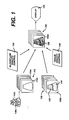
- a sequence of ultrasonic blood flow images is shown at 110 and a corresponding sequence of ultrasonic B mode, or tissue images is shown at 120.
- Each sequence of images is a plurality of planar images taken through different slice planes of a volumetric region of a patient. In this illustration the sequence is in order of depth through the volumetric region; the top image is taken through the front of the region, for instance, and successive images were acquired at progressive depths from the front toward the back of the region.
- the images in the blood flow image sequence exhibit close spatial correspondence to images of the tissue image sequence. That is, the position of the flow in the blood flow images corresponds to the position of the blood vessels in the tissue images. This correspondence is most easily achieved by acquiring the image data of both sequences at the same time. This may be done by time interleaving Doppler and B mode line acquisitions or interleaving frames of Doppler information with frames of B mode information, for instance.
- This spatial correspondence of the two types of image information allows them to be combined in a three dimensional display rendering which accurately depicts the spatial relationship of the tissue and blood flow in the volume from which the images were acquired.
- the problem cited by the '929 patent as insoluble is the obscuring of internal structure and blood flow of an organ by the B mode pixels of the surface of the organ. For instance, suppose that the first image 120a of the tissue image sequence depicts tissue of two adjacent organs 122 and 124, in particular, tissue that is closest to the viewer in a three dimensional presentation. Suppose also that image 110c in the interior of the corresponding blood flow image sequence 110 shows a blood flow network in the interior of the two organs, as shown in image frame 112c.
- tissue structure of tissue image 120a will be closest to the viewer and hence will obscure the internal blood flow networks 126c of the organs 122 and 124.
- the '929 patent does not recognize the possibility that the clinician can be provided the clinical information he seeks in a three dimensional rendering other than one in which the tissue structure is depicted as opaque surfaces.
- the present inventor has realized that clinically useful three dimensional presentations can be formed in which the tissue is depicted as a semi-transparent or sparsely completed body. Such a depiction, the present inventor has found, will show the extent of tissue structures by the continuity of the pattern of the acoustic texture and anatomical borders of the tissue. Since different tissues will return ultrasonic echoes with distinct and differing frequency spectra, intensity and duration, differing echo characteristics can be used to differentiate tissue types in a three dimensional ultrasonic image.
- the three dimensional rendering of image frame 150a of the three dimensional image sequence 150 is formed by controlling the transparency (opacity) of the two types of image information by means of blood flow opacity control 130 and tissue opacity control 140.
- the opacity controls are capable of separately controlling the display opacity of the two types of image information.
- the viewer will be able to examine the internal vascular network 126c of the two organs 122,124 and determine the extent of the internal vasculature of each organ in the three dimensional presentation 128.
- the boundary and extent of each organ is defined by the extent of the illustrated texture of the tissue seen in the three dimensional image.
- a sequence 150 of three dimensional projection images is formed, each image from the perspective of a different viewing angle, as described in U.S. Pat. 5,485,842.
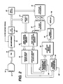
- a scanhead 10 includes a transducer array 12 which transmits ultrasonic pulses into a patient and receives back ultrasonic echoes.
- the transducer array 12 is pulsed and echoes received by a transmitter/receiver 14.
- Ultrasonic beams transmitted and received by the transducer array are steered and focused under control of a beamformer 16, which processes echo signals from a plurality of elements to form scanlines of coherent echo signals.
- the received echo signals are quadrature detected and filtered by an I,Q filter 20, then processed for either B mode or Doppler display.
- the I and Q samples are coupled to a detector and grayscale mapping circuit 24, which produces grayscale signals with brightness corresponding to the intensity of received echo signals.
- the grayscale signals are stored in a tissue image memory 40 together with information bearing their spatial relationship to the image field.
- scanlines of grayscale signals are acquired to form a full planar image, and a sequence of B mode planar tissue images is stored in tissue image memory 40.
- the image planes retain their spatial orientation to each other by being stored in the time or spatial sequence in which they were acquired.
- Doppler processing the I and Q samples undergo highpass filtering in a wall filter 22, then are assembled in ensembles of Doppler data in a Doppler processor 30.
- the data ensembles are processed by a Doppler technique such as autocorrelation or Fourier transform processing to produce Doppler signals of parameters such as Doppler power (P), velocity (v), or variance ( ⁇ ).
- the Doppler signals are processed to remove motion artifacts in a flash suppresser 32, then stored in a Doppler image memory together with information bearing their spatial relationship to the image field.
- scanlines of Doppler signals are acquired to form a full planar image, and a sequence of Doppler images is stored in a Doppler image partition of image memory 40, or a separate memory.
- the Doppler images retain their spatial orientation to each other by being stored in the time or spatial sequence in which they were acquired.
- the tissue and Doppler image information is then scan converted into the desired image format, such as a sector format, in a scan converter 42.
- Separate scan converters may be employed to rapidly scan convert tissue and Doppler images simultaneously, or a single scan converter may be used by time multiplexing the processing of the two types of image information.
- Scan conversion may also be merged into the rendering operations, whereby the two functions are carried out together (i.e., convert polar coordinate scanlines directly into a rectilinearly addressed three dimensional image.)
- the separate tissue and Doppler (blood flow) image information is processed together to render a composite three dimensional presentation.
- the ultrasound system operator enters rendering parameters into rendering parameter storage areas 52 and 54 by means of a user interface (UIF). These parameters control the manner in which each type of image information will be processed in the rendering. For instance, the user may enter values for the opacity and contrast to be imparted to each type of image information.
- the parameters for the two types of image information may be separately determined, so that the different types of image information can be processed differently. This enables, for example, the tissue image information to be displayed with greater transparency than the blood flow information, or with greater or lesser contrast.
- the spatially corresponding tissue and blood flow image information is rendered together in accordance with the rendering parameters to form a three dimensional image presentation in a 3D composite image rendering processor 50.
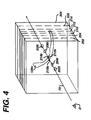
- a preferred technique of three dimensional rendering is illustrated by FIGURE 4, which depicts a three dimensional overlay of a number of planar blood flow and tissue images 202-214.
- a three dimensional projection image may be formed by combining spatially acquired planar images.
- both tissue and blood flow image information is rendered together by producing a composite pixel from the pixels of the overlaid images which are encountered when vectors along a viewing direction are directed through the images.
- a vector 70 passes through the image planes in a given direction, encountering tissue information on planes 202, 204, 206, and 208, and blood flow information on planes 210, 212, and 214.
- the first pixel of information encountered along the vector is a tissue pixel identified as 202t on image plane 202. This pixel is nearest to a viewer D who is observing the three dimensional structure from the origin of the vector 70.
- the next pixel encountered is tissue pixel 204t on image plane 204.
- three blood flow pixels of a blood vessel 126v are encountered on image planes 210, 212, and 214. Behind the blood vessel the vector 70 intersects a tissue pixel 206t on image plane 206 and a final tissue pixel 208t on the back image plane 208.
- tissue pixels of a given intensity are given an opacity which is semi transparent, and less than that of a blood flow pixel of the same intensity.
- two curves 302 and 304 are shown of the parameters for opacity control of the blood flow and tissue information, respectively.
- Curve 302 shows that, as the intensity or velocity or power or grayscale level of the Doppler information signals increases, the degree of opacification of the blood flow pixels increases, reaching a maximum of 100% opacity.
- the tissue information increases with grayscale level or intensity or power to a maximum opacity of 70% in the example. Thus, tissue pixels will never be fully opaque, but always semi transparent.
- the preferred rendering algorithm in addition to variable opacity weighting, also weights the contributions of the tissue and blood flow information in the three dimensional display by the depth of the image planes in the plane sequence (i.e., in the body).
- the preferred algorithm is of the form where P t and P b are tissue and blood flow pixel values and a t and a b are opacity weighting coefficients for tissue and blood flow pixels, and a d is a depth weighting coefficient.
- the weighting coefficients a t and a b are functionally variable for each pixel as shown by the curves of FIGURE 3, and may additionally or alternatively be functionally related to the spatial location of the pixels, the grayscale or color pixel values, or the relationship of color to grayscale pixel levels or variation.
- image information along the vector v can be weighted by opacity and depth to arrive at a composite three dimensional rendering, a result which cannot be attained by separately rendering the tissue and blood flow information, then combining the renderings. All of the pixel information encountered along the vector 70 is weighted and summed in this manner.
- the first tissue pixel 202t will have the greatest depth coefficient a d , since it is closest to the viewer D, but will also be weighted by its gray level and will be semi transparent. Its contribution to the final 3D pixel P 3D will be determined by these two weighting coefficients.
- the next tissue pixel 204t will have a lesser depth coefficient than the first pixel 202t, since it is further from the viewer.
- the following blood flow pixels 201b, 212b, and 214b may have significant opacity weighting coefficients a b because they are blood flow pixels, but will have lesser depth coefficients a d .
- the blood flow may still be visible behind the semi transparent tissue pixels, and the 3D pixel P 3D could be a light pink color in the three dimensional display, where red signifies blood flow and brightness signifies tissue.
- the rendered pixel would thus be a depth dependent composite of both characteristics.
- a three dimensional image of these two structures may reveal a stippled image texture in the region of the placenta and a more striated image texture in the region of the uterine wall.
- the clinician would be able to determine blood flow in the placenta by that part of the three dimensional vessel structure which is surrounded by a stippled image texture, as compared to vasculature of the uterine wall which is surrounded by a more striated texture.
- the clinician is able to assess the vasculature of the placenta under what are usually clinically problematic conditions.
- the present three dimensional display technique can be performed with any type of Doppler flow information, such as power Doppler or velocity (color flow) information.
- Doppler flow information such as power Doppler or velocity (color flow) information.
- the ultrasonic Doppler and B mode echo information can be obtained by gating the acquisition of both types of information to a phase of the pulsatile flow of blood
- excellent three dimensional renderings of pulsatile structures may be obtained by utilizing ungated power Doppler signal acquisition and gated B mode acquisition. For instance, if the region being imaged is moving in synchronism with the heartbeat cycle, the tissue information is gathered in synchronism with a flow gating signal such as an ECG heart gate signal. Tissue signal information is thus acquired during a constant phase of the heart cycle.
- the power Doppler signals need not be gated, but can be acquired at all other times when the tissue signals are not being acquired. Essentially, power Doppler information is acquired as rapidly and often as possible, while tissue information is acquired only at the desired phase of the heart cycle. After the power Doppler information has been temporally averaged, a combined rendering of tissue and blood flow information displays the blood flow information in spatial correspondence with the tissue structure.
- a semi transparent three dimensional presentation of tissue structure may also be obtained by use of a spatially dependent opacity function in the three dimensional rendering operation.
- Opacity can be dependent upon a segmentation function, for example.
- FIGURE 5 illustrates two opacity curves 306,308 which are a function of tissue segmentation or geometry. The curves could correspond to the tissue of organs 122 and 124 in image 120a of FIGURE 1, for example.
- a geometric opacity function can be used which omits tissue information in alternate pixels of a three dimensional rendering to produce a sparsely completed tissue structure. Such a rendering will enable a viewer to seen inside a tissue structure, since the tissue structure will thereby be given a gapped or perforated appearance.
- the preferred rendering technique of opacity and depth weighting of tissue and blood flow gives a smoother, more pleasing appearance to the three dimensional display presentation.
Landscapes
- Health & Medical Sciences (AREA)
- Life Sciences & Earth Sciences (AREA)
- Physics & Mathematics (AREA)
- Engineering & Computer Science (AREA)
- Radar, Positioning & Navigation (AREA)
- Remote Sensing (AREA)
- Animal Behavior & Ethology (AREA)
- Medical Informatics (AREA)
- Veterinary Medicine (AREA)
- Biophysics (AREA)
- Nuclear Medicine, Radiotherapy & Molecular Imaging (AREA)
- Pathology (AREA)
- Radiology & Medical Imaging (AREA)
- Biomedical Technology (AREA)
- Heart & Thoracic Surgery (AREA)
- Public Health (AREA)
- Molecular Biology (AREA)
- Surgery (AREA)
- Acoustics & Sound (AREA)
- General Health & Medical Sciences (AREA)
- General Physics & Mathematics (AREA)
- Computer Networks & Wireless Communication (AREA)
- Hematology (AREA)
- Ultra Sonic Daignosis Equipment (AREA)
Applications Claiming Priority (2)
| Application Number | Priority Date | Filing Date | Title |
|---|---|---|---|
| US1395196P | 1996-03-22 | 1996-03-22 | |
| US13951 | 1996-03-22 |
Publications (3)
| Publication Number | Publication Date |
|---|---|
| EP0797106A2 true EP0797106A2 (de) | 1997-09-24 |
| EP0797106A3 EP0797106A3 (de) | 1999-09-15 |
| EP0797106B1 EP0797106B1 (de) | 2006-04-26 |
Family
ID=21762700
Family Applications (1)
| Application Number | Title | Priority Date | Filing Date |
|---|---|---|---|
| EP97301921A Expired - Lifetime EP0797106B1 (de) | 1996-03-22 | 1997-03-21 | Medizinische diagnostische dreidimensionale Ultraschall-Abbildung von Gewebetextur und Vaskulatur |
Country Status (6)
| Country | Link |
|---|---|
| US (1) | US5720291A (de) |
| EP (1) | EP0797106B1 (de) |
| JP (1) | JP4107695B2 (de) |
| AT (1) | ATE324596T1 (de) |
| DE (1) | DE69735737T2 (de) |
| NO (1) | NO971314L (de) |
Cited By (9)
| Publication number | Priority date | Publication date | Assignee | Title |
|---|---|---|---|---|
| EP0844496A3 (de) * | 1996-11-26 | 1999-09-08 | Advanced Technology Laboratories, Inc. | Wiedergabe von dreidimensionellen Ultraschall-Bilder für Diagnostik aus Strömungs- und Gewebebilder |
| US6116244A (en) * | 1998-06-02 | 2000-09-12 | Acuson Corporation | Ultrasonic system and method for three-dimensional imaging with opacity control |
| EP1046929A3 (de) * | 1999-04-23 | 2001-09-05 | General Electric Company | Verfahren und Vorrichtung zur dreidimensionalen Ultraschall-Bilddarstellung mittels Volumendarstellung mit verbesserten Oberflächen |
| WO2001069282A3 (en) * | 2000-03-17 | 2002-01-10 | Koninkl Philips Electronics Nv | Spatially compounded three dimensional ultrasonic images |
| US6755787B2 (en) | 1998-06-02 | 2004-06-29 | Acuson Corporation | Medical diagnostic ultrasound system and method for versatile processing |
| EP1739627A1 (de) * | 2005-06-28 | 2007-01-03 | Medison Co., Ltd. | Verfahren und Vorrichtung zur Anzeige eines Strömungsfarbbildes auf einem diagnostischen Ultraschallsystem |
| WO2008044173A1 (en) * | 2006-10-13 | 2008-04-17 | Koninklijke Philips Electronics N.V. | 3d ultrasonic color flow imaging with grayscale invert |
| WO2012064413A1 (en) * | 2010-11-12 | 2012-05-18 | Boston Scientific Scimed, Inc. | Systems and methods for making and using rotational transducers for concurrently imaging blood flow and tissue |
| CN103230258A (zh) * | 2013-04-12 | 2013-08-07 | 华中科技大学 | 基于有限体积法的扩散光学断层成像正向问题处理方法 |
Families Citing this family (136)
| Publication number | Priority date | Publication date | Assignee | Title |
|---|---|---|---|---|
| JPH0733200U (ja) * | 1993-12-06 | 1995-06-20 | 大森機械工業株式会社 | 便座用シート |
| US5608690A (en) * | 1995-03-02 | 1997-03-04 | Acuson Corporation | Transmit beamformer with frequency dependent focus |
| WO1997034530A1 (en) * | 1996-03-18 | 1997-09-25 | Furuno Electric Company, Limited | Ultrasonic diagnostic device |
| JP4065327B2 (ja) * | 1996-10-08 | 2008-03-26 | 株式会社日立メディコ | 投影像表示方法および装置 |
| US5961460A (en) * | 1997-04-11 | 1999-10-05 | Acuson Corporation | Ultrasound imaging enhancement methods and systems |
| US6102864A (en) * | 1997-05-07 | 2000-08-15 | General Electric Company | Three-dimensional ultrasound imaging of velocity and power data using average or median pixel projections |
| US5895358A (en) * | 1997-05-07 | 1999-04-20 | General Electric Company | Method and apparatus for mapping color flow velocity data into display intensities |
| US5928151A (en) * | 1997-08-22 | 1999-07-27 | Acuson Corporation | Ultrasonic system and method for harmonic imaging in three dimensions |
| US5876343A (en) * | 1997-09-23 | 1999-03-02 | Scimed Life Systems, Inc. | Methods and apparatus for blood speckle detection in an intravascular ultrasound imaging system |
| JPH11164833A (ja) | 1997-09-30 | 1999-06-22 | Toshiba Corp | 医用画像診断装置 |
| US5882315A (en) * | 1997-12-23 | 1999-03-16 | Acuson Corporation | Ultrasonic imaging method and image for doppler tissue parameters |
| US6102858A (en) * | 1998-04-23 | 2000-08-15 | General Electric Company | Method and apparatus for three-dimensional ultrasound imaging using contrast agents and harmonic echoes |
| US6280387B1 (en) * | 1998-05-06 | 2001-08-28 | Siemens Medical Systems, Inc. | Three-dimensional tissue/flow ultrasound imaging system |
| US6241675B1 (en) | 1998-06-09 | 2001-06-05 | Volumetrics Medical Imaging | Methods and systems for determining velocity of tissue using three dimensional ultrasound data |
| JP3905644B2 (ja) * | 1998-06-15 | 2007-04-18 | 東芝医用システムエンジニアリング株式会社 | 3次元超音波システム |
| USD426298S (en) * | 1998-07-20 | 2000-06-06 | 3M Innovative Properties Company | Full-face respirator lens |
| US6245017B1 (en) * | 1998-10-30 | 2001-06-12 | Kabushiki Kaisha Toshiba | 3D ultrasonic diagnostic apparatus |
| US6352509B1 (en) * | 1998-11-16 | 2002-03-05 | Kabushiki Kaisha Toshiba | Three-dimensional ultrasonic diagnosis apparatus |
| JP4574768B2 (ja) * | 1998-11-16 | 2010-11-04 | 株式会社東芝 | 3次元超音波診断装置 |
| IL128904A0 (en) * | 1999-03-09 | 2000-02-17 | Inta Medics Ltd | Ultrasound pulsatility imaging |
| US6322505B1 (en) | 1999-06-08 | 2001-11-27 | Acuson Corporation | Medical diagnostic ultrasound system and method for post processing |
| US6264609B1 (en) | 1999-09-15 | 2001-07-24 | Wake Forest University | Ultrasound apparatus and method for tissue characterization |
| US6443894B1 (en) | 1999-09-29 | 2002-09-03 | Acuson Corporation | Medical diagnostic ultrasound system and method for mapping surface data for three dimensional imaging |
| US6515657B1 (en) | 2000-02-11 | 2003-02-04 | Claudio I. Zanelli | Ultrasonic imager |
| US6760486B1 (en) * | 2000-03-28 | 2004-07-06 | General Electric Company | Flash artifact suppression in two-dimensional ultrasound imaging |
| US6454715B2 (en) | 2000-04-11 | 2002-09-24 | Scimed Life Systems, Inc. | Methods and apparatus for blood speckle detection in an intravascular ultrasound imaging system |
| US6468216B1 (en) * | 2000-08-24 | 2002-10-22 | Kininklijke Philips Electronics N.V. | Ultrasonic diagnostic imaging of the coronary arteries |
| US6544175B1 (en) * | 2000-09-15 | 2003-04-08 | Koninklijke Philips Electronics N.V. | Ultrasound apparatus and methods for display of a volume using interlaced data |
| US6875177B2 (en) * | 2000-11-15 | 2005-04-05 | Aloka Co., Ltd. | Ultrasonic diagnostic apparatus |
| WO2002095653A2 (en) * | 2001-05-18 | 2002-11-28 | Mayo Foundation For Medical Education And Research | Ultrasound laboratory information management system and method |
| US6692438B2 (en) | 2001-12-18 | 2004-02-17 | Koninklijke Philips Electronics Nv | Ultrasonic imaging system and method for displaying tissue perfusion and other parameters varying with time |
| US20050075567A1 (en) * | 2001-12-18 | 2005-04-07 | Koninklijke Philips Electronics N.V. | Ultrasonic diagnostic imaging system with assisted border tracing |
| US6780155B2 (en) | 2001-12-18 | 2004-08-24 | Koninklijke Philips Electronics | Method and system for ultrasound blood flow imaging and volume flow calculations |
| JP2003224203A (ja) * | 2002-01-28 | 2003-08-08 | Hitachi Ltd | 半導体集積回路装置およびその製造方法 |
| US6776760B2 (en) * | 2002-03-06 | 2004-08-17 | Alfred E. Mann Institute For Biomedical Engineering At The University Of Southern California | Multi-mode processing for ultrasonic imaging |
| JP4130114B2 (ja) * | 2002-10-09 | 2008-08-06 | 株式会社日立メディコ | 超音波イメージング装置及び超音波信号処理方法 |
| JP2006505321A (ja) * | 2002-11-06 | 2006-02-16 | コーニンクレッカ フィリップス エレクトロニクス エヌ ヴィ | 運動部分の3次元撮像用フェーズドアレイ音響システム |
| US6692441B1 (en) * | 2002-11-12 | 2004-02-17 | Koninklijke Philips Electronics N.V. | System for identifying a volume of interest in a volume rendered ultrasound image |
| US8521260B2 (en) * | 2002-12-02 | 2013-08-27 | Yeda Research And Development Co. Ltd. | Characterization of arteriosclerosis by optical imaging |
| US7037263B2 (en) * | 2003-08-20 | 2006-05-02 | Siemens Medical Solutions Usa, Inc. | Computing spatial derivatives for medical diagnostic imaging methods and systems |
| EP1684638B1 (de) | 2003-11-07 | 2018-05-16 | Koninklijke Philips N.V. | Verfahren für die ultraschall-perfusionsdarstellung |
| JP2005143733A (ja) * | 2003-11-13 | 2005-06-09 | Toshiba Corp | 超音波診断装置、3次元画像データ表示装置及び3次元画像データ表示方法 |
| US20050107704A1 (en) * | 2003-11-14 | 2005-05-19 | Von Behren Patrick L. | Motion analysis methods and systems for medical diagnostic ultrasound |
| US20070055161A1 (en) * | 2003-12-03 | 2007-03-08 | Koninklijke Philips Electronics N.V. | Ultrasonic imaging system and method for simulataneous display of blood flow and perfusion parameters |
| CN1894594B (zh) * | 2003-12-16 | 2010-05-26 | 皇家飞利浦电子股份有限公司 | 具有自动控制穿透度、分辨率和帧速率的超声诊断成像系统 |
| US8021300B2 (en) * | 2004-06-16 | 2011-09-20 | Siemens Medical Solutions Usa, Inc. | Three-dimensional fly-through systems and methods using ultrasound data |
| US8160315B2 (en) * | 2004-09-13 | 2012-04-17 | Hitachi Medical Corporation | Ultrasonic imaging apparatus and projection image generating method |
| US8234923B2 (en) * | 2004-09-20 | 2012-08-07 | Innervision Medical Technologies Inc. | Systems and methods for ultrasound imaging |
| JP2008524739A (ja) * | 2004-12-21 | 2008-07-10 | コーニンクレッカ フィリップス エレクトロニクス エヌ ヴィ | データセットのハンドリング |
| JP2008539905A (ja) * | 2005-05-06 | 2008-11-20 | エダ リサーチ アンド ディベロップメント カンパニー,リミティド | 心臓周期に関連する血管における赤血球の運動の撮像および分析 |
| DE102005022345A1 (de) * | 2005-05-13 | 2006-11-16 | Siemens Ag | Verfahren zur Erzeugung und Darstellung von Untersuchungsbildern und zugehöriger Ultraschallkatheter |
| KR100748585B1 (ko) * | 2005-07-15 | 2007-08-13 | 주식회사 메디슨 | 부가정보를 이용하여 영상을 구성하는 초음파 시스템 |
| US7914451B2 (en) * | 2005-09-15 | 2011-03-29 | Innervision Medical Technologies Inc. | Determining attributes using ultrasound |
| EP1935344B1 (de) * | 2005-10-07 | 2013-03-13 | Hitachi Medical Corporation | Bildanzeigemethode und medizinisches bild-diagnosesystem |
| US7839403B2 (en) * | 2005-12-19 | 2010-11-23 | Siemens Aktiengesellschaft | Simultaneous generation of different data sets from a single acquisition run and dual rendering of images |
| DE102006001884A1 (de) * | 2006-01-13 | 2007-07-19 | Siemens Ag | Verfahren zur bildlichen Unterstützung einer Führung eines in einen Hohlraum eines Untersuchungsobjekts mindestens teilweise eingeführten medizinischen Instruments |
| WO2007092054A2 (en) | 2006-02-06 | 2007-08-16 | Specht Donald F | Method and apparatus to visualize the coronary arteries using ultrasound |
| US9612142B2 (en) * | 2006-04-27 | 2017-04-04 | General Electric Company | Method and system for measuring flow through a heart valve |
| US8446410B2 (en) * | 2006-05-11 | 2013-05-21 | Anatomage Inc. | Apparatus for generating volumetric image and matching color textured external surface |
| CN101489489B (zh) | 2006-07-14 | 2013-01-02 | 皇家飞利浦电子股份有限公司 | 超声成像系统中用于组织、记录和显示图像的系统和方法 |
| WO2008017998A2 (en) | 2006-08-11 | 2008-02-14 | Koninklijke Philips Electronics, N.V. | Ultrasound system for cerebral blood flow imaging and microbubble-enhanced blood clot lysis |
| JP4881112B2 (ja) * | 2006-09-19 | 2012-02-22 | 株式会社東芝 | 超音波診断装置及び画像データ生成方法 |
| EP2088932B1 (de) | 2006-10-25 | 2020-04-08 | Maui Imaging, Inc. | Verfahren und vorrichtung zur herstellung von ultraschallbildern mithilfe mehrerer öffnungen |
| US8160395B2 (en) * | 2006-11-22 | 2012-04-17 | General Electric Company | Method and apparatus for synchronizing corresponding landmarks among a plurality of images |
| US8244015B2 (en) * | 2006-11-22 | 2012-08-14 | General Electric Company | Methods and apparatus for detecting aneurysm in vasculatures |
| JP5022700B2 (ja) * | 2006-12-27 | 2012-09-12 | 株式会社東芝 | 超音波診断装置 |
| JP4907382B2 (ja) * | 2007-02-23 | 2012-03-28 | ジーイー・メディカル・システムズ・グローバル・テクノロジー・カンパニー・エルエルシー | 超音波画像表示方法および超音波診断装置 |
| US20090156947A1 (en) * | 2007-05-22 | 2009-06-18 | Seward James B | Knowledgebased image informatics system and method |
| US7991450B2 (en) | 2007-07-02 | 2011-08-02 | General Electric Company | Methods and systems for volume fusion in diagnostic imaging |
| JP2009011711A (ja) * | 2007-07-09 | 2009-01-22 | Toshiba Corp | 超音波診断装置 |
| JP5390081B2 (ja) * | 2007-07-25 | 2014-01-15 | 株式会社東芝 | 超音波診断装置、超音波画像処理装置及び超音波画像処理プログラム |
| US7912264B2 (en) | 2007-08-03 | 2011-03-22 | Siemens Medical Solutions Usa, Inc. | Multi-volume rendering of single mode data in medical diagnostic imaging |
| US9247926B2 (en) | 2010-04-14 | 2016-02-02 | Maui Imaging, Inc. | Concave ultrasound transducers and 3D arrays |
| US9788813B2 (en) | 2010-10-13 | 2017-10-17 | Maui Imaging, Inc. | Multiple aperture probe internal apparatus and cable assemblies |
| US9339256B2 (en) | 2007-10-01 | 2016-05-17 | Maui Imaging, Inc. | Determining material stiffness using multiple aperture ultrasound |
| US9282945B2 (en) | 2009-04-14 | 2016-03-15 | Maui Imaging, Inc. | Calibration of ultrasound probes |
| US8403862B2 (en) * | 2007-12-20 | 2013-03-26 | Yeda Research And Development Co. Ltd. | Time-based imaging |
| JP4376944B2 (ja) * | 2008-04-03 | 2009-12-02 | 富士フイルム株式会社 | 中間画像生成方法および装置ならびにプログラム |
| US8131044B2 (en) * | 2008-04-08 | 2012-03-06 | General Electric Company | Method and apparatus for determining the effectiveness of an image transformation process |
| JP5366612B2 (ja) | 2008-05-20 | 2013-12-11 | 株式会社東芝 | 画像処理装置、画像処理方法および画像処理プログラム |
| US8425422B2 (en) * | 2008-06-06 | 2013-04-23 | Siemens Medical Solutions Usa, Inc. | Adaptive volume rendering for ultrasound color flow diagnostic imaging |
| JP5764058B2 (ja) * | 2008-07-10 | 2015-08-12 | コーニンクレッカ フィリップス エヌ ヴェ | 心臓の同時性及び生存能力の超音波評価 |
| JP5485373B2 (ja) | 2009-04-14 | 2014-05-07 | マウイ イマギング,インコーポレーテッド | 複数開口の超音波アレイ位置合せ装置 |
| KR101068917B1 (ko) * | 2009-05-18 | 2011-09-30 | 삼성메디슨 주식회사 | 기관 표시를 위한 초음파 진단기 및 방법 |
| BR112012006930A2 (pt) | 2009-10-01 | 2019-09-24 | Koninl Philips Electronics Nv | método para a avaliação de uma condiçã de um fígado e sistema para a avaliação de uma condição de um fígado |
| US20180365825A1 (en) * | 2009-10-05 | 2018-12-20 | Carolyn M. Salafia | Automated placental measurement |
| JP5683868B2 (ja) * | 2009-10-08 | 2015-03-11 | 株式会社東芝 | 超音波診断装置、超音波画像処理装置、超音波画像処理方法、及び超音波画像処理プログラム |
| CN102711625B (zh) * | 2010-01-18 | 2015-03-25 | 株式会社日立医疗器械 | 超声波诊断装置以及超声波图像显示方法 |
| EP2535003A4 (de) * | 2010-02-09 | 2017-03-15 | Hitachi, Ltd. | Ultraschalldiagnosevorrichtung und ultraschallbildanzeigeverfahren |
| KR102322776B1 (ko) | 2010-02-18 | 2021-11-04 | 마우이 이미징, 인코포레이티드 | 초음파 이미지를 구성하는 방법 및 이를 위한 다중-개구 초음파 이미징 시스템 |
| US8582865B2 (en) * | 2010-04-28 | 2013-11-12 | General Electric Company | Ultrasound imaging with ray casting and software-based image reconstruction |
| CN102917648B (zh) | 2010-05-26 | 2015-05-20 | 皇家飞利浦电子股份有限公司 | 高体积速率3d 超声诊断成像 |
| WO2011148275A1 (en) | 2010-05-26 | 2011-12-01 | Koninklijke Philips Electronics N.V. | High volume rate 3d ultrasonic diagnostic imaging of the heart |
| JP5815541B2 (ja) | 2010-09-29 | 2015-11-17 | 株式会社日立メディコ | 超音波診断装置、超音波画像表示方法、および、プログラム |
| WO2012042423A1 (en) | 2010-09-30 | 2012-04-05 | Koninklijke Philips Electronics N.V. | Monitoring and control of microbubble cavitation in therapeutic ultrasound |
| WO2012049586A2 (en) | 2010-10-11 | 2012-04-19 | Koninklijke Philips Electronics N.V. | Micro-particulate identification and classification in ultrasound images |
| WO2012053514A1 (ja) * | 2010-10-19 | 2012-04-26 | 株式会社 東芝 | 超音波診断装置、超音波画像処理装置及び超音波画像処理方法 |
| US9179892B2 (en) * | 2010-11-08 | 2015-11-10 | General Electric Company | System and method for ultrasound imaging |
| KR101313220B1 (ko) | 2010-11-23 | 2013-09-30 | 삼성메디슨 주식회사 | 특성 곡선 정보에 기초하여 컬러 도플러 모드 영상을 제공하는 초음파 시스템 및 방법 |
| CN102551791B (zh) * | 2010-12-17 | 2016-04-27 | 深圳迈瑞生物医疗电子股份有限公司 | 一种超声成像方法和装置 |
| DE102011005237A1 (de) * | 2011-03-08 | 2012-09-13 | Siemens Aktiengesellschaft | Selektive Visualisierung eines 3-dimensionalen Volumendatensatzes |
| JP6407719B2 (ja) | 2011-12-01 | 2018-10-17 | マウイ イマギング,インコーポレーテッド | ピングベース及び多数開口ドップラー超音波を用いた運動の検出 |
| JP2015503404A (ja) | 2011-12-29 | 2015-02-02 | マウイ イマギング,インコーポレーテッド | 任意経路のmモード超音波イメージング |
| WO2013111813A1 (ja) * | 2012-01-27 | 2013-08-01 | 株式会社 東芝 | 医用画像処理装置 |
| WO2013121341A1 (en) | 2012-02-13 | 2013-08-22 | Koninklijke Philips Electronics N.V. | Simultaneous ultrasonic viewing of 3d volume from multiple directions |
| EP2833791B1 (de) | 2012-03-26 | 2022-12-21 | Maui Imaging, Inc. | Verfahren zur verbesserung der ultraschallbildqualität durch anwendung von gewichtungsfaktoren |
| EP3730966B1 (de) | 2012-05-22 | 2023-07-12 | Koninklijke Philips N.V. | Einstellung einer ultraschallbildanzeige für fernanzeigeendgerät |
| CN104620128B (zh) | 2012-08-10 | 2017-06-23 | 毛伊图像公司 | 多孔径超声探头的校准 |
| BR112015004443A2 (pt) | 2012-09-01 | 2017-07-04 | Koninklijke Philips Nv | método para o uso de informações ultrassônicas para o tratamento de patologias por ablação, e sistema para o uso de informações ultrassônicas para tratamento de patologias por ablação |
| CN104582585B (zh) | 2012-09-01 | 2017-09-26 | 皇家飞利浦有限公司 | 用于消融规划的超声体积流量测量 |
| US9986969B2 (en) | 2012-09-06 | 2018-06-05 | Maui Imaging, Inc. | Ultrasound imaging system memory architecture |
| CN103676827A (zh) | 2012-09-06 | 2014-03-26 | Ip音乐集团有限公司 | 用于远程控制音频设备的系统和方法 |
| WO2014160291A1 (en) | 2013-03-13 | 2014-10-02 | Maui Imaging, Inc. | Alignment of ultrasound transducer arrays and multiple aperture probe assembly |
| US9883848B2 (en) | 2013-09-13 | 2018-02-06 | Maui Imaging, Inc. | Ultrasound imaging using apparent point-source transmit transducer |
| US10401493B2 (en) | 2014-08-18 | 2019-09-03 | Maui Imaging, Inc. | Network-based ultrasound imaging system |
| US9655592B2 (en) * | 2014-11-21 | 2017-05-23 | General Electric Corporation | Method and apparatus for rendering an ultrasound image |
| KR102367194B1 (ko) * | 2014-12-31 | 2022-02-25 | 삼성메디슨 주식회사 | 초음파 진단 장치 및 그 동작방법 |
| WO2016142204A1 (en) | 2015-03-10 | 2016-09-15 | Koninklijke Philips N.V. | Ultrasonic diagnosis of cardiac performance using heart model chamber segmentation with user control |
| RU2708317C2 (ru) | 2015-03-10 | 2019-12-05 | Конинклейке Филипс Н.В. | Ультразвуковая диагностика работы сердца посредством сегментации камеры с одной степенью свободы |
| CN107613878B (zh) | 2015-03-30 | 2021-04-06 | 毛伊图像公司 | 用于检测物体运动的超声成像系统和方法 |
| WO2016157072A1 (en) | 2015-03-30 | 2016-10-06 | Koninklijke Philips N.V. | Ultrasonic transducer array for sonothrombolysis treatment and monitoring |
| CN108289655B (zh) | 2015-12-02 | 2022-03-18 | 皇家飞利浦有限公司 | 具有中轴弯曲和横向偏心的心脏的超声心脏评估 |
| EP4697068A3 (de) | 2016-01-27 | 2026-04-22 | Maui Imaging, Inc. | Ultraschallbildgebung mit spärlichen arraysonden |
| WO2017149027A1 (en) | 2016-03-01 | 2017-09-08 | Koninklijke Philips N.V. | Automated ultrasonic measurement of nuchal fold translucency |
| US11911218B2 (en) | 2016-03-30 | 2024-02-27 | Koninklijke Philips N.V. | Two dimensional ultrasonic array transducer with one dimensional patches |
| WO2017168279A1 (en) | 2016-03-30 | 2017-10-05 | Koninklijke Philips N.V. | Two dimensional ultrasonic array transducer with one dimensional subpatches |
| WO2017207815A1 (en) | 2016-06-02 | 2017-12-07 | Koninklijke Philips N.V. | Ultrasound systems with time compression and time multiplexing of received ultrasound signals |
| EP3551288A1 (de) | 2016-12-07 | 2019-10-16 | Koninklijke Philips N.V. | Behandlungsplanung für ultraschall-sonothrombolyse |
| US20200107811A1 (en) | 2016-12-19 | 2020-04-09 | Koninklijke Philips N.V. | Ultrasonic transducer array monitoring during transcranial ultrasound procedures |
| EP3602502B1 (de) | 2017-03-29 | 2023-11-08 | Koninklijke Philips N.V. | Eingebettete virtuelle lichtquelle in einem 3d-volumen, das mit fadenkreuzen mit mpr-ansicht verknüpft ist |
| US10705210B2 (en) * | 2017-05-31 | 2020-07-07 | B-K Medical Aps | Three-dimensional (3-D) imaging with a row-column addressed (RCA) transducer array using synthetic aperture sequential beamforming (SASB) |
| JP7602015B2 (ja) * | 2020-08-07 | 2024-12-17 | オックスフォード ユニヴァーシティ イノヴェーション リミテッド | 超音波方法 |
| JP7724853B2 (ja) | 2020-10-21 | 2025-08-18 | マウイ イマギング,インコーポレーテッド | 多数開口超音波を用いた組織の特徴付けのためのシステム及び方法 |
| EP4236811A4 (de) | 2020-11-02 | 2024-10-09 | Maui Imaging, Inc. | Systeme und verfahren zur verbesserung der ultraschallbildqualität |
| US20230005133A1 (en) * | 2021-06-24 | 2023-01-05 | Carolyn M Salafia | Automated placental measurement |
| US12056873B2 (en) | 2021-12-21 | 2024-08-06 | GE Precision Healthcare LLC | Method and system for automatically analyzing placenta insufficiency in a curved topographical ultrasound image slice |
Family Cites Families (14)
| Publication number | Priority date | Publication date | Assignee | Title |
|---|---|---|---|---|
| JPS5849137A (ja) * | 1981-09-18 | 1983-03-23 | 株式会社東芝 | 超音波血流測定装置 |
| US4630612A (en) * | 1983-05-25 | 1986-12-23 | Aloka Co., Ltd. | Ultrasonic diagnostic apparatus |
| US4694434A (en) * | 1984-06-12 | 1987-09-15 | Von Ramm Olaf T | Three-dimensional imaging system |
| US4794932A (en) * | 1986-03-31 | 1989-01-03 | Kabushiki Kaisha Toshiba | Conversion control circuit for image display apparatus |
| US5159931A (en) * | 1988-11-25 | 1992-11-03 | Riccardo Pini | Apparatus for obtaining a three-dimensional reconstruction of anatomic structures through the acquisition of echographic images |
| DE3914619A1 (de) * | 1989-05-03 | 1990-11-08 | Kontron Elektronik | Vorrichtung zur transoesophagealen echokardiographie |
| US5282471A (en) * | 1991-07-31 | 1994-02-01 | Kabushiki Kaisha Toshiba | Ultrasonic imaging system capable of displaying 3-dimensional angiogram in real time mode |
| JP3187148B2 (ja) * | 1991-08-26 | 2001-07-11 | 株式会社東芝 | 超音波診断装置 |
| US5396890A (en) * | 1993-09-30 | 1995-03-14 | Siemens Medical Systems, Inc. | Three-dimensional scan converter for ultrasound imaging |
| US5365929A (en) * | 1993-10-04 | 1994-11-22 | Advanced Technology Laboratories, Inc. | Multiple sample volume spectral Doppler |
| JP3833282B2 (ja) * | 1994-06-24 | 2006-10-11 | 株式会社東芝 | 超音波診断装置 |
| US5476097A (en) * | 1994-10-13 | 1995-12-19 | Advanced Technology Laboratories, Inc. | Simultaneous ultrasonic imaging and Doppler display system |
| US5474073A (en) * | 1994-11-22 | 1995-12-12 | Advanced Technology Laboratories, Inc. | Ultrasonic diagnostic scanning for three dimensional display |
| US5485842A (en) * | 1994-11-30 | 1996-01-23 | Advanced Technology Laboratories, Inc. | Ultrasonic diagnostic scan conversion for three dimensional display processing |
-
1996
- 1996-04-29 US US08/638,710 patent/US5720291A/en not_active Expired - Lifetime
-
1997
- 1997-03-20 NO NO971314A patent/NO971314L/no unknown
- 1997-03-21 EP EP97301921A patent/EP0797106B1/de not_active Expired - Lifetime
- 1997-03-21 JP JP08556497A patent/JP4107695B2/ja not_active Expired - Lifetime
- 1997-03-21 AT AT97301921T patent/ATE324596T1/de not_active IP Right Cessation
- 1997-03-21 DE DE69735737T patent/DE69735737T2/de not_active Expired - Lifetime
Cited By (13)
| Publication number | Priority date | Publication date | Assignee | Title |
|---|---|---|---|---|
| EP0844496A3 (de) * | 1996-11-26 | 1999-09-08 | Advanced Technology Laboratories, Inc. | Wiedergabe von dreidimensionellen Ultraschall-Bilder für Diagnostik aus Strömungs- und Gewebebilder |
| US6116244A (en) * | 1998-06-02 | 2000-09-12 | Acuson Corporation | Ultrasonic system and method for three-dimensional imaging with opacity control |
| US6755787B2 (en) | 1998-06-02 | 2004-06-29 | Acuson Corporation | Medical diagnostic ultrasound system and method for versatile processing |
| EP1046929A3 (de) * | 1999-04-23 | 2001-09-05 | General Electric Company | Verfahren und Vorrichtung zur dreidimensionalen Ultraschall-Bilddarstellung mittels Volumendarstellung mit verbesserten Oberflächen |
| WO2001069282A3 (en) * | 2000-03-17 | 2002-01-10 | Koninkl Philips Electronics Nv | Spatially compounded three dimensional ultrasonic images |
| US7621872B2 (en) | 2005-06-28 | 2009-11-24 | Medison Co., Ltd. | Method and apparatus for displaying a color flow image in an ultrasound diagnostic system |
| EP1739627A1 (de) * | 2005-06-28 | 2007-01-03 | Medison Co., Ltd. | Verfahren und Vorrichtung zur Anzeige eines Strömungsfarbbildes auf einem diagnostischen Ultraschallsystem |
| KR100825054B1 (ko) | 2005-06-28 | 2008-04-28 | 주식회사 메디슨 | 컬러 플로우 영상을 촬상하는 방법 및 초음파 진단 시스템 |
| WO2008044173A1 (en) * | 2006-10-13 | 2008-04-17 | Koninklijke Philips Electronics N.V. | 3d ultrasonic color flow imaging with grayscale invert |
| WO2012064413A1 (en) * | 2010-11-12 | 2012-05-18 | Boston Scientific Scimed, Inc. | Systems and methods for making and using rotational transducers for concurrently imaging blood flow and tissue |
| US8591421B2 (en) | 2010-11-12 | 2013-11-26 | Boston Scientific Scimed, Inc. | Systems and methods for making and using rotational transducers for concurrently imaging blood flow and tissue |
| US9603584B2 (en) | 2010-11-12 | 2017-03-28 | Boston Scientific Scimed, Inc. | Systems and methods for intravascular imaging with rotational transducers for concurrently imaging blood flow and tissue |
| CN103230258A (zh) * | 2013-04-12 | 2013-08-07 | 华中科技大学 | 基于有限体积法的扩散光学断层成像正向问题处理方法 |
Also Published As
| Publication number | Publication date |
|---|---|
| DE69735737T2 (de) | 2007-05-16 |
| US5720291A (en) | 1998-02-24 |
| JPH09262236A (ja) | 1997-10-07 |
| NO971314D0 (no) | 1997-03-20 |
| NO971314L (no) | 1997-09-23 |
| EP0797106B1 (de) | 2006-04-26 |
| EP0797106A3 (de) | 1999-09-15 |
| JP4107695B2 (ja) | 2008-06-25 |
| ATE324596T1 (de) | 2006-05-15 |
| DE69735737D1 (de) | 2006-06-01 |
Similar Documents
| Publication | Publication Date | Title |
|---|---|---|
| EP0797106B1 (de) | Medizinische diagnostische dreidimensionale Ultraschall-Abbildung von Gewebetextur und Vaskulatur | |
| US5515856A (en) | Method for generating anatomical M-mode displays | |
| EP3905960B1 (de) | Systeme und verfahren zur kontrastverstärkten bildgebung | |
| EP2082262B1 (de) | 3d-ultraschallfarbflussbildgebung mit grauskalaumkehrung | |
| US20070055161A1 (en) | Ultrasonic imaging system and method for simulataneous display of blood flow and perfusion parameters | |
| US5396890A (en) | Three-dimensional scan converter for ultrasound imaging | |
| EP2016905B1 (de) | Diagnostische Ultraschallvorrichtung | |
| EP2030570B1 (de) | Bildverarbeitungsvorrichtung | |
| EP0844496A2 (de) | Wiedergabe von dreidimensionellen Ultraschall-Bilder für Diagnostik aus Strömungs- und Gewebebilder | |
| US6544178B1 (en) | Methods and systems for volume rendering using ultrasound data | |
| US20110125016A1 (en) | Fetal rendering in medical diagnostic ultrasound | |
| WO2009044316A1 (en) | System and method for real-time multi-slice acquisition and display of medical ultrasound images | |
| EP4076208B1 (de) | Systeme und verfahren zur beurteilung einer plazenta | |
| US7907758B2 (en) | Method and system for maintaining consistent anatomic views in displayed image data | |
| US20050075567A1 (en) | Ultrasonic diagnostic imaging system with assisted border tracing | |
| EP1739627B1 (de) | Verfahren und Vorrichtung zur Anzeige eines Strömungsfarbbildes auf einem diagnostischen Ultraschallsystem | |
| JP2007518512A (ja) | 心筋灌流を表示するための画像分割 | |
| JP3505281B2 (ja) | 超音波3次元画像形成装置 | |
| JP2001061839A (ja) | 超音波診断装置及び超音波画像処理装置 | |
| JP3977779B2 (ja) | 超音波診断装置 | |
| WO2023186948A1 (en) | Systems and methods for color mapping for contrast enhanced ultrasound parametric imaging |
Legal Events
| Date | Code | Title | Description |
|---|---|---|---|
| PUAI | Public reference made under article 153(3) epc to a published international application that has entered the european phase |
Free format text: ORIGINAL CODE: 0009012 |
|
| AK | Designated contracting states |
Kind code of ref document: A2 Designated state(s): AT BE CH DE DK ES FR GB GR IE IT LI LU MC NL PT SE |
|
| PUAL | Search report despatched |
Free format text: ORIGINAL CODE: 0009013 |
|
| AK | Designated contracting states |
Kind code of ref document: A3 Designated state(s): AT BE CH DE DK ES FR GB GR IE IT LI LU MC NL PT SE |
|
| 17P | Request for examination filed |
Effective date: 20000315 |
|
| 17Q | First examination report despatched |
Effective date: 20030829 |
|
| RIC1 | Information provided on ipc code assigned before grant |
Ipc: 7A 61B 8/06 A |
|
| GRAP | Despatch of communication of intention to grant a patent |
Free format text: ORIGINAL CODE: EPIDOSNIGR1 |
|
| RIC1 | Information provided on ipc code assigned before grant |
Ipc: 7A 61B 8/06 B Ipc: 7G 01S 15/89 A |
|
| GRAS | Grant fee paid |
Free format text: ORIGINAL CODE: EPIDOSNIGR3 |
|
| GRAA | (expected) grant |
Free format text: ORIGINAL CODE: 0009210 |
|
| AK | Designated contracting states |
Kind code of ref document: B1 Designated state(s): AT BE CH DE DK ES FR GB GR IE IT LI LU MC NL PT SE |
|
| PG25 | Lapsed in a contracting state [announced via postgrant information from national office to epo] |
Ref country code: NL Free format text: LAPSE BECAUSE OF FAILURE TO SUBMIT A TRANSLATION OF THE DESCRIPTION OR TO PAY THE FEE WITHIN THE PRESCRIBED TIME-LIMIT Effective date: 20060426 Ref country code: LI Free format text: LAPSE BECAUSE OF FAILURE TO SUBMIT A TRANSLATION OF THE DESCRIPTION OR TO PAY THE FEE WITHIN THE PRESCRIBED TIME-LIMIT Effective date: 20060426 Ref country code: IT Free format text: LAPSE BECAUSE OF FAILURE TO SUBMIT A TRANSLATION OF THE DESCRIPTION OR TO PAY THE FEE WITHIN THE PRESCRIBED TIME-LIMIT;WARNING: LAPSES OF ITALIAN PATENTS WITH EFFECTIVE DATE BEFORE 2007 MAY HAVE OCCURRED AT ANY TIME BEFORE 2007. THE CORRECT EFFECTIVE DATE MAY BE DIFFERENT FROM THE ONE RECORDED. Effective date: 20060426 Ref country code: CH Free format text: LAPSE BECAUSE OF FAILURE TO SUBMIT A TRANSLATION OF THE DESCRIPTION OR TO PAY THE FEE WITHIN THE PRESCRIBED TIME-LIMIT Effective date: 20060426 Ref country code: BE Free format text: LAPSE BECAUSE OF FAILURE TO SUBMIT A TRANSLATION OF THE DESCRIPTION OR TO PAY THE FEE WITHIN THE PRESCRIBED TIME-LIMIT Effective date: 20060426 |
|
| REG | Reference to a national code |
Ref country code: GB Ref legal event code: FG4D |
|
| REG | Reference to a national code |
Ref country code: IE Ref legal event code: FG4D |
|
| REF | Corresponds to: |
Ref document number: 69735737 Country of ref document: DE Date of ref document: 20060601 Kind code of ref document: P |
|
| PG25 | Lapsed in a contracting state [announced via postgrant information from national office to epo] |
Ref country code: SE Free format text: LAPSE BECAUSE OF FAILURE TO SUBMIT A TRANSLATION OF THE DESCRIPTION OR TO PAY THE FEE WITHIN THE PRESCRIBED TIME-LIMIT Effective date: 20060726 Ref country code: DK Free format text: LAPSE BECAUSE OF FAILURE TO SUBMIT A TRANSLATION OF THE DESCRIPTION OR TO PAY THE FEE WITHIN THE PRESCRIBED TIME-LIMIT Effective date: 20060726 |
|
| PG25 | Lapsed in a contracting state [announced via postgrant information from national office to epo] |
Ref country code: ES Free format text: LAPSE BECAUSE OF FAILURE TO SUBMIT A TRANSLATION OF THE DESCRIPTION OR TO PAY THE FEE WITHIN THE PRESCRIBED TIME-LIMIT Effective date: 20060806 |
|
| PG25 | Lapsed in a contracting state [announced via postgrant information from national office to epo] |
Ref country code: PT Free format text: LAPSE BECAUSE OF FAILURE TO SUBMIT A TRANSLATION OF THE DESCRIPTION OR TO PAY THE FEE WITHIN THE PRESCRIBED TIME-LIMIT Effective date: 20060926 |
|
| REG | Reference to a national code |
Ref country code: CH Ref legal event code: PL |
|
| NLV1 | Nl: lapsed or annulled due to failure to fulfill the requirements of art. 29p and 29m of the patents act | ||
| ET | Fr: translation filed | ||
| PGFP | Annual fee paid to national office [announced via postgrant information from national office to epo] |
Ref country code: AT Payment date: 20070323 Year of fee payment: 11 |
|
| PLBE | No opposition filed within time limit |
Free format text: ORIGINAL CODE: 0009261 |
|
| STAA | Information on the status of an ep patent application or granted ep patent |
Free format text: STATUS: NO OPPOSITION FILED WITHIN TIME LIMIT |
|
| 26N | No opposition filed |
Effective date: 20070129 |
|
| PG25 | Lapsed in a contracting state [announced via postgrant information from national office to epo] |
Ref country code: MC Free format text: LAPSE BECAUSE OF NON-PAYMENT OF DUE FEES Effective date: 20070331 Ref country code: IE Free format text: LAPSE BECAUSE OF NON-PAYMENT OF DUE FEES Effective date: 20070321 |
|
| PG25 | Lapsed in a contracting state [announced via postgrant information from national office to epo] |
Ref country code: GR Free format text: LAPSE BECAUSE OF FAILURE TO SUBMIT A TRANSLATION OF THE DESCRIPTION OR TO PAY THE FEE WITHIN THE PRESCRIBED TIME-LIMIT Effective date: 20060727 |
|
| PGFP | Annual fee paid to national office [announced via postgrant information from national office to epo] |
Ref country code: IT Payment date: 20080227 Year of fee payment: 12 |
|
| PG25 | Lapsed in a contracting state [announced via postgrant information from national office to epo] |
Ref country code: AT Free format text: LAPSE BECAUSE OF NON-PAYMENT OF DUE FEES Effective date: 20080321 |
|
| PG25 | Lapsed in a contracting state [announced via postgrant information from national office to epo] |
Ref country code: LU Free format text: LAPSE BECAUSE OF NON-PAYMENT OF DUE FEES Effective date: 20070321 |
|
| PG25 | Lapsed in a contracting state [announced via postgrant information from national office to epo] |
Ref country code: IT Free format text: LAPSE BECAUSE OF NON-PAYMENT OF DUE FEES Effective date: 20090321 |
|
| REG | Reference to a national code |
Ref country code: FR Ref legal event code: PLFP Year of fee payment: 20 |
|
| PGFP | Annual fee paid to national office [announced via postgrant information from national office to epo] |
Ref country code: FR Payment date: 20160331 Year of fee payment: 20 Ref country code: GB Payment date: 20160331 Year of fee payment: 20 |
|
| PGFP | Annual fee paid to national office [announced via postgrant information from national office to epo] |
Ref country code: DE Payment date: 20160531 Year of fee payment: 20 |
|
| REG | Reference to a national code |
Ref country code: DE Ref legal event code: R071 Ref document number: 69735737 Country of ref document: DE |
|
| REG | Reference to a national code |
Ref country code: GB Ref legal event code: PE20 Expiry date: 20170320 |
|
| PG25 | Lapsed in a contracting state [announced via postgrant information from national office to epo] |
Ref country code: GB Free format text: LAPSE BECAUSE OF EXPIRATION OF PROTECTION Effective date: 20170320 |